Superior mesenteric plexus Stock Photos and Images
(21)See superior mesenteric plexus stock video clipsQuick filters:
Superior mesenteric plexus Stock Photos and Images
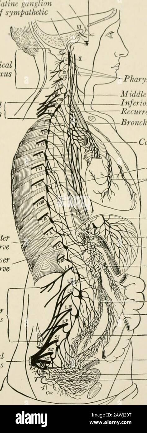 The anatomy of the nervous system, from the standpoint of development and function . Cervicalplexus Brachialplexus Greater Jffisplanchnic nerve ^K Lesser splanchnic nerve Lumbarplexus Sacralpie cus. Pharyngeal plexus Middle cervical ganglion of sympatheticInferior cervical gang, of sympatheticRecurrent nervtBronchial plexus Cardiac plexus Esophageal plexus^Coronary plexu i Left vagus nerve Gastric plexusCeliac plexus Superior mesenteric plexus —j-Aortic plexus^—Inferior mesenteric plexus Hypogastric plexus Pelvic plexus BladderVesical plexus Fig. 33. Fig. 34. Fig. 33.—General view of the cen Stock Photohttps://www.alamy.com/image-license-details/?v=1https://www.alamy.com/the-anatomy-of-the-nervous-system-from-the-standpoint-of-development-and-function-cervicalplexus-brachialplexus-greater-jffisplanchnic-nerve-k-lesser-splanchnic-nerve-lumbarplexus-sacralpie-cus-pharyngeal-plexus-middle-cervical-ganglion-of-sympatheticinferior-cervical-gang-of-sympatheticrecurrent-nervtbronchial-plexus-cardiac-plexus-esophageal-plexuscoronary-plexu-i-left-vagus-nerve-gastric-plexusceliac-plexus-superior-mesenteric-plexus-j-aortic-plexusinferior-mesenteric-plexus-hypogastric-plexus-pelvic-plexus-bladdervesical-plexus-fig-33-fig-34-fig-33general-view-of-the-cen-image342760120.html
RM2AWJ20T–The anatomy of the nervous system, from the standpoint of development and function . Cervicalplexus Brachialplexus Greater Jffisplanchnic nerve ^K Lesser splanchnic nerve Lumbarplexus Sacralpie cus. Pharyngeal plexus Middle cervical ganglion of sympatheticInferior cervical gang, of sympatheticRecurrent nervtBronchial plexus Cardiac plexus Esophageal plexus^Coronary plexu i Left vagus nerve Gastric plexusCeliac plexus Superior mesenteric plexus —j-Aortic plexus^—Inferior mesenteric plexus Hypogastric plexus Pelvic plexus BladderVesical plexus Fig. 33. Fig. 34. Fig. 33.—General view of the cen
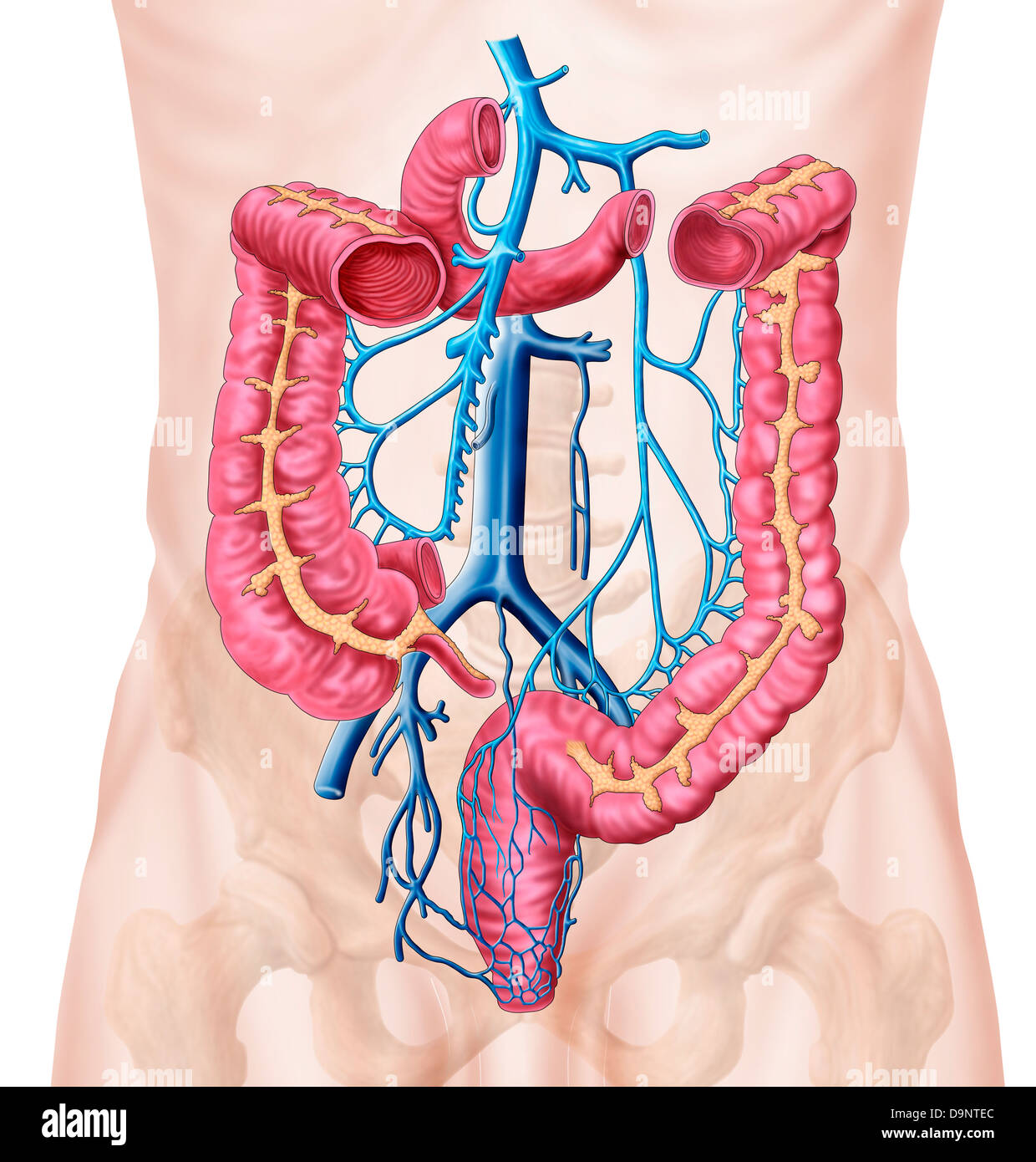 Anatomy of human abdominal vein system. Stock Photohttps://www.alamy.com/image-license-details/?v=1https://www.alamy.com/stock-photo-anatomy-of-human-abdominal-vein-system-57643220.html
RFD9NTEC–Anatomy of human abdominal vein system.
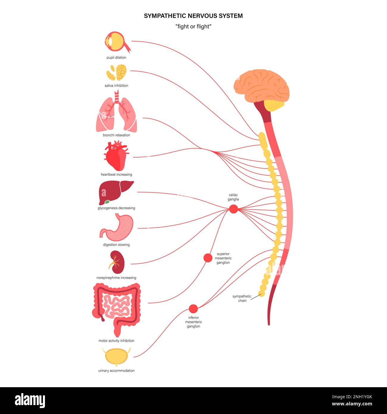 Sympathetic nervous system, illustration Stock Photohttps://www.alamy.com/image-license-details/?v=1https://www.alamy.com/sympathetic-nervous-system-illustration-image526803779.html
RF2NH1YGK–Sympathetic nervous system, illustration
 Archive image from page 1246 of Cunningham's Text-book of anatomy (1914). Cunningham's Text-book of anatomy cunninghamstextb00cunn Year: 1914 ( (LECUM AND VERMIFOBM PEOCESS. 1213 Nerves.—The nerves come from the superior mesenteric plexus, an offshoot of the cceliac plexus, and from the inferior mesenteric, a derivative of the aortic plexus. The arrangement is similar to that of the nerves of the small intestine. INTESTINUM (LECUM AND PKOCESSUS VERMIFORMIS. Intestinum Caecum.—After leaving the pelvic cavity, as already described, terminal portion of the small intestine passes upwards, backwar Stock Photohttps://www.alamy.com/image-license-details/?v=1https://www.alamy.com/archive-image-from-page-1246-of-cunninghams-text-book-of-anatomy-1914-cunninghams-text-book-of-anatomy-cunninghamstextb00cunn-year-1914-lecum-and-vermifobm-peocess-1213-nervesthe-nerves-come-from-the-superior-mesenteric-plexus-an-offshoot-of-the-cceliac-plexus-and-from-the-inferior-mesenteric-a-derivative-of-the-aortic-plexus-the-arrangement-is-similar-to-that-of-the-nerves-of-the-small-intestine-intestinum-lecum-and-pkocessus-vermiformis-intestinum-caecumafter-leaving-the-pelvic-cavity-as-already-described-terminal-portion-of-the-small-intestine-passes-upwards-backwar-image264068618.html
RMW9HA62–Archive image from page 1246 of Cunningham's Text-book of anatomy (1914). Cunningham's Text-book of anatomy cunninghamstextb00cunn Year: 1914 ( (LECUM AND VERMIFOBM PEOCESS. 1213 Nerves.—The nerves come from the superior mesenteric plexus, an offshoot of the cceliac plexus, and from the inferior mesenteric, a derivative of the aortic plexus. The arrangement is similar to that of the nerves of the small intestine. INTESTINUM (LECUM AND PKOCESSUS VERMIFORMIS. Intestinum Caecum.—After leaving the pelvic cavity, as already described, terminal portion of the small intestine passes upwards, backwar
 . Cunningham's Text-book of anatomy. Anatomy. (LECUM AND VERMIFOBM PEOCESS. 1213 Nerves.—The nerves come from the superior mesenteric plexus, an offshoot of the cceliac plexus, and from the inferior mesenteric, a derivative of the aortic plexus. The arrangement is similar to that of the nerves of the small intestine. INTESTINUM (LECUM AND PKOCESSUS VERMIFORMIS. Intestinum Caecum.—After leaving the pelvic cavity, as already described, terminal portion of the small intestine passes upwards, backwards, and to right, and opens, by the ileo- cecal orifice, into the large in- testine some 2| inches Stock Photohttps://www.alamy.com/image-license-details/?v=1https://www.alamy.com/cunninghams-text-book-of-anatomy-anatomy-lecum-and-vermifobm-peocess-1213-nervesthe-nerves-come-from-the-superior-mesenteric-plexus-an-offshoot-of-the-cceliac-plexus-and-from-the-inferior-mesenteric-a-derivative-of-the-aortic-plexus-the-arrangement-is-similar-to-that-of-the-nerves-of-the-small-intestine-intestinum-lecum-and-pkocessus-vermiformis-intestinum-caecumafter-leaving-the-pelvic-cavity-as-already-described-terminal-portion-of-the-small-intestine-passes-upwards-backwards-and-to-right-and-opens-by-the-ileo-cecal-orifice-into-the-large-in-testine-some-2-inches-image216340135.html
RMPFY41B–. Cunningham's Text-book of anatomy. Anatomy. (LECUM AND VERMIFOBM PEOCESS. 1213 Nerves.—The nerves come from the superior mesenteric plexus, an offshoot of the cceliac plexus, and from the inferior mesenteric, a derivative of the aortic plexus. The arrangement is similar to that of the nerves of the small intestine. INTESTINUM (LECUM AND PKOCESSUS VERMIFORMIS. Intestinum Caecum.—After leaving the pelvic cavity, as already described, terminal portion of the small intestine passes upwards, backwards, and to right, and opens, by the ileo- cecal orifice, into the large in- testine some 2| inches
 . Diseases of the rectum and anus: designed for students and practitioners of medicine. ein. The Lymphatics of the large intestine are in two sets: onelying under the crypts of Lieberkiihn and the other in the sub-mucosa. The lymphatics of the sigmoid colon empty into thelumbar glands, and those of the other part of the large in-testine open into the mesenteric glands. ANATOMY AND PHYSIOLOGY 5 The large intestine receives its nerve-supply from thesympathetic system. The filaments going to the cecum, theascending and the first half of the transverse colon are fromthe superior mesenteric plexus, Stock Photohttps://www.alamy.com/image-license-details/?v=1https://www.alamy.com/diseases-of-the-rectum-and-anus-designed-for-students-and-practitioners-of-medicine-ein-the-lymphatics-of-the-large-intestine-are-in-two-sets-onelying-under-the-crypts-of-lieberkiihn-and-the-other-in-the-sub-mucosa-the-lymphatics-of-the-sigmoid-colon-empty-into-thelumbar-glands-and-those-of-the-other-part-of-the-large-in-testine-open-into-the-mesenteric-glands-anatomy-and-physiology-5-the-large-intestine-receives-its-nerve-supply-from-thesympathetic-system-the-filaments-going-to-the-cecum-theascending-and-the-first-half-of-the-transverse-colon-are-fromthe-superior-mesenteric-plexus-image336832329.html
RM2AG0121–. Diseases of the rectum and anus: designed for students and practitioners of medicine. ein. The Lymphatics of the large intestine are in two sets: onelying under the crypts of Lieberkiihn and the other in the sub-mucosa. The lymphatics of the sigmoid colon empty into thelumbar glands, and those of the other part of the large in-testine open into the mesenteric glands. ANATOMY AND PHYSIOLOGY 5 The large intestine receives its nerve-supply from thesympathetic system. The filaments going to the cecum, theascending and the first half of the transverse colon are fromthe superior mesenteric plexus,
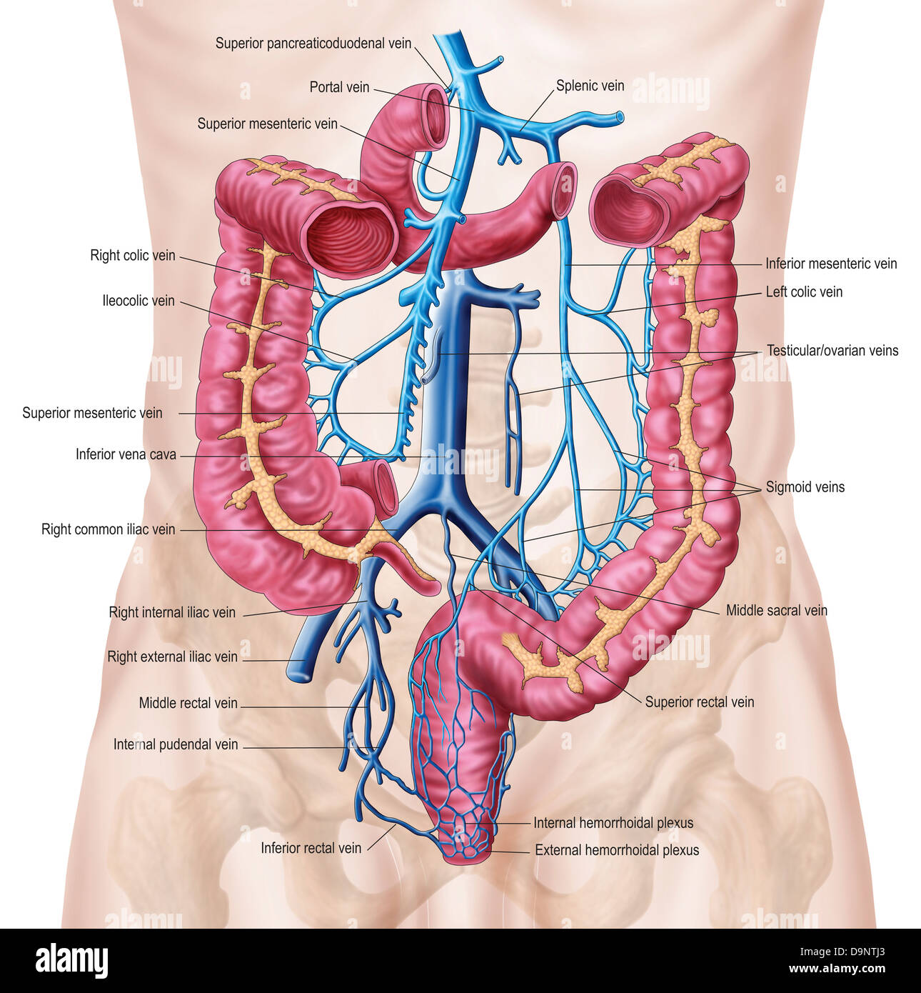 Anatomy of human abdominal vein system. Stock Photohttps://www.alamy.com/image-license-details/?v=1https://www.alamy.com/stock-photo-anatomy-of-human-abdominal-vein-system-57643323.html
RFD9NTJ3–Anatomy of human abdominal vein system.
 . Cunningham's Text-book of anatomy. Anatomy. 76-i THE NEKVOUS SYSTEM. detached portion at the lower end is named the aortico-renal ganglion. Other small scattered masses of cells are present in the cceliac plexus. At the upper end the cceliac ganglion receives the greater splanchnic nerve. The aortico-renal ganglion % i JK/*° .Tfliujm ---Greater splanchriic'ii'i ⢠â ⢠â¢mkr â - Abdominal sympathetic chain *V%£ ; «a^ â - -Cceliac ganglion Suprarenal plexus Smaller splanchnic nerve -4 'ijxlift- *> S»" "Lowest splanchnic nerve Superior mesenteric plexus-*4| .'; -^rrW^ * Aortic Stock Photohttps://www.alamy.com/image-license-details/?v=1https://www.alamy.com/cunninghams-text-book-of-anatomy-anatomy-76-i-the-nekvous-system-detached-portion-at-the-lower-end-is-named-the-aortico-renal-ganglion-other-small-scattered-masses-of-cells-are-present-in-the-cceliac-plexus-at-the-upper-end-the-cceliac-ganglion-receives-the-greater-splanchnic-nerve-the-aortico-renal-ganglion-i-jk-tfliujm-greater-splanchriiciii-mkr-abdominal-sympathetic-chain-v-a-cceliac-ganglion-suprarenal-plexus-smaller-splanchnic-nerve-4-ijxlift-gt-squot-quotlowest-splanchnic-nerve-superior-mesenteric-plexus-4-rrw-aortic-image216345239.html
RMPFYAFK–. Cunningham's Text-book of anatomy. Anatomy. 76-i THE NEKVOUS SYSTEM. detached portion at the lower end is named the aortico-renal ganglion. Other small scattered masses of cells are present in the cceliac plexus. At the upper end the cceliac ganglion receives the greater splanchnic nerve. The aortico-renal ganglion % i JK/*° .Tfliujm ---Greater splanchriic'ii'i ⢠â ⢠â¢mkr â - Abdominal sympathetic chain *V%£ ; «a^ â - -Cceliac ganglion Suprarenal plexus Smaller splanchnic nerve -4 'ijxlift- *> S»" "Lowest splanchnic nerve Superior mesenteric plexus-*4| .'; -^rrW^ * Aortic
 A series of engravings, explaining the course of the nerves : with an address to young physicians on the study of the nerves . , formed by the SplanchnicNerve. (6. This Number is placed upon the Trunk of the Cceliac Artery; but surrounding the vessels, we see a great net-work of Nerves, viz. the Solar Plexus.* The Par Vagum coming down with the (Esophagus into the Abdomen. They are seen extensively distributed over the Stomach; some of their lesser twigs are at the same time seen to join the Solar Plexus. 17. The Superior Mesenteric Plexus, formed by the Solar Plexus being continued down upon Stock Photohttps://www.alamy.com/image-license-details/?v=1https://www.alamy.com/a-series-of-engravings-explaining-the-course-of-the-nerves-with-an-address-to-young-physicians-on-the-study-of-the-nerves-formed-by-the-splanchnicnerve-6-this-number-is-placed-upon-the-trunk-of-the-cceliac-artery-but-surrounding-the-vessels-we-see-a-great-net-work-of-nerves-viz-the-solar-plexus-the-par-vagum-coming-down-with-the-esophagus-into-the-abdomen-they-are-seen-extensively-distributed-over-the-stomach-some-of-their-lesser-twigs-are-at-the-same-time-seen-to-join-the-solar-plexus-17-the-superior-mesenteric-plexus-formed-by-the-solar-plexus-being-continued-down-upon-image343186820.html
RM2AX9E84–A series of engravings, explaining the course of the nerves : with an address to young physicians on the study of the nerves . , formed by the SplanchnicNerve. (6. This Number is placed upon the Trunk of the Cceliac Artery; but surrounding the vessels, we see a great net-work of Nerves, viz. the Solar Plexus.* The Par Vagum coming down with the (Esophagus into the Abdomen. They are seen extensively distributed over the Stomach; some of their lesser twigs are at the same time seen to join the Solar Plexus. 17. The Superior Mesenteric Plexus, formed by the Solar Plexus being continued down upon
 The laws and mechanics of circulation, with the principle involved in animal movement . Pig. 108.—Nerves of the Mesentery (reduced).—Bougery, etc. 1, Root of superiormesenteric artery and nervous plexus (a portion of the transverse colon is excised,in order to show this circumstance) ; 2, superior mesenteric plexus ; 5, 5, continua-tion of same over the walls of the vessels to the intestines ; A, intestines ; B, caecum ;C, appendix vermiformis ; D, ascending colon ; E, transverse colon ; F, descendingcolon. 274 NEliVES TO THE INTESTINES.. Fig. 109.—Solar Plexus (reduced).—Bougery, etc. The let Stock Photohttps://www.alamy.com/image-license-details/?v=1https://www.alamy.com/the-laws-and-mechanics-of-circulation-with-the-principle-involved-in-animal-movement-pig-108nerves-of-the-mesentery-reducedbougery-etc-1-root-of-superiormesenteric-artery-and-nervous-plexus-a-portion-of-the-transverse-colon-is-excisedin-order-to-show-this-circumstance-2-superior-mesenteric-plexus-5-5-continua-tion-of-same-over-the-walls-of-the-vessels-to-the-intestines-a-intestines-b-caecum-c-appendix-vermiformis-d-ascending-colon-e-transverse-colon-f-descendingcolon-274-nelives-to-the-intestines-fig-109solar-plexus-reducedbougery-etc-the-let-image339954433.html
RM2AN279N–The laws and mechanics of circulation, with the principle involved in animal movement . Pig. 108.—Nerves of the Mesentery (reduced).—Bougery, etc. 1, Root of superiormesenteric artery and nervous plexus (a portion of the transverse colon is excised,in order to show this circumstance) ; 2, superior mesenteric plexus ; 5, 5, continua-tion of same over the walls of the vessels to the intestines ; A, intestines ; B, caecum ;C, appendix vermiformis ; D, ascending colon ; E, transverse colon ; F, descendingcolon. 274 NEliVES TO THE INTESTINES.. Fig. 109.—Solar Plexus (reduced).—Bougery, etc. The let
 . Cunningham's Text-book of anatomy. Anatomy. (LECUM AND VERMIFOBM PEOCESS. 1213 Nerves.—The nerves come from the superior mesenteric plexus, an offshoot of the cceliac plexus, and from the inferior mesenteric, a derivative of the aortic plexus. The arrangement is similar to that of the nerves of the small intestine. INTESTINUM (LECUM AND PKOCESSUS VERMIFORMIS. Intestinum Caecum.—After leaving the pelvic cavity, as already described, terminal portion of the small intestine passes upwards, backwards, and to right, and opens, by the ileo- cecal orifice, into the large in- testine some 2| inches Stock Photohttps://www.alamy.com/image-license-details/?v=1https://www.alamy.com/cunninghams-text-book-of-anatomy-anatomy-lecum-and-vermifobm-peocess-1213-nervesthe-nerves-come-from-the-superior-mesenteric-plexus-an-offshoot-of-the-cceliac-plexus-and-from-the-inferior-mesenteric-a-derivative-of-the-aortic-plexus-the-arrangement-is-similar-to-that-of-the-nerves-of-the-small-intestine-intestinum-lecum-and-pkocessus-vermiformis-intestinum-caecumafter-leaving-the-pelvic-cavity-as-already-described-terminal-portion-of-the-small-intestine-passes-upwards-backwards-and-to-right-and-opens-by-the-ileo-cecal-orifice-into-the-large-in-testine-some-2-inches-image231849004.html
RMRD5HNG–. Cunningham's Text-book of anatomy. Anatomy. (LECUM AND VERMIFOBM PEOCESS. 1213 Nerves.—The nerves come from the superior mesenteric plexus, an offshoot of the cceliac plexus, and from the inferior mesenteric, a derivative of the aortic plexus. The arrangement is similar to that of the nerves of the small intestine. INTESTINUM (LECUM AND PKOCESSUS VERMIFORMIS. Intestinum Caecum.—After leaving the pelvic cavity, as already described, terminal portion of the small intestine passes upwards, backwards, and to right, and opens, by the ileo- cecal orifice, into the large in- testine some 2| inches
 . Anatomy in a nutshell : a treatise on human anatomy in its relation to osteopathy. Human anatomy; Osteopathic medicine; Osteopathic Medicine; Anatomy. 421) ANATOMY IN A NUTSHELL. from the cceliac plexus, the right vagus nerve, and the left semilunar ganglion. It distributes branches to the pancreatic and left gastro-epiploic plexus,also to the substance of the spl< i n. Superior Mesenteric Plexus. This plexus emerges from under cover of the pancreas and surrounds the trunk of the superior ni< seiiti ric artery. A few ganglia which are found around this artery are called ganglia mesente Stock Photohttps://www.alamy.com/image-license-details/?v=1https://www.alamy.com/anatomy-in-a-nutshell-a-treatise-on-human-anatomy-in-its-relation-to-osteopathy-human-anatomy-osteopathic-medicine-osteopathic-medicine-anatomy-421-anatomy-in-a-nutshell-from-the-cceliac-plexus-the-right-vagus-nerve-and-the-left-semilunar-ganglion-it-distributes-branches-to-the-pancreatic-and-left-gastro-epiploic-plexusalso-to-the-substance-of-the-spllt-i-n-superior-mesenteric-plexus-this-plexus-emerges-from-under-cover-of-the-pancreas-and-surrounds-the-trunk-of-the-superior-nilt-seiiti-ric-artery-a-few-ganglia-which-are-found-around-this-artery-are-called-ganglia-mesente-image236802596.html
RMRN783G–. Anatomy in a nutshell : a treatise on human anatomy in its relation to osteopathy. Human anatomy; Osteopathic medicine; Osteopathic Medicine; Anatomy. 421) ANATOMY IN A NUTSHELL. from the cceliac plexus, the right vagus nerve, and the left semilunar ganglion. It distributes branches to the pancreatic and left gastro-epiploic plexus,also to the substance of the spl< i n. Superior Mesenteric Plexus. This plexus emerges from under cover of the pancreas and surrounds the trunk of the superior ni< seiiti ric artery. A few ganglia which are found around this artery are called ganglia mesente
 . Cunningham's Text-book of anatomy. Anatomy. 76-i THE NEKVOUS SYSTEM. detached portion at the lower end is named the aortico-renal ganglion. Other small scattered masses of cells are present in the cceliac plexus. At the upper end the cceliac ganglion receives the greater splanchnic nerve. The aortico-renal ganglion % i JK/*° .Tfliujm ---Greater splanchriic'ii'i ⢠â ⢠â¢mkr â - Abdominal sympathetic chain *V%£ ; «a^ â - -Cceliac ganglion Suprarenal plexus Smaller splanchnic nerve -4 'ijxlift- *> S»" "Lowest splanchnic nerve Superior mesenteric plexus-*4| .'; -^rrW^ * Aortic Stock Photohttps://www.alamy.com/image-license-details/?v=1https://www.alamy.com/cunninghams-text-book-of-anatomy-anatomy-76-i-the-nekvous-system-detached-portion-at-the-lower-end-is-named-the-aortico-renal-ganglion-other-small-scattered-masses-of-cells-are-present-in-the-cceliac-plexus-at-the-upper-end-the-cceliac-ganglion-receives-the-greater-splanchnic-nerve-the-aortico-renal-ganglion-i-jk-tfliujm-greater-splanchriiciii-mkr-abdominal-sympathetic-chain-v-a-cceliac-ganglion-suprarenal-plexus-smaller-splanchnic-nerve-4-ijxlift-gt-squot-quotlowest-splanchnic-nerve-superior-mesenteric-plexus-4-rrw-aortic-image231868944.html
RMRD6F5M–. Cunningham's Text-book of anatomy. Anatomy. 76-i THE NEKVOUS SYSTEM. detached portion at the lower end is named the aortico-renal ganglion. Other small scattered masses of cells are present in the cceliac plexus. At the upper end the cceliac ganglion receives the greater splanchnic nerve. The aortico-renal ganglion % i JK/*° .Tfliujm ---Greater splanchriic'ii'i ⢠â ⢠â¢mkr â - Abdominal sympathetic chain *V%£ ; «a^ â - -Cceliac ganglion Suprarenal plexus Smaller splanchnic nerve -4 'ijxlift- *> S»" "Lowest splanchnic nerve Superior mesenteric plexus-*4| .'; -^rrW^ * Aortic
 Archives of neurology and psychopathology. . nal sympatheticnerve, known as the superior mesenteric nerve, themiddle mesenteric nerve, and the inferior mesentericnerve. The inferior mesenteric ganglion gives off the hypogas-tric nerves, one for each side, which course with thehypogastric plexus (sympathetic supply). The latterreceives additional supply on each side by a direct branchfrom the second, another from the third sacral nerves(Nawrocki and Skabitschewski). Chapter IV. ARCHITECTURE AND MORPHOLOGICAL ORGANIZATION OF THESYMPATHETIC. General Morphological Interrelation of the SympatheticS Stock Photohttps://www.alamy.com/image-license-details/?v=1https://www.alamy.com/archives-of-neurology-and-psychopathology-nal-sympatheticnerve-known-as-the-superior-mesenteric-nerve-themiddle-mesenteric-nerve-and-the-inferior-mesentericnerve-the-inferior-mesenteric-ganglion-gives-off-the-hypogas-tric-nerves-one-for-each-side-which-course-with-thehypogastric-plexus-sympathetic-supply-the-latterreceives-additional-supply-on-each-side-by-a-direct-branchfrom-the-second-another-from-the-third-sacral-nervesnawrocki-and-skabitschewski-chapter-iv-architecture-and-morphological-organization-of-thesympathetic-general-morphological-interrelation-of-the-sympathetics-image342771575.html
RM2AWJGHY–Archives of neurology and psychopathology. . nal sympatheticnerve, known as the superior mesenteric nerve, themiddle mesenteric nerve, and the inferior mesentericnerve. The inferior mesenteric ganglion gives off the hypogas-tric nerves, one for each side, which course with thehypogastric plexus (sympathetic supply). The latterreceives additional supply on each side by a direct branchfrom the second, another from the third sacral nerves(Nawrocki and Skabitschewski). Chapter IV. ARCHITECTURE AND MORPHOLOGICAL ORGANIZATION OF THESYMPATHETIC. General Morphological Interrelation of the SympatheticS
 A manual of anatomy . formed by branches from therenal and aortic plexuses; branches pass to the testes. {b) The ovarian plexuses are formed in the same manner and sendbranches to the ovaries, oviducts and borders of the uterus. Theycommunicate with the uterine plexus. 9. The aortic plexus is situated upon the front and sides of theabdominal aorta between the superior and inferior mesentericarteries. It is made up of branches from the solar plexus and thelumbar sympathetic gangha. It distributes branches to the sper-matic, inferior mesenteric, suprarenal, renal, and hypogastric plexusesand to Stock Photohttps://www.alamy.com/image-license-details/?v=1https://www.alamy.com/a-manual-of-anatomy-formed-by-branches-from-therenal-and-aortic-plexuses-branches-pass-to-the-testes-b-the-ovarian-plexuses-are-formed-in-the-same-manner-and-sendbranches-to-the-ovaries-oviducts-and-borders-of-the-uterus-theycommunicate-with-the-uterine-plexus-9-the-aortic-plexus-is-situated-upon-the-front-and-sides-of-theabdominal-aorta-between-the-superior-and-inferior-mesentericarteries-it-is-made-up-of-branches-from-the-solar-plexus-and-thelumbar-sympathetic-gangha-it-distributes-branches-to-the-sper-matic-inferior-mesenteric-suprarenal-renal-and-hypogastric-plexusesand-to-image343317203.html
RM2AXFCGK–A manual of anatomy . formed by branches from therenal and aortic plexuses; branches pass to the testes. {b) The ovarian plexuses are formed in the same manner and sendbranches to the ovaries, oviducts and borders of the uterus. Theycommunicate with the uterine plexus. 9. The aortic plexus is situated upon the front and sides of theabdominal aorta between the superior and inferior mesentericarteries. It is made up of branches from the solar plexus and thelumbar sympathetic gangha. It distributes branches to the sper-matic, inferior mesenteric, suprarenal, renal, and hypogastric plexusesand to
 Abdominal surgery . foramen of Winslow. In theanterior edge of the foramen we find: the hepatic artery infront; the hepatic and cystic ducts, and the origin of the ductuscholedochus, in the middle; the portal vein behind the branchesof the great sympathetic, and the end of the right vagus.Behind the stomach we have the second and third portions ofthe duodenum, the pancreas, the coeliac axis and its branches,the superior mesenteric artery surrounded by lymphatic glands,and the solar plexus ; and behind all, the aorta and vena cava,resting on the vertebral column. Right Hypochondriac (II.)—Chief Stock Photohttps://www.alamy.com/image-license-details/?v=1https://www.alamy.com/abdominal-surgery-foramen-of-winslow-in-theanterior-edge-of-the-foramen-we-find-the-hepatic-artery-infront-the-hepatic-and-cystic-ducts-and-the-origin-of-the-ductuscholedochus-in-the-middle-the-portal-vein-behind-the-branchesof-the-great-sympathetic-and-the-end-of-the-right-vagusbehind-the-stomach-we-have-the-second-and-third-portions-ofthe-duodenum-the-pancreas-the-coeliac-axis-and-its-branchesthe-superior-mesenteric-artery-surrounded-by-lymphatic-glandsand-the-solar-plexus-and-behind-all-the-aorta-and-vena-cavaresting-on-the-vertebral-column-right-hypochondriac-iichief-image339148597.html
RM2AKNFDW–Abdominal surgery . foramen of Winslow. In theanterior edge of the foramen we find: the hepatic artery infront; the hepatic and cystic ducts, and the origin of the ductuscholedochus, in the middle; the portal vein behind the branchesof the great sympathetic, and the end of the right vagus.Behind the stomach we have the second and third portions ofthe duodenum, the pancreas, the coeliac axis and its branches,the superior mesenteric artery surrounded by lymphatic glands,and the solar plexus ; and behind all, the aorta and vena cava,resting on the vertebral column. Right Hypochondriac (II.)—Chief
 A manual of anatomy . rior pancreaticoduodenale (from thegastroduodenale of the hepatic artery), the inferior pancreaticoduo-denale (from the superior mesenteric artery) and pancreatic branchesfrom the splenic and hepatic arteries. The veins empty into thesuperior mesenteric, splenic and portal veins. The nerves are mainlysympathetic from the solar plexus by way of the celiac, splenic andsuperior mesenteric plexuses. CHAPTER VIII THE URINARY SYSTEM The urinary system comprises the two kidneys, two ureters, theMadder and the urethra. THE KJDITEYS Each kidney {ren) is a large, bean-shaped organ, Stock Photohttps://www.alamy.com/image-license-details/?v=1https://www.alamy.com/a-manual-of-anatomy-rior-pancreaticoduodenale-from-thegastroduodenale-of-the-hepatic-artery-the-inferior-pancreaticoduo-denale-from-the-superior-mesenteric-artery-and-pancreatic-branchesfrom-the-splenic-and-hepatic-arteries-the-veins-empty-into-thesuperior-mesenteric-splenic-and-portal-veins-the-nerves-are-mainlysympathetic-from-the-solar-plexus-by-way-of-the-celiac-splenic-andsuperior-mesenteric-plexuses-chapter-viii-the-urinary-system-the-urinary-system-comprises-the-two-kidneys-two-ureters-themadder-and-the-urethra-the-kjditeys-each-kidney-ren-is-a-large-bean-shaped-organ-image343343358.html
RM2AXGHXP–A manual of anatomy . rior pancreaticoduodenale (from thegastroduodenale of the hepatic artery), the inferior pancreaticoduo-denale (from the superior mesenteric artery) and pancreatic branchesfrom the splenic and hepatic arteries. The veins empty into thesuperior mesenteric, splenic and portal veins. The nerves are mainlysympathetic from the solar plexus by way of the celiac, splenic andsuperior mesenteric plexuses. CHAPTER VIII THE URINARY SYSTEM The urinary system comprises the two kidneys, two ureters, theMadder and the urethra. THE KJDITEYS Each kidney {ren) is a large, bean-shaped organ,
 . Human physiology. ing to the inci.sive fora-men. 5. Recurrent branch or Vidian nerve dividing into the carotid and petrosal branches. 6. Poste-rior palatine branches. 7. Lingual nerve joined by the chorda tympani. 8. Portio dura of the seventhpair. 9. Superior cervical ganglion. 10 Middle cervical ganglion. 11. Inferior cervical ganglion. 12.Roots of the great splanchnic nerve arising from the dorsal ganglia. 13. Lesser splanchnic nerve. 14.Renal plexus. 15. Solar plexus. 16. Mesenteric plexus. 17. Lumbar ganglia. 18. Sacral ganglia. 19.Vesical plexus. 20. Rectal plexus. 21. Lumbar plexus (c Stock Photohttps://www.alamy.com/image-license-details/?v=1https://www.alamy.com/human-physiology-ing-to-the-incisive-fora-men-5-recurrent-branch-or-vidian-nerve-dividing-into-the-carotid-and-petrosal-branches-6-poste-rior-palatine-branches-7-lingual-nerve-joined-by-the-chorda-tympani-8-portio-dura-of-the-seventhpair-9-superior-cervical-ganglion-10-middle-cervical-ganglion-11-inferior-cervical-ganglion-12roots-of-the-great-splanchnic-nerve-arising-from-the-dorsal-ganglia-13-lesser-splanchnic-nerve-14renal-plexus-15-solar-plexus-16-mesenteric-plexus-17-lumbar-ganglia-18-sacral-ganglia-19vesical-plexus-20-rectal-plexus-21-lumbar-plexus-c-image336599167.html
RM2AFHBJR–. Human physiology. ing to the inci.sive fora-men. 5. Recurrent branch or Vidian nerve dividing into the carotid and petrosal branches. 6. Poste-rior palatine branches. 7. Lingual nerve joined by the chorda tympani. 8. Portio dura of the seventhpair. 9. Superior cervical ganglion. 10 Middle cervical ganglion. 11. Inferior cervical ganglion. 12.Roots of the great splanchnic nerve arising from the dorsal ganglia. 13. Lesser splanchnic nerve. 14.Renal plexus. 15. Solar plexus. 16. Mesenteric plexus. 17. Lumbar ganglia. 18. Sacral ganglia. 19.Vesical plexus. 20. Rectal plexus. 21. Lumbar plexus (c
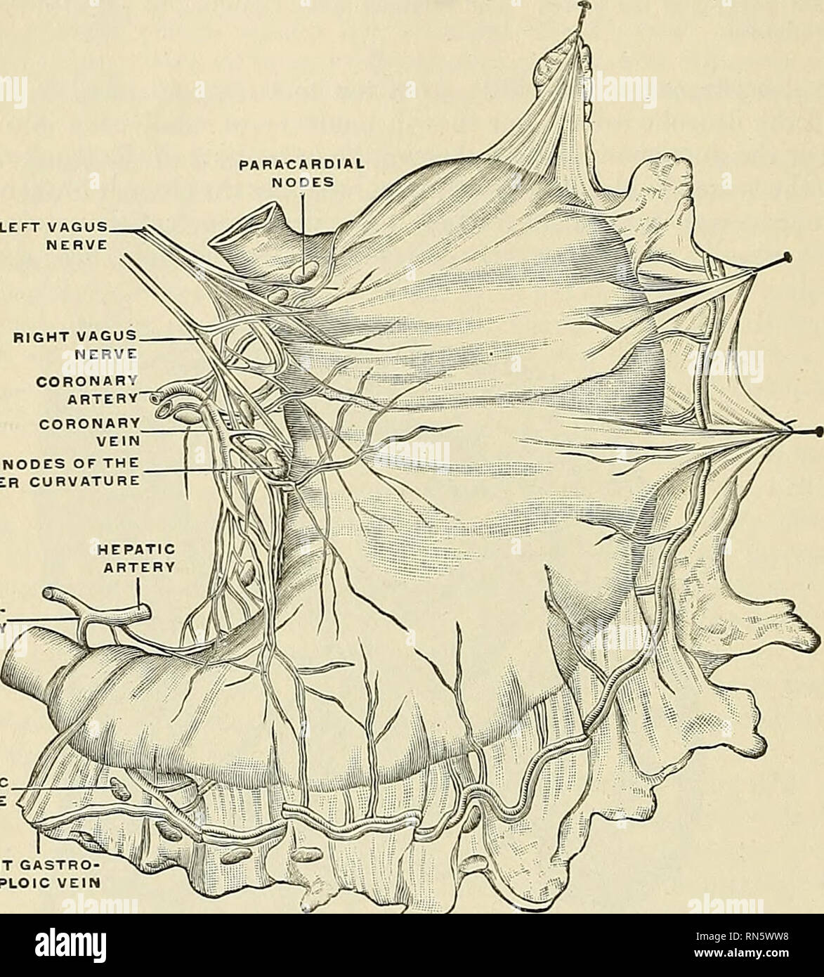 . Anatomy, descriptive and applied. Anatomy. 792 THE VASCULAR SYSTEMS around the superior hemorrhoidal artery; and (c) a pararectal group in contact with the muscle coat of the rectum. Their afFerents drain the descending colon, sigmoid flexure, and upper portion of the rectum; their efFerents pass to the inferior mesenteric nodes. SUBPYLORI NODE /^. "M J Fig. 570.—General view of the subperitoneal lymphatic plexus of the stomach prepared by the nicthod of Gerota. (Cun^o.) The Lymphatic Vessels of the Abdominal and Pelvic Viscera, These consist of: (1) Those of the subdiaphragmatic portio Stock Photohttps://www.alamy.com/image-license-details/?v=1https://www.alamy.com/anatomy-descriptive-and-applied-anatomy-792-the-vascular-systems-around-the-superior-hemorrhoidal-artery-and-c-a-pararectal-group-in-contact-with-the-muscle-coat-of-the-rectum-their-afferents-drain-the-descending-colon-sigmoid-flexure-and-upper-portion-of-the-rectum-their-efferents-pass-to-the-inferior-mesenteric-nodes-subpylori-node-quotm-j-fig-570general-view-of-the-subperitoneal-lymphatic-plexus-of-the-stomach-prepared-by-the-nicthod-of-gerota-cuno-the-lymphatic-vessels-of-the-abdominal-and-pelvic-viscera-these-consist-of-1-those-of-the-subdiaphragmatic-portio-image236772628.html
RMRN5WW8–. Anatomy, descriptive and applied. Anatomy. 792 THE VASCULAR SYSTEMS around the superior hemorrhoidal artery; and (c) a pararectal group in contact with the muscle coat of the rectum. Their afFerents drain the descending colon, sigmoid flexure, and upper portion of the rectum; their efFerents pass to the inferior mesenteric nodes. SUBPYLORI NODE /^. "M J Fig. 570.—General view of the subperitoneal lymphatic plexus of the stomach prepared by the nicthod of Gerota. (Cun^o.) The Lymphatic Vessels of the Abdominal and Pelvic Viscera, These consist of: (1) Those of the subdiaphragmatic portio
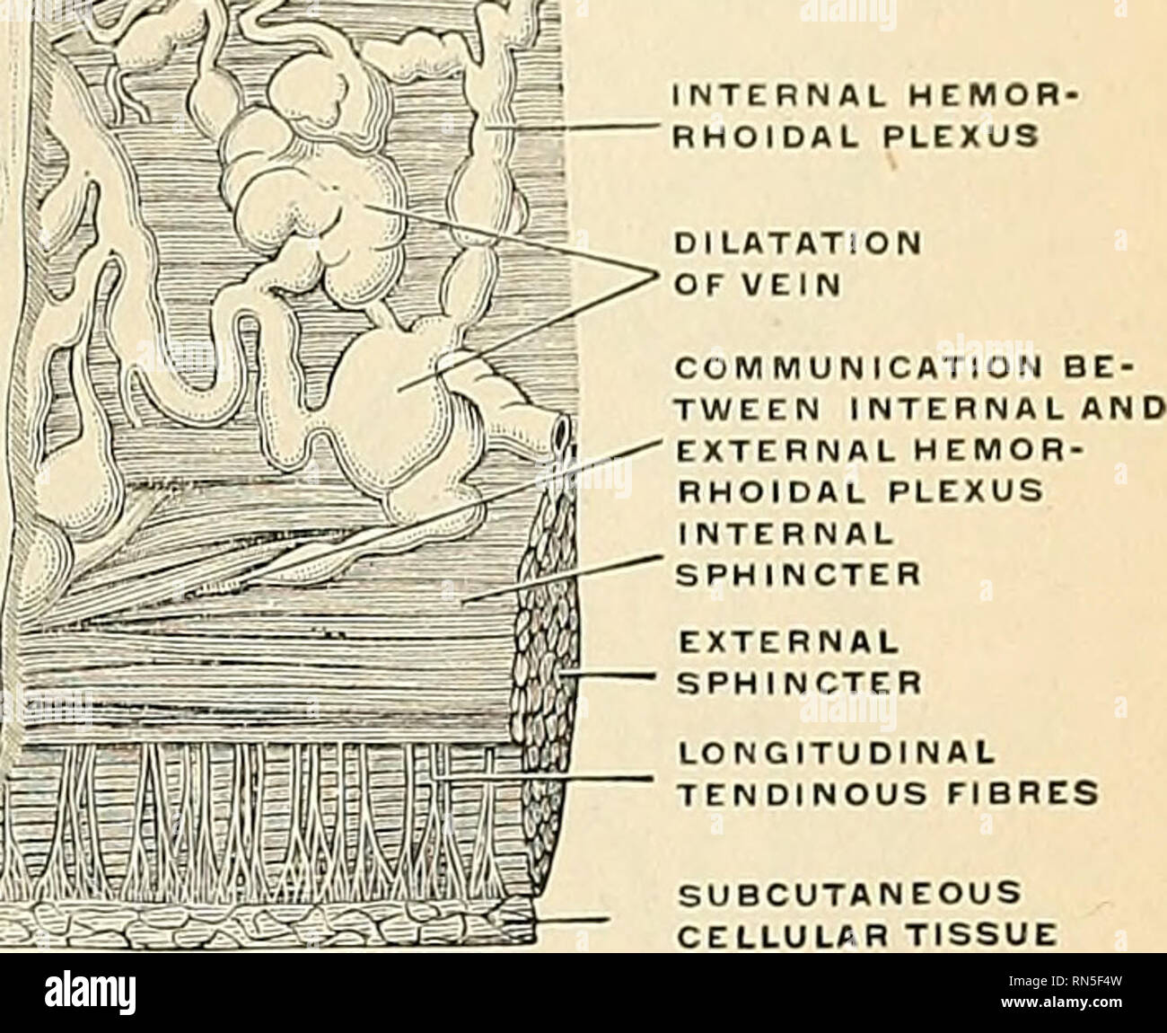 . Anatomy, descriptive and applied. Anatomy. THE RECTUM 1311 (Fig. 1075). The ascending colon is supplied by the right colic, and the transverse colon by the middle colic branch of the superior mesenteric. The descending colon is supi^lied by the ^^.Ly. %;^^^. LULAH TISSUE Fig. 1074.—Inner wall ot the lower end of the rectum and anus. On the right the mucous membrane has been removed to show the dilatation of the veins and how they pass through the muscular wall to anastomose with the external hemorrhoidal plexus. (Luschka.). Please note that these images are extracted from scanned page ima Stock Photohttps://www.alamy.com/image-license-details/?v=1https://www.alamy.com/anatomy-descriptive-and-applied-anatomy-the-rectum-1311-fig-1075-the-ascending-colon-is-supplied-by-the-right-colic-and-the-transverse-colon-by-the-middle-colic-branch-of-the-superior-mesenteric-the-descending-colon-is-supilied-by-the-ly-lulah-tissue-fig-1074inner-wall-ot-the-lower-end-of-the-rectum-and-anus-on-the-right-the-mucous-membrane-has-been-removed-to-show-the-dilatation-of-the-veins-and-how-they-pass-through-the-muscular-wall-to-anastomose-with-the-external-hemorrhoidal-plexus-luschka-please-note-that-these-images-are-extracted-from-scanned-page-ima-image236764217.html
RMRN5F4W–. Anatomy, descriptive and applied. Anatomy. THE RECTUM 1311 (Fig. 1075). The ascending colon is supplied by the right colic, and the transverse colon by the middle colic branch of the superior mesenteric. The descending colon is supi^lied by the ^^.Ly. %;^^^. LULAH TISSUE Fig. 1074.—Inner wall ot the lower end of the rectum and anus. On the right the mucous membrane has been removed to show the dilatation of the veins and how they pass through the muscular wall to anastomose with the external hemorrhoidal plexus. (Luschka.). Please note that these images are extracted from scanned page ima
 . Chordate morphology. Morphology (Animals); Chordata. chordo tympani branch parotid salivary gland sphenopalatine ganglion palatine^ tympanic plexus ciliary ganglion ,,, I* 'i ''2,3 lacrimal gland eye part of sympathetic chain (including stellate ganglion) left ouf nuclei in spinal cord thoracics (lumbar outflow) sacral outflow. â â â I- submaxillary submaxillory salivary ,. ' ' . , ' ganglion gland otic ganglion bladder sphincter bladder superior mesenteric ganglion Figure 13-3. Autonomic system of the tetrapod. (After Goodrich, 1930) lature of the third arch (hyoid), and the parotid sahv Stock Photohttps://www.alamy.com/image-license-details/?v=1https://www.alamy.com/chordate-morphology-morphology-animals-chordata-chordo-tympani-branch-parotid-salivary-gland-sphenopalatine-ganglion-palatine-tympanic-plexus-ciliary-ganglion-i-i-23-lacrimal-gland-eye-part-of-sympathetic-chain-including-stellate-ganglion-left-ouf-nuclei-in-spinal-cord-thoracics-lumbar-outflow-sacral-outflow-i-submaxillary-submaxillory-salivary-ganglion-gland-otic-ganglion-bladder-sphincter-bladder-superior-mesenteric-ganglion-figure-13-3-autonomic-system-of-the-tetrapod-after-goodrich-1930-lature-of-the-third-arch-hyoid-and-the-parotid-sahv-image234949410.html
RMRJ6TAA–. Chordate morphology. Morphology (Animals); Chordata. chordo tympani branch parotid salivary gland sphenopalatine ganglion palatine^ tympanic plexus ciliary ganglion ,,, I* 'i ''2,3 lacrimal gland eye part of sympathetic chain (including stellate ganglion) left ouf nuclei in spinal cord thoracics (lumbar outflow) sacral outflow. â â â I- submaxillary submaxillory salivary ,. ' ' . , ' ganglion gland otic ganglion bladder sphincter bladder superior mesenteric ganglion Figure 13-3. Autonomic system of the tetrapod. (After Goodrich, 1930) lature of the third arch (hyoid), and the parotid sahv