Quick filters:
Symbiotic cells Stock Photos and Images
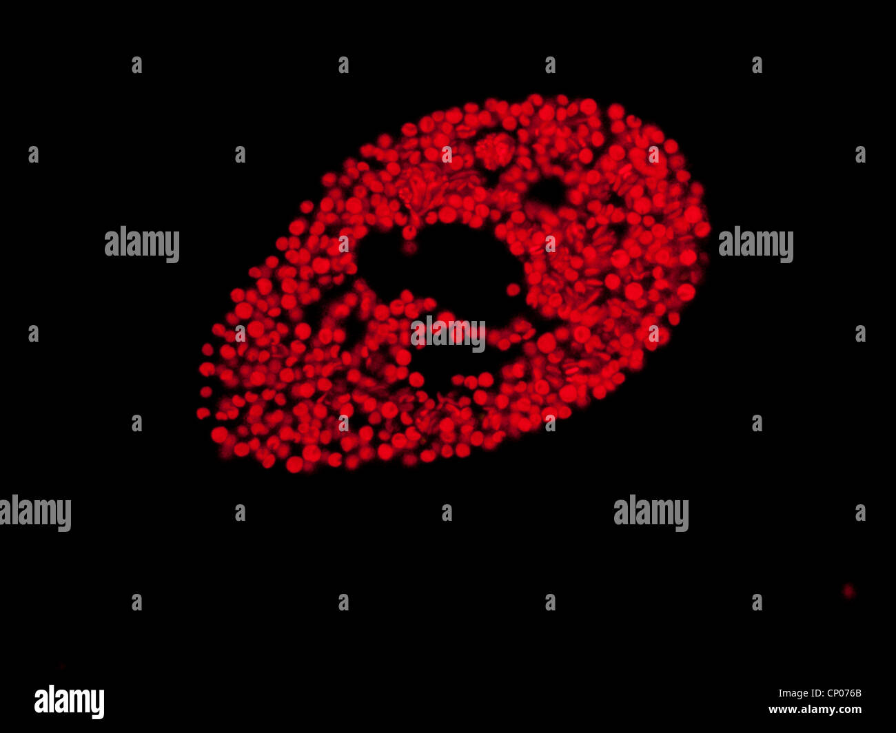 green slipper animalcule (Paramecium bursaria), with symbiotic zoochlorella, Germany Stock Photohttps://www.alamy.com/image-license-details/?v=1https://www.alamy.com/stock-photo-green-slipper-animalcule-paramecium-bursaria-with-symbiotic-zoochlorella-47948835.html
RMCP076B–green slipper animalcule (Paramecium bursaria), with symbiotic zoochlorella, Germany
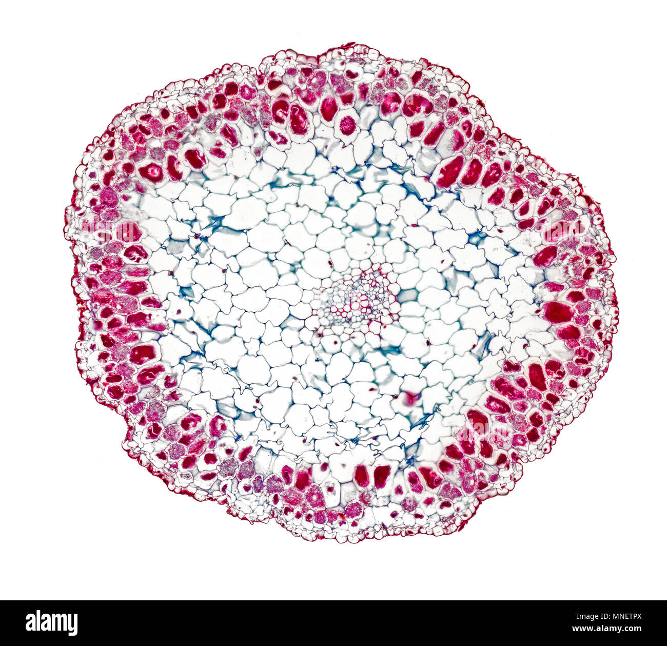 Birds nest orchid root, TS, mycorrhiza is a symbiotic association between a fungus and the roots of a vascular host plant. brightfield photomicrograph Stock Photohttps://www.alamy.com/image-license-details/?v=1https://www.alamy.com/birds-nest-orchid-root-ts-mycorrhiza-is-a-symbiotic-association-between-a-fungus-and-the-roots-of-a-vascular-host-plant-brightfield-photomicrograph-image185338242.html
RMMNETPX–Birds nest orchid root, TS, mycorrhiza is a symbiotic association between a fungus and the roots of a vascular host plant. brightfield photomicrograph
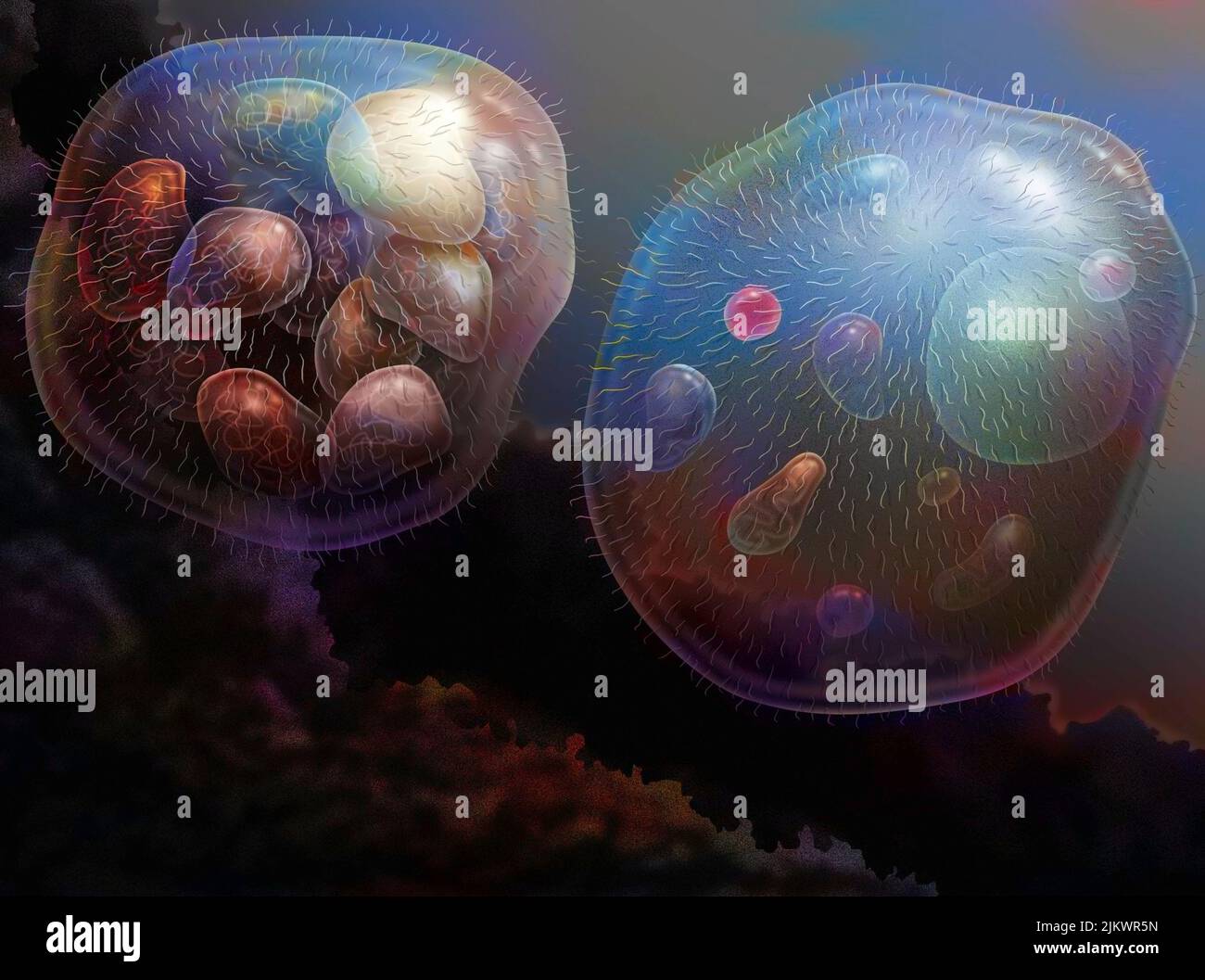 The nucleus cells would come from the association of several bacteria. Stock Photohttps://www.alamy.com/image-license-details/?v=1https://www.alamy.com/the-nucleus-cells-would-come-from-the-association-of-several-bacteria-image476925393.html
RF2JKWR5N–The nucleus cells would come from the association of several bacteria.
 Giant Green Anemone, Anthopleura xanthogrammica, in tide pool at Point of Arches in Olympic National Park, Washington State, USA Stock Photohttps://www.alamy.com/image-license-details/?v=1https://www.alamy.com/giant-green-anemone-anthopleura-xanthogrammica-in-tide-pool-at-point-of-arches-in-olympic-national-park-washington-state-usa-image527140226.html
RM2NHH8MJ–Giant Green Anemone, Anthopleura xanthogrammica, in tide pool at Point of Arches in Olympic National Park, Washington State, USA
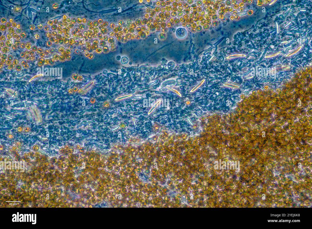 Symbiotic algae (zooxanthellae) and stinging cells from Aiptasia sp., probably belonging to the speceis Breviolum dendrogyrum. Stock Photohttps://www.alamy.com/image-license-details/?v=1https://www.alamy.com/symbiotic-algae-zooxanthellae-and-stinging-cells-from-aiptasia-sp-probably-belonging-to-the-speceis-breviolum-dendrogyrum-image628578812.html
RM2YEJ6K8–Symbiotic algae (zooxanthellae) and stinging cells from Aiptasia sp., probably belonging to the speceis Breviolum dendrogyrum.
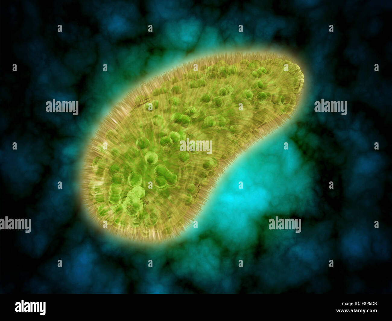 Microscopic view of Paramecium bursaria, a species of ciliate protozoan that has a mutualistic symbiotic relationship with green Stock Photohttps://www.alamy.com/image-license-details/?v=1https://www.alamy.com/stock-photo-microscopic-view-of-paramecium-bursaria-a-species-of-ciliate-protozoan-74246743.html
RFE8P6DB–Microscopic view of Paramecium bursaria, a species of ciliate protozoan that has a mutualistic symbiotic relationship with green
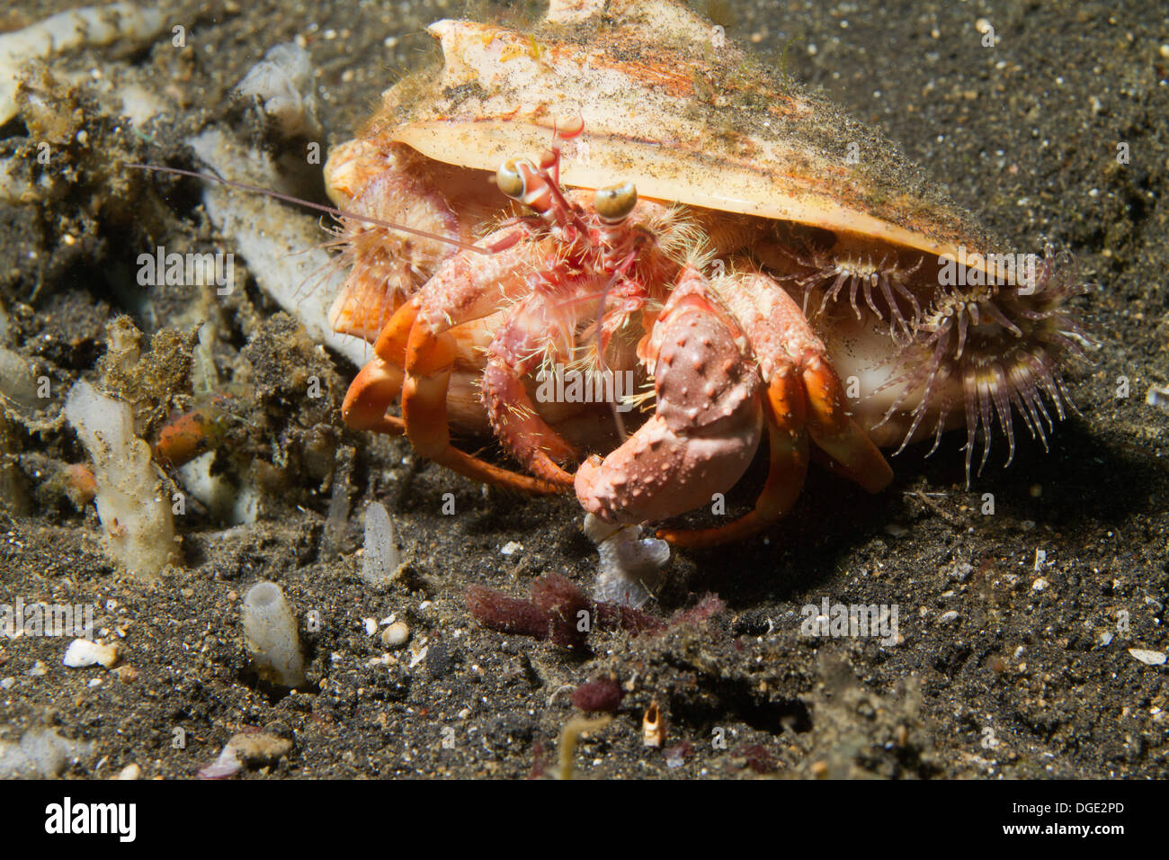 Anemone Hermit Crab carries symbiotic anemones on it's shell for protection.(Dardanus pedunculatus).Lembeh Straits,Indonesia Stock Photohttps://www.alamy.com/image-license-details/?v=1https://www.alamy.com/anemone-hermit-crab-carries-symbiotic-anemones-on-its-shell-for-protectiondardanus-image61775125.html
RMDGE2PD–Anemone Hermit Crab carries symbiotic anemones on it's shell for protection.(Dardanus pedunculatus).Lembeh Straits,Indonesia
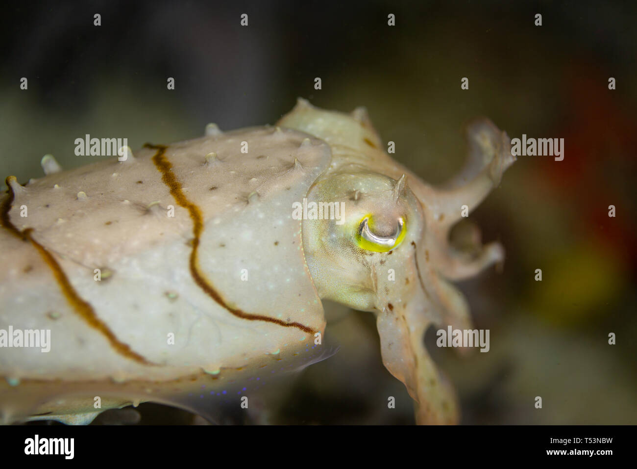 Closeup of a broadclub cuttlefish, Sepia latimanus,special pigment cells called chromatophores to turn white to blend with sand Stock Photohttps://www.alamy.com/image-license-details/?v=1https://www.alamy.com/closeup-of-a-broadclub-cuttlefish-sepia-latimanusspecial-pigment-cells-called-chromatophores-to-turn-white-to-blend-with-sand-image244101085.html
RMT53NBW–Closeup of a broadclub cuttlefish, Sepia latimanus,special pigment cells called chromatophores to turn white to blend with sand
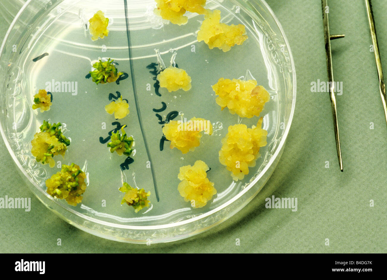 Alfalfa embryos forming from cells. Stock Photohttps://www.alamy.com/image-license-details/?v=1https://www.alamy.com/stock-photo-alfalfa-embryos-forming-from-cells-19967127.html
RMB4DG7K–Alfalfa embryos forming from cells.
 Pale cyan petals-like foliose lichen on rock, recent rains revived the vegetative body, natural macro background Stock Photohttps://www.alamy.com/image-license-details/?v=1https://www.alamy.com/pale-cyan-petals-like-foliose-lichen-on-rock-recent-rains-revived-the-vegetative-body-natural-macro-background-image459107006.html
RF2HJX3JP–Pale cyan petals-like foliose lichen on rock, recent rains revived the vegetative body, natural macro background
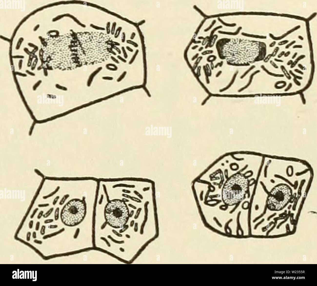 Archive image from page 134 of The cytoplasm of the plant. The cytoplasm of the plant cell cytoplasmofplant00guil Year: 1941 SKY, DuESBERG, LEVI, and MiLOViDOV, is today abandoned by PoR- TIER himself. Nevertheless it had the merit of initiating investi- gations which have produced methods by which chondriosomes can be distinguished in cells from symbiotic and parasitic bacteria, CowDRY and Olitsky, Duesberg, and Milovidov have described methods by which, in the cells of nodules of legumes and in the adipose cells of cockroaches, symbiotic bacteria can be distin- guished from the chondriosom Stock Photohttps://www.alamy.com/image-license-details/?v=1https://www.alamy.com/archive-image-from-page-134-of-the-cytoplasm-of-the-plant-the-cytoplasm-of-the-plant-cell-cytoplasmofplant00guil-year-1941-sky-duesberg-levi-and-milovidov-is-today-abandoned-by-por-tier-himself-nevertheless-it-had-the-merit-of-initiating-investi-gations-which-have-produced-methods-by-which-chondriosomes-can-be-distinguished-in-cells-from-symbiotic-and-parasitic-bacteria-cowdry-and-olitsky-duesberg-and-milovidov-have-described-methods-by-which-in-the-cells-of-nodules-of-legumes-and-in-the-adipose-cells-of-cockroaches-symbiotic-bacteria-can-be-distin-guished-from-the-chondriosom-image259454771.html
RMW2355R–Archive image from page 134 of The cytoplasm of the plant. The cytoplasm of the plant cell cytoplasmofplant00guil Year: 1941 SKY, DuESBERG, LEVI, and MiLOViDOV, is today abandoned by PoR- TIER himself. Nevertheless it had the merit of initiating investi- gations which have produced methods by which chondriosomes can be distinguished in cells from symbiotic and parasitic bacteria, CowDRY and Olitsky, Duesberg, and Milovidov have described methods by which, in the cells of nodules of legumes and in the adipose cells of cockroaches, symbiotic bacteria can be distin- guished from the chondriosom
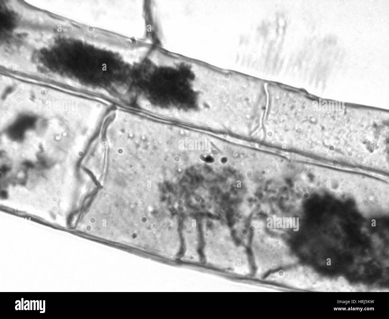 Arbuscules, Corn Root Cells Stock Photohttps://www.alamy.com/image-license-details/?v=1https://www.alamy.com/stock-photo-arbuscules-corn-root-cells-135009277.html
RMHRJ5KW–Arbuscules, Corn Root Cells
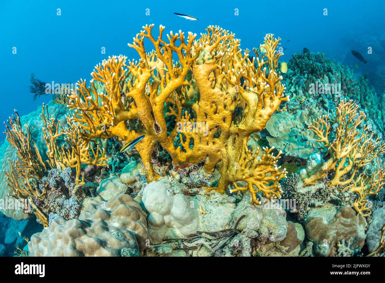 Fire coral, Millepora dichotoma, are similar to coral but are not true corals. They are instead more closely related to hydrozoans, making them hydroc Stock Photohttps://www.alamy.com/image-license-details/?v=1https://www.alamy.com/fire-coral-millepora-dichotoma-are-similar-to-coral-but-are-not-true-corals-they-are-instead-more-closely-related-to-hydrozoans-making-them-hydroc-image474469435.html
RM2JFWXGY–Fire coral, Millepora dichotoma, are similar to coral but are not true corals. They are instead more closely related to hydrozoans, making them hydroc
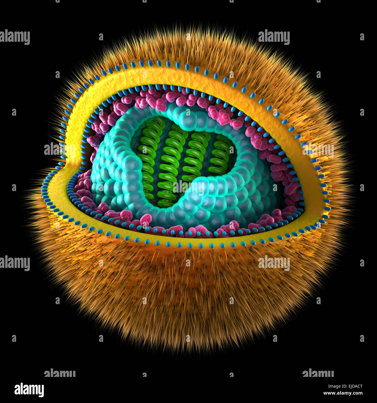 Herpes Simplex Cell - isolated on white Stock Photohttps://www.alamy.com/image-license-details/?v=1https://www.alamy.com/stock-photo-herpes-simplex-cell-isolated-on-white-80198856.html
RFEJDACT–Herpes Simplex Cell - isolated on white
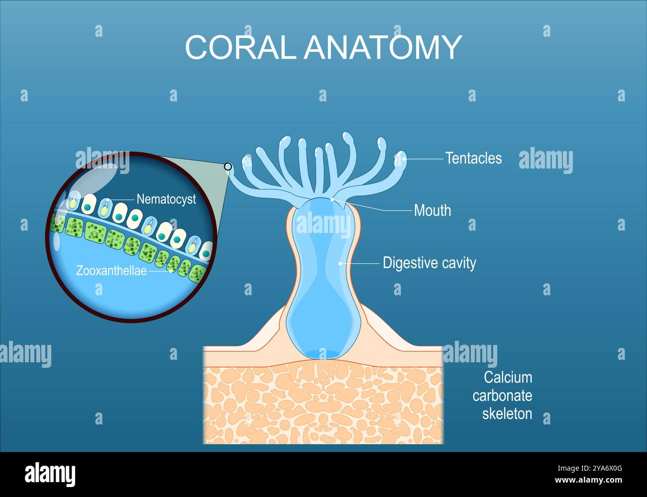 Coral anatomy. Cross section and structure of a polyp with digestive cavity, Mouth, and Tentacles. Close-up of epithelium with Zooxanthellae, and stin Stock Vectorhttps://www.alamy.com/image-license-details/?v=1https://www.alamy.com/coral-anatomy-cross-section-and-structure-of-a-polyp-with-digestive-cavity-mouth-and-tentacles-close-up-of-epithelium-with-zooxanthellae-and-stin-image625871920.html
RF2YA6X0G–Coral anatomy. Cross section and structure of a polyp with digestive cavity, Mouth, and Tentacles. Close-up of epithelium with Zooxanthellae, and stin
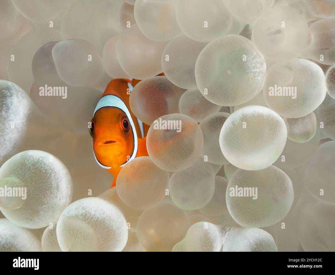 Juvenile western clown anemone fish hiding amongst the tentacles of a bubble tip anemone Stock Photohttps://www.alamy.com/image-license-details/?v=1https://www.alamy.com/juvenile-western-clown-anemone-fish-hiding-amongst-the-tentacles-of-a-bubble-tip-anemone-image627323156.html
RM2YCH12C–Juvenile western clown anemone fish hiding amongst the tentacles of a bubble tip anemone
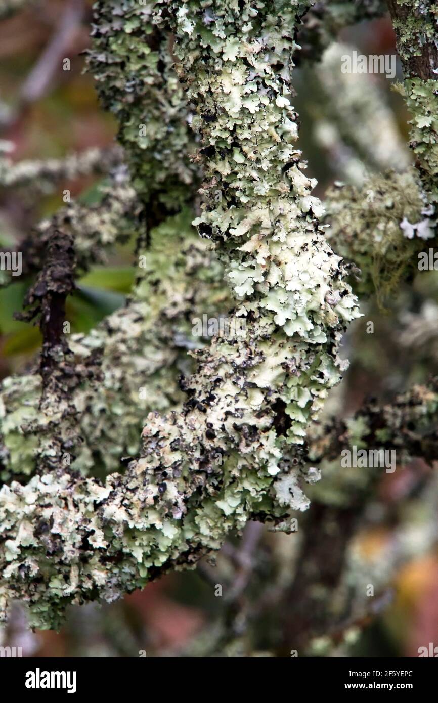 Close-up of leaflike white green lichen covering a tree branch Stock Photohttps://www.alamy.com/image-license-details/?v=1https://www.alamy.com/close-up-of-leaflike-white-green-lichen-covering-a-tree-branch-image416726420.html
RF2F5YEPC–Close-up of leaflike white green lichen covering a tree branch
 Lichen on an old tree in the New Forest, Hampshire, UK. Stock Photohttps://www.alamy.com/image-license-details/?v=1https://www.alamy.com/stock-image-lichen-on-an-old-tree-in-the-new-forest-hampshire-uk-167201377.html
RMKM0K1N–Lichen on an old tree in the New Forest, Hampshire, UK.
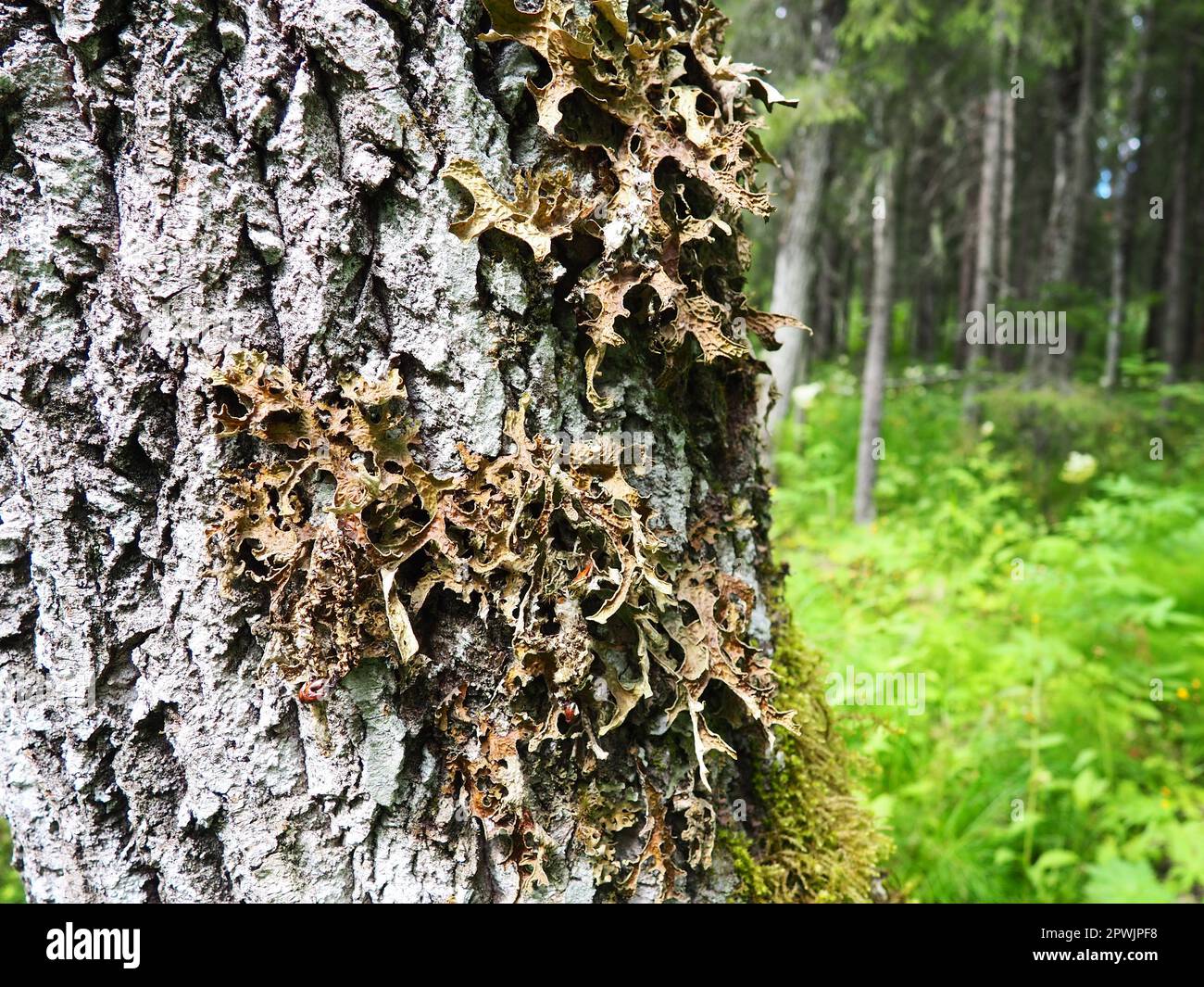 Moss and lichens on the bark of a tree in a spruce taiga forest. Karelia, Orzega. Lobaria Lobaria is a genus of lichenized ascomycetes belonging to th Stock Photohttps://www.alamy.com/image-license-details/?v=1https://www.alamy.com/moss-and-lichens-on-the-bark-of-a-tree-in-a-spruce-taiga-forest-karelia-orzega-lobaria-lobaria-is-a-genus-of-lichenized-ascomycetes-belonging-to-th-image549300620.html
RF2PWJPF8–Moss and lichens on the bark of a tree in a spruce taiga forest. Karelia, Orzega. Lobaria Lobaria is a genus of lichenized ascomycetes belonging to th
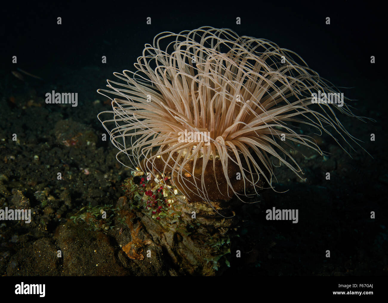 Tube anemone, Cerianthid, in Tulamben, Bali Sea, Indonesia Stock Photohttps://www.alamy.com/image-license-details/?v=1https://www.alamy.com/stock-photo-tube-anemone-cerianthid-in-tulamben-bali-sea-indonesia-89906282.html
RFF67GAJ–Tube anemone, Cerianthid, in Tulamben, Bali Sea, Indonesia
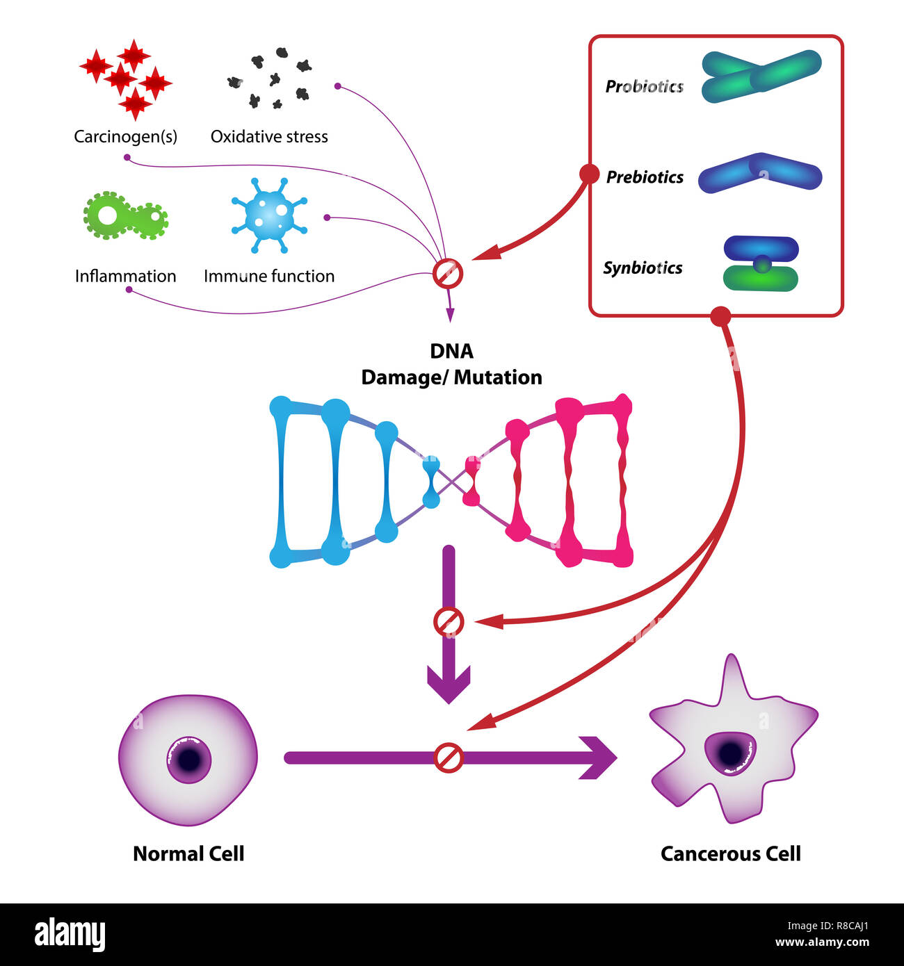 Probiotic bacteria prevent DNA damage and mutation, prevent the formation of cancer cells. Medical illustration Stock Photohttps://www.alamy.com/image-license-details/?v=1https://www.alamy.com/probiotic-bacteria-prevent-dna-damage-and-mutation-prevent-the-formation-of-cancer-cells-medical-illustration-image228923801.html
RFR8CAJ1–Probiotic bacteria prevent DNA damage and mutation, prevent the formation of cancer cells. Medical illustration
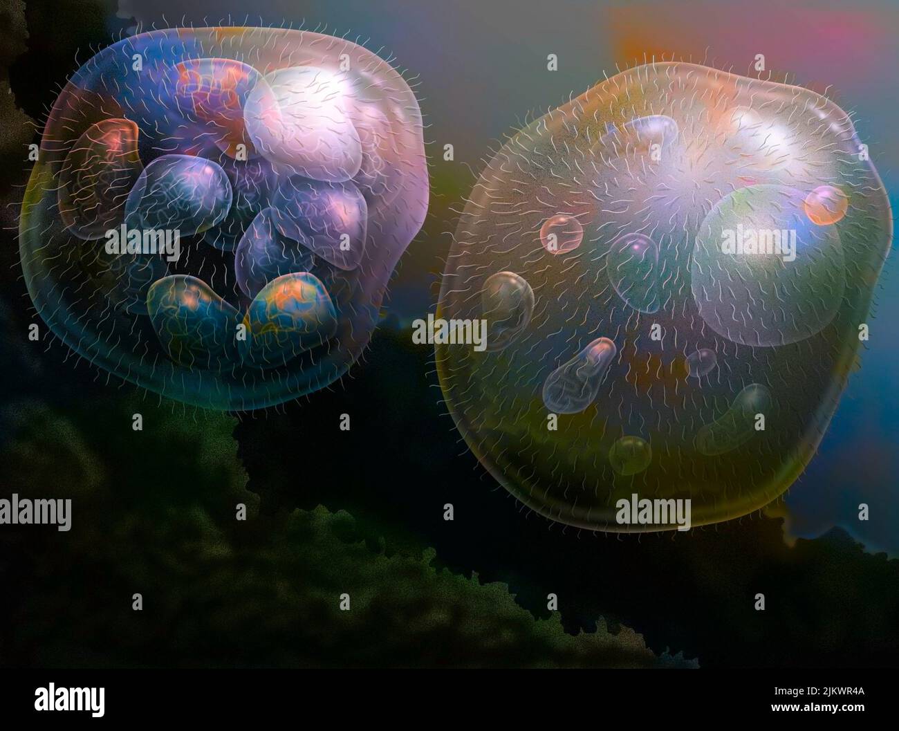 The nucleus cells would come from the association of several bacteria. Stock Photohttps://www.alamy.com/image-license-details/?v=1https://www.alamy.com/the-nucleus-cells-would-come-from-the-association-of-several-bacteria-image476925354.html
RF2JKWR4A–The nucleus cells would come from the association of several bacteria.
 Giant Green Anemone, Anthopleura xanthogrammica, in tide pool at Point of Arches in Olympic National Park, Washington State, USA Stock Photohttps://www.alamy.com/image-license-details/?v=1https://www.alamy.com/giant-green-anemone-anthopleura-xanthogrammica-in-tide-pool-at-point-of-arches-in-olympic-national-park-washington-state-usa-image527139949.html
RM2NHH8AN–Giant Green Anemone, Anthopleura xanthogrammica, in tide pool at Point of Arches in Olympic National Park, Washington State, USA
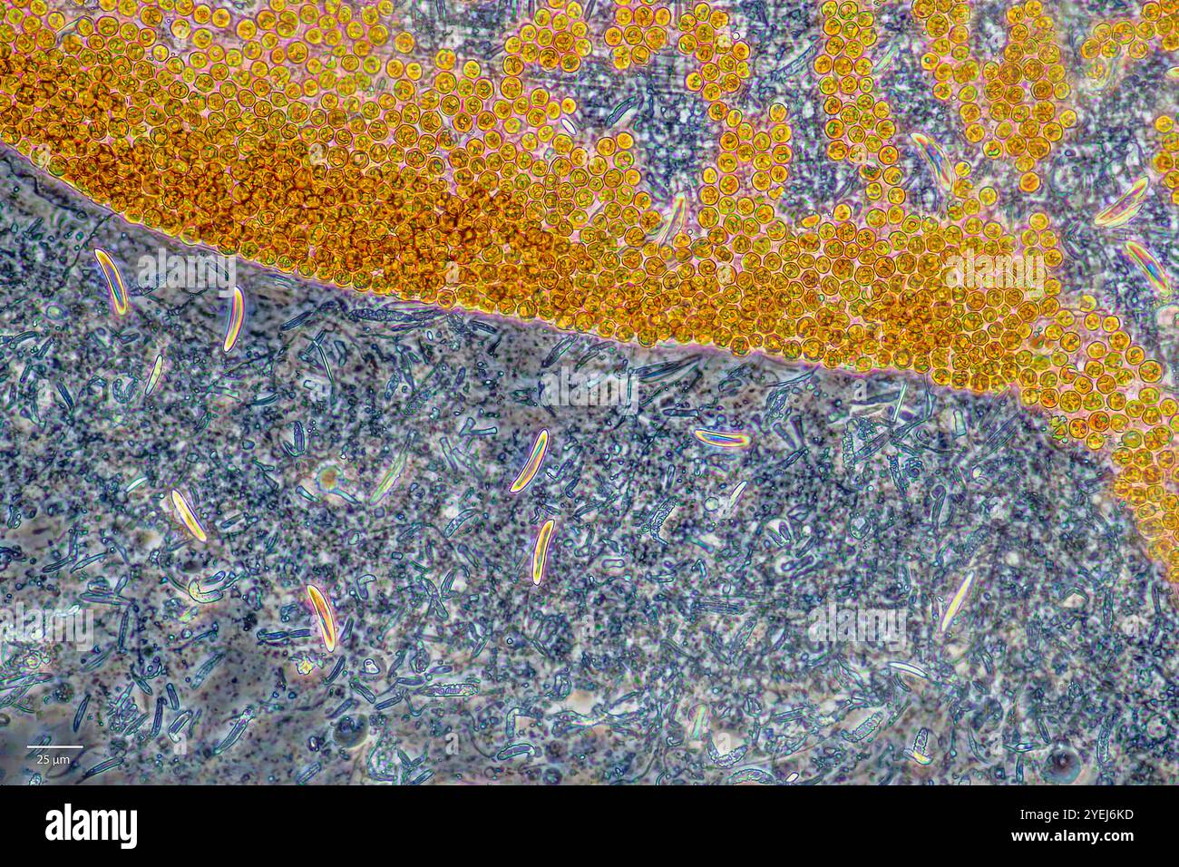 Symbiotic algae (zooxanthellae) and stinging cells from Aiptasia sp., probably belonging to the speceis Breviolum dendrogyrum. Stock Photohttps://www.alamy.com/image-license-details/?v=1https://www.alamy.com/symbiotic-algae-zooxanthellae-and-stinging-cells-from-aiptasia-sp-probably-belonging-to-the-speceis-breviolum-dendrogyrum-image628578817.html
RM2YEJ6KD–Symbiotic algae (zooxanthellae) and stinging cells from Aiptasia sp., probably belonging to the speceis Breviolum dendrogyrum.
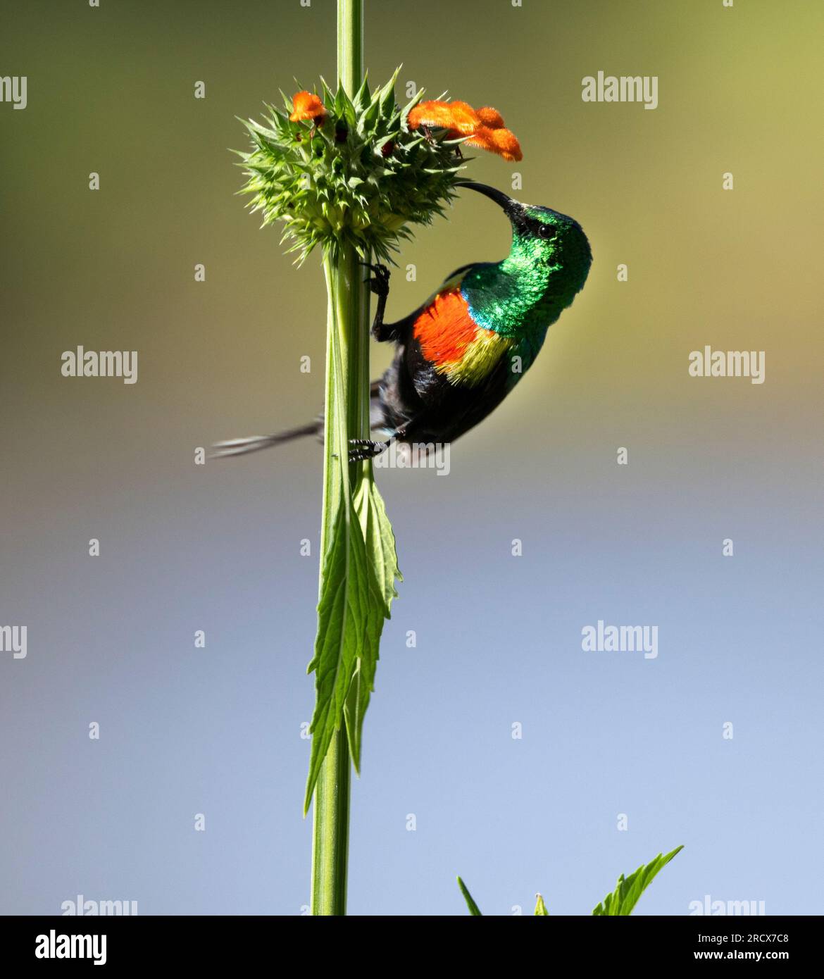 The stunning breeding colours of the male Beautiful Sunbird are provided by light refracted from surface cells known as gyroids. Stock Photohttps://www.alamy.com/image-license-details/?v=1https://www.alamy.com/the-stunning-breeding-colours-of-the-male-beautiful-sunbird-are-provided-by-light-refracted-from-surface-cells-known-as-gyroids-image558684232.html
RM2RCX7C8–The stunning breeding colours of the male Beautiful Sunbird are provided by light refracted from surface cells known as gyroids.
 bee extracts honey from the comb, nestled within the intricate network of cells. Concept: Symbiotic Harmony Stock Photohttps://www.alamy.com/image-license-details/?v=1https://www.alamy.com/bee-extracts-honey-from-the-comb-nestled-within-the-intricate-network-of-cells-concept-symbiotic-harmony-image597259955.html
RF2WKKF4K–bee extracts honey from the comb, nestled within the intricate network of cells. Concept: Symbiotic Harmony
 The beautiful iridescent plumage of the male Red-chested Sunbird refracts the sunlight through special suface cells of the feathers known as gyroides Stock Photohttps://www.alamy.com/image-license-details/?v=1https://www.alamy.com/the-beautiful-iridescent-plumage-of-the-male-red-chested-sunbird-refracts-the-sunlight-through-special-suface-cells-of-the-feathers-known-as-gyroides-image351846571.html
RM2BCBYTY–The beautiful iridescent plumage of the male Red-chested Sunbird refracts the sunlight through special suface cells of the feathers known as gyroides
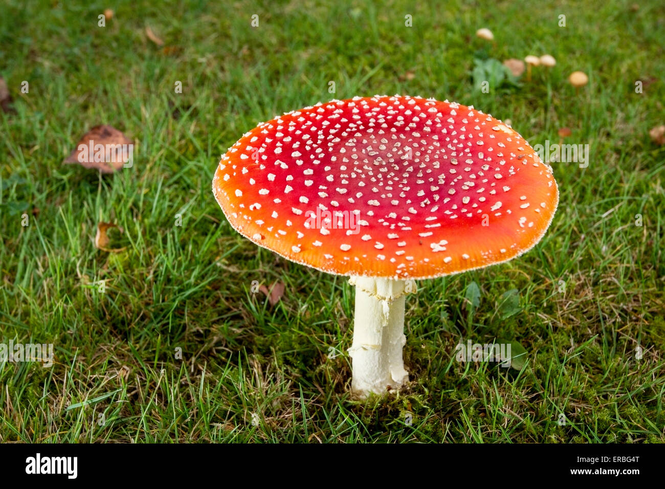 fly agaric (Amanita muscaria) fruiting body, on grass lawn, in autumn, Norfolk, England, United Kingdom Stock Photohttps://www.alamy.com/image-license-details/?v=1https://www.alamy.com/stock-photo-fly-agaric-amanita-muscaria-fruiting-body-on-grass-lawn-in-autumn-83232712.html
RMERBG4T–fly agaric (Amanita muscaria) fruiting body, on grass lawn, in autumn, Norfolk, England, United Kingdom
 Pale cyan petals-like foliose lichen on rock, recent rains revived the vegetative body, natural macro background Stock Photohttps://www.alamy.com/image-license-details/?v=1https://www.alamy.com/pale-cyan-petals-like-foliose-lichen-on-rock-recent-rains-revived-the-vegetative-body-natural-macro-background-image459107020.html
RF2HJX3K8–Pale cyan petals-like foliose lichen on rock, recent rains revived the vegetative body, natural macro background
 The gyroide cells in the outer layers of the feathers refract light to give the male Western Violet-backed Sunbird it spectacular metallic sheen. Stock Photohttps://www.alamy.com/image-license-details/?v=1https://www.alamy.com/the-gyroide-cells-in-the-outer-layers-of-the-feathers-refract-light-to-give-the-male-western-violet-backed-sunbird-it-spectacular-metallic-sheen-image329568240.html
RM2A453J8–The gyroide cells in the outer layers of the feathers refract light to give the male Western Violet-backed Sunbird it spectacular metallic sheen.
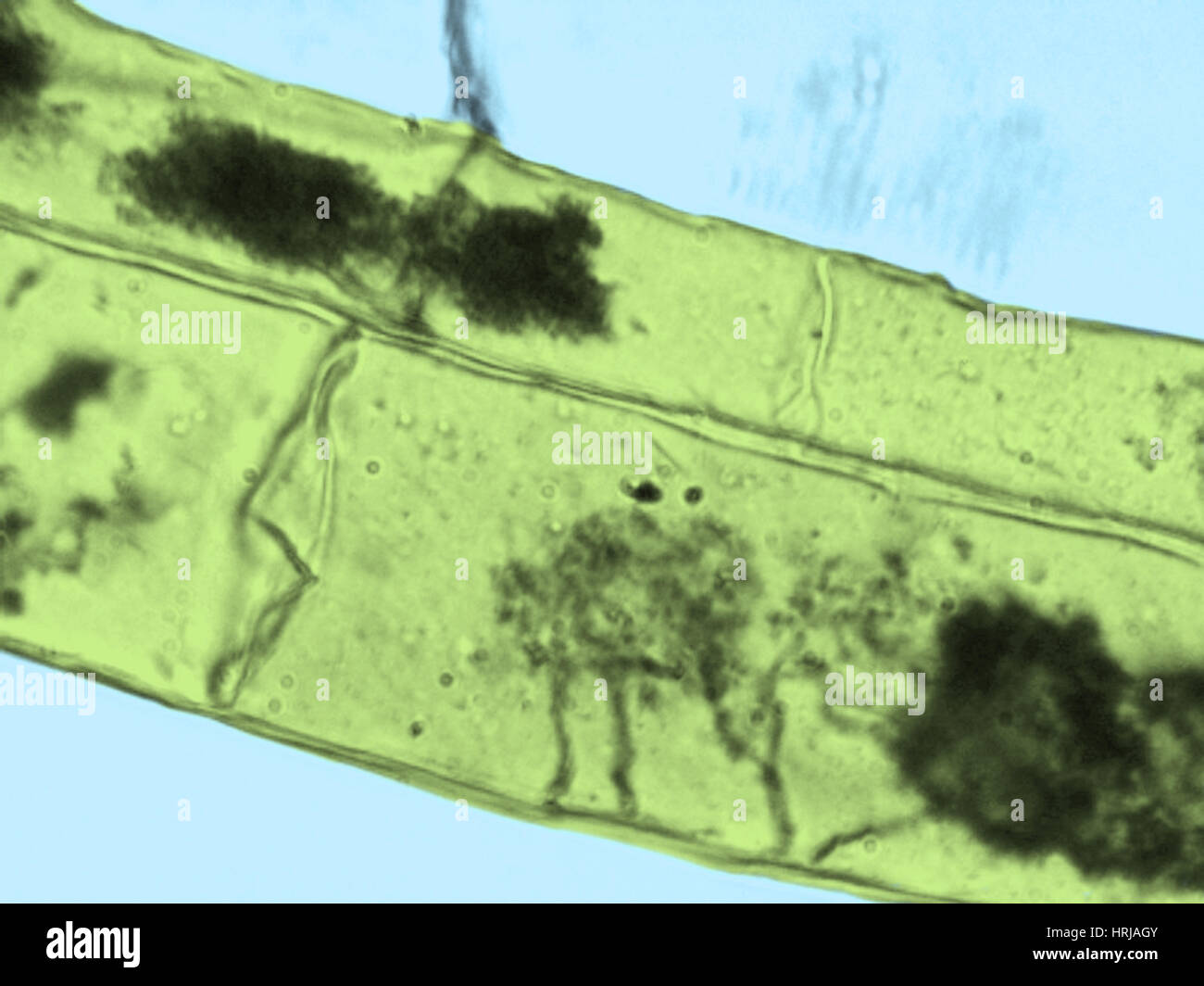 Arbuscules, Corn Root Cells Stock Photohttps://www.alamy.com/image-license-details/?v=1https://www.alamy.com/stock-photo-arbuscules-corn-root-cells-135013115.html
RMHRJAGY–Arbuscules, Corn Root Cells
 Pale cyan petals-like foliose lichen on rock, recent rains revived the vegetative body, natural macro background Stock Photohttps://www.alamy.com/image-license-details/?v=1https://www.alamy.com/pale-cyan-petals-like-foliose-lichen-on-rock-recent-rains-revived-the-vegetative-body-natural-macro-background-image470034809.html
RF2J8KX5D–Pale cyan petals-like foliose lichen on rock, recent rains revived the vegetative body, natural macro background
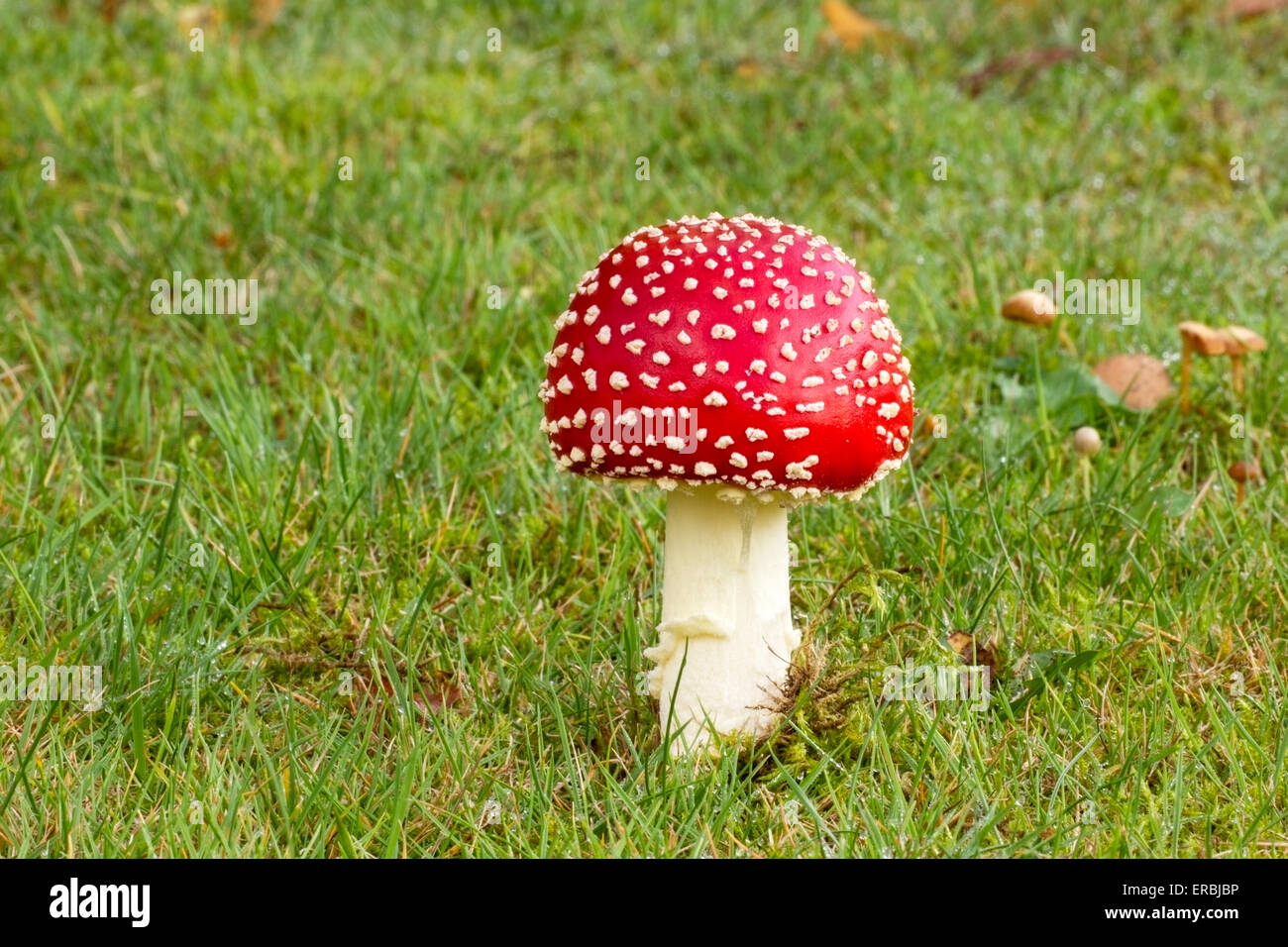 fly agaric (Amanita muscaria) fruiting body, on grass lawn, in autumn, Norfolk, England, United Kingdom Stock Photohttps://www.alamy.com/image-license-details/?v=1https://www.alamy.com/stock-photo-fly-agaric-amanita-muscaria-fruiting-body-on-grass-lawn-in-autumn-83234474.html
RMERBJBP–fly agaric (Amanita muscaria) fruiting body, on grass lawn, in autumn, Norfolk, England, United Kingdom
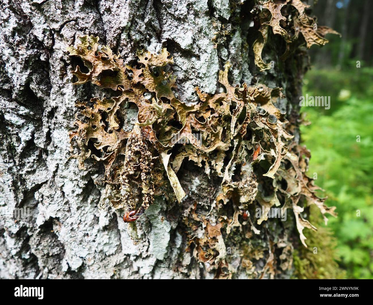 Moss and lichens on the bark of a tree in a spruce taiga forest. Karelia, Orzega. Lobaria Lobaria is a genus of lichenized ascomycetes belonging to Stock Photohttps://www.alamy.com/image-license-details/?v=1https://www.alamy.com/moss-and-lichens-on-the-bark-of-a-tree-in-a-spruce-taiga-forest-karelia-orzega-lobaria-lobaria-is-a-genus-of-lichenized-ascomycetes-belonging-to-image598669727.html
RF2WNYN9K–Moss and lichens on the bark of a tree in a spruce taiga forest. Karelia, Orzega. Lobaria Lobaria is a genus of lichenized ascomycetes belonging to
 Portrait of a Western clown anemone fish looking out from a beautiful red and green anemone Stock Photohttps://www.alamy.com/image-license-details/?v=1https://www.alamy.com/portrait-of-a-western-clown-anemone-fish-looking-out-from-a-beautiful-red-and-green-anemone-image627323304.html
RM2YCH17M–Portrait of a Western clown anemone fish looking out from a beautiful red and green anemone
 Clam (Tridacna sp.), detail of mantle. Microscopic algal cells are contained in the mantle. Myrmidon Reef, Queensland, Australia Stock Photohttps://www.alamy.com/image-license-details/?v=1https://www.alamy.com/clam-tridacna-sp-detail-of-mantle-microscopic-algal-cells-are-contained-in-the-mantle-myrmidon-reef-queensland-australia-image333916800.html
RM2AB7680–Clam (Tridacna sp.), detail of mantle. Microscopic algal cells are contained in the mantle. Myrmidon Reef, Queensland, Australia
 Pink anemone fish nestles in pink tipped bubble tip anemone Stock Photohttps://www.alamy.com/image-license-details/?v=1https://www.alamy.com/pink-anemone-fish-nestles-in-pink-tipped-bubble-tip-anemone-image627466130.html
RM2YCRFCJ–Pink anemone fish nestles in pink tipped bubble tip anemone
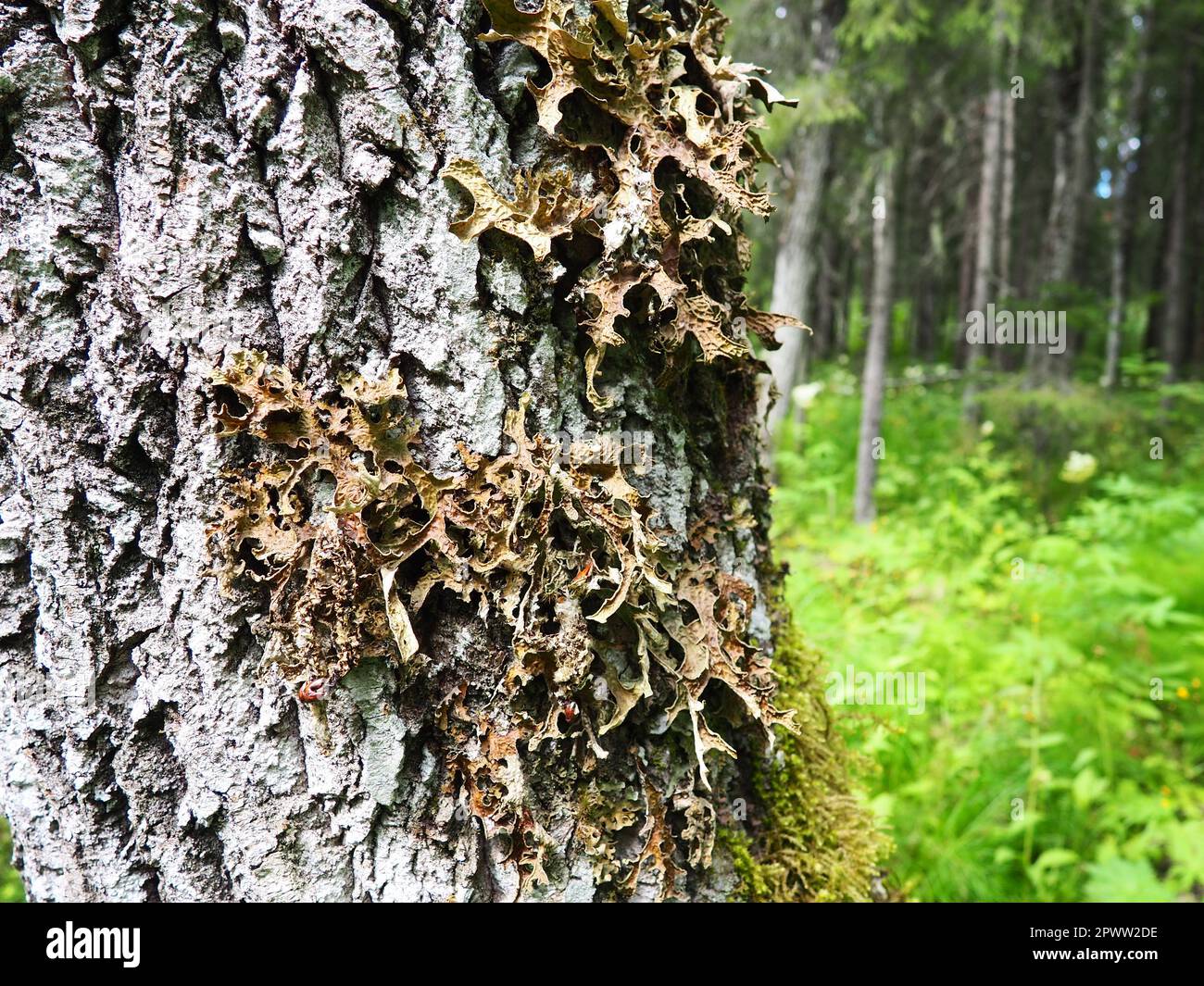 Moss and lichens on the bark of a tree in a spruce taiga forest. Karelia, Orzega. Lobaria Lobaria is a genus of lichenized ascomycetes belonging to th Stock Photohttps://www.alamy.com/image-license-details/?v=1https://www.alamy.com/moss-and-lichens-on-the-bark-of-a-tree-in-a-spruce-taiga-forest-karelia-orzega-lobaria-lobaria-is-a-genus-of-lichenized-ascomycetes-belonging-to-th-image549438554.html
RF2PWW2DE–Moss and lichens on the bark of a tree in a spruce taiga forest. Karelia, Orzega. Lobaria Lobaria is a genus of lichenized ascomycetes belonging to th
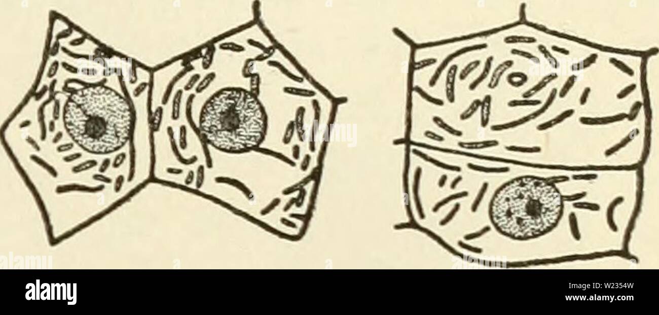 Archive image from page 134 of The cytoplasm of the plant. The cytoplasm of the plant cell cytoplasmofplant00guil Year: 1941 Chapter XI 119 — Role of Ghondriosomes SKY, DuESBERG, LEVI, and MiLOViDOV, is today abandoned by PoR- TIER himself. Nevertheless it had the merit of initiating investi- gations which have produced methods by which chondriosomes can be distinguished in cells from symbiotic and parasitic bacteria, CowDRY and Olitsky, Duesberg, and Milovidov have described methods by which, in the cells of nodules of legumes and in the adipose cells of cockroaches, symbiotic bacteria c Stock Photohttps://www.alamy.com/image-license-details/?v=1https://www.alamy.com/archive-image-from-page-134-of-the-cytoplasm-of-the-plant-the-cytoplasm-of-the-plant-cell-cytoplasmofplant00guil-year-1941-chapter-xi-119-role-of-ghondriosomes-sky-duesberg-levi-and-milovidov-is-today-abandoned-by-por-tier-himself-nevertheless-it-had-the-merit-of-initiating-investi-gations-which-have-produced-methods-by-which-chondriosomes-can-be-distinguished-in-cells-from-symbiotic-and-parasitic-bacteria-cowdry-and-olitsky-duesberg-and-milovidov-have-described-methods-by-which-in-the-cells-of-nodules-of-legumes-and-in-the-adipose-cells-of-cockroaches-symbiotic-bacteria-c-image259454745.html
RMW2354W–Archive image from page 134 of The cytoplasm of the plant. The cytoplasm of the plant cell cytoplasmofplant00guil Year: 1941 Chapter XI 119 — Role of Ghondriosomes SKY, DuESBERG, LEVI, and MiLOViDOV, is today abandoned by PoR- TIER himself. Nevertheless it had the merit of initiating investi- gations which have produced methods by which chondriosomes can be distinguished in cells from symbiotic and parasitic bacteria, CowDRY and Olitsky, Duesberg, and Milovidov have described methods by which, in the cells of nodules of legumes and in the adipose cells of cockroaches, symbiotic bacteria c
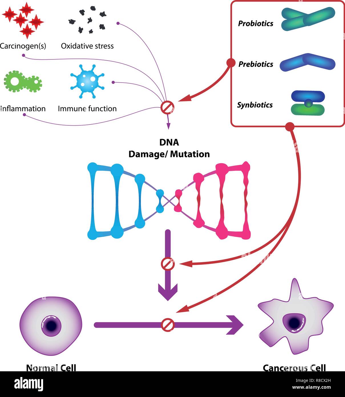 Probiotic bacteria prevent DNA damage and mutation, prevent the formation of cancer cells. Medical vector illustration Stock Vectorhttps://www.alamy.com/image-license-details/?v=1https://www.alamy.com/probiotic-bacteria-prevent-dna-damage-and-mutation-prevent-the-formation-of-cancer-cells-medical-vector-illustration-image228935913.html
RFR8CX2H–Probiotic bacteria prevent DNA damage and mutation, prevent the formation of cancer cells. Medical vector illustration
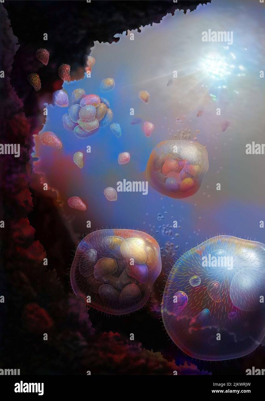 The nucleus cells would come from the association of several bacteria (prokaryotes). Stock Photohttps://www.alamy.com/image-license-details/?v=1https://www.alamy.com/the-nucleus-cells-would-come-from-the-association-of-several-bacteria-prokaryotes-image476925761.html
RF2JKWRJW–The nucleus cells would come from the association of several bacteria (prokaryotes).
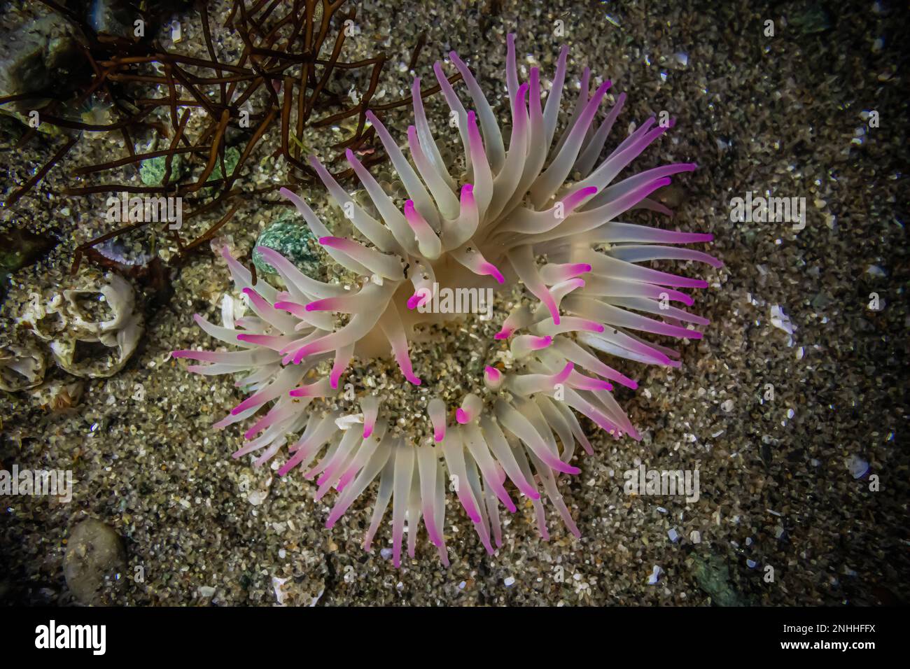 Aggregating Anemone, Anthopleura elegantissima, at Point of arches in Olympic National Park, Washington State, USA Stock Photohttps://www.alamy.com/image-license-details/?v=1https://www.alamy.com/aggregating-anemone-anthopleura-elegantissima-at-point-of-arches-in-olympic-national-park-washington-state-usa-image527145582.html
RM2NHHFFX–Aggregating Anemone, Anthopleura elegantissima, at Point of arches in Olympic National Park, Washington State, USA
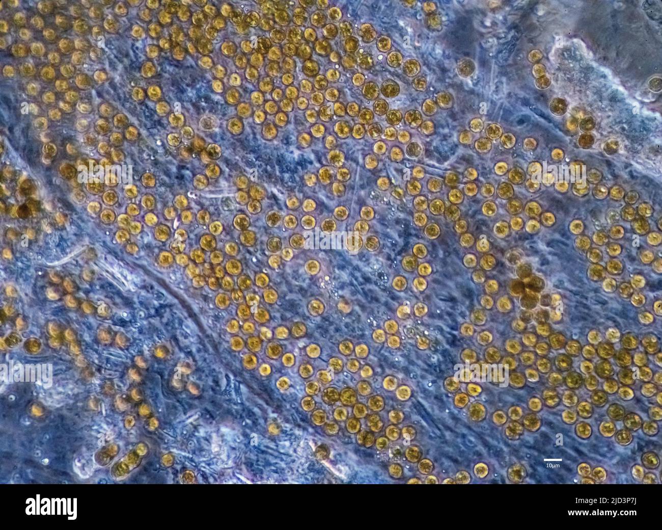 Symbiotic algae (zooxanthellae) from Aiptasia sp., probably belonging to the spaceis Breviolum dendrogyrum. The individual cells are in average about Stock Photohttps://www.alamy.com/image-license-details/?v=1https://www.alamy.com/symbiotic-algae-zooxanthellae-from-aiptasia-sp-probably-belonging-to-the-spaceis-breviolum-dendrogyrum-the-individual-cells-are-in-average-about-image472753782.html
RM2JD3P7J–Symbiotic algae (zooxanthellae) from Aiptasia sp., probably belonging to the spaceis Breviolum dendrogyrum. The individual cells are in average about
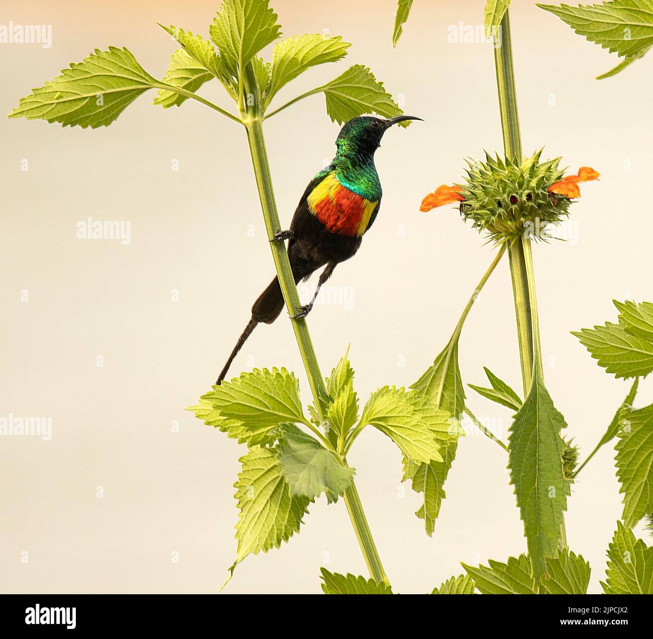 Male Beautiful Sunbird Stock Photohttps://www.alamy.com/image-license-details/?v=1https://www.alamy.com/male-beautiful-sunbird-image478480634.html
RM2JPCJX2–Male Beautiful Sunbird
 Bacteria , germs on hand. 3d rendering. Isolated on solid color background Stock Photohttps://www.alamy.com/image-license-details/?v=1https://www.alamy.com/bacteria-germs-on-hand-3d-rendering-isolated-on-solid-color-background-image271117328.html
RFWN2CWM–Bacteria , germs on hand. 3d rendering. Isolated on solid color background
 Tube Anemone (Cerianthid) with Anemone Shrimps (Periclimenes brevicarpalis) Bunaken North Sulawesi, Indonesia Stock Photohttps://www.alamy.com/image-license-details/?v=1https://www.alamy.com/tube-anemone-cerianthid-with-anemone-shrimps-periclimenes-brevicarpalis-image8391587.html
RMAJBGX4–Tube Anemone (Cerianthid) with Anemone Shrimps (Periclimenes brevicarpalis) Bunaken North Sulawesi, Indonesia
 White soft coral. Marine animal that looks like a water plant. Elongated branched tubes. Alcyonacea common names for subset of this order are sea fans Stock Photohttps://www.alamy.com/image-license-details/?v=1https://www.alamy.com/white-soft-coral-marine-animal-that-looks-like-a-water-plant-elongated-branched-tubes-alcyonacea-common-names-for-subset-of-this-order-are-sea-fans-image332485389.html
RF2A8X0E5–White soft coral. Marine animal that looks like a water plant. Elongated branched tubes. Alcyonacea common names for subset of this order are sea fans
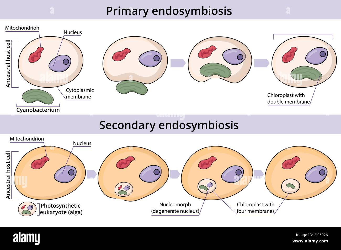 Primary and secondary endosymbiosis. Cell engulfs and absorbs a prokaryotic cell. An eukaryotic cell engulfs and absorbs another eukaryotic cell. Stock Vectorhttps://www.alamy.com/image-license-details/?v=1https://www.alamy.com/primary-and-secondary-endosymbiosis-cell-engulfs-and-absorbs-a-prokaryotic-cell-an-eukaryotic-cell-engulfs-and-absorbs-another-eukaryotic-cell-image470350670.html
RF2J96926–Primary and secondary endosymbiosis. Cell engulfs and absorbs a prokaryotic cell. An eukaryotic cell engulfs and absorbs another eukaryotic cell.
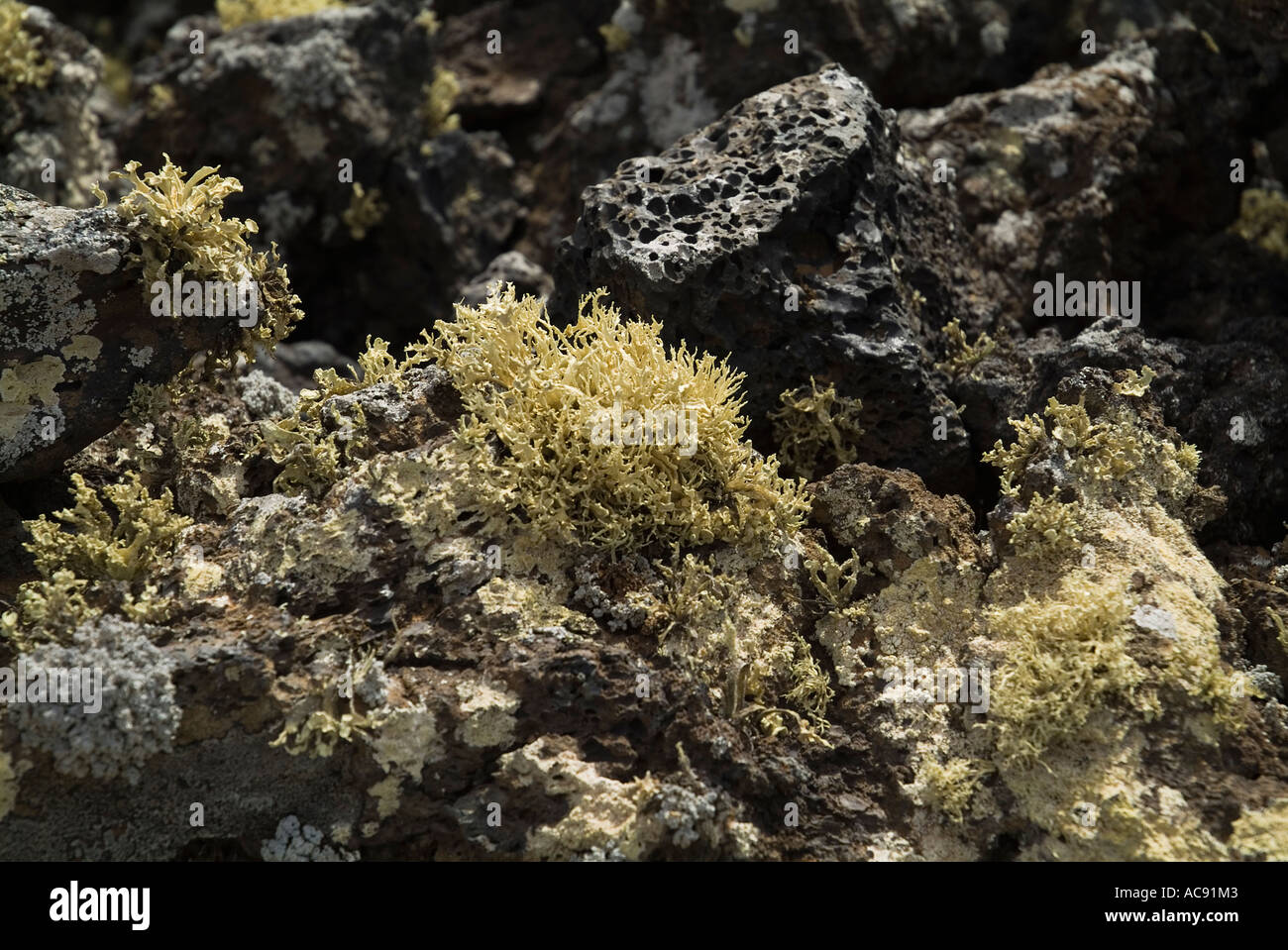 dh LAVA LANZAROTE Volcanic Aa Aa lava bed with lichen fungi algae leafy magma rocks geology Stock Photohttps://www.alamy.com/image-license-details/?v=1https://www.alamy.com/dh-lava-lanzarote-volcanic-aa-aa-lava-bed-with-lichen-fungi-algae-image7474690.html
RMAC91M3–dh LAVA LANZAROTE Volcanic Aa Aa lava bed with lichen fungi algae leafy magma rocks geology
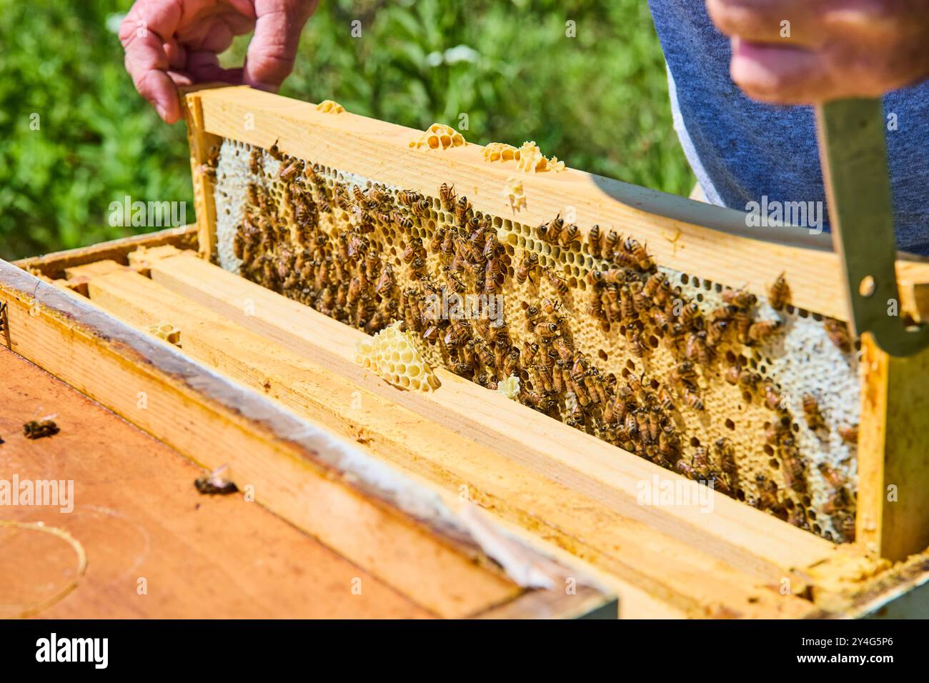 Beekeeper Inspects Hive Frame with Busy Honeybees Eye-Level View Stock Photohttps://www.alamy.com/image-license-details/?v=1https://www.alamy.com/beekeeper-inspects-hive-frame-with-busy-honeybees-eye-level-view-image622387646.html
RF2Y4G5P6–Beekeeper Inspects Hive Frame with Busy Honeybees Eye-Level View
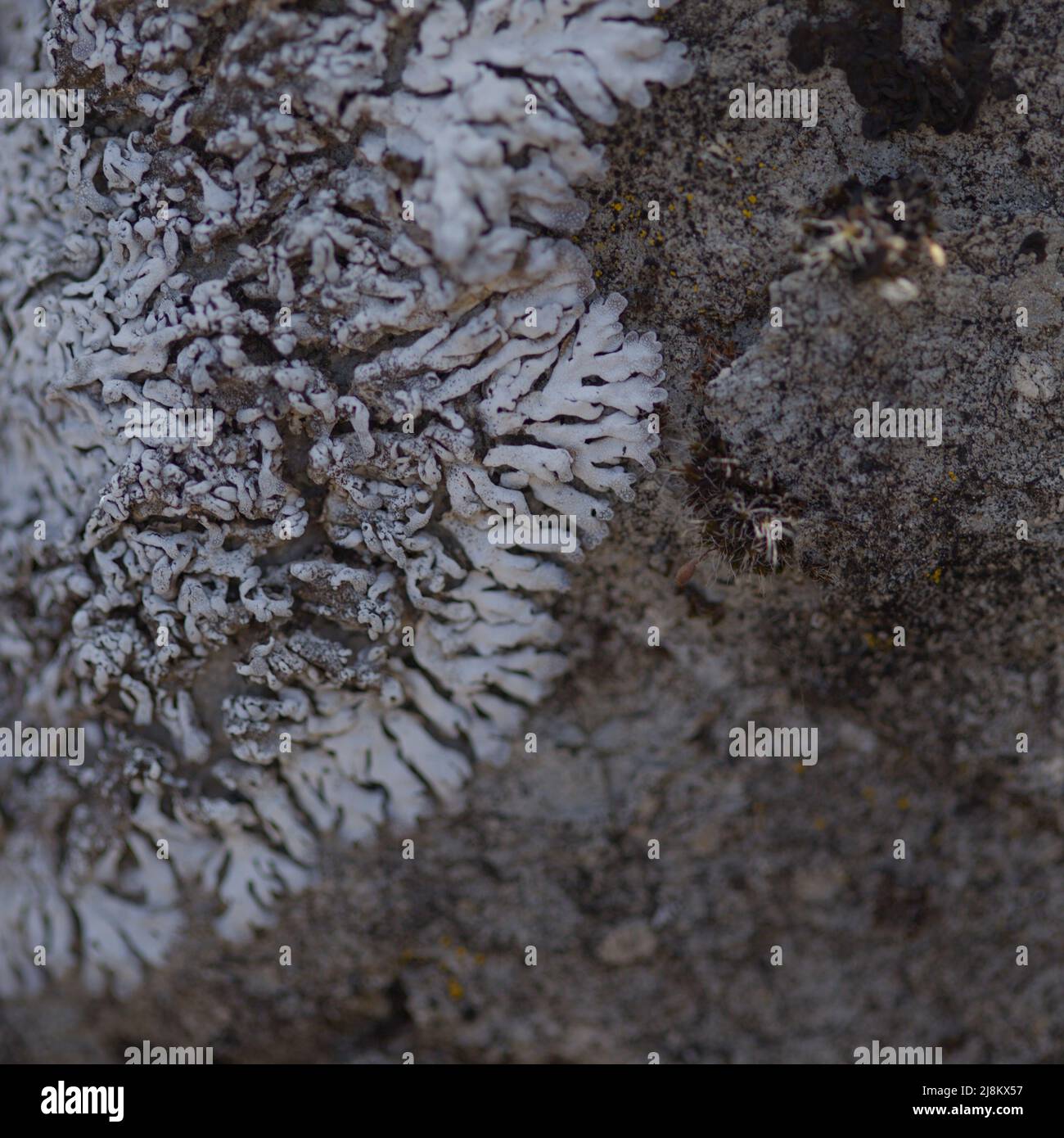 Pale cyan petals-like foliose lichen on rock, recent rains revived the vegetative body, natural macro background Stock Photohttps://www.alamy.com/image-license-details/?v=1https://www.alamy.com/pale-cyan-petals-like-foliose-lichen-on-rock-recent-rains-revived-the-vegetative-body-natural-macro-background-image470034803.html
RF2J8KX57–Pale cyan petals-like foliose lichen on rock, recent rains revived the vegetative body, natural macro background
 fly agaric (Amanita muscaria) fruiting body, on grass lawn, in autumn, Norfolk, England, United Kingdom Stock Photohttps://www.alamy.com/image-license-details/?v=1https://www.alamy.com/stock-photo-fly-agaric-amanita-muscaria-fruiting-body-on-grass-lawn-in-autumn-83234470.html
RMERBJBJ–fly agaric (Amanita muscaria) fruiting body, on grass lawn, in autumn, Norfolk, England, United Kingdom
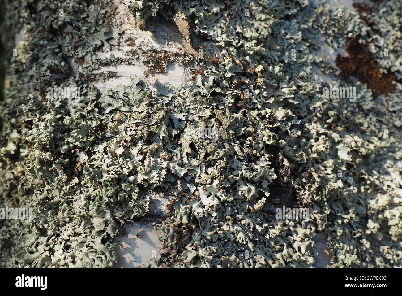 Parmelia sulcata is a foliose lichen in the family Parmeliaceae tolerant of pollution, has cosmopolitan distribution, the most common lichens. It Stock Photohttps://www.alamy.com/image-license-details/?v=1https://www.alamy.com/parmelia-sulcata-is-a-foliose-lichen-in-the-family-parmeliaceae-tolerant-of-pollution-has-cosmopolitan-distribution-the-most-common-lichens-it-image598926079.html
RF2WPBC93–Parmelia sulcata is a foliose lichen in the family Parmeliaceae tolerant of pollution, has cosmopolitan distribution, the most common lichens. It
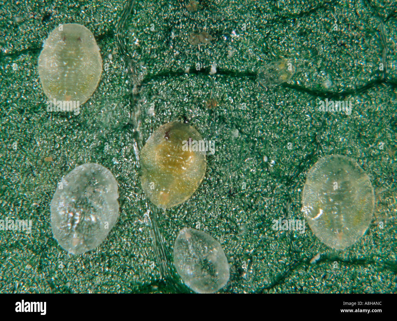 Cotton whitefly Bemisia tabaci scales larvae on cotton leaf Stock Photohttps://www.alamy.com/image-license-details/?v=1https://www.alamy.com/cotton-whitefly-bemisia-tabaci-scales-larvae-on-cotton-leaf-image6917339.html
RMA8HANC–Cotton whitefly Bemisia tabaci scales larvae on cotton leaf
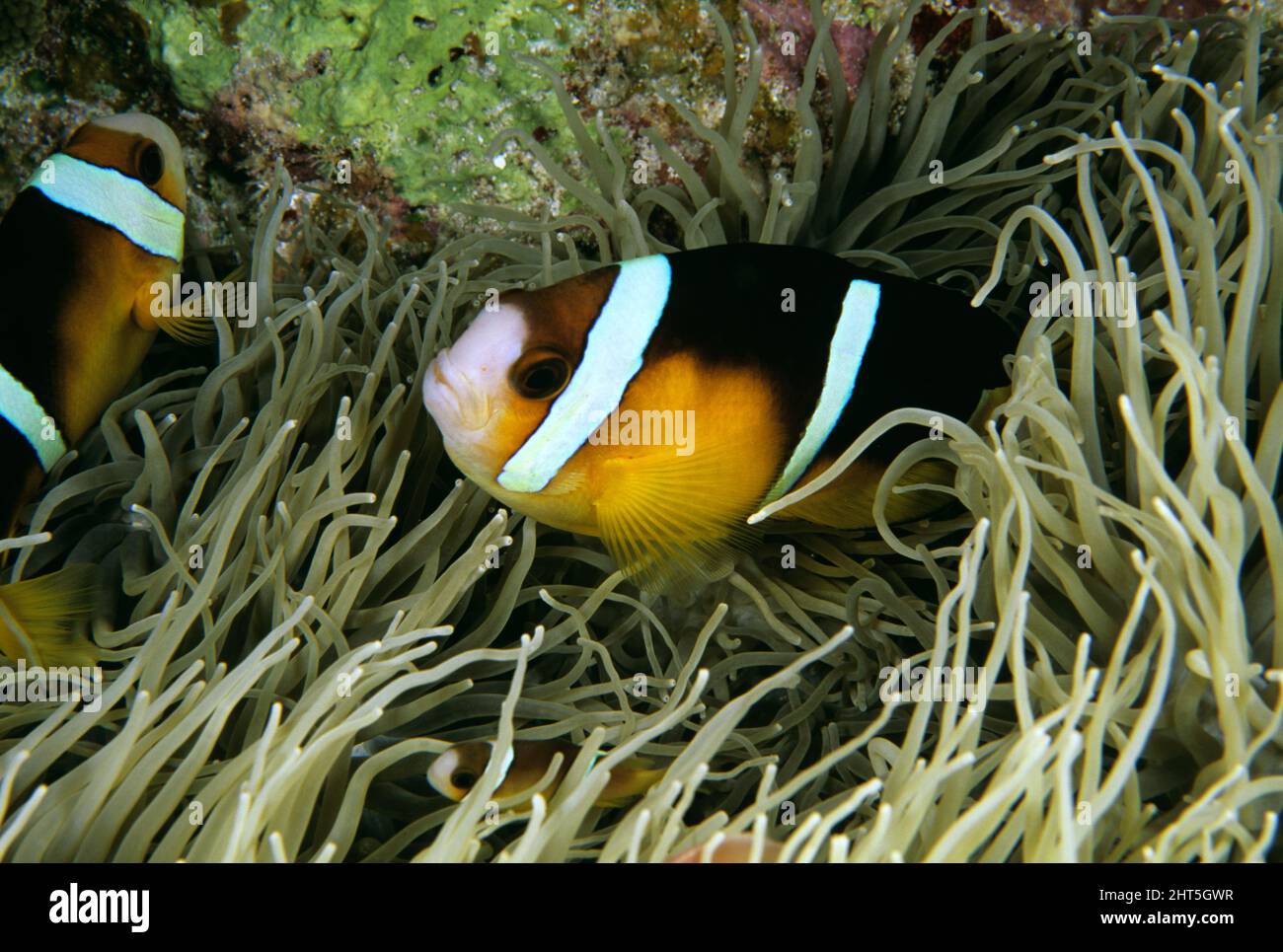 Clark’s anemonefish (Amphiprion clarkii), dwell within anemone tentacles, immune to their stinging cells. Western Australia Stock Photohttps://www.alamy.com/image-license-details/?v=1https://www.alamy.com/clarks-anemonefish-amphiprion-clarkii-dwell-within-anemone-tentacles-immune-to-their-stinging-cells-western-australia-image462344339.html
RM2HT5GWR–Clark’s anemonefish (Amphiprion clarkii), dwell within anemone tentacles, immune to their stinging cells. Western Australia
 . The Biological bulletin. Biology; Zoology; Biology; Marine Biology. SYM32 LOCALIZATION IN ANTHOPLEVRA 345. Figure 3. Transmission electron micrographs of immunogold-labeled sections ol tentacles trom symbiotic Aiithnpleiim elef>uiiti.siitui. I A) Illustrates the presence of dinoflagellates within host gastrodermal cells. (Bi Enlargement of boxed area in A. showing gold spheres associated with the multiple layers of membrane that surround the dinoflagellates. The arrows delineate the margins of the multiple membranes surrounding the dinofiagellate. Preimmune controls showed virtually no g Stock Photohttps://www.alamy.com/image-license-details/?v=1https://www.alamy.com/the-biological-bulletin-biology-zoology-biology-marine-biology-sym32-localization-in-anthoplevra-345-figure-3-transmission-electron-micrographs-of-immunogold-labeled-sections-ol-tentacles-trom-symbiotic-aiithnpleiim-elefgtuiitisiitui-i-a-illustrates-the-presence-of-dinoflagellates-within-host-gastrodermal-cells-bi-enlargement-of-boxed-area-in-a-showing-gold-spheres-associated-with-the-multiple-layers-of-membrane-that-surround-the-dinoflagellates-the-arrows-delineate-the-margins-of-the-multiple-membranes-surrounding-the-dinofiagellate-preimmune-controls-showed-virtually-no-g-image234627399.html
RMRHM5HY–. The Biological bulletin. Biology; Zoology; Biology; Marine Biology. SYM32 LOCALIZATION IN ANTHOPLEVRA 345. Figure 3. Transmission electron micrographs of immunogold-labeled sections ol tentacles trom symbiotic Aiithnpleiim elef>uiiti.siitui. I A) Illustrates the presence of dinoflagellates within host gastrodermal cells. (Bi Enlargement of boxed area in A. showing gold spheres associated with the multiple layers of membrane that surround the dinoflagellates. The arrows delineate the margins of the multiple membranes surrounding the dinofiagellate. Preimmune controls showed virtually no g
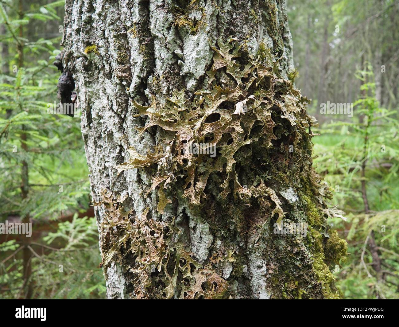 Moss and lichens on the bark of a tree in a spruce taiga forest. Karelia, Orzega. Lobaria Lobaria is a genus of lichenized ascomycetes belonging to th Stock Photohttps://www.alamy.com/image-license-details/?v=1https://www.alamy.com/moss-and-lichens-on-the-bark-of-a-tree-in-a-spruce-taiga-forest-karelia-orzega-lobaria-lobaria-is-a-genus-of-lichenized-ascomycetes-belonging-to-th-image549300572.html
RF2PWJPDG–Moss and lichens on the bark of a tree in a spruce taiga forest. Karelia, Orzega. Lobaria Lobaria is a genus of lichenized ascomycetes belonging to th
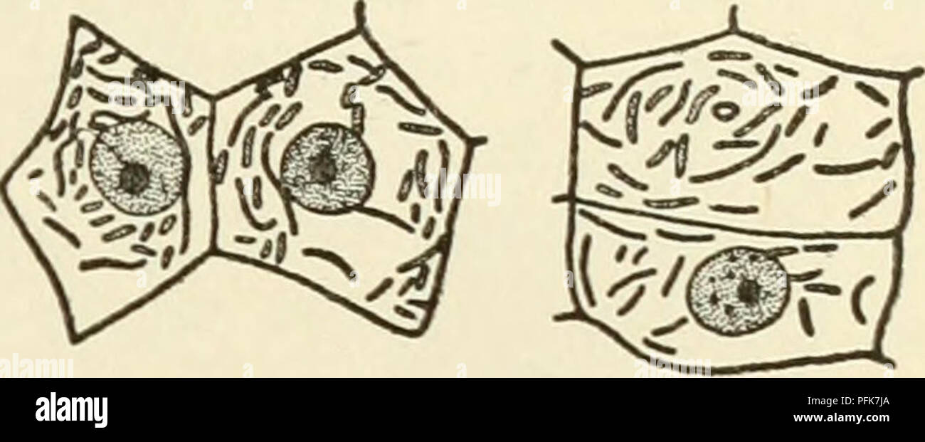 . The cytoplasm of the plant cell. Plant cells and tissues; Protoplasm. Chapter XI 119 — Role of Ghondriosomes. SKY, DuESBERG, LEVI, and MiLOViDOV, is today abandoned by PoR- TIER himself. Nevertheless it had the merit of initiating investi- gations which have produced methods by which chondriosomes can be distinguished in cells from symbiotic and parasitic bacteria, CowDRY and Olitsky, Duesberg, and Milovidov have described methods by which, in the cells of nodules of legumes and in the adipose cells of cockroaches, symbiotic bacteria can be distin- guished from the chondriosomes by means of Stock Photohttps://www.alamy.com/image-license-details/?v=1https://www.alamy.com/the-cytoplasm-of-the-plant-cell-plant-cells-and-tissues-protoplasm-chapter-xi-119-role-of-ghondriosomes-sky-duesberg-levi-and-milovidov-is-today-abandoned-by-por-tier-himself-nevertheless-it-had-the-merit-of-initiating-investi-gations-which-have-produced-methods-by-which-chondriosomes-can-be-distinguished-in-cells-from-symbiotic-and-parasitic-bacteria-cowdry-and-olitsky-duesberg-and-milovidov-have-described-methods-by-which-in-the-cells-of-nodules-of-legumes-and-in-the-adipose-cells-of-cockroaches-symbiotic-bacteria-can-be-distin-guished-from-the-chondriosomes-by-means-of-image216167346.html
RMPFK7JA–. The cytoplasm of the plant cell. Plant cells and tissues; Protoplasm. Chapter XI 119 — Role of Ghondriosomes. SKY, DuESBERG, LEVI, and MiLOViDOV, is today abandoned by PoR- TIER himself. Nevertheless it had the merit of initiating investi- gations which have produced methods by which chondriosomes can be distinguished in cells from symbiotic and parasitic bacteria, CowDRY and Olitsky, Duesberg, and Milovidov have described methods by which, in the cells of nodules of legumes and in the adipose cells of cockroaches, symbiotic bacteria can be distin- guished from the chondriosomes by means of
 White lichen and green moss abstract close-up intimate nature photograph Stock Photohttps://www.alamy.com/image-license-details/?v=1https://www.alamy.com/white-lichen-and-green-moss-abstract-close-up-intimate-nature-photograph-image336481868.html
RM2AFC21G–White lichen and green moss abstract close-up intimate nature photograph
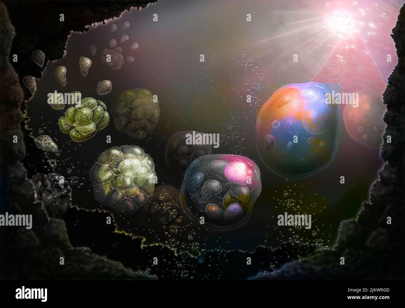 The nucleus cells would come from the association of several bacteria (prokaryotes). Stock Photohttps://www.alamy.com/image-license-details/?v=1https://www.alamy.com/the-nucleus-cells-would-come-from-the-association-of-several-bacteria-prokaryotes-image476925693.html
RF2JKWRGD–The nucleus cells would come from the association of several bacteria (prokaryotes).
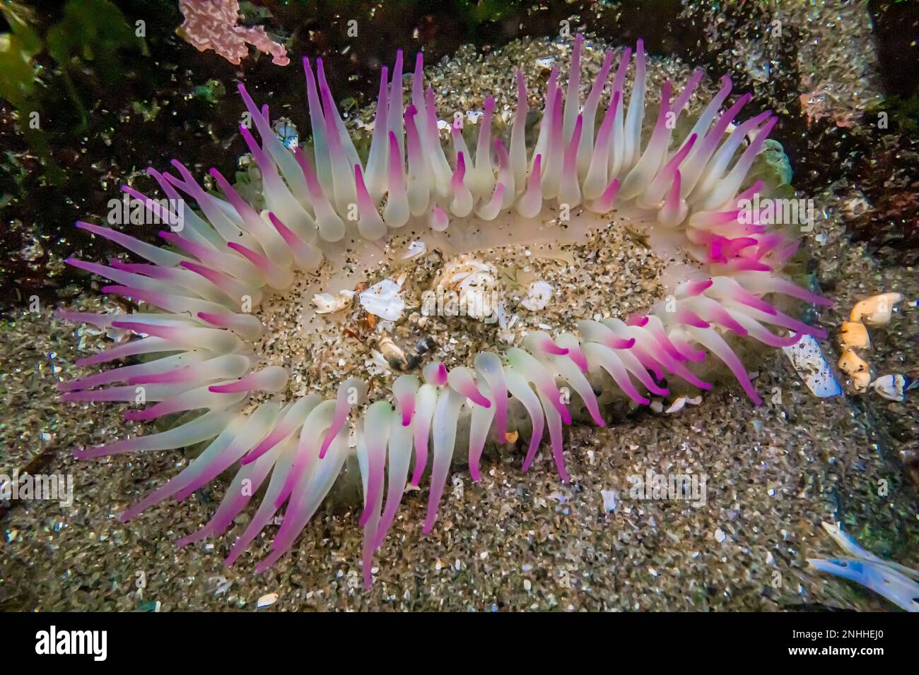 Aggregating Anemone, Anthopleura elegantissima, at Point of arches in Olympic National Park, Washington State, USA Stock Photohttps://www.alamy.com/image-license-details/?v=1https://www.alamy.com/aggregating-anemone-anthopleura-elegantissima-at-point-of-arches-in-olympic-national-park-washington-state-usa-image527144856.html
RM2NHHEJ0–Aggregating Anemone, Anthopleura elegantissima, at Point of arches in Olympic National Park, Washington State, USA
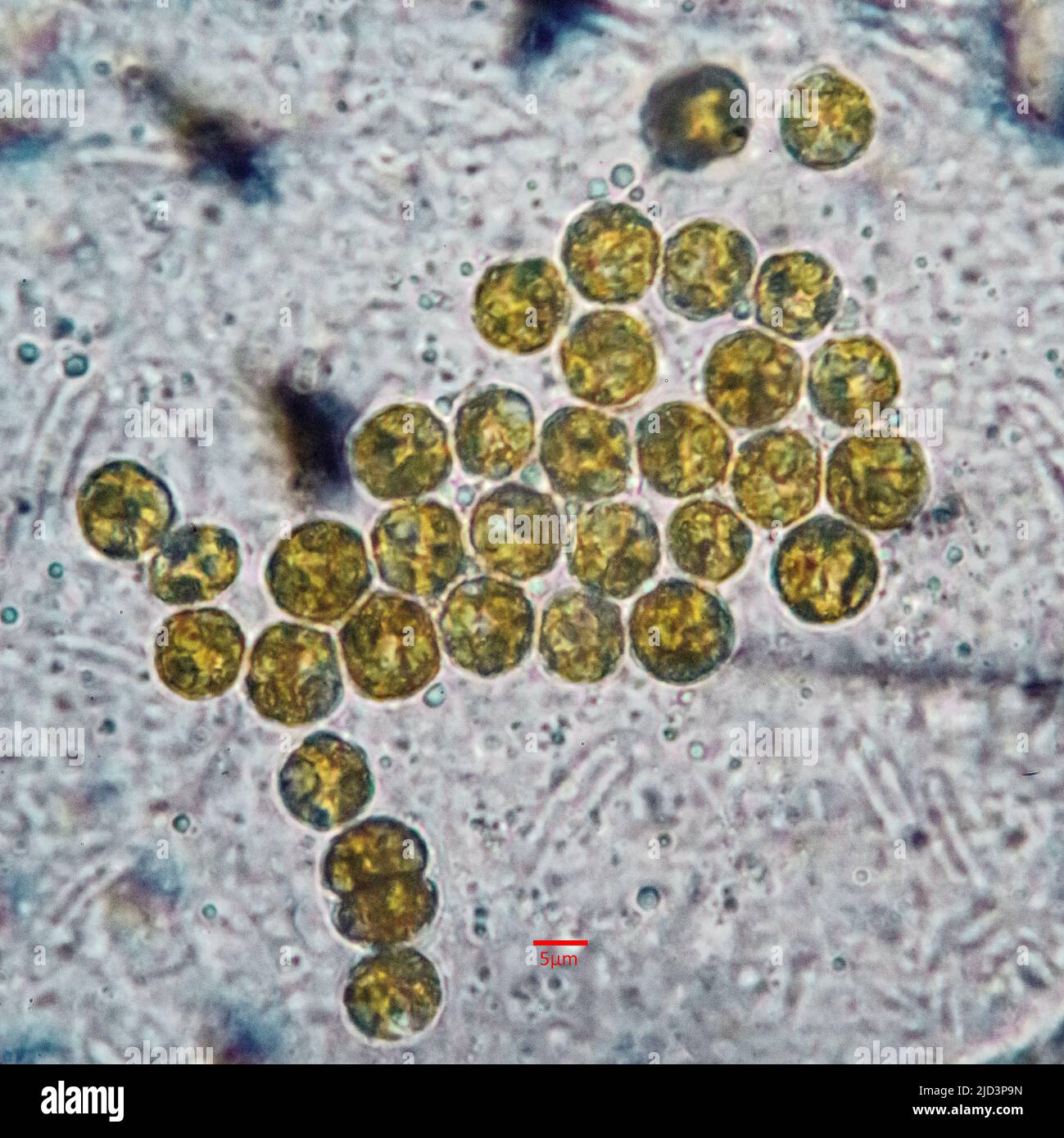 Symbiotic algae (zooxanthellae) from Aiptasia sp., probably belonging to the spaceis Breviolum dendrogyrum. The individual cells are in average about Stock Photohttps://www.alamy.com/image-license-details/?v=1https://www.alamy.com/symbiotic-algae-zooxanthellae-from-aiptasia-sp-probably-belonging-to-the-spaceis-breviolum-dendrogyrum-the-individual-cells-are-in-average-about-image472753841.html
RM2JD3P9N–Symbiotic algae (zooxanthellae) from Aiptasia sp., probably belonging to the spaceis Breviolum dendrogyrum. The individual cells are in average about
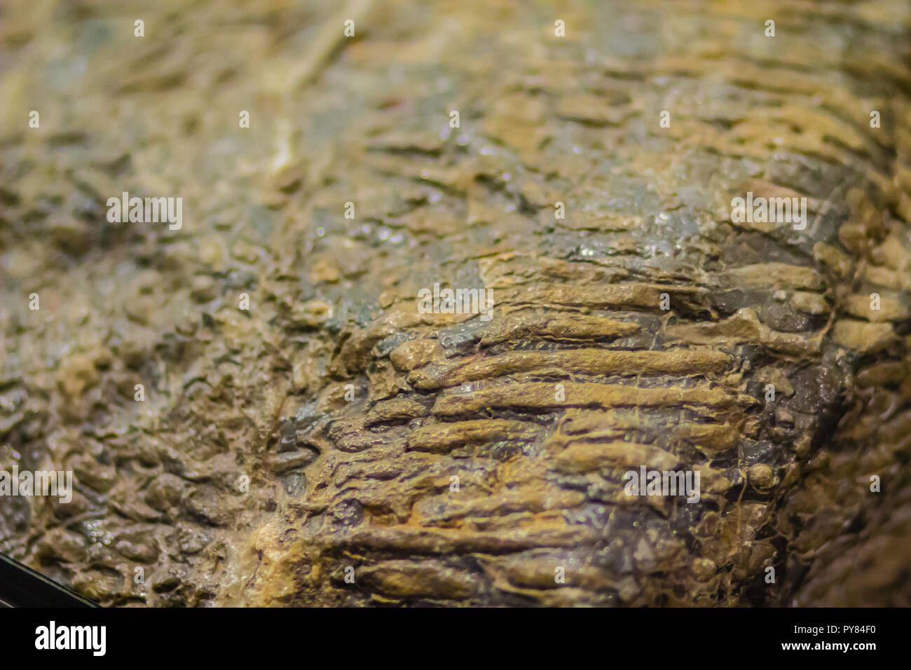 Scleractinia coral fossil specimen for education. Scleractinia, also called stony corals or hard corals. Scleractinian corals evolved from soft-bodied Stock Photohttps://www.alamy.com/image-license-details/?v=1https://www.alamy.com/scleractinia-coral-fossil-specimen-for-education-scleractinia-also-called-stony-corals-or-hard-corals-scleractinian-corals-evolved-from-soft-bodied-image223299300.html
RFPY84F0–Scleractinia coral fossil specimen for education. Scleractinia, also called stony corals or hard corals. Scleractinian corals evolved from soft-bodied
 Bacteria , germs on hand. 3d rendering. Isolated on solid color background Stock Photohttps://www.alamy.com/image-license-details/?v=1https://www.alamy.com/bacteria-germs-on-hand-3d-rendering-isolated-on-solid-color-background-image271117360.html
RFWN2CXT–Bacteria , germs on hand. 3d rendering. Isolated on solid color background
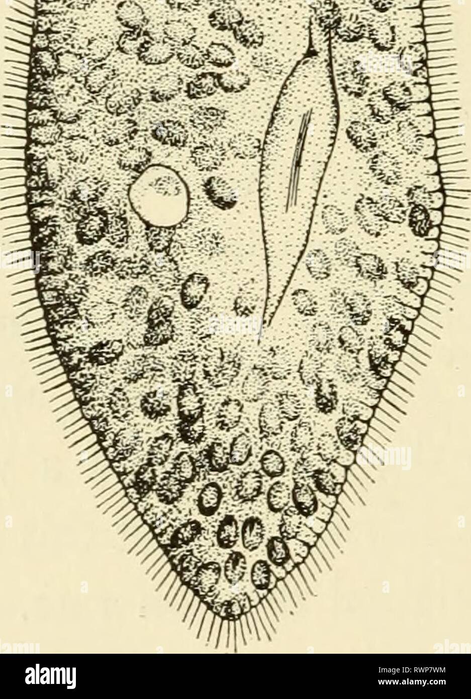 Elements of biology, with special Elements of biology, with special reference to their rôle in the lives of animals elementsofbiolog00buch Year: 1933 ZOOCHLORELLA Fig. 22.—ParamcEciitm btirsaria containing symbiotic plant cells, Zoochlorella. (Re-drawn after Conn.) Symbiosis. Certain species of Paramoecium, for example, Paramoocium bursaria, are frequently infected with a green plant cell growth (Fig. 22). This small unicellular green plant, Zoochlo- rella, is found in sufficient numbers to give the animal a green color. This relation between the host paramoecium and the invader plant cell Stock Photohttps://www.alamy.com/image-license-details/?v=1https://www.alamy.com/elements-of-biology-with-special-elements-of-biology-with-special-reference-to-their-rle-in-the-lives-of-animals-elementsofbiolog00buch-year-1933-zoochlorella-fig-22paramceciitm-btirsaria-containing-symbiotic-plant-cells-zoochlorella-re-drawn-after-conn-symbiosis-certain-species-of-paramoecium-for-example-paramoocium-bursaria-are-frequently-infected-with-a-green-plant-cell-growth-fig-22-this-small-unicellular-green-plant-zoochlo-rella-is-found-in-sufficient-numbers-to-give-the-animal-a-green-color-this-relation-between-the-host-paramoecium-and-the-invader-plant-cell-image239590336.html
RMRWP7WM–Elements of biology, with special Elements of biology, with special reference to their rôle in the lives of animals elementsofbiolog00buch Year: 1933 ZOOCHLORELLA Fig. 22.—ParamcEciitm btirsaria containing symbiotic plant cells, Zoochlorella. (Re-drawn after Conn.) Symbiosis. Certain species of Paramoecium, for example, Paramoocium bursaria, are frequently infected with a green plant cell growth (Fig. 22). This small unicellular green plant, Zoochlo- rella, is found in sufficient numbers to give the animal a green color. This relation between the host paramoecium and the invader plant cell
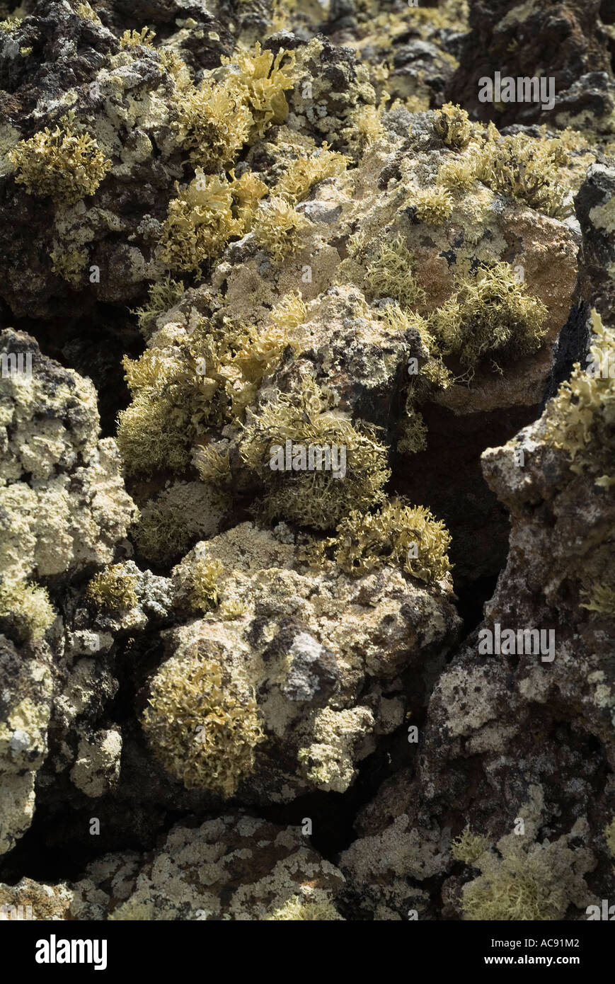 dh LAVA LANZAROTE Volcanic Aa Aa lava bed with lichen fungi geology clinker ahah fungus Stock Photohttps://www.alamy.com/image-license-details/?v=1https://www.alamy.com/dh-lava-lanzarote-volcanic-aa-aa-lava-bed-with-lichen-fungi-geology-image7474689.html
RMAC91M2–dh LAVA LANZAROTE Volcanic Aa Aa lava bed with lichen fungi geology clinker ahah fungus
 Pale cyan petals-like foliose lichen on rock, recent rains revived the vegetative body, natural macro background Stock Photohttps://www.alamy.com/image-license-details/?v=1https://www.alamy.com/pale-cyan-petals-like-foliose-lichen-on-rock-recent-rains-revived-the-vegetative-body-natural-macro-background-image470034825.html
RF2J8KX61–Pale cyan petals-like foliose lichen on rock, recent rains revived the vegetative body, natural macro background
 fly agaric (Amanita muscaria) fruiting body, on grass lawn, in autumn, Norfolk, England, United Kingdom Stock Photohttps://www.alamy.com/image-license-details/?v=1https://www.alamy.com/stock-photo-fly-agaric-amanita-muscaria-fruiting-body-on-grass-lawn-in-autumn-83234472.html
RMERBJBM–fly agaric (Amanita muscaria) fruiting body, on grass lawn, in autumn, Norfolk, England, United Kingdom
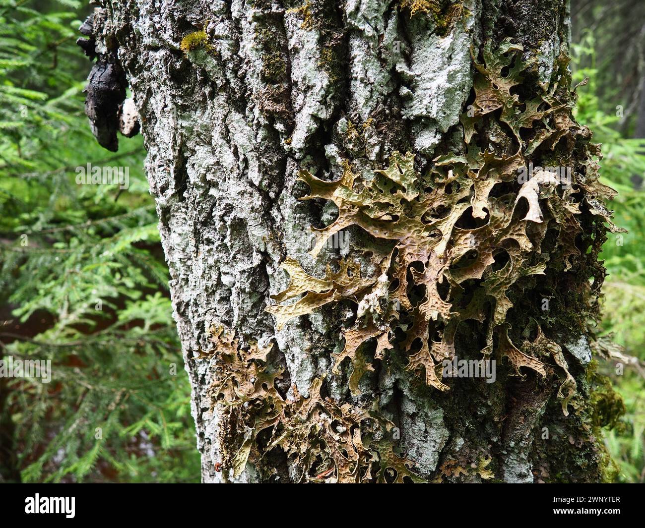 Moss and lichens on the bark of a tree in a spruce taiga forest. Karelia, Orzega. Lobaria Lobaria is a genus of lichenized ascomycetes belonging to Stock Photohttps://www.alamy.com/image-license-details/?v=1https://www.alamy.com/moss-and-lichens-on-the-bark-of-a-tree-in-a-spruce-taiga-forest-karelia-orzega-lobaria-lobaria-is-a-genus-of-lichenized-ascomycetes-belonging-to-image598672223.html
RF2WNYTER–Moss and lichens on the bark of a tree in a spruce taiga forest. Karelia, Orzega. Lobaria Lobaria is a genus of lichenized ascomycetes belonging to
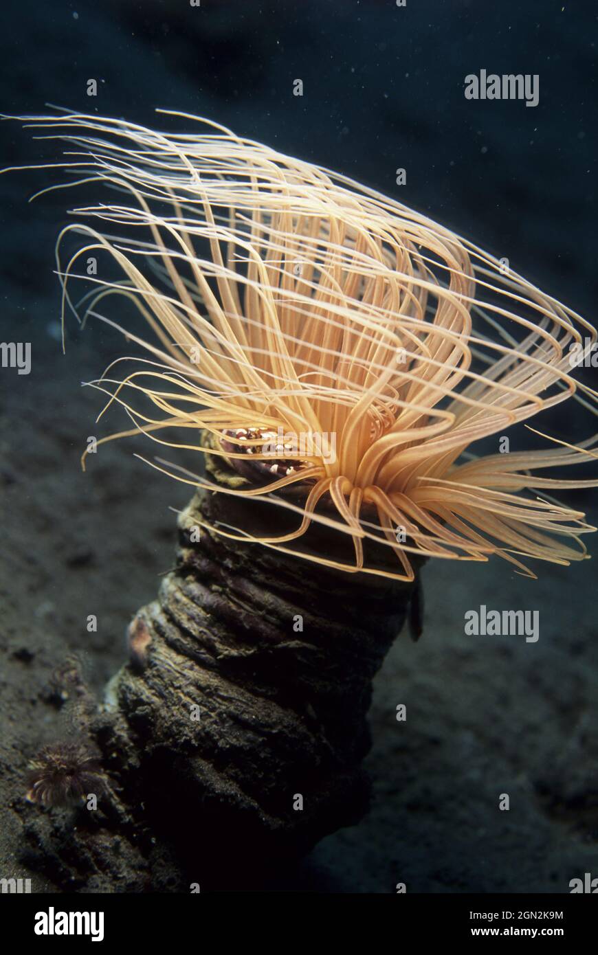 Burrowing anemone (Cerianthus sp.), with a commensal Porcelain crab nestled in the tentacles that are in two whorls, the outer, longer, stinging ones Stock Photohttps://www.alamy.com/image-license-details/?v=1https://www.alamy.com/burrowing-anemone-cerianthus-sp-with-a-commensal-porcelain-crab-nestled-in-the-tentacles-that-are-in-two-whorls-the-outer-longer-stinging-ones-image443226048.html
RM2GN2K9M–Burrowing anemone (Cerianthus sp.), with a commensal Porcelain crab nestled in the tentacles that are in two whorls, the outer, longer, stinging ones
 . The Biological bulletin. Biology; Zoology; Biology; Marine Biology. CELL DETACHMENT IN SYMBIOTIC CNIDARIANS 329. Figure 5. Transmission electron micrographs showing degradation of the host cells released by Pocillopora damicomis in response to cold stress. After dissociation from the epithelium, the host cell plasma membrane ruptures (A) and the cytoplasmic constituents are free to disperse in the seawater (B). ZX, zooxanthella; RPM. ruptured host cell plasma membrane; CC. host cell cytoplasmic constituents; IZX, isolated zooxanthella. Bar = I nm. mitochondria (Fig. 4). Once released as a re Stock Photohttps://www.alamy.com/image-license-details/?v=1https://www.alamy.com/the-biological-bulletin-biology-zoology-biology-marine-biology-cell-detachment-in-symbiotic-cnidarians-329-figure-5-transmission-electron-micrographs-showing-degradation-of-the-host-cells-released-by-pocillopora-damicomis-in-response-to-cold-stress-after-dissociation-from-the-epithelium-the-host-cell-plasma-membrane-ruptures-a-and-the-cytoplasmic-constituents-are-free-to-disperse-in-the-seawater-b-zx-zooxanthella-rpm-ruptured-host-cell-plasma-membrane-cc-host-cell-cytoplasmic-constituents-izx-isolated-zooxanthella-bar-=-i-nm-mitochondria-fig-4-once-released-as-a-re-image234633075.html
RMRHMCTK–. The Biological bulletin. Biology; Zoology; Biology; Marine Biology. CELL DETACHMENT IN SYMBIOTIC CNIDARIANS 329. Figure 5. Transmission electron micrographs showing degradation of the host cells released by Pocillopora damicomis in response to cold stress. After dissociation from the epithelium, the host cell plasma membrane ruptures (A) and the cytoplasmic constituents are free to disperse in the seawater (B). ZX, zooxanthella; RPM. ruptured host cell plasma membrane; CC. host cell cytoplasmic constituents; IZX, isolated zooxanthella. Bar = I nm. mitochondria (Fig. 4). Once released as a re
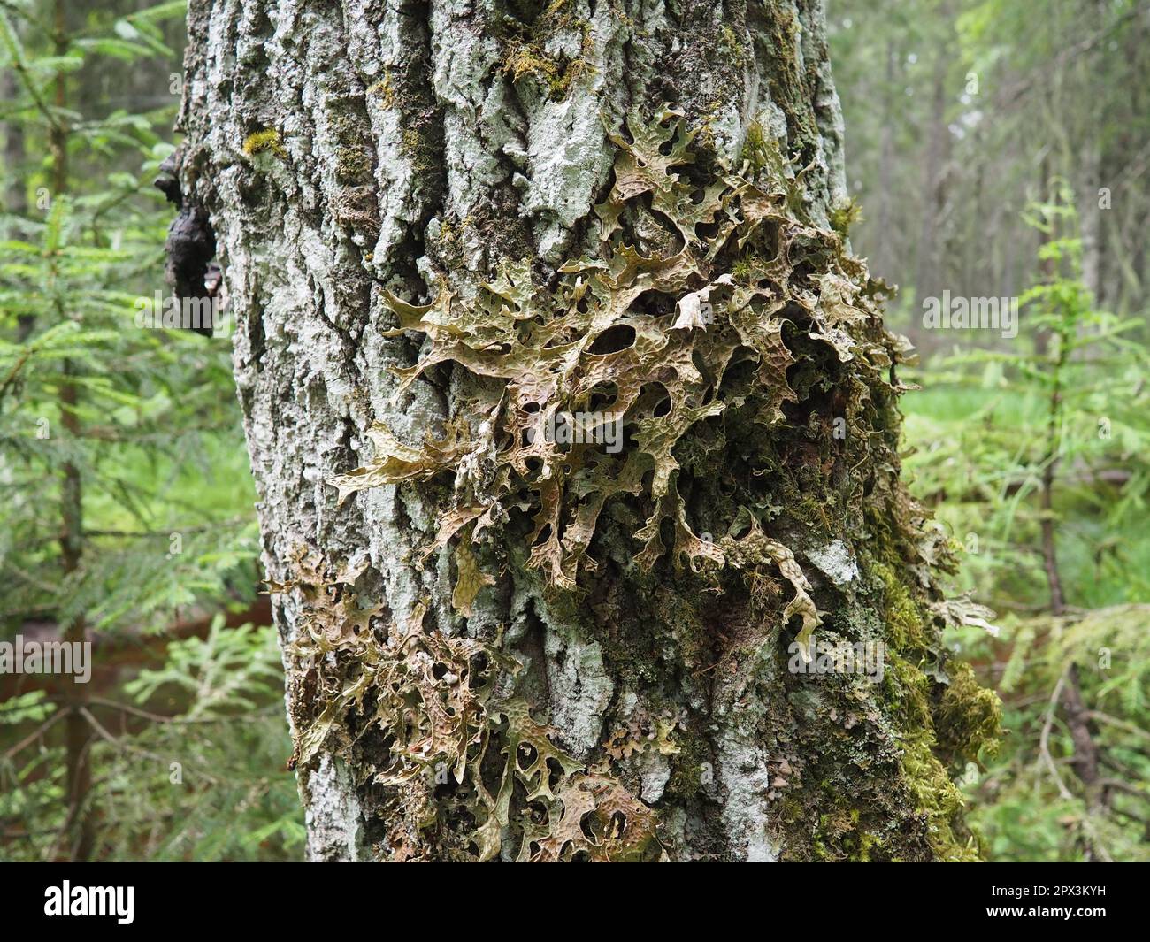 Moss and lichens on the bark of a tree in a spruce taiga forest. Karelia, Orzega. Lobaria Lobaria is a genus of lichenized ascomycetes belonging to th Stock Photohttps://www.alamy.com/image-license-details/?v=1https://www.alamy.com/moss-and-lichens-on-the-bark-of-a-tree-in-a-spruce-taiga-forest-karelia-orzega-lobaria-lobaria-is-a-genus-of-lichenized-ascomycetes-belonging-to-th-image549583989.html
RF2PX3KYH–Moss and lichens on the bark of a tree in a spruce taiga forest. Karelia, Orzega. Lobaria Lobaria is a genus of lichenized ascomycetes belonging to th
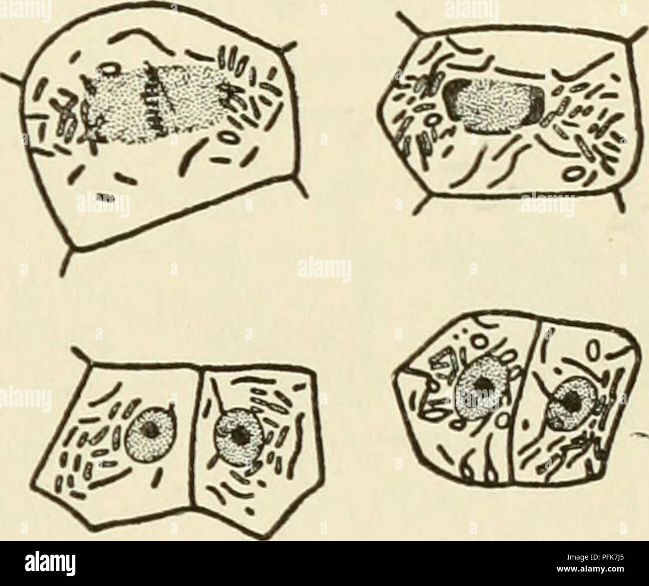 . The cytoplasm of the plant cell. Plant cells and tissues; Protoplasm. SKY, DuESBERG, LEVI, and MiLOViDOV, is today abandoned by PoR- TIER himself. Nevertheless it had the merit of initiating investi- gations which have produced methods by which chondriosomes can be distinguished in cells from symbiotic and parasitic bacteria, CowDRY and Olitsky, Duesberg, and Milovidov have described methods by which, in the cells of nodules of legumes and in the adipose cells of cockroaches, symbiotic bacteria can be distin- guished from the chondriosomes by means of differential staining. By these methods Stock Photohttps://www.alamy.com/image-license-details/?v=1https://www.alamy.com/the-cytoplasm-of-the-plant-cell-plant-cells-and-tissues-protoplasm-sky-duesberg-levi-and-milovidov-is-today-abandoned-by-por-tier-himself-nevertheless-it-had-the-merit-of-initiating-investi-gations-which-have-produced-methods-by-which-chondriosomes-can-be-distinguished-in-cells-from-symbiotic-and-parasitic-bacteria-cowdry-and-olitsky-duesberg-and-milovidov-have-described-methods-by-which-in-the-cells-of-nodules-of-legumes-and-in-the-adipose-cells-of-cockroaches-symbiotic-bacteria-can-be-distin-guished-from-the-chondriosomes-by-means-of-differential-staining-by-these-methods-image216167341.html
RMPFK7J5–. The cytoplasm of the plant cell. Plant cells and tissues; Protoplasm. SKY, DuESBERG, LEVI, and MiLOViDOV, is today abandoned by PoR- TIER himself. Nevertheless it had the merit of initiating investi- gations which have produced methods by which chondriosomes can be distinguished in cells from symbiotic and parasitic bacteria, CowDRY and Olitsky, Duesberg, and Milovidov have described methods by which, in the cells of nodules of legumes and in the adipose cells of cockroaches, symbiotic bacteria can be distin- guished from the chondriosomes by means of differential staining. By these methods
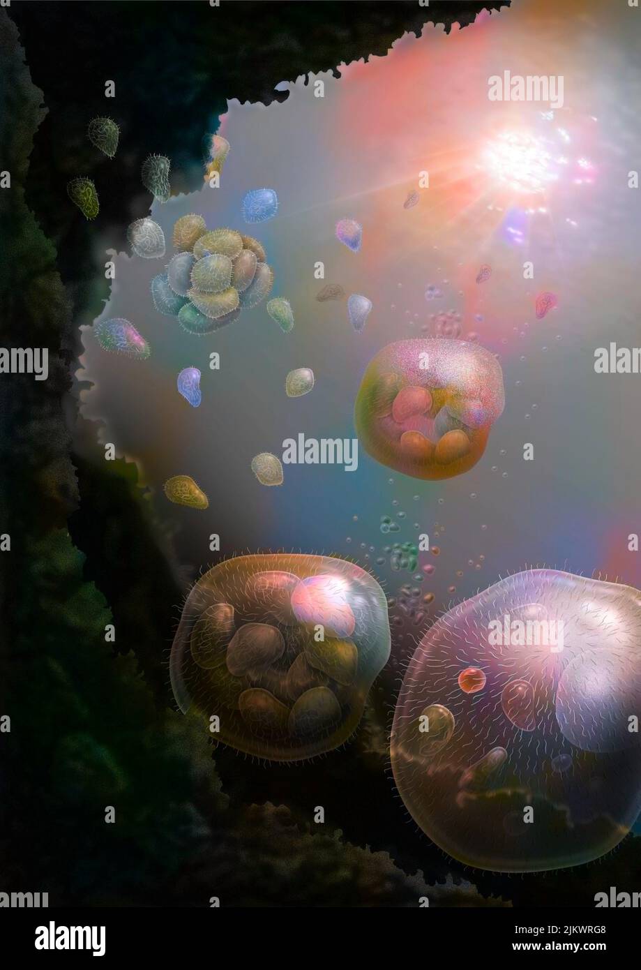 The nucleus cells would come from the association of several bacteria (prokaryotes). Stock Photohttps://www.alamy.com/image-license-details/?v=1https://www.alamy.com/the-nucleus-cells-would-come-from-the-association-of-several-bacteria-prokaryotes-image476925688.html
RF2JKWRG8–The nucleus cells would come from the association of several bacteria (prokaryotes).
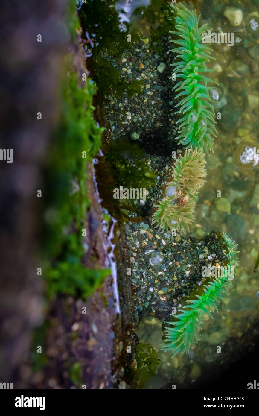 Column of Giant Green Anemone, Anthopleura xanthogrammica, in tide pool at Point of Arches in Olympic National Park, Washington State, USA Stock Photohttps://www.alamy.com/image-license-details/?v=1https://www.alamy.com/column-of-giant-green-anemone-anthopleura-xanthogrammica-in-tide-pool-at-point-of-arches-in-olympic-national-park-washington-state-usa-image527146175.html
RM2NHHG93–Column of Giant Green Anemone, Anthopleura xanthogrammica, in tide pool at Point of Arches in Olympic National Park, Washington State, USA
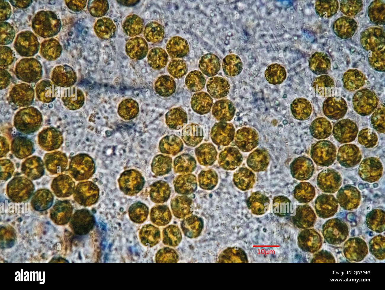 Symbiotic algae (zooxanthellae) from Aiptasia sp., probably belonging to the spaceis Breviolum dendrogyrum. The individual cells are in average about Stock Photohttps://www.alamy.com/image-license-details/?v=1https://www.alamy.com/symbiotic-algae-zooxanthellae-from-aiptasia-sp-probably-belonging-to-the-spaceis-breviolum-dendrogyrum-the-individual-cells-are-in-average-about-image472753696.html
RM2JD3P4G–Symbiotic algae (zooxanthellae) from Aiptasia sp., probably belonging to the spaceis Breviolum dendrogyrum. The individual cells are in average about
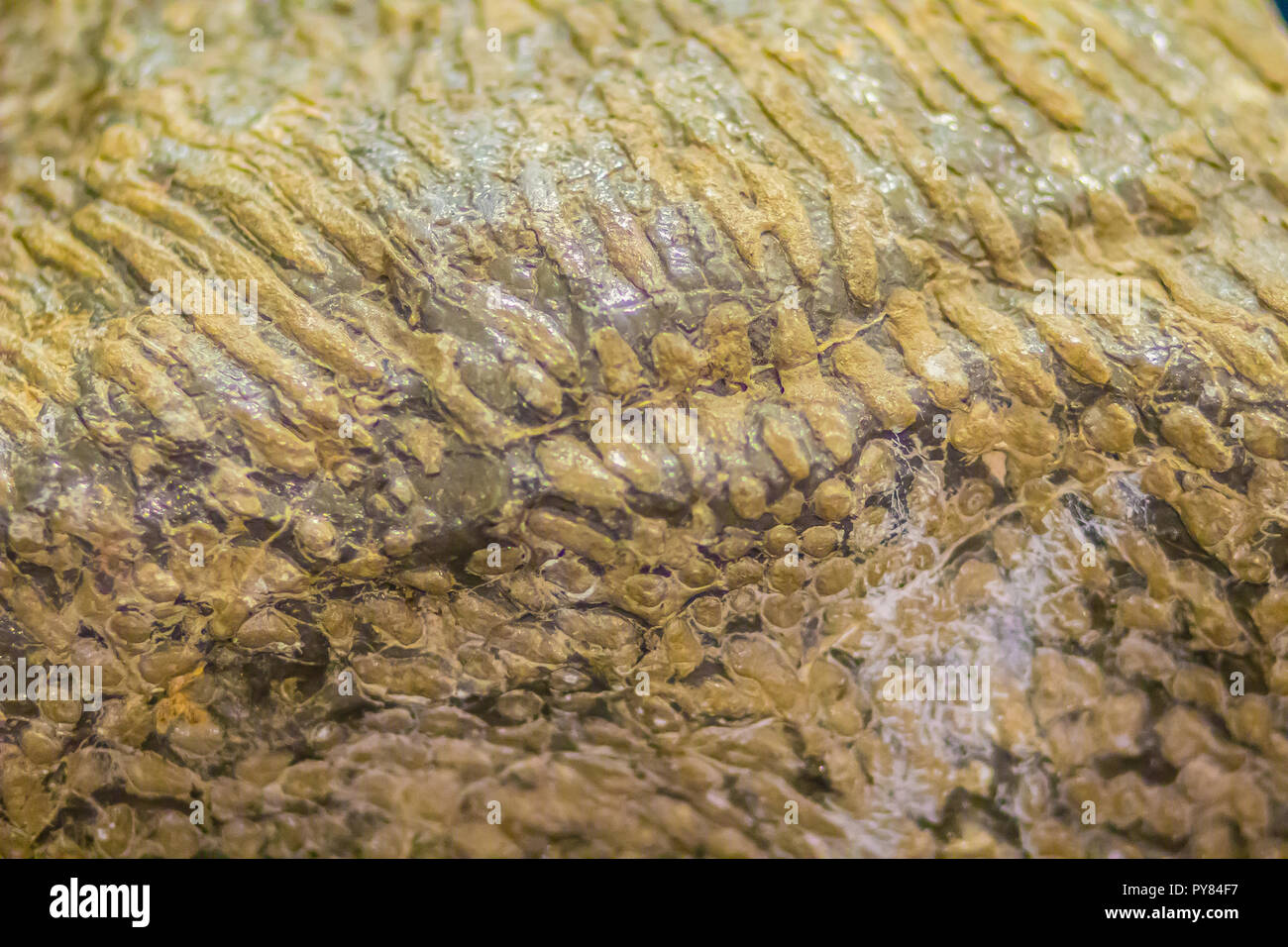 Scleractinia coral fossil specimen for education. Scleractinia, also called stony corals or hard corals. Scleractinian corals evolved from soft-bodied Stock Photohttps://www.alamy.com/image-license-details/?v=1https://www.alamy.com/scleractinia-coral-fossil-specimen-for-education-scleractinia-also-called-stony-corals-or-hard-corals-scleractinian-corals-evolved-from-soft-bodied-image223299307.html
RFPY84F7–Scleractinia coral fossil specimen for education. Scleractinia, also called stony corals or hard corals. Scleractinian corals evolved from soft-bodied
 Bacteria , germs on hand. 3d rendering. Isolated on solid color background Stock Photohttps://www.alamy.com/image-license-details/?v=1https://www.alamy.com/bacteria-germs-on-hand-3d-rendering-isolated-on-solid-color-background-image271117176.html
RFWN2CM8–Bacteria , germs on hand. 3d rendering. Isolated on solid color background
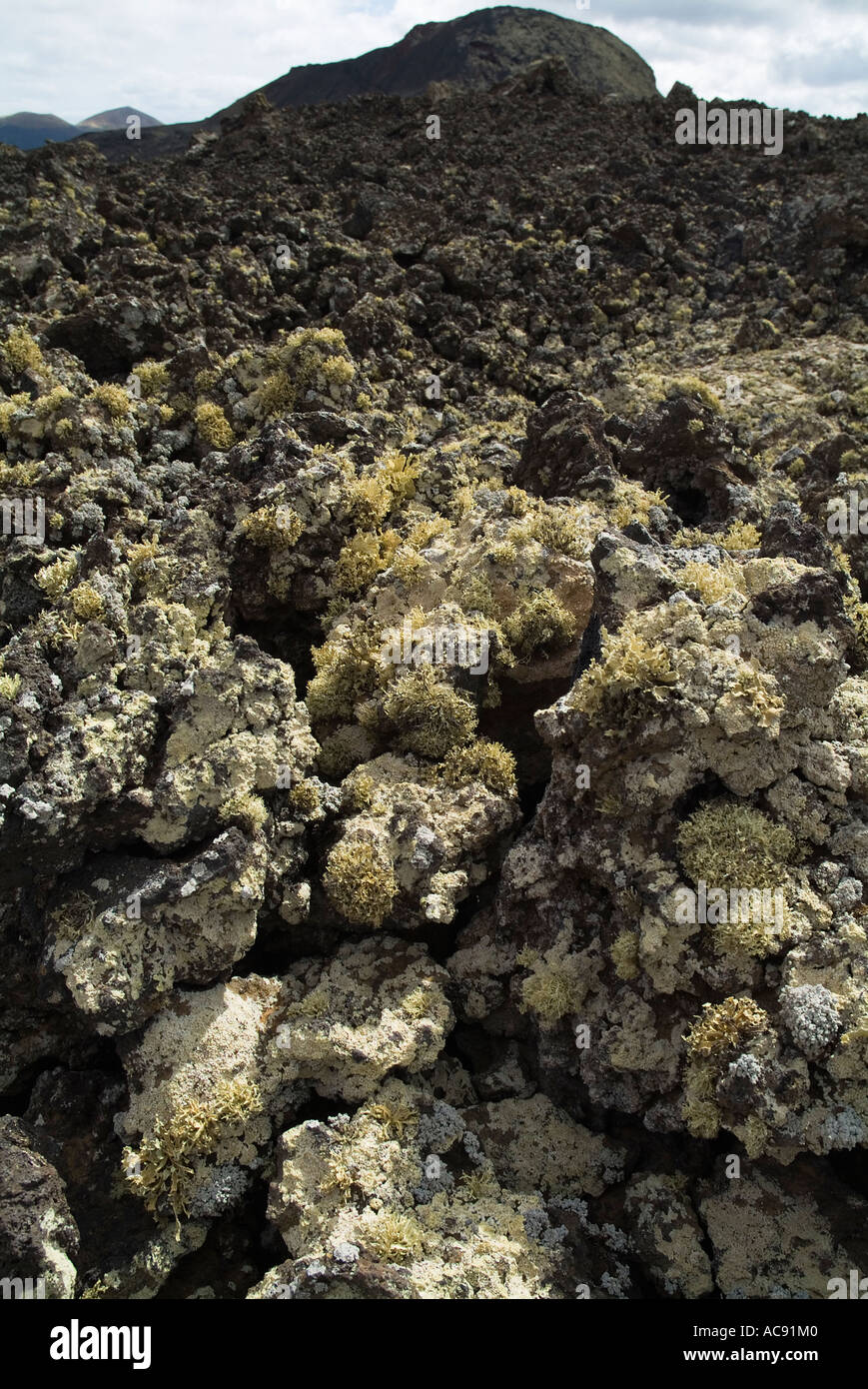 dh LAVA LANZAROTE Volcanic Aa Aa lava bed field with lichen fungi clinker rock Stock Photohttps://www.alamy.com/image-license-details/?v=1https://www.alamy.com/dh-lava-lanzarote-volcanic-aa-aa-lava-bed-field-with-lichen-fungi-image7474687.html
RMAC91M0–dh LAVA LANZAROTE Volcanic Aa Aa lava bed field with lichen fungi clinker rock
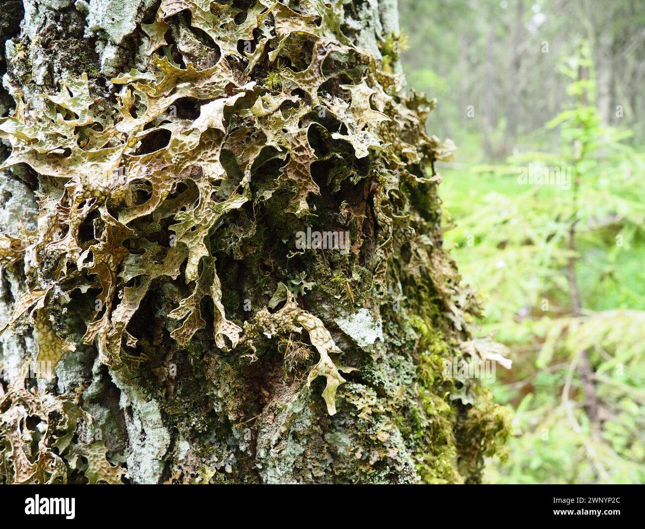 Moss and lichens on the bark of a tree in a spruce taiga forest. Karelia, Orzega. Lobaria Lobaria is a genus of lichenized ascomycetes belonging to Stock Photohttps://www.alamy.com/image-license-details/?v=1https://www.alamy.com/moss-and-lichens-on-the-bark-of-a-tree-in-a-spruce-taiga-forest-karelia-orzega-lobaria-lobaria-is-a-genus-of-lichenized-ascomycetes-belonging-to-image598670308.html
RF2WNYP2C–Moss and lichens on the bark of a tree in a spruce taiga forest. Karelia, Orzega. Lobaria Lobaria is a genus of lichenized ascomycetes belonging to
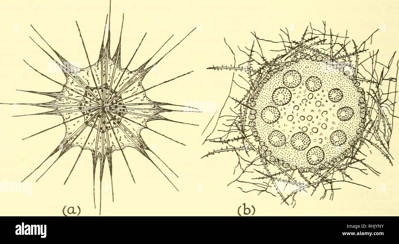 . The biology of marine animals. Marine animals; Physiology, Comparative. 612 THE BIOLOGY OF MARINE ANIMALS and megalospheres, which reproduce asexually and sexually. The large asexual bodies or amoebulae which arise by multiple fission of the parent carry away some of the symbionts, but the gametes are free of algae and the microspheres must be reinfected from without. A few marine ciliates and flagellates also contain symbiotic algae. The peritrich Spatostyla sertulariarum encloses a small number of zooxanthellae which are passed to the daughter cells during cell division. Of particular inte Stock Photohttps://www.alamy.com/image-license-details/?v=1https://www.alamy.com/the-biology-of-marine-animals-marine-animals-physiology-comparative-612-the-biology-of-marine-animals-and-megalospheres-which-reproduce-asexually-and-sexually-the-large-asexual-bodies-or-amoebulae-which-arise-by-multiple-fission-of-the-parent-carry-away-some-of-the-symbionts-but-the-gametes-are-free-of-algae-and-the-microspheres-must-be-reinfected-from-without-a-few-marine-ciliates-and-flagellates-also-contain-symbiotic-algae-the-peritrich-spatostyla-sertulariarum-encloses-a-small-number-of-zooxanthellae-which-are-passed-to-the-daughter-cells-during-cell-division-of-particular-inte-image234600855.html
RMRHJYNY–. The biology of marine animals. Marine animals; Physiology, Comparative. 612 THE BIOLOGY OF MARINE ANIMALS and megalospheres, which reproduce asexually and sexually. The large asexual bodies or amoebulae which arise by multiple fission of the parent carry away some of the symbionts, but the gametes are free of algae and the microspheres must be reinfected from without. A few marine ciliates and flagellates also contain symbiotic algae. The peritrich Spatostyla sertulariarum encloses a small number of zooxanthellae which are passed to the daughter cells during cell division. Of particular inte
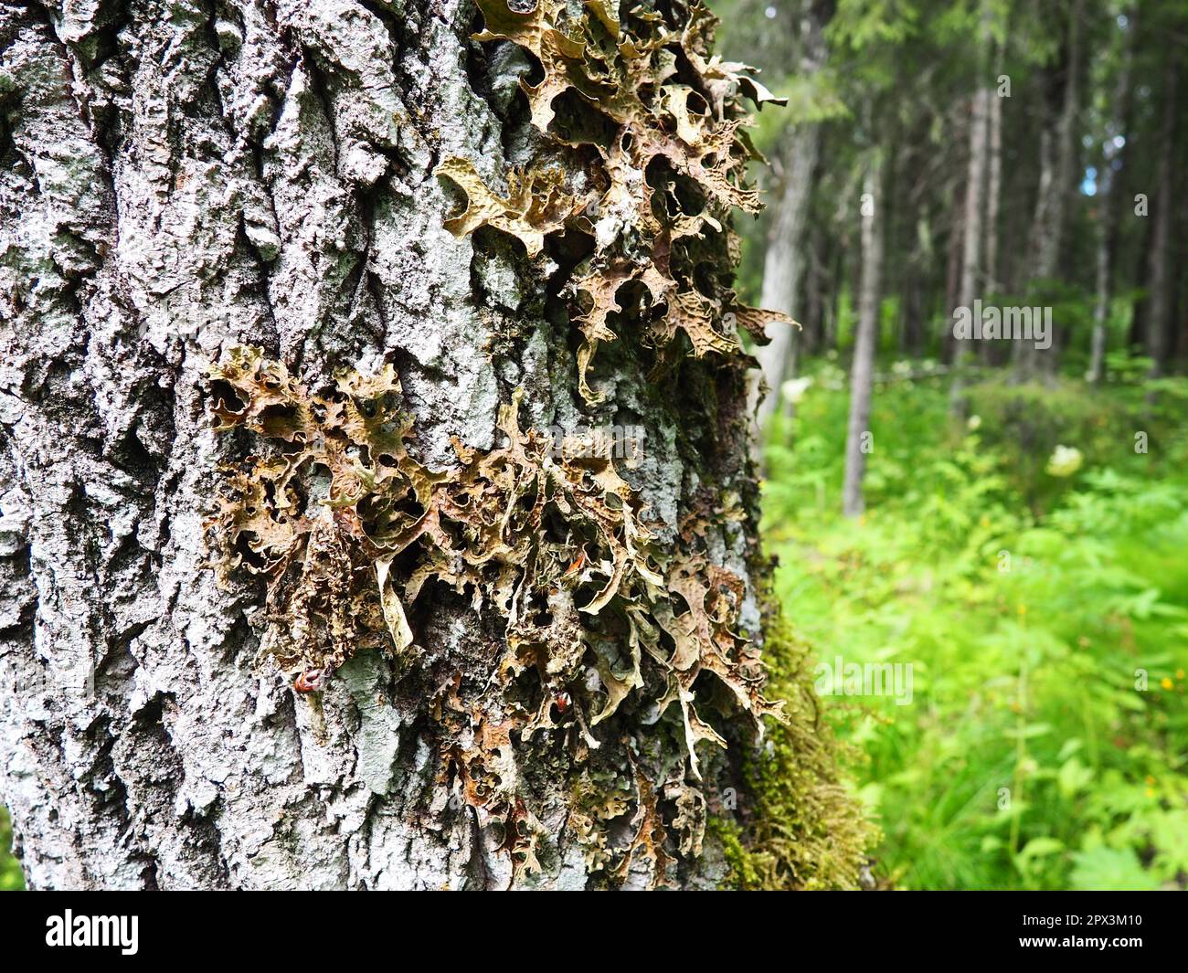 Moss and lichens on the bark of a tree in a spruce taiga forest. Karelia, Orzega. Lobaria Lobaria is a genus of lichenized ascomycetes belonging to th Stock Photohttps://www.alamy.com/image-license-details/?v=1https://www.alamy.com/moss-and-lichens-on-the-bark-of-a-tree-in-a-spruce-taiga-forest-karelia-orzega-lobaria-lobaria-is-a-genus-of-lichenized-ascomycetes-belonging-to-th-image549584028.html
RF2PX3M10–Moss and lichens on the bark of a tree in a spruce taiga forest. Karelia, Orzega. Lobaria Lobaria is a genus of lichenized ascomycetes belonging to th
 . Bacteria in relation to soil fertility. Soil microbiology; Bacteriology, Agricultural. SYMBIOTIC NITROGEN FIXATION 159 This fuel is silently passed down to those tiny dynamos, the bac- teria, which fill the nodules on all the roots. Here the carbohy- drates are burned, and the resulting energy used to cause the lazy nitrogen to join hands with hydrogen and oxy- gen. Some of the resulting protein is used in the construction of more bacterial cells, but most of it is passed on to the plant and then be- comes a part of its tissues and later its seeds. The bacteria probably receive this fuel in Stock Photohttps://www.alamy.com/image-license-details/?v=1https://www.alamy.com/bacteria-in-relation-to-soil-fertility-soil-microbiology-bacteriology-agricultural-symbiotic-nitrogen-fixation-159-this-fuel-is-silently-passed-down-to-those-tiny-dynamos-the-bac-teria-which-fill-the-nodules-on-all-the-roots-here-the-carbohy-drates-are-burned-and-the-resulting-energy-used-to-cause-the-lazy-nitrogen-to-join-hands-with-hydrogen-and-oxy-gen-some-of-the-resulting-protein-is-used-in-the-construction-of-more-bacterial-cells-but-most-of-it-is-passed-on-to-the-plant-and-then-be-comes-a-part-of-its-tissues-and-later-its-seeds-the-bacteria-probably-receive-this-fuel-in-image216288940.html
RMPFTPN0–. Bacteria in relation to soil fertility. Soil microbiology; Bacteriology, Agricultural. SYMBIOTIC NITROGEN FIXATION 159 This fuel is silently passed down to those tiny dynamos, the bac- teria, which fill the nodules on all the roots. Here the carbohy- drates are burned, and the resulting energy used to cause the lazy nitrogen to join hands with hydrogen and oxy- gen. Some of the resulting protein is used in the construction of more bacterial cells, but most of it is passed on to the plant and then be- comes a part of its tissues and later its seeds. The bacteria probably receive this fuel in
 The nucleus cells would come from the association of several bacteria (prokaryotes). Stock Photohttps://www.alamy.com/image-license-details/?v=1https://www.alamy.com/the-nucleus-cells-would-come-from-the-association-of-several-bacteria-prokaryotes-image476925651.html
RF2JKWREY–The nucleus cells would come from the association of several bacteria (prokaryotes).
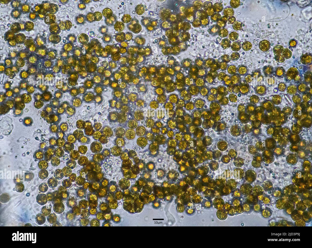 Symbiotic algae (zooxanthellae) from Aiptasia sp., probably belonging to the spaceis Breviolum dendrogyrum. The individual cells are in average about Stock Photohttps://www.alamy.com/image-license-details/?v=1https://www.alamy.com/symbiotic-algae-zooxanthellae-from-aiptasia-sp-probably-belonging-to-the-spaceis-breviolum-dendrogyrum-the-individual-cells-are-in-average-about-image472753838.html
RM2JD3P9J–Symbiotic algae (zooxanthellae) from Aiptasia sp., probably belonging to the spaceis Breviolum dendrogyrum. The individual cells are in average about
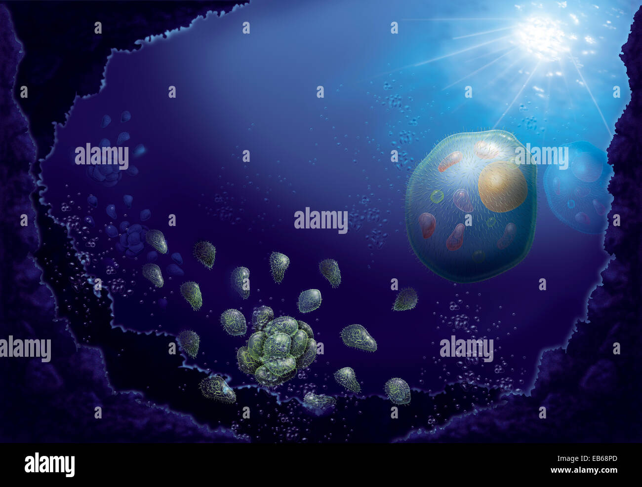 EVOLUTION, DRAWING Stock Photohttps://www.alamy.com/image-license-details/?v=1https://www.alamy.com/stock-photo-evolution-drawing-75741301.html
RMEB68PD–EVOLUTION, DRAWING
 Bacteria , germs on hand. 3d rendering. Isolated on solid color background Stock Photohttps://www.alamy.com/image-license-details/?v=1https://www.alamy.com/bacteria-germs-on-hand-3d-rendering-isolated-on-solid-color-background-image271117216.html
RFWN2CNM–Bacteria , germs on hand. 3d rendering. Isolated on solid color background
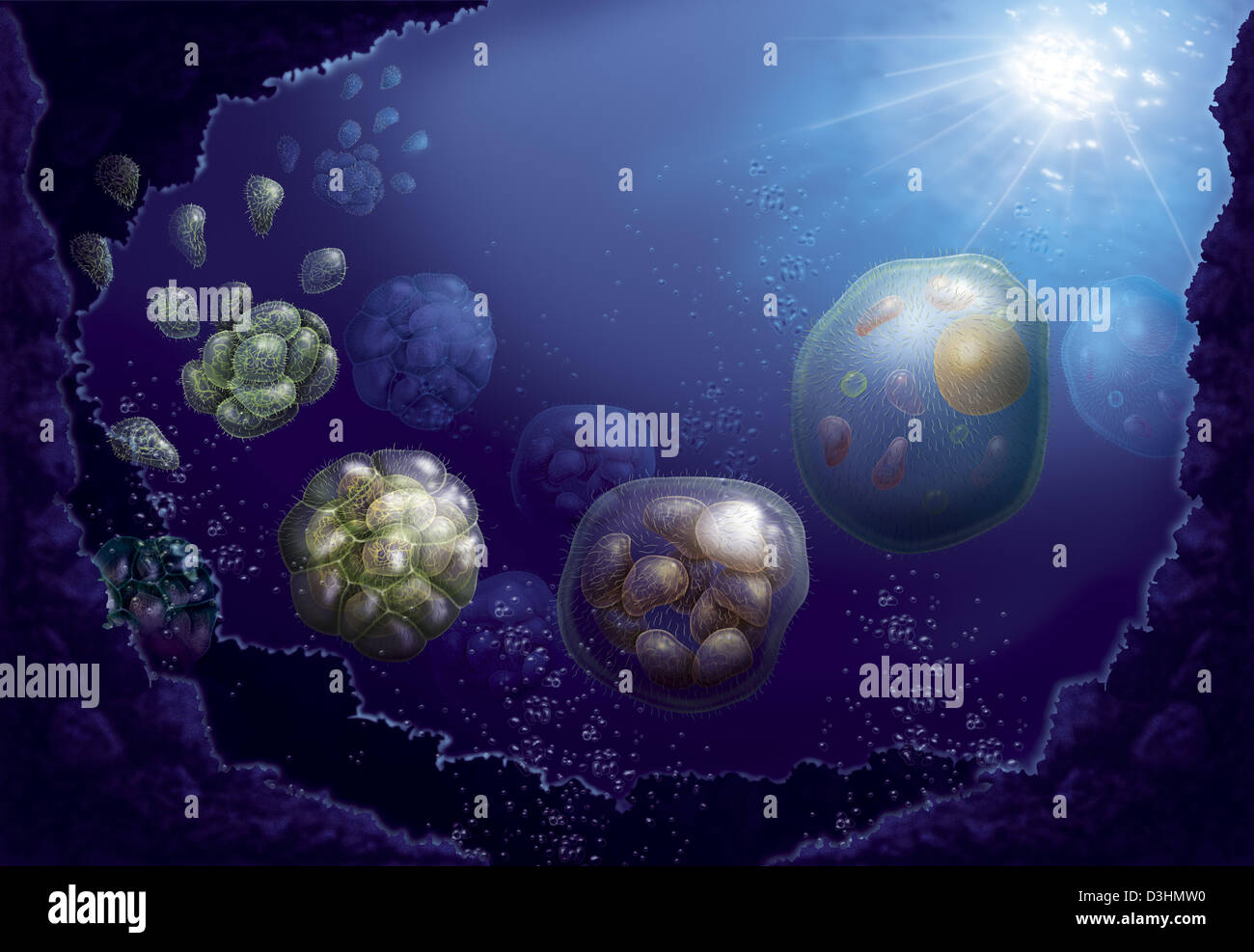 CELL, DRAWING Stock Photohttps://www.alamy.com/image-license-details/?v=1https://www.alamy.com/stock-photo-cell-drawing-53864636.html
RMD3HMW0–CELL, DRAWING
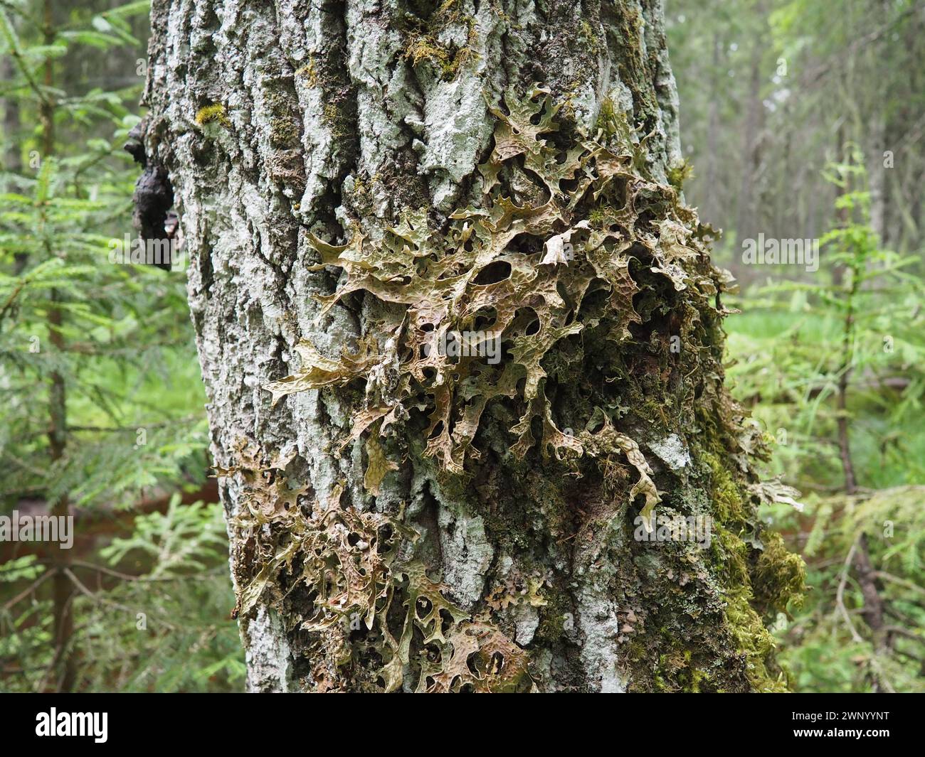 Moss and lichens on the bark of a tree in a spruce taiga forest. Karelia, Orzega. Lobaria Lobaria is a genus of lichenized ascomycetes belonging to Stock Photohttps://www.alamy.com/image-license-details/?v=1https://www.alamy.com/moss-and-lichens-on-the-bark-of-a-tree-in-a-spruce-taiga-forest-karelia-orzega-lobaria-lobaria-is-a-genus-of-lichenized-ascomycetes-belonging-to-image598674772.html
RF2WNYYNT–Moss and lichens on the bark of a tree in a spruce taiga forest. Karelia, Orzega. Lobaria Lobaria is a genus of lichenized ascomycetes belonging to
 . The biology of the Protozoa. Protozoa; Protozoa. 258 BIOLOGY OF THE PROTOZOA are symbiotic as in the yellow cells of Radiolaria and Foraminifera (Zooxanthelhe especially ChrysldeUa). Some are also ectoparasitic on diatoms, filamentous algte and Crustacea; others are endopara- sitic in water-dwelling animals such as Gammarus and allied Crustacea, and on rotifers. Order I. CHRYSOMONADIDA. The Chrysomonads are characterized b' the presence of yellow- brown chromatophores; by the presence of oils and highly refractile granules (leucosin); by spore formation in cysts and by the silicious composi Stock Photohttps://www.alamy.com/image-license-details/?v=1https://www.alamy.com/the-biology-of-the-protozoa-protozoa-protozoa-258-biology-of-the-protozoa-are-symbiotic-as-in-the-yellow-cells-of-radiolaria-and-foraminifera-zooxanthelhe-especially-chrysldeua-some-are-also-ectoparasitic-on-diatoms-filamentous-algte-and-crustacea-others-are-endopara-sitic-in-water-dwelling-animals-such-as-gammarus-and-allied-crustacea-and-on-rotifers-order-i-chrysomonadida-the-chrysomonads-are-characterized-b-the-presence-of-yellow-brown-chromatophores-by-the-presence-of-oils-and-highly-refractile-granules-leucosin-by-spore-formation-in-cysts-and-by-the-silicious-composi-image234602961.html
RMRHK2D5–. The biology of the Protozoa. Protozoa; Protozoa. 258 BIOLOGY OF THE PROTOZOA are symbiotic as in the yellow cells of Radiolaria and Foraminifera (Zooxanthelhe especially ChrysldeUa). Some are also ectoparasitic on diatoms, filamentous algte and Crustacea; others are endopara- sitic in water-dwelling animals such as Gammarus and allied Crustacea, and on rotifers. Order I. CHRYSOMONADIDA. The Chrysomonads are characterized b' the presence of yellow- brown chromatophores; by the presence of oils and highly refractile granules (leucosin); by spore formation in cysts and by the silicious composi
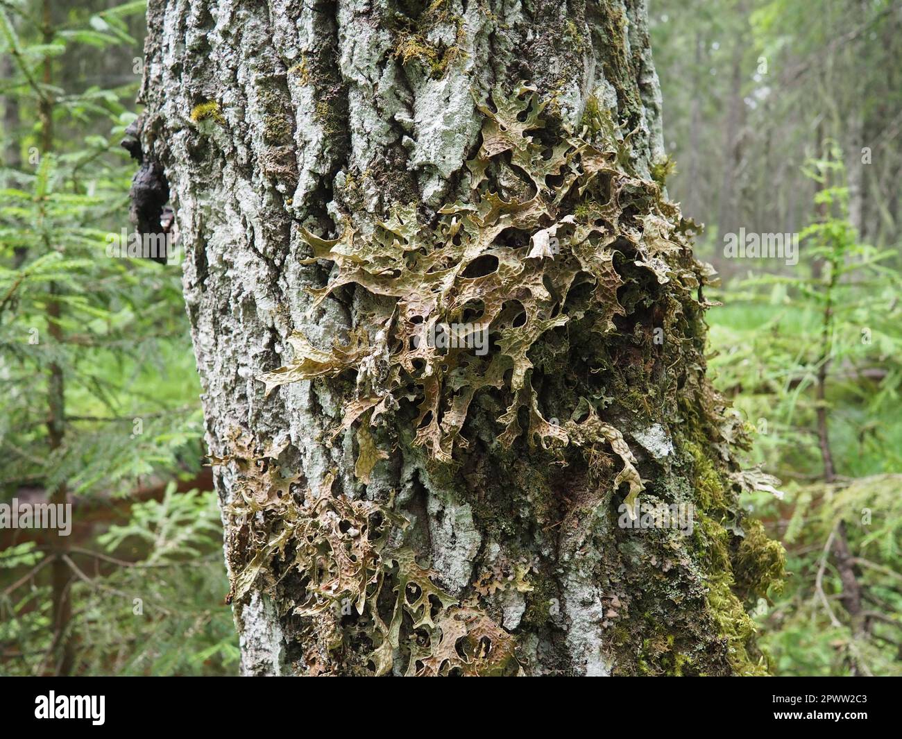 Moss and lichens on the bark of a tree in a spruce taiga forest. Karelia, Orzega. Lobaria Lobaria is a genus of lichenized ascomycetes belonging to th Stock Photohttps://www.alamy.com/image-license-details/?v=1https://www.alamy.com/moss-and-lichens-on-the-bark-of-a-tree-in-a-spruce-taiga-forest-karelia-orzega-lobaria-lobaria-is-a-genus-of-lichenized-ascomycetes-belonging-to-th-image549438515.html
RF2PWW2C3–Moss and lichens on the bark of a tree in a spruce taiga forest. Karelia, Orzega. Lobaria Lobaria is a genus of lichenized ascomycetes belonging to th
 . Report to the government of Baroda on the marine zoology of Okhamandal in Kattiawar . Marine animals. HORN ELL—THE PRESENCE OF SYMBIOTIC ALG^ IN MELIBE 147 apparently algal, were quantities of clear, ronndecl, colourless cells filled with refractive granules (Figs. 4 and 4a). The above notes were made during a rough examination of the living tissue. As I was greatly pressed for time, they are necessarily superficial; time and opportunity to carry the inquiry further have both been lacking. For some time I was unable to satisfy myself as to the nature of the organism. As the handful of its se Stock Photohttps://www.alamy.com/image-license-details/?v=1https://www.alamy.com/report-to-the-government-of-baroda-on-the-marine-zoology-of-okhamandal-in-kattiawar-marine-animals-horn-ellthe-presence-of-symbiotic-alg-in-melibe-147-apparently-algal-were-quantities-of-clear-ronndecl-colourless-cells-filled-with-refractive-granules-figs-4-and-4a-the-above-notes-were-made-during-a-rough-examination-of-the-living-tissue-as-i-was-greatly-pressed-for-time-they-are-necessarily-superficial-time-and-opportunity-to-carry-the-inquiry-further-have-both-been-lacking-for-some-time-i-was-unable-to-satisfy-myself-as-to-the-nature-of-the-organism-as-the-handful-of-its-se-image216398059.html
RMPG1NX3–. Report to the government of Baroda on the marine zoology of Okhamandal in Kattiawar . Marine animals. HORN ELL—THE PRESENCE OF SYMBIOTIC ALG^ IN MELIBE 147 apparently algal, were quantities of clear, ronndecl, colourless cells filled with refractive granules (Figs. 4 and 4a). The above notes were made during a rough examination of the living tissue. As I was greatly pressed for time, they are necessarily superficial; time and opportunity to carry the inquiry further have both been lacking. For some time I was unable to satisfy myself as to the nature of the organism. As the handful of its se
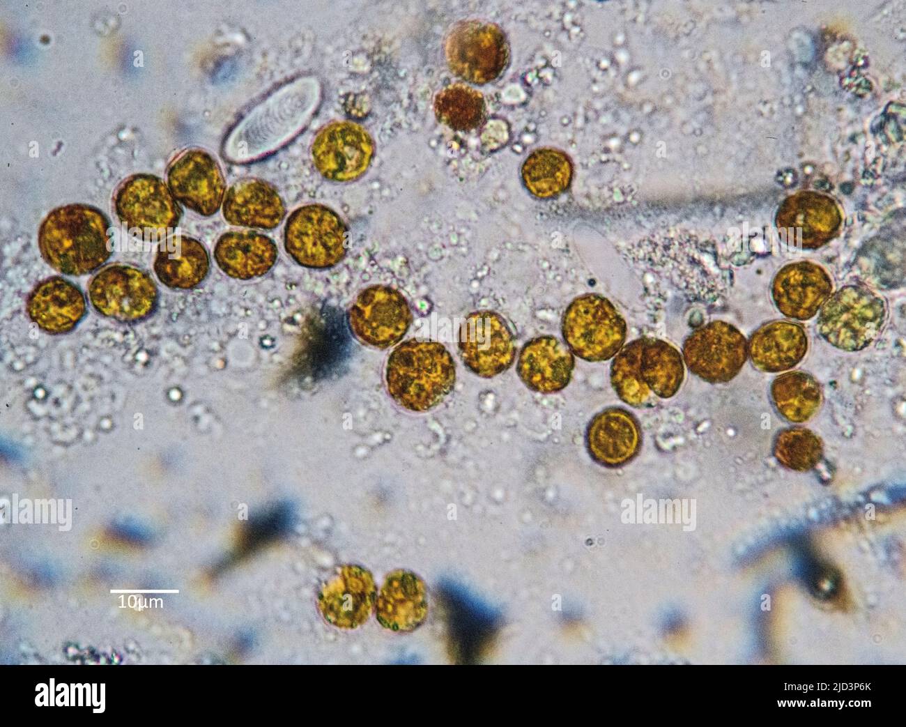 Symbiotic algae (zooxanthellae) from a zoanthid (Zoanthidea) from the Indo-Pacific. The individual cells are in average about 9 µm in diameter. Stock Photohttps://www.alamy.com/image-license-details/?v=1https://www.alamy.com/symbiotic-algae-zooxanthellae-from-a-zoanthid-zoanthidea-from-the-indo-pacific-the-individual-cells-are-in-average-about-9-m-in-diameter-image472753755.html
RM2JD3P6K–Symbiotic algae (zooxanthellae) from a zoanthid (Zoanthidea) from the Indo-Pacific. The individual cells are in average about 9 µm in diameter.
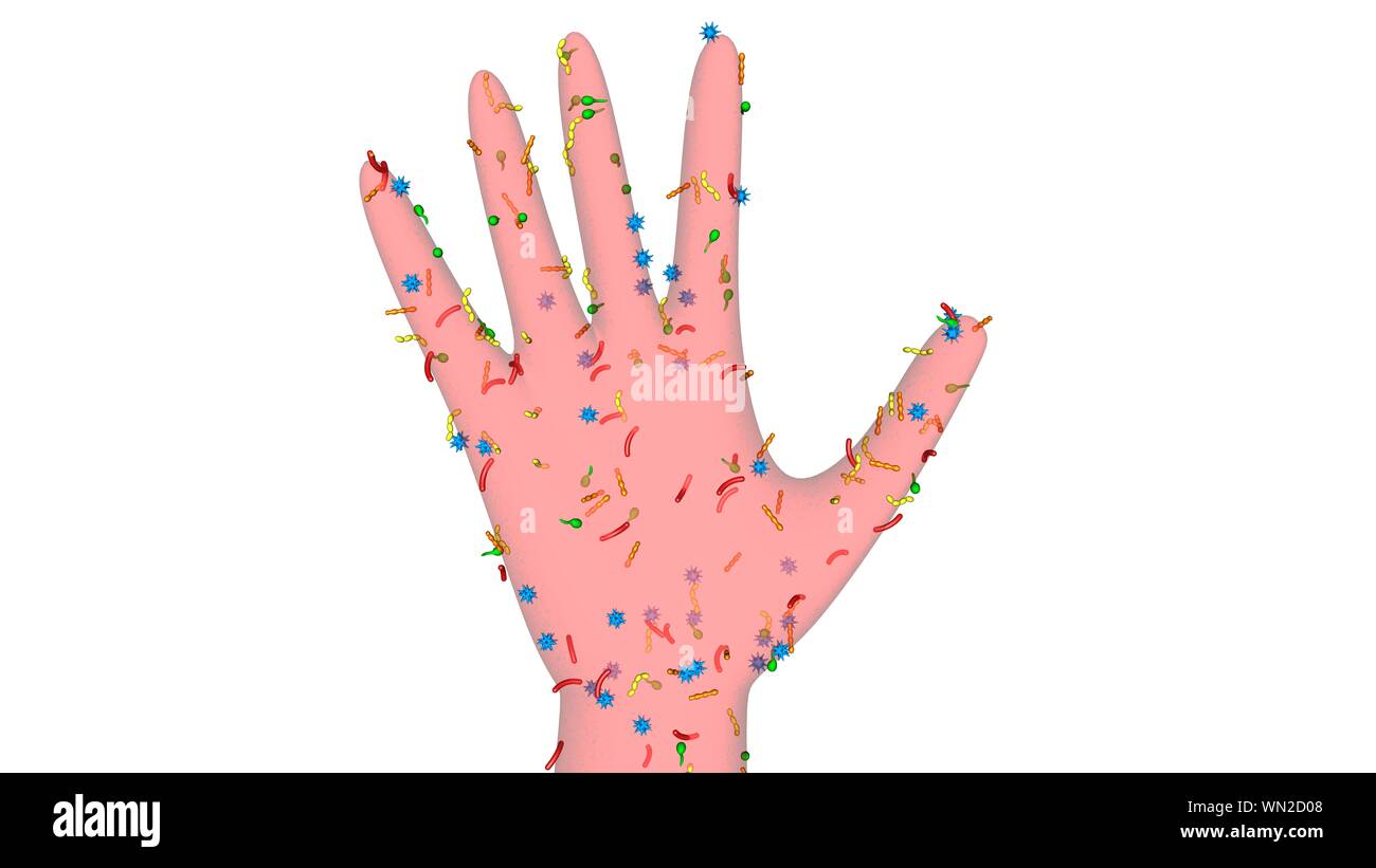 Bacteria , germs on hand. 3d rendering. Isolated on solid color background Stock Photohttps://www.alamy.com/image-license-details/?v=1https://www.alamy.com/bacteria-germs-on-hand-3d-rendering-isolated-on-solid-color-background-image271117400.html
RFWN2D08–Bacteria , germs on hand. 3d rendering. Isolated on solid color background
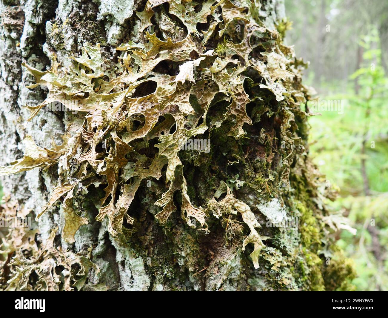 Moss and lichens on the bark of a tree in a spruce taiga forest. Karelia, Orzega. Lobaria Lobaria is a genus of lichenized ascomycetes belonging to Stock Photohttps://www.alamy.com/image-license-details/?v=1https://www.alamy.com/moss-and-lichens-on-the-bark-of-a-tree-in-a-spruce-taiga-forest-karelia-orzega-lobaria-lobaria-is-a-genus-of-lichenized-ascomycetes-belonging-to-image598665452.html
RF2WNYFW0–Moss and lichens on the bark of a tree in a spruce taiga forest. Karelia, Orzega. Lobaria Lobaria is a genus of lichenized ascomycetes belonging to
 . The cytoplasm of the plant cell. Plant cells and tissues; Protoplasm. Chapter XI 119 — Role of Ghondriosomes. SKY, DuESBERG, LEVI, and MiLOViDOV, is today abandoned by PoR- TIER himself. Nevertheless it had the merit of initiating investi- gations which have produced methods by which chondriosomes can be distinguished in cells from symbiotic and parasitic bacteria, CowDRY and Olitsky, Duesberg, and Milovidov have described methods by which, in the cells of nodules of legumes and in the adipose cells of cockroaches, symbiotic bacteria can be distin- guished from the chondriosomes by means of Stock Photohttps://www.alamy.com/image-license-details/?v=1https://www.alamy.com/the-cytoplasm-of-the-plant-cell-plant-cells-and-tissues-protoplasm-chapter-xi-119-role-of-ghondriosomes-sky-duesberg-levi-and-milovidov-is-today-abandoned-by-por-tier-himself-nevertheless-it-had-the-merit-of-initiating-investi-gations-which-have-produced-methods-by-which-chondriosomes-can-be-distinguished-in-cells-from-symbiotic-and-parasitic-bacteria-cowdry-and-olitsky-duesberg-and-milovidov-have-described-methods-by-which-in-the-cells-of-nodules-of-legumes-and-in-the-adipose-cells-of-cockroaches-symbiotic-bacteria-can-be-distin-guished-from-the-chondriosomes-by-means-of-image231803691.html
RMRD3FY7–. The cytoplasm of the plant cell. Plant cells and tissues; Protoplasm. Chapter XI 119 — Role of Ghondriosomes. SKY, DuESBERG, LEVI, and MiLOViDOV, is today abandoned by PoR- TIER himself. Nevertheless it had the merit of initiating investi- gations which have produced methods by which chondriosomes can be distinguished in cells from symbiotic and parasitic bacteria, CowDRY and Olitsky, Duesberg, and Milovidov have described methods by which, in the cells of nodules of legumes and in the adipose cells of cockroaches, symbiotic bacteria can be distin- guished from the chondriosomes by means of
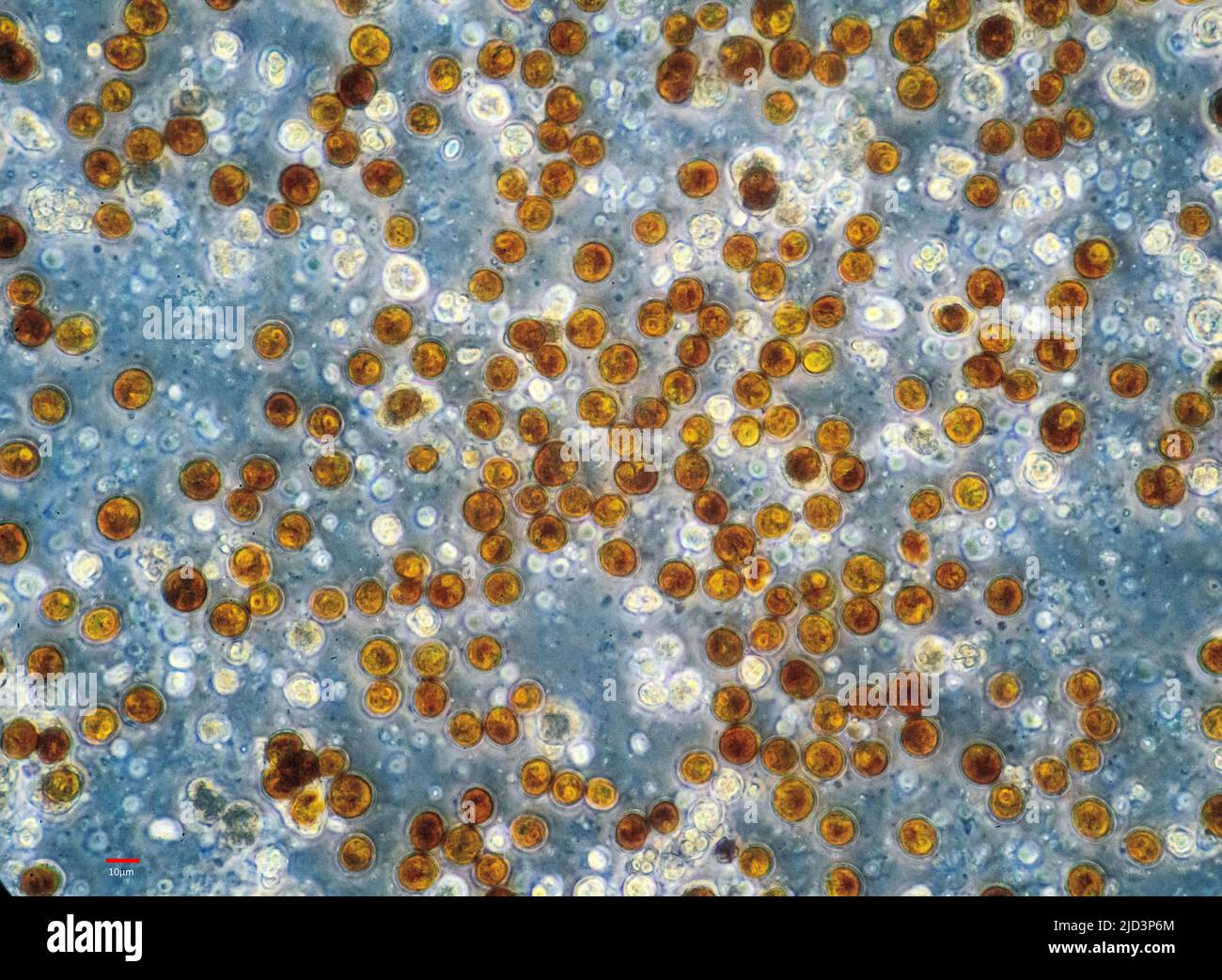 Zooxanthellae from a soft coral (probably Sinularia sp.). The individual cells are bout 10-12 µm in diameter. Stock Photohttps://www.alamy.com/image-license-details/?v=1https://www.alamy.com/zooxanthellae-from-a-soft-coral-probably-sinularia-sp-the-individual-cells-are-bout-10-12-m-in-diameter-image472753756.html
RM2JD3P6M–Zooxanthellae from a soft coral (probably Sinularia sp.). The individual cells are bout 10-12 µm in diameter.
 Bacteria , germs on hand. 3d rendering. Isolated on solid color background Stock Photohttps://www.alamy.com/image-license-details/?v=1https://www.alamy.com/bacteria-germs-on-hand-3d-rendering-isolated-on-solid-color-background-image271117266.html
RFWN2CRE–Bacteria , germs on hand. 3d rendering. Isolated on solid color background
 . The cytoplasm of the plant cell. Plant cells and tissues; Protoplasm. SKY, DuESBERG, LEVI, and MiLOViDOV, is today abandoned by PoR- TIER himself. Nevertheless it had the merit of initiating investi- gations which have produced methods by which chondriosomes can be distinguished in cells from symbiotic and parasitic bacteria, CowDRY and Olitsky, Duesberg, and Milovidov have described methods by which, in the cells of nodules of legumes and in the adipose cells of cockroaches, symbiotic bacteria can be distin- guished from the chondriosomes by means of differential staining. By these methods Stock Photohttps://www.alamy.com/image-license-details/?v=1https://www.alamy.com/the-cytoplasm-of-the-plant-cell-plant-cells-and-tissues-protoplasm-sky-duesberg-levi-and-milovidov-is-today-abandoned-by-por-tier-himself-nevertheless-it-had-the-merit-of-initiating-investi-gations-which-have-produced-methods-by-which-chondriosomes-can-be-distinguished-in-cells-from-symbiotic-and-parasitic-bacteria-cowdry-and-olitsky-duesberg-and-milovidov-have-described-methods-by-which-in-the-cells-of-nodules-of-legumes-and-in-the-adipose-cells-of-cockroaches-symbiotic-bacteria-can-be-distin-guished-from-the-chondriosomes-by-means-of-differential-staining-by-these-methods-image231803685.html
RMRD3FY1–. The cytoplasm of the plant cell. Plant cells and tissues; Protoplasm. SKY, DuESBERG, LEVI, and MiLOViDOV, is today abandoned by PoR- TIER himself. Nevertheless it had the merit of initiating investi- gations which have produced methods by which chondriosomes can be distinguished in cells from symbiotic and parasitic bacteria, CowDRY and Olitsky, Duesberg, and Milovidov have described methods by which, in the cells of nodules of legumes and in the adipose cells of cockroaches, symbiotic bacteria can be distin- guished from the chondriosomes by means of differential staining. By these methods
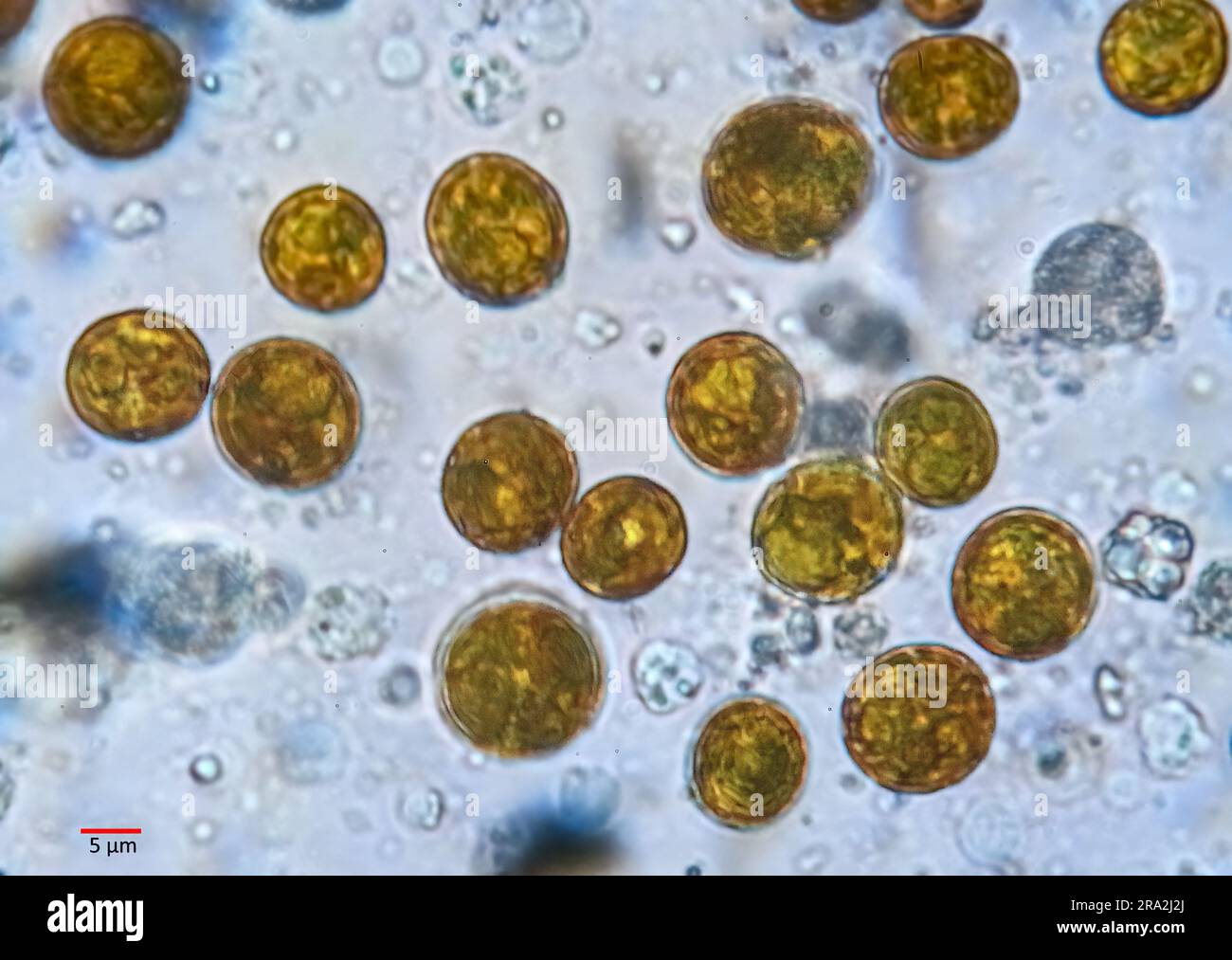 Zooxanthellae from a soft coral (probably Sinularia sp.). The individual cells are bout 10-12 µm in diameter. Stock Photohttps://www.alamy.com/image-license-details/?v=1https://www.alamy.com/zooxanthellae-from-a-soft-coral-probably-sinularia-sp-the-individual-cells-are-bout-10-12-m-in-diameter-image556936426.html
RM2RA2J2J–Zooxanthellae from a soft coral (probably Sinularia sp.). The individual cells are bout 10-12 µm in diameter.