Quick filters:
Tensor fascia lata Stock Photos and Images
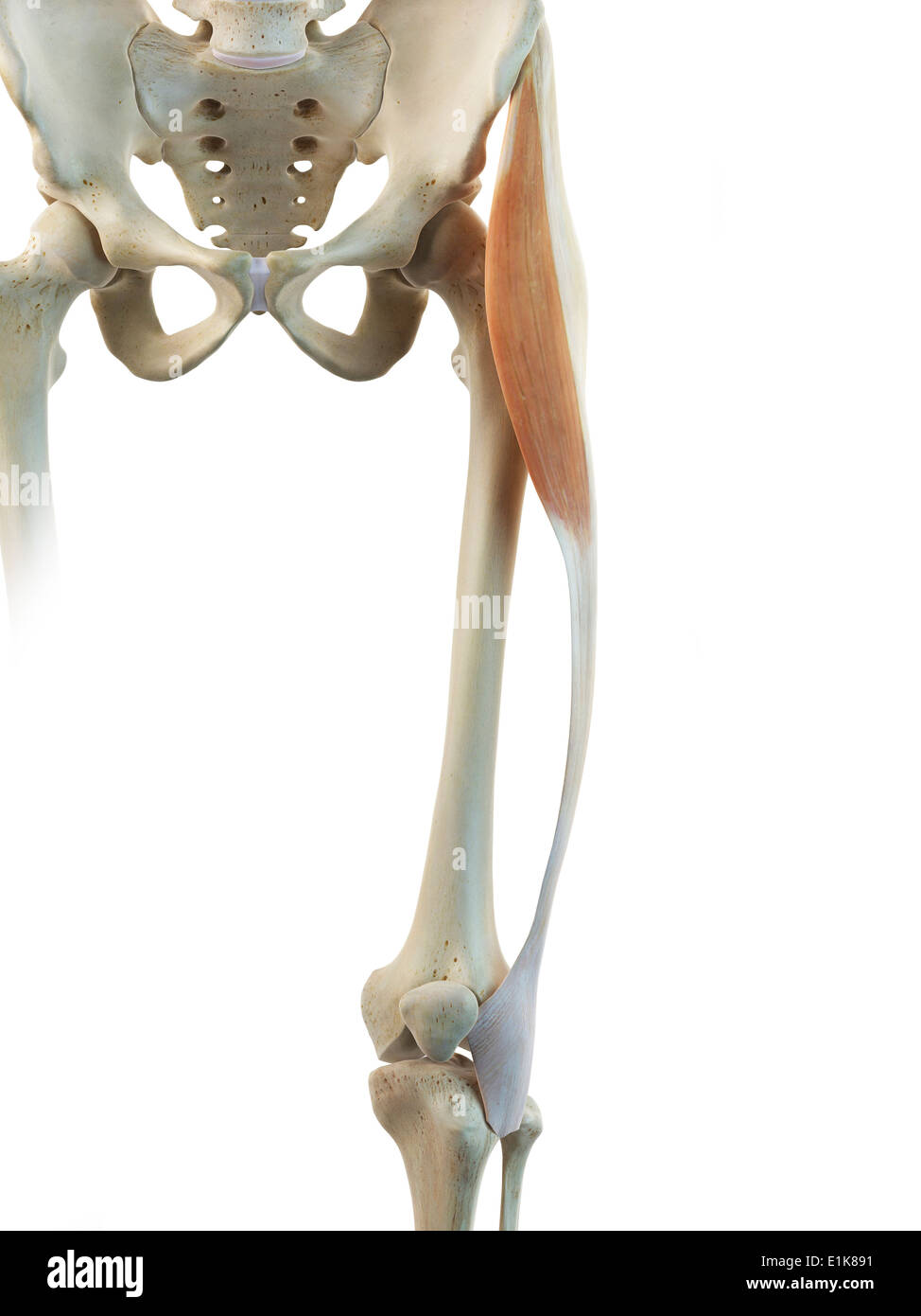 Human tensor fascia lata muscle computer artwork. Stock Photohttps://www.alamy.com/image-license-details/?v=1https://www.alamy.com/human-tensor-fascia-lata-muscle-computer-artwork-image69879741.html
RFE1K891–Human tensor fascia lata muscle computer artwork.
 medically accurate muscle illustration of the tensor fascia lata Stock Photohttps://www.alamy.com/image-license-details/?v=1https://www.alamy.com/stock-photo-medically-accurate-muscle-illustration-of-the-tensor-fascia-lata-89754534.html
RFF60JR2–medically accurate muscle illustration of the tensor fascia lata
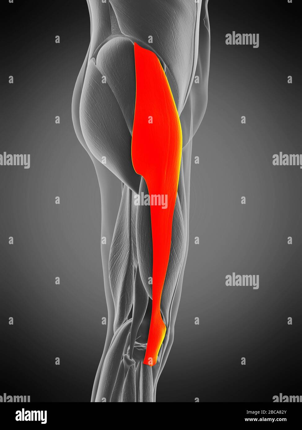 Tensor fascia lata muscle, illustration. Stock Photohttps://www.alamy.com/image-license-details/?v=1https://www.alamy.com/tensor-fascia-lata-muscle-illustration-image351809107.html
RF2BCA82Y–Tensor fascia lata muscle, illustration.
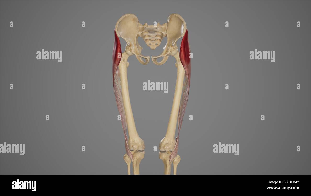 Medical Illustration of Tensor Fascia Lata Muscle Stock Photohttps://www.alamy.com/image-license-details/?v=1https://www.alamy.com/medical-illustration-of-tensor-fascia-lata-muscle-image490198491.html
RF2KDED4Y–Medical Illustration of Tensor Fascia Lata Muscle
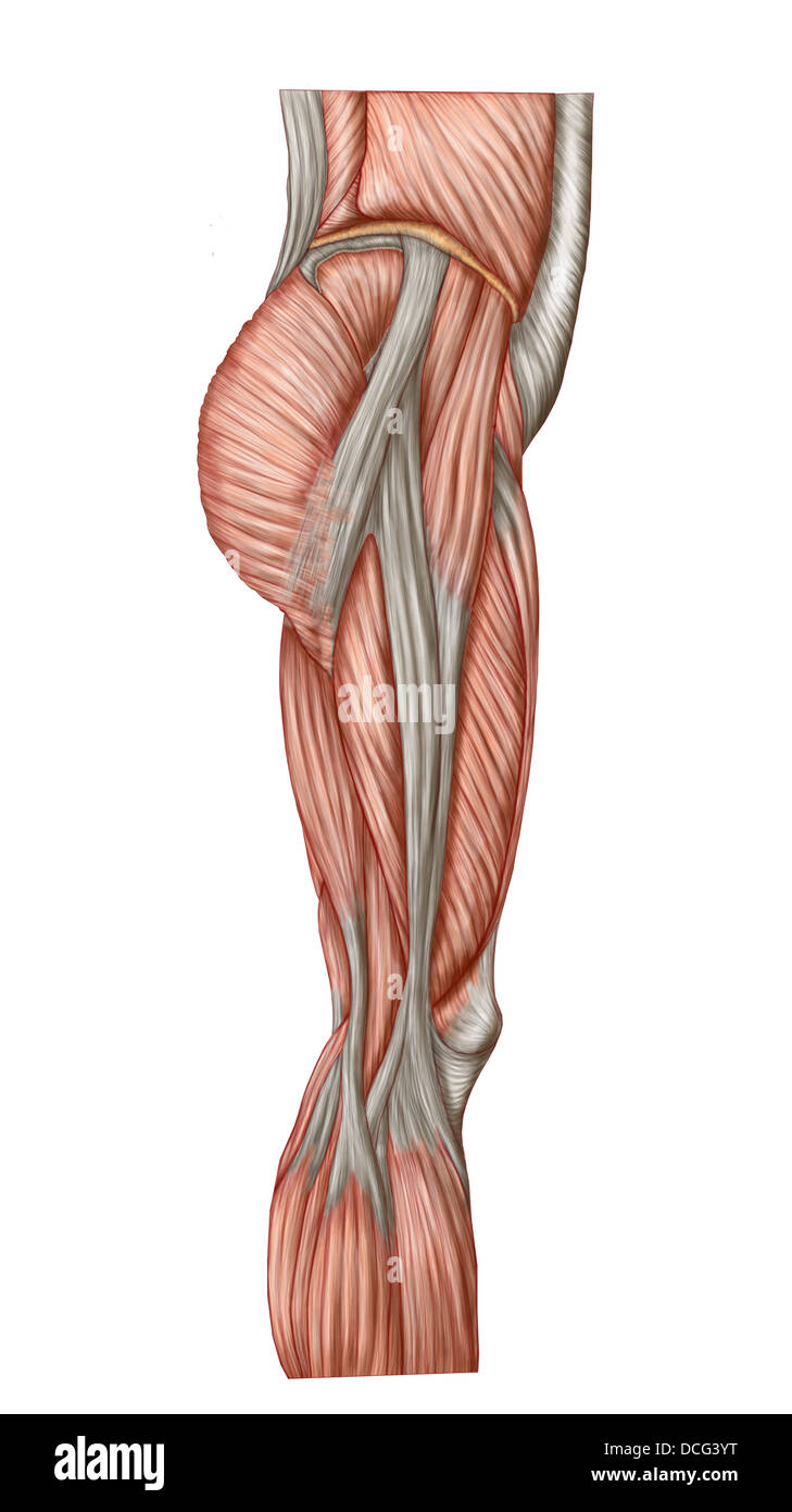 Anatomy of human thigh muscles, anterior view. Stock Photohttps://www.alamy.com/image-license-details/?v=1https://www.alamy.com/stock-photo-anatomy-of-human-thigh-muscles-anterior-view-59361340.html
RFDCG3YT–Anatomy of human thigh muscles, anterior view.
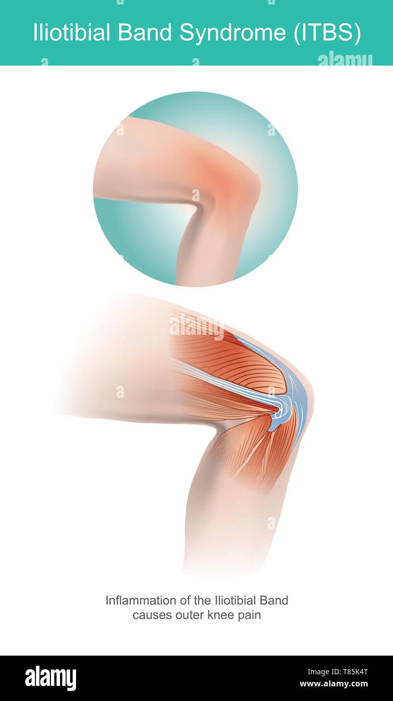 The Iliotibial Band is a longitudinal fibrous reinforcement of the fascia lata in a knee muscle. Part of anatomy human body. Illustration. Stock Vectorhttps://www.alamy.com/image-license-details/?v=1https://www.alamy.com/the-iliotibial-band-is-a-longitudinal-fibrous-reinforcement-of-the-fascia-lata-in-a-knee-muscle-part-of-anatomy-human-body-illustration-image245987192.html
RFT85K4T–The Iliotibial Band is a longitudinal fibrous reinforcement of the fascia lata in a knee muscle. Part of anatomy human body. Illustration.
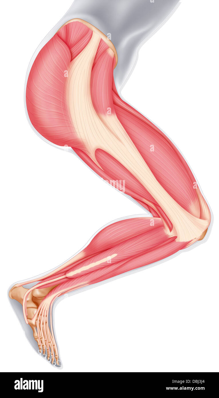 LOWER LIMB MUSCLE, DRAWING Stock Photohttps://www.alamy.com/image-license-details/?v=1https://www.alamy.com/stock-photo-lower-limb-muscle-drawing-58790316.html
RMDBJ3J4–LOWER LIMB MUSCLE, DRAWING
 3D illustration of TENSOR title on a medical document Stock Photohttps://www.alamy.com/image-license-details/?v=1https://www.alamy.com/3d-illustration-of-tensor-title-on-a-medical-document-image333093658.html
RF2A9WMA2–3D illustration of TENSOR title on a medical document
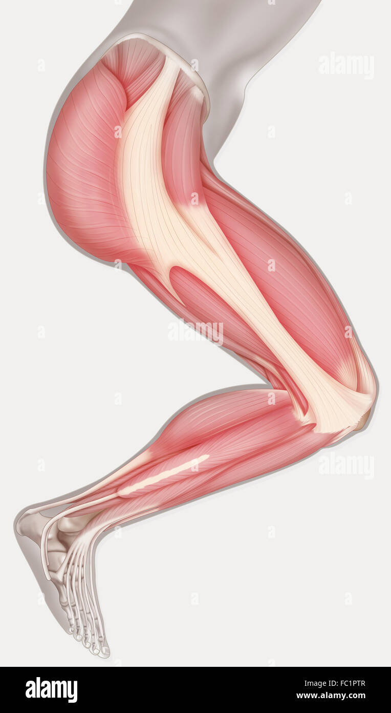 LOWER LIMB MUSCLE, DRAWING Stock Photohttps://www.alamy.com/image-license-details/?v=1https://www.alamy.com/stock-photo-lower-limb-muscle-drawing-93467607.html
RMFC1PTR–LOWER LIMB MUSCLE, DRAWING
![. The physiology of domestic animals ... Physiology, Comparative; Veterinary physiology. PHYSIOLOGY OF THE DOMESTIC ANIMALS. muscular exertion is required in walking on level ground. In ascending an incline, however, the active limb has at each step to elevate the weight of the body by extending the knee- and ankle-joint by the thigh-exten- sors and calf-muscles, therefore greatly increasing the muscular power required. During walking the trunk leans toward the active leg, owing to the contraction of the glutei muscles and the tensor fascia lata, and is inclined slight]}' torward to overcome t Stock Photo . The physiology of domestic animals ... Physiology, Comparative; Veterinary physiology. PHYSIOLOGY OF THE DOMESTIC ANIMALS. muscular exertion is required in walking on level ground. In ascending an incline, however, the active limb has at each step to elevate the weight of the body by extending the knee- and ankle-joint by the thigh-exten- sors and calf-muscles, therefore greatly increasing the muscular power required. During walking the trunk leans toward the active leg, owing to the contraction of the glutei muscles and the tensor fascia lata, and is inclined slight]}' torward to overcome t Stock Photo](https://c8.alamy.com/comp/RE3FR1/the-physiology-of-domestic-animals-physiology-comparative-veterinary-physiology-physiology-of-the-domestic-animals-muscular-exertion-is-required-in-walking-on-level-ground-in-ascending-an-incline-however-the-active-limb-has-at-each-step-to-elevate-the-weight-of-the-body-by-extending-the-knee-and-ankle-joint-by-the-thigh-exten-sors-and-calf-muscles-therefore-greatly-increasing-the-muscular-power-required-during-walking-the-trunk-leans-toward-the-active-leg-owing-to-the-contraction-of-the-glutei-muscles-and-the-tensor-fascia-lata-and-is-inclined-slight-torward-to-overcome-t-RE3FR1.jpg) . The physiology of domestic animals ... Physiology, Comparative; Veterinary physiology. PHYSIOLOGY OF THE DOMESTIC ANIMALS. muscular exertion is required in walking on level ground. In ascending an incline, however, the active limb has at each step to elevate the weight of the body by extending the knee- and ankle-joint by the thigh-exten- sors and calf-muscles, therefore greatly increasing the muscular power required. During walking the trunk leans toward the active leg, owing to the contraction of the glutei muscles and the tensor fascia lata, and is inclined slight]}' torward to overcome t Stock Photohttps://www.alamy.com/image-license-details/?v=1https://www.alamy.com/the-physiology-of-domestic-animals-physiology-comparative-veterinary-physiology-physiology-of-the-domestic-animals-muscular-exertion-is-required-in-walking-on-level-ground-in-ascending-an-incline-however-the-active-limb-has-at-each-step-to-elevate-the-weight-of-the-body-by-extending-the-knee-and-ankle-joint-by-the-thigh-exten-sors-and-calf-muscles-therefore-greatly-increasing-the-muscular-power-required-during-walking-the-trunk-leans-toward-the-active-leg-owing-to-the-contraction-of-the-glutei-muscles-and-the-tensor-fascia-lata-and-is-inclined-slight-torward-to-overcome-t-image232418229.html
. The physiology of domestic animals ... Physiology, Comparative; Veterinary physiology. PHYSIOLOGY OF THE DOMESTIC ANIMALS. muscular exertion is required in walking on level ground. In ascending an incline, however, the active limb has at each step to elevate the weight of the body by extending the knee- and ankle-joint by the thigh-exten- sors and calf-muscles, therefore greatly increasing the muscular power required. During walking the trunk leans toward the active leg, owing to the contraction of the glutei muscles and the tensor fascia lata, and is inclined slight]}' torward to overcome t Stock Photohttps://www.alamy.com/image-license-details/?v=1https://www.alamy.com/the-physiology-of-domestic-animals-physiology-comparative-veterinary-physiology-physiology-of-the-domestic-animals-muscular-exertion-is-required-in-walking-on-level-ground-in-ascending-an-incline-however-the-active-limb-has-at-each-step-to-elevate-the-weight-of-the-body-by-extending-the-knee-and-ankle-joint-by-the-thigh-exten-sors-and-calf-muscles-therefore-greatly-increasing-the-muscular-power-required-during-walking-the-trunk-leans-toward-the-active-leg-owing-to-the-contraction-of-the-glutei-muscles-and-the-tensor-fascia-lata-and-is-inclined-slight-torward-to-overcome-t-image232418229.htmlRMRE3FR1–. The physiology of domestic animals ... Physiology, Comparative; Veterinary physiology. PHYSIOLOGY OF THE DOMESTIC ANIMALS. muscular exertion is required in walking on level ground. In ascending an incline, however, the active limb has at each step to elevate the weight of the body by extending the knee- and ankle-joint by the thigh-exten- sors and calf-muscles, therefore greatly increasing the muscular power required. During walking the trunk leans toward the active leg, owing to the contraction of the glutei muscles and the tensor fascia lata, and is inclined slight]}' torward to overcome t
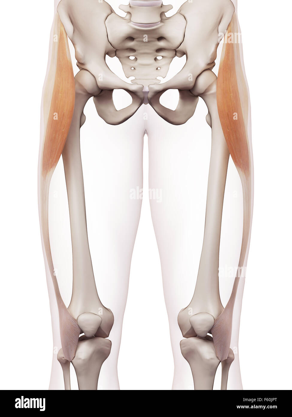 medically accurate muscle illustration of the tensor fascia lata Stock Photohttps://www.alamy.com/image-license-details/?v=1https://www.alamy.com/stock-photo-medically-accurate-muscle-illustration-of-the-tensor-fascia-lata-89754528.html
RFF60JPT–medically accurate muscle illustration of the tensor fascia lata
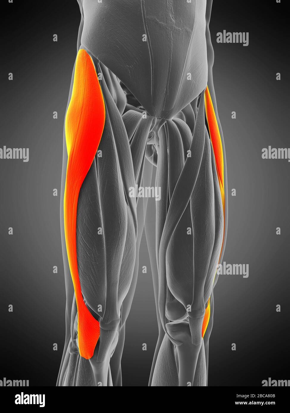 Tensor fascia lata muscle, illustration. Stock Photohttps://www.alamy.com/image-license-details/?v=1https://www.alamy.com/tensor-fascia-lata-muscle-illustration-image351809035.html
RF2BCA80B–Tensor fascia lata muscle, illustration.
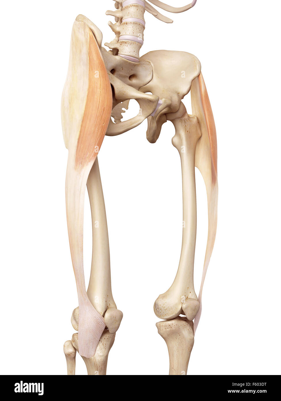 medical accurate illustration of the tensor fascia lata Stock Photohttps://www.alamy.com/image-license-details/?v=1https://www.alamy.com/stock-photo-medical-accurate-illustration-of-the-tensor-fascia-lata-89742516.html
RFF603DT–medical accurate illustration of the tensor fascia lata
 Human leg muscles, illustration. tensor fascia lata Stock Photohttps://www.alamy.com/image-license-details/?v=1https://www.alamy.com/stock-photo-human-leg-muscles-illustration-tensor-fascia-lata-118698782.html
RFGW35E6–Human leg muscles, illustration. tensor fascia lata
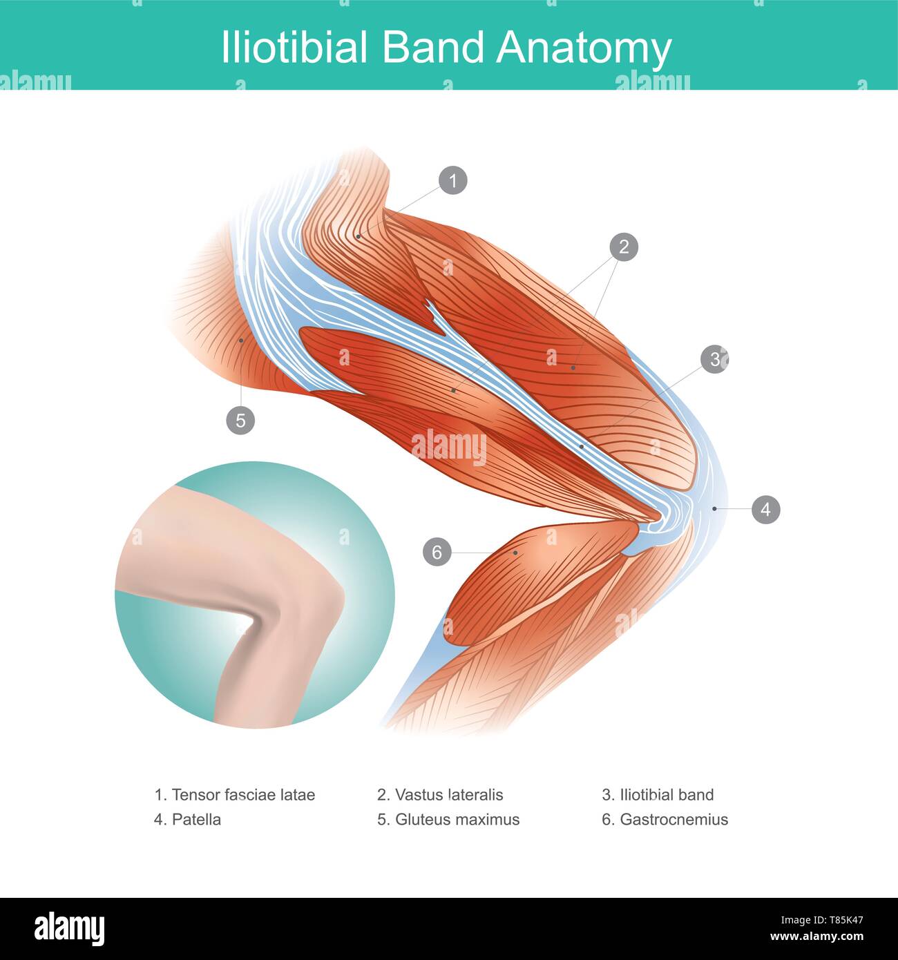 The Iliotibial Band is a longitudinal fibrous reinforcement of the fascia lata in a knee muscle. Part of anatomy human body. Illustration. Stock Vectorhttps://www.alamy.com/image-license-details/?v=1https://www.alamy.com/the-iliotibial-band-is-a-longitudinal-fibrous-reinforcement-of-the-fascia-lata-in-a-knee-muscle-part-of-anatomy-human-body-illustration-image245987175.html
RFT85K47–The Iliotibial Band is a longitudinal fibrous reinforcement of the fascia lata in a knee muscle. Part of anatomy human body. Illustration.
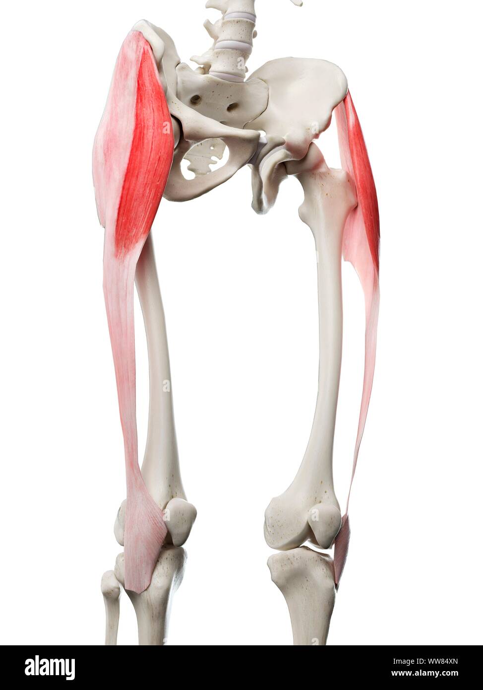 Tensor fascia lata muscle, illustration Stock Photohttps://www.alamy.com/image-license-details/?v=1https://www.alamy.com/tensor-fascia-lata-muscle-illustration-image273701421.html
RFWW84XN–Tensor fascia lata muscle, illustration
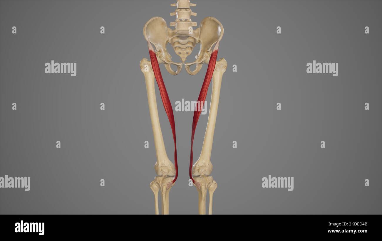 Medical Illustration of Sartorius Muscle Stock Photohttps://www.alamy.com/image-license-details/?v=1https://www.alamy.com/medical-illustration-of-sartorius-muscle-image490198475.html
RF2KDED4B–Medical Illustration of Sartorius Muscle
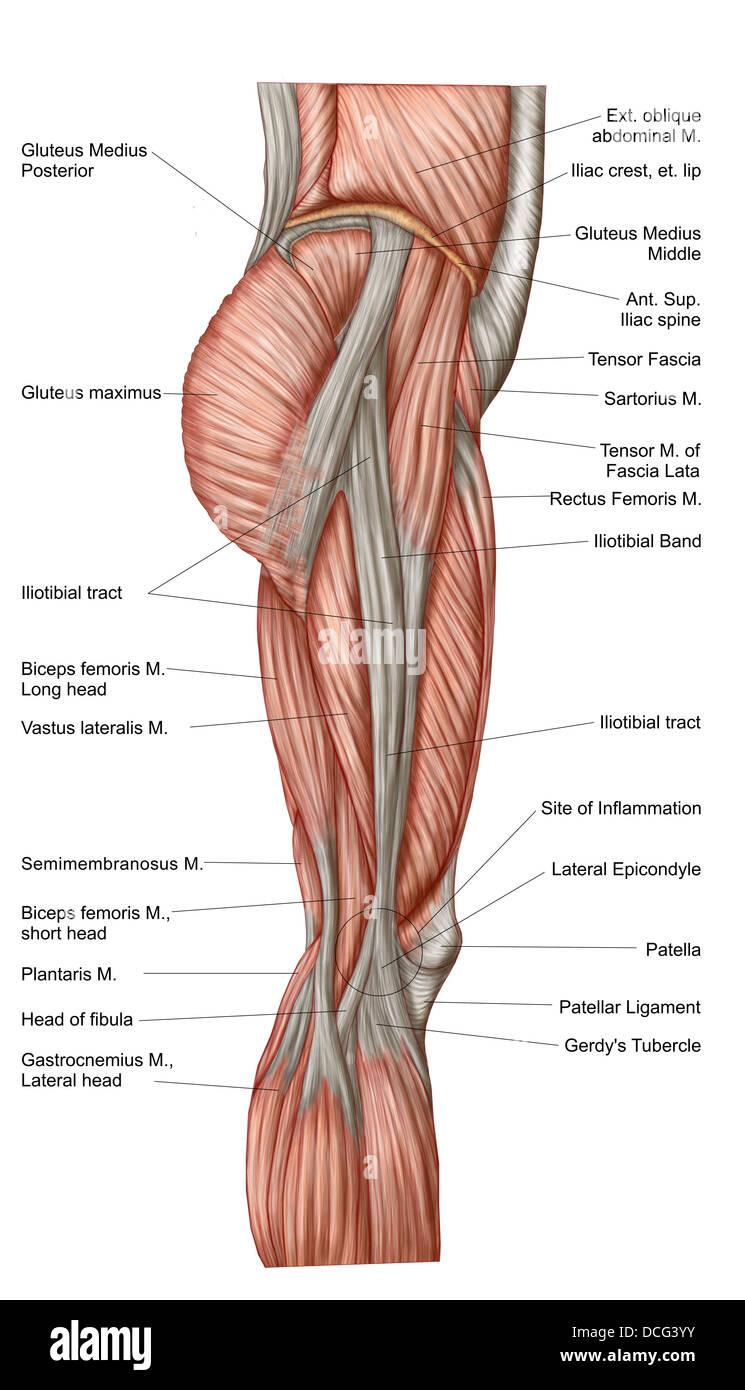 Anatomy of human thigh muscles, anterior view. Stock Photohttps://www.alamy.com/image-license-details/?v=1https://www.alamy.com/stock-photo-anatomy-of-human-thigh-muscles-anterior-view-59361343.html
RFDCG3YY–Anatomy of human thigh muscles, anterior view.
 . The anatomy of the domestic animals. Veterinary anatomy. THE STIFLE JOINT 23.5 biceps femoris, but it also furnishes insertion to the tensor fascia; lata; by means of the fascia lata, which blends with it. The femoro-tibial articulation (Articulatio femoro-tibialis) is formed between the condyles of the femur, the j)roximal enil of the tibia, and the interposed articular menisci or semilunar cail^ilages. Articular Surfaces.—The condyles of the femur are slightly oljlique in tlirec- tion. The articular surface of the lateral one is more strongly curv-ed than that of the medial one; the latter Stock Photohttps://www.alamy.com/image-license-details/?v=1https://www.alamy.com/the-anatomy-of-the-domestic-animals-veterinary-anatomy-the-stifle-joint-235-biceps-femoris-but-it-also-furnishes-insertion-to-the-tensor-fascia-lata-by-means-of-the-fascia-lata-which-blends-with-it-the-femoro-tibial-articulation-articulatio-femoro-tibialis-is-formed-between-the-condyles-of-the-femur-the-jroximal-enil-of-the-tibia-and-the-interposed-articular-menisci-or-semilunar-caililages-articular-surfacesthe-condyles-of-the-femur-are-slightly-oljlique-in-tlirec-tion-the-articular-surface-of-the-lateral-one-is-more-strongly-curv-ed-than-that-of-the-medial-one-the-latter-image236762853.html
RMRN5DC5–. The anatomy of the domestic animals. Veterinary anatomy. THE STIFLE JOINT 23.5 biceps femoris, but it also furnishes insertion to the tensor fascia; lata; by means of the fascia lata, which blends with it. The femoro-tibial articulation (Articulatio femoro-tibialis) is formed between the condyles of the femur, the j)roximal enil of the tibia, and the interposed articular menisci or semilunar cail^ilages. Articular Surfaces.—The condyles of the femur are slightly oljlique in tlirec- tion. The articular surface of the lateral one is more strongly curv-ed than that of the medial one; the latter
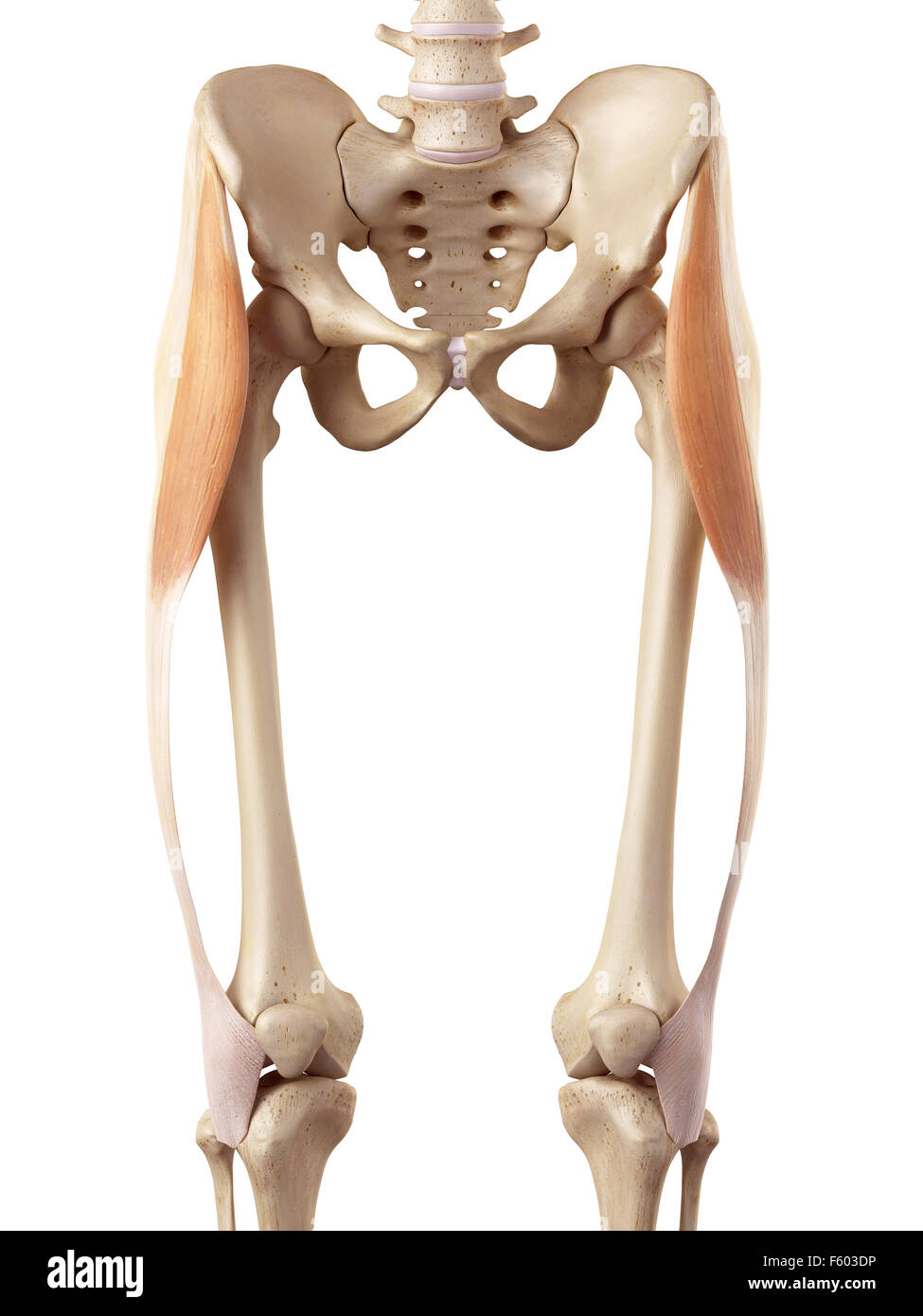 medical accurate illustration of the tensor fascia lata Stock Photohttps://www.alamy.com/image-license-details/?v=1https://www.alamy.com/stock-photo-medical-accurate-illustration-of-the-tensor-fascia-lata-89742514.html
RFF603DP–medical accurate illustration of the tensor fascia lata
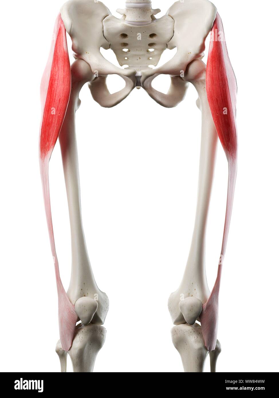 Tensor fascia lata muscle, illustration Stock Photohttps://www.alamy.com/image-license-details/?v=1https://www.alamy.com/tensor-fascia-lata-muscle-illustration-image273701397.html
RFWW84WW–Tensor fascia lata muscle, illustration
 Horse tensor fascia lata muscle, illustration Stock Photohttps://www.alamy.com/image-license-details/?v=1https://www.alamy.com/horse-tensor-fascia-lata-muscle-illustration-image273702714.html
RFWW86GX–Horse tensor fascia lata muscle, illustration
 . The anatomy of the domestic animals. Veterinary anatomy. BRANCHES OF THE ABDOMINAL AORTA 667 Internal iliac lymph i Circumflex iliac vessels External iliac lymph gla Remnant of inguinal ligament Posterior branch of circum- flex iliac artery Lateral cutaneous nerve of thigh External spermatic nerrc Tensor fascia: lata: Femoral nerve and ante- rior femoral vessels Femoral artery Prefemoral lymph glands Deep inguinal lymph glands Fascia lata Saphenous nerves Saphenous vessels Medial patellar ligament Middle patellar ligament. Fig. 576.—Dissection of Pelvis, Thigh, .nd Phoxlm.^l P.rt of Leg of Stock Photohttps://www.alamy.com/image-license-details/?v=1https://www.alamy.com/the-anatomy-of-the-domestic-animals-veterinary-anatomy-branches-of-the-abdominal-aorta-667-internal-iliac-lymph-i-circumflex-iliac-vessels-external-iliac-lymph-gla-remnant-of-inguinal-ligament-posterior-branch-of-circum-flex-iliac-artery-lateral-cutaneous-nerve-of-thigh-external-spermatic-nerrc-tensor-fascia-lata-femoral-nerve-and-ante-rior-femoral-vessels-femoral-artery-prefemoral-lymph-glands-deep-inguinal-lymph-glands-fascia-lata-saphenous-nerves-saphenous-vessels-medial-patellar-ligament-middle-patellar-ligament-fig-576dissection-of-pelvis-thigh-nd-phoxlml-prt-of-leg-of-image236759542.html
RMRN595X–. The anatomy of the domestic animals. Veterinary anatomy. BRANCHES OF THE ABDOMINAL AORTA 667 Internal iliac lymph i Circumflex iliac vessels External iliac lymph gla Remnant of inguinal ligament Posterior branch of circum- flex iliac artery Lateral cutaneous nerve of thigh External spermatic nerrc Tensor fascia: lata: Femoral nerve and ante- rior femoral vessels Femoral artery Prefemoral lymph glands Deep inguinal lymph glands Fascia lata Saphenous nerves Saphenous vessels Medial patellar ligament Middle patellar ligament. Fig. 576.—Dissection of Pelvis, Thigh, .nd Phoxlm.^l P.rt of Leg of
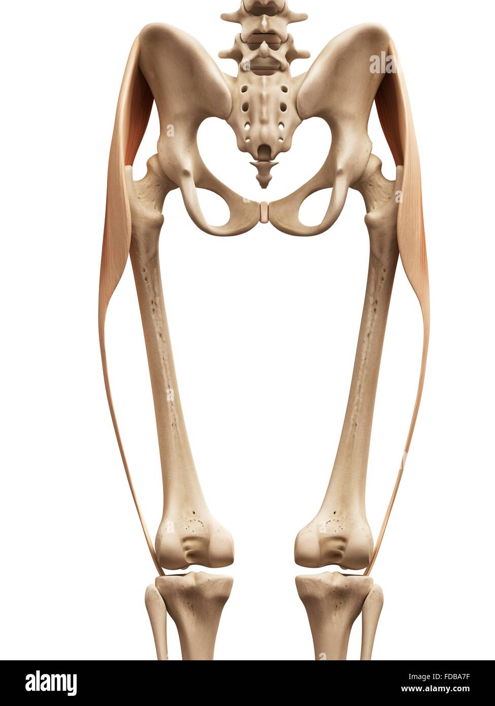 Human leg muscles (tensor fascia lata), illustration. Stock Photohttps://www.alamy.com/image-license-details/?v=1https://www.alamy.com/stock-photo-human-leg-muscles-tensor-fascia-lata-illustration-94291891.html
RFFDBA7F–Human leg muscles (tensor fascia lata), illustration.
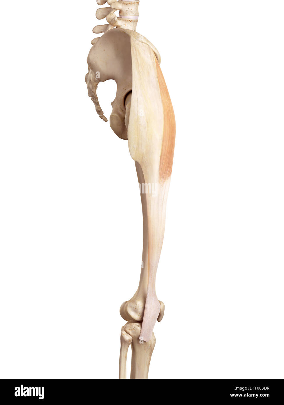 medical accurate illustration of the tensor fascia lata Stock Photohttps://www.alamy.com/image-license-details/?v=1https://www.alamy.com/stock-photo-medical-accurate-illustration-of-the-tensor-fascia-lata-89742515.html
RFF603DR–medical accurate illustration of the tensor fascia lata
 . Chordate anatomy. Chordata; Anatomy, Comparative. 2o6 CHORDATE ANATOMY stomes (Petromyzon) do all the somites produce myotomes. In the higher forms the series is broken in the ear region.. FLEX. CARPI ULNARIS FLEX. CARPI RAD. LOXB.s BRACHIORADIAL.' Da CARPI RAD LONGUS- PALMARIS LONGUS. BRACHIALIS TRICEPS â¢â '- CORACO BRACHIALIS-' TERES MAJOR' LATISSIMUS DORS I SERRATUS ANTERIOR LINEA ALBA RECTUS ABDOMINIS EXTERNAL OBLIQUE ILIUM TENSOR FASCIA LATA ILIOPSOAS RECTINEUSâ PUBIS 'â RECTUS FEMORIS- SARTORIUS Mljl ADDUCTOR LONGUS - -"" "' ADDUCTOR MAGNUS- VASTUS LATERALIS PERONEU Stock Photohttps://www.alamy.com/image-license-details/?v=1https://www.alamy.com/chordate-anatomy-chordata-anatomy-comparative-2o6-chordate-anatomy-stomes-petromyzon-do-all-the-somites-produce-myotomes-in-the-higher-forms-the-series-is-broken-in-the-ear-region-flex-carpi-ulnaris-flex-carpi-rad-loxbs-brachioradial-da-carpi-rad-longus-palmaris-longus-brachialis-triceps-coraco-brachialis-teres-major-latissimus-dors-i-serratus-anterior-linea-alba-rectus-abdominis-external-oblique-ilium-tensor-fascia-lata-iliopsoas-rectineus-pubis-rectus-femoris-sartorius-mljl-adductor-longus-quotquot-quot-adductor-magnus-vastus-lateralis-peroneu-image234909530.html
RMRJ51E2–. Chordate anatomy. Chordata; Anatomy, Comparative. 2o6 CHORDATE ANATOMY stomes (Petromyzon) do all the somites produce myotomes. In the higher forms the series is broken in the ear region.. FLEX. CARPI ULNARIS FLEX. CARPI RAD. LOXB.s BRACHIORADIAL.' Da CARPI RAD LONGUS- PALMARIS LONGUS. BRACHIALIS TRICEPS â¢â '- CORACO BRACHIALIS-' TERES MAJOR' LATISSIMUS DORS I SERRATUS ANTERIOR LINEA ALBA RECTUS ABDOMINIS EXTERNAL OBLIQUE ILIUM TENSOR FASCIA LATA ILIOPSOAS RECTINEUSâ PUBIS 'â RECTUS FEMORIS- SARTORIUS Mljl ADDUCTOR LONGUS - -"" "' ADDUCTOR MAGNUS- VASTUS LATERALIS PERONEU
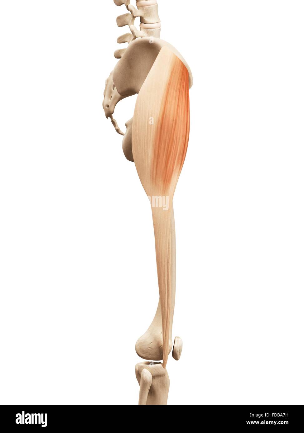 Human leg muscles (tensor fascia lata), illustration. Stock Photohttps://www.alamy.com/image-license-details/?v=1https://www.alamy.com/stock-photo-human-leg-muscles-tensor-fascia-lata-illustration-94291893.html
RFFDBA7H–Human leg muscles (tensor fascia lata), illustration.
 medical accurate illustration of the tensor fascia lata Stock Photohttps://www.alamy.com/image-license-details/?v=1https://www.alamy.com/stock-photo-medical-accurate-illustration-of-the-tensor-fascia-lata-89742517.html
RFF603DW–medical accurate illustration of the tensor fascia lata
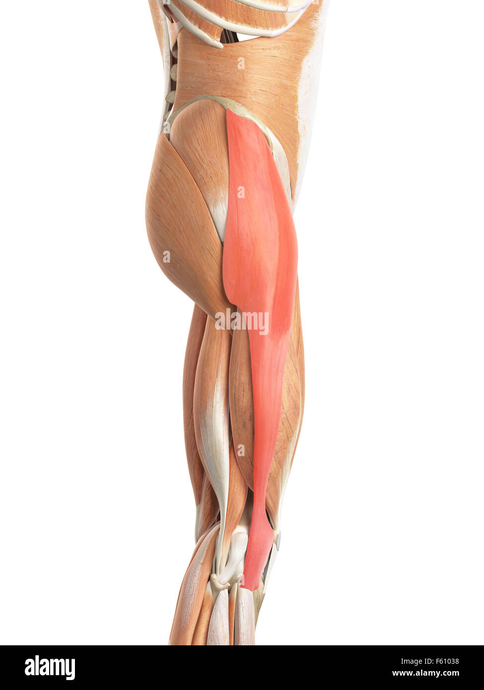 medically accurate illustration of the tensor fascia lata Stock Photohttps://www.alamy.com/image-license-details/?v=1https://www.alamy.com/stock-photo-medically-accurate-illustration-of-the-tensor-fascia-lata-89761820.html
RFF61038–medically accurate illustration of the tensor fascia lata
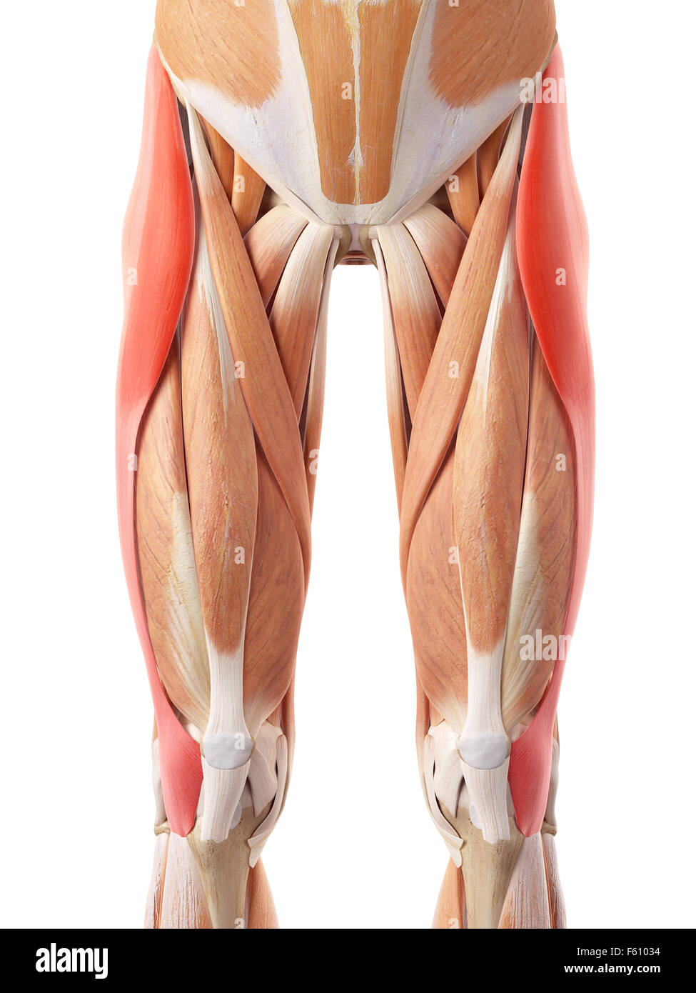 medically accurate illustration of the tensor fascia lata Stock Photohttps://www.alamy.com/image-license-details/?v=1https://www.alamy.com/stock-photo-medically-accurate-illustration-of-the-tensor-fascia-lata-89761816.html
RFF61034–medically accurate illustration of the tensor fascia lata
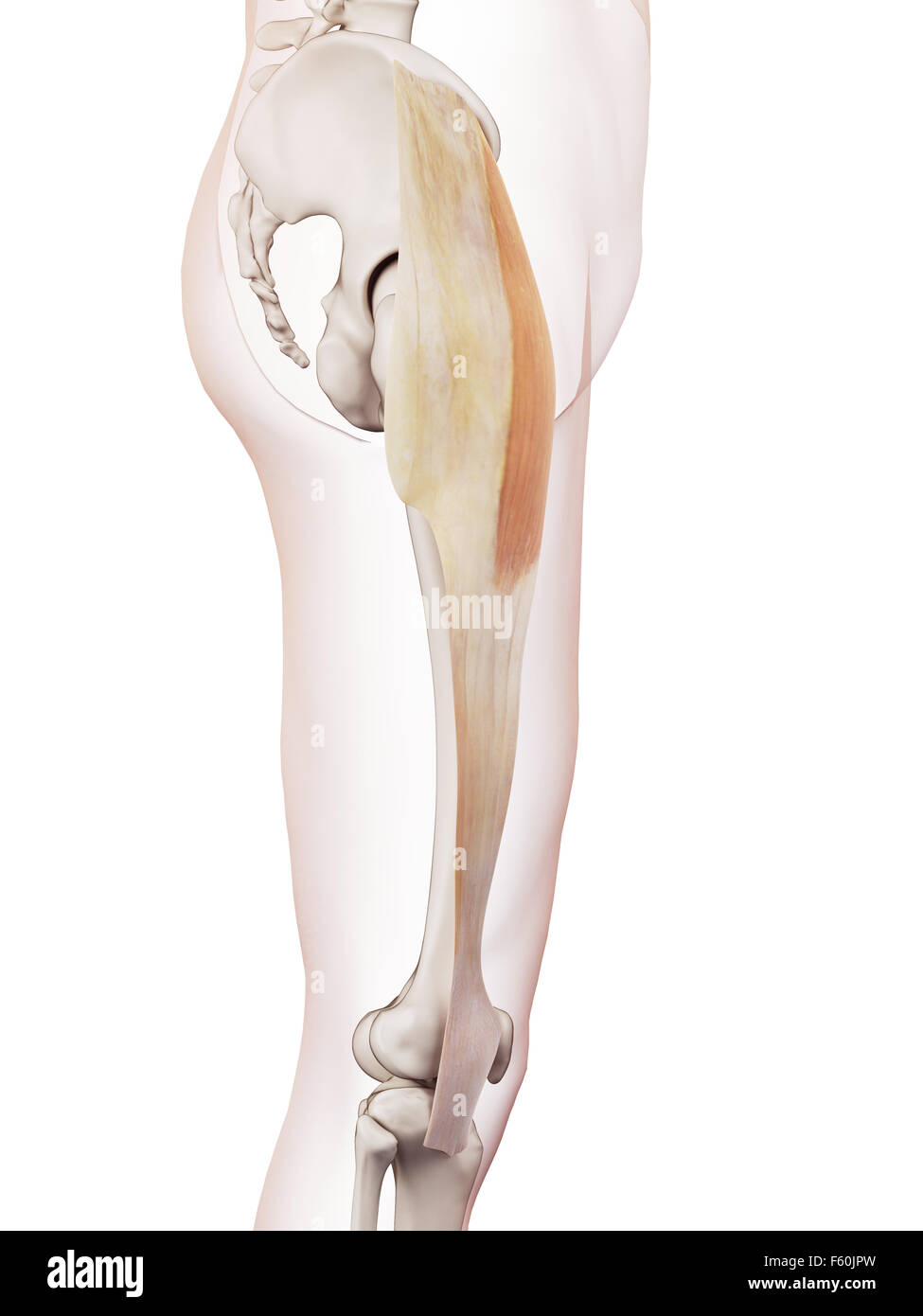 medically accurate muscle illustration of the tensor fascia lata Stock Photohttps://www.alamy.com/image-license-details/?v=1https://www.alamy.com/stock-photo-medically-accurate-muscle-illustration-of-the-tensor-fascia-lata-89754529.html
RFF60JPW–medically accurate muscle illustration of the tensor fascia lata
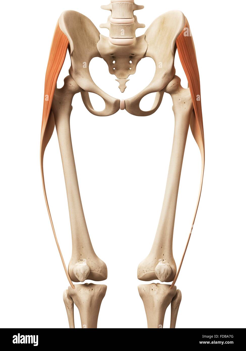 Human leg muscles (tensor fascia lata), illustration. Stock Photohttps://www.alamy.com/image-license-details/?v=1https://www.alamy.com/stock-photo-human-leg-muscles-tensor-fascia-lata-illustration-94291892.html
RFFDBA7G–Human leg muscles (tensor fascia lata), illustration.
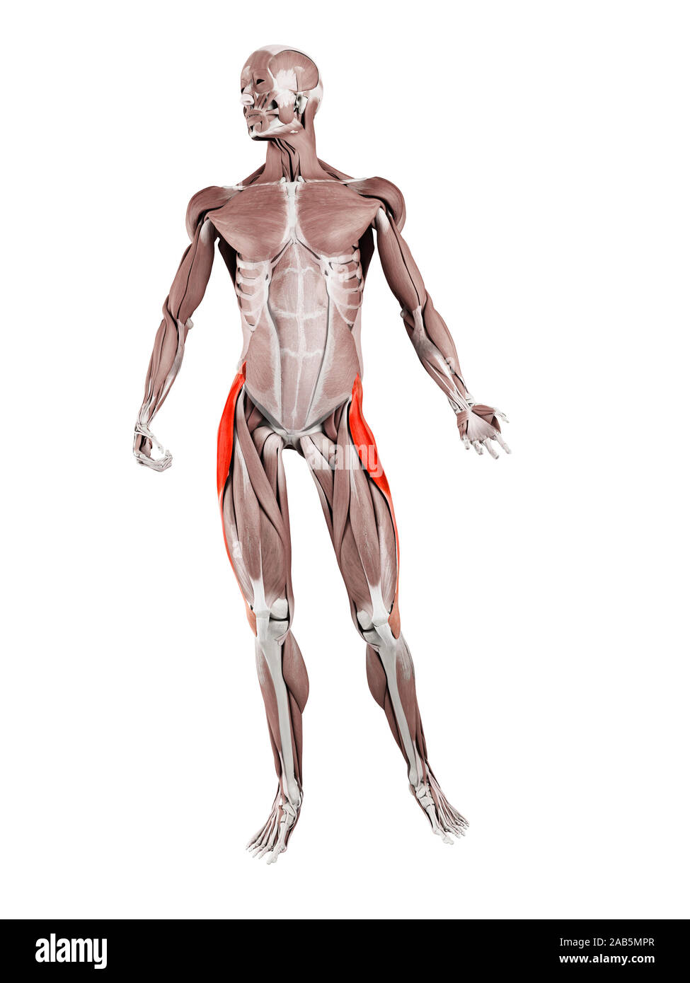 3d rendered muscle illustration of the tensor fascia lata Stock Photohttps://www.alamy.com/image-license-details/?v=1https://www.alamy.com/3d-rendered-muscle-illustration-of-the-tensor-fascia-lata-image333884287.html
RF2AB5MPR–3d rendered muscle illustration of the tensor fascia lata
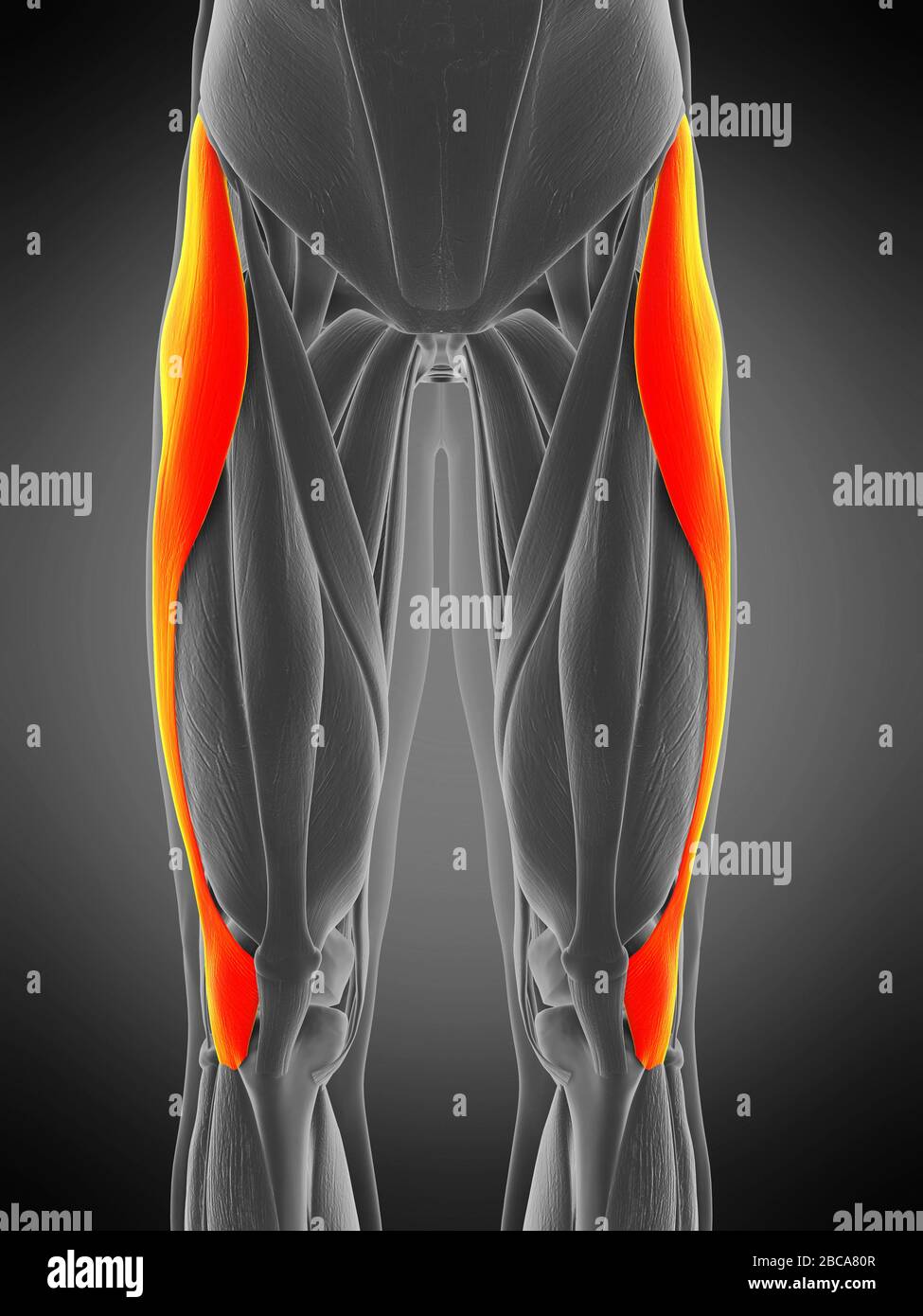 Tensor fascia lata muscle, illustration. Stock Photohttps://www.alamy.com/image-license-details/?v=1https://www.alamy.com/tensor-fascia-lata-muscle-illustration-image351809047.html
RF2BCA80R–Tensor fascia lata muscle, illustration.
 3d rendered muscle illustration of the tensor fascia lata Stock Photohttps://www.alamy.com/image-license-details/?v=1https://www.alamy.com/3d-rendered-muscle-illustration-of-the-tensor-fascia-lata-image333875573.html
RF2AB59KH–3d rendered muscle illustration of the tensor fascia lata
 Diseases of the nervous system : a text-book of neurology and psychiatry . extremity. The platysma myoides, sternocleidomastoid, trapezius, and oblique musclesof the abdomen, gluteus medius, and tensor of the fascia lata have been removed. Theshading and cross-hatching follow the muscles and innervation as in Figs. 11 and 12.(After Dejerine.) UPPER EXTREMITIES 47 The interossei and lumbricales muscles of the hand flex the proximalphalanges, and extend the middle and terminal phalanges. The dorsal Inferior maxillaru I Post, branchcervical nerves Ci-njjN. Rectus anticusand longus collisC.P.N. Sc Stock Photohttps://www.alamy.com/image-license-details/?v=1https://www.alamy.com/diseases-of-the-nervous-system-a-text-book-of-neurology-and-psychiatry-extremity-the-platysma-myoides-sternocleidomastoid-trapezius-and-oblique-musclesof-the-abdomen-gluteus-medius-and-tensor-of-the-fascia-lata-have-been-removed-theshading-and-cross-hatching-follow-the-muscles-and-innervation-as-in-figs-11-and-12after-dejerine-upper-extremities-47-the-interossei-and-lumbricales-muscles-of-the-hand-flex-the-proximalphalanges-and-extend-the-middle-and-terminal-phalanges-the-dorsal-inferior-maxillaru-i-post-branchcervical-nerves-ci-njjn-rectus-anticusand-longus-colliscpn-sc-image339073266.html
RM2AKJ3BE–Diseases of the nervous system : a text-book of neurology and psychiatry . extremity. The platysma myoides, sternocleidomastoid, trapezius, and oblique musclesof the abdomen, gluteus medius, and tensor of the fascia lata have been removed. Theshading and cross-hatching follow the muscles and innervation as in Figs. 11 and 12.(After Dejerine.) UPPER EXTREMITIES 47 The interossei and lumbricales muscles of the hand flex the proximalphalanges, and extend the middle and terminal phalanges. The dorsal Inferior maxillaru I Post, branchcervical nerves Ci-njjN. Rectus anticusand longus collisC.P.N. Sc
 Tensor fascia lata muscle, illustration. Stock Photohttps://www.alamy.com/image-license-details/?v=1https://www.alamy.com/tensor-fascia-lata-muscle-illustration-image351809103.html
RF2BCA82R–Tensor fascia lata muscle, illustration.
 3d rendered muscle illustration of the tensor fascia lata Stock Photohttps://www.alamy.com/image-license-details/?v=1https://www.alamy.com/3d-rendered-muscle-illustration-of-the-tensor-fascia-lata-image333875230.html
RF2AB597A–3d rendered muscle illustration of the tensor fascia lata
 A text-book of clinical anatomy : for students and practitioners . drop a linevertically downward from the anterior superior spine of the ilium. Marka second oblique line from the same point to the upper border of thetrochanter, and a third horizontal line from the trochanter to join thefirst-mentioned vertical line. These three lines form Bryants triangle,which is also a standard measurement in hip injuries (see below). 4. Note the position of the gluteal fold; the lower border of thegluteus maximus lies a little above it. Also palpate the firm band (ilio-tibial) of fascia lata (tensor fascia Stock Photohttps://www.alamy.com/image-license-details/?v=1https://www.alamy.com/a-text-book-of-clinical-anatomy-for-students-and-practitioners-drop-a-linevertically-downward-from-the-anterior-superior-spine-of-the-ilium-marka-second-oblique-line-from-the-same-point-to-the-upper-border-of-thetrochanter-and-a-third-horizontal-line-from-the-trochanter-to-join-thefirst-mentioned-vertical-line-these-three-lines-form-bryants-trianglewhich-is-also-a-standard-measurement-in-hip-injuries-see-below-4-note-the-position-of-the-gluteal-fold-the-lower-border-of-thegluteus-maximus-lies-a-little-above-it-also-palpate-the-firm-band-ilio-tibial-of-fascia-lata-tensor-fascia-image340193821.html
RM2AND4K9–A text-book of clinical anatomy : for students and practitioners . drop a linevertically downward from the anterior superior spine of the ilium. Marka second oblique line from the same point to the upper border of thetrochanter, and a third horizontal line from the trochanter to join thefirst-mentioned vertical line. These three lines form Bryants triangle,which is also a standard measurement in hip injuries (see below). 4. Note the position of the gluteal fold; the lower border of thegluteus maximus lies a little above it. Also palpate the firm band (ilio-tibial) of fascia lata (tensor fascia
 3d rendered medically accurate illustration of the tensor fascia lata Stock Photohttps://www.alamy.com/image-license-details/?v=1https://www.alamy.com/3d-rendered-medically-accurate-illustration-of-the-tensor-fascia-lata-image257796997.html
RFTYBJKH–3d rendered medically accurate illustration of the tensor fascia lata
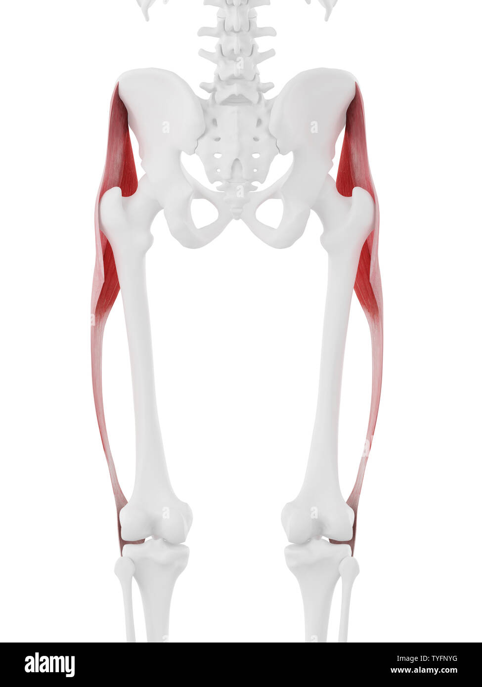 3d rendered medically accurate illustration of the Tensor Fascia Lata Stock Photohttps://www.alamy.com/image-license-details/?v=1https://www.alamy.com/3d-rendered-medically-accurate-illustration-of-the-tensor-fascia-lata-image257887380.html
RFTYFNYG–3d rendered medically accurate illustration of the Tensor Fascia Lata
 Tensor fascia lata muscle, illustration. Stock Photohttps://www.alamy.com/image-license-details/?v=1https://www.alamy.com/tensor-fascia-lata-muscle-illustration-image351809043.html
RF2BCA80K–Tensor fascia lata muscle, illustration.
 Illustration of the tensor fascia lata muscles. Stock Photohttps://www.alamy.com/image-license-details/?v=1https://www.alamy.com/stock-photo-illustration-of-the-tensor-fascia-lata-muscles-126900907.html
RFHACRB7–Illustration of the tensor fascia lata muscles.
![. The anatomy of the domestic animals. Veterinary anatomy. THE STIFLE JOINT 235 biceps femoris, but it also furnishes insertion to the tensor fasciae latse by means of the fascia lata, which blends with it. The femoro-tibial articulation (Articulatio femoro-tibialis) is formed between tlie condyles of the femur, the ])roximal end of the tibia, antl tlie interposed articular menisci or semilunar cartilages. Articular Surfaces.—The condyles of the femur are slightly oblique in direc- tion. The articular surface of the lateral one is more strongly curved than that of the medial one; the latter is Stock Photo . The anatomy of the domestic animals. Veterinary anatomy. THE STIFLE JOINT 235 biceps femoris, but it also furnishes insertion to the tensor fasciae latse by means of the fascia lata, which blends with it. The femoro-tibial articulation (Articulatio femoro-tibialis) is formed between tlie condyles of the femur, the ])roximal end of the tibia, antl tlie interposed articular menisci or semilunar cartilages. Articular Surfaces.—The condyles of the femur are slightly oblique in direc- tion. The articular surface of the lateral one is more strongly curved than that of the medial one; the latter is Stock Photo](https://c8.alamy.com/comp/RN5DB2/the-anatomy-of-the-domestic-animals-veterinary-anatomy-the-stifle-joint-235-biceps-femoris-but-it-also-furnishes-insertion-to-the-tensor-fasciae-latse-by-means-of-the-fascia-lata-which-blends-with-it-the-femoro-tibial-articulation-articulatio-femoro-tibialis-is-formed-between-tlie-condyles-of-the-femur-the-roximal-end-of-the-tibia-antl-tlie-interposed-articular-menisci-or-semilunar-cartilages-articular-surfacesthe-condyles-of-the-femur-are-slightly-oblique-in-direc-tion-the-articular-surface-of-the-lateral-one-is-more-strongly-curved-than-that-of-the-medial-one-the-latter-is-RN5DB2.jpg) . The anatomy of the domestic animals. Veterinary anatomy. THE STIFLE JOINT 235 biceps femoris, but it also furnishes insertion to the tensor fasciae latse by means of the fascia lata, which blends with it. The femoro-tibial articulation (Articulatio femoro-tibialis) is formed between tlie condyles of the femur, the ])roximal end of the tibia, antl tlie interposed articular menisci or semilunar cartilages. Articular Surfaces.—The condyles of the femur are slightly oblique in direc- tion. The articular surface of the lateral one is more strongly curved than that of the medial one; the latter is Stock Photohttps://www.alamy.com/image-license-details/?v=1https://www.alamy.com/the-anatomy-of-the-domestic-animals-veterinary-anatomy-the-stifle-joint-235-biceps-femoris-but-it-also-furnishes-insertion-to-the-tensor-fasciae-latse-by-means-of-the-fascia-lata-which-blends-with-it-the-femoro-tibial-articulation-articulatio-femoro-tibialis-is-formed-between-tlie-condyles-of-the-femur-the-roximal-end-of-the-tibia-antl-tlie-interposed-articular-menisci-or-semilunar-cartilages-articular-surfacesthe-condyles-of-the-femur-are-slightly-oblique-in-direc-tion-the-articular-surface-of-the-lateral-one-is-more-strongly-curved-than-that-of-the-medial-one-the-latter-is-image236762822.html
. The anatomy of the domestic animals. Veterinary anatomy. THE STIFLE JOINT 235 biceps femoris, but it also furnishes insertion to the tensor fasciae latse by means of the fascia lata, which blends with it. The femoro-tibial articulation (Articulatio femoro-tibialis) is formed between tlie condyles of the femur, the ])roximal end of the tibia, antl tlie interposed articular menisci or semilunar cartilages. Articular Surfaces.—The condyles of the femur are slightly oblique in direc- tion. The articular surface of the lateral one is more strongly curved than that of the medial one; the latter is Stock Photohttps://www.alamy.com/image-license-details/?v=1https://www.alamy.com/the-anatomy-of-the-domestic-animals-veterinary-anatomy-the-stifle-joint-235-biceps-femoris-but-it-also-furnishes-insertion-to-the-tensor-fasciae-latse-by-means-of-the-fascia-lata-which-blends-with-it-the-femoro-tibial-articulation-articulatio-femoro-tibialis-is-formed-between-tlie-condyles-of-the-femur-the-roximal-end-of-the-tibia-antl-tlie-interposed-articular-menisci-or-semilunar-cartilages-articular-surfacesthe-condyles-of-the-femur-are-slightly-oblique-in-direc-tion-the-articular-surface-of-the-lateral-one-is-more-strongly-curved-than-that-of-the-medial-one-the-latter-is-image236762822.htmlRMRN5DB2–. The anatomy of the domestic animals. Veterinary anatomy. THE STIFLE JOINT 235 biceps femoris, but it also furnishes insertion to the tensor fasciae latse by means of the fascia lata, which blends with it. The femoro-tibial articulation (Articulatio femoro-tibialis) is formed between tlie condyles of the femur, the ])roximal end of the tibia, antl tlie interposed articular menisci or semilunar cartilages. Articular Surfaces.—The condyles of the femur are slightly oblique in direc- tion. The articular surface of the lateral one is more strongly curved than that of the medial one; the latter is
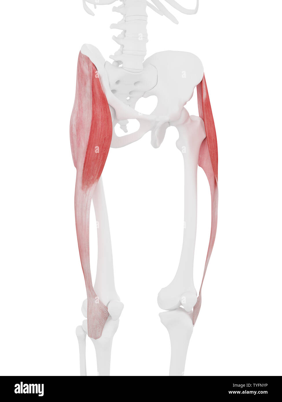 3d rendered medically accurate illustration of the Tensor Fascia Lata Stock Photohttps://www.alamy.com/image-license-details/?v=1https://www.alamy.com/3d-rendered-medically-accurate-illustration-of-the-tensor-fascia-lata-image257887386.html
RFTYFNYP–3d rendered medically accurate illustration of the Tensor Fascia Lata
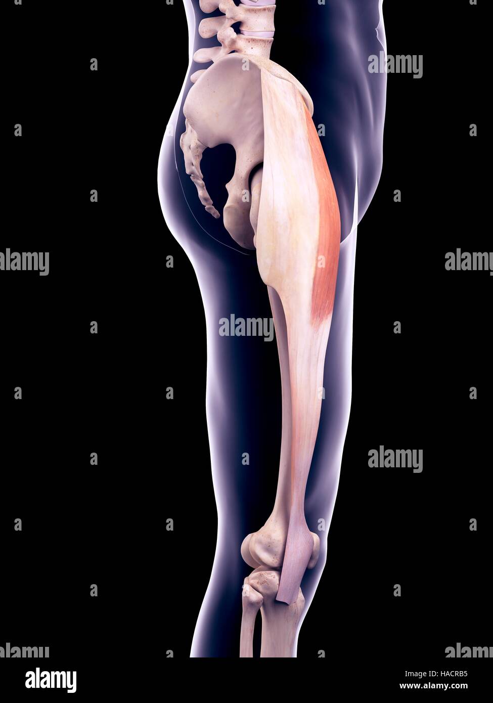 Illustration of the tensor fascia lata muscle. Stock Photohttps://www.alamy.com/image-license-details/?v=1https://www.alamy.com/stock-photo-illustration-of-the-tensor-fascia-lata-muscle-126900905.html
RFHACRB5–Illustration of the tensor fascia lata muscle.
 . The anatomy of the domestic animals. Veterinary anatomy. 330 FASCI.E AND MUSCLES OF THE HORSE nishes insertion to fibers of the vasti. The tendon of insertion is formed bj' the union of these tendinous layers on the lower part of the muscle. The lower portion of the muscle is pennate, the fibers on either side converging on the tendon at an acute angle. Relations.—Medially, the iliacus, sartorius, and vastus medialis; laterally, the tensor fascise latae, glutei, and vastus lateralis; posteriorly, the hip joint and the vastus intermedius; anteriorly, the fascia lata and the skin. The anterior Stock Photohttps://www.alamy.com/image-license-details/?v=1https://www.alamy.com/the-anatomy-of-the-domestic-animals-veterinary-anatomy-330-fascie-and-muscles-of-the-horse-nishes-insertion-to-fibers-of-the-vasti-the-tendon-of-insertion-is-formed-bj-the-union-of-these-tendinous-layers-on-the-lower-part-of-the-muscle-the-lower-portion-of-the-muscle-is-pennate-the-fibers-on-either-side-converging-on-the-tendon-at-an-acute-angle-relationsmedially-the-iliacus-sartorius-and-vastus-medialis-laterally-the-tensor-fascise-latae-glutei-and-vastus-lateralis-posteriorly-the-hip-joint-and-the-vastus-intermedius-anteriorly-the-fascia-lata-and-the-skin-the-anterior-image236800991.html
RMRN7627–. The anatomy of the domestic animals. Veterinary anatomy. 330 FASCI.E AND MUSCLES OF THE HORSE nishes insertion to fibers of the vasti. The tendon of insertion is formed bj' the union of these tendinous layers on the lower part of the muscle. The lower portion of the muscle is pennate, the fibers on either side converging on the tendon at an acute angle. Relations.—Medially, the iliacus, sartorius, and vastus medialis; laterally, the tensor fascise latae, glutei, and vastus lateralis; posteriorly, the hip joint and the vastus intermedius; anteriorly, the fascia lata and the skin. The anterior
 3d rendered medically accurate illustration of the Tensor Fascia Lata Stock Photohttps://www.alamy.com/image-license-details/?v=1https://www.alamy.com/3d-rendered-medically-accurate-illustration-of-the-tensor-fascia-lata-image257887400.html
RFTYFP08–3d rendered medically accurate illustration of the Tensor Fascia Lata
 Illustration of the tensor fascia lata muscles. Stock Photohttps://www.alamy.com/image-license-details/?v=1https://www.alamy.com/stock-photo-illustration-of-the-tensor-fascia-lata-muscles-126900655.html
RFHACR27–Illustration of the tensor fascia lata muscles.
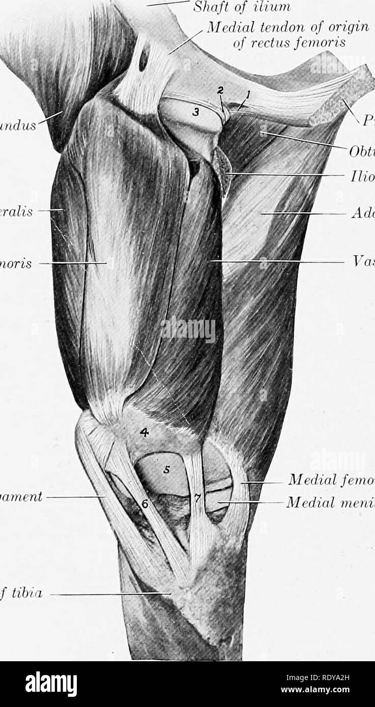 . The anatomy of the domestic animals . Veterinary anatomy. 330 FASCIA AND MUSCLES OF THE HORSE nishes insertion to fibers of tlie vasti. Tlie tendon of insertion is formed by the union of these tendinous layers on the lower part of the muscle. The lower portion of the muscle is pennate, the fibers on either side converging on the tendon at an acute angle. Relations.—NLedially, the ihacus, sartorius, and vastus mediahs; laterally, the tensor fascia latse, glutei, and vastus lateralis; posteriorly, the hip joint and the vastus intermedins; anteriorly, the fascia lata and the skin. The anterior Stock Photohttps://www.alamy.com/image-license-details/?v=1https://www.alamy.com/the-anatomy-of-the-domestic-animals-veterinary-anatomy-330-fascia-and-muscles-of-the-horse-nishes-insertion-to-fibers-of-tlie-vasti-tlie-tendon-of-insertion-is-formed-by-the-union-of-these-tendinous-layers-on-the-lower-part-of-the-muscle-the-lower-portion-of-the-muscle-is-pennate-the-fibers-on-either-side-converging-on-the-tendon-at-an-acute-angle-relationsnledially-the-ihacus-sartorius-and-vastus-mediahs-laterally-the-tensor-fascia-latse-glutei-and-vastus-lateralis-posteriorly-the-hip-joint-and-the-vastus-intermedins-anteriorly-the-fascia-lata-and-the-skin-the-anterior-image232325929.html
RMRDYA2H–. The anatomy of the domestic animals . Veterinary anatomy. 330 FASCIA AND MUSCLES OF THE HORSE nishes insertion to fibers of tlie vasti. Tlie tendon of insertion is formed by the union of these tendinous layers on the lower part of the muscle. The lower portion of the muscle is pennate, the fibers on either side converging on the tendon at an acute angle. Relations.—NLedially, the ihacus, sartorius, and vastus mediahs; laterally, the tensor fascia latse, glutei, and vastus lateralis; posteriorly, the hip joint and the vastus intermedins; anteriorly, the fascia lata and the skin. The anterior
 3d rendered medically accurate illustration of the Tensor Fascia Lata Stock Photohttps://www.alamy.com/image-license-details/?v=1https://www.alamy.com/3d-rendered-medically-accurate-illustration-of-the-tensor-fascia-lata-image257739600.html
RFTY91DM–3d rendered medically accurate illustration of the Tensor Fascia Lata
 Illustration of the tensor fascia lata muscle. Stock Photohttps://www.alamy.com/image-license-details/?v=1https://www.alamy.com/stock-photo-illustration-of-the-tensor-fascia-lata-muscle-126900906.html
RFHACRB6–Illustration of the tensor fascia lata muscle.
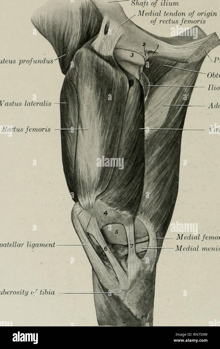 . The anatomy of the domestic animals. Veterinary anatomy. 330 FASCIA AND MUSCLES OF THE HORSE nishes insertion to fibers of the vasti. The tendon of insertion is formed by the union of these tendinous layers on the lower part of the muscle. The lower portion of the muscle is pennate, the fibers on either side converging on the tendon at an acute angle. Relations.—Medially, the iliacus, sartorius, and vastus medialis; laterally, the tensor fasciae latae, glutei, and vastus lateralis; posteriorly, the hip joint and the vastus intermedius; anteriorly, the fascia lata and the skin. The anterior f Stock Photohttps://www.alamy.com/image-license-details/?v=1https://www.alamy.com/the-anatomy-of-the-domestic-animals-veterinary-anatomy-330-fascia-and-muscles-of-the-horse-nishes-insertion-to-fibers-of-the-vasti-the-tendon-of-insertion-is-formed-by-the-union-of-these-tendinous-layers-on-the-lower-part-of-the-muscle-the-lower-portion-of-the-muscle-is-pennate-the-fibers-on-either-side-converging-on-the-tendon-at-an-acute-angle-relationsmedially-the-iliacus-sartorius-and-vastus-medialis-laterally-the-tensor-fasciae-latae-glutei-and-vastus-lateralis-posteriorly-the-hip-joint-and-the-vastus-intermedius-anteriorly-the-fascia-lata-and-the-skin-the-anterior-f-image236800897.html
RMRN75XW–. The anatomy of the domestic animals. Veterinary anatomy. 330 FASCIA AND MUSCLES OF THE HORSE nishes insertion to fibers of the vasti. The tendon of insertion is formed by the union of these tendinous layers on the lower part of the muscle. The lower portion of the muscle is pennate, the fibers on either side converging on the tendon at an acute angle. Relations.—Medially, the iliacus, sartorius, and vastus medialis; laterally, the tensor fasciae latae, glutei, and vastus lateralis; posteriorly, the hip joint and the vastus intermedius; anteriorly, the fascia lata and the skin. The anterior f
 3d rendered medically accurate illustration of the Tensor Fascia Lata Stock Photohttps://www.alamy.com/image-license-details/?v=1https://www.alamy.com/3d-rendered-medically-accurate-illustration-of-the-tensor-fascia-lata-image257887434.html
RFTYFP1E–3d rendered medically accurate illustration of the Tensor Fascia Lata
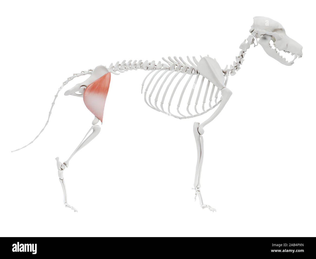 3d rendered illustration of the dog muscle anatomy - tensor fascia lata Stock Photohttps://www.alamy.com/image-license-details/?v=1https://www.alamy.com/3d-rendered-illustration-of-the-dog-muscle-anatomy-tensor-fascia-lata-image333864013.html
RF2AB4PXN–3d rendered illustration of the dog muscle anatomy - tensor fascia lata
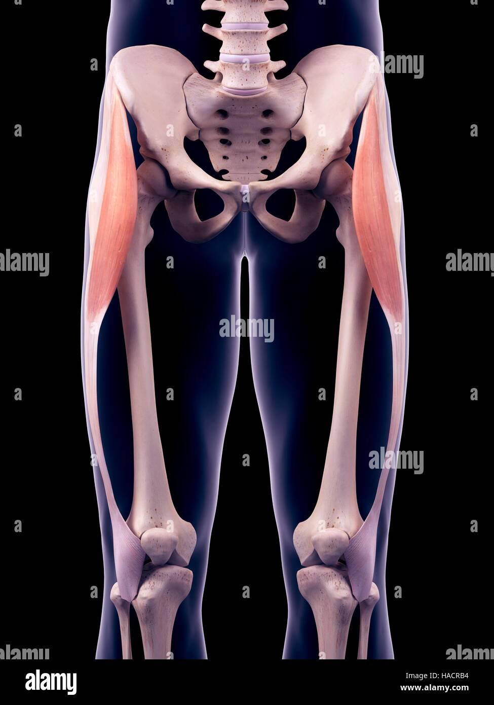 Illustration of the tensor fascia lata muscles. Stock Photohttps://www.alamy.com/image-license-details/?v=1https://www.alamy.com/stock-photo-illustration-of-the-tensor-fascia-lata-muscles-126900904.html
RFHACRB4–Illustration of the tensor fascia lata muscles.
 . The topographical anatomy of the limbs of the horse. Horses; Physiology. THE LIMBS OF THE HOESE 119 Dissection.—The deep gluteal muscle must be reflected in order that tbe origin of the rectus femoris and capsularis muscles may be thoroughly wifh wivwr^"""'*^ ^'^^'" "°^ *° '"J"'"^ t'^^ capsule of the hip joint, with which the deep gluteal muscle is intimately associated. A strong sheet of fascia connects the tensor of the fascia lata with ni.Ll! TV, the ihum, and covers the lateral margin of the iliacus muscle, ihis must be cut away. M. glutfcus medius Stock Photohttps://www.alamy.com/image-license-details/?v=1https://www.alamy.com/the-topographical-anatomy-of-the-limbs-of-the-horse-horses-physiology-the-limbs-of-the-hoese-119-dissectionthe-deep-gluteal-muscle-must-be-reflected-in-order-that-tbe-origin-of-the-rectus-femoris-and-capsularis-muscles-may-be-thoroughly-wifh-wivwrquotquotquot-quot-quot-quotjquotquot-t-capsule-of-the-hip-joint-with-which-the-deep-gluteal-muscle-is-intimately-associated-a-strong-sheet-of-fascia-connects-the-tensor-of-the-fascia-lata-with-nill!-tv-the-ihum-and-covers-the-lateral-margin-of-the-iliacus-muscle-ihis-must-be-cut-away-m-glutfcus-medius-image232425275.html
RMRE3TPK–. The topographical anatomy of the limbs of the horse. Horses; Physiology. THE LIMBS OF THE HOESE 119 Dissection.—The deep gluteal muscle must be reflected in order that tbe origin of the rectus femoris and capsularis muscles may be thoroughly wifh wivwr^"""'*^ ^'^^'" "°^ *° '"J"'"^ t'^^ capsule of the hip joint, with which the deep gluteal muscle is intimately associated. A strong sheet of fascia connects the tensor of the fascia lata with ni.Ll! TV, the ihum, and covers the lateral margin of the iliacus muscle, ihis must be cut away. M. glutfcus medius
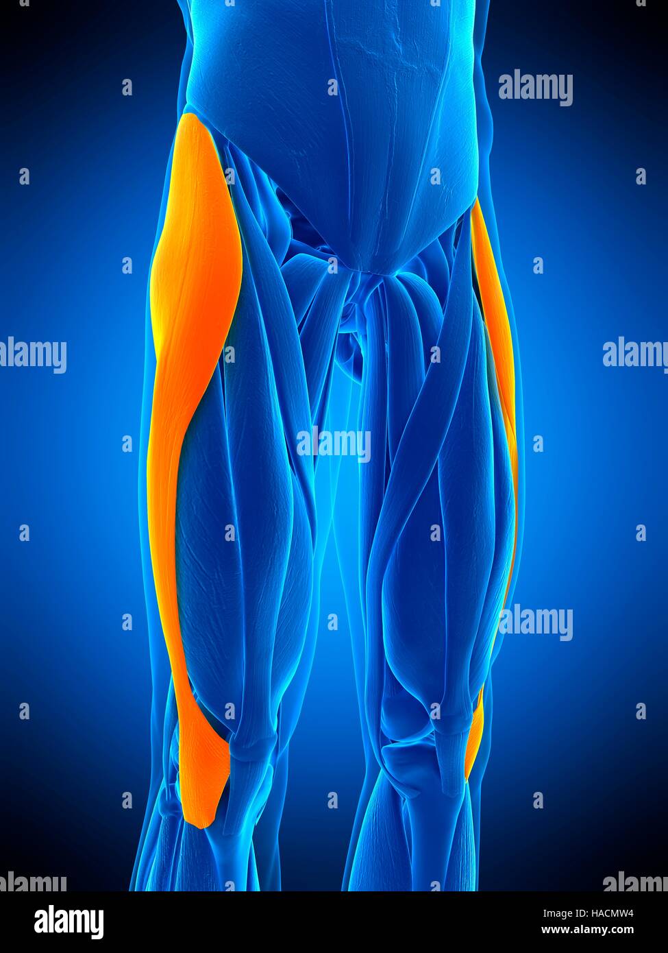 Illustration of the tensor fascia lata muscle. Stock Photohttps://www.alamy.com/image-license-details/?v=1https://www.alamy.com/stock-photo-illustration-of-the-tensor-fascia-lata-muscle-126898944.html
RFHACMW4–Illustration of the tensor fascia lata muscle.
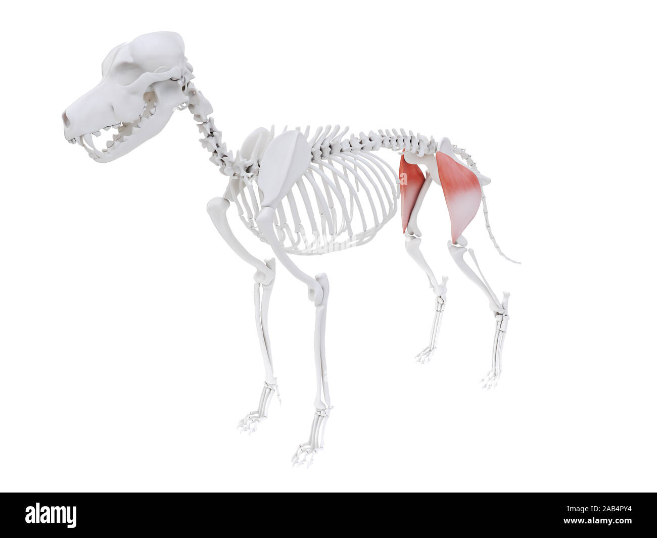 3d rendered illustration of the dog muscle anatomy - tensor fascia lata Stock Photohttps://www.alamy.com/image-license-details/?v=1https://www.alamy.com/3d-rendered-illustration-of-the-dog-muscle-anatomy-tensor-fascia-lata-image333864024.html
RF2AB4PY4–3d rendered illustration of the dog muscle anatomy - tensor fascia lata
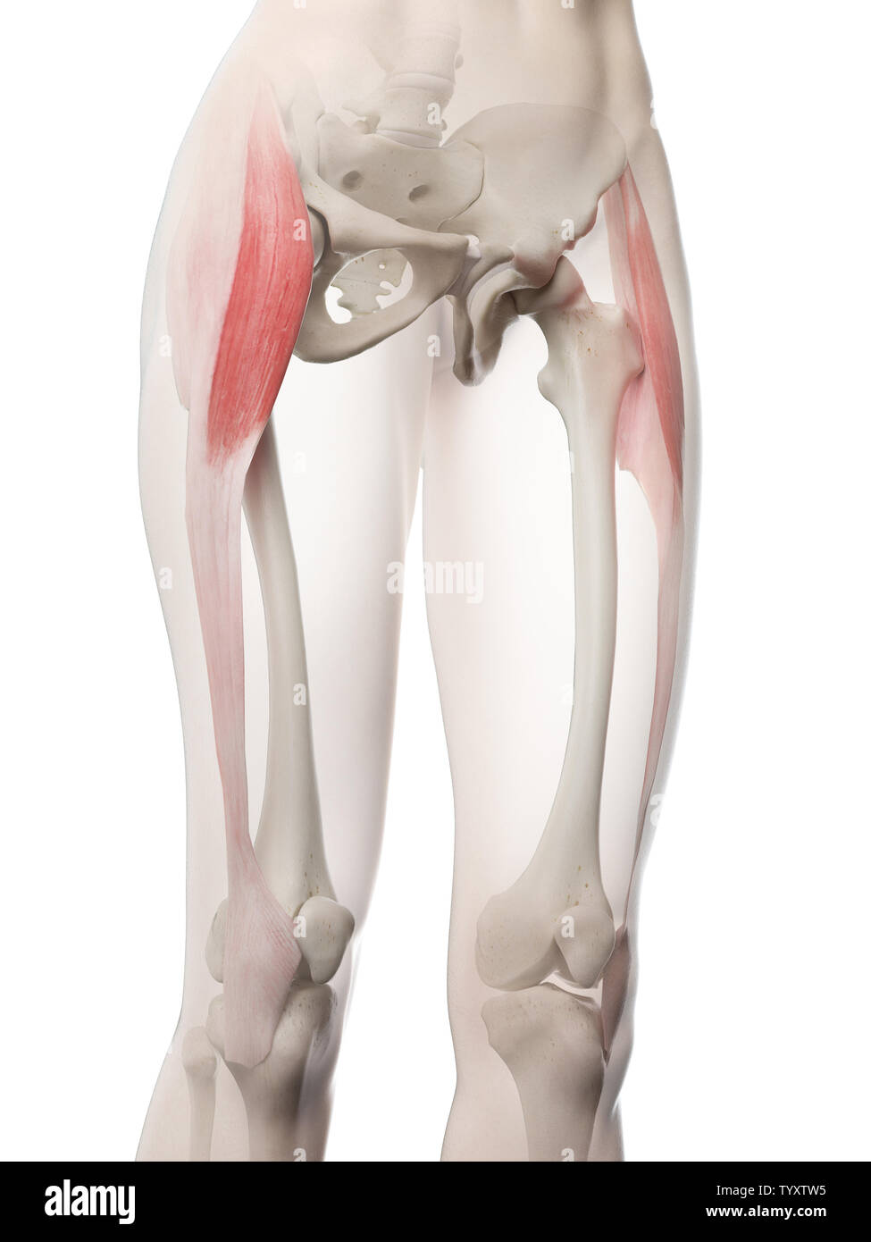 3d rendered medically accurate illustration of a womans Tensor Fascia Lata Stock Photohttps://www.alamy.com/image-license-details/?v=1https://www.alamy.com/3d-rendered-medically-accurate-illustration-of-a-womans-tensor-fascia-lata-image258131137.html
RFTYXTW5–3d rendered medically accurate illustration of a womans Tensor Fascia Lata
 . The anatomy of the domestic animals . Veterinary anatomy. ANTERIOR MUSCLES OP THE THIGH 331 Insertion.—(1) The lateral part of the anterior surface of the patella; (2) the tendon of the rectus femoris. Action.—To extend the stifle joint. Structure.—The fibers are directed downward and forward, many being in- serted into the tendinous sheet which covers the side of the rectus. A bursa is usually present between the distal end and the patella. Relations.—Laterally, the fascia lata and skin, tensor fasciae latae, superficial Fold of flank Tensor fascicc latce Vastus medialis Sartorius Saphenous Stock Photohttps://www.alamy.com/image-license-details/?v=1https://www.alamy.com/the-anatomy-of-the-domestic-animals-veterinary-anatomy-anterior-muscles-op-the-thigh-331-insertion1-the-lateral-part-of-the-anterior-surface-of-the-patella-2-the-tendon-of-the-rectus-femoris-actionto-extend-the-stifle-joint-structurethe-fibers-are-directed-downward-and-forward-many-being-in-serted-into-the-tendinous-sheet-which-covers-the-side-of-the-rectus-a-bursa-is-usually-present-between-the-distal-end-and-the-patella-relationslaterally-the-fascia-lata-and-skin-tensor-fasciae-latae-superficial-fold-of-flank-tensor-fascicc-latce-vastus-medialis-sartorius-saphenous-image232325924.html
RMRDYA2C–. The anatomy of the domestic animals . Veterinary anatomy. ANTERIOR MUSCLES OP THE THIGH 331 Insertion.—(1) The lateral part of the anterior surface of the patella; (2) the tendon of the rectus femoris. Action.—To extend the stifle joint. Structure.—The fibers are directed downward and forward, many being in- serted into the tendinous sheet which covers the side of the rectus. A bursa is usually present between the distal end and the patella. Relations.—Laterally, the fascia lata and skin, tensor fasciae latae, superficial Fold of flank Tensor fascicc latce Vastus medialis Sartorius Saphenous
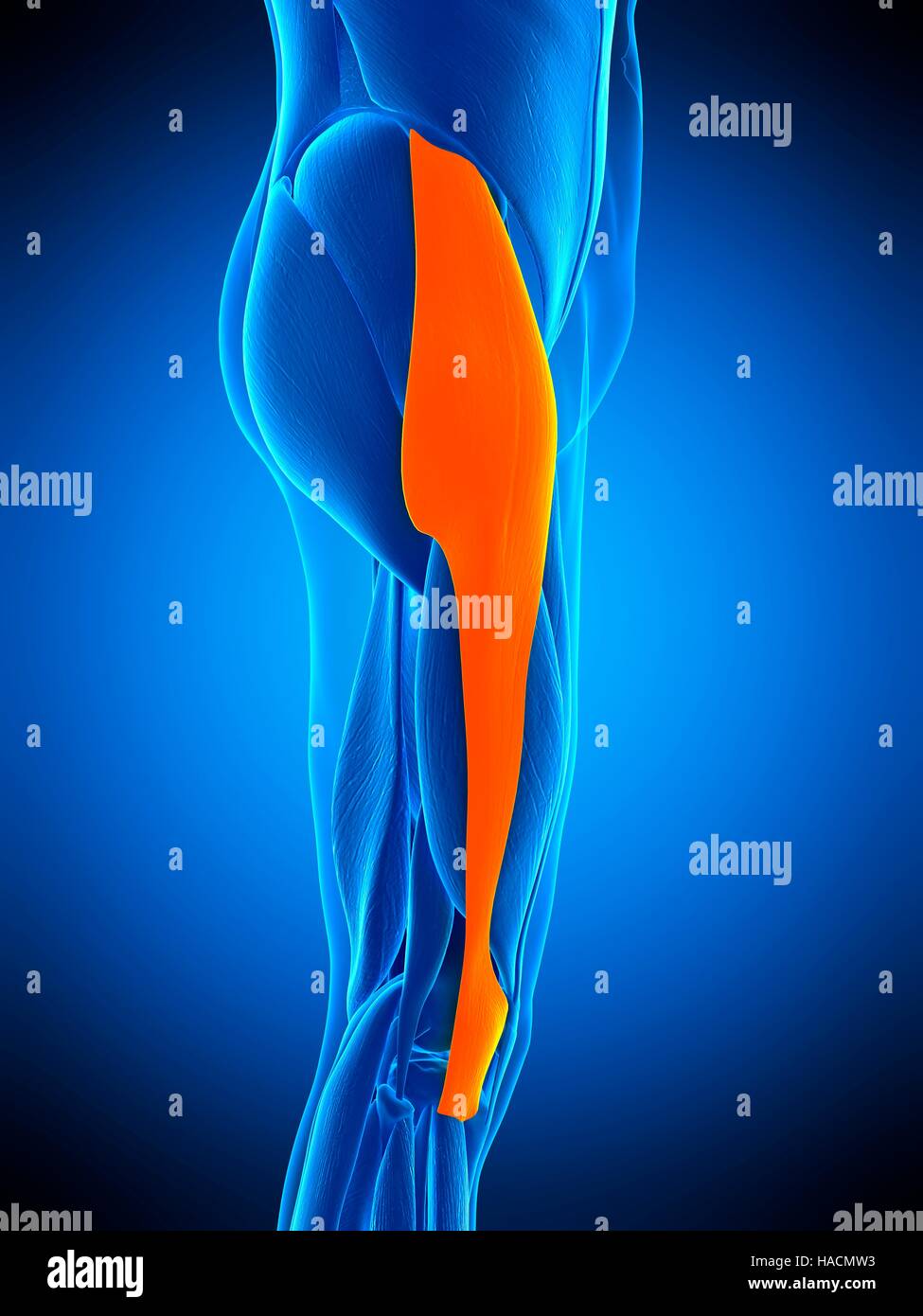 Illustration of the tensor fascia lata muscle. Stock Photohttps://www.alamy.com/image-license-details/?v=1https://www.alamy.com/stock-photo-illustration-of-the-tensor-fascia-lata-muscle-126898943.html
RFHACMW3–Illustration of the tensor fascia lata muscle.
 3d rendered medically accurate illustration of a womans Tensor Fascia Lata Stock Photohttps://www.alamy.com/image-license-details/?v=1https://www.alamy.com/3d-rendered-medically-accurate-illustration-of-a-womans-tensor-fascia-lata-image258130811.html
RFTYXTDF–3d rendered medically accurate illustration of a womans Tensor Fascia Lata
 . The anatomy of the domestic animals. Veterinary anatomy. ANTERIOR MUSCLES OF THE THIGH 331 Insertion.—(1) The lateral part of the anterior surface of the patella; (2) the tendon of the rectus femoris. Action.—To extend the stifle joint. Structure.—The fibers are directed downward and forward, many being in- serted into the tendinous sheet which covers the side of the rectus. A bursa is usually present between the distal end and the patella. Relations.—Laterally, the fascia lata and skin, tensor fascis lata, superficial Fold of flank Tensor fascice latcB Vaslus mcdialis. Vastus lateralis Vast Stock Photohttps://www.alamy.com/image-license-details/?v=1https://www.alamy.com/the-anatomy-of-the-domestic-animals-veterinary-anatomy-anterior-muscles-of-the-thigh-331-insertion1-the-lateral-part-of-the-anterior-surface-of-the-patella-2-the-tendon-of-the-rectus-femoris-actionto-extend-the-stifle-joint-structurethe-fibers-are-directed-downward-and-forward-many-being-in-serted-into-the-tendinous-sheet-which-covers-the-side-of-the-rectus-a-bursa-is-usually-present-between-the-distal-end-and-the-patella-relationslaterally-the-fascia-lata-and-skin-tensor-fascis-lata-superficial-fold-of-flank-tensor-fascice-latcb-vaslus-mcdialis-vastus-lateralis-vast-image236800918.html
RMRN75YJ–. The anatomy of the domestic animals. Veterinary anatomy. ANTERIOR MUSCLES OF THE THIGH 331 Insertion.—(1) The lateral part of the anterior surface of the patella; (2) the tendon of the rectus femoris. Action.—To extend the stifle joint. Structure.—The fibers are directed downward and forward, many being in- serted into the tendinous sheet which covers the side of the rectus. A bursa is usually present between the distal end and the patella. Relations.—Laterally, the fascia lata and skin, tensor fascis lata, superficial Fold of flank Tensor fascice latcB Vaslus mcdialis. Vastus lateralis Vast
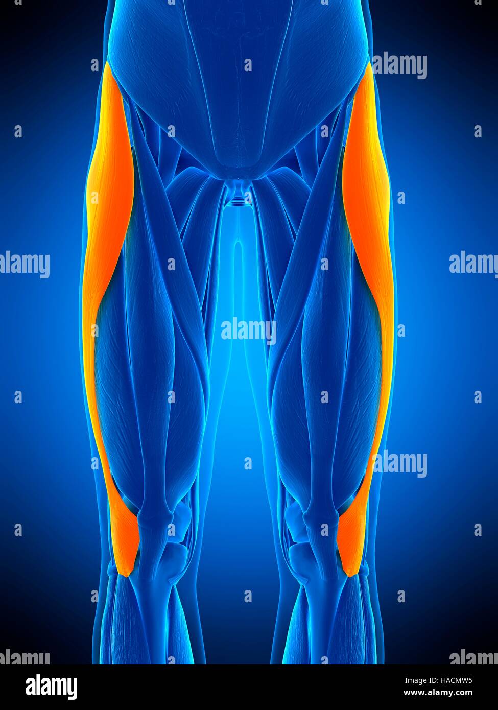 Illustration of the tensor fascia lata muscle. Stock Photohttps://www.alamy.com/image-license-details/?v=1https://www.alamy.com/stock-photo-illustration-of-the-tensor-fascia-lata-muscle-126898945.html
RFHACMW5–Illustration of the tensor fascia lata muscle.
 Tensor fascia lata muscle, illustration Stock Photohttps://www.alamy.com/image-license-details/?v=1https://www.alamy.com/tensor-fascia-lata-muscle-illustration-image332908533.html
RF2A9H86D–Tensor fascia lata muscle, illustration
 3d rendered medically accurate illustration of a womans Tensor Fascia Lata Stock Photohttps://www.alamy.com/image-license-details/?v=1https://www.alamy.com/3d-rendered-medically-accurate-illustration-of-a-womans-tensor-fascia-lata-image258130772.html
RFTYXTC4–3d rendered medically accurate illustration of a womans Tensor Fascia Lata
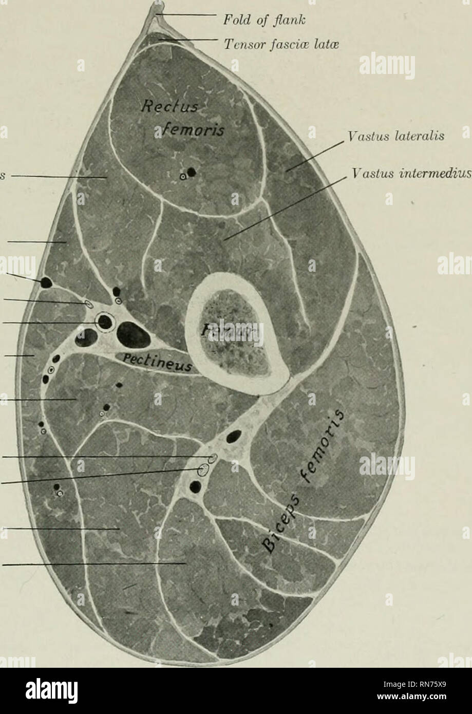 . The anatomy of the domestic animals. Veterinary anatomy. ANTERIOR MUSCLES OF THE THIGH 331 Insertion.—(1) The lateral part of the anterior surface of the patella; (2) the tendon of the rectus femoris. Action.—To extend the stifle joint. Structure.—The fibers are directed downward and forward, many being in- serted into the tendinous sheet which covers the side of the rectus. A bursa is usually present between the distal end and the patella. Relations.—Laterally, the fascia lata and skin, tensor fasciae latae, superficial Fold of flank Tctisor fascia: latoe Vujstus mcdiali Sartorius Saphenous Stock Photohttps://www.alamy.com/image-license-details/?v=1https://www.alamy.com/the-anatomy-of-the-domestic-animals-veterinary-anatomy-anterior-muscles-of-the-thigh-331-insertion1-the-lateral-part-of-the-anterior-surface-of-the-patella-2-the-tendon-of-the-rectus-femoris-actionto-extend-the-stifle-joint-structurethe-fibers-are-directed-downward-and-forward-many-being-in-serted-into-the-tendinous-sheet-which-covers-the-side-of-the-rectus-a-bursa-is-usually-present-between-the-distal-end-and-the-patella-relationslaterally-the-fascia-lata-and-skin-tensor-fasciae-latae-superficial-fold-of-flank-tctisor-fascia-latoe-vujstus-mcdiali-sartorius-saphenous-image236800881.html
RMRN75X9–. The anatomy of the domestic animals. Veterinary anatomy. ANTERIOR MUSCLES OF THE THIGH 331 Insertion.—(1) The lateral part of the anterior surface of the patella; (2) the tendon of the rectus femoris. Action.—To extend the stifle joint. Structure.—The fibers are directed downward and forward, many being in- serted into the tendinous sheet which covers the side of the rectus. A bursa is usually present between the distal end and the patella. Relations.—Laterally, the fascia lata and skin, tensor fasciae latae, superficial Fold of flank Tctisor fascia: latoe Vujstus mcdiali Sartorius Saphenous
 Tensor fascia lata muscle, illustration Stock Photohttps://www.alamy.com/image-license-details/?v=1https://www.alamy.com/tensor-fascia-lata-muscle-illustration-image332908395.html
RF2A9H81F–Tensor fascia lata muscle, illustration
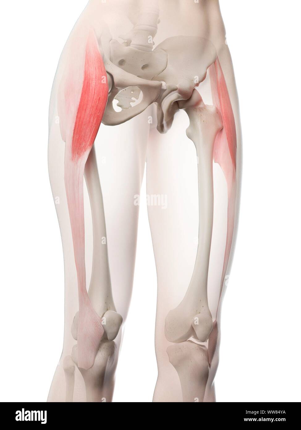 Tensor fascia lata muscle, illustration Stock Photohttps://www.alamy.com/image-license-details/?v=1https://www.alamy.com/tensor-fascia-lata-muscle-illustration-image273701438.html
RFWW84YA–Tensor fascia lata muscle, illustration
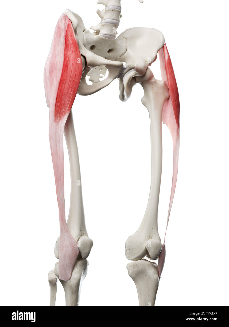 3d rendered medically accurate illustration of a womans Tensor Fascia Lata Stock Photohttps://www.alamy.com/image-license-details/?v=1https://www.alamy.com/3d-rendered-medically-accurate-illustration-of-a-womans-tensor-fascia-lata-image258131167.html
RFTYXTX7–3d rendered medically accurate illustration of a womans Tensor Fascia Lata
 . The comparative anatomy of the domesticated animals. Veterinary anatomy. 282 THE MUSCLES. superior extremity of that bone, and assists in rearing. In the first instance it acts as a lever of the first order; in the second, as one of the third order. Fig. 129.. SUPERFICIAL MUSCLES OF THE CROUP AND THIGH. 1, Middle glutens, or gluteus maximus; 2, Anterior spinous process of ilium ; 3, Muscle of the fascia lata, or tensor vaginse; 4, Superficial gluteus, or gluteus externus; *, Great trochanter of femur ; 5, Fascia lata; 6, Patella, with insertion of rectus; 7, Long vastus, or adductor magnus; Stock Photohttps://www.alamy.com/image-license-details/?v=1https://www.alamy.com/the-comparative-anatomy-of-the-domesticated-animals-veterinary-anatomy-282-the-muscles-superior-extremity-of-that-bone-and-assists-in-rearing-in-the-first-instance-it-acts-as-a-lever-of-the-first-order-in-the-second-as-one-of-the-third-order-fig-129-superficial-muscles-of-the-croup-and-thigh-1-middle-glutens-or-gluteus-maximus-2-anterior-spinous-process-of-ilium-3-muscle-of-the-fascia-lata-or-tensor-vaginse-4-superficial-gluteus-or-gluteus-externus-great-trochanter-of-femur-5-fascia-lata-6-patella-with-insertion-of-rectus-7-long-vastus-or-adductor-magnus-image232680637.html
RMREFEEN–. The comparative anatomy of the domesticated animals. Veterinary anatomy. 282 THE MUSCLES. superior extremity of that bone, and assists in rearing. In the first instance it acts as a lever of the first order; in the second, as one of the third order. Fig. 129.. SUPERFICIAL MUSCLES OF THE CROUP AND THIGH. 1, Middle glutens, or gluteus maximus; 2, Anterior spinous process of ilium ; 3, Muscle of the fascia lata, or tensor vaginse; 4, Superficial gluteus, or gluteus externus; *, Great trochanter of femur ; 5, Fascia lata; 6, Patella, with insertion of rectus; 7, Long vastus, or adductor magnus;
 Tensor fascia lata muscle, illustration Stock Photohttps://www.alamy.com/image-license-details/?v=1https://www.alamy.com/tensor-fascia-lata-muscle-illustration-image273701402.html
RFWW84X2–Tensor fascia lata muscle, illustration
 3d rendered medically accurate illustration of a womans Tensor Fascia Lata Stock Photohttps://www.alamy.com/image-license-details/?v=1https://www.alamy.com/3d-rendered-medically-accurate-illustration-of-a-womans-tensor-fascia-lata-image258131119.html
RFTYXTTF–3d rendered medically accurate illustration of a womans Tensor Fascia Lata
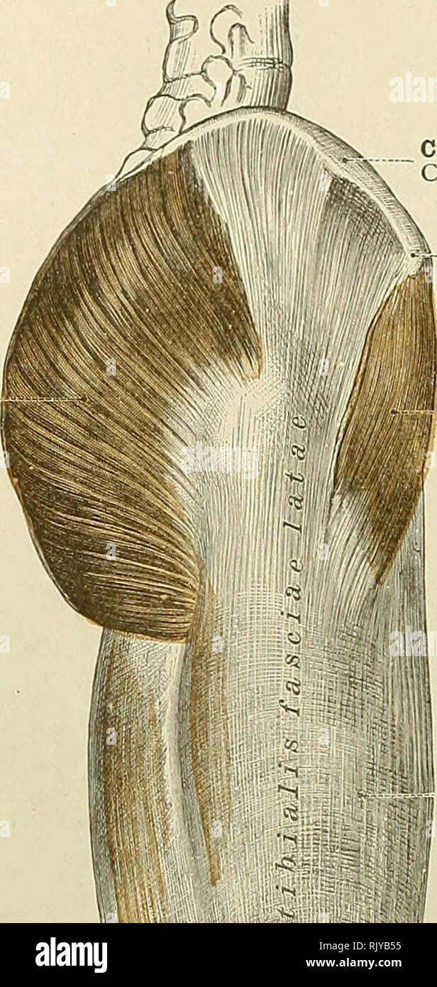 . An atlas of human anatomy for students and physicians. Anatomy. 338 THE MUSCLES OF THE LOWER EXTREMITY Gluteus maximus muscle M. glutjeus maximus. Crest of the ilium Crista iliaca Anterior superior spine of the ilium Spina iliaca anterior superior Tensor vaginse femoris or tensor fasciae femoris muscle M. tensor fasciae latas Deep fascia of the thigh, or fascia lata (superficial layer) Fascia lata (lamina superficialis) Deep fascia of the thigh, or fascia- lata (superficial layer) Fascia lata (lamina superficialis). Please note that these images are extracted from scanned page images that ma Stock Photohttps://www.alamy.com/image-license-details/?v=1https://www.alamy.com/an-atlas-of-human-anatomy-for-students-and-physicians-anatomy-338-the-muscles-of-the-lower-extremity-gluteus-maximus-muscle-m-glutjeus-maximus-crest-of-the-ilium-crista-iliaca-anterior-superior-spine-of-the-ilium-spina-iliaca-anterior-superior-tensor-vaginse-femoris-or-tensor-fasciae-femoris-muscle-m-tensor-fasciae-latas-deep-fascia-of-the-thigh-or-fascia-lata-superficial-layer-fascia-lata-lamina-superficialis-deep-fascia-of-the-thigh-or-fascia-lata-superficial-layer-fascia-lata-lamina-superficialis-please-note-that-these-images-are-extracted-from-scanned-page-images-that-ma-image235400065.html
RMRJYB55–. An atlas of human anatomy for students and physicians. Anatomy. 338 THE MUSCLES OF THE LOWER EXTREMITY Gluteus maximus muscle M. glutjeus maximus. Crest of the ilium Crista iliaca Anterior superior spine of the ilium Spina iliaca anterior superior Tensor vaginse femoris or tensor fasciae femoris muscle M. tensor fasciae latas Deep fascia of the thigh, or fascia lata (superficial layer) Fascia lata (lamina superficialis) Deep fascia of the thigh, or fascia- lata (superficial layer) Fascia lata (lamina superficialis). Please note that these images are extracted from scanned page images that ma
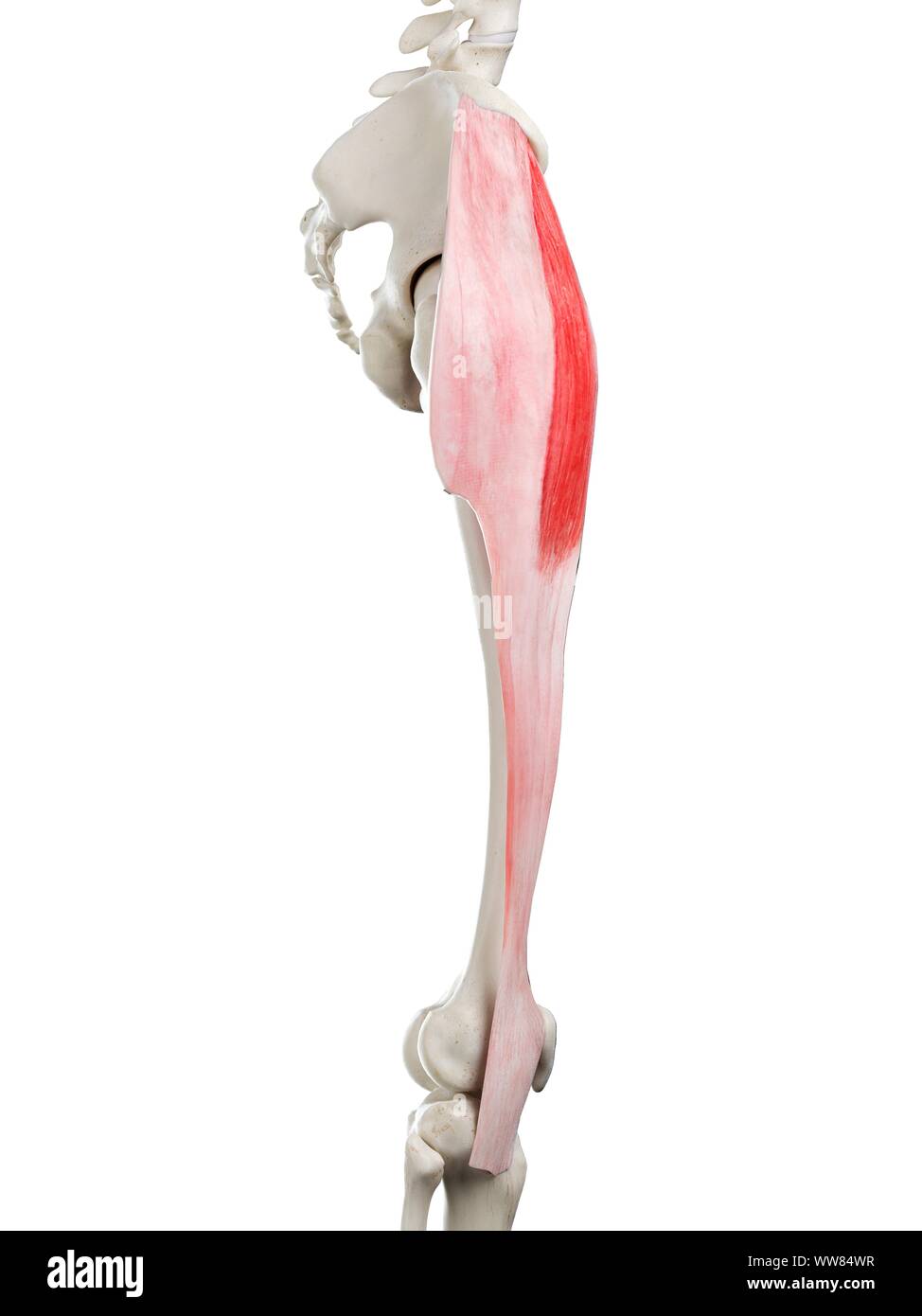 Tensor fascia lata muscle, illustration Stock Photohttps://www.alamy.com/image-license-details/?v=1https://www.alamy.com/tensor-fascia-lata-muscle-illustration-image273701395.html
RFWW84WR–Tensor fascia lata muscle, illustration
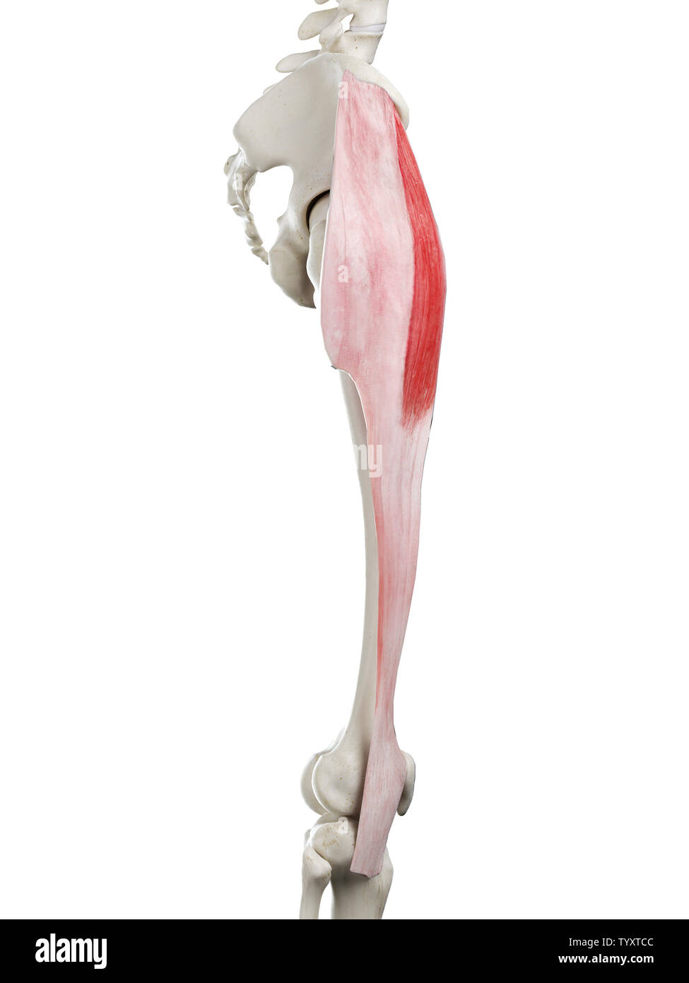 3d rendered medically accurate illustration of a womans Tensor Fascia Lata Stock Photohttps://www.alamy.com/image-license-details/?v=1https://www.alamy.com/3d-rendered-medically-accurate-illustration-of-a-womans-tensor-fascia-lata-image258130780.html
RFTYXTCC–3d rendered medically accurate illustration of a womans Tensor Fascia Lata
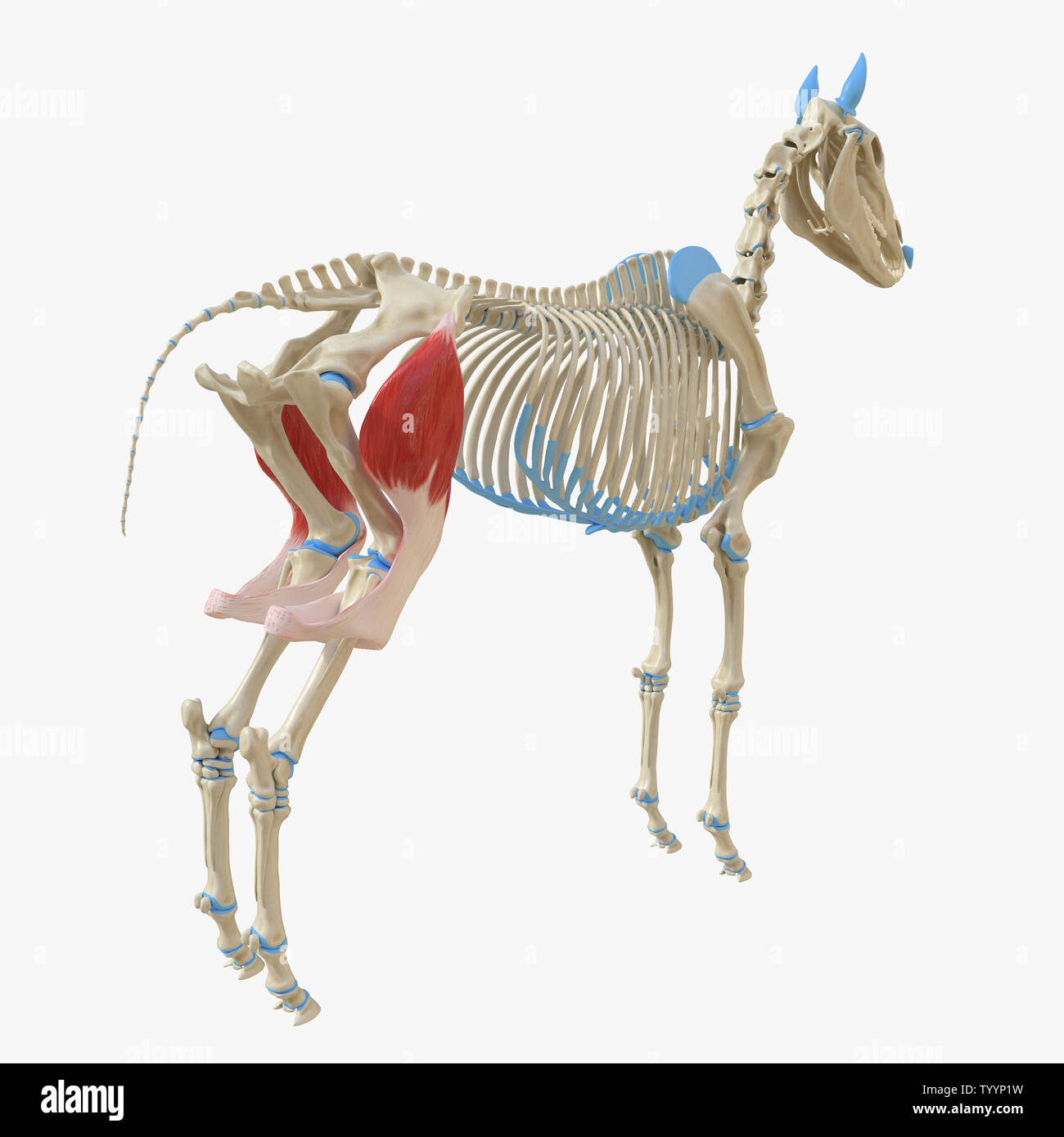 3d rendered medically accurate illustration of the equine muscle anatomy - Tensor Fascia Lata Stock Photohttps://www.alamy.com/image-license-details/?v=1https://www.alamy.com/3d-rendered-medically-accurate-illustration-of-the-equine-muscle-anatomy-tensor-fascia-lata-image258150869.html
RFTYYP1W–3d rendered medically accurate illustration of the equine muscle anatomy - Tensor Fascia Lata
 . An atlas of human anatomy for students and physicians. Anatomy. Crest of the ilium Crista iliaca Anterior superior spine of the ilium Spina iliaca anterior superior Tensor vaginse femoris or tensor fasciae femoris muscle M. tensor fasciae latas Deep fascia of the thigh, or fascia lata (superficial layer) Fascia lata (lamina superficialis) Deep fascia of the thigh, or fascia- lata (superficial layer) Fascia lata (lamina superficialis). Iliotibial band or ligament Fig. 587.—Deep Fascia of the Thigh, or Fascia Lata, seen from the Outer Side, with THE Thickened Portion of this Fascia, known as t Stock Photohttps://www.alamy.com/image-license-details/?v=1https://www.alamy.com/an-atlas-of-human-anatomy-for-students-and-physicians-anatomy-crest-of-the-ilium-crista-iliaca-anterior-superior-spine-of-the-ilium-spina-iliaca-anterior-superior-tensor-vaginse-femoris-or-tensor-fasciae-femoris-muscle-m-tensor-fasciae-latas-deep-fascia-of-the-thigh-or-fascia-lata-superficial-layer-fascia-lata-lamina-superficialis-deep-fascia-of-the-thigh-or-fascia-lata-superficial-layer-fascia-lata-lamina-superficialis-iliotibial-band-or-ligament-fig-587deep-fascia-of-the-thigh-or-fascia-lata-seen-from-the-outer-side-with-the-thickened-portion-of-this-fascia-known-as-t-image235400048.html
RMRJYB4G–. An atlas of human anatomy for students and physicians. Anatomy. Crest of the ilium Crista iliaca Anterior superior spine of the ilium Spina iliaca anterior superior Tensor vaginse femoris or tensor fasciae femoris muscle M. tensor fasciae latas Deep fascia of the thigh, or fascia lata (superficial layer) Fascia lata (lamina superficialis) Deep fascia of the thigh, or fascia- lata (superficial layer) Fascia lata (lamina superficialis). Iliotibial band or ligament Fig. 587.—Deep Fascia of the Thigh, or Fascia Lata, seen from the Outer Side, with THE Thickened Portion of this Fascia, known as t
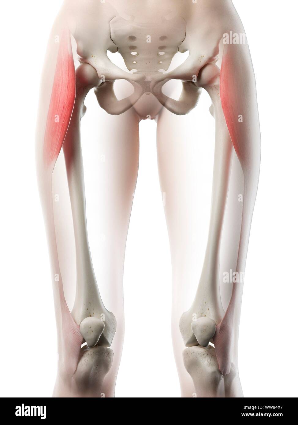 Tensor fascia lata muscle, illustration Stock Photohttps://www.alamy.com/image-license-details/?v=1https://www.alamy.com/tensor-fascia-lata-muscle-illustration-image273701407.html
RFWW84X7–Tensor fascia lata muscle, illustration
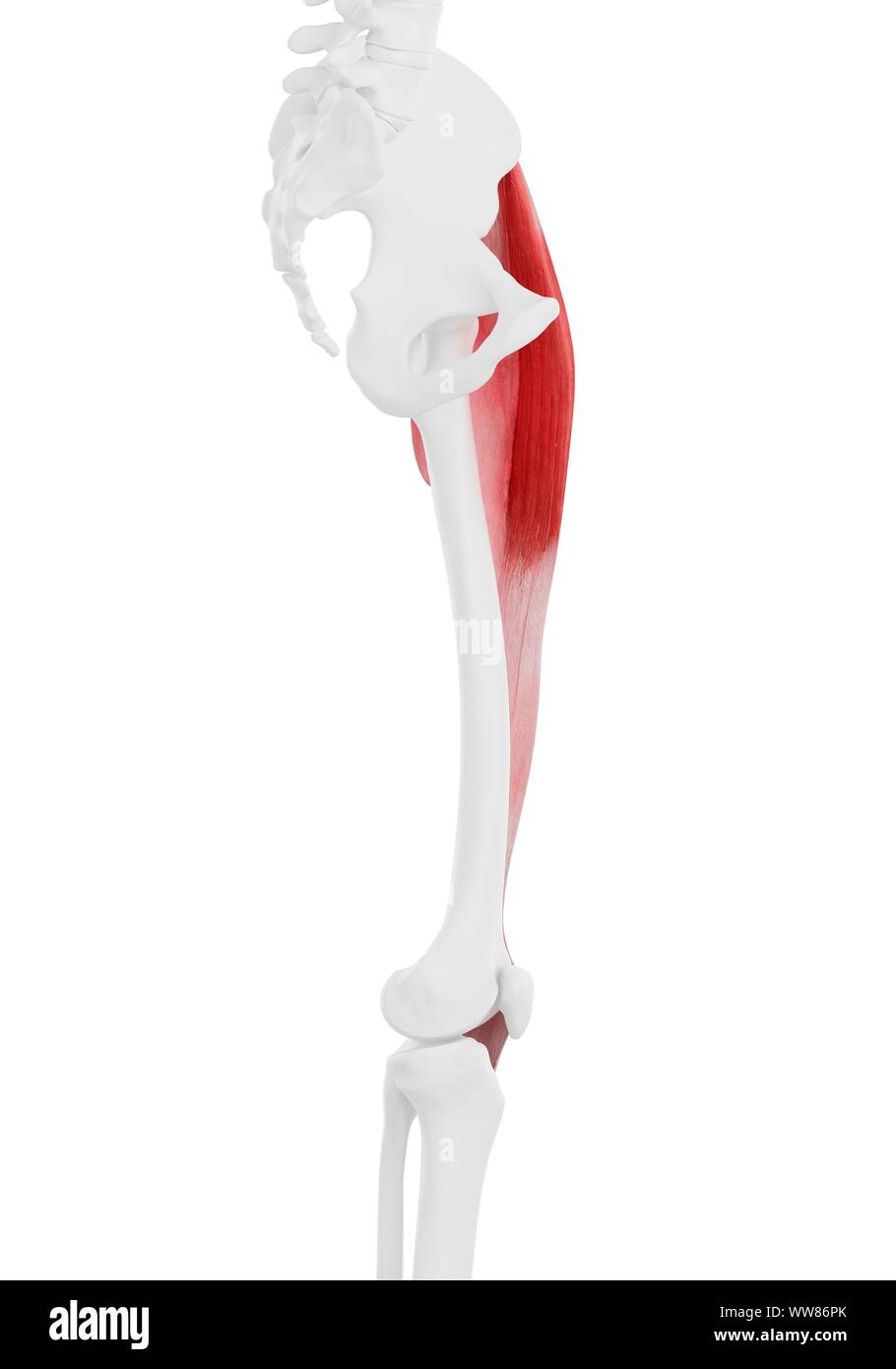 Tensor fascia lata muscle, illustration Stock Photohttps://www.alamy.com/image-license-details/?v=1https://www.alamy.com/tensor-fascia-lata-muscle-illustration-image273702875.html
RFWW86PK–Tensor fascia lata muscle, illustration
 3d rendered medically accurate illustration of the equine muscle anatomy - Tensor Fascia Lata Stock Photohttps://www.alamy.com/image-license-details/?v=1https://www.alamy.com/3d-rendered-medically-accurate-illustration-of-the-equine-muscle-anatomy-tensor-fascia-lata-image258150846.html
RFTYYP12–3d rendered medically accurate illustration of the equine muscle anatomy - Tensor Fascia Lata
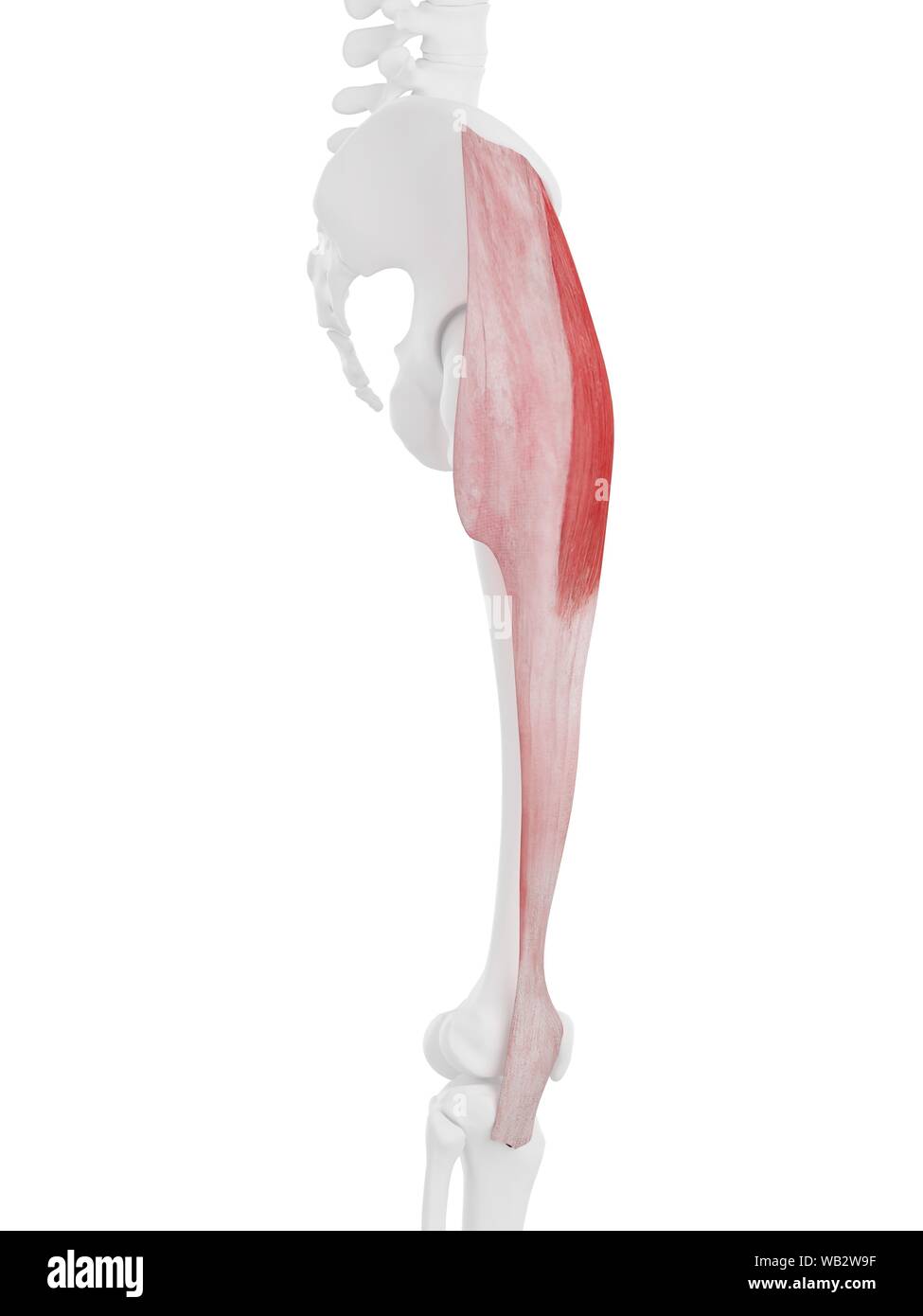 Tensor fascia lata muscle, computer illustration. Stock Photohttps://www.alamy.com/image-license-details/?v=1https://www.alamy.com/tensor-fascia-lata-muscle-computer-illustration-image264980507.html
RFWB2W9F–Tensor fascia lata muscle, computer illustration.
 . The comparative anatomy of the domesticated animals. Veterinary anatomy. 282 THE MUSCLES. superior extremity of that bone, and assists in rearing. In the first instance it acts as a lever of the first order; in the second, as one of the third order. Fig. 129.. SUPERFICIAL MUSCLES OF THE CROUP AND THIGH. 1, Middle glutens, or gluteus maximus; 2, Anterior spinous process of ilium; 3, Muscle of the fascia lata, or tensor vaginas; 4, Superficial gluteus, or gluteus externus; *, Great trochanter of femur; 5, Fascia lata; 6, Patella, with insertion of rectus; 7, Long vastus, or addncfio^ magnus; 8 Stock Photohttps://www.alamy.com/image-license-details/?v=1https://www.alamy.com/the-comparative-anatomy-of-the-domesticated-animals-veterinary-anatomy-282-the-muscles-superior-extremity-of-that-bone-and-assists-in-rearing-in-the-first-instance-it-acts-as-a-lever-of-the-first-order-in-the-second-as-one-of-the-third-order-fig-129-superficial-muscles-of-the-croup-and-thigh-1-middle-glutens-or-gluteus-maximus-2-anterior-spinous-process-of-ilium-3-muscle-of-the-fascia-lata-or-tensor-vaginas-4-superficial-gluteus-or-gluteus-externus-great-trochanter-of-femur-5-fascia-lata-6-patella-with-insertion-of-rectus-7-long-vastus-or-addncfio-magnus-8-image237848858.html
RMRPXXJ2–. The comparative anatomy of the domesticated animals. Veterinary anatomy. 282 THE MUSCLES. superior extremity of that bone, and assists in rearing. In the first instance it acts as a lever of the first order; in the second, as one of the third order. Fig. 129.. SUPERFICIAL MUSCLES OF THE CROUP AND THIGH. 1, Middle glutens, or gluteus maximus; 2, Anterior spinous process of ilium; 3, Muscle of the fascia lata, or tensor vaginas; 4, Superficial gluteus, or gluteus externus; *, Great trochanter of femur; 5, Fascia lata; 6, Patella, with insertion of rectus; 7, Long vastus, or addncfio^ magnus; 8
 Tensor fascia lata muscle, computer illustration. Stock Photohttps://www.alamy.com/image-license-details/?v=1https://www.alamy.com/tensor-fascia-lata-muscle-computer-illustration-image264980559.html
RFWB2WBB–Tensor fascia lata muscle, computer illustration.
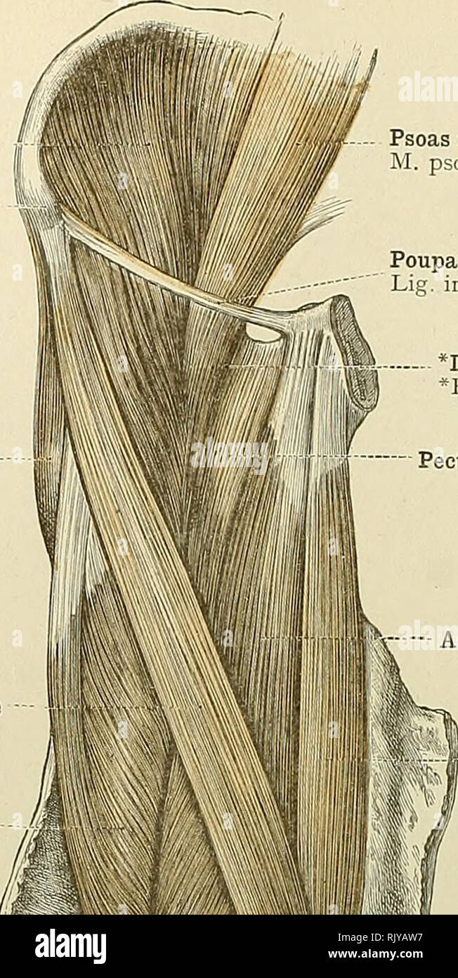 . An atlas of human anatomy for students and physicians. Anatomy. 350 THE MUSCLES OF THE LOWER EXTREMITY 'IhJ Iliacus muscle Anterior superior spine of the ilium Spina iliaca anterior superior Tensor vaginae femoris or tensor fasciae femoris muscle M. tensor fascia; latse Sartorius muscle Rectus femoris muscle Vastus internus muscle M. vastus medialis Deep fascia of the thigh, or fascia lata Prepatellax subcutaneous bursa Bursa pr^patellaris subcutanea Infrapatellar subcutaneous bursa Bursa infrapatellaris subcutanea. Psoas magnus muscle W. psoas major Poupart's ligament (superficial femoral a Stock Photohttps://www.alamy.com/image-license-details/?v=1https://www.alamy.com/an-atlas-of-human-anatomy-for-students-and-physicians-anatomy-350-the-muscles-of-the-lower-extremity-ihj-iliacus-muscle-anterior-superior-spine-of-the-ilium-spina-iliaca-anterior-superior-tensor-vaginae-femoris-or-tensor-fasciae-femoris-muscle-m-tensor-fascia-latse-sartorius-muscle-rectus-femoris-muscle-vastus-internus-muscle-m-vastus-medialis-deep-fascia-of-the-thigh-or-fascia-lata-prepatellax-subcutaneous-bursa-bursa-prpatellaris-subcutanea-infrapatellar-subcutaneous-bursa-bursa-infrapatellaris-subcutanea-psoas-magnus-muscle-w-psoas-major-pouparts-ligament-superficial-femoral-a-image235399843.html
RMRJYAW7–. An atlas of human anatomy for students and physicians. Anatomy. 350 THE MUSCLES OF THE LOWER EXTREMITY 'IhJ Iliacus muscle Anterior superior spine of the ilium Spina iliaca anterior superior Tensor vaginae femoris or tensor fasciae femoris muscle M. tensor fascia; latse Sartorius muscle Rectus femoris muscle Vastus internus muscle M. vastus medialis Deep fascia of the thigh, or fascia lata Prepatellax subcutaneous bursa Bursa pr^patellaris subcutanea Infrapatellar subcutaneous bursa Bursa infrapatellaris subcutanea. Psoas magnus muscle W. psoas major Poupart's ligament (superficial femoral a
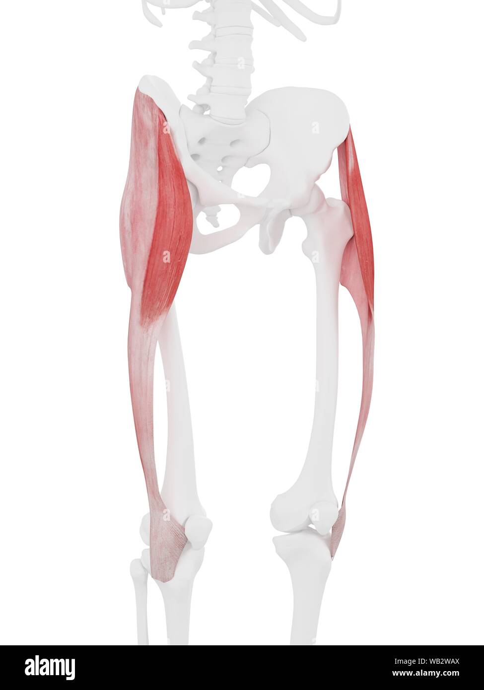 Tensor fascia lata muscle, computer illustration. Stock Photohttps://www.alamy.com/image-license-details/?v=1https://www.alamy.com/tensor-fascia-lata-muscle-computer-illustration-image264980546.html
RFWB2WAX–Tensor fascia lata muscle, computer illustration.
 . The comparative anatomy of the domesticated animals. Veterinary anatomy. 282 THE MUSCLES. superior extremity of that bone, and assists in rearing. In the fii-st instance it acts as a lever of the first order; in the second, as one of the third order. Fig. 129.. SUPERFICIAL MUSCLES OE THE CEOUP AUD THIGH. 1, Middle glutens, or gluteus maximus; 2, Anterior spinous process of ilium; 3, Muscle of the fascia lata, or tensor vaginse; 4, Superficial gluteus, or gluteus ^ externus; *, Great trochanter of femur; 5, Fascia lata; 6, Patella, with insertion of rectus; 7, Long vastus, or adductor magnus; Stock Photohttps://www.alamy.com/image-license-details/?v=1https://www.alamy.com/the-comparative-anatomy-of-the-domesticated-animals-veterinary-anatomy-282-the-muscles-superior-extremity-of-that-bone-and-assists-in-rearing-in-the-fii-st-instance-it-acts-as-a-lever-of-the-first-order-in-the-second-as-one-of-the-third-order-fig-129-superficial-muscles-oe-the-ceoup-aud-thigh-1-middle-glutens-or-gluteus-maximus-2-anterior-spinous-process-of-ilium-3-muscle-of-the-fascia-lata-or-tensor-vaginse-4-superficial-gluteus-or-gluteus-externus-great-trochanter-of-femur-5-fascia-lata-6-patella-with-insertion-of-rectus-7-long-vastus-or-adductor-magnus-image232453091.html
RMRE5483–. The comparative anatomy of the domesticated animals. Veterinary anatomy. 282 THE MUSCLES. superior extremity of that bone, and assists in rearing. In the fii-st instance it acts as a lever of the first order; in the second, as one of the third order. Fig. 129.. SUPERFICIAL MUSCLES OE THE CEOUP AUD THIGH. 1, Middle glutens, or gluteus maximus; 2, Anterior spinous process of ilium; 3, Muscle of the fascia lata, or tensor vaginse; 4, Superficial gluteus, or gluteus ^ externus; *, Great trochanter of femur; 5, Fascia lata; 6, Patella, with insertion of rectus; 7, Long vastus, or adductor magnus;
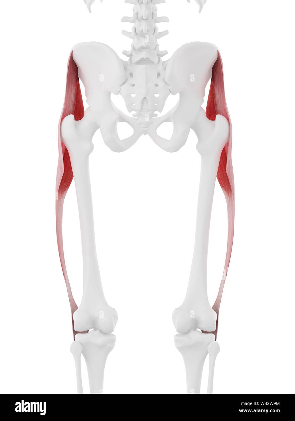 Tensor fascia lata muscle, computer illustration. Stock Photohttps://www.alamy.com/image-license-details/?v=1https://www.alamy.com/tensor-fascia-lata-muscle-computer-illustration-image264980512.html
RFWB2W9M–Tensor fascia lata muscle, computer illustration.
 Horse tensor fascia lata muscle, illustration Stock Photohttps://www.alamy.com/image-license-details/?v=1https://www.alamy.com/horse-tensor-fascia-lata-muscle-illustration-image273702709.html
RFWW86GN–Horse tensor fascia lata muscle, illustration
 . The anatomy of the domestic animals . Veterinary anatomy. BRANCHES OF THE ABDOMINAL AORTA 667 Internal iliac lymph gla Circumflex iliac vessels External iliac lymph glands Remnant of ingiiiiml ligament Posterior branch of circum- flex iliac artery Lateral cutaneous nerve of thigh External spermatic nerve Tensor fascice latce Femoral nerve and ante- rior femoral vessels Femoral artery Prefemoral lymph glands Deep inguinal lymph glands Fascia lata Medial patellar ligament Middle patellar ligament. Saphenous nerves Saphenous vessels Fig. 576.—Dissection of Pelvis, Thigh, and Proximal Part o Stock Photohttps://www.alamy.com/image-license-details/?v=1https://www.alamy.com/the-anatomy-of-the-domestic-animals-veterinary-anatomy-branches-of-the-abdominal-aorta-667-internal-iliac-lymph-gla-circumflex-iliac-vessels-external-iliac-lymph-glands-remnant-of-ingiiiiml-ligament-posterior-branch-of-circum-flex-iliac-artery-lateral-cutaneous-nerve-of-thigh-external-spermatic-nerve-tensor-fascice-latce-femoral-nerve-and-ante-rior-femoral-vessels-femoral-artery-prefemoral-lymph-glands-deep-inguinal-lymph-glands-fascia-lata-medial-patellar-ligament-middle-patellar-ligament-saphenous-nerves-saphenous-vessels-fig-576dissection-of-pelvis-thigh-and-proximal-part-o-image232323543.html
RMRDY71B–. The anatomy of the domestic animals . Veterinary anatomy. BRANCHES OF THE ABDOMINAL AORTA 667 Internal iliac lymph gla Circumflex iliac vessels External iliac lymph glands Remnant of ingiiiiml ligament Posterior branch of circum- flex iliac artery Lateral cutaneous nerve of thigh External spermatic nerve Tensor fascice latce Femoral nerve and ante- rior femoral vessels Femoral artery Prefemoral lymph glands Deep inguinal lymph glands Fascia lata Medial patellar ligament Middle patellar ligament. Saphenous nerves Saphenous vessels Fig. 576.—Dissection of Pelvis, Thigh, and Proximal Part o
 Horse tensor fascia lata muscle, illustration Stock Photohttps://www.alamy.com/image-license-details/?v=1https://www.alamy.com/horse-tensor-fascia-lata-muscle-illustration-image273702638.html
RFWW86E6–Horse tensor fascia lata muscle, illustration
 Tensor fascia lata muscle, computer illustration. Stock Photohttps://www.alamy.com/image-license-details/?v=1https://www.alamy.com/tensor-fascia-lata-muscle-computer-illustration-image333579291.html
RF2AAKRP3–Tensor fascia lata muscle, computer illustration.
 . The comparative anatomy of the domesticated animals. Veterinary anatomy. MUSCLES OF THE ANTERIOR FEMORAL REGION IN MAN. 1, Crest of the ilium; 2, Its antero-superior spinous process; 3, Gluteus medius; 4, Tensor vaginee femoris; 5, Sartorius; 6, Rectus; 7, Vastus externus; 8, Vastus internus; 9, Patella; 10, lliacus internus; 11, Psoas magnus; 12, Pectineus; 13, Adductor longus; 14, Portion of adductor magnus; 15, Gracilis.. MUSCLES OF THE POSTERIOR FEMORAL AND GLUTEAL REGION IN MAN. 1, Gluteus medius ; 2, Gluteus maximus ; 3, Vastus externus, covered by fascia lata; 4, Long head of biceps; Stock Photohttps://www.alamy.com/image-license-details/?v=1https://www.alamy.com/the-comparative-anatomy-of-the-domesticated-animals-veterinary-anatomy-muscles-of-the-anterior-femoral-region-in-man-1-crest-of-the-ilium-2-its-antero-superior-spinous-process-3-gluteus-medius-4-tensor-vaginee-femoris-5-sartorius-6-rectus-7-vastus-externus-8-vastus-internus-9-patella-10-lliacus-internus-11-psoas-magnus-12-pectineus-13-adductor-longus-14-portion-of-adductor-magnus-15-gracilis-muscles-of-the-posterior-femoral-and-gluteal-region-in-man-1-gluteus-medius-2-gluteus-maximus-3-vastus-externus-covered-by-fascia-lata-4-long-head-of-biceps-image232680599.html
RMREFEDB–. The comparative anatomy of the domesticated animals. Veterinary anatomy. MUSCLES OF THE ANTERIOR FEMORAL REGION IN MAN. 1, Crest of the ilium; 2, Its antero-superior spinous process; 3, Gluteus medius; 4, Tensor vaginee femoris; 5, Sartorius; 6, Rectus; 7, Vastus externus; 8, Vastus internus; 9, Patella; 10, lliacus internus; 11, Psoas magnus; 12, Pectineus; 13, Adductor longus; 14, Portion of adductor magnus; 15, Gracilis.. MUSCLES OF THE POSTERIOR FEMORAL AND GLUTEAL REGION IN MAN. 1, Gluteus medius ; 2, Gluteus maximus ; 3, Vastus externus, covered by fascia lata; 4, Long head of biceps;
 Tensor fascia lata muscle, illustration Stock Photohttps://www.alamy.com/image-license-details/?v=1https://www.alamy.com/tensor-fascia-lata-muscle-illustration-image328921508.html
RF2A33JMM–Tensor fascia lata muscle, illustration
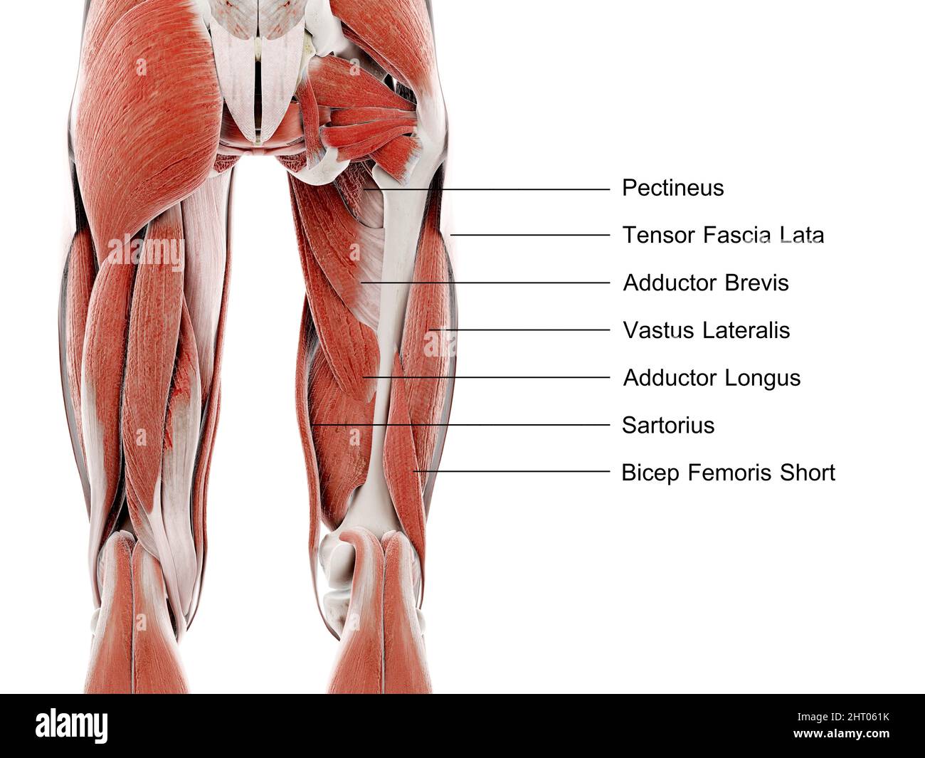
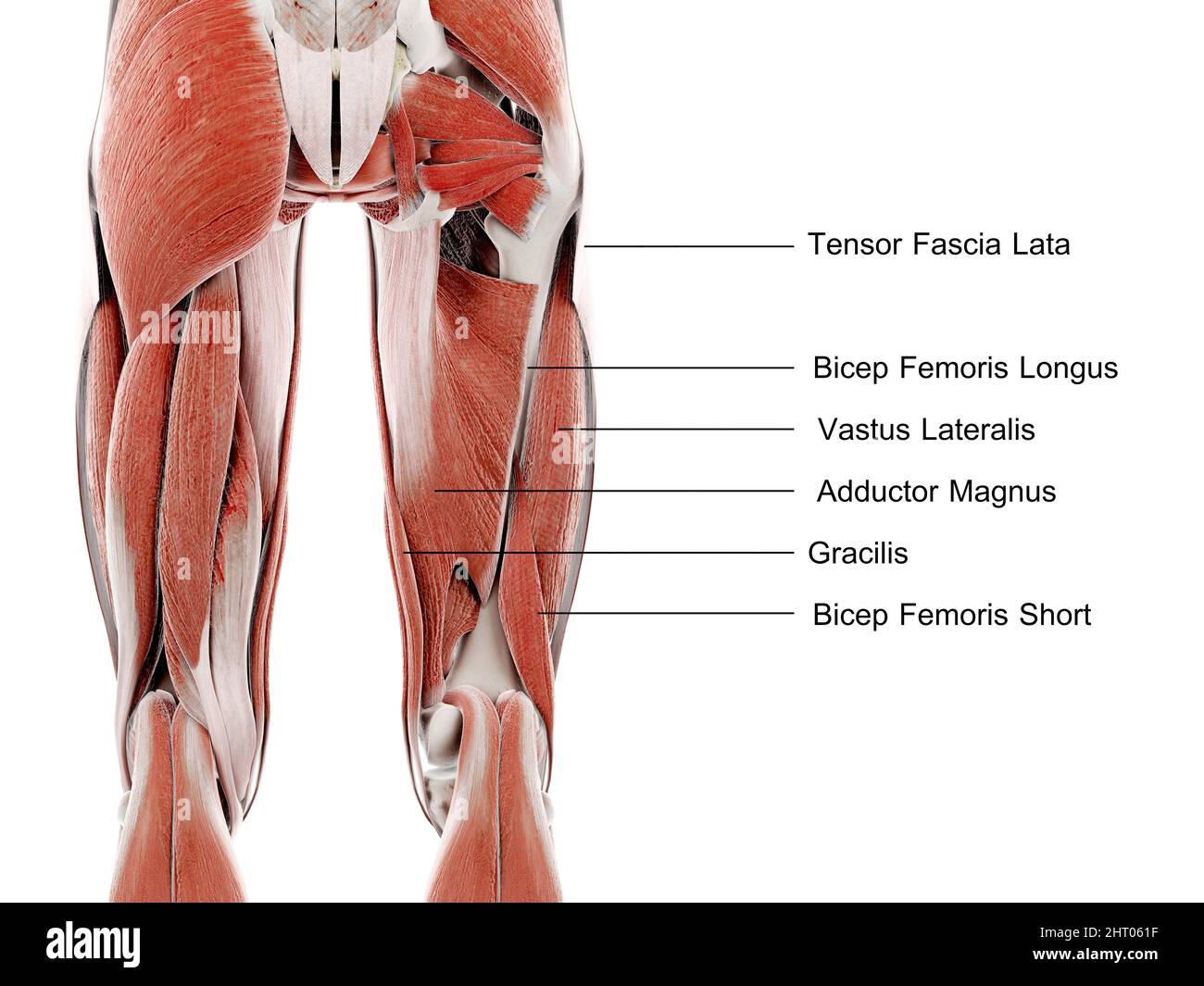 Muscles of the upper leg, illustration Stock Photohttps://www.alamy.com/image-license-details/?v=1https://www.alamy.com/muscles-of-the-upper-leg-illustration-image462226059.html
RF2HT061F–Muscles of the upper leg, illustration
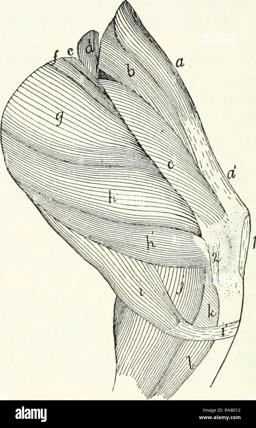 . Anatomy of the cat. Cats; Mammals. 200 THE MUSCLES. This depression is called the iliopectineal fossa; it contains the femoral vein and artery and saphenous nerve imbedded in fat (Fig. 127). The medial edge of the adductor longus is in relation with the integument; the lateral edge with the pec-. FiG. 92.—Second Layer of Muscles on the Medial Side of the Thigh. a, M. tensor fascise lata;; a', fascia lata; b, M. rectus feiiioris; c, M. vastus mcdi- alis; </, M. iliopsoas (cut); t", M. pectincus;/", M. adductor longus; g, M. adductor femoris; h, //, M. semimembranosus; /, M. semit Stock Photohttps://www.alamy.com/image-license-details/?v=1https://www.alamy.com/anatomy-of-the-cat-cats-mammals-200-the-muscles-this-depression-is-called-the-iliopectineal-fossa-it-contains-the-femoral-vein-and-artery-and-saphenous-nerve-imbedded-in-fat-fig-127-the-medial-edge-of-the-adductor-longus-is-in-relation-with-the-integument-the-lateral-edge-with-the-pec-fig-92second-layer-of-muscles-on-the-medial-side-of-the-thigh-a-m-tensor-fascise-lata-a-fascia-lata-b-m-rectus-feiiioris-c-m-vastus-mcdi-alis-lt-m-iliopsoas-cut-tquot-m-pectincusquot-m-adductor-longus-g-m-adductor-femoris-h-m-semimembranosus-m-semit-image236818206.html
RMRN8012–. Anatomy of the cat. Cats; Mammals. 200 THE MUSCLES. This depression is called the iliopectineal fossa; it contains the femoral vein and artery and saphenous nerve imbedded in fat (Fig. 127). The medial edge of the adductor longus is in relation with the integument; the lateral edge with the pec-. FiG. 92.—Second Layer of Muscles on the Medial Side of the Thigh. a, M. tensor fascise lata;; a', fascia lata; b, M. rectus feiiioris; c, M. vastus mcdi- alis; </, M. iliopsoas (cut); t", M. pectincus;/", M. adductor longus; g, M. adductor femoris; h, //, M. semimembranosus; /, M. semit
 Muscles of the upper leg, illustration Stock Photohttps://www.alamy.com/image-license-details/?v=1https://www.alamy.com/muscles-of-the-upper-leg-illustration-image462226063.html
RF2HT061K–Muscles of the upper leg, illustration
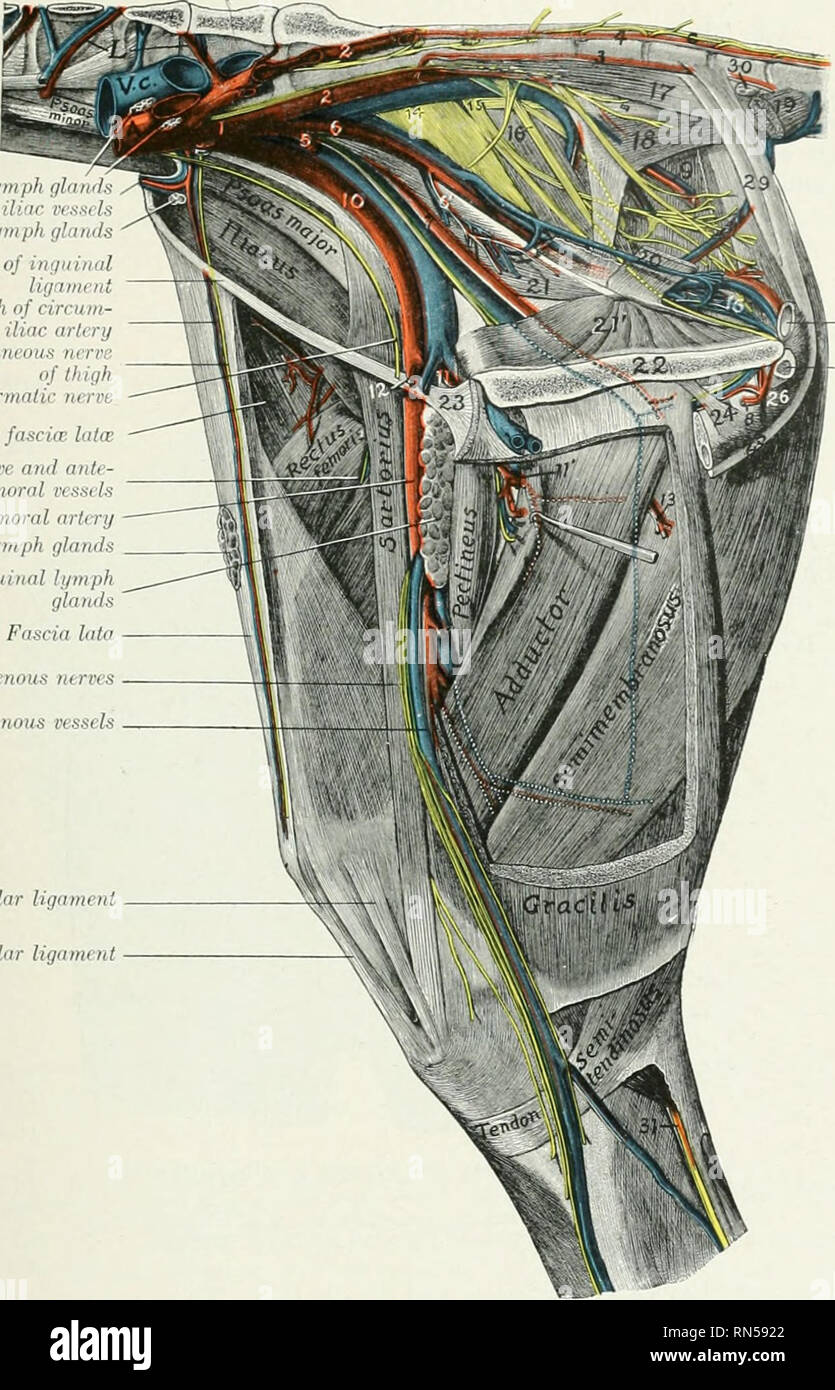 . The anatomy of the domestic animals. Veterinary anatomy. BRANCHES OF THE ABDOMINAL AORTA 667 Internal iliac lymph gland: CiTcumflex iliac vessels EiUrnaliliaclymph ijlands Remnant of ini/iiinal U,r.,mn,t Posteriiir lirnin-li of lininn- jhx ilinr „rl,ni Lateral eiilaiinnix nrrrr oflhigh External spermatic nerre Tensor fasciw lalw Femoral nerve atul onl(- rior femoral vessels Femoral arttry Prefemoral lymph glands Deep inguinal hpnph glands Fascia lata Saphenous nerves Saphenous vessels Medial patellar ligament Middle patellar ligament. Fig. 576.—Dissectiox of Pelvis, Thigh, and Proximal Part Stock Photohttps://www.alamy.com/image-license-details/?v=1https://www.alamy.com/the-anatomy-of-the-domestic-animals-veterinary-anatomy-branches-of-the-abdominal-aorta-667-internal-iliac-lymph-gland-citcumflex-iliac-vessels-eiurnaliliaclymph-ijlands-remnant-of-iniiiinal-urmnt-posteriiir-lirnin-li-of-lininn-jhx-ilinr-rlni-lateral-eiilaiinnix-nrrrr-oflhigh-external-spermatic-nerre-tensor-fasciw-lalw-femoral-nerve-atul-onl-rior-femoral-vessels-femoral-arttry-prefemoral-lymph-glands-deep-inguinal-hpnph-glands-fascia-lata-saphenous-nerves-saphenous-vessels-medial-patellar-ligament-middle-patellar-ligament-fig-576dissectiox-of-pelvis-thigh-and-proximal-part-image236759434.html
RMRN5922–. The anatomy of the domestic animals. Veterinary anatomy. BRANCHES OF THE ABDOMINAL AORTA 667 Internal iliac lymph gland: CiTcumflex iliac vessels EiUrnaliliaclymph ijlands Remnant of ini/iiinal U,r.,mn,t Posteriiir lirnin-li of lininn- jhx ilinr „rl,ni Lateral eiilaiinnix nrrrr oflhigh External spermatic nerre Tensor fasciw lalw Femoral nerve atul onl(- rior femoral vessels Femoral arttry Prefemoral lymph glands Deep inguinal hpnph glands Fascia lata Saphenous nerves Saphenous vessels Medial patellar ligament Middle patellar ligament. Fig. 576.—Dissectiox of Pelvis, Thigh, and Proximal Part
