Quick filters:
Testis Stock Photos and Images
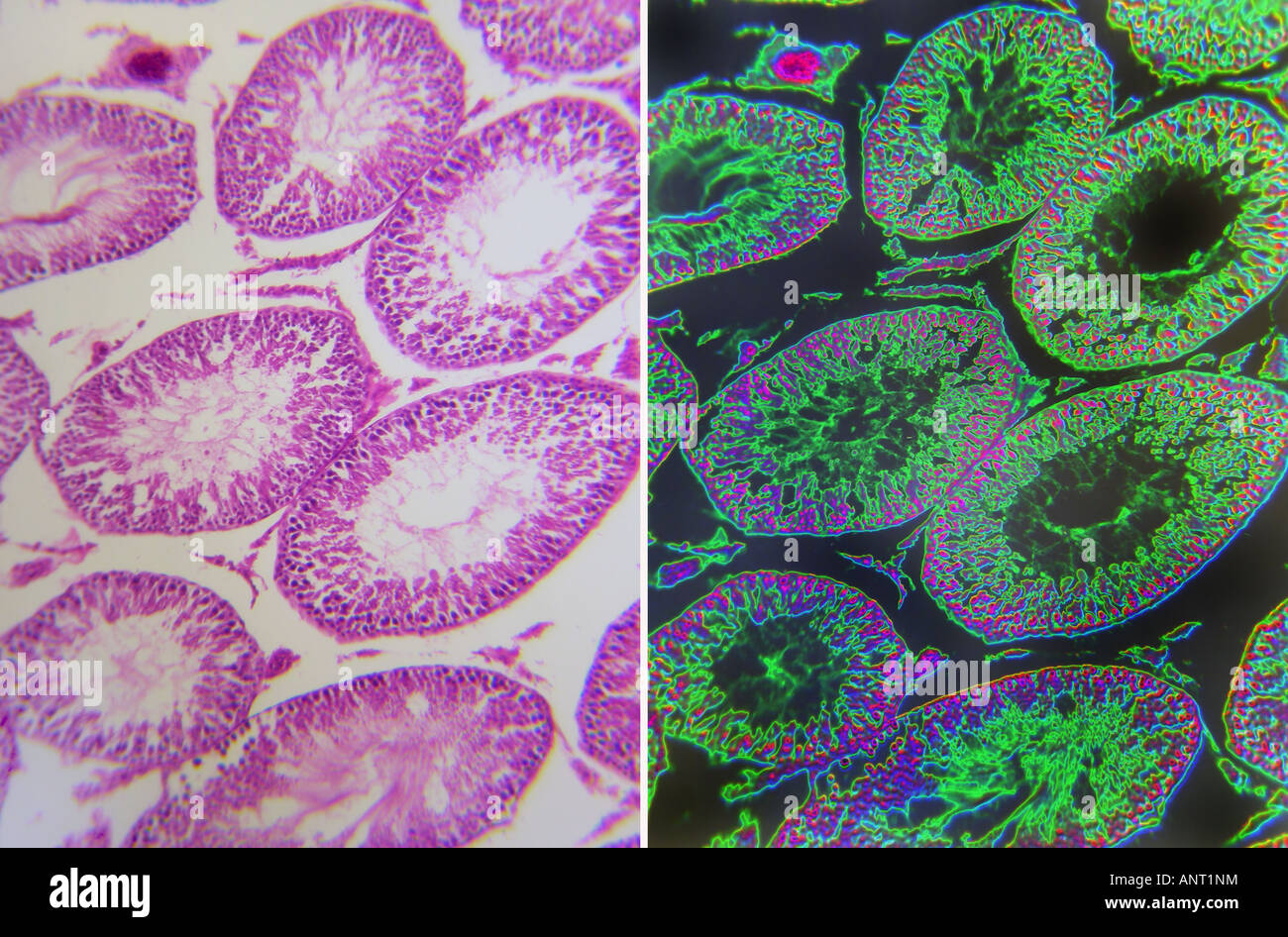 testis tissue comparative views Stock Photohttps://www.alamy.com/image-license-details/?v=1https://www.alamy.com/stock-photo-testis-tissue-comparative-views-15592783.html
RFANT1NM–testis tissue comparative views
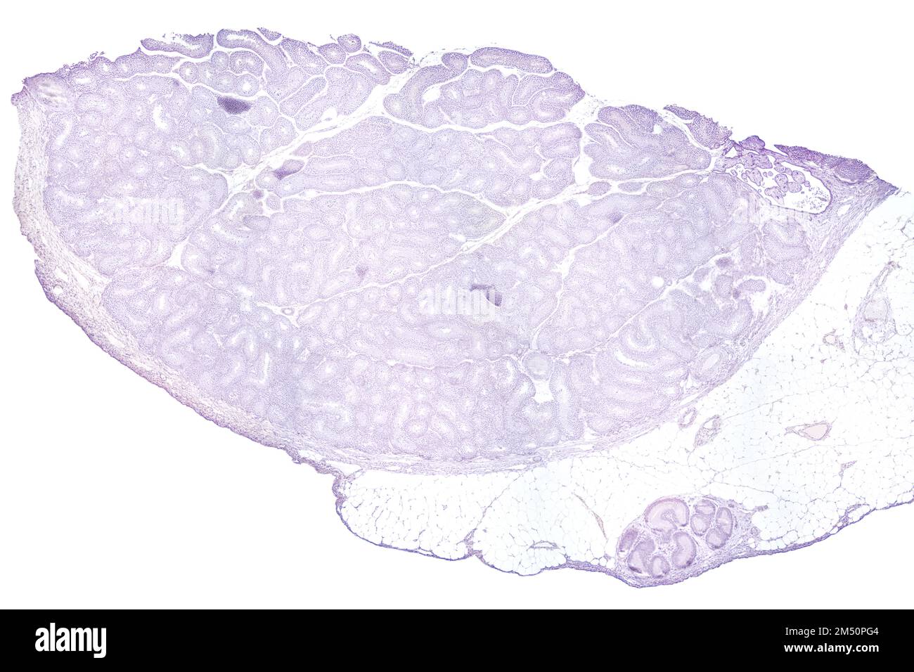 Testis, transverse section, 20X light micrograph. Testicle, the male reproductive gland, under the light microscope, T.S. Stock Photohttps://www.alamy.com/image-license-details/?v=1https://www.alamy.com/testis-transverse-section-20x-light-micrograph-testicle-the-male-reproductive-gland-under-the-light-microscope-ts-image502191652.html
RF2M50PG4–Testis, transverse section, 20X light micrograph. Testicle, the male reproductive gland, under the light microscope, T.S.
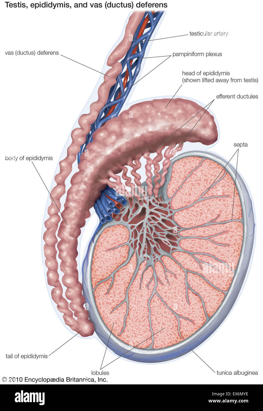 Testis, epididymis, and vas (ductus) deferens Stock Photohttps://www.alamy.com/image-license-details/?v=1https://www.alamy.com/stock-photo-testis-epididymis-and-vas-ductus-deferens-84970690.html
RMEX6MYE–Testis, epididymis, and vas (ductus) deferens
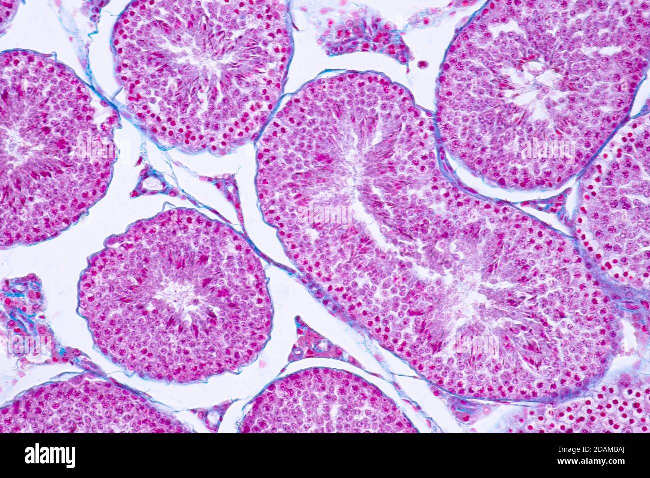 Human testis, light micrograph. Stock Photohttps://www.alamy.com/image-license-details/?v=1https://www.alamy.com/human-testis-light-micrograph-image385222618.html
RF2DAMBAJ–Human testis, light micrograph.
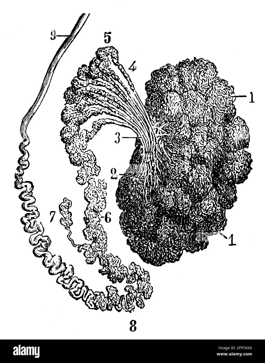 Tunica albuginea of testis. Illustration of the 19th century. Germany. White background. Stock Photohttps://www.alamy.com/image-license-details/?v=1https://www.alamy.com/tunica-albuginea-of-testis-illustration-of-the-19th-century-germany-white-background-image419115580.html
RF2F9TA5G–Tunica albuginea of testis. Illustration of the 19th century. Germany. White background.
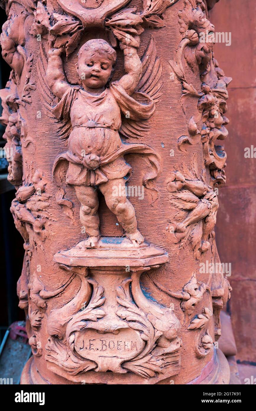 Architectural detail on pillar outside house, Clareville Street, Kensington, London, UK, dated 1884 with Sol Mea Testis (Let the sun be wi Stock Photohttps://www.alamy.com/image-license-details/?v=1https://www.alamy.com/architectural-detail-on-pillar-outside-house-clareville-street-kensington-london-uk-dated-1884-with-sol-mea-testis-let-the-sun-be-wi-image431042669.html
RM2G17K91–Architectural detail on pillar outside house, Clareville Street, Kensington, London, UK, dated 1884 with Sol Mea Testis (Let the sun be wi
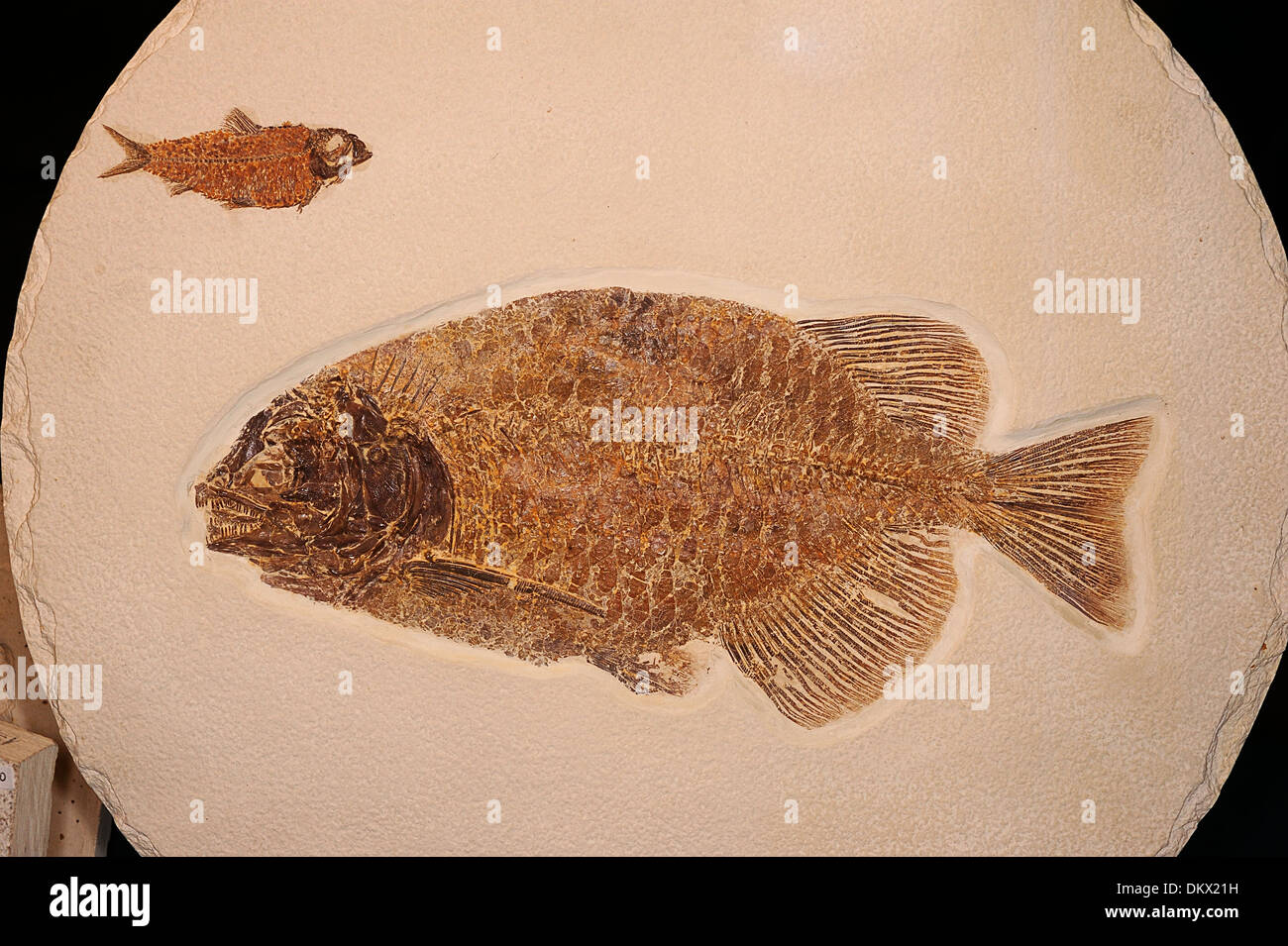 Fossil fish Phareodus testis, Laggerstatten, the Green River Formation, Wyoming USA Eocene Age Stock Photohttps://www.alamy.com/image-license-details/?v=1https://www.alamy.com/fossil-fish-phareodus-testis-laggerstatten-the-green-river-formation-image63881933.html
RMDKX21H–Fossil fish Phareodus testis, Laggerstatten, the Green River Formation, Wyoming USA Eocene Age
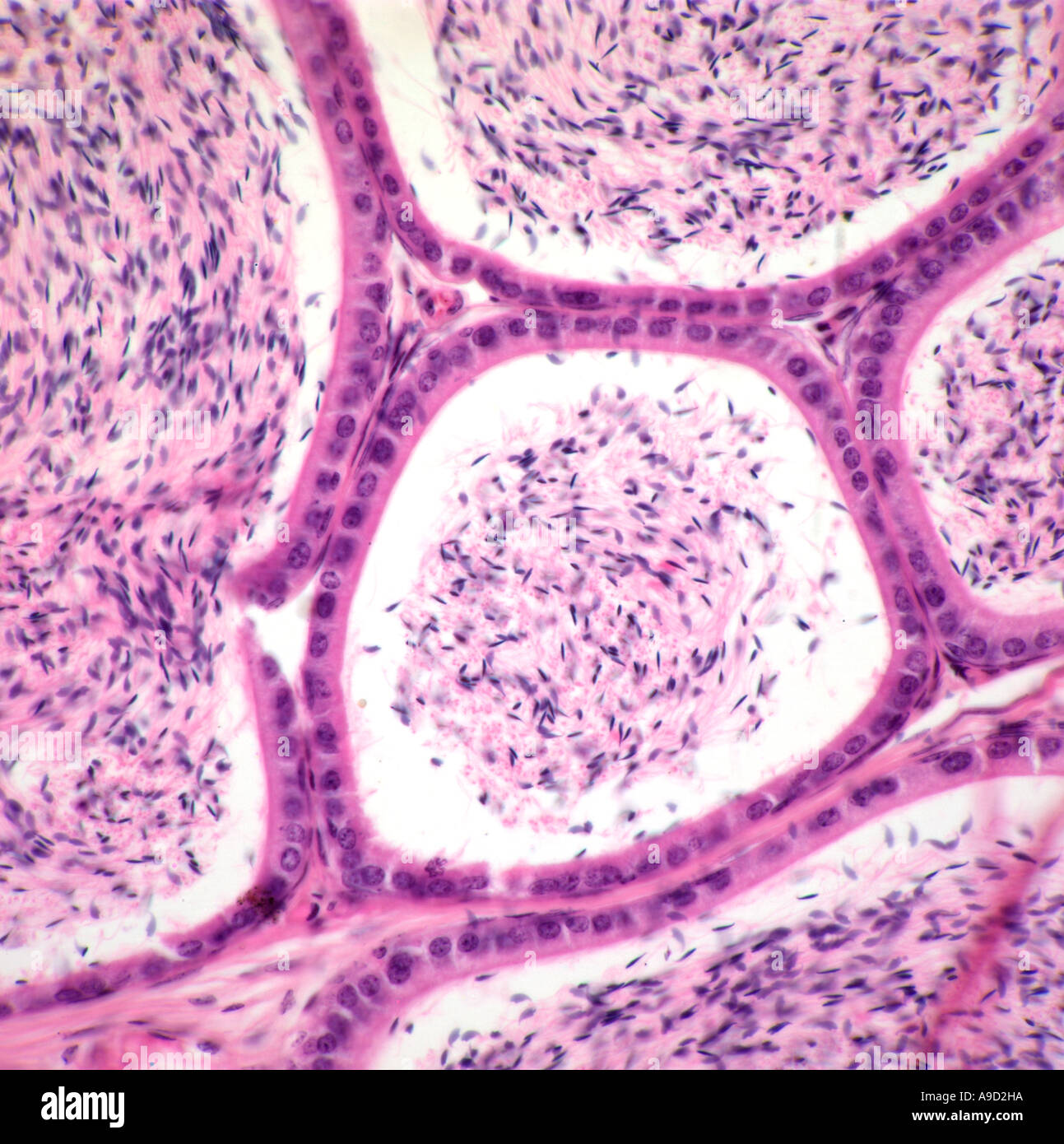 Human testis section showing spematozoa production Stock Photohttps://www.alamy.com/image-license-details/?v=1https://www.alamy.com/human-testis-section-showing-spematozoa-production-image7044761.html
RMA9D2HA–Human testis section showing spematozoa production
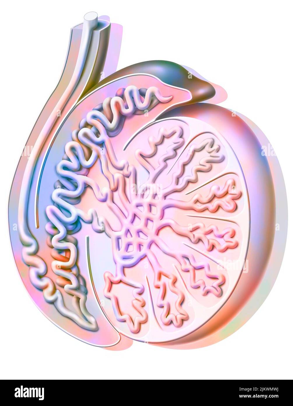 Sagittal section of a testicle showing the seminiferous tube. Stock Photohttps://www.alamy.com/image-license-details/?v=1https://www.alamy.com/sagittal-section-of-a-testicle-showing-the-seminiferous-tube-image476923598.html
RF2JKWMWJ–Sagittal section of a testicle showing the seminiferous tube.
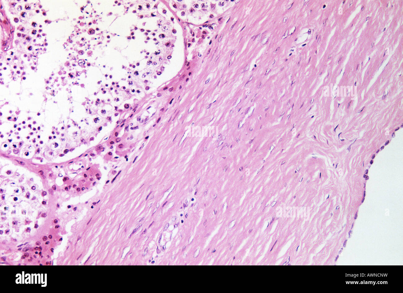 Testis Stock Photohttps://www.alamy.com/image-license-details/?v=1https://www.alamy.com/stock-photo-testis-16621956.html
RFAWNCNW–Testis
 Concept of right sided scrotal swelling or painful testis. Idea of shouting, needing help, trauma or pain. Isolated on white. Stock Photohttps://www.alamy.com/image-license-details/?v=1https://www.alamy.com/concept-of-right-sided-scrotal-swelling-or-painful-testis-idea-of-shouting-needing-help-trauma-or-pain-isolated-on-white-image485347512.html
RF2K5HDKM–Concept of right sided scrotal swelling or painful testis. Idea of shouting, needing help, trauma or pain. Isolated on white.
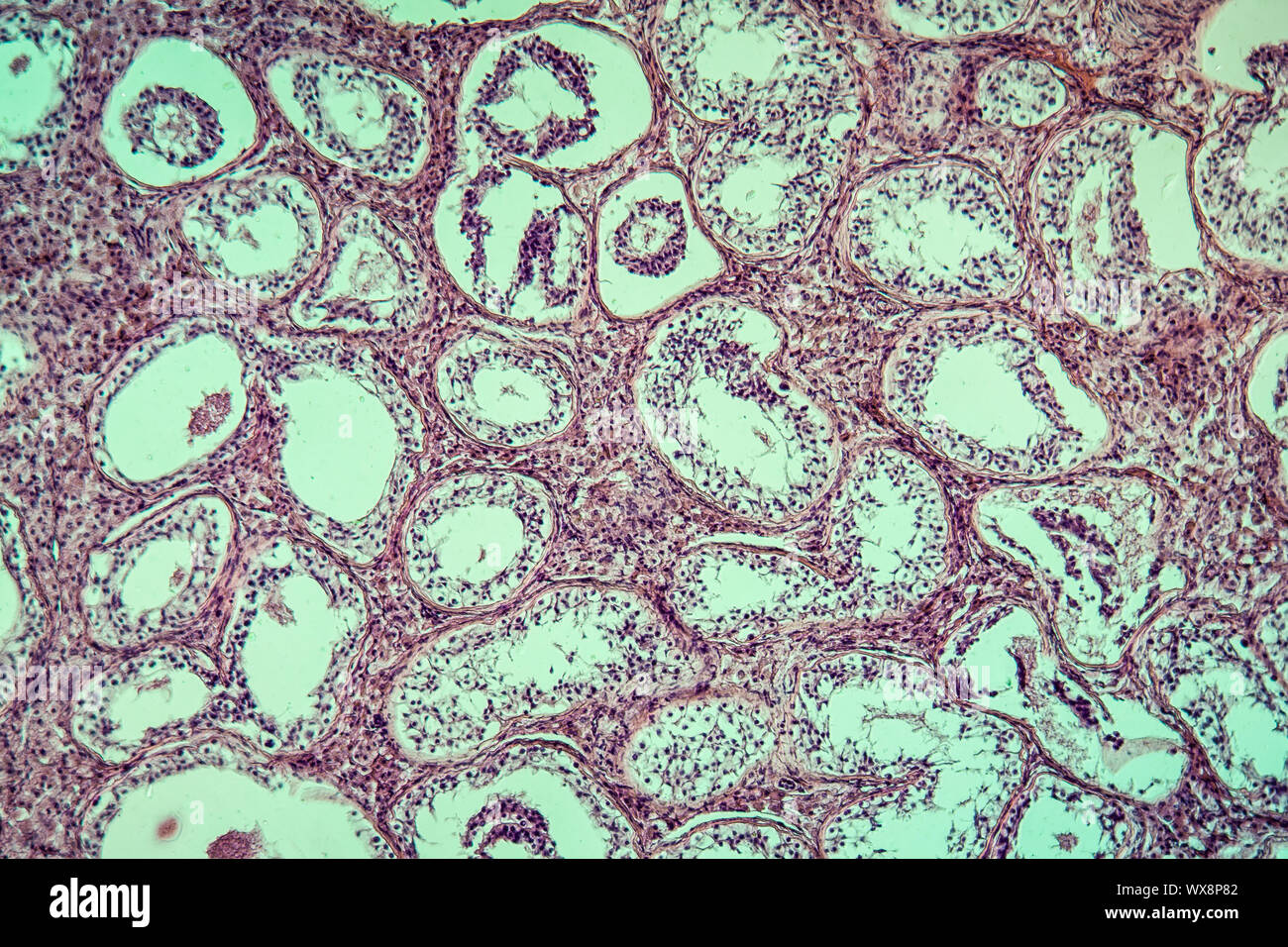 Inguinal testis diseased tissue 100x Stock Photohttps://www.alamy.com/image-license-details/?v=1https://www.alamy.com/inguinal-testis-diseased-tissue-100x-image274329666.html
RMWX8P82–Inguinal testis diseased tissue 100x
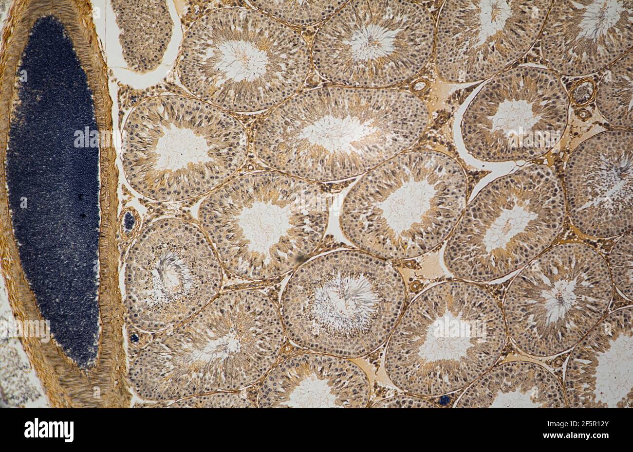 mammal testis cells shown in transverse section under a light microscope Stock Photohttps://www.alamy.com/image-license-details/?v=1https://www.alamy.com/mammal-testis-cells-shown-in-transverse-section-under-a-light-microscope-image416627875.html
RF2F5R12Y–mammal testis cells shown in transverse section under a light microscope
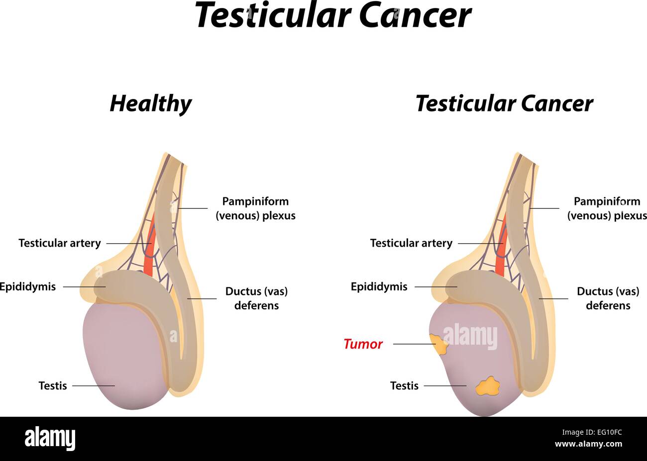 Testicular Cancer Stock Vectorhttps://www.alamy.com/image-license-details/?v=1https://www.alamy.com/stock-photo-testicular-cancer-78698352.html
RFEG10FC–Testicular Cancer
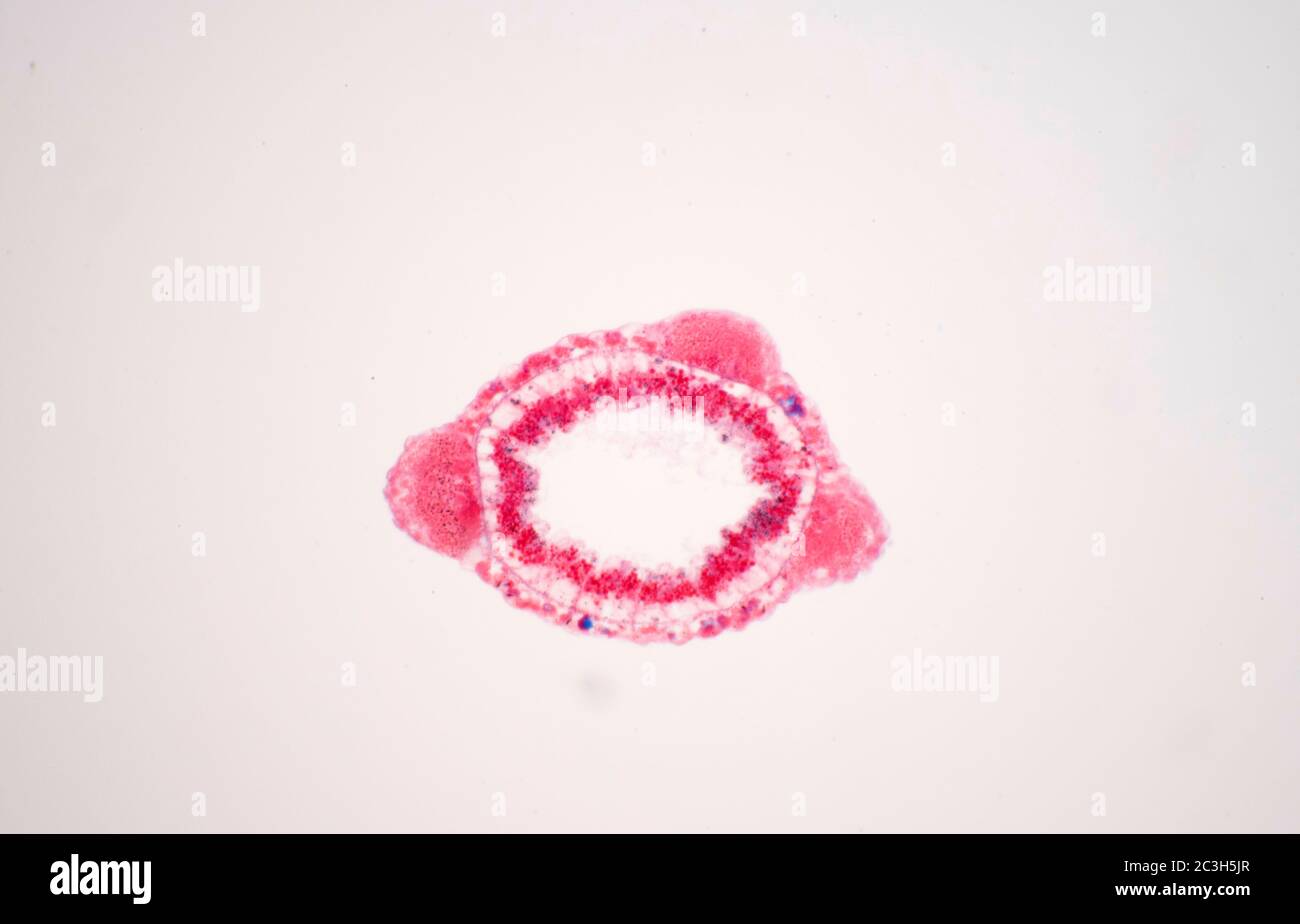 Hydra Testis, transverse section Stock Photohttps://www.alamy.com/image-license-details/?v=1https://www.alamy.com/hydra-testis-transverse-section-image363639327.html
RM2C3H5JR–Hydra Testis, transverse section
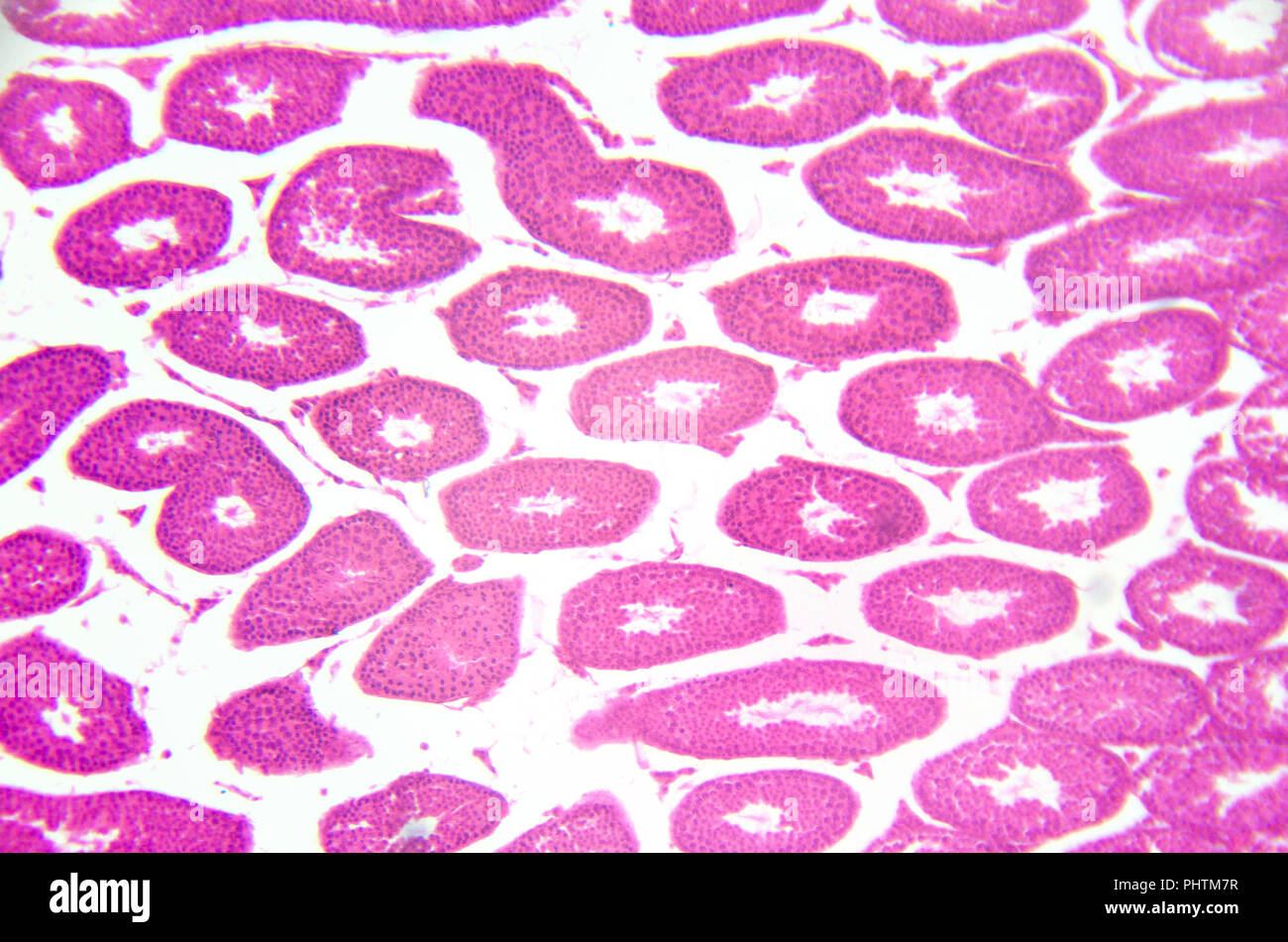 Microscopy photography. Testis, seminiferous tubules, cross section. Stock Photohttps://www.alamy.com/image-license-details/?v=1https://www.alamy.com/microscopy-photography-testis-seminiferous-tubules-cross-section-image217516315.html
RFPHTM7R–Microscopy photography. Testis, seminiferous tubules, cross section.
 Kate Winslet attending Mario Testi's 'Todo o Nada' photography exhibition opening at the Thyssen-Bornemisza Museum in Madrid Stock Photohttps://www.alamy.com/image-license-details/?v=1https://www.alamy.com/stock-photo-kate-winslet-attending-mario-testis-todo-o-nada-photography-exhibition-52002996.html
RMD0GX9T–Kate Winslet attending Mario Testi's 'Todo o Nada' photography exhibition opening at the Thyssen-Bornemisza Museum in Madrid
 Phareodus Testis.Dinòpolis. Paleontological Museum of Teruel, Paleontological Foundation of Teruel. Aragon. spain. Stock Photohttps://www.alamy.com/image-license-details/?v=1https://www.alamy.com/phareodus-testisdinpolis-paleontological-museum-of-teruel-paleontological-foundation-of-teruel-aragon-spain-image620324543.html
RM2Y1667Y–Phareodus Testis.Dinòpolis. Paleontological Museum of Teruel, Paleontological Foundation of Teruel. Aragon. spain.
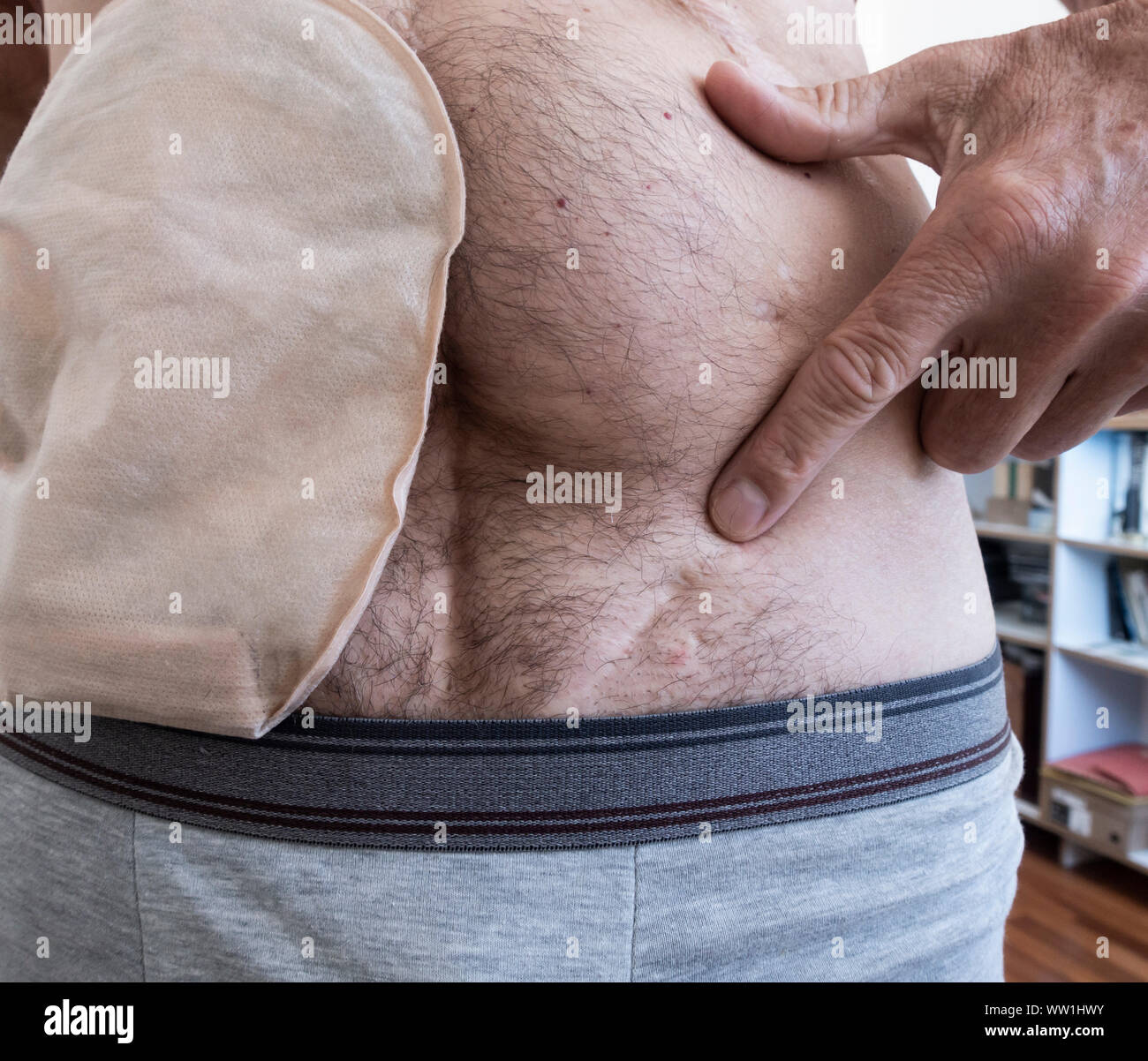 60 year old male wearing ileostomy bag points to scar on left lower abdomen, the result of an operation to remove a testicle (orchiectomy) in 1979 Stock Photohttps://www.alamy.com/image-license-details/?v=1https://www.alamy.com/60-year-old-male-wearing-ileostomy-bag-points-to-scar-on-left-lower-abdomen-the-result-of-an-operation-to-remove-a-testicle-orchiectomy-in-1979-image273557927.html
RMWW1HWY–60 year old male wearing ileostomy bag points to scar on left lower abdomen, the result of an operation to remove a testicle (orchiectomy) in 1979
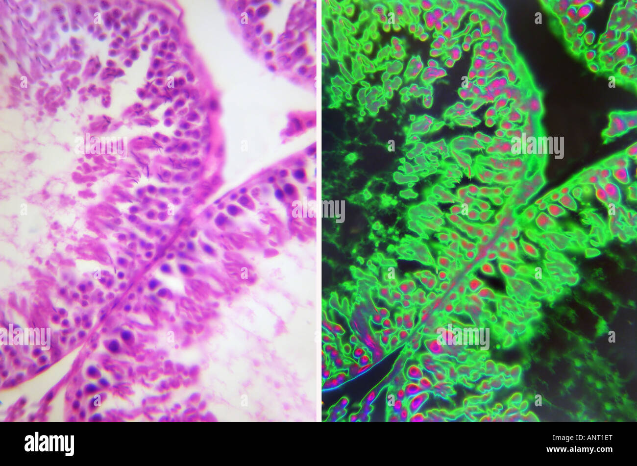 Testis tissue fe013 Stock Photohttps://www.alamy.com/image-license-details/?v=1https://www.alamy.com/stock-photo-testis-tissue-fe013-15592703.html
RFANT1ET–Testis tissue fe013
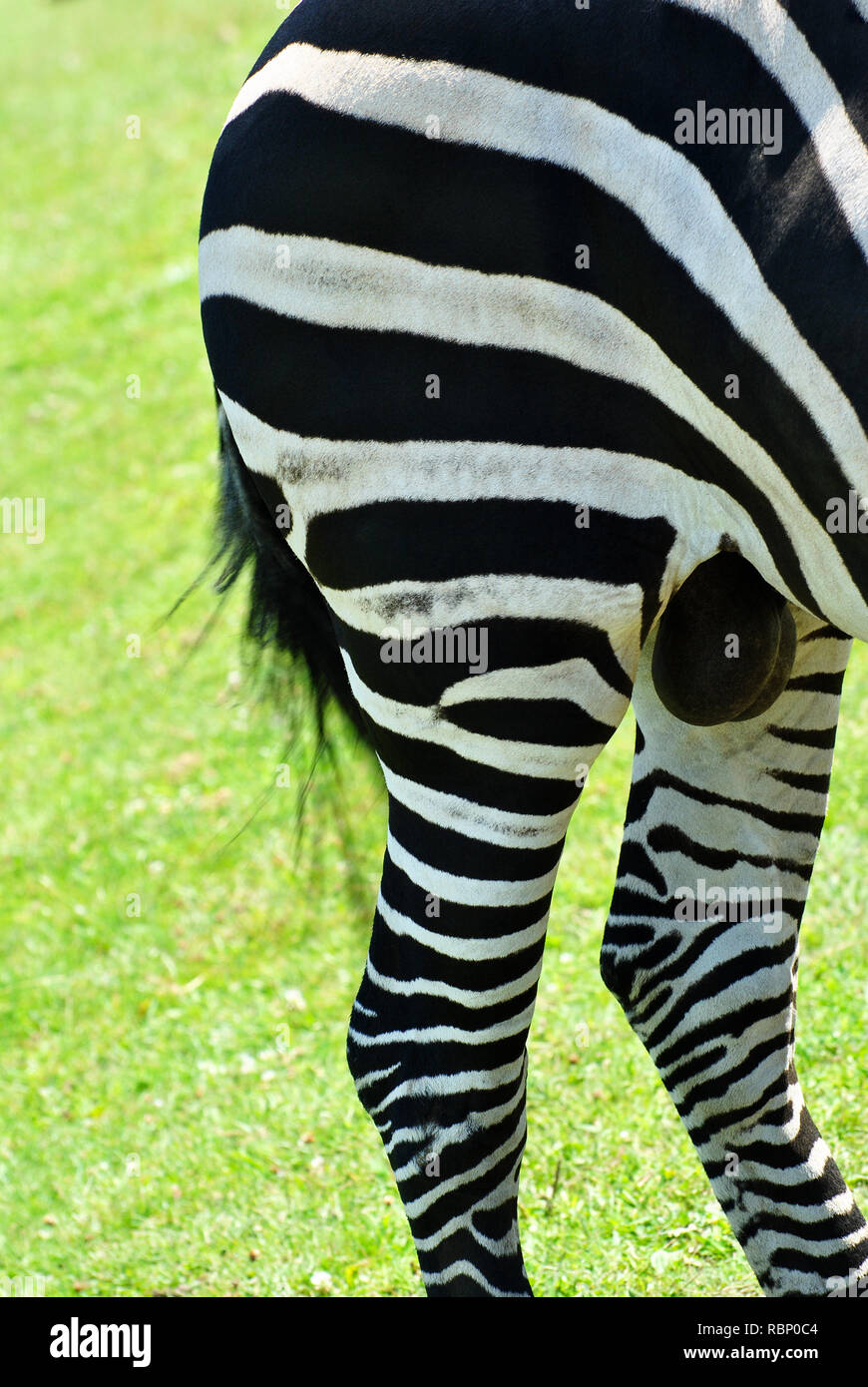 Close Up of the Hind End of a Wild Zebra Stock Photohttps://www.alamy.com/image-license-details/?v=1https://www.alamy.com/close-up-of-the-hind-end-of-a-wild-zebra-image230979284.html
RFRBP0C4–Close Up of the Hind End of a Wild Zebra
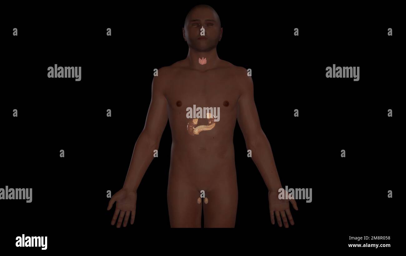 Anterior view of male endocrine system,3D rendering Stock Photohttps://www.alamy.com/image-license-details/?v=1https://www.alamy.com/anterior-view-of-male-endocrine-system3d-rendering-image504522964.html
RF2M8R058–Anterior view of male endocrine system,3D rendering
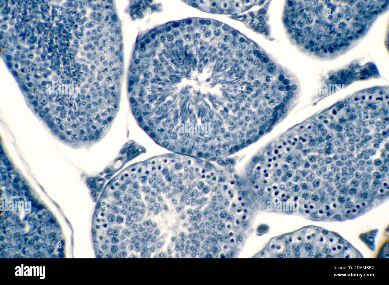 Human testis, light micrograph. Seen here are spermatogonia, spermatocytes undergoing meiosis, spermatids, and spermatozoa. Stock Photohttps://www.alamy.com/image-license-details/?v=1https://www.alamy.com/human-testis-light-micrograph-seen-here-are-spermatogonia-spermatocytes-undergoing-meiosis-spermatids-and-spermatozoa-image385222630.html
RF2DAMBB2–Human testis, light micrograph. Seen here are spermatogonia, spermatocytes undergoing meiosis, spermatids, and spermatozoa.
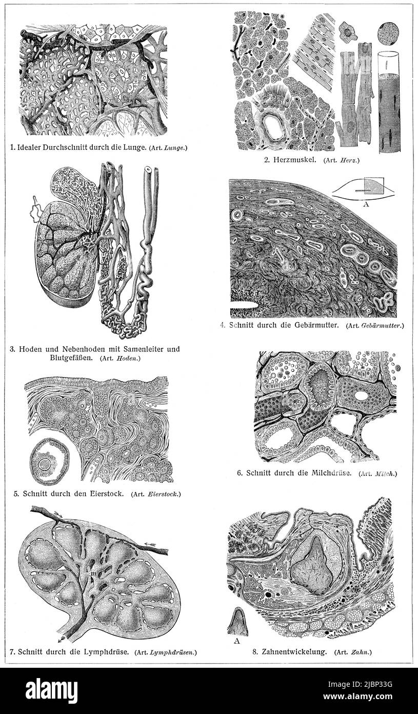 Cross section of human internal organs. Publication of the book 'Meyers Konversations-Lexikon', Volume 2, Leipzig, Germany, 1910 Stock Photohttps://www.alamy.com/image-license-details/?v=1https://www.alamy.com/cross-section-of-human-internal-organs-publication-of-the-book-meyers-konversations-lexikon-volume-2-leipzig-germany-1910-image471926548.html
RF2JBP33G–Cross section of human internal organs. Publication of the book 'Meyers Konversations-Lexikon', Volume 2, Leipzig, Germany, 1910
 Architectural detail on pillar outside house, Clareville Street, Kensington, London, UK, dated 1884 with Sol Mea Testis (Let the sun be wi Stock Photohttps://www.alamy.com/image-license-details/?v=1https://www.alamy.com/architectural-detail-on-pillar-outside-house-clareville-street-kensington-london-uk-dated-1884-with-sol-mea-testis-let-the-sun-be-wi-image431042671.html
RM2G17K93–Architectural detail on pillar outside house, Clareville Street, Kensington, London, UK, dated 1884 with Sol Mea Testis (Let the sun be wi
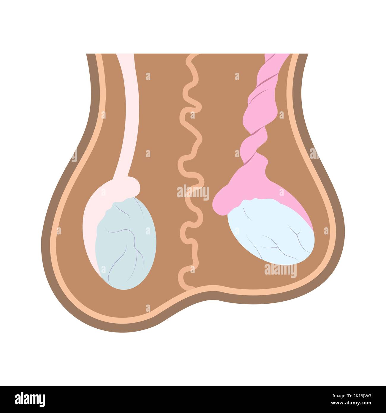 Illustration of normal testicle and testicle torsion in scrotum. Medical chart of reproductive organ anatomy. Stock Vectorhttps://www.alamy.com/image-license-details/?v=1https://www.alamy.com/illustration-of-normal-testicle-and-testicle-torsion-in-scrotum-medical-chart-of-reproductive-organ-anatomy-image482695404.html
RF2K18JWG–Illustration of normal testicle and testicle torsion in scrotum. Medical chart of reproductive organ anatomy.
 Portrait of ram Stock Photohttps://www.alamy.com/image-license-details/?v=1https://www.alamy.com/portrait-of-ram-image242400506.html
RFT2A88X–Portrait of ram
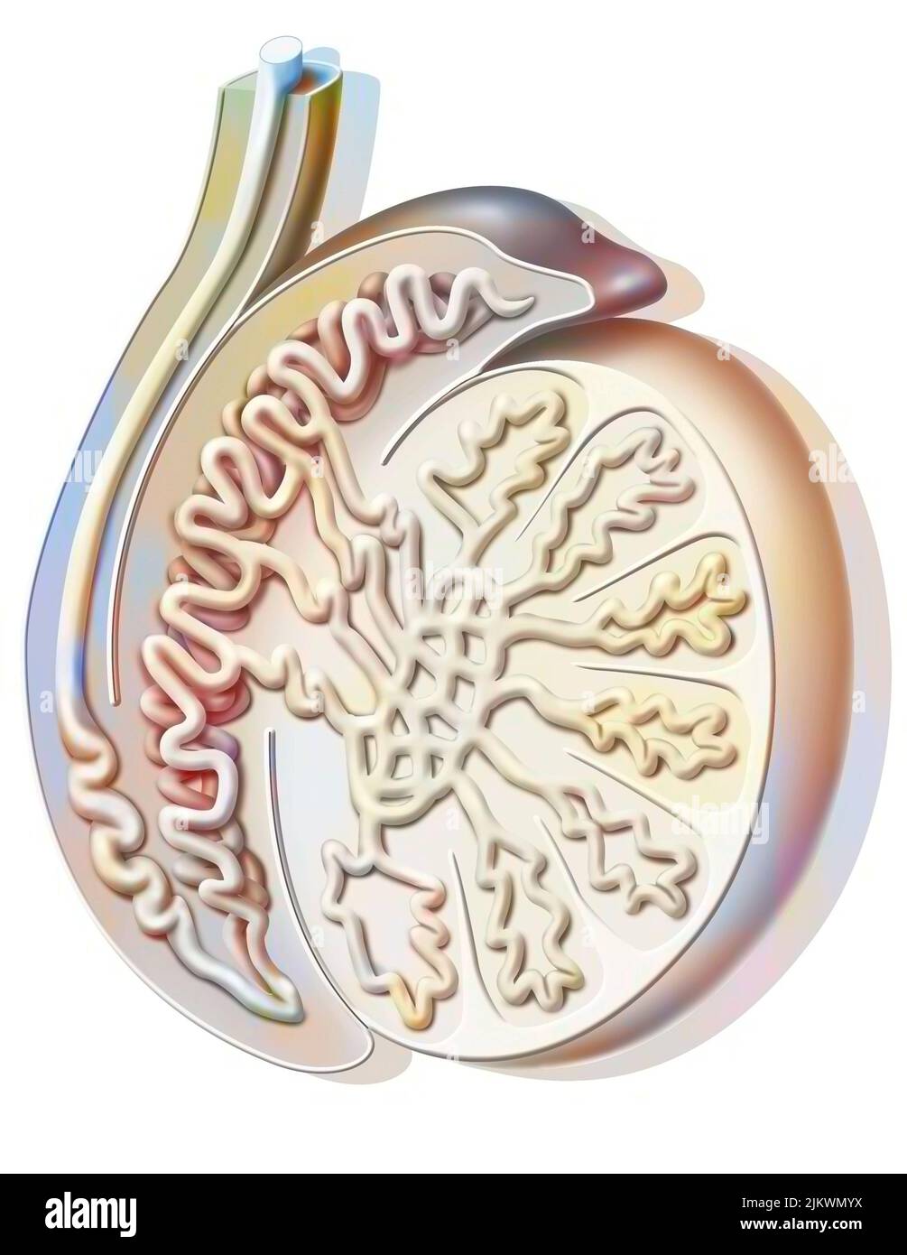 Sagittal section of a testicle showing the seminiferous tube. Stock Photohttps://www.alamy.com/image-license-details/?v=1https://www.alamy.com/sagittal-section-of-a-testicle-showing-the-seminiferous-tube-image476923662.html
RF2JKWMYX–Sagittal section of a testicle showing the seminiferous tube.
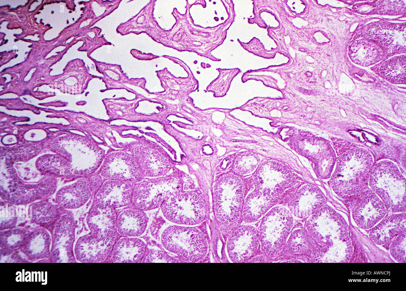 Testis Stock Photohttps://www.alamy.com/image-license-details/?v=1https://www.alamy.com/stock-photo-testis-16621961.html
RFAWNCPJ–Testis
 Concept of right sided scrotal inflammation or testicular pain. Sad or crying facial expression. With text space. Isolated on white. Stock Photohttps://www.alamy.com/image-license-details/?v=1https://www.alamy.com/concept-of-right-sided-scrotal-inflammation-or-testicular-pain-sad-or-crying-facial-expression-with-text-space-isolated-on-white-image436916555.html
RF2GAR7EK–Concept of right sided scrotal inflammation or testicular pain. Sad or crying facial expression. With text space. Isolated on white.
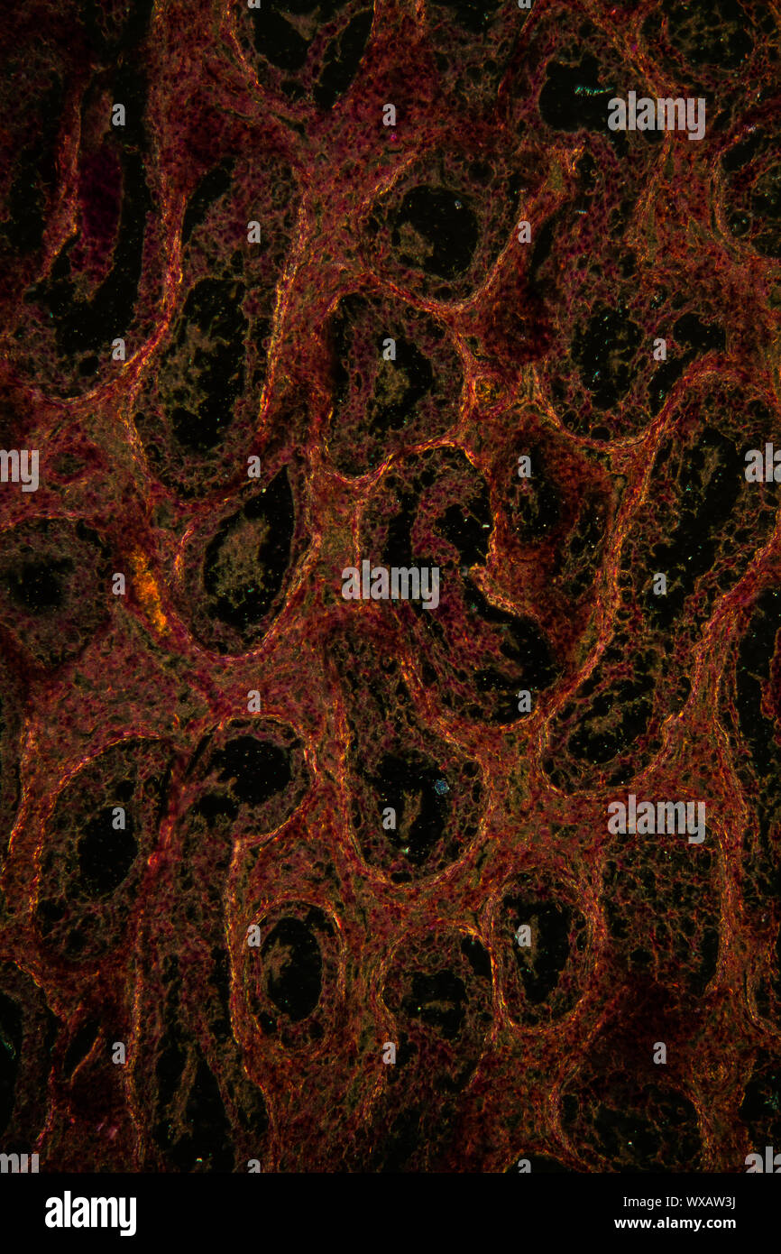 Inguinal testis thin section under the microscope 100x Stock Photohttps://www.alamy.com/image-license-details/?v=1https://www.alamy.com/inguinal-testis-thin-section-under-the-microscope-100x-image274375798.html
RMWXAW3J–Inguinal testis thin section under the microscope 100x
 meiosis in Locust testis transverse section, under a light microscope at 10 times magnification. Stock Photohttps://www.alamy.com/image-license-details/?v=1https://www.alamy.com/meiosis-in-locust-testis-transverse-section-under-a-light-microscope-at-10-times-magnification-image415796706.html
RF2F4D4XA–meiosis in Locust testis transverse section, under a light microscope at 10 times magnification.
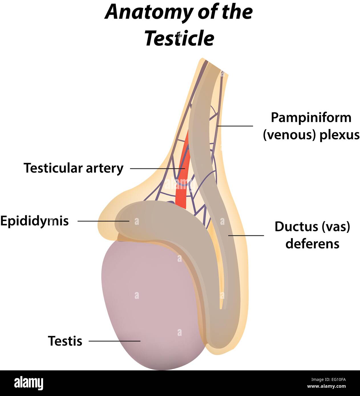 Anatomy of the Testicle Stock Vectorhttps://www.alamy.com/image-license-details/?v=1https://www.alamy.com/stock-photo-anatomy-of-the-testicle-78698350.html
RFEG10FA–Anatomy of the Testicle
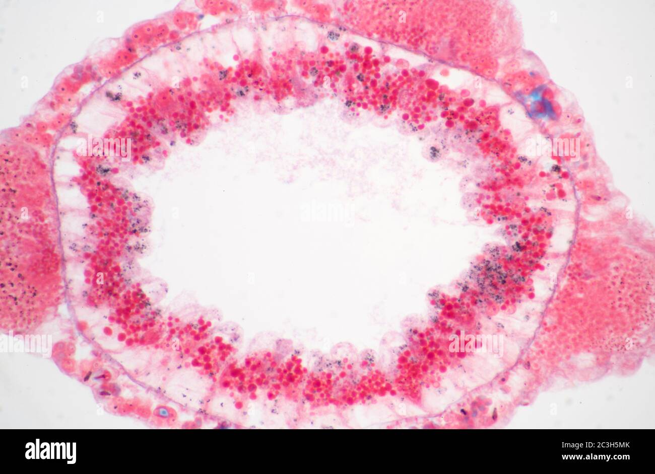 Hydra Testis, transverse section Stock Photohttps://www.alamy.com/image-license-details/?v=1https://www.alamy.com/hydra-testis-transverse-section-image363639379.html
RM2C3H5MK–Hydra Testis, transverse section
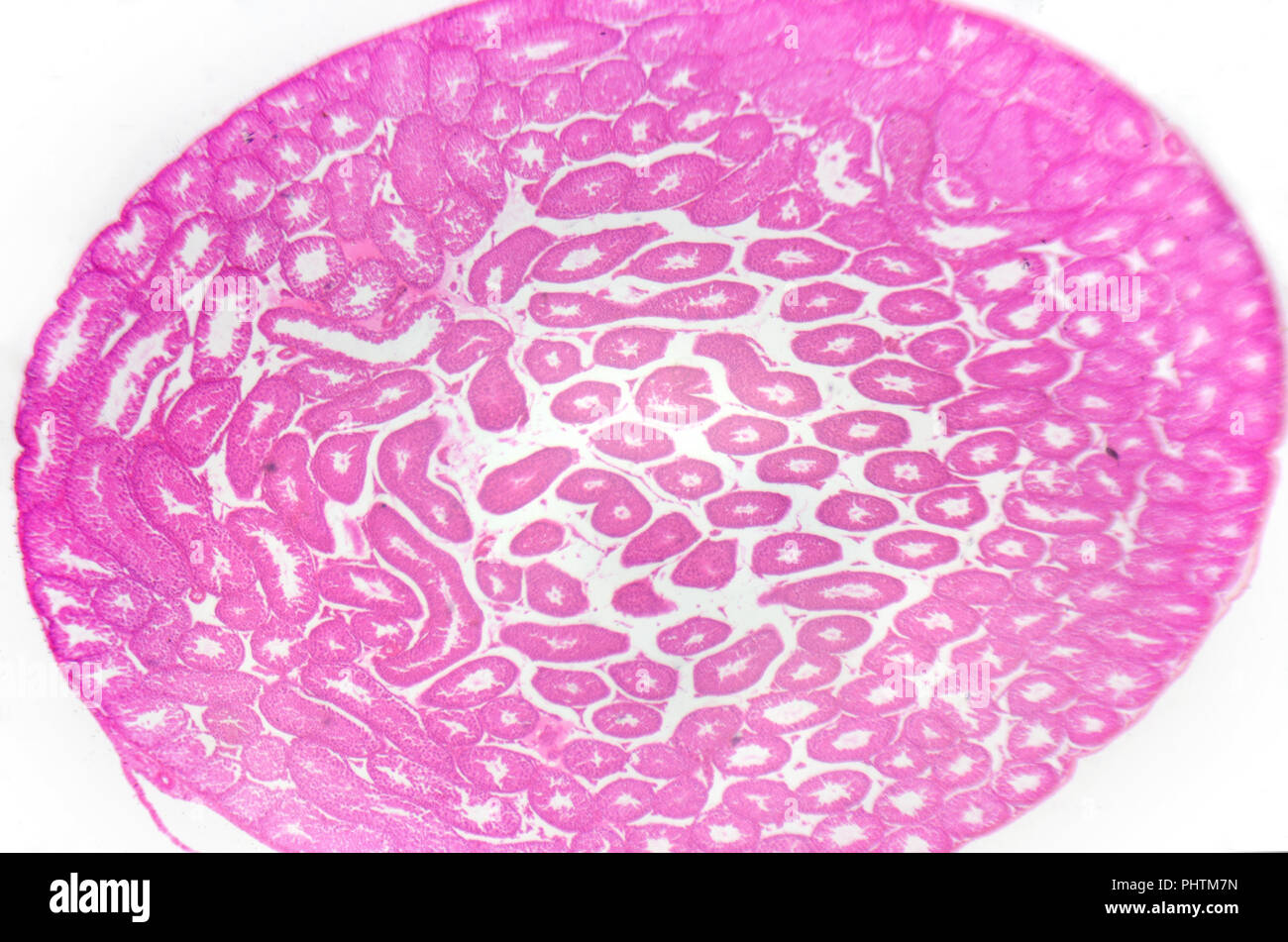 Microscopy photography. Testis, seminiferous tubules, cross section. Stock Photohttps://www.alamy.com/image-license-details/?v=1https://www.alamy.com/microscopy-photography-testis-seminiferous-tubules-cross-section-image217516313.html
RFPHTM7N–Microscopy photography. Testis, seminiferous tubules, cross section.
 Clusterin: Immunoperoxidase staining of formalin-fixed, paraffin-embedded mouse male reproductive tract showing cytoplasm and cell membranous staining in the Sertoli cells of the testis, as well as cells in the epididymis and prostate Stock Photohttps://www.alamy.com/image-license-details/?v=1https://www.alamy.com/stock-photo-clusterin-immunoperoxidase-staining-of-formalin-fixed-paraffin-embedded-84969861.html
RMEX6KWW–Clusterin: Immunoperoxidase staining of formalin-fixed, paraffin-embedded mouse male reproductive tract showing cytoplasm and cell membranous staining in the Sertoli cells of the testis, as well as cells in the epididymis and prostate
 Section of the Testis of a Dog showing portions of seminal tube vintage line drawing or engraving illustration. Stock Vectorhttps://www.alamy.com/image-license-details/?v=1https://www.alamy.com/section-of-the-testis-of-a-dog-showing-portions-of-seminal-tube-vintage-line-drawing-or-engraving-illustration-image244563802.html
RFT5TRHE–Section of the Testis of a Dog showing portions of seminal tube vintage line drawing or engraving illustration.
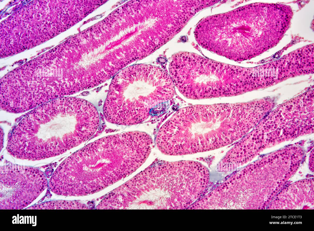 Human testicle or testis section showing seminiferous tubules, Leydig cells, Sertoli cells, spermatocytes, spermatogonia, spermatides and spermatozoon Stock Photohttps://www.alamy.com/image-license-details/?v=1https://www.alamy.com/human-testicle-or-testis-section-showing-seminiferous-tubules-leydig-cells-sertoli-cells-spermatocytes-spermatogonia-spermatides-and-spermatozoon-image575626803.html
RF2TCE1T3–Human testicle or testis section showing seminiferous tubules, Leydig cells, Sertoli cells, spermatocytes, spermatogonia, spermatides and spermatozoon
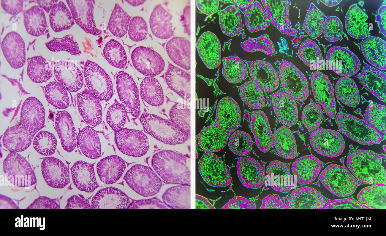 Low power view of testis tissue Stock Photohttps://www.alamy.com/image-license-details/?v=1https://www.alamy.com/stock-photo-low-power-view-of-testis-tissue-15592747.html
RFANT1JM–Low power view of testis tissue
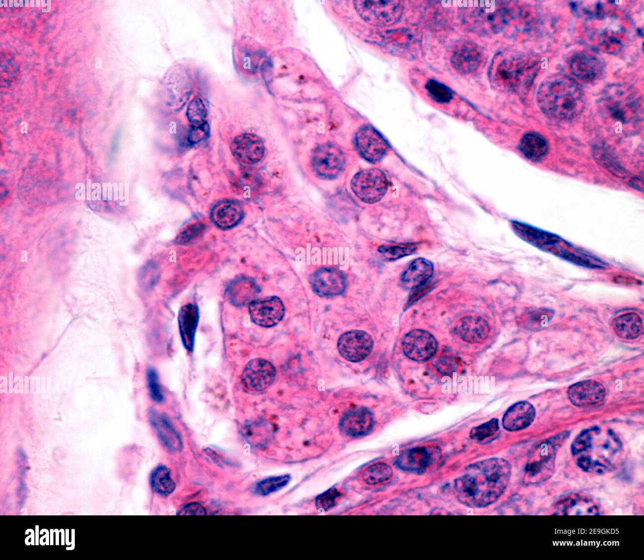 High magnification micrograph of a group of interstitial Leydig cells, in the testis. They are polyhedral cells of eosinophilic cytoplasm that show pi Stock Photohttps://www.alamy.com/image-license-details/?v=1https://www.alamy.com/high-magnification-micrograph-of-a-group-of-interstitial-leydig-cells-in-the-testis-they-are-polyhedral-cells-of-eosinophilic-cytoplasm-that-show-pi-image401736865.html
RF2E9GKD5–High magnification micrograph of a group of interstitial Leydig cells, in the testis. They are polyhedral cells of eosinophilic cytoplasm that show pi
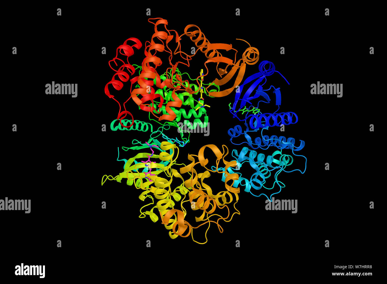 Phosphorylase b kinase gamma catalytic chain, testis/liver isoform, an enzyme that in humans is encoded by the PHKG2 gene. 3d rendering. Stock Photohttps://www.alamy.com/image-license-details/?v=1https://www.alamy.com/phosphorylase-b-kinase-gamma-catalytic-chain-testisliver-isoform-an-enzyme-that-in-humans-is-encoded-by-the-phkg2-gene-3d-rendering-image262849980.html
RFW7HRR8–Phosphorylase b kinase gamma catalytic chain, testis/liver isoform, an enzyme that in humans is encoded by the PHKG2 gene. 3d rendering.
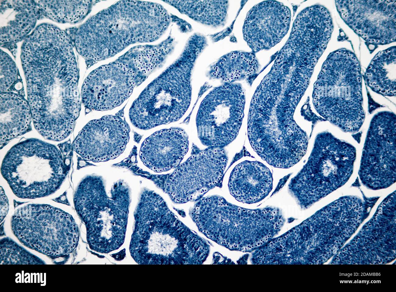 Human testis, light micrograph. Seen here are spermatogonia, spermatocytes undergoing meiosis, spermatids, and spermatozoa. Stock Photohttps://www.alamy.com/image-license-details/?v=1https://www.alamy.com/human-testis-light-micrograph-seen-here-are-spermatogonia-spermatocytes-undergoing-meiosis-spermatids-and-spermatozoa-image385222634.html
RF2DAMBB6–Human testis, light micrograph. Seen here are spermatogonia, spermatocytes undergoing meiosis, spermatids, and spermatozoa.
 Semi-schematic representation of the male urinary and reproductive organs. Illustration of the 19th century. Germany. White background. Stock Photohttps://www.alamy.com/image-license-details/?v=1https://www.alamy.com/semi-schematic-representation-of-the-male-urinary-and-reproductive-organs-illustration-of-the-19th-century-germany-white-background-image419115594.html
RF2F9TA62–Semi-schematic representation of the male urinary and reproductive organs. Illustration of the 19th century. Germany. White background.
 salamander testis Stock Photohttps://www.alamy.com/image-license-details/?v=1https://www.alamy.com/salamander-testis-image225718393.html
RFR36A35–salamander testis
 Phareodus Testis.Dinòpolis. Paleontological Museum of Teruel, Paleontological Foundation of Teruel. Aragon. spain. Stock Photohttps://www.alamy.com/image-license-details/?v=1https://www.alamy.com/phareodus-testisdinpolis-paleontological-museum-of-teruel-paleontological-foundation-of-teruel-aragon-spain-image619877234.html
RM2Y0DRMJ–Phareodus Testis.Dinòpolis. Paleontological Museum of Teruel, Paleontological Foundation of Teruel. Aragon. spain.
 Portrait of ram Stock Photohttps://www.alamy.com/image-license-details/?v=1https://www.alamy.com/portrait-of-ram-image248238300.html
RFTBT6DG–Portrait of ram
 Фemale African medical students, young graduates in research medical test laboratory perform various testis on patient samples. Microscopic analysis b Stock Photohttps://www.alamy.com/image-license-details/?v=1https://www.alamy.com/emale-african-medical-students-young-graduates-in-research-medical-test-laboratory-perform-various-testis-on-patient-samples-microscopic-analysis-b-image350214740.html
RF2B9NJD8–Фemale African medical students, young graduates in research medical test laboratory perform various testis on patient samples. Microscopic analysis b
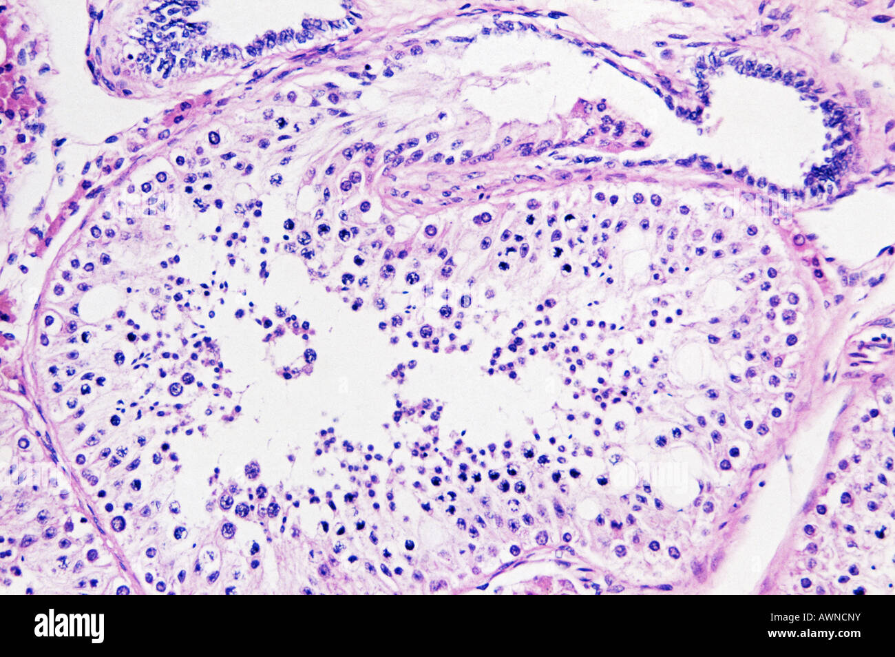 Testis Stock Photohttps://www.alamy.com/image-license-details/?v=1https://www.alamy.com/stock-photo-testis-16621958.html
RFAWNCNY–Testis
 Concept of right sided scrotal swelling or testicular pain. Comparing standard chicken eggs and bantam eggs. With text space. Isolated on white. Stock Photohttps://www.alamy.com/image-license-details/?v=1https://www.alamy.com/concept-of-right-sided-scrotal-swelling-or-testicular-pain-comparing-standard-chicken-eggs-and-bantam-eggs-with-text-space-isolated-on-white-image436916550.html
RF2GAR7EE–Concept of right sided scrotal swelling or testicular pain. Comparing standard chicken eggs and bantam eggs. With text space. Isolated on white.
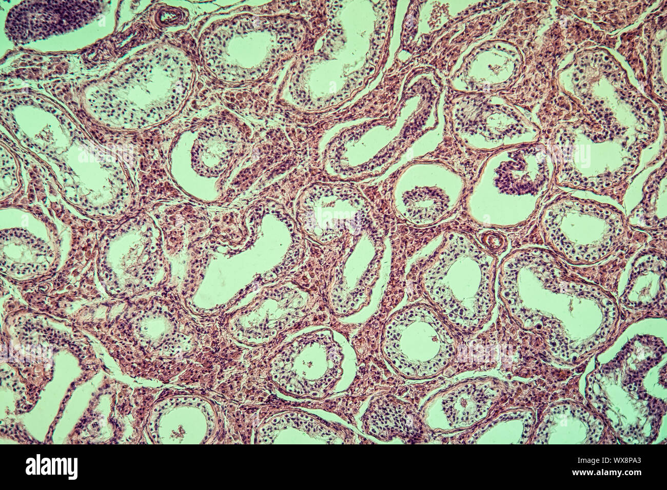 Inguinal testis diseased tissue 100x Stock Photohttps://www.alamy.com/image-license-details/?v=1https://www.alamy.com/inguinal-testis-diseased-tissue-100x-image274329723.html
RMWX8PA3–Inguinal testis diseased tissue 100x
 Lans gekleed met een hemd / Graanhalm met vaatwerk; Restat Ex Victore Orientis / Victoria Testis; Symbola Divina et Humana Pontificum Imperatorum Regum.An emblem with two performances. Above a landscape with a lance dressed in a shirt. Under a landscape with one grain hall from which a Christian crockery grows. The inventions of Sultan Saladin and Bayezid II concern. Stock Photohttps://www.alamy.com/image-license-details/?v=1https://www.alamy.com/lans-gekleed-met-een-hemd-graanhalm-met-vaatwerk-restat-ex-victore-orientis-victoria-testis-symbola-divina-et-humana-pontificum-imperatorum-reguman-emblem-with-two-performances-above-a-landscape-with-a-lance-dressed-in-a-shirt-under-a-landscape-with-one-grain-hall-from-which-a-christian-crockery-grows-the-inventions-of-sultan-saladin-and-bayezid-ii-concern-image414454048.html
RM2F280A8–Lans gekleed met een hemd / Graanhalm met vaatwerk; Restat Ex Victore Orientis / Victoria Testis; Symbola Divina et Humana Pontificum Imperatorum Regum.An emblem with two performances. Above a landscape with a lance dressed in a shirt. Under a landscape with one grain hall from which a Christian crockery grows. The inventions of Sultan Saladin and Bayezid II concern.
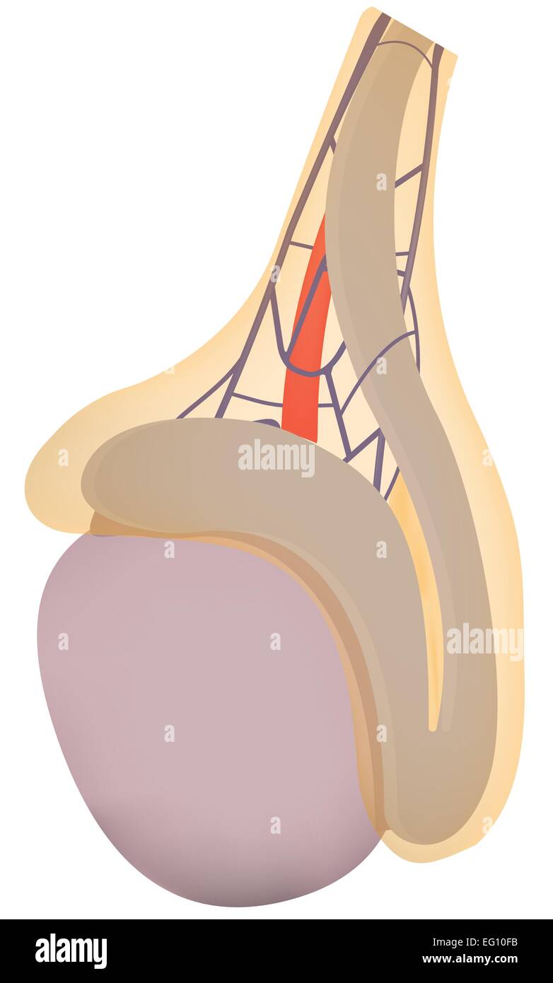 Testicle Stock Vectorhttps://www.alamy.com/image-license-details/?v=1https://www.alamy.com/stock-photo-testicle-78698351.html
RFEG10FB–Testicle
 Phareodus testis fish fossil Stock Photohttps://www.alamy.com/image-license-details/?v=1https://www.alamy.com/stock-photo-phareodus-testis-fish-fossil-32204146.html
RMBTB0M2–Phareodus testis fish fossil
 Daria Werbowy Mario Testi and Kate Winslet attending Mario Testi's 'Todo o Nada' photography exhibition opening at the Stock Photohttps://www.alamy.com/image-license-details/?v=1https://www.alamy.com/stock-photo-daria-werbowy-mario-testi-and-kate-winslet-attending-mario-testis-52003043.html
RMD0GXBF–Daria Werbowy Mario Testi and Kate Winslet attending Mario Testi's 'Todo o Nada' photography exhibition opening at the
 Epididymis of testis Semithin section Stained Brightfield HFW 200um Stock Photohttps://www.alamy.com/image-license-details/?v=1https://www.alamy.com/epididymis-of-testis-semithin-section-stained-brightfield-hfw-200um-image769715.html
RMABBEB3–Epididymis of testis Semithin section Stained Brightfield HFW 200um
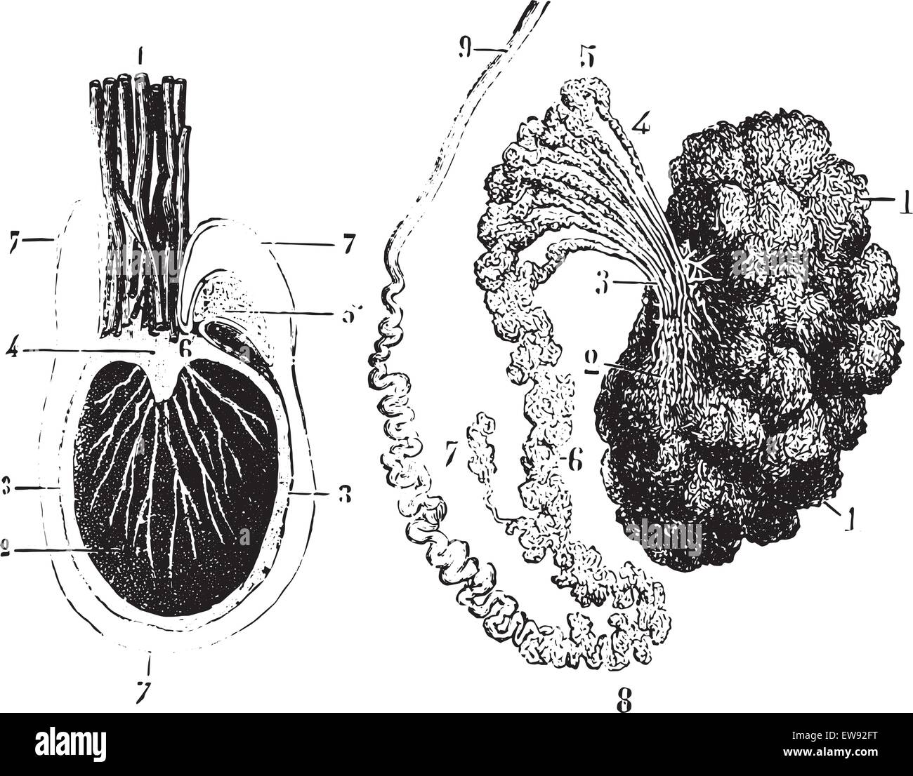 Cross section of the testis, epididymis and tunica vaginalis, vintage engraved illustration. Usual Medicine Dictionary by Dr Lab Stock Vectorhttps://www.alamy.com/image-license-details/?v=1https://www.alamy.com/stock-photo-cross-section-of-the-testis-epididymis-and-tunica-vaginalis-vintage-84407452.html
RFEW92FT–Cross section of the testis, epididymis and tunica vaginalis, vintage engraved illustration. Usual Medicine Dictionary by Dr Lab
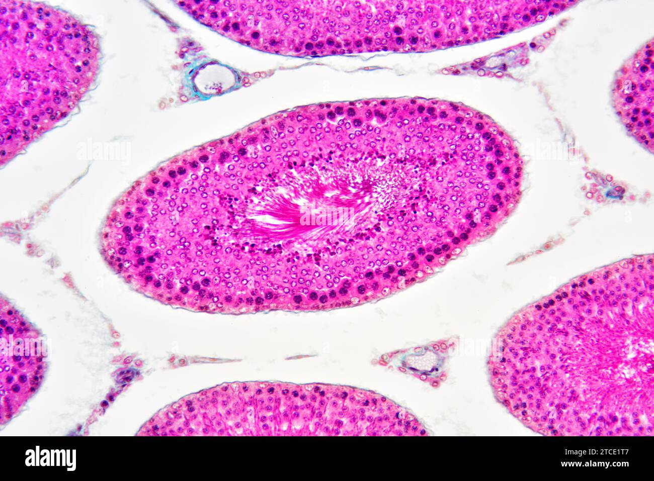 Human testicle or testis section showing seminiferous tubules, Leydig cells, Sertoli cells, spermatocytes, spermatogonia, spermatides and spermatozoon Stock Photohttps://www.alamy.com/image-license-details/?v=1https://www.alamy.com/human-testicle-or-testis-section-showing-seminiferous-tubules-leydig-cells-sertoli-cells-spermatocytes-spermatogonia-spermatides-and-spermatozoon-image575626807.html
RF2TCE1T7–Human testicle or testis section showing seminiferous tubules, Leydig cells, Sertoli cells, spermatocytes, spermatogonia, spermatides and spermatozoon
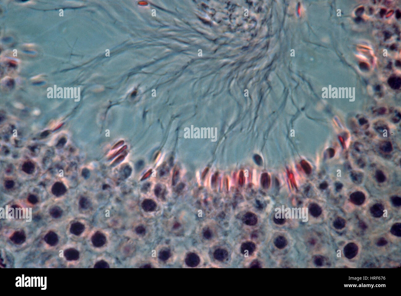 Monkey Testis (LM) Stock Photohttps://www.alamy.com/image-license-details/?v=1https://www.alamy.com/stock-photo-monkey-testis-lm-134943850.html
RMHRF676–Monkey Testis (LM)
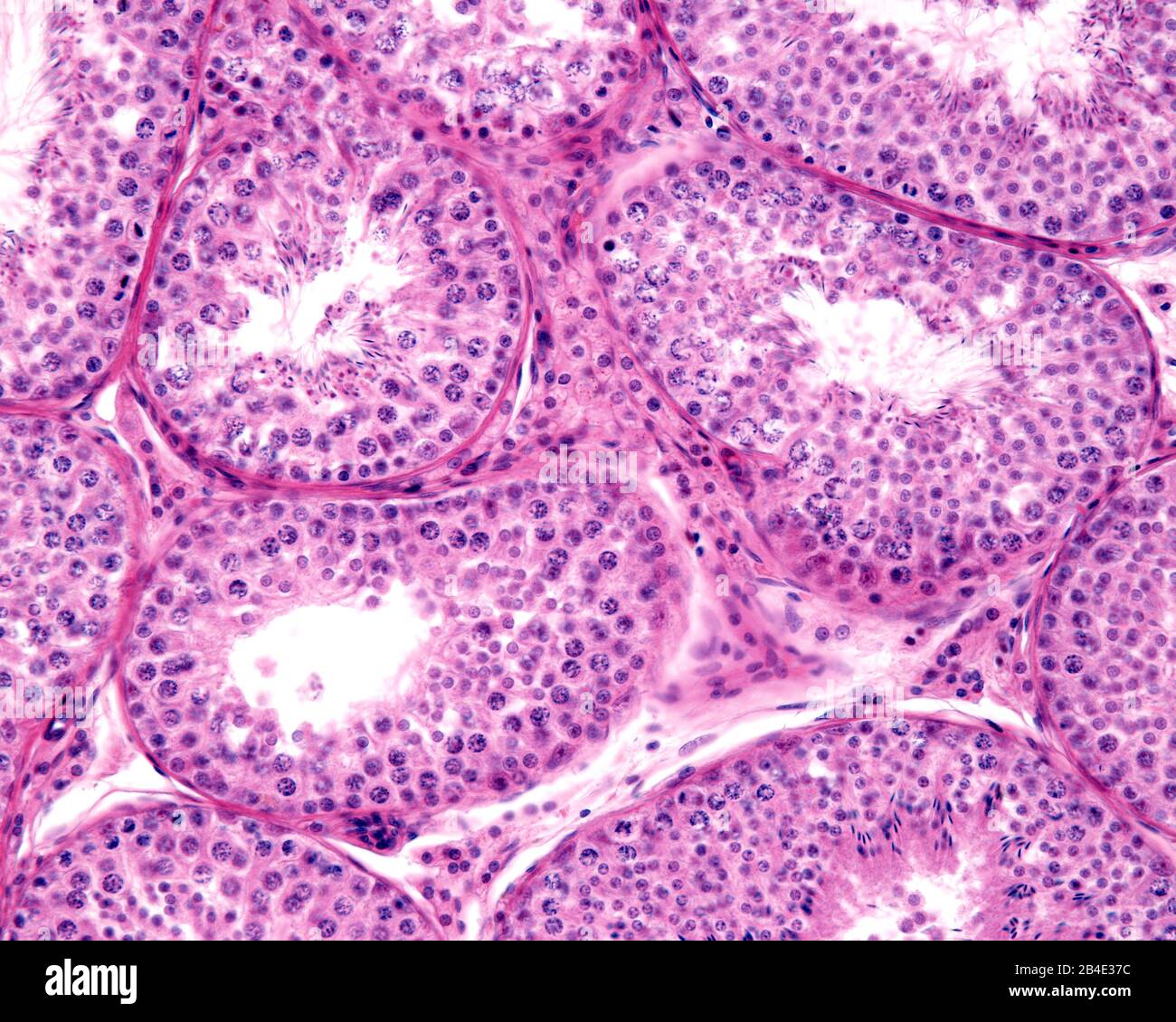 Seminiferous tubules of human testis. Male germinal epithelium shows spermatogonia, spermatocytes in meiosis, spermatids, and spermatozoa with tails p Stock Photohttps://www.alamy.com/image-license-details/?v=1https://www.alamy.com/seminiferous-tubules-of-human-testis-male-germinal-epithelium-shows-spermatogonia-spermatocytes-in-meiosis-spermatids-and-spermatozoa-with-tails-p-image346975872.html
RF2B4E37C–Seminiferous tubules of human testis. Male germinal epithelium shows spermatogonia, spermatocytes in meiosis, spermatids, and spermatozoa with tails p
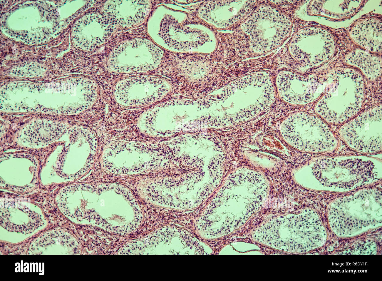 inguinal testis diseased tissue 100x Stock Photohttps://www.alamy.com/image-license-details/?v=1https://www.alamy.com/inguinal-testis-diseased-tissue-100x-image227729314.html
RFR6DY1P–inguinal testis diseased tissue 100x
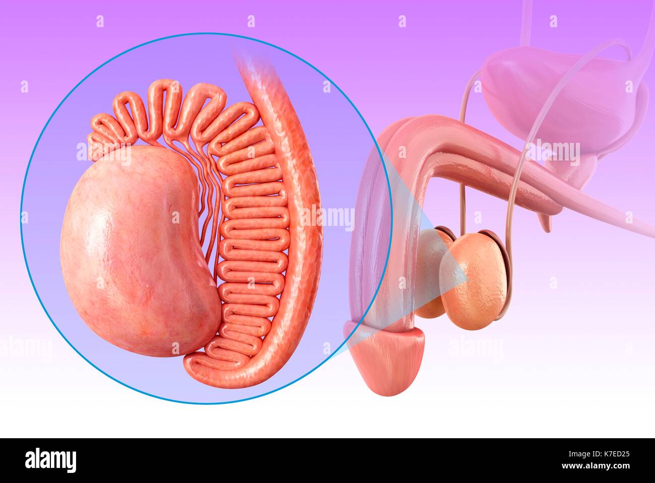 Illustration of male testis anatomy. Stock Photohttps://www.alamy.com/image-license-details/?v=1https://www.alamy.com/illustration-of-male-testis-anatomy-image159513485.html
RFK7ED25–Illustration of male testis anatomy.
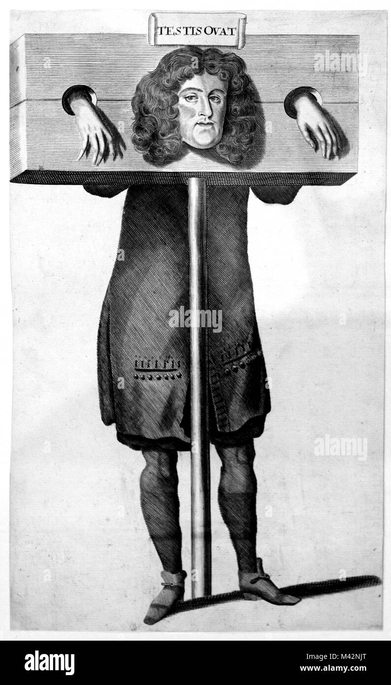 Titus Oates in the Pillory, after Robert White, c.1685. Engraving of Titus Oates (1649-1705), who fabricated the 'Popish Plot', a supposed Catholic conspiracy to kill King Charles II. Stock Photohttps://www.alamy.com/image-license-details/?v=1https://www.alamy.com/stock-photo-titus-oates-in-the-pillory-after-robert-white-c1685-engraving-of-titus-174623200.html
RMM42NJT–Titus Oates in the Pillory, after Robert White, c.1685. Engraving of Titus Oates (1649-1705), who fabricated the 'Popish Plot', a supposed Catholic conspiracy to kill King Charles II.
 Concept of Yes, Undescended Testicles: Do you know about it? write on a book isolated on Wooden Table. Stock Photohttps://www.alamy.com/image-license-details/?v=1https://www.alamy.com/concept-of-yes-undescended-testicles-do-you-know-about-it-write-on-a-book-isolated-on-wooden-table-image468421601.html
RF2J62CEW–Concept of Yes, Undescended Testicles: Do you know about it? write on a book isolated on Wooden Table.
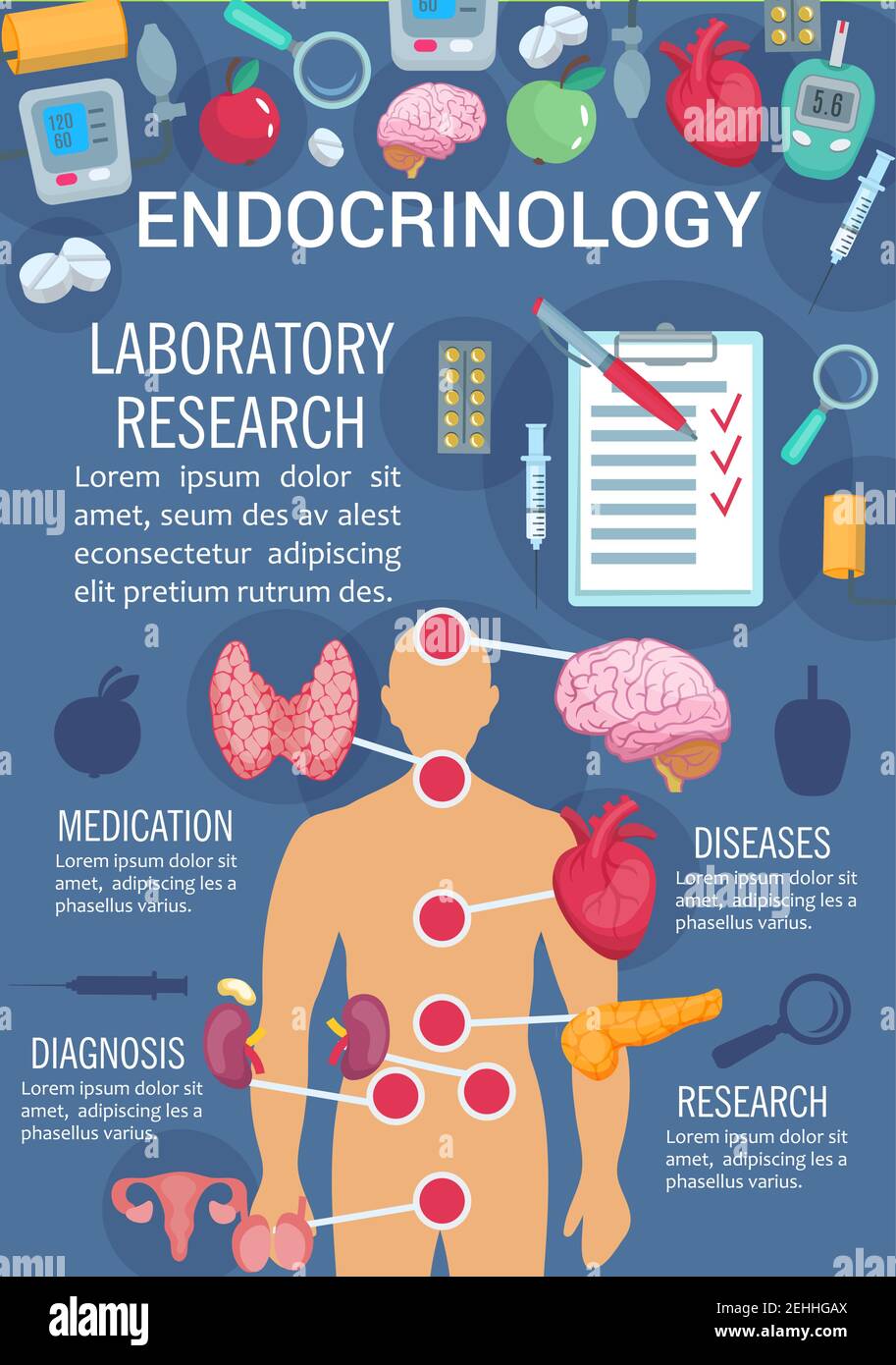 Endocrinology medicine poster of human endocrine system anatomy diagram. Thyroid gland, pancreas and hypothalamus, ovary, testis and thymus poster for Stock Vectorhttps://www.alamy.com/image-license-details/?v=1https://www.alamy.com/endocrinology-medicine-poster-of-human-endocrine-system-anatomy-diagram-thyroid-gland-pancreas-and-hypothalamus-ovary-testis-and-thymus-poster-for-image406673650.html
RF2EHHGAX–Endocrinology medicine poster of human endocrine system anatomy diagram. Thyroid gland, pancreas and hypothalamus, ovary, testis and thymus poster for
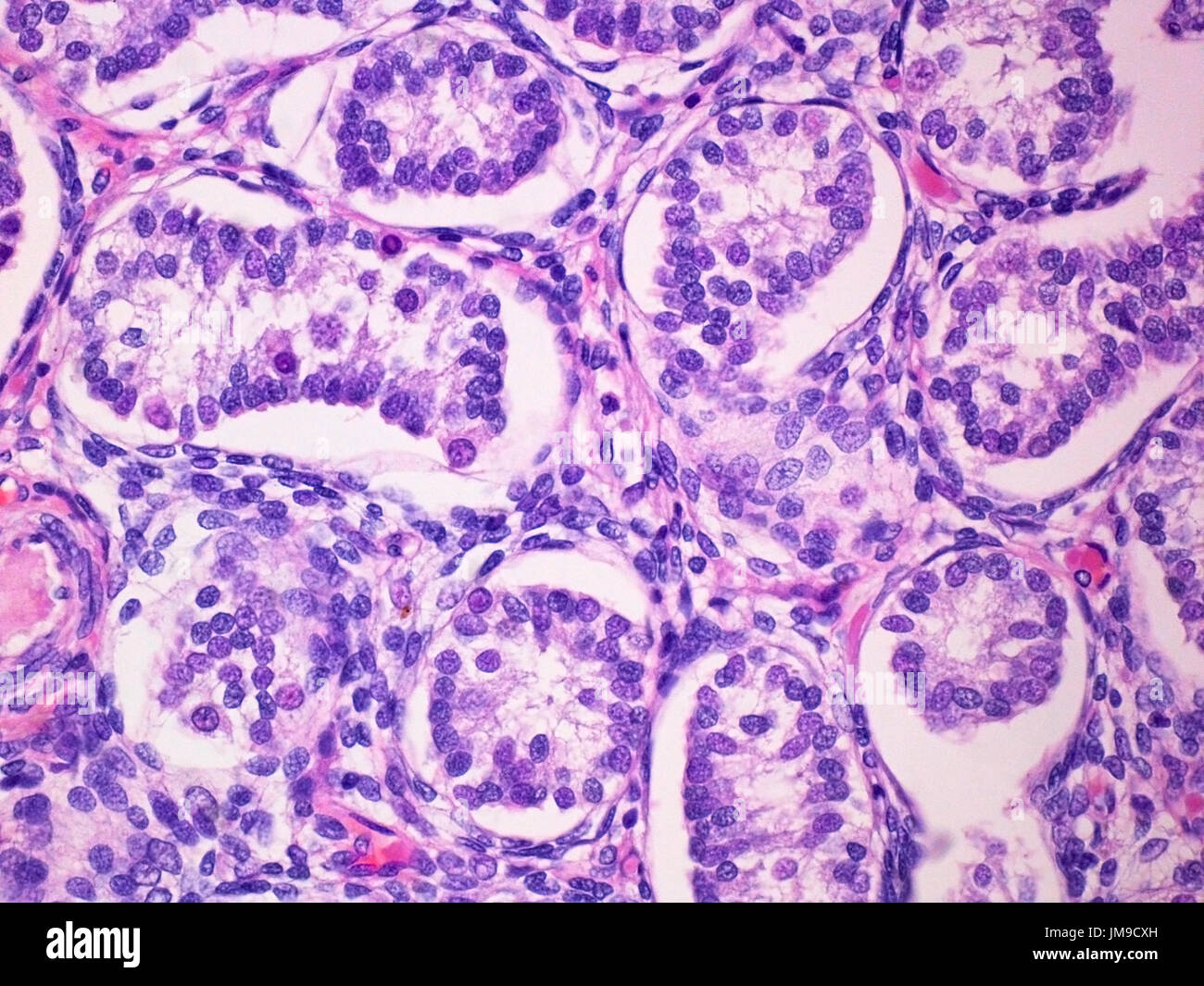 Seminiferous Tubules of a Prepubertal Male Child Testicle Viewed at 400x Magnification with Haemotoxylin and Eosin Staining. Stock Photohttps://www.alamy.com/image-license-details/?v=1https://www.alamy.com/seminiferous-tubules-of-a-prepubertal-male-child-testicle-viewed-at-image150183785.html
RFJM9CXH–Seminiferous Tubules of a Prepubertal Male Child Testicle Viewed at 400x Magnification with Haemotoxylin and Eosin Staining.
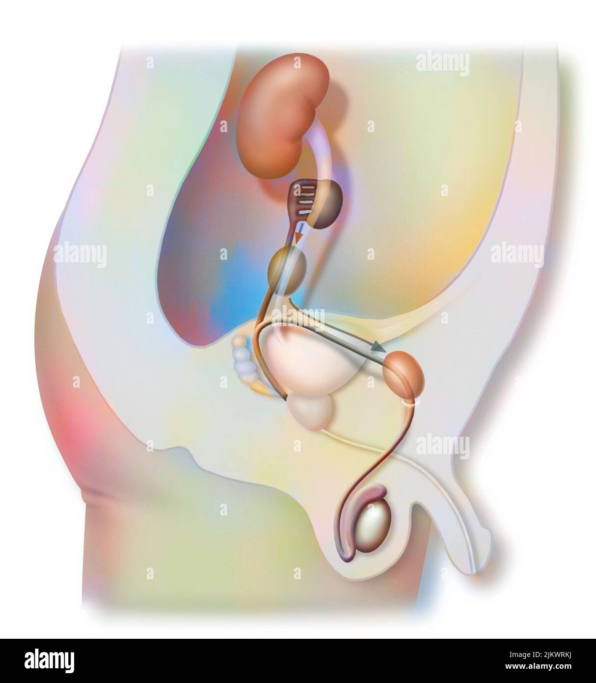 Descent of the testicles: they take the inguinal canal in order to lodge in the scrotal region. Stock Photohttps://www.alamy.com/image-license-details/?v=1https://www.alamy.com/descent-of-the-testicles-they-take-the-inguinal-canal-in-order-to-lodge-in-the-scrotal-region-image476925782.html
RF2JKWRKJ–Descent of the testicles: they take the inguinal canal in order to lodge in the scrotal region.
 Orchid 01 Stock Photohttps://www.alamy.com/image-license-details/?v=1https://www.alamy.com/orchid-01-image3544408.html
RMA3TN59–Orchid 01
 Concept of right sided scrotal swelling or testicular pain. With text space. Isolated on white. Stock Photohttps://www.alamy.com/image-license-details/?v=1https://www.alamy.com/concept-of-right-sided-scrotal-swelling-or-testicular-pain-with-text-space-isolated-on-white-image485346898.html
RF2K5HCWP–Concept of right sided scrotal swelling or testicular pain. With text space. Isolated on white.
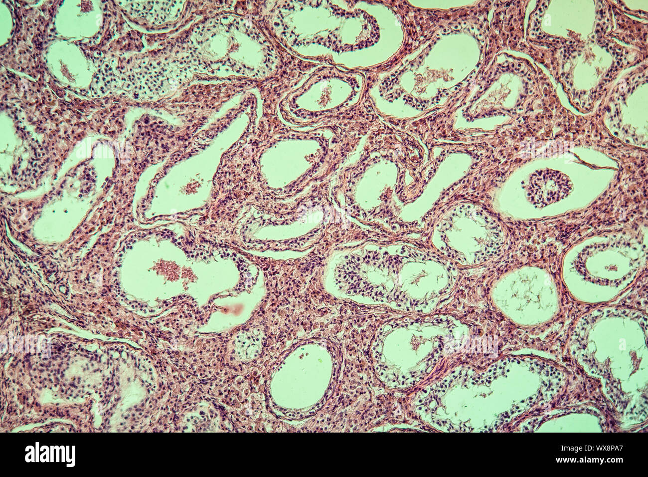 Inguinal testis diseased tissue 100x Stock Photohttps://www.alamy.com/image-license-details/?v=1https://www.alamy.com/inguinal-testis-diseased-tissue-100x-image274329727.html
RMWX8PA7–Inguinal testis diseased tissue 100x
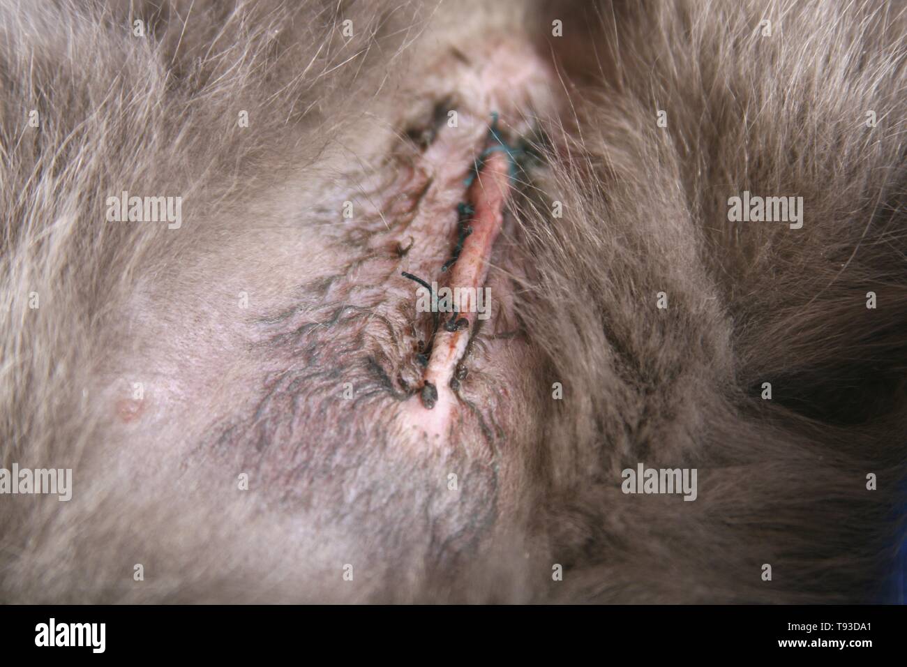 postoperative scar after neutering at undescended testicle Stock Photohttps://www.alamy.com/image-license-details/?v=1https://www.alamy.com/postoperative-scar-after-neutering-at-undescended-testicle-image246553385.html
RMT93DA1–postoperative scar after neutering at undescended testicle
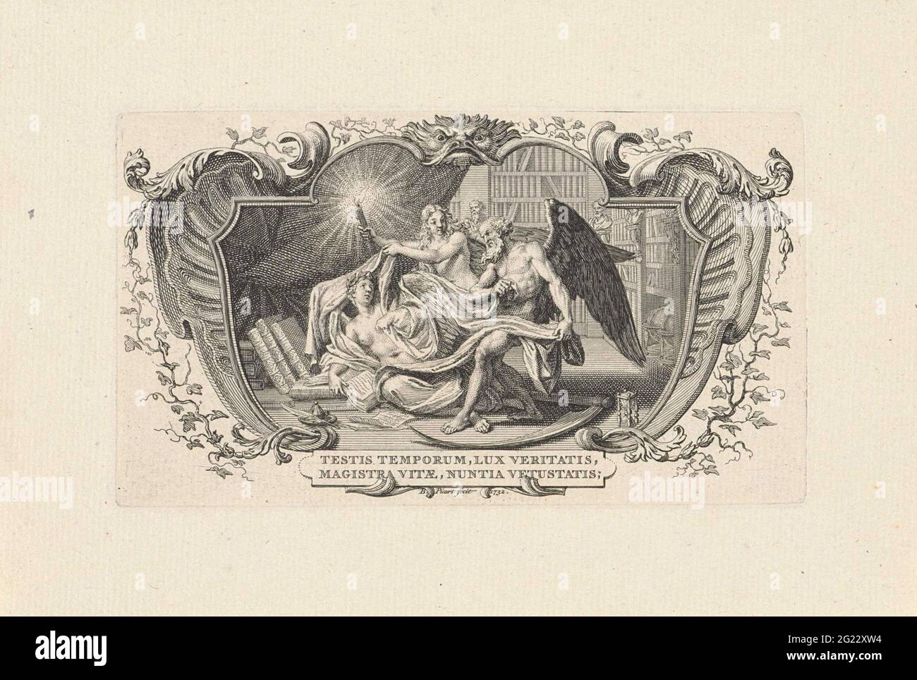 History Dismatches by father's time and truth. Father time and the light of the truth evaluate history in a study. Illustration of the motto under the performance: Testis Temporum Lux Veritatis Magistra Vite Nuntia vetus. The performance is caught in an ornamental frame. Stock Photohttps://www.alamy.com/image-license-details/?v=1https://www.alamy.com/history-dismatches-by-fathers-time-and-truth-father-time-and-the-light-of-the-truth-evaluate-history-in-a-study-illustration-of-the-motto-under-the-performance-testis-temporum-lux-veritatis-magistra-vite-nuntia-vetus-the-performance-is-caught-in-an-ornamental-frame-image431553504.html
RM2G22XW4–History Dismatches by father's time and truth. Father time and the light of the truth evaluate history in a study. Illustration of the motto under the performance: Testis Temporum Lux Veritatis Magistra Vite Nuntia vetus. The performance is caught in an ornamental frame.
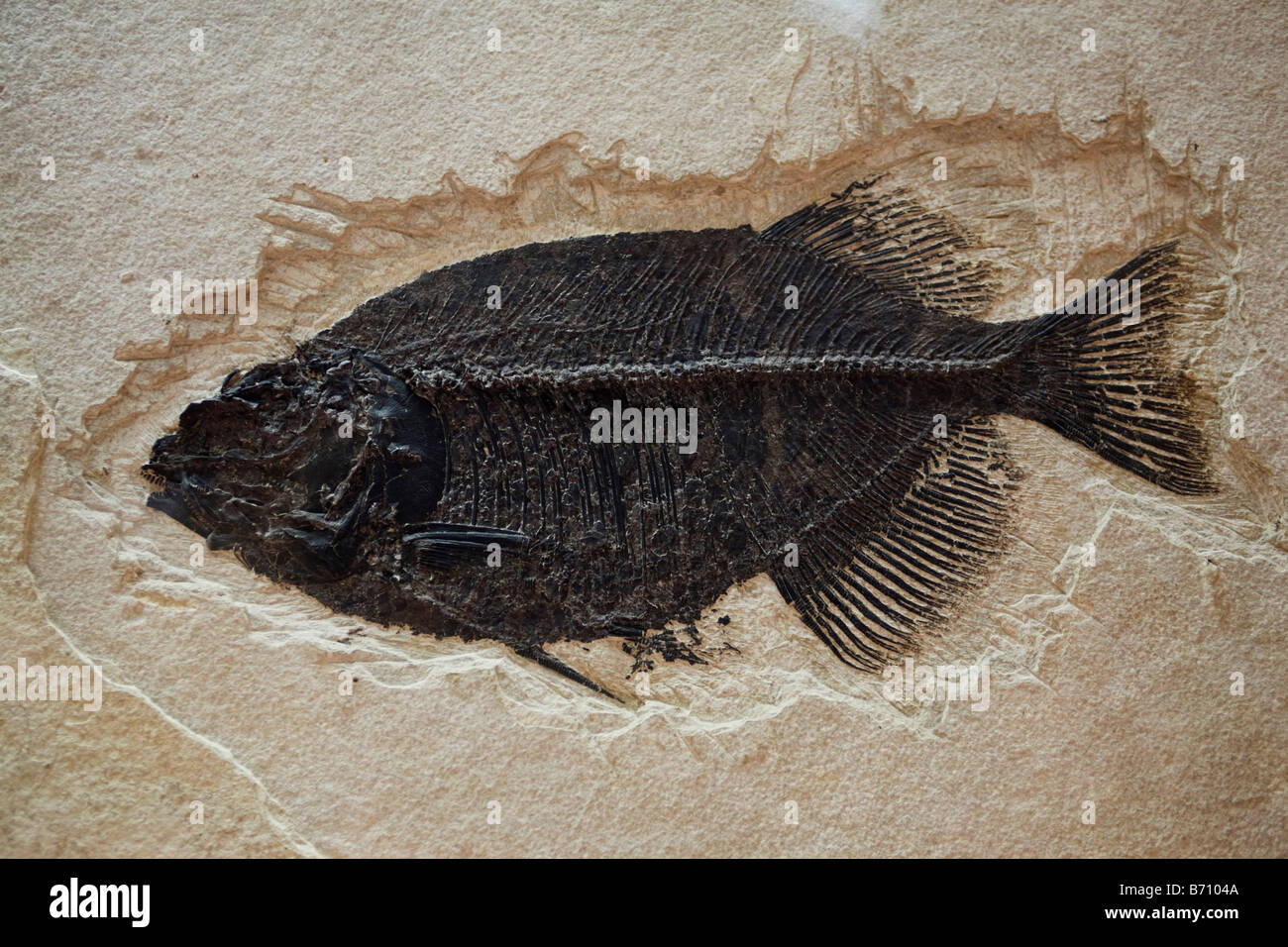 Fossil of fish from the Eocene period found in North America's Wyoming Stock Photohttps://www.alamy.com/image-license-details/?v=1https://www.alamy.com/stock-photo-fossil-of-fish-from-the-eocene-period-found-in-north-americas-wyoming-21535034.html
RMB7104A–Fossil of fish from the Eocene period found in North America's Wyoming
 Daria Werbowy Mario Testi and Kate Winslet attending Mario Testi's 'Todo o Nada' photography exhibition opening at the Stock Photohttps://www.alamy.com/image-license-details/?v=1https://www.alamy.com/stock-photo-daria-werbowy-mario-testi-and-kate-winslet-attending-mario-testis-52003010.html
RMD0GXAA–Daria Werbowy Mario Testi and Kate Winslet attending Mario Testi's 'Todo o Nada' photography exhibition opening at the
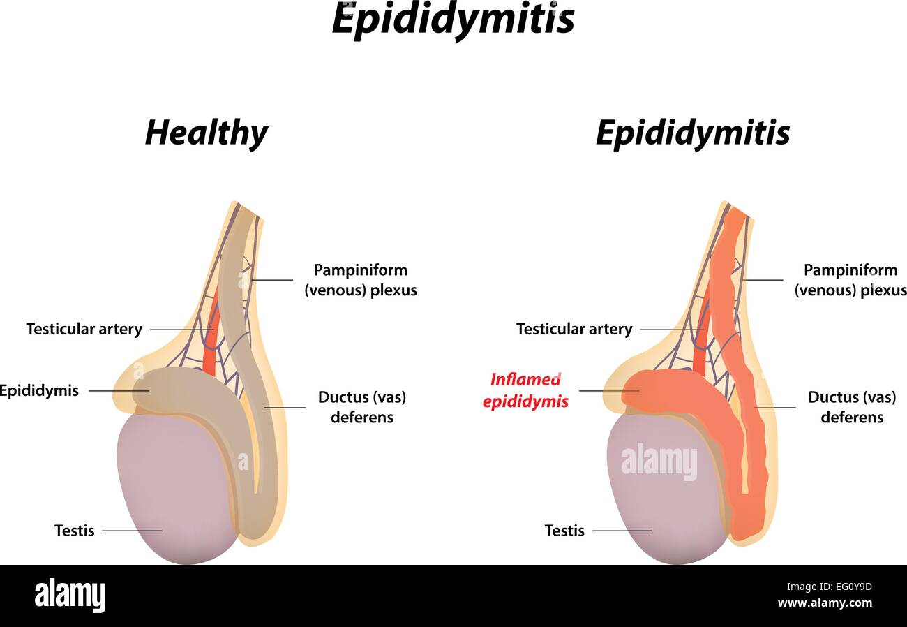 Epididymitis Stock Vectorhttps://www.alamy.com/image-license-details/?v=1https://www.alamy.com/stock-photo-epididymitis-78697401.html
RFEG0Y9D–Epididymitis
 Cross section of the testis, epididymis and tunica vaginalis, vintage engraved illustration. Usual Medicine Dictionary by Dr Lab Stock Vectorhttps://www.alamy.com/image-license-details/?v=1https://www.alamy.com/stock-photo-cross-section-of-the-testis-epididymis-and-tunica-vaginalis-vintage-84419687.html
RFEW9J4R–Cross section of the testis, epididymis and tunica vaginalis, vintage engraved illustration. Usual Medicine Dictionary by Dr Lab
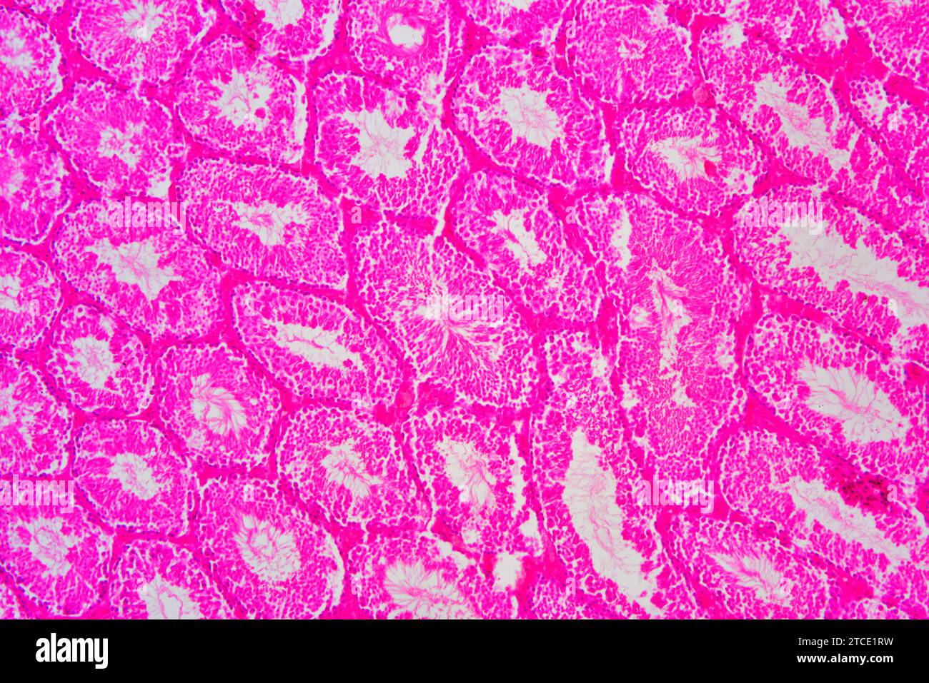 Human testicle or testis section showing seminiferous tubules, Leydig cells, Sertoli cells, spermatocytes, spermatogonia, spermatides and spermatozoon Stock Photohttps://www.alamy.com/image-license-details/?v=1https://www.alamy.com/human-testicle-or-testis-section-showing-seminiferous-tubules-leydig-cells-sertoli-cells-spermatocytes-spermatogonia-spermatides-and-spermatozoon-image575626797.html
RF2TCE1RW–Human testicle or testis section showing seminiferous tubules, Leydig cells, Sertoli cells, spermatocytes, spermatogonia, spermatides and spermatozoon
 The enlarged scrotum of a middle-aged man due to a hydrocele, an accumulation of fluid in the sac surrounding the testes. Stock Photohttps://www.alamy.com/image-license-details/?v=1https://www.alamy.com/the-enlarged-scrotum-of-a-middle-aged-man-due-to-a-hydrocele-an-accumulation-of-fluid-in-the-sac-surrounding-the-testes-image353693002.html
RM2BFC30X–The enlarged scrotum of a middle-aged man due to a hydrocele, an accumulation of fluid in the sac surrounding the testes.
 Epithelial lining of a seminiferous tubule of rat testis. On the lumen tubule, sperm tails are identified. Deeper into the epithelium, meiotic phases Stock Photohttps://www.alamy.com/image-license-details/?v=1https://www.alamy.com/epithelial-lining-of-a-seminiferous-tubule-of-rat-testis-on-the-lumen-tubule-sperm-tails-are-identified-deeper-into-the-epithelium-meiotic-phases-image346977455.html
RF2B4E57Y–Epithelial lining of a seminiferous tubule of rat testis. On the lumen tubule, sperm tails are identified. Deeper into the epithelium, meiotic phases
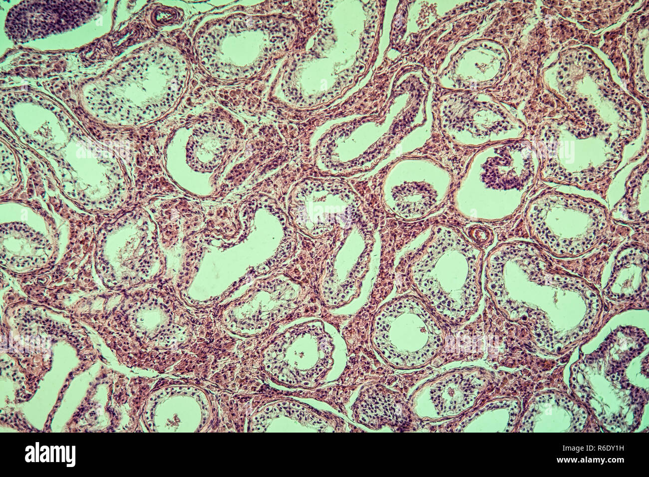 inguinal testis diseased tissue 100x Stock Photohttps://www.alamy.com/image-license-details/?v=1https://www.alamy.com/inguinal-testis-diseased-tissue-100x-image227729309.html
RFR6DY1H–inguinal testis diseased tissue 100x
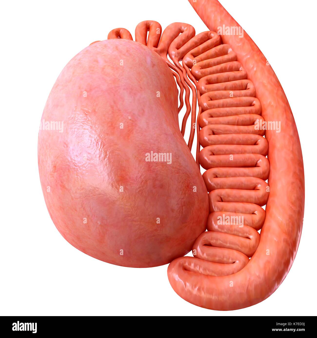 Illustration of male testis anatomy. Stock Photohttps://www.alamy.com/image-license-details/?v=1https://www.alamy.com/illustration-of-male-testis-anatomy-image159513526.html
RFK7ED3J–Illustration of male testis anatomy.
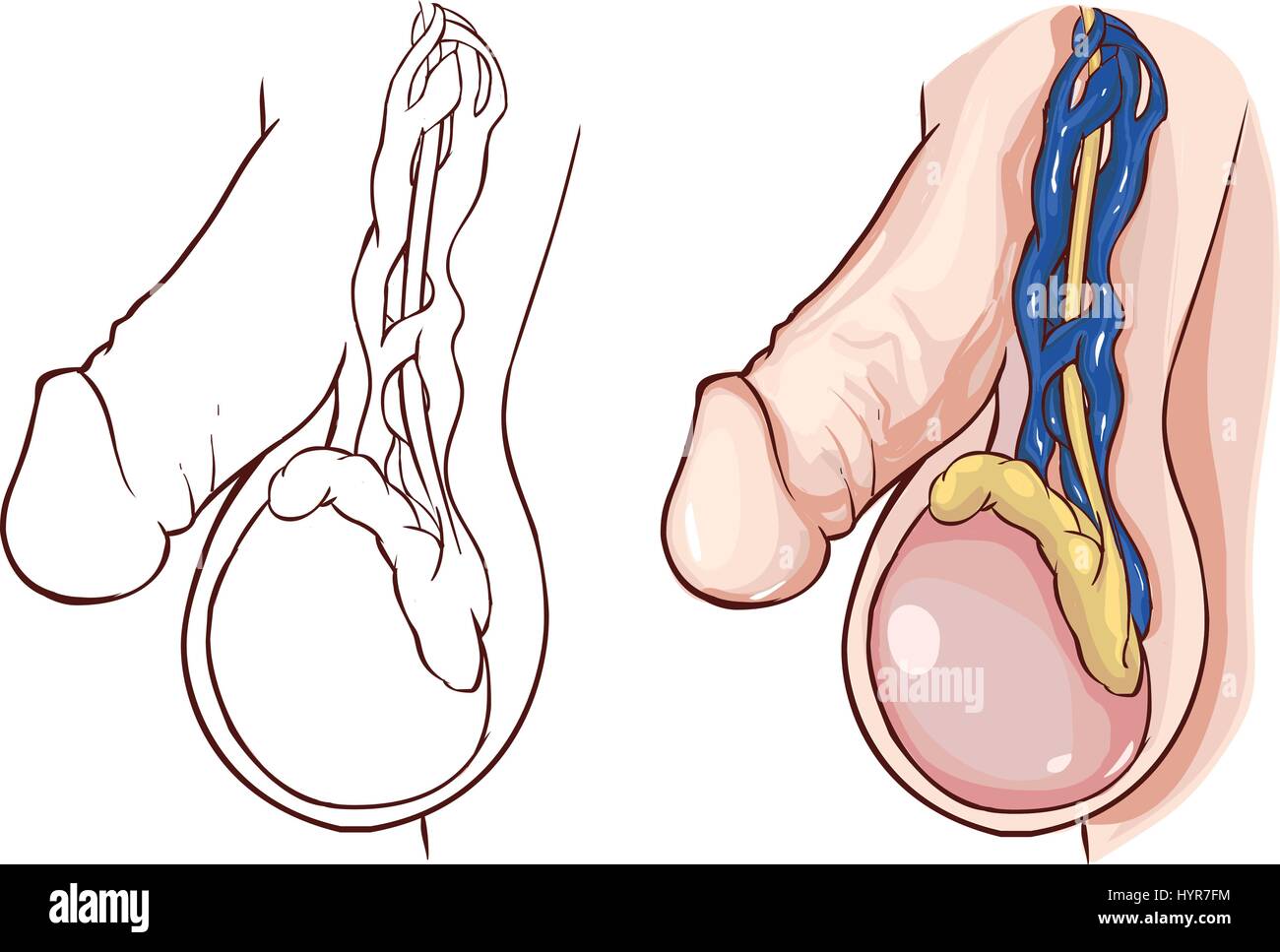 white background vector illustration of a Varicocele Stock Vectorhttps://www.alamy.com/image-license-details/?v=1https://www.alamy.com/stock-photo-white-background-vector-illustration-of-a-varicocele-137579112.html
RFHYR7FM–white background vector illustration of a Varicocele
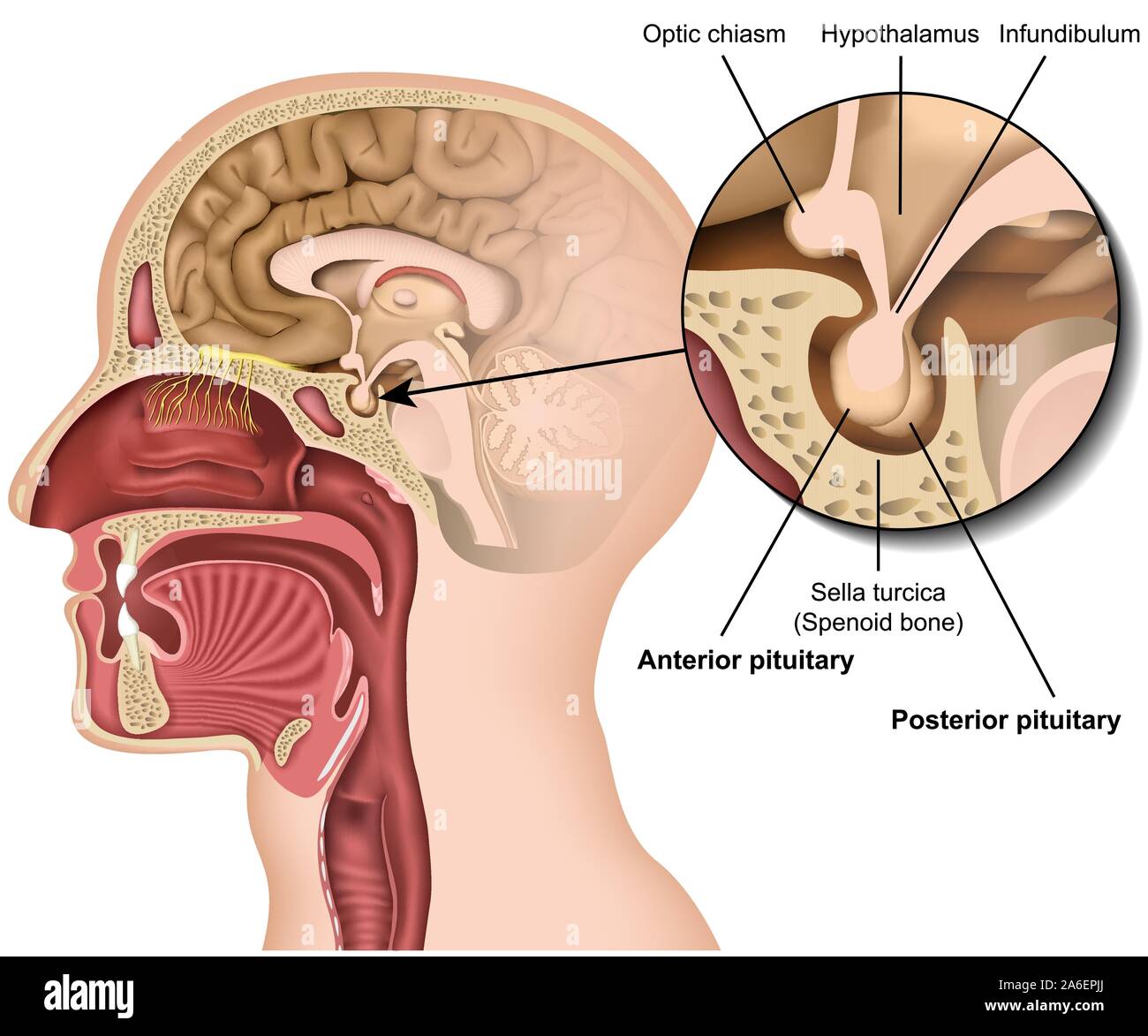 Pituitary gland anatomy 3d medical vector illustration isolated on white background hypothalamus in human brain eps 10 infographic Stock Vectorhttps://www.alamy.com/image-license-details/?v=1https://www.alamy.com/pituitary-gland-anatomy-3d-medical-vector-illustration-isolated-on-white-background-hypothalamus-in-human-brain-eps-10-infographic-image331010026.html
RF2A6EPJJ–Pituitary gland anatomy 3d medical vector illustration isolated on white background hypothalamus in human brain eps 10 infographic
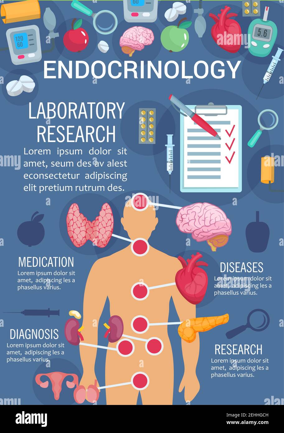 Endocrinology medicine poster of human endocrine system anatomy diagram. Thyroid gland, pancreas and hypothalamus, ovary, testis and thymus poster for Stock Vectorhttps://www.alamy.com/image-license-details/?v=1https://www.alamy.com/endocrinology-medicine-poster-of-human-endocrine-system-anatomy-diagram-thyroid-gland-pancreas-and-hypothalamus-ovary-testis-and-thymus-poster-for-image406673697.html
RF2EHHGCH–Endocrinology medicine poster of human endocrine system anatomy diagram. Thyroid gland, pancreas and hypothalamus, ovary, testis and thymus poster for
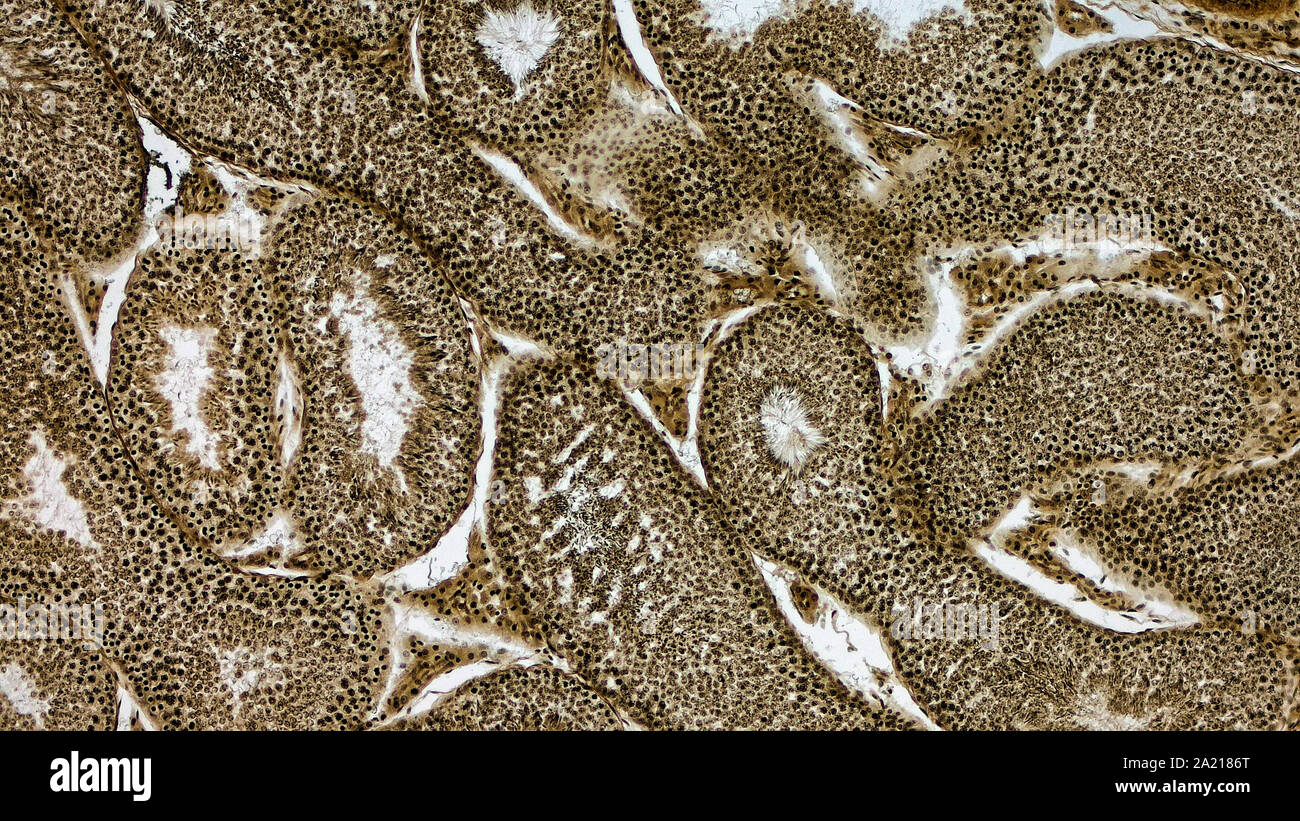 Histologically stained cross-section ofa rabbit testicle. Stock Photohttps://www.alamy.com/image-license-details/?v=1https://www.alamy.com/histologically-stained-cross-section-ofa-rabbit-testicle-image328254720.html
RM2A2186T–Histologically stained cross-section ofa rabbit testicle.
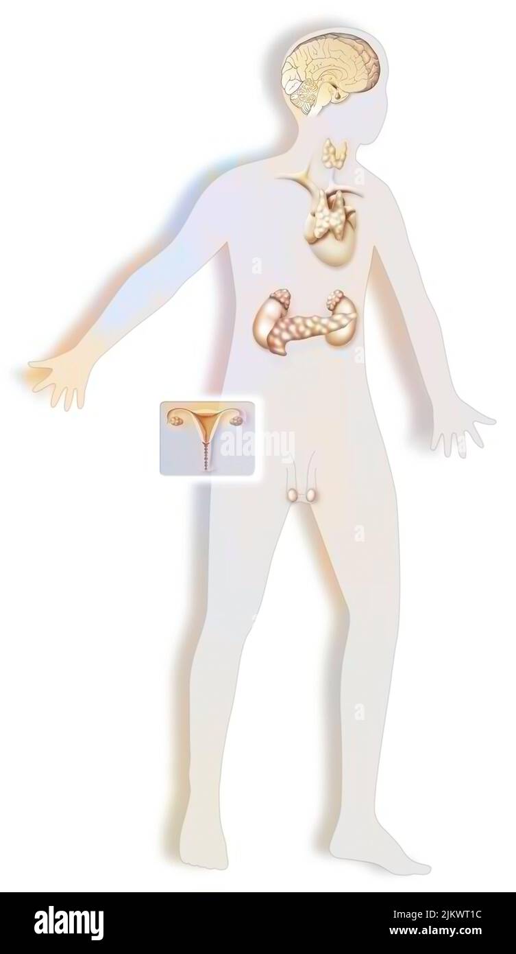 Hormonal glands in a boy with pituitary gland, thyroid gland. Stock Photohttps://www.alamy.com/image-license-details/?v=1https://www.alamy.com/hormonal-glands-in-a-boy-with-pituitary-gland-thyroid-gland-image476926056.html
RF2JKWT1C–Hormonal glands in a boy with pituitary gland, thyroid gland.
 Testosterone boost increase, improve fertility, masculine bro science and recovery balls tanning concept with two eggs on a red light therapy spotligh Stock Photohttps://www.alamy.com/image-license-details/?v=1https://www.alamy.com/testosterone-boost-increase-improve-fertility-masculine-bro-science-and-recovery-balls-tanning-concept-with-two-eggs-on-a-red-light-therapy-spotligh-image471931044.html
RF2JBP8T4–Testosterone boost increase, improve fertility, masculine bro science and recovery balls tanning concept with two eggs on a red light therapy spotligh
 Concept of right sided scrotal swelling or testicular pain. With text space. Isolated on white. Stock Photohttps://www.alamy.com/image-license-details/?v=1https://www.alamy.com/concept-of-right-sided-scrotal-swelling-or-testicular-pain-with-text-space-isolated-on-white-image485347515.html
RF2K5HDKR–Concept of right sided scrotal swelling or testicular pain. With text space. Isolated on white.
 Inguinal testis diseased tissue 100x Stock Photohttps://www.alamy.com/image-license-details/?v=1https://www.alamy.com/inguinal-testis-diseased-tissue-100x-image274329699.html
RMWX8P97–Inguinal testis diseased tissue 100x
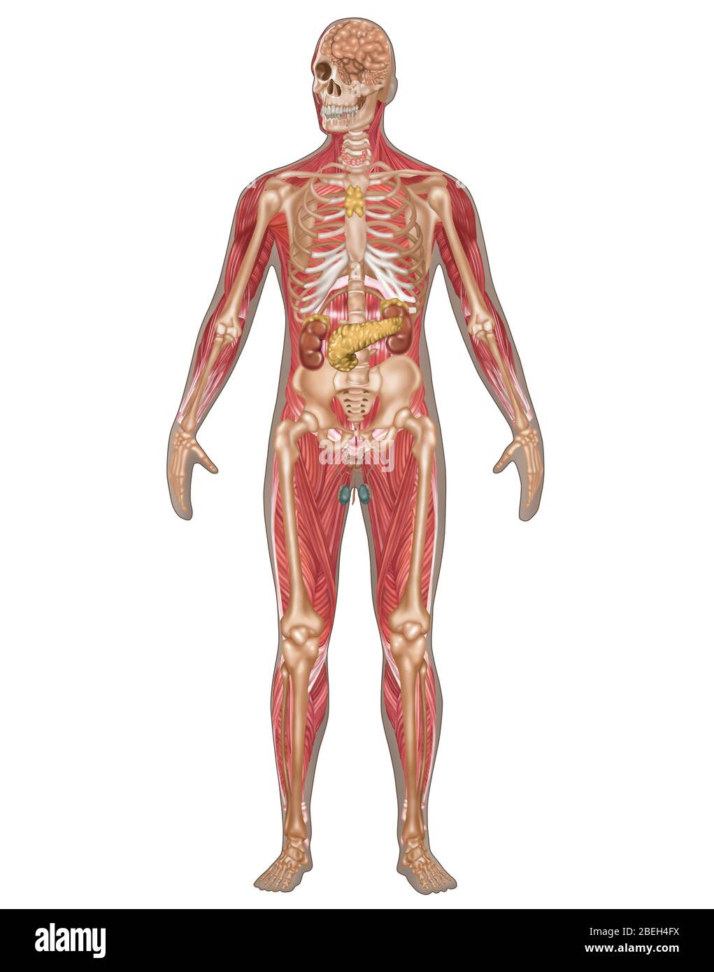 Endocrine, Skeletal & Muscular Systems, Male Stock Photohttps://www.alamy.com/image-license-details/?v=1https://www.alamy.com/endocrine-skeletal-muscular-systems-male-image353189310.html
RF2BEH4FX–Endocrine, Skeletal & Muscular Systems, Male
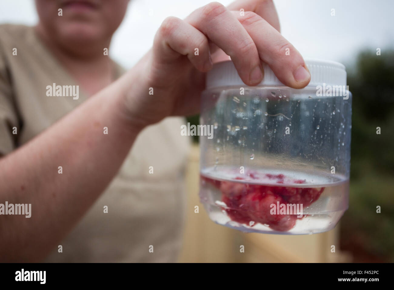 Wild elephant (Loxodonta africana), vas deferens, removed during keyhole surgery in the bush. Private game reserve in Limpopo, South Africa, April 2011. Winner of the IUCN grant in the Melvita Nature Images Awards Competition 2013 Stock Photohttps://www.alamy.com/image-license-details/?v=1https://www.alamy.com/stock-photo-wild-elephant-loxodonta-africana-vas-deferens-removed-during-keyhole-88622420.html
RMF452PC–Wild elephant (Loxodonta africana), vas deferens, removed during keyhole surgery in the bush. Private game reserve in Limpopo, South Africa, April 2011. Winner of the IUCN grant in the Melvita Nature Images Awards Competition 2013
 Human Kidneys Anatomy Stock Photohttps://www.alamy.com/image-license-details/?v=1https://www.alamy.com/human-kidneys-anatomy-image214597687.html
RFPD3NF3–Human Kidneys Anatomy
 Daria Werbowy, Mario Testi and Kate Winslet attending Mario Testi's 'Todo o Nada' photography exhibition opening at the Stock Photohttps://www.alamy.com/image-license-details/?v=1https://www.alamy.com/stock-photo-daria-werbowy-mario-testi-and-kate-winslet-attending-mario-testis-52003004.html
RMD0GXA4–Daria Werbowy, Mario Testi and Kate Winslet attending Mario Testi's 'Todo o Nada' photography exhibition opening at the
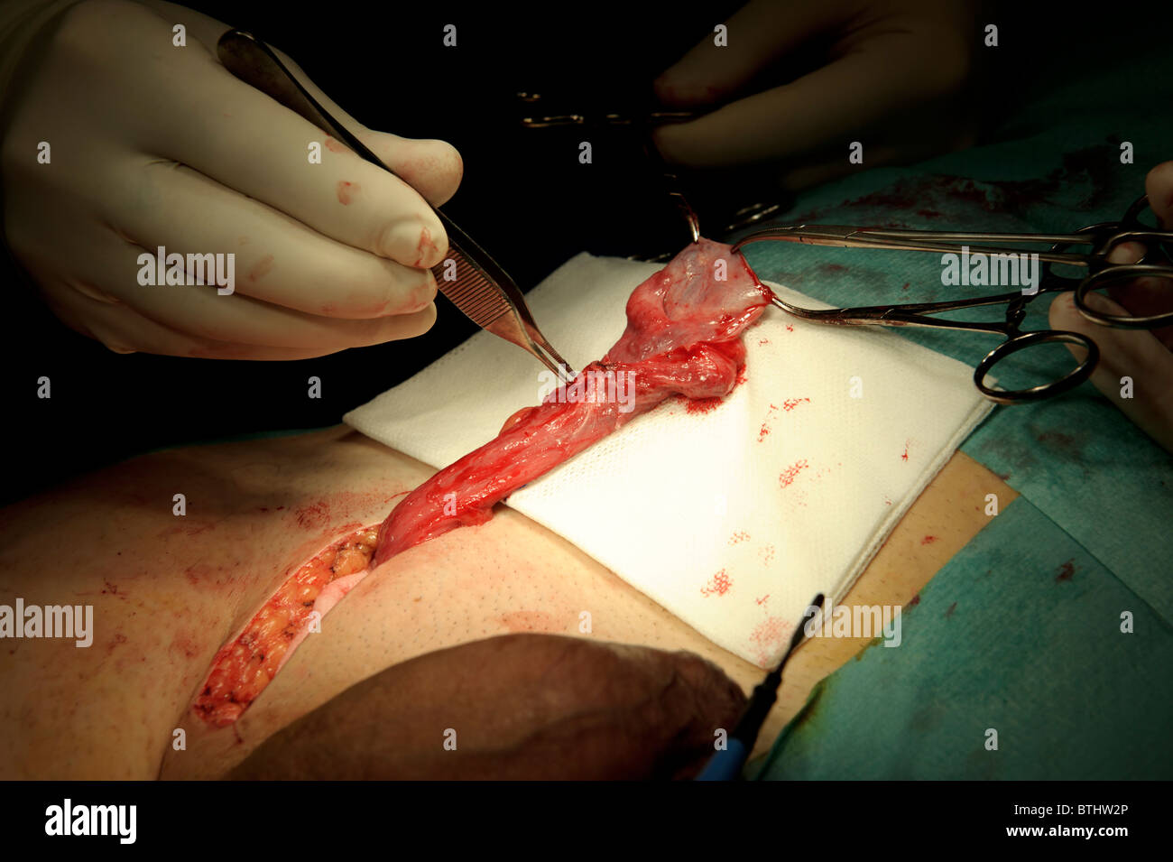 Testicular tumor removal surgery, urology Stock Photohttps://www.alamy.com/image-license-details/?v=1https://www.alamy.com/stock-photo-testicular-tumor-removal-surgery-urology-32354974.html
RMBTHW2P–Testicular tumor removal surgery, urology
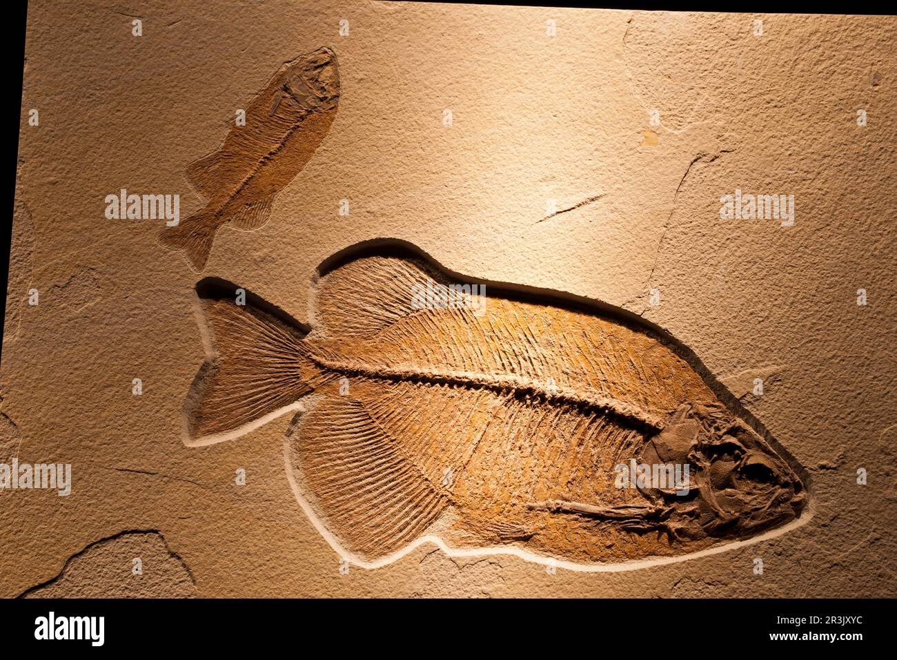 Phareodus Testis.Dinòpolis. Museo paleontólogico de Teruel, fundación conjunto Paleontólogico de Teruel. Aragon. españa. Stock Photohttps://www.alamy.com/image-license-details/?v=1https://www.alamy.com/phareodus-testisdinpolis-museo-paleontlogico-de-teruel-fundacin-conjunto-paleontlogico-de-teruel-aragon-espaa-image552992032.html
RM2R3JXYC–Phareodus Testis.Dinòpolis. Museo paleontólogico de Teruel, fundación conjunto Paleontólogico de Teruel. Aragon. españa.
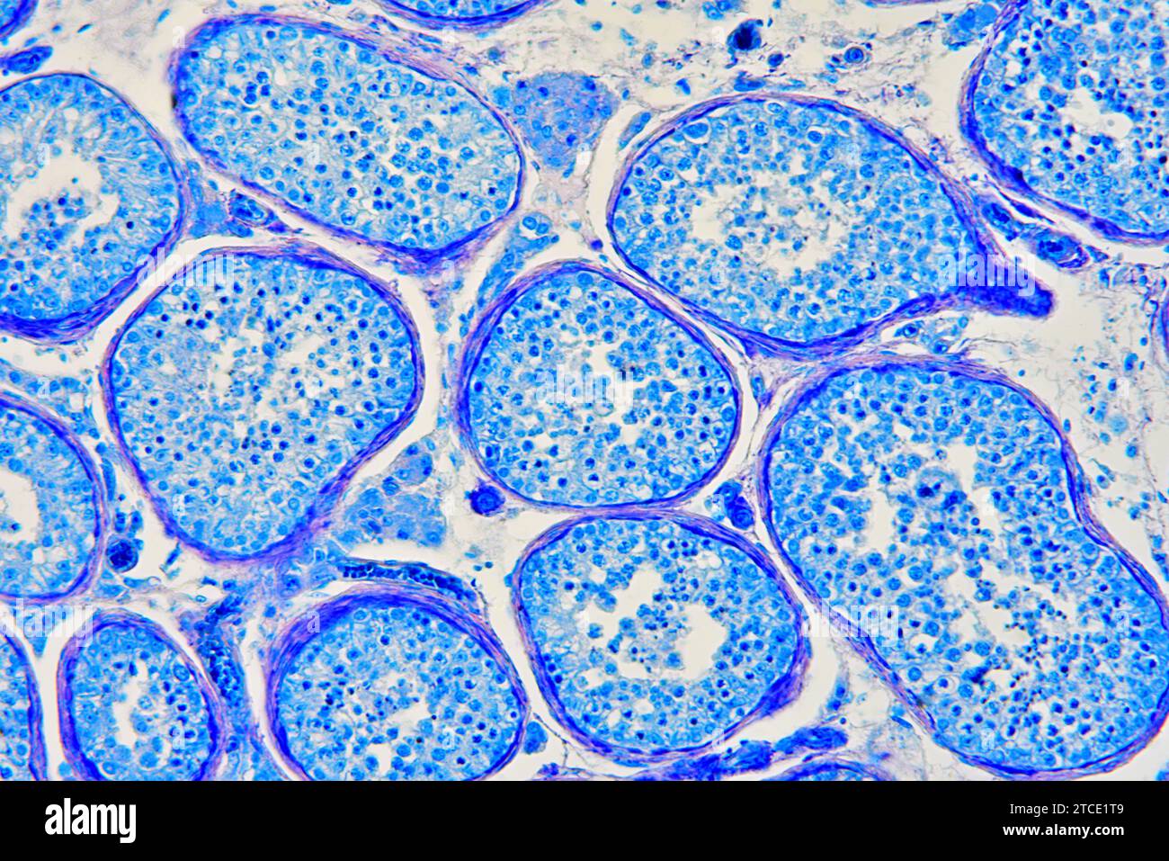 Human testicle or testis section showing seminiferous tubules, Leydig cells, Sertoli cells, spermatocytes and spermatogonia. Optical microscope X200. Stock Photohttps://www.alamy.com/image-license-details/?v=1https://www.alamy.com/human-testicle-or-testis-section-showing-seminiferous-tubules-leydig-cells-sertoli-cells-spermatocytes-and-spermatogonia-optical-microscope-x200-image575626809.html
RF2TCE1T9–Human testicle or testis section showing seminiferous tubules, Leydig cells, Sertoli cells, spermatocytes and spermatogonia. Optical microscope X200.
 The enlarged scrotum of a middle-aged man due to a hydrocele, an accumulation of fluid in the sac surrounding the testes. Stock Photohttps://www.alamy.com/image-license-details/?v=1https://www.alamy.com/the-enlarged-scrotum-of-a-middle-aged-man-due-to-a-hydrocele-an-accumulation-of-fluid-in-the-sac-surrounding-the-testes-image353692998.html
RM2BFC30P–The enlarged scrotum of a middle-aged man due to a hydrocele, an accumulation of fluid in the sac surrounding the testes.
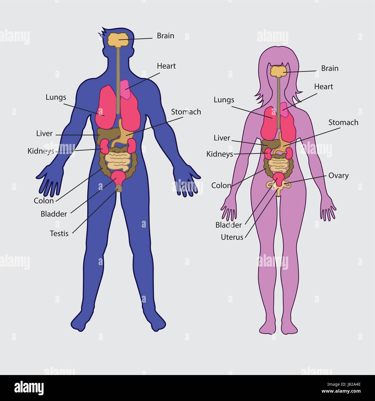 Basic human internal organs vector Stock Vectorhttps://www.alamy.com/image-license-details/?v=1https://www.alamy.com/stock-photo-basic-human-internal-organs-vector-142652062.html
RFJ82A4E–Basic human internal organs vector
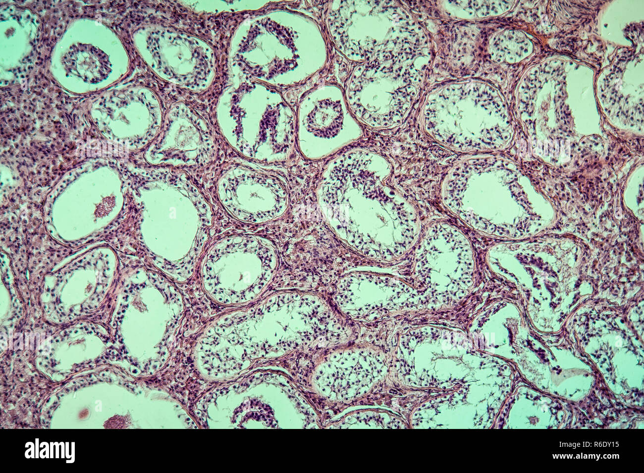 inguinal testis diseased tissue 100x Stock Photohttps://www.alamy.com/image-license-details/?v=1https://www.alamy.com/inguinal-testis-diseased-tissue-100x-image227729297.html
RFR6DY15–inguinal testis diseased tissue 100x
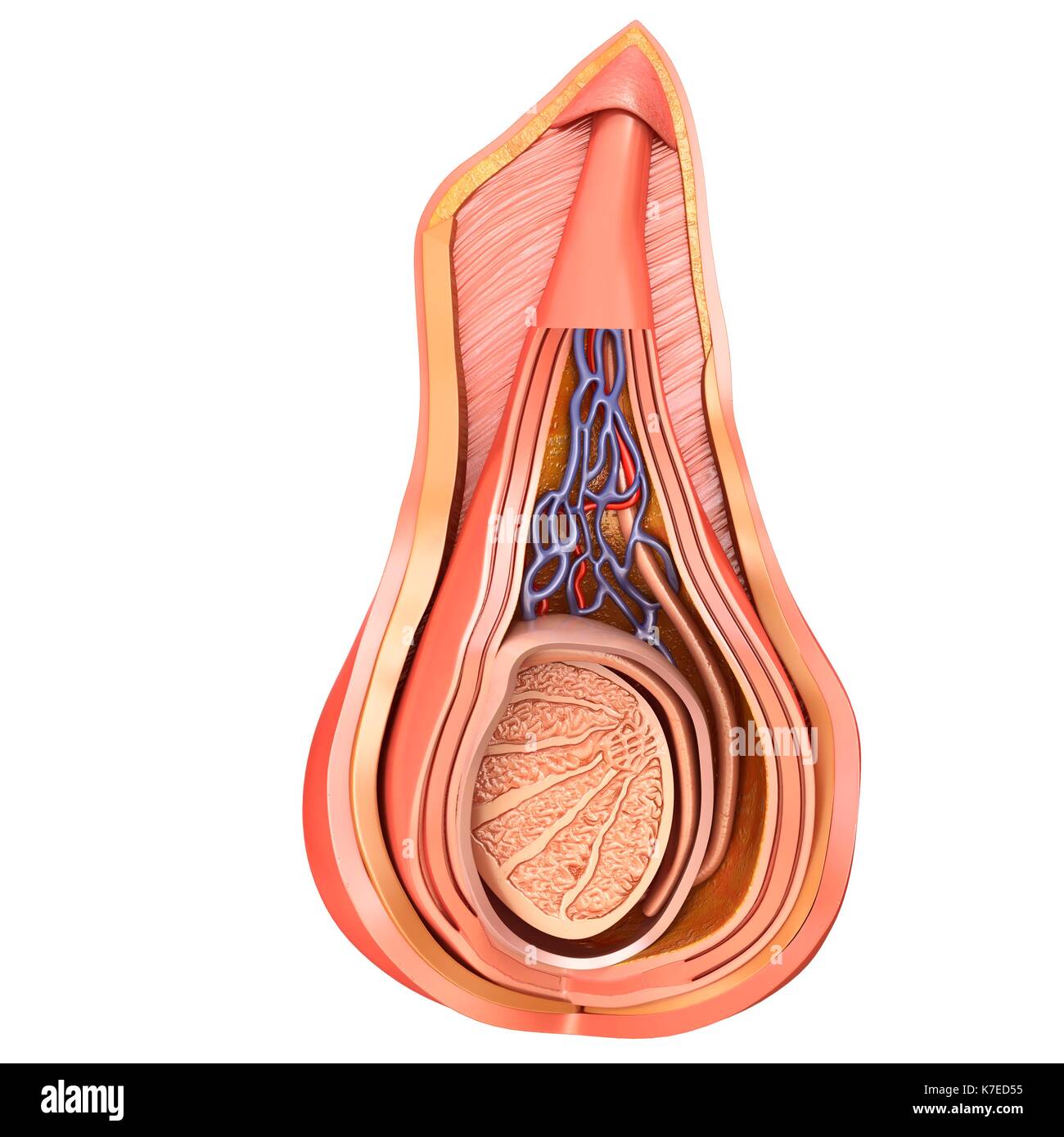 Illustration of scrotal layers of testis. Stock Photohttps://www.alamy.com/image-license-details/?v=1https://www.alamy.com/illustration-of-scrotal-layers-of-testis-image159513569.html
RFK7ED55–Illustration of scrotal layers of testis.
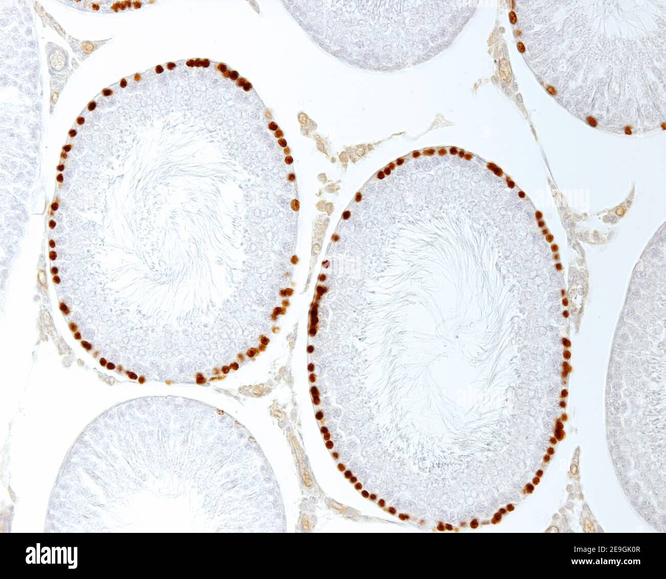 Labelling with bromodeoxiuridine (BrdU) of proliferating cells (spermatogonia) of seminiferous tubules. Note the differences in the labelling intensit Stock Photohttps://www.alamy.com/image-license-details/?v=1https://www.alamy.com/labelling-with-bromodeoxiuridine-brdu-of-proliferating-cells-spermatogonia-of-seminiferous-tubules-note-the-differences-in-the-labelling-intensit-image401736519.html
RF2E9GK0R–Labelling with bromodeoxiuridine (BrdU) of proliferating cells (spermatogonia) of seminiferous tubules. Note the differences in the labelling intensit