The muscle fiber and the myofibril Stock Photos and Images
(139)See the muscle fiber and the myofibril stock video clipsQuick filters:
The muscle fiber and the myofibril Stock Photos and Images
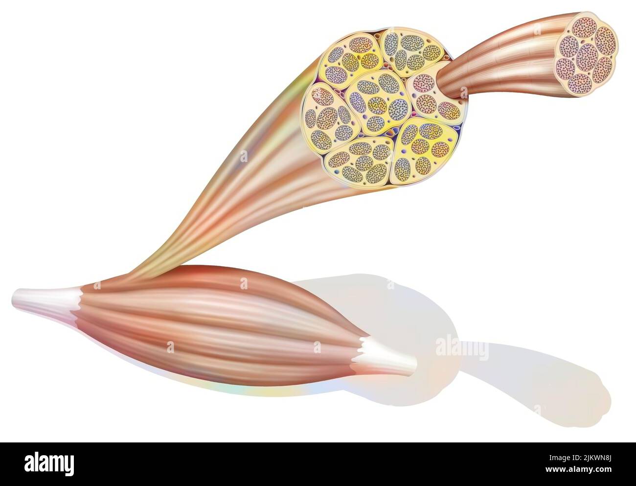 From muscle to muscle fiber: tendon, muscle, muscle fiber. Stock Photohttps://www.alamy.com/image-license-details/?v=1https://www.alamy.com/from-muscle-to-muscle-fiber-tendon-muscle-muscle-fiber-image476923906.html
RF2JKWN8J–From muscle to muscle fiber: tendon, muscle, muscle fiber.
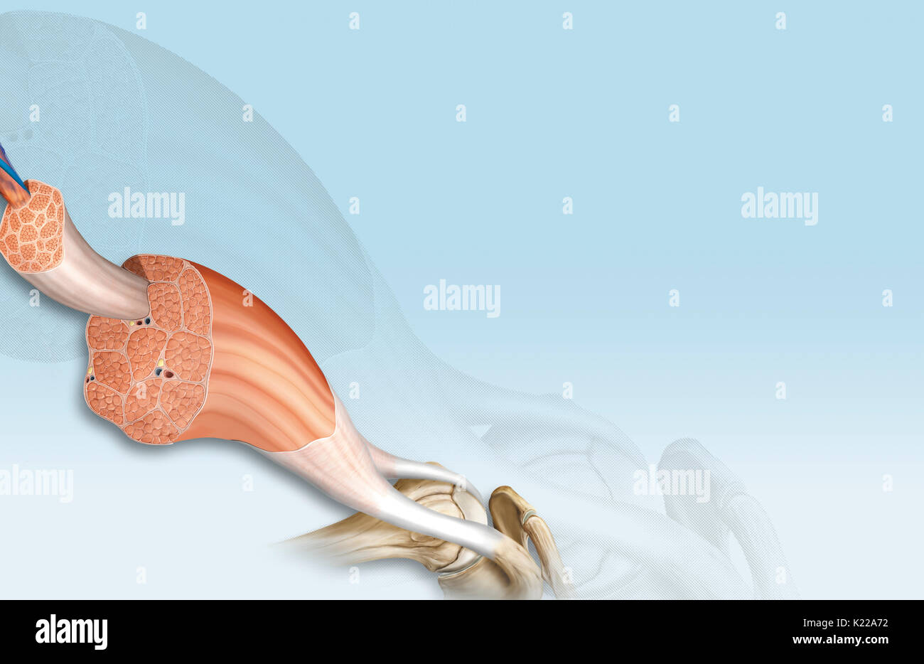 This image shows the structure of a skeletal muscle, revealing the muscle fibers bundle, the motor neuron, the muscle fiber and the myofibril. Stock Photohttps://www.alamy.com/image-license-details/?v=1https://www.alamy.com/this-image-shows-the-structure-of-a-skeletal-muscle-revealing-the-image156174566.html
RMK22A72–This image shows the structure of a skeletal muscle, revealing the muscle fibers bundle, the motor neuron, the muscle fiber and the myofibril.
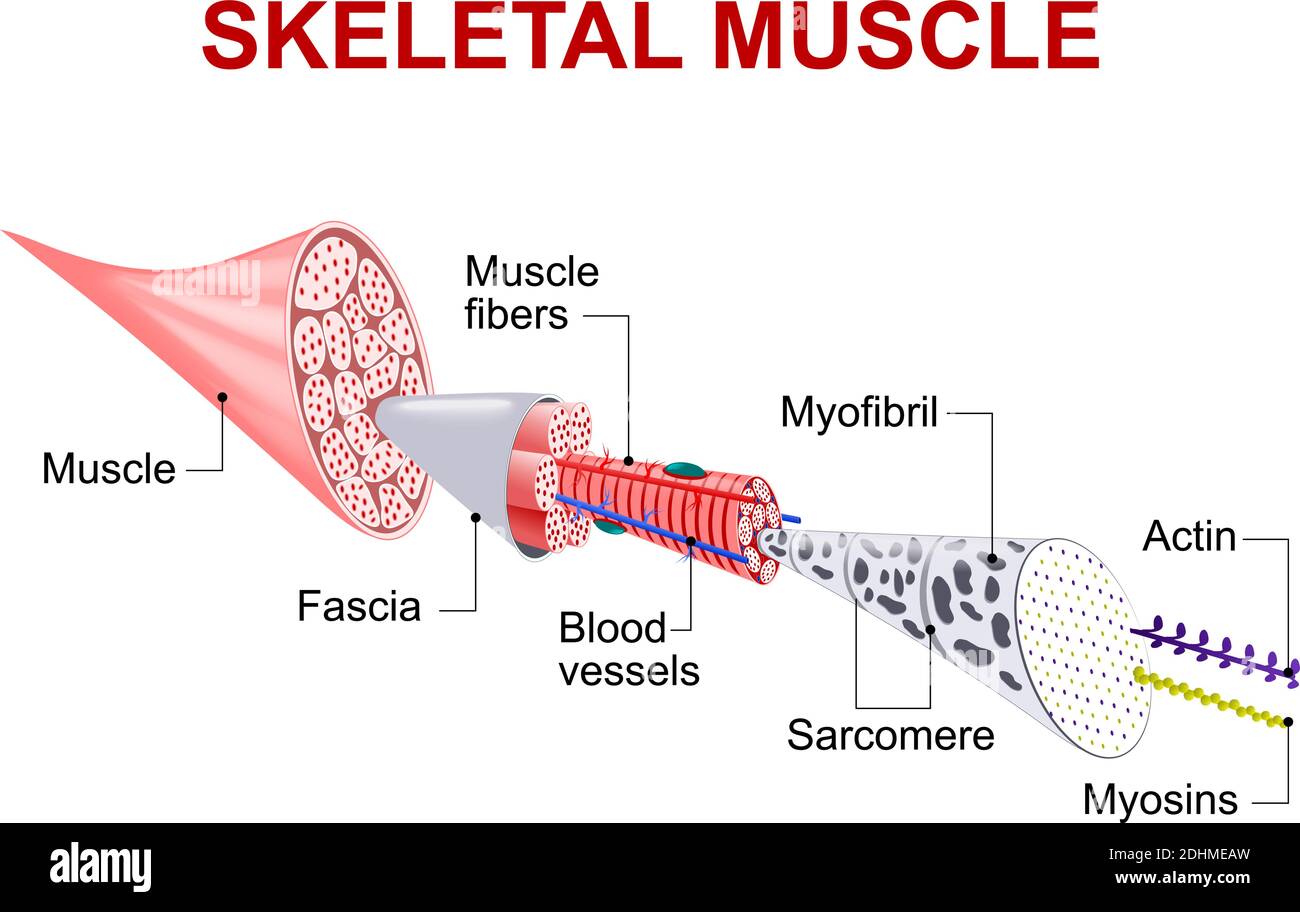 Each skeletal muscle fiber has many bundles of myofilaments. Each bundle is called a myofibril. This is what gives the muscle its striated appearance. Stock Vectorhttps://www.alamy.com/image-license-details/?v=1https://www.alamy.com/each-skeletal-muscle-fiber-has-many-bundles-of-myofilaments-each-bundle-is-called-a-myofibril-this-is-what-gives-the-muscle-its-striated-appearance-image389527569.html
RF2DHMEAW–Each skeletal muscle fiber has many bundles of myofilaments. Each bundle is called a myofibril. This is what gives the muscle its striated appearance.
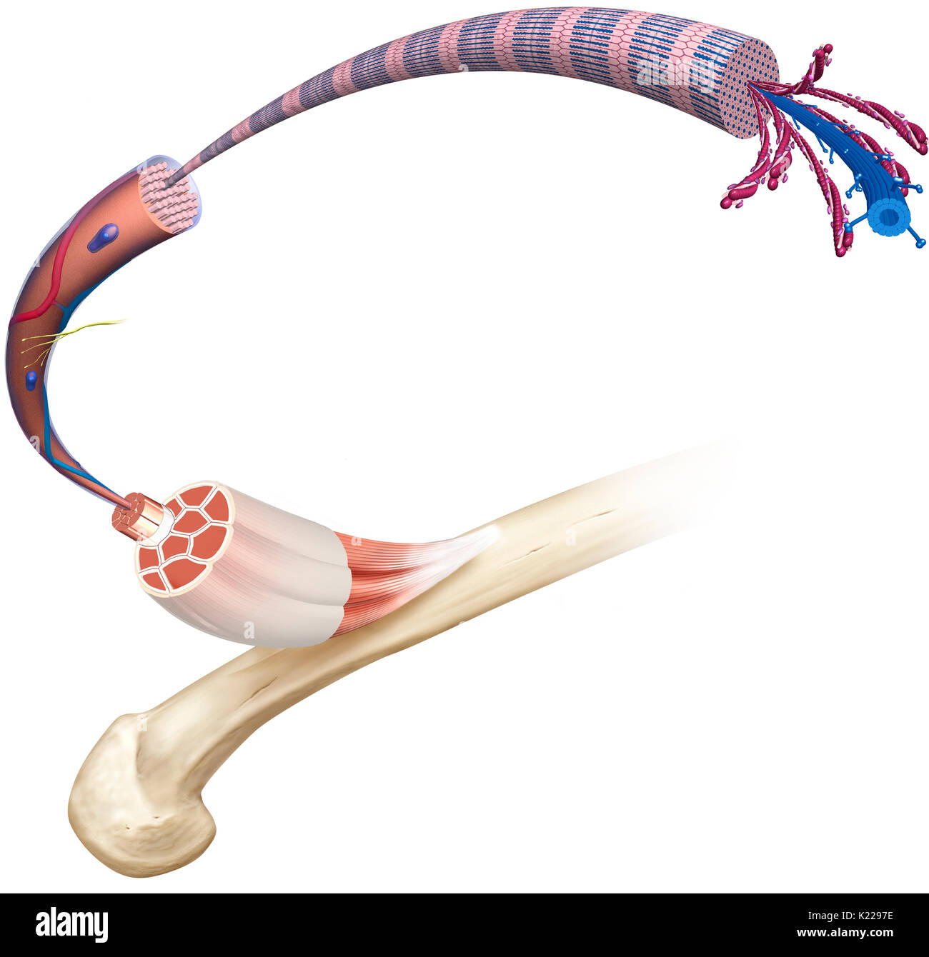 This image shows the structure of a skeletal muscle, revealing the muscle fibers bundle, the motor neuron, the muscle fiber and the myofibril. Stock Photohttps://www.alamy.com/image-license-details/?v=1https://www.alamy.com/this-image-shows-the-structure-of-a-skeletal-muscle-revealing-the-image156173794.html
RMK2297E–This image shows the structure of a skeletal muscle, revealing the muscle fibers bundle, the motor neuron, the muscle fiber and the myofibril.
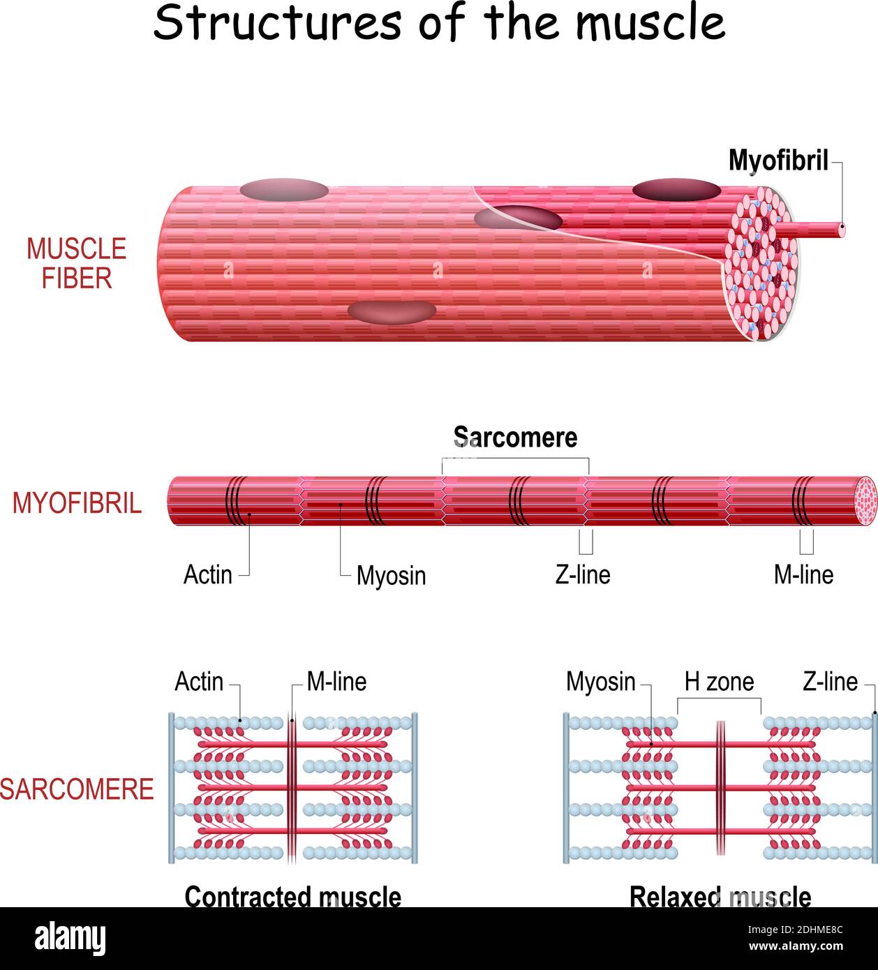 Structure Skeletal Muscle. myofibril with thin and thick filament. close up of a sarcomere. Muscles contract by sliding the myosin and actin filaments Stock Vectorhttps://www.alamy.com/image-license-details/?v=1https://www.alamy.com/structure-skeletal-muscle-myofibril-with-thin-and-thick-filament-close-up-of-a-sarcomere-muscles-contract-by-sliding-the-myosin-and-actin-filaments-image389527500.html
RF2DHME8C–Structure Skeletal Muscle. myofibril with thin and thick filament. close up of a sarcomere. Muscles contract by sliding the myosin and actin filaments
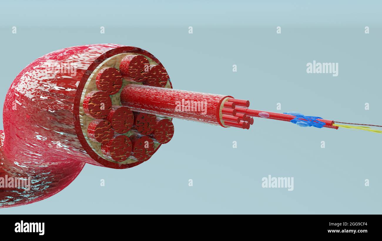 3d Illustration of Muscle Type: Heart muscle - cross section through muscle with muscle fibers visible - 3D Rendering Stock Photohttps://www.alamy.com/image-license-details/?v=1https://www.alamy.com/3d-illustration-of-muscle-type-heart-muscle-cross-section-through-muscle-with-muscle-fibers-visible-3d-rendering-image440301096.html
RM2GG9CF4–3d Illustration of Muscle Type: Heart muscle - cross section through muscle with muscle fibers visible - 3D Rendering
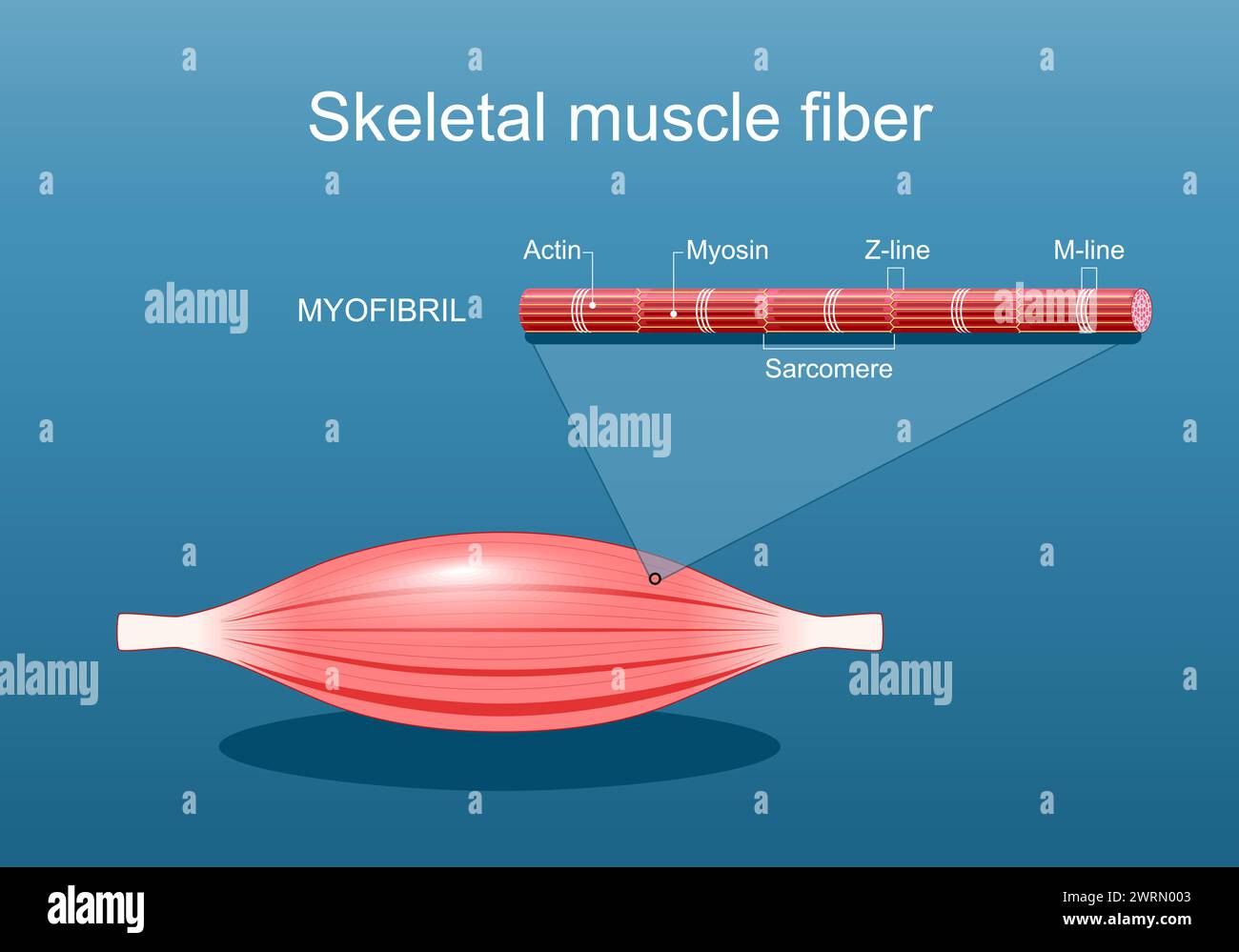 Anatomy of a Skeletal muscle fiber. Myofibril structure include Myosin, Z-line, M-line, Actin filaments, and Sarcomere. Isometric flat vector Illustra Stock Vectorhttps://www.alamy.com/image-license-details/?v=1https://www.alamy.com/anatomy-of-a-skeletal-muscle-fiber-myofibril-structure-include-myosin-z-line-m-line-actin-filaments-and-sarcomere-isometric-flat-vector-illustra-image599750595.html
RF2WRN003–Anatomy of a Skeletal muscle fiber. Myofibril structure include Myosin, Z-line, M-line, Actin filaments, and Sarcomere. Isometric flat vector Illustra
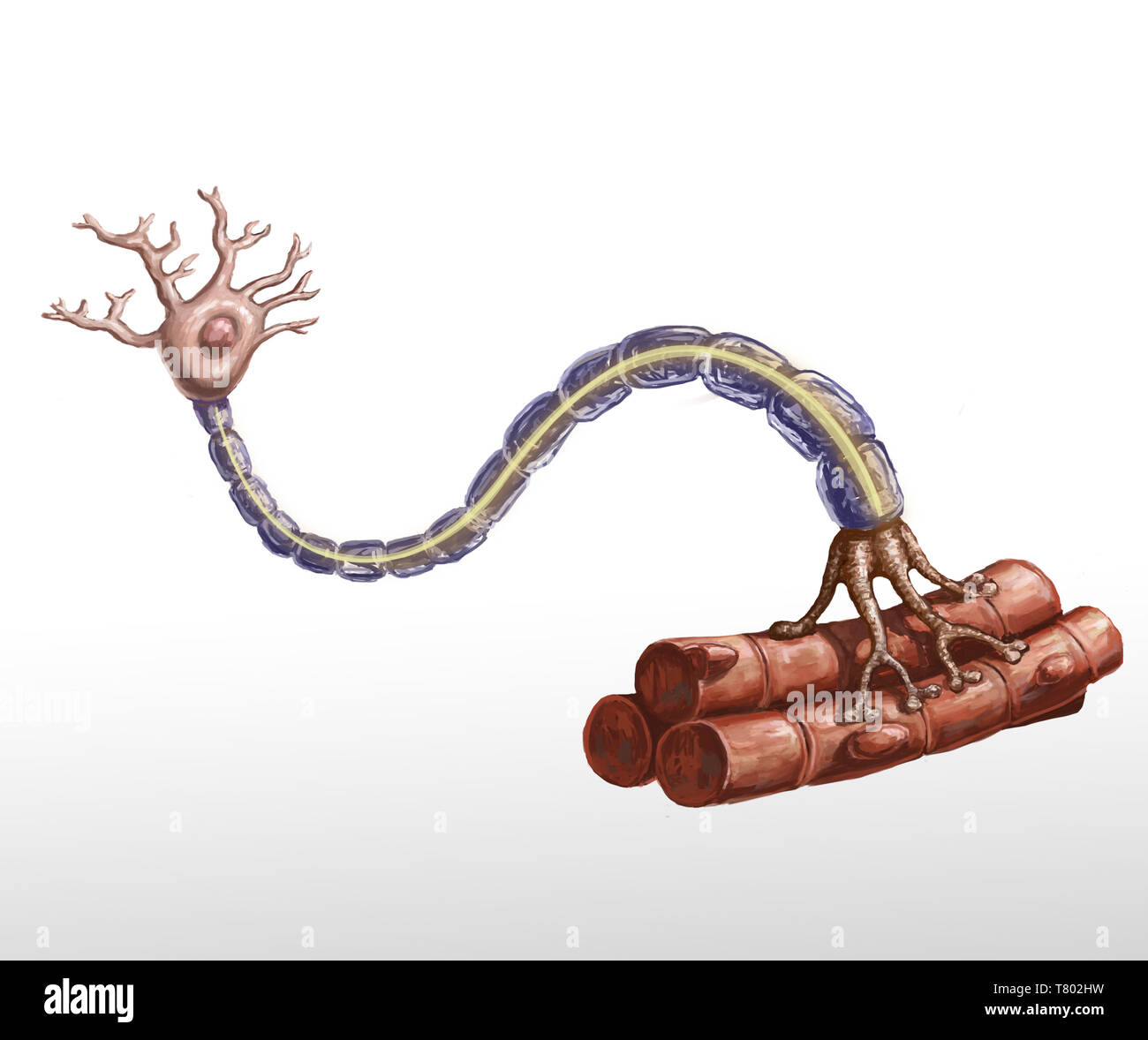 Motor Neuron and Muscle Fiber Illustration Stock Photohttps://www.alamy.com/image-license-details/?v=1https://www.alamy.com/motor-neuron-and-muscle-fiber-illustration-image245864469.html
RMT802HW–Motor Neuron and Muscle Fiber Illustration
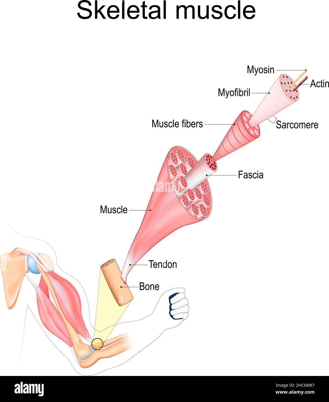 Skeletal Muscle anatomy. structure of Muscle fibers from Fascia and Tendon to Actin and Myosin. Vector poster Stock Vectorhttps://www.alamy.com/image-license-details/?v=1https://www.alamy.com/skeletal-muscle-anatomy-structure-of-muscle-fibers-from-fascia-and-tendon-to-actin-and-myosin-vector-poster-image454993063.html
RF2HC6M87–Skeletal Muscle anatomy. structure of Muscle fibers from Fascia and Tendon to Actin and Myosin. Vector poster
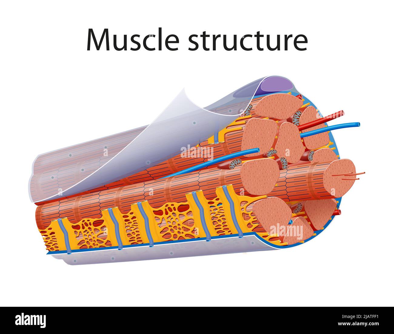 Illustration of Structure Skeletal Muscle Anatomy Stock Photohttps://www.alamy.com/image-license-details/?v=1https://www.alamy.com/illustration-of-structure-skeletal-muscle-anatomy-image471365525.html
RF2JATFF1–Illustration of Structure Skeletal Muscle Anatomy
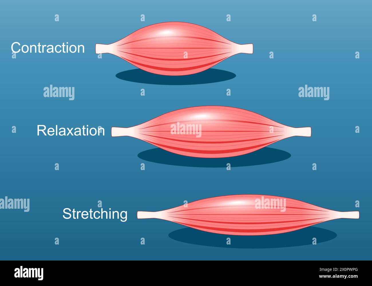 Muscle relaxation, stretching, and contraction. Close-up of a Skeletal muscle fiber. Isometric flat vector Illustration Stock Vectorhttps://www.alamy.com/image-license-details/?v=1https://www.alamy.com/muscle-relaxation-stretching-and-contraction-close-up-of-a-skeletal-muscle-fiber-isometric-flat-vector-illustration-image602866056.html
RF2X0PWPG–Muscle relaxation, stretching, and contraction. Close-up of a Skeletal muscle fiber. Isometric flat vector Illustration
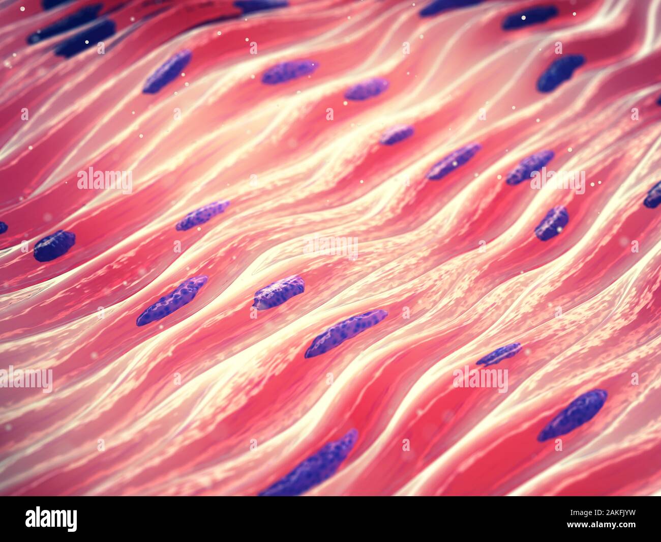 Myocytes, Group of smooth muscle cells Stock Photohttps://www.alamy.com/image-license-details/?v=1https://www.alamy.com/myocytes-group-of-smooth-muscle-cells-image339019629.html
RF2AKFJYW–Myocytes, Group of smooth muscle cells
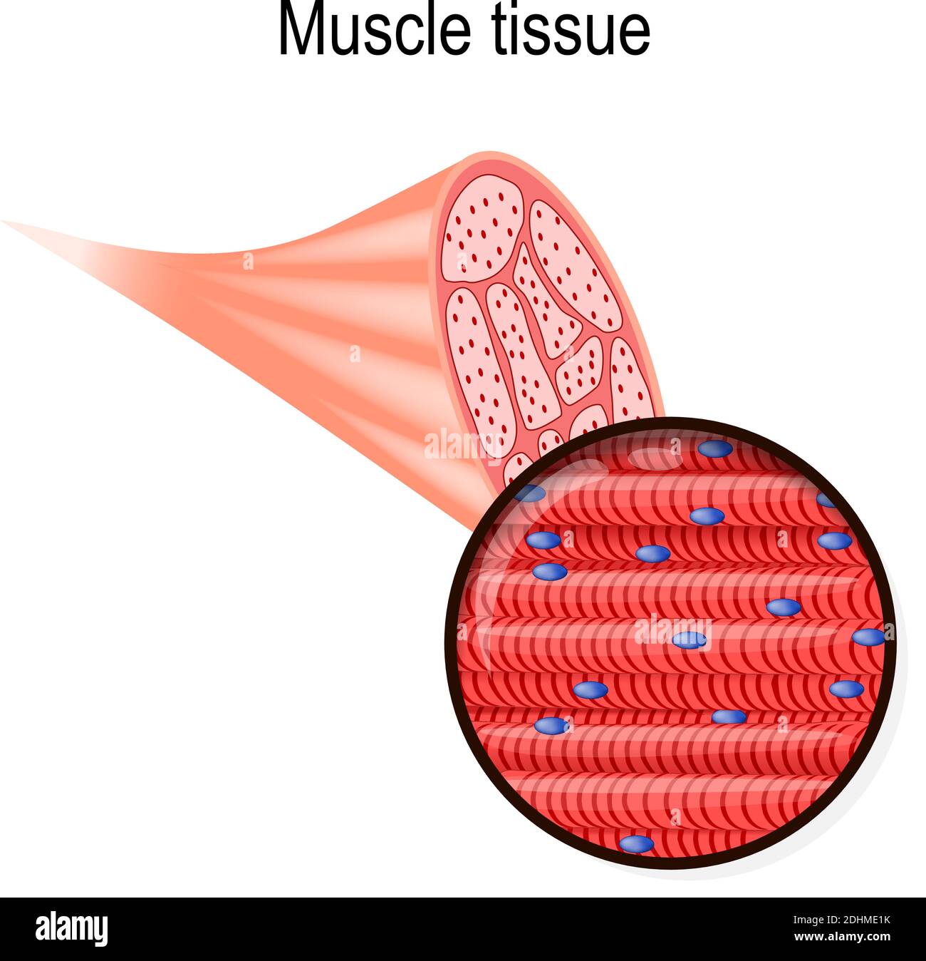 skeletal muscle. Tissue and fiber. Part of the biceps and close-up of muscle fibers. Vector illustration for biological, medical, science use Stock Vectorhttps://www.alamy.com/image-license-details/?v=1https://www.alamy.com/skeletal-muscle-tissue-and-fiber-part-of-the-biceps-and-close-up-of-muscle-fibers-vector-illustration-for-biological-medical-science-use-image389527311.html
RF2DHME1K–skeletal muscle. Tissue and fiber. Part of the biceps and close-up of muscle fibers. Vector illustration for biological, medical, science use
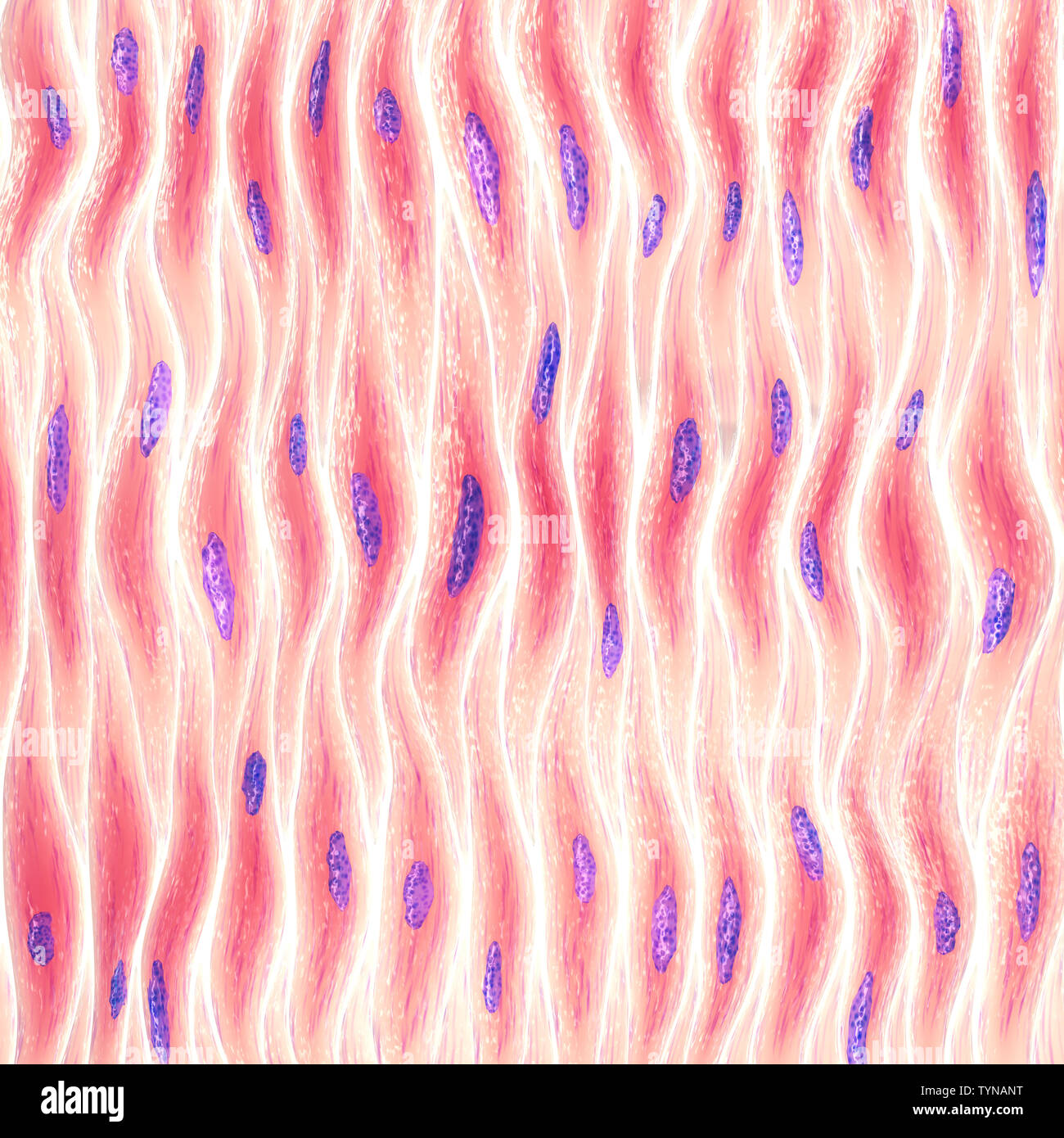 Myocytes, Group of smooth muscle cells Stock Photohttps://www.alamy.com/image-license-details/?v=1https://www.alamy.com/myocytes-group-of-smooth-muscle-cells-image258010308.html
RFTYNANT–Myocytes, Group of smooth muscle cells
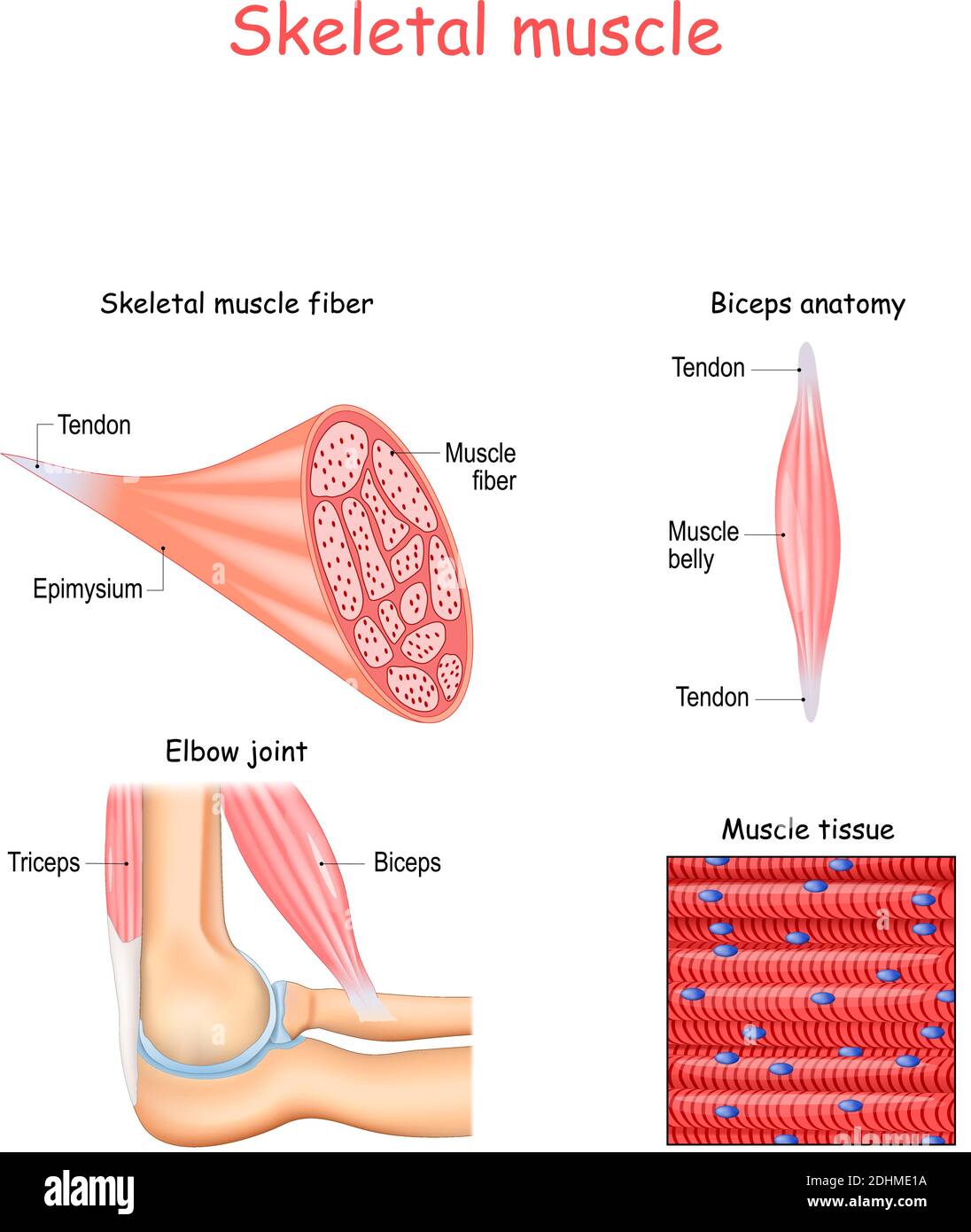 Structure of skeletal muscle fibers. Biceps and Triceps anatomy. Background of muscle tissue. Set of vectors illustrations for education use Stock Vectorhttps://www.alamy.com/image-license-details/?v=1https://www.alamy.com/structure-of-skeletal-muscle-fibers-biceps-and-triceps-anatomy-background-of-muscle-tissue-set-of-vectors-illustrations-for-education-use-image389527302.html
RF2DHME1A–Structure of skeletal muscle fibers. Biceps and Triceps anatomy. Background of muscle tissue. Set of vectors illustrations for education use
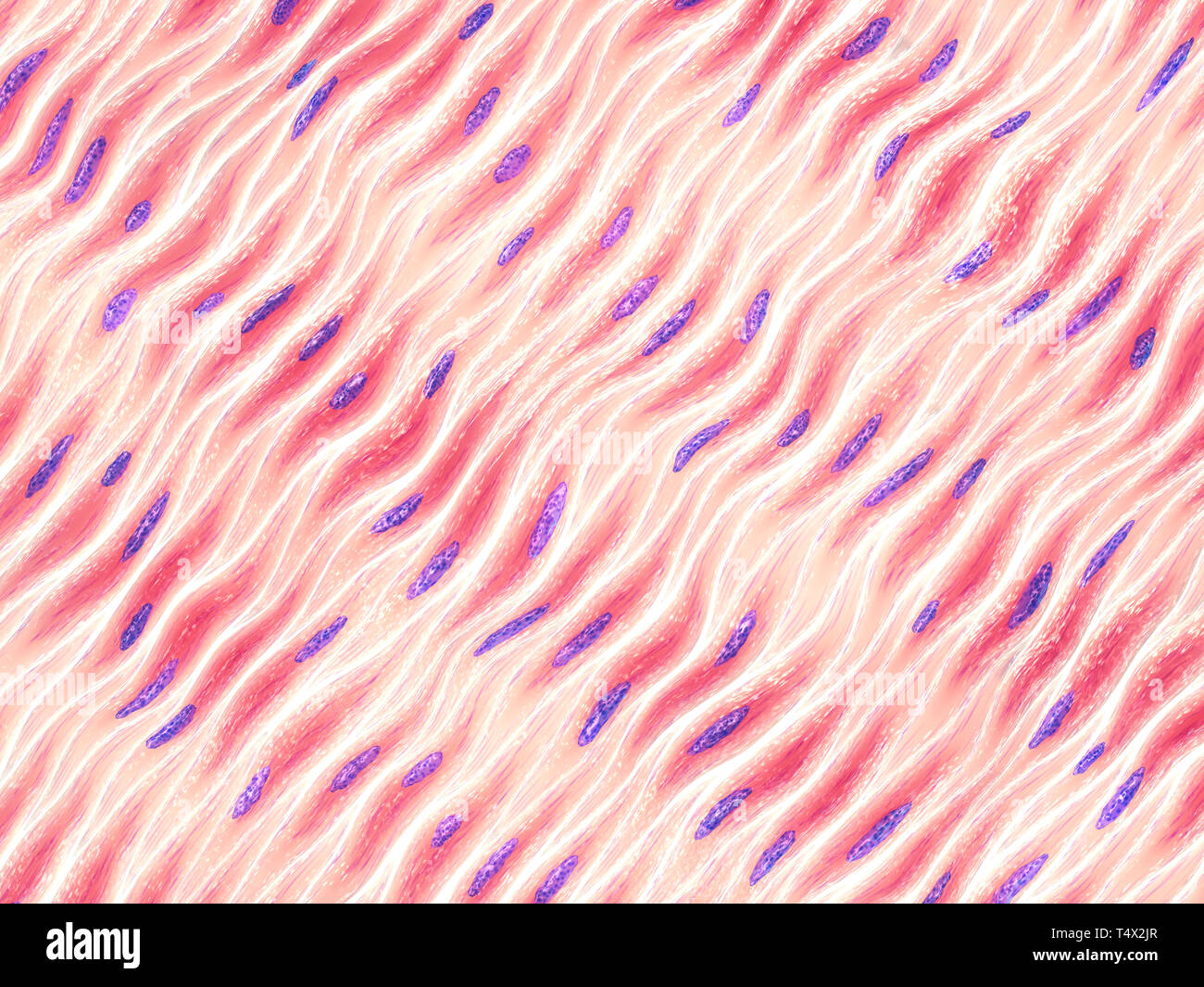 Myocytes, Group of smooth muscle cells Stock Photohttps://www.alamy.com/image-license-details/?v=1https://www.alamy.com/myocytes-group-of-smooth-muscle-cells-image243976623.html
RFT4X2JR–Myocytes, Group of smooth muscle cells
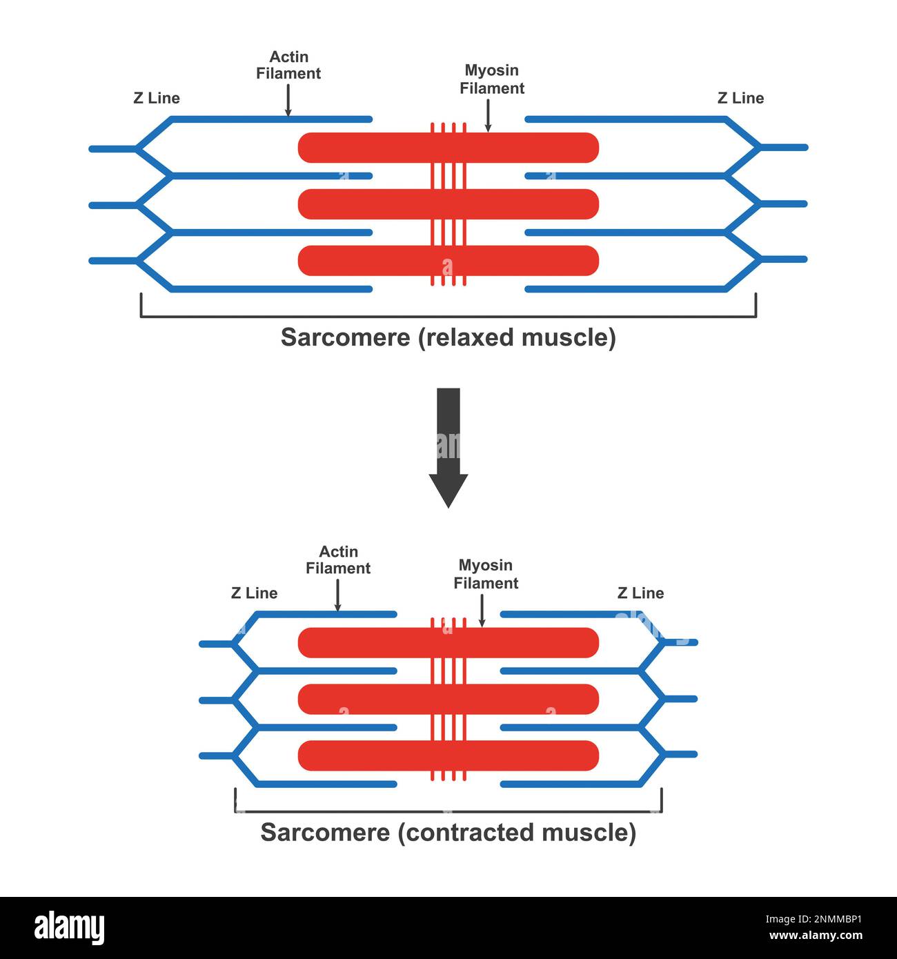 Contraction and relaxation of muscle, illustration Stock Photohttps://www.alamy.com/image-license-details/?v=1https://www.alamy.com/contraction-and-relaxation-of-muscle-illustration-image529052441.html
RF2NMMBP1–Contraction and relaxation of muscle, illustration
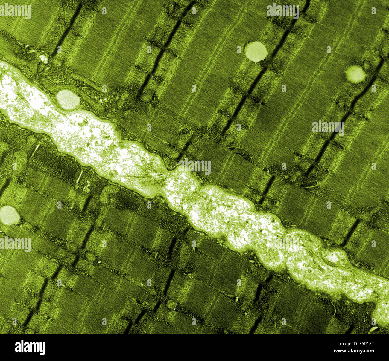 Transmission electron micrograph (TEM) of a thin longitudinal section cut through an area of human skeletal muscle tissue. Stock Photohttps://www.alamy.com/image-license-details/?v=1https://www.alamy.com/stock-photo-transmission-electron-micrograph-tem-of-a-thin-longitudinal-section-72420680.html
RME5R18T–Transmission electron micrograph (TEM) of a thin longitudinal section cut through an area of human skeletal muscle tissue.
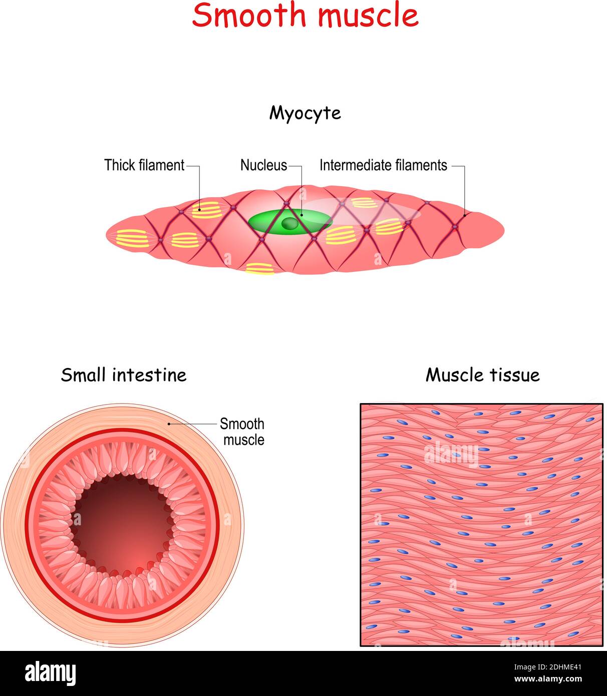 Structure of smooth muscle fibers. anatomy of Myocyte. Background of smooth muscle tissue. Set of vectors illustrations Stock Vectorhttps://www.alamy.com/image-license-details/?v=1https://www.alamy.com/structure-of-smooth-muscle-fibers-anatomy-of-myocyte-background-of-smooth-muscle-tissue-set-of-vectors-illustrations-image389527377.html
RF2DHME41–Structure of smooth muscle fibers. anatomy of Myocyte. Background of smooth muscle tissue. Set of vectors illustrations
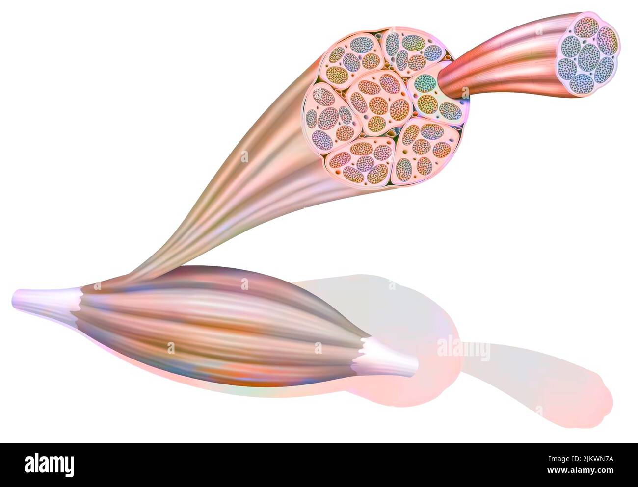 From muscle to muscle fiber: tendon, muscle, muscle fiber. Stock Photohttps://www.alamy.com/image-license-details/?v=1https://www.alamy.com/from-muscle-to-muscle-fiber-tendon-muscle-muscle-fiber-image476923870.html
RF2JKWN7A–From muscle to muscle fiber: tendon, muscle, muscle fiber.
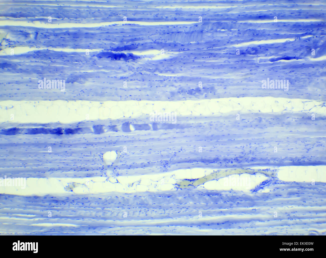 Skeletal muscle tissue longitudinal section under a microscope, Skeletal muscle L.S., 400x Stock Photohttps://www.alamy.com/image-license-details/?v=1https://www.alamy.com/stock-photo-skeletal-muscle-tissue-longitudinal-section-under-a-microscope-skeletal-81101689.html
RFEKXE0W–Skeletal muscle tissue longitudinal section under a microscope, Skeletal muscle L.S., 400x
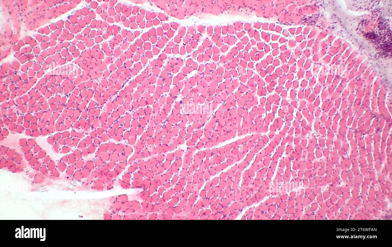 Microscopic structure of skeletal muscle tissue. Сross section of muscle fibers. Haematoxylin end eosin stain. Magnification: x200 Stock Photohttps://www.alamy.com/image-license-details/?v=1https://www.alamy.com/microscopic-structure-of-skeletal-muscle-tissue-ross-section-of-muscle-fibers-haematoxylin-end-eosin-stain-magnification-x200-image572190941.html
RF2T6WFAN–Microscopic structure of skeletal muscle tissue. Сross section of muscle fibers. Haematoxylin end eosin stain. Magnification: x200
 This image shows the structure of a skeletal muscle, revealing the muscle fibers bundle, the motor neuron, the muscle fiber and the myofibril. Stock Photohttps://www.alamy.com/image-license-details/?v=1https://www.alamy.com/this-image-shows-the-structure-of-a-skeletal-muscle-revealing-the-image156174431.html
RMK22A27–This image shows the structure of a skeletal muscle, revealing the muscle fibers bundle, the motor neuron, the muscle fiber and the myofibril.
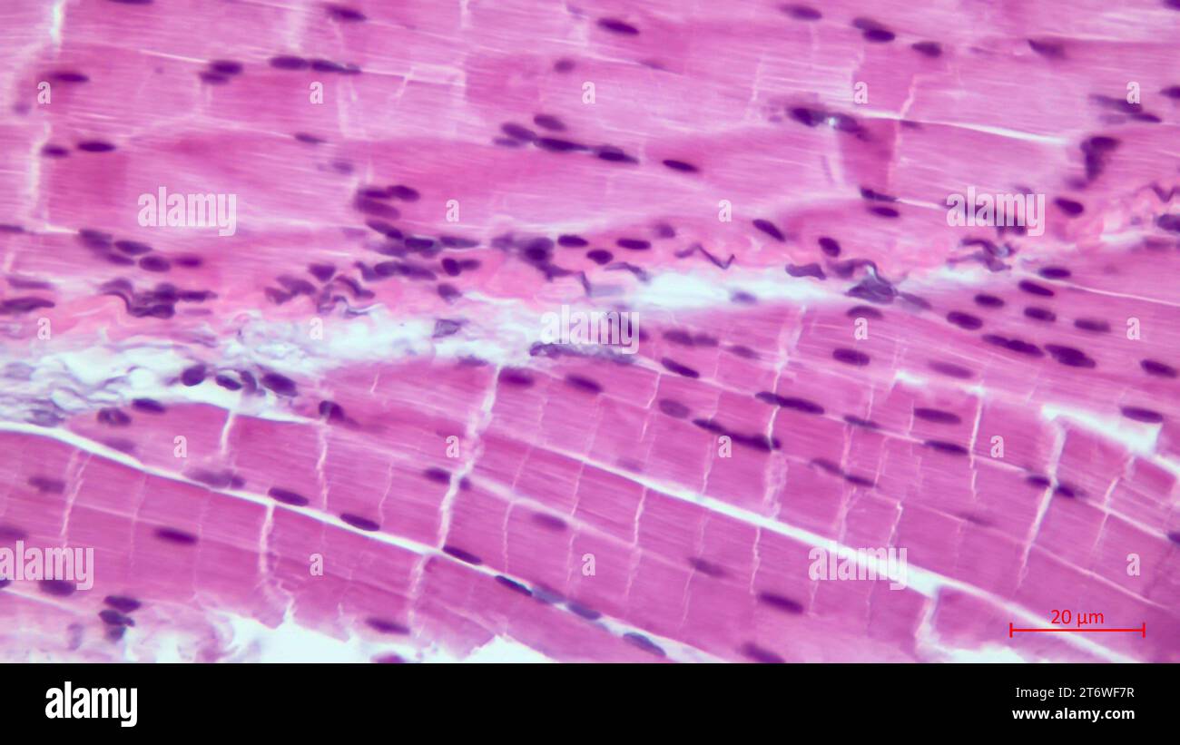 Light micrograph of a section through skeletal muscle. Muscle fibre fascicles. Haematoxylin end eosin stain. Magnification: x200 Stock Photohttps://www.alamy.com/image-license-details/?v=1https://www.alamy.com/light-micrograph-of-a-section-through-skeletal-muscle-muscle-fibre-fascicles-haematoxylin-end-eosin-stain-magnification-x200-image572190859.html
RF2T6WF7R–Light micrograph of a section through skeletal muscle. Muscle fibre fascicles. Haematoxylin end eosin stain. Magnification: x200
 Myocytes, Group of smooth muscle cells on white background Stock Photohttps://www.alamy.com/image-license-details/?v=1https://www.alamy.com/myocytes-group-of-smooth-muscle-cells-on-white-background-image364091923.html
RF2C49PXY–Myocytes, Group of smooth muscle cells on white background
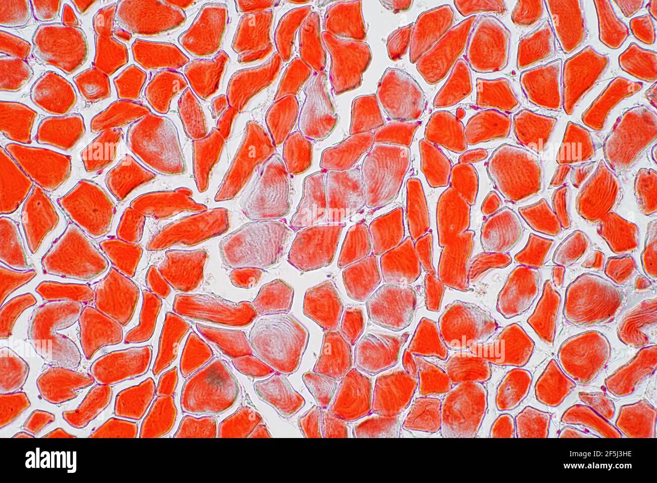 Skeletal muscle, light micrograph Stock Photohttps://www.alamy.com/image-license-details/?v=1https://www.alamy.com/skeletal-muscle-light-micrograph-image416520090.html
RF2F5J3HE–Skeletal muscle, light micrograph
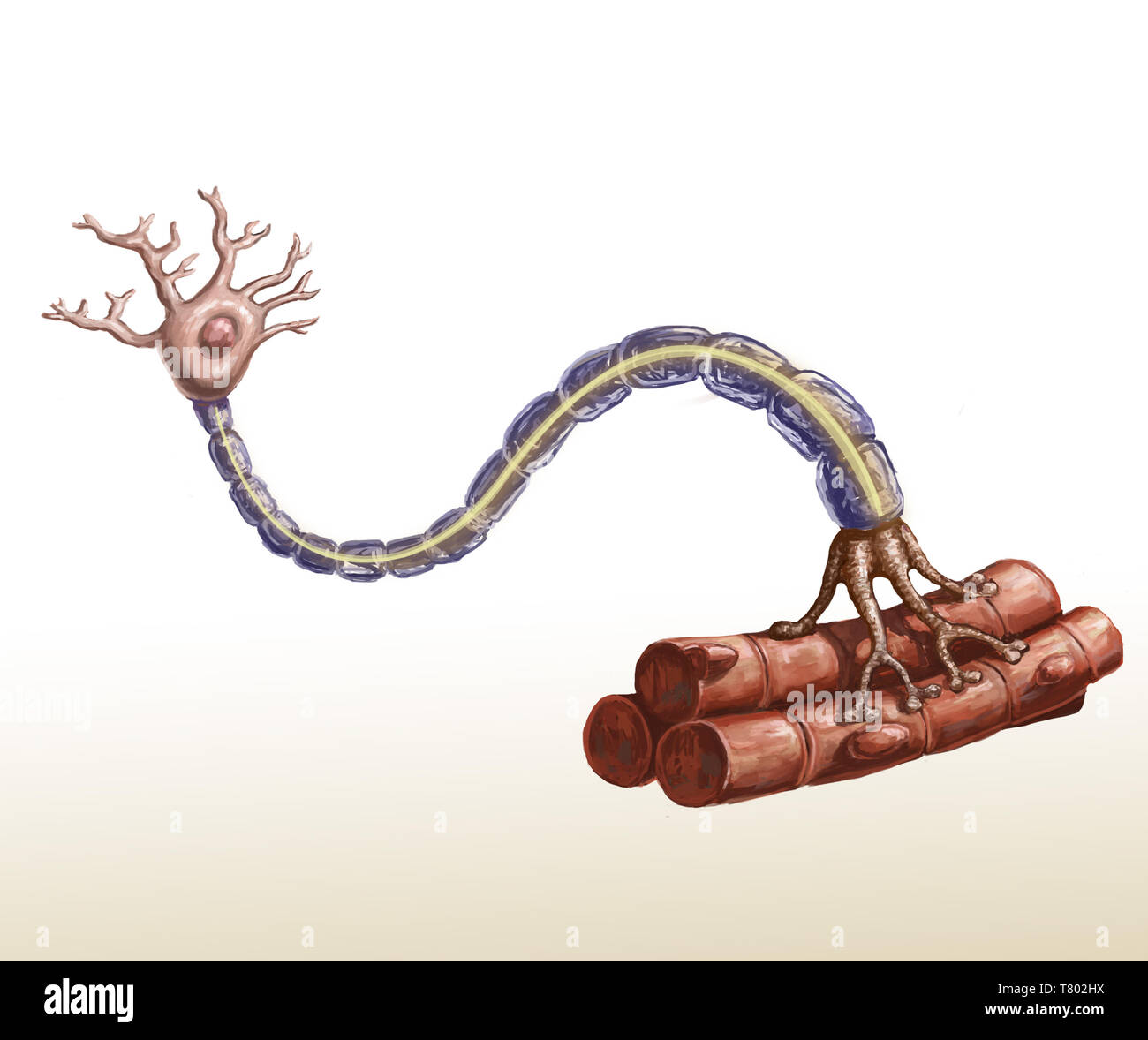 Motor Neuron and Muscle Fiber Illustration Stock Photohttps://www.alamy.com/image-license-details/?v=1https://www.alamy.com/motor-neuron-and-muscle-fiber-illustration-image245864470.html
RMT802HX–Motor Neuron and Muscle Fiber Illustration
 3d rendering of close up thin filaments, Stock Photohttps://www.alamy.com/image-license-details/?v=1https://www.alamy.com/3d-rendering-of-close-up-thin-filaments-image595072602.html
RF2WG3W4X–3d rendering of close up thin filaments,
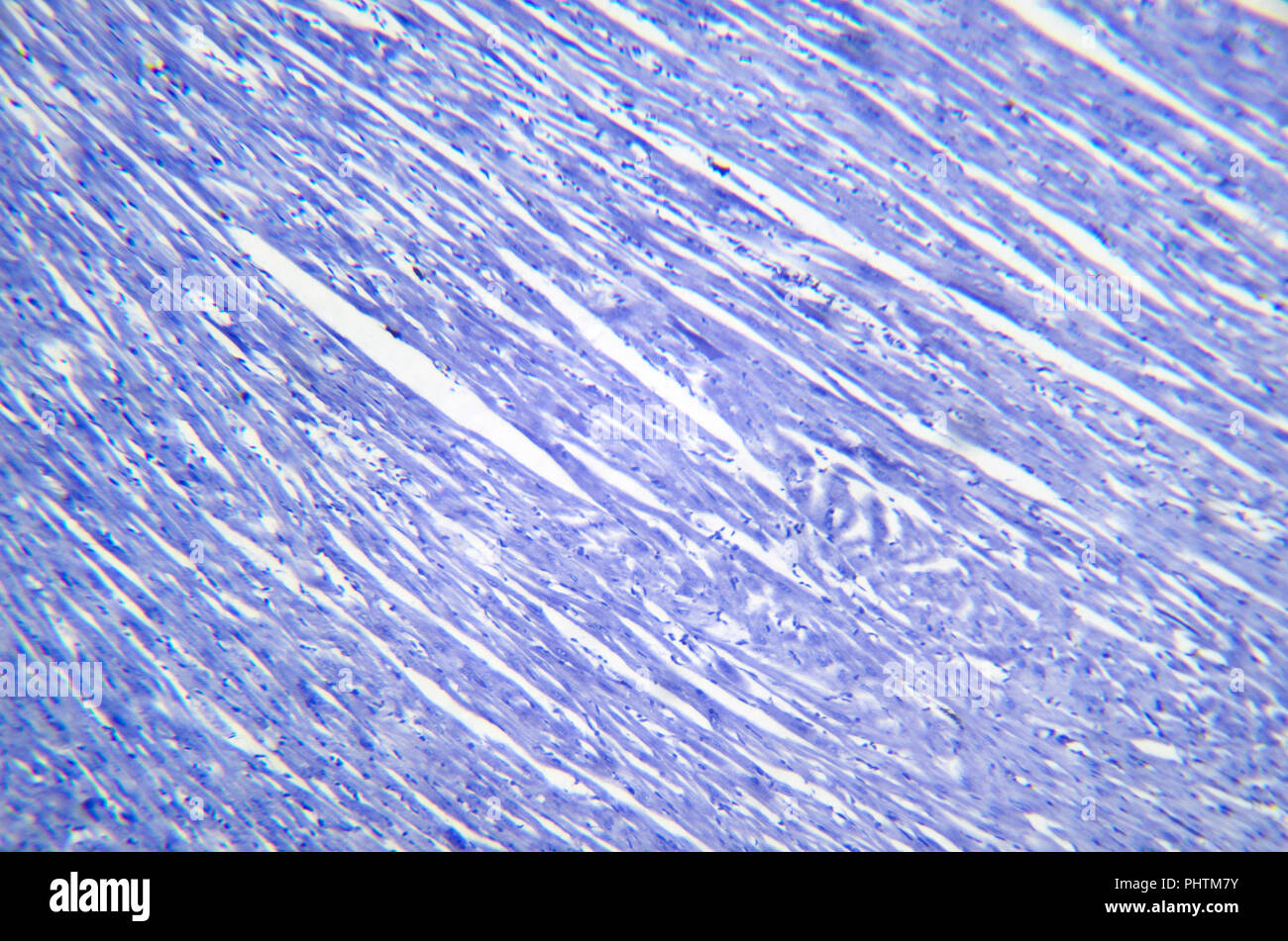 Microscopy photography. Cardiac muscle section. Stock Photohttps://www.alamy.com/image-license-details/?v=1https://www.alamy.com/microscopy-photography-cardiac-muscle-section-image217516319.html
RFPHTM7Y–Microscopy photography. Cardiac muscle section.
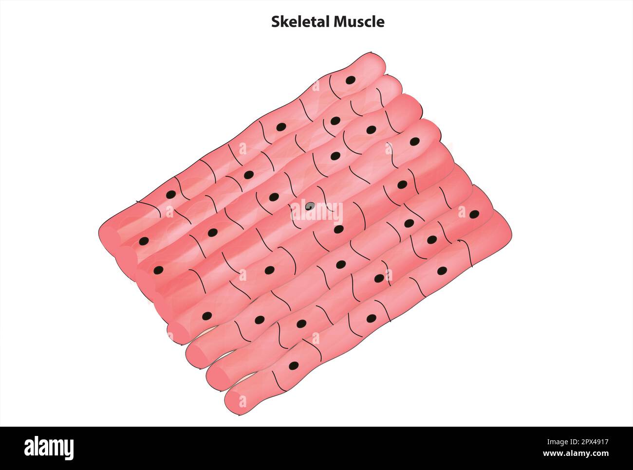 Skeletal muscle tissue Stock Vectorhttps://www.alamy.com/image-license-details/?v=1https://www.alamy.com/skeletal-muscle-tissue-image549597363.html
RF2PX4917–Skeletal muscle tissue
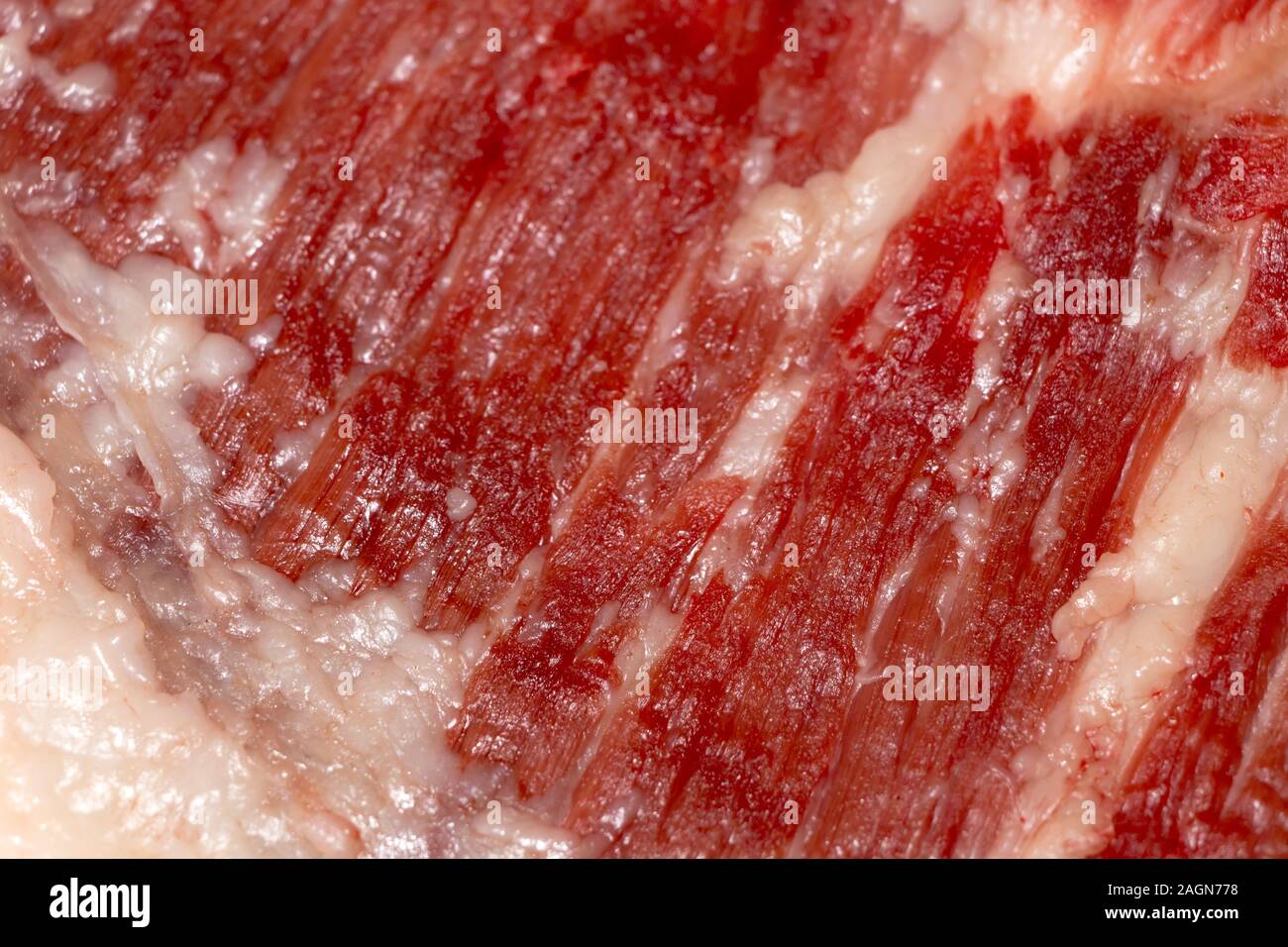 Education anatomy and Histological sample of Muscle tissue and Adipose tissue close up. Selective focus. Animal tissues Stock Photohttps://www.alamy.com/image-license-details/?v=1https://www.alamy.com/education-anatomy-and-histological-sample-of-muscle-tissue-and-adipose-tissue-close-up-selective-focus-animal-tissues-image337298172.html
RF2AGN778–Education anatomy and Histological sample of Muscle tissue and Adipose tissue close up. Selective focus. Animal tissues
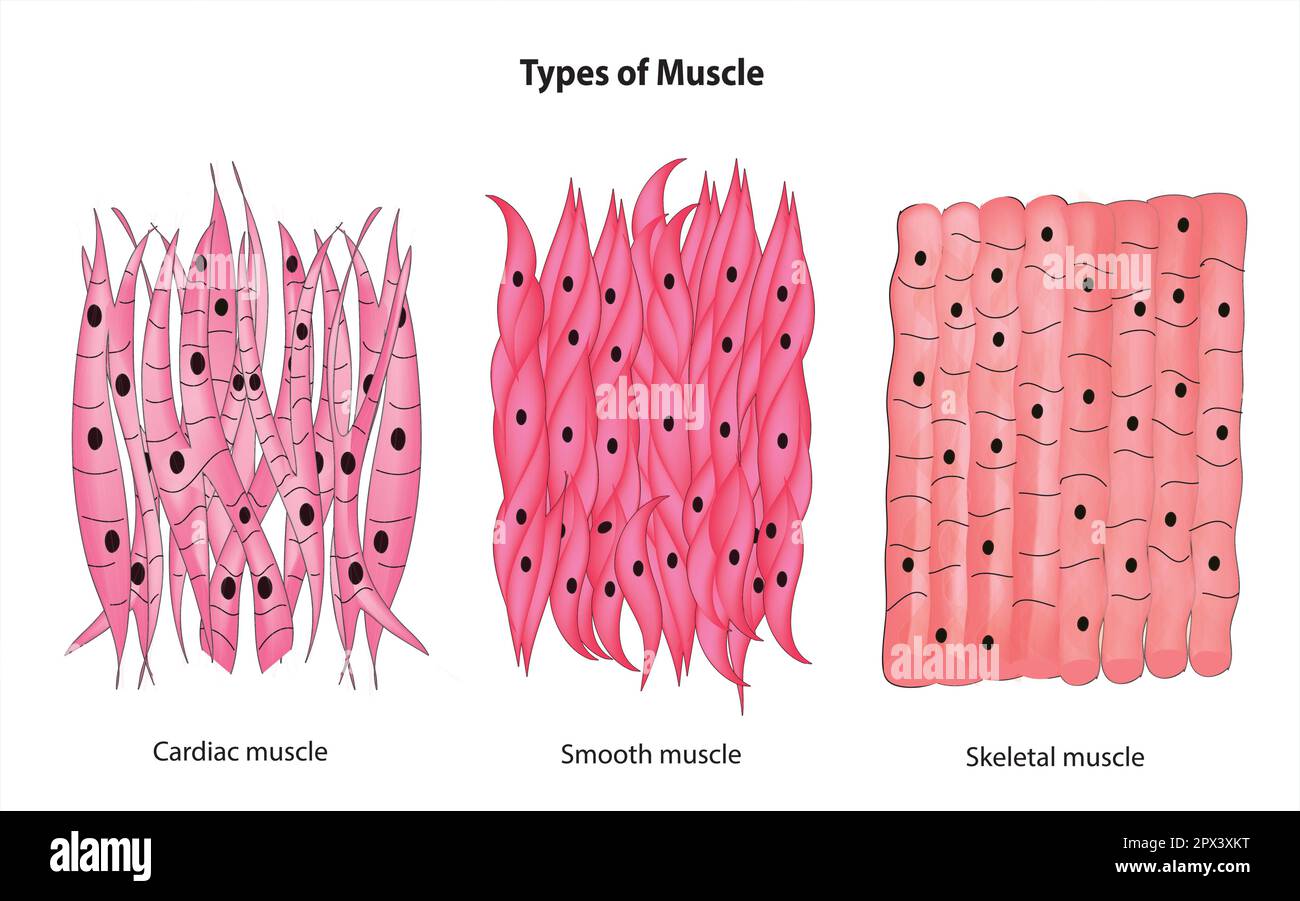 Types of muscle Stock Vectorhttps://www.alamy.com/image-license-details/?v=1https://www.alamy.com/types-of-muscle-image549589260.html
RF2PX3XKT–Types of muscle
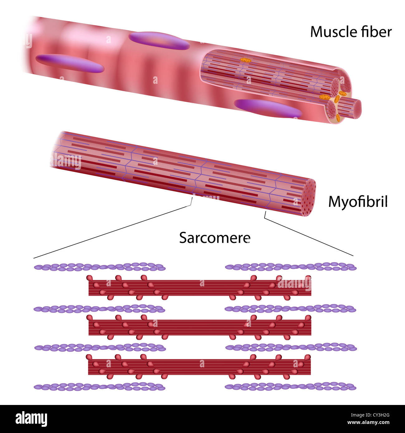 Structure of skeletal muscle fiber Stock Photohttps://www.alamy.com/image-license-details/?v=1https://www.alamy.com/stock-photo-structure-of-skeletal-muscle-fiber-51095704.html
RFCY3H2G–Structure of skeletal muscle fiber
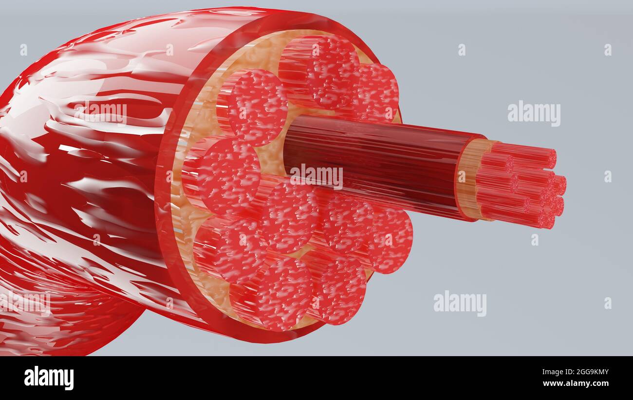 3d Illustration of Muscle Type: Heart muscle - cross section through muscle with muscle fibers visible - 3D Rendering Stock Photohttps://www.alamy.com/image-license-details/?v=1https://www.alamy.com/3d-illustration-of-muscle-type-heart-muscle-cross-section-through-muscle-with-muscle-fibers-visible-3d-rendering-image440306747.html
RM2GG9KMY–3d Illustration of Muscle Type: Heart muscle - cross section through muscle with muscle fibers visible - 3D Rendering
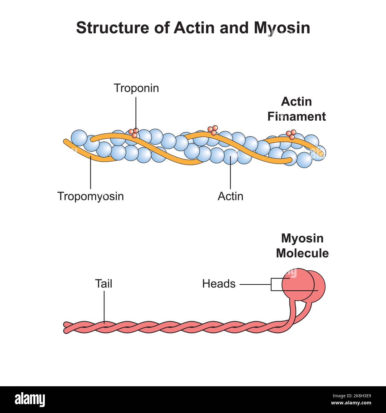 Scientific Designing of Actin and Myosin Structure. Colorful Symbols. Vector Illustration. Stock Vectorhttps://www.alamy.com/image-license-details/?v=1https://www.alamy.com/scientific-designing-of-actin-and-myosin-structure-colorful-symbols-vector-illustration-image487183489.html
RF2K8H3E9–Scientific Designing of Actin and Myosin Structure. Colorful Symbols. Vector Illustration.
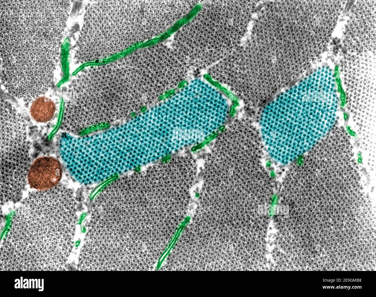 False colour transmission electron microscope (TEM) micrograph showing cross-sectioned myofibrils at the A-band level and two sections (blue) at the H Stock Photohttps://www.alamy.com/image-license-details/?v=1https://www.alamy.com/false-colour-transmission-electron-microscope-tem-micrograph-showing-cross-sectioned-myofibrils-at-the-a-band-level-and-two-sections-blue-at-the-h-image401737596.html
RF2E9GMB8–False colour transmission electron microscope (TEM) micrograph showing cross-sectioned myofibrils at the A-band level and two sections (blue) at the H
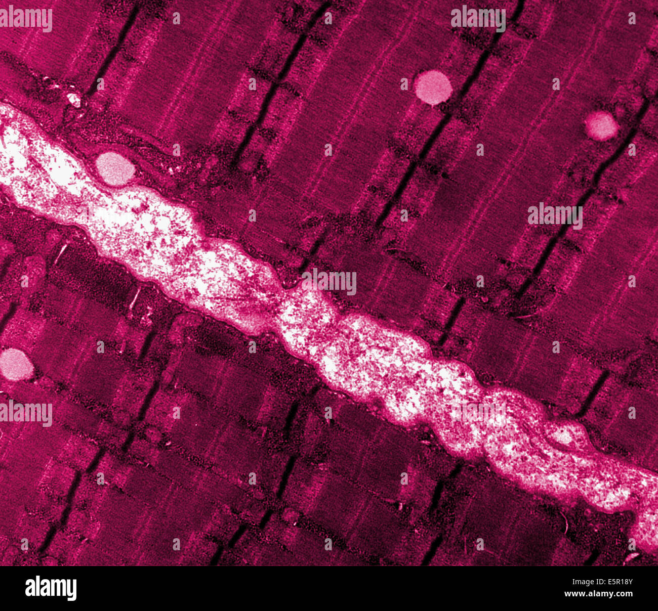 Transmission electron micrograph (TEM) of a thin longitudinal section cut through an area of human skeletal muscle tissue. Stock Photohttps://www.alamy.com/image-license-details/?v=1https://www.alamy.com/stock-photo-transmission-electron-micrograph-tem-of-a-thin-longitudinal-section-72420683.html
RME5R18Y–Transmission electron micrograph (TEM) of a thin longitudinal section cut through an area of human skeletal muscle tissue.
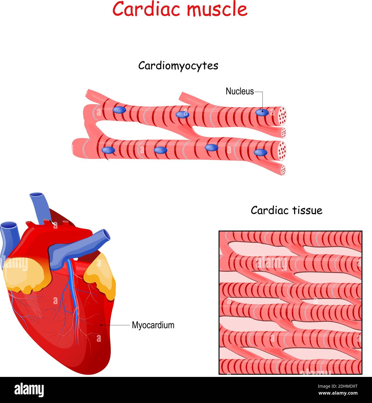 Structure of Cardiac muscle fibers. anatomy of cardiomyocyte. Background of heart muscle tissue. Set of vectors illustrations Stock Vectorhttps://www.alamy.com/image-license-details/?v=1https://www.alamy.com/structure-of-cardiac-muscle-fibers-anatomy-of-cardiomyocyte-background-of-heart-muscle-tissue-set-of-vectors-illustrations-image389527232.html
RF2DHMDXT–Structure of Cardiac muscle fibers. anatomy of cardiomyocyte. Background of heart muscle tissue. Set of vectors illustrations
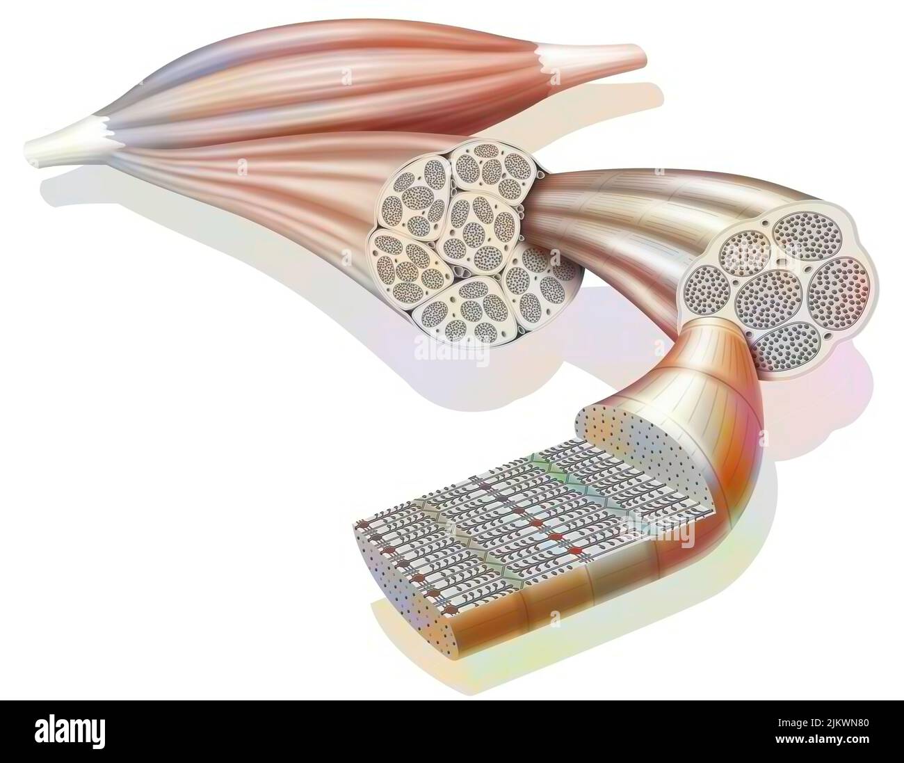 From muscle to muscle fiber: tendon, muscle, muscle fiber. Stock Photohttps://www.alamy.com/image-license-details/?v=1https://www.alamy.com/from-muscle-to-muscle-fiber-tendon-muscle-muscle-fiber-image476923888.html
RF2JKWN80–From muscle to muscle fiber: tendon, muscle, muscle fiber.
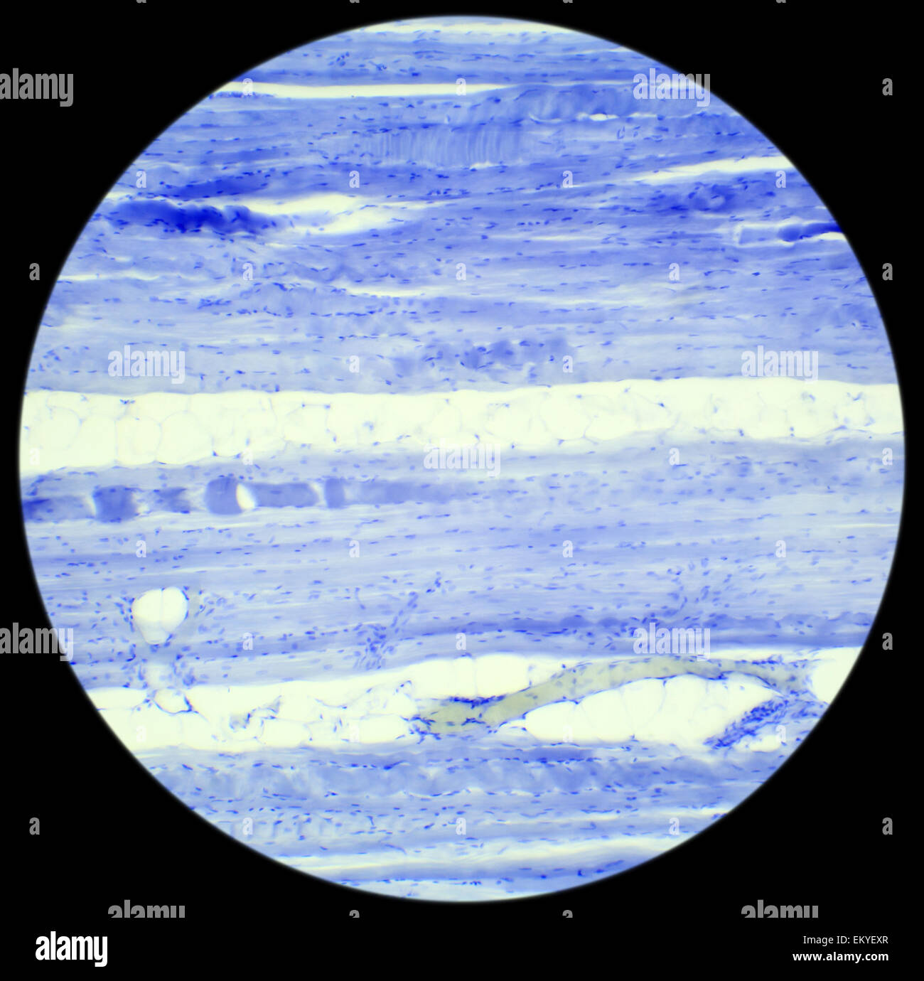 Skeletal muscle tissue longitudinal section under a microscope, Skeletal muscle L.S., 400x Stock Photohttps://www.alamy.com/image-license-details/?v=1https://www.alamy.com/stock-photo-skeletal-muscle-tissue-longitudinal-section-under-a-microscope-skeletal-81124367.html
RFEKYEXR–Skeletal muscle tissue longitudinal section under a microscope, Skeletal muscle L.S., 400x
 Transmission electron micrograph (TEM) of the skeletal muscle also called musculus skeleti of a hamster tongu, Magnification Stock Photohttps://www.alamy.com/image-license-details/?v=1https://www.alamy.com/stock-photo-transmission-electron-micrograph-tem-of-the-skeletal-muscle-also-called-72420975.html
RME5R1KB–Transmission electron micrograph (TEM) of the skeletal muscle also called musculus skeleti of a hamster tongu, Magnification
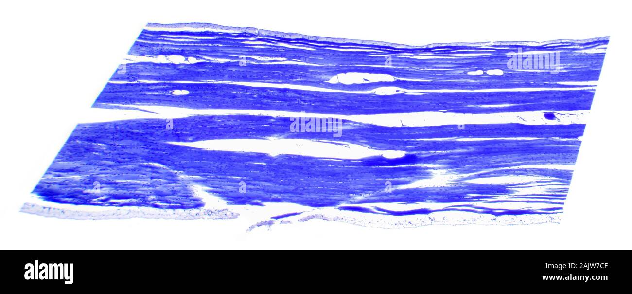 Skeletal muscle tissue cross-section under a microscope, Skeletal muscle C.S. Stock Photohttps://www.alamy.com/image-license-details/?v=1https://www.alamy.com/skeletal-muscle-tissue-cross-section-under-a-microscope-skeletal-muscle-cs-image338615439.html
RF2AJW7CF–Skeletal muscle tissue cross-section under a microscope, Skeletal muscle C.S.
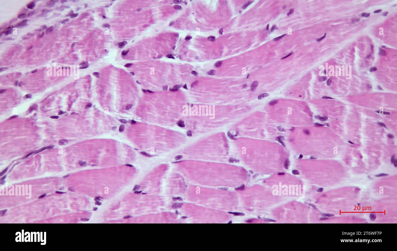 Light micrograph of a section through skeletal muscle. Muscle fibre fascicles. Haematoxylin end eosin stain. Magnification: x200 Stock Photohttps://www.alamy.com/image-license-details/?v=1https://www.alamy.com/light-micrograph-of-a-section-through-skeletal-muscle-muscle-fibre-fascicles-haematoxylin-end-eosin-stain-magnification-x200-image572190858.html
RF2T6WF7P–Light micrograph of a section through skeletal muscle. Muscle fibre fascicles. Haematoxylin end eosin stain. Magnification: x200
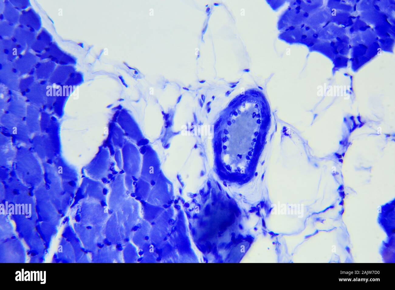 Skeletal muscle tissue longitudinal section under a microscope, Skeletal muscle L.S. Stock Photohttps://www.alamy.com/image-license-details/?v=1https://www.alamy.com/skeletal-muscle-tissue-longitudinal-section-under-a-microscope-skeletal-muscle-ls-image338615452.html
RF2AJW7D0–Skeletal muscle tissue longitudinal section under a microscope, Skeletal muscle L.S.
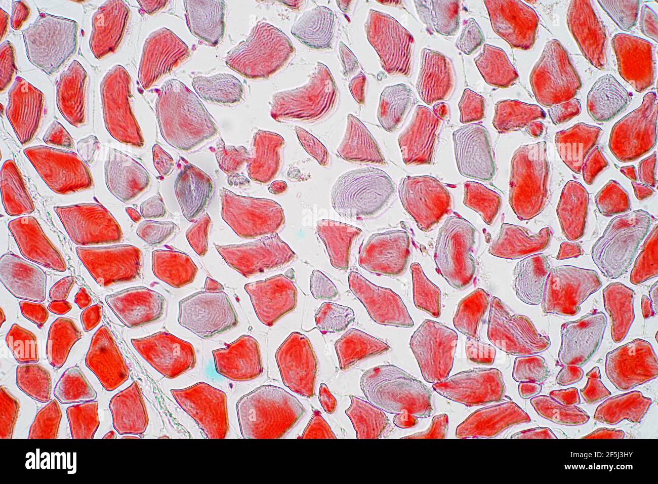 Skeletal muscle, light micrograph Stock Photohttps://www.alamy.com/image-license-details/?v=1https://www.alamy.com/skeletal-muscle-light-micrograph-image416520103.html
RF2F5J3HY–Skeletal muscle, light micrograph
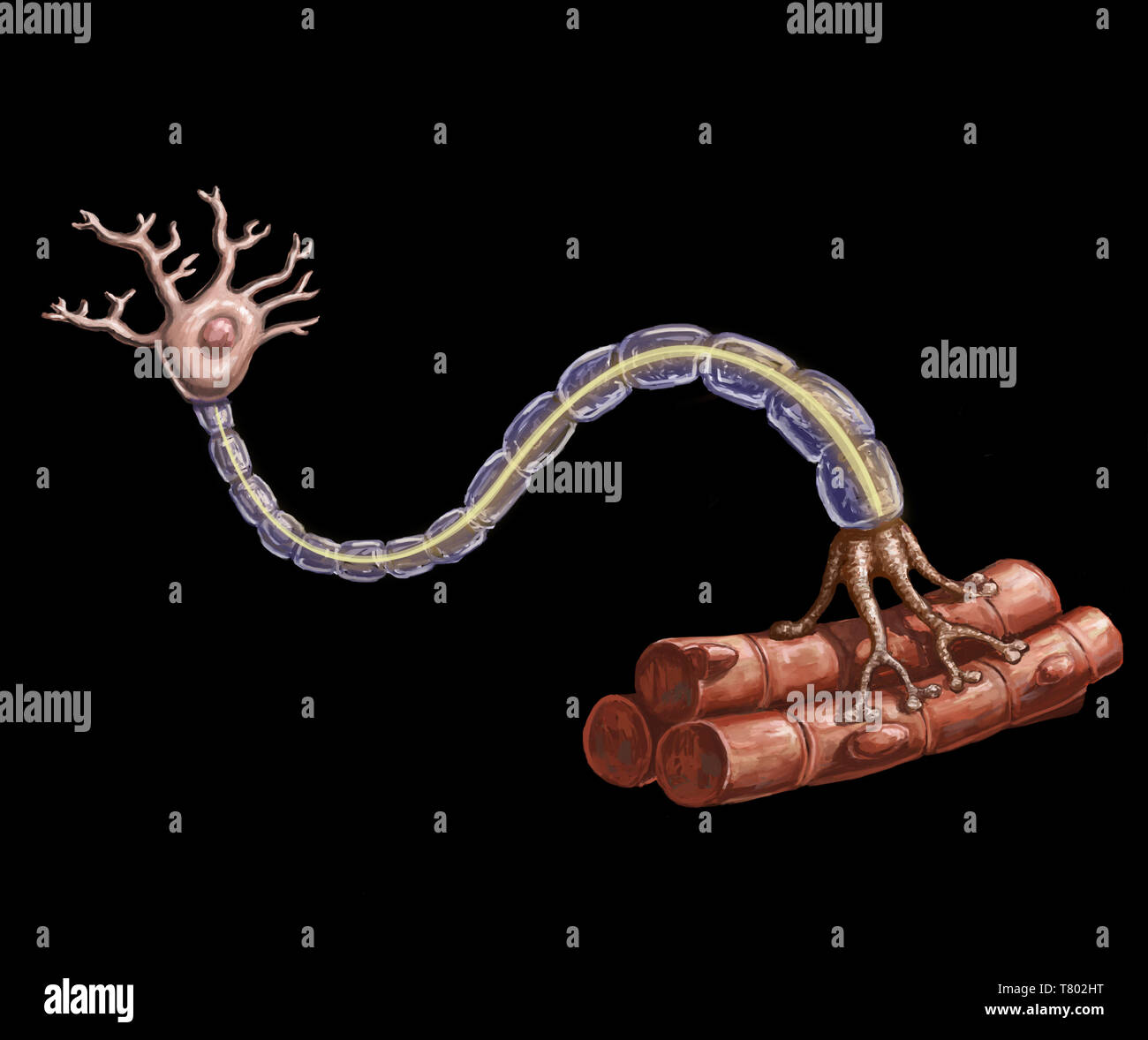 Motor Neuron and Muscle Fiber Illustration Stock Photohttps://www.alamy.com/image-license-details/?v=1https://www.alamy.com/motor-neuron-and-muscle-fiber-illustration-image245864468.html
RMT802HT–Motor Neuron and Muscle Fiber Illustration
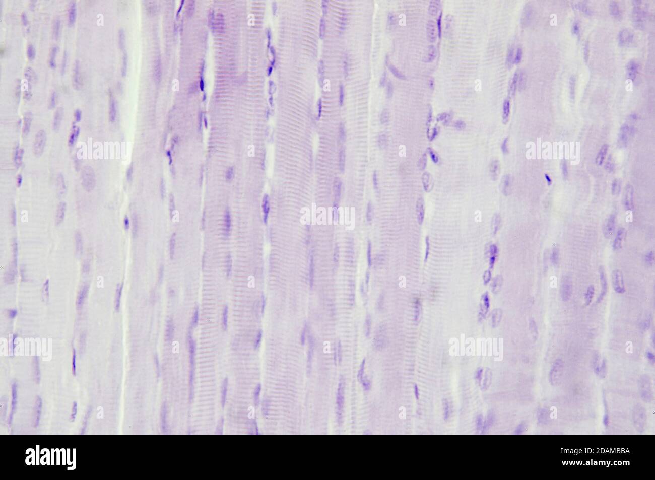 Skeletal muscle, light micrograph. Stock Photohttps://www.alamy.com/image-license-details/?v=1https://www.alamy.com/skeletal-muscle-light-micrograph-image385222638.html
RF2DAMBBA–Skeletal muscle, light micrograph.
 3d rendering of close up thin filaments, Stock Photohttps://www.alamy.com/image-license-details/?v=1https://www.alamy.com/3d-rendering-of-close-up-thin-filaments-image595072605.html
RF2WG3W51–3d rendering of close up thin filaments,
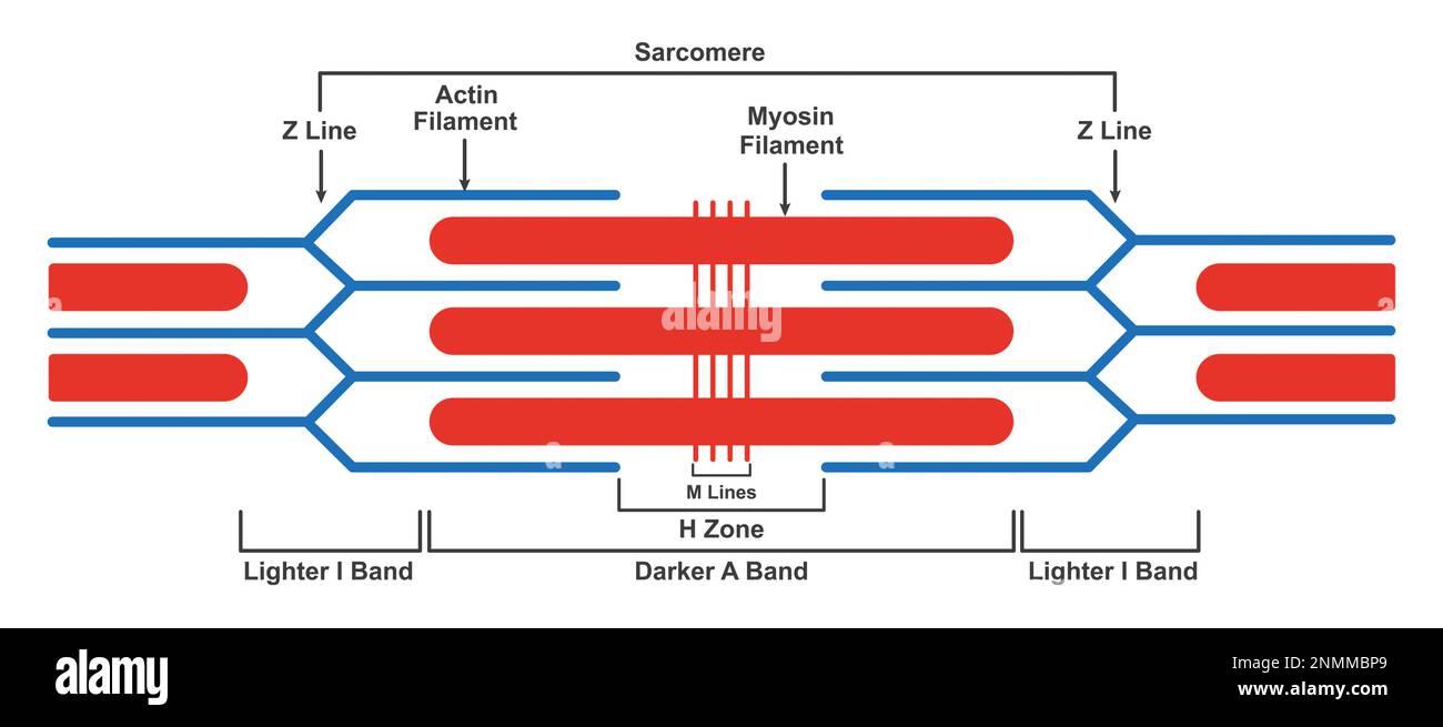 Sarcomere structure, illustration Stock Photohttps://www.alamy.com/image-license-details/?v=1https://www.alamy.com/sarcomere-structure-illustration-image529052449.html
RF2NMMBP9–Sarcomere structure, illustration
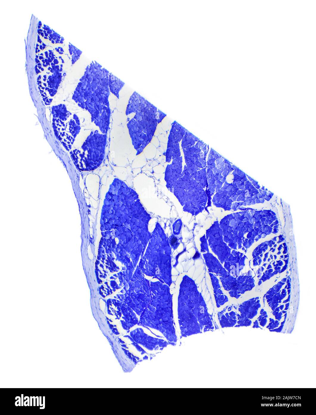 Skeletal muscle tissue longitudinal section under a microscope, Skeletal muscle L.S. Stock Photohttps://www.alamy.com/image-license-details/?v=1https://www.alamy.com/skeletal-muscle-tissue-longitudinal-section-under-a-microscope-skeletal-muscle-ls-image338615445.html
RF2AJW7CN–Skeletal muscle tissue longitudinal section under a microscope, Skeletal muscle L.S.
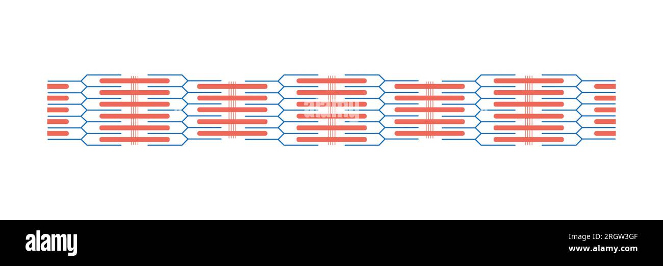 Sarcomere structure, illustration Stock Photohttps://www.alamy.com/image-license-details/?v=1https://www.alamy.com/sarcomere-structure-illustration-image561117887.html
RF2RGW3GF–Sarcomere structure, illustration
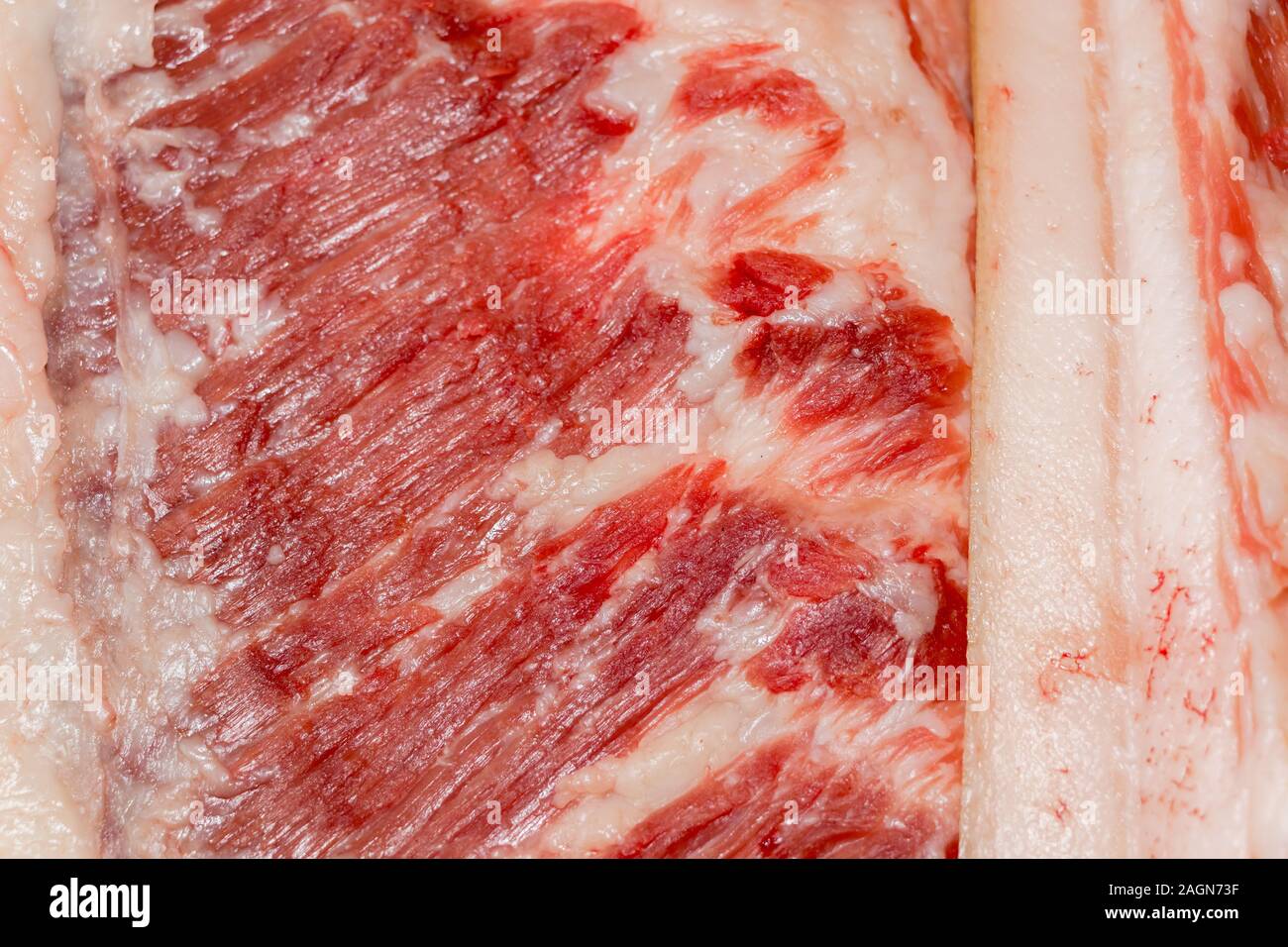 Education anatomy and Histological sample of Muscle tissue and Adipose tissue close up. Selective focus. Animal tissues Stock Photohttps://www.alamy.com/image-license-details/?v=1https://www.alamy.com/education-anatomy-and-histological-sample-of-muscle-tissue-and-adipose-tissue-close-up-selective-focus-animal-tissues-image337298067.html
RF2AGN73F–Education anatomy and Histological sample of Muscle tissue and Adipose tissue close up. Selective focus. Animal tissues
 Skeletal muscle structure Stock Photohttps://www.alamy.com/image-license-details/?v=1https://www.alamy.com/stock-photo-skeletal-muscle-structure-51023138.html
RFCY08EX–Skeletal muscle structure
 Scientific Designing of Myosin Molecule Structure. Colorful Symbols. Vector Illustration. Stock Vectorhttps://www.alamy.com/image-license-details/?v=1https://www.alamy.com/scientific-designing-of-myosin-molecule-structure-colorful-symbols-vector-illustration-image487183314.html
RF2K8H382–Scientific Designing of Myosin Molecule Structure. Colorful Symbols. Vector Illustration.
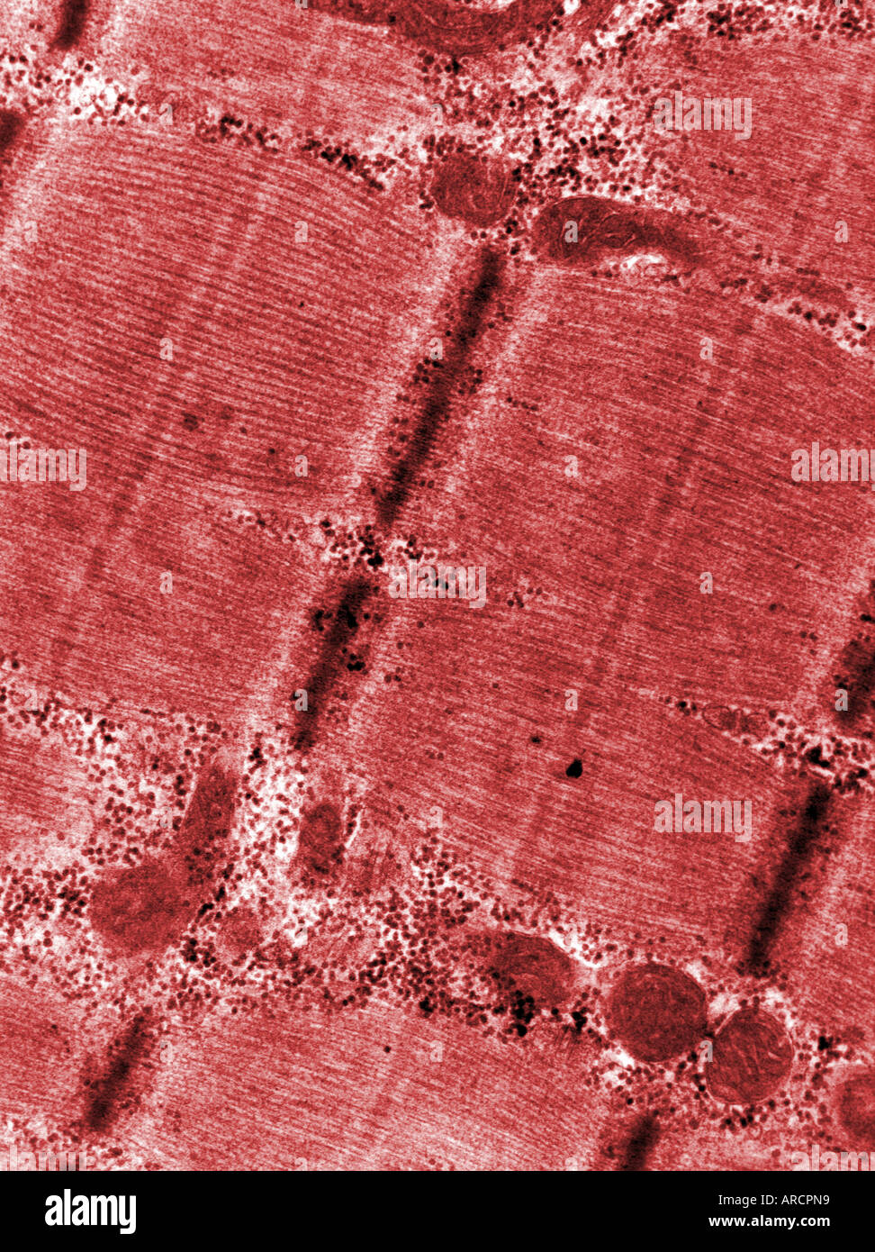 A transmission electron micrograph (TEM) of myofibrils in skeletal muscle fibre, Stock Photohttps://www.alamy.com/image-license-details/?v=1https://www.alamy.com/a-transmission-electron-micrograph-tem-of-myofibrils-in-skeletal-muscle-image9150680.html
RMARCPN9–A transmission electron micrograph (TEM) of myofibrils in skeletal muscle fibre,
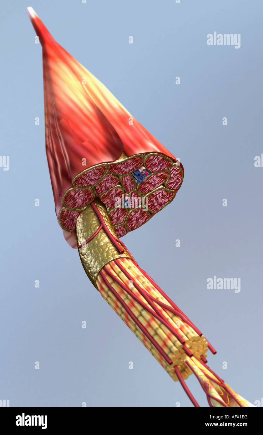 Muscle structure Stock Photohttps://www.alamy.com/image-license-details/?v=1https://www.alamy.com/stock-photo-muscle-structure-14030967.html
RFAFX1EG–Muscle structure
 . The Biological bulletin. Biology; Zoology; Biology; Marine Biology. Fiber Myofibril Triad (Perifibrillar) Triad (Intrafibrillar). Fiber Myofibril Triad (Perifibrillar) Figure 4. Stylized cross-sectional views of single muscle fibers amongst crustaceans, showing distribution of triads and subdivision into myonbrils. A. Single fiber not subdivided into myofibrils and with scat- tered triads as in the ostracod Cypnclopsis vidua (Fahrenbach. 1964) and the cephalocand lliilchin.wniella macracantha (present report). B. Single fiber subdivided into myofibrils by longitudinally running sarcoplasmic Stock Photohttps://www.alamy.com/image-license-details/?v=1https://www.alamy.com/the-biological-bulletin-biology-zoology-biology-marine-biology-fiber-myofibril-triad-perifibrillar-triad-intrafibrillar-fiber-myofibril-triad-perifibrillar-figure-4-stylized-cross-sectional-views-of-single-muscle-fibers-amongst-crustaceans-showing-distribution-of-triads-and-subdivision-into-myonbrils-a-single-fiber-not-subdivided-into-myofibrils-and-with-scat-tered-triads-as-in-the-ostracod-cypnclopsis-vidua-fahrenbach-1964-and-the-cephalocand-lliilchinwniella-macracantha-present-report-b-single-fiber-subdivided-into-myofibrils-by-longitudinally-running-sarcoplasmic-image234643370.html
RMRHMX0A–. The Biological bulletin. Biology; Zoology; Biology; Marine Biology. Fiber Myofibril Triad (Perifibrillar) Triad (Intrafibrillar). Fiber Myofibril Triad (Perifibrillar) Figure 4. Stylized cross-sectional views of single muscle fibers amongst crustaceans, showing distribution of triads and subdivision into myonbrils. A. Single fiber not subdivided into myofibrils and with scat- tered triads as in the ostracod Cypnclopsis vidua (Fahrenbach. 1964) and the cephalocand lliilchin.wniella macracantha (present report). B. Single fiber subdivided into myofibrils by longitudinally running sarcoplasmic
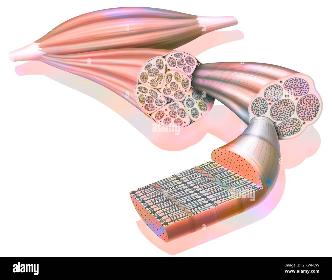 From muscle to muscle fiber: tendon, muscle, muscle fiber. Stock Photohttps://www.alamy.com/image-license-details/?v=1https://www.alamy.com/from-muscle-to-muscle-fiber-tendon-muscle-muscle-fiber-image476923885.html
RF2JKWN7W–From muscle to muscle fiber: tendon, muscle, muscle fiber.
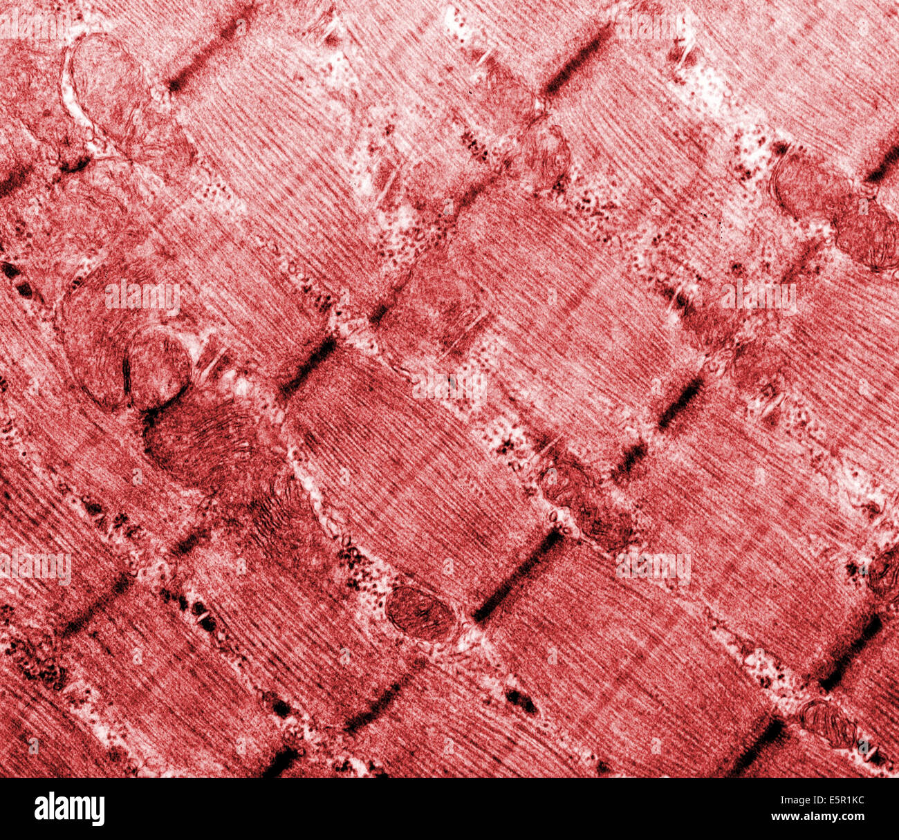 Transmission electron micrograph (TEM) of the skeletal muscle also called musculus skeleti of a hamster tongue, Magnification Stock Photohttps://www.alamy.com/image-license-details/?v=1https://www.alamy.com/stock-photo-transmission-electron-micrograph-tem-of-the-skeletal-muscle-also-called-72420976.html
RME5R1KC–Transmission electron micrograph (TEM) of the skeletal muscle also called musculus skeleti of a hamster tongue, Magnification
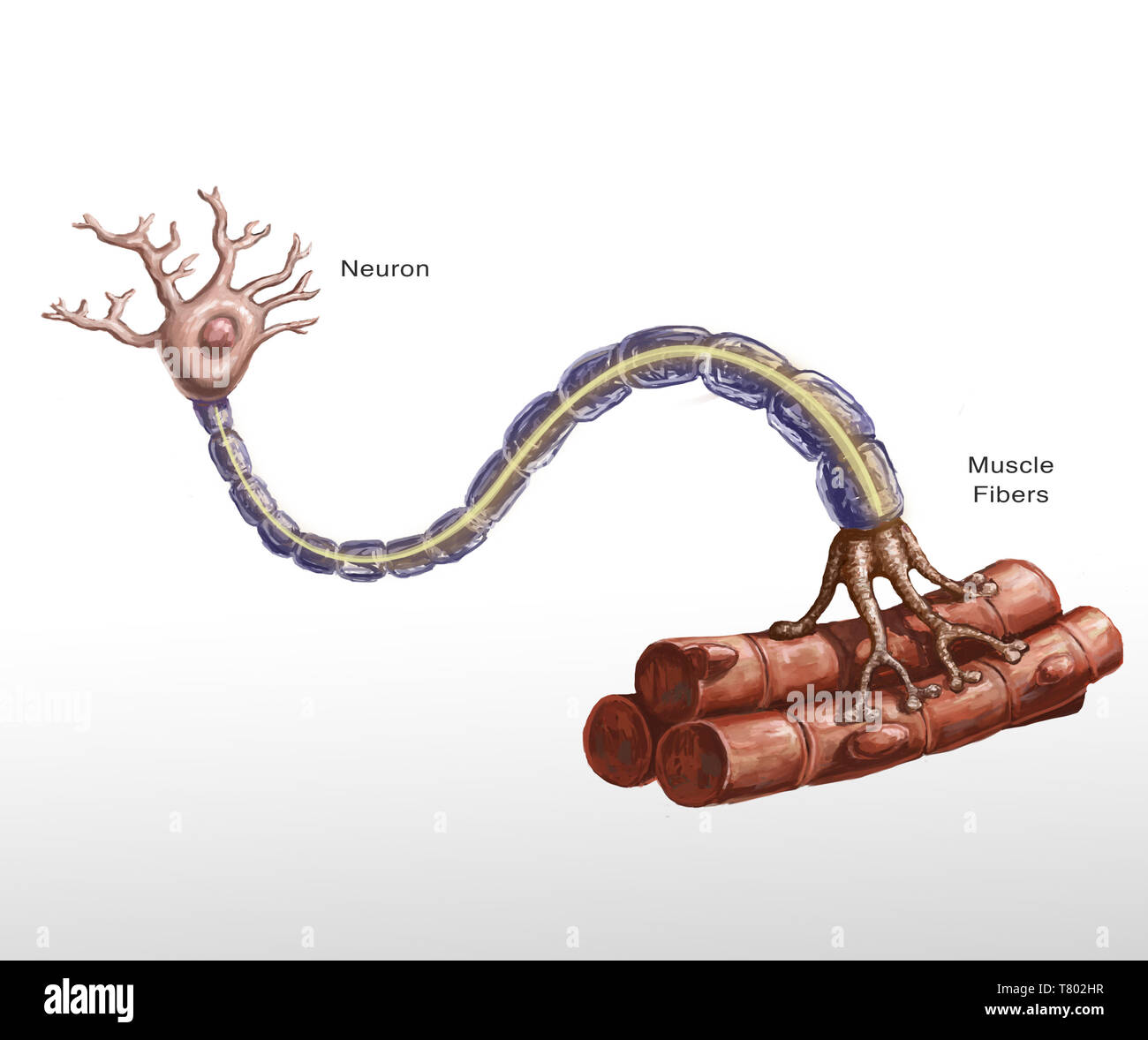 Motor Neuron and Muscle Fiber Illustration Stock Photohttps://www.alamy.com/image-license-details/?v=1https://www.alamy.com/motor-neuron-and-muscle-fiber-illustration-image245864467.html
RMT802HR–Motor Neuron and Muscle Fiber Illustration
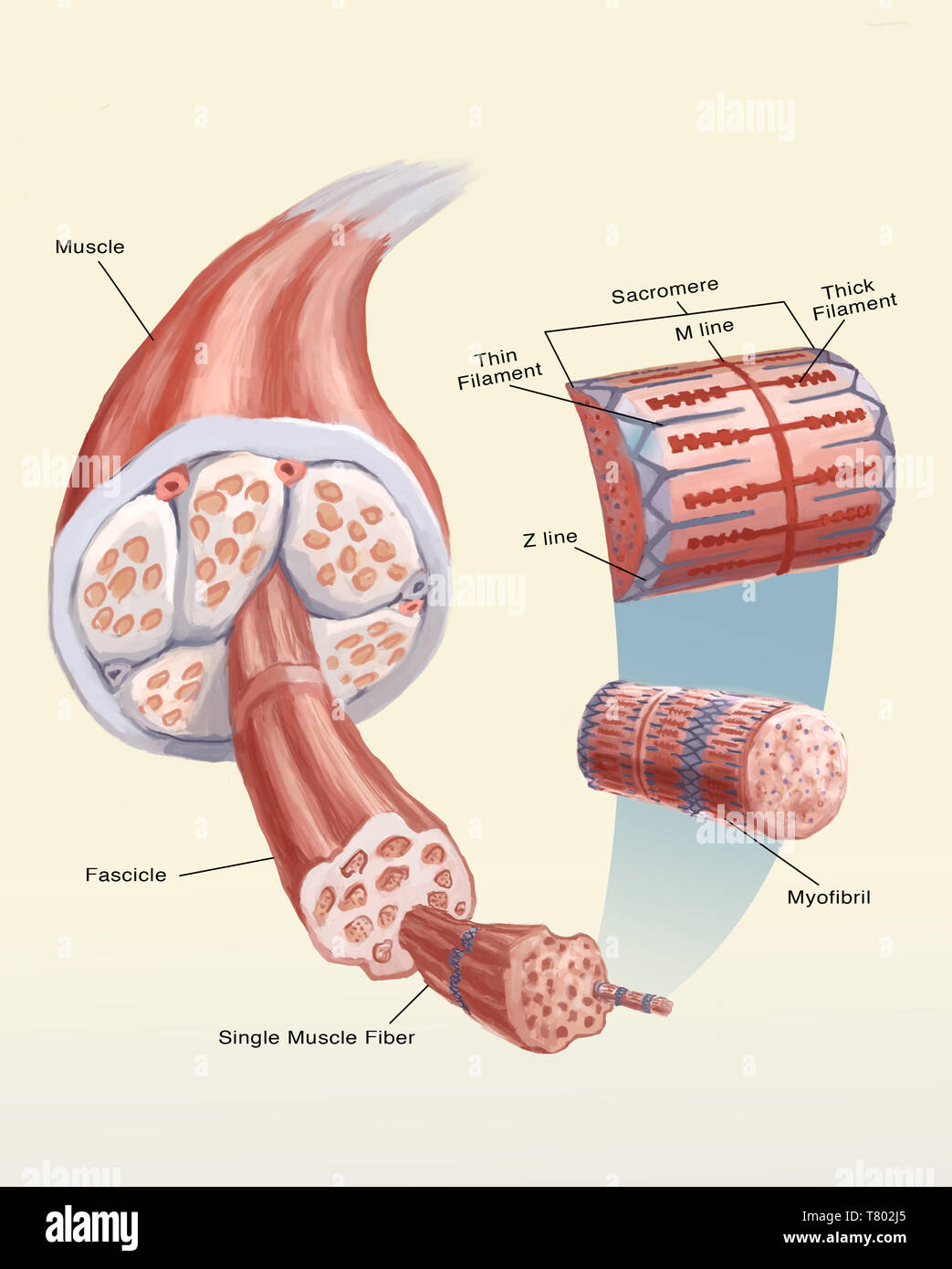 Muscle Cell, Illustration Stock Photohttps://www.alamy.com/image-license-details/?v=1https://www.alamy.com/muscle-cell-illustration-image245864477.html
RMT802J5–Muscle Cell, Illustration
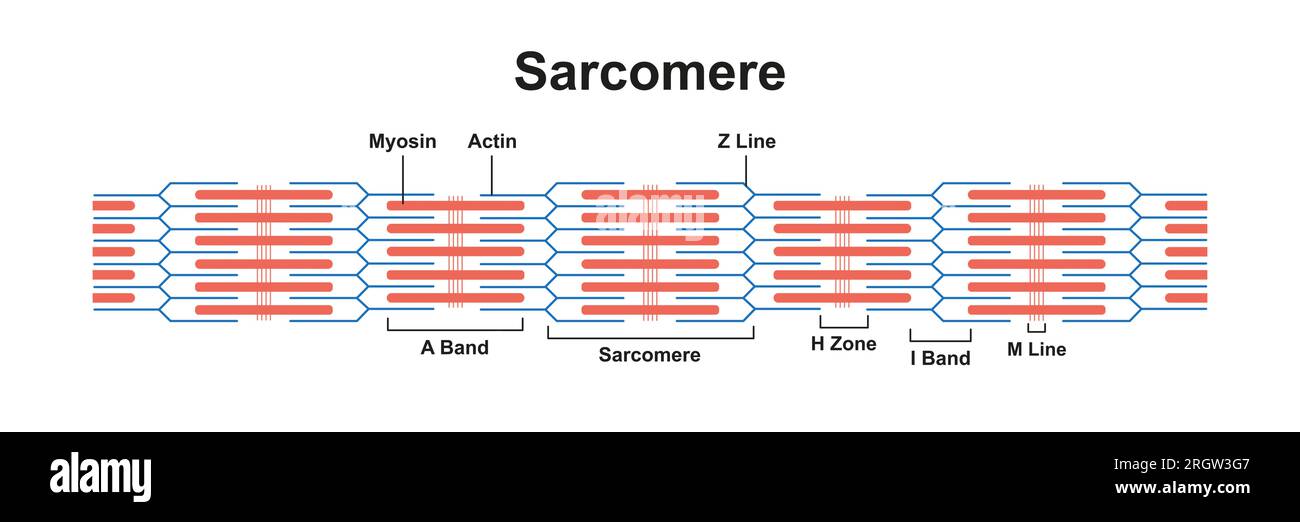 Sarcomere structure, illustration Stock Photohttps://www.alamy.com/image-license-details/?v=1https://www.alamy.com/sarcomere-structure-illustration-image561117879.html
RF2RGW3G7–Sarcomere structure, illustration
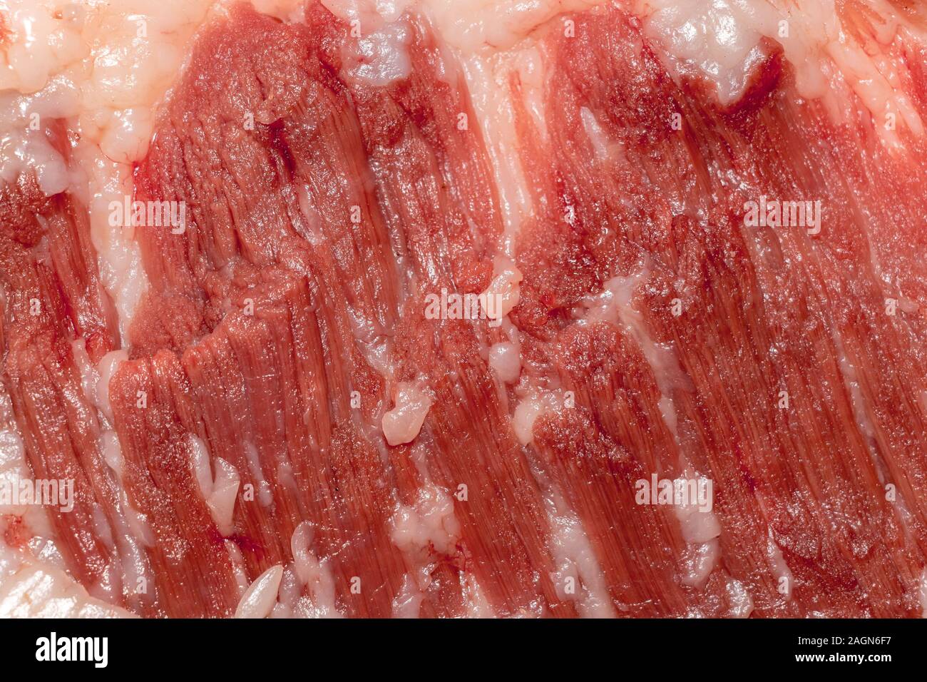 Education anatomy and Histological sample of Muscle tissue and Adipose tissue close up. Selective focus. Animal tissues Stock Photohttps://www.alamy.com/image-license-details/?v=1https://www.alamy.com/education-anatomy-and-histological-sample-of-muscle-tissue-and-adipose-tissue-close-up-selective-focus-animal-tissues-image337297611.html
RF2AGN6F7–Education anatomy and Histological sample of Muscle tissue and Adipose tissue close up. Selective focus. Animal tissues
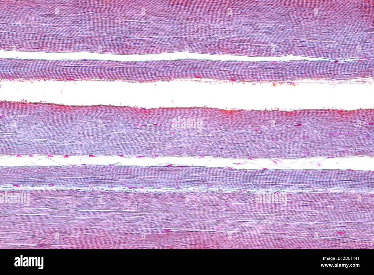 Human skeletal muscle, light micrograph Stock Photohttps://www.alamy.com/image-license-details/?v=1https://www.alamy.com/human-skeletal-muscle-light-micrograph-image387258481.html
RF2DE1441–Human skeletal muscle, light micrograph
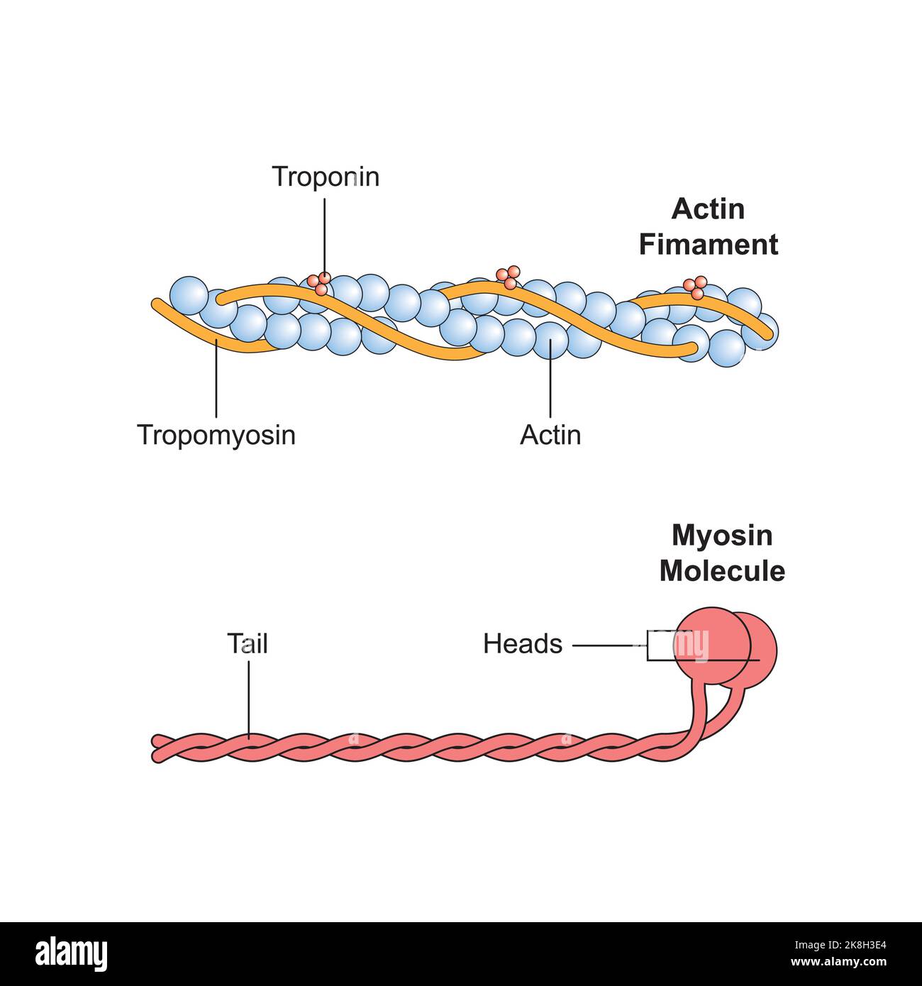 Scientific Designing of Actin and Myosin Structure. Colorful Symbols. Vector Illustration. Stock Vectorhttps://www.alamy.com/image-license-details/?v=1https://www.alamy.com/scientific-designing-of-actin-and-myosin-structure-colorful-symbols-vector-illustration-image487183484.html
RF2K8H3E4–Scientific Designing of Actin and Myosin Structure. Colorful Symbols. Vector Illustration.
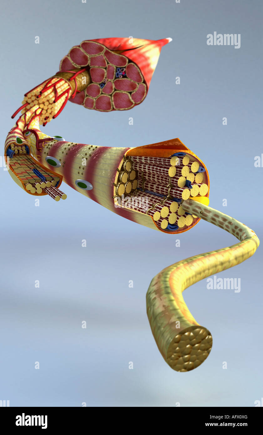 Muscle structure Stock Photohttps://www.alamy.com/image-license-details/?v=1https://www.alamy.com/stock-photo-muscle-structure-14030775.html
RFAFX0XG–Muscle structure
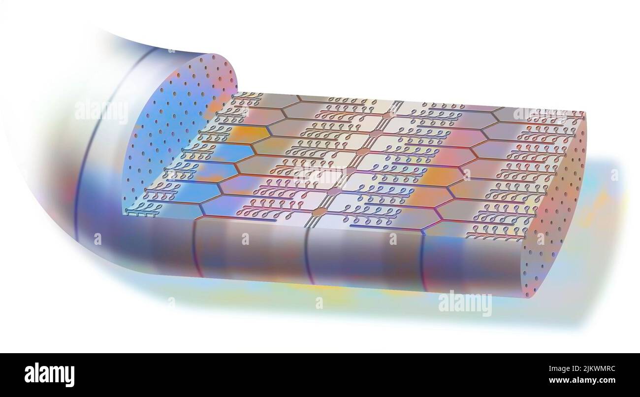 Contracted myofibril, made up of myofilaments and proteins, allowing muscle contraction. Stock Photohttps://www.alamy.com/image-license-details/?v=1https://www.alamy.com/contracted-myofibril-made-up-of-myofilaments-and-proteins-allowing-muscle-contraction-image476923536.html
RF2JKWMRC–Contracted myofibril, made up of myofilaments and proteins, allowing muscle contraction.
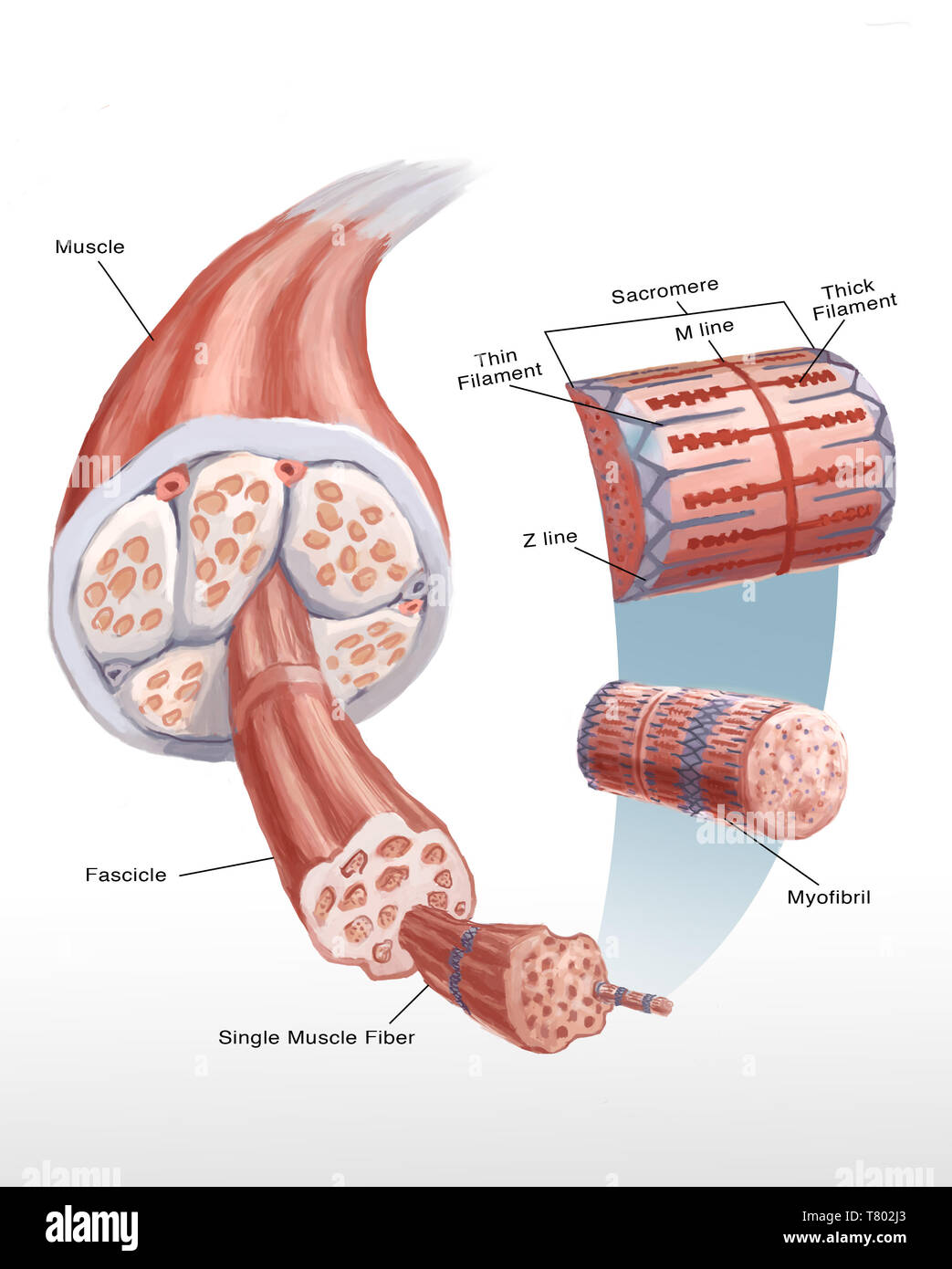 Muscle Cell, Illustration Stock Photohttps://www.alamy.com/image-license-details/?v=1https://www.alamy.com/muscle-cell-illustration-image245864475.html
RMT802J3–Muscle Cell, Illustration
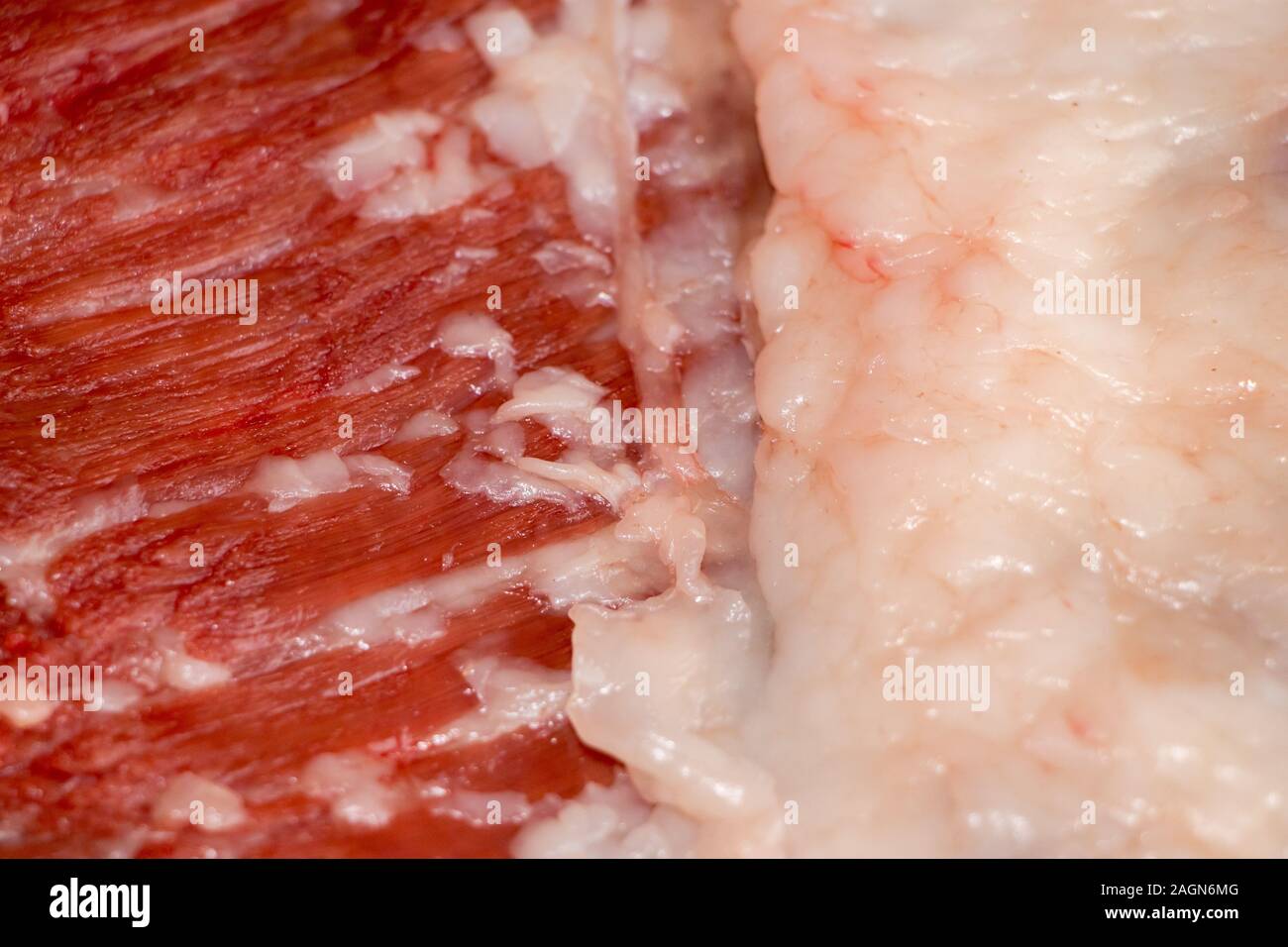 Education anatomy and Histological sample of Muscle tissue and Adipose tissue close up. Selective focus. Animal tissues Stock Photohttps://www.alamy.com/image-license-details/?v=1https://www.alamy.com/education-anatomy-and-histological-sample-of-muscle-tissue-and-adipose-tissue-close-up-selective-focus-animal-tissues-image337297760.html
RF2AGN6MG–Education anatomy and Histological sample of Muscle tissue and Adipose tissue close up. Selective focus. Animal tissues
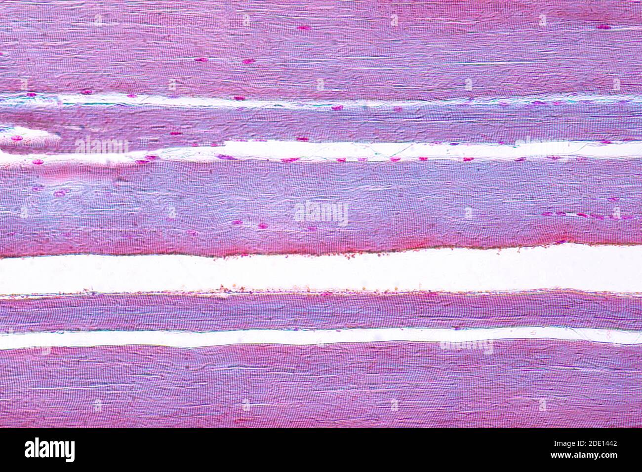 Human skeletal muscle, light micrograph Stock Photohttps://www.alamy.com/image-license-details/?v=1https://www.alamy.com/human-skeletal-muscle-light-micrograph-image387258482.html
RF2DE1442–Human skeletal muscle, light micrograph
 Scientific Designing of Actin and Myosin Structure. Colorful Symbols. Vector Illustration. Stock Vectorhttps://www.alamy.com/image-license-details/?v=1https://www.alamy.com/scientific-designing-of-actin-and-myosin-structure-colorful-symbols-vector-illustration-image487183397.html
RF2K8H3B1–Scientific Designing of Actin and Myosin Structure. Colorful Symbols. Vector Illustration.
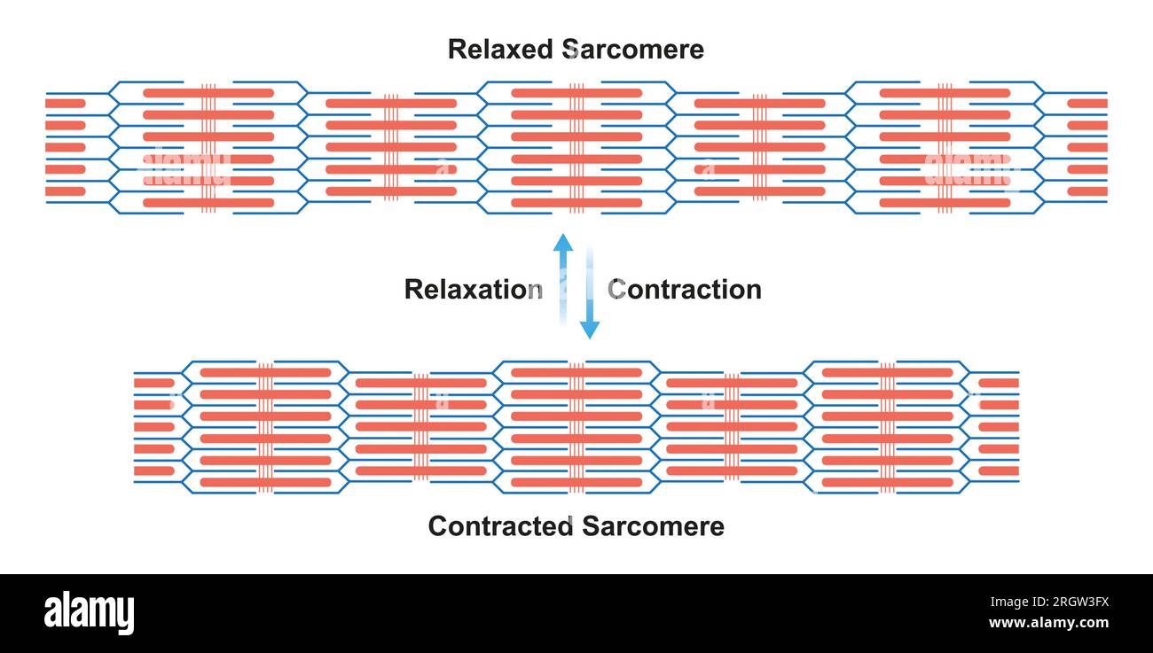 Contracted and relaxed sarcomeres, illustration Stock Photohttps://www.alamy.com/image-license-details/?v=1https://www.alamy.com/contracted-and-relaxed-sarcomeres-illustration-image561117870.html
RF2RGW3FX–Contracted and relaxed sarcomeres, illustration
 Muscle structure Stock Photohttps://www.alamy.com/image-license-details/?v=1https://www.alamy.com/stock-photo-muscle-structure-14033062.html
RFAFX7MR–Muscle structure
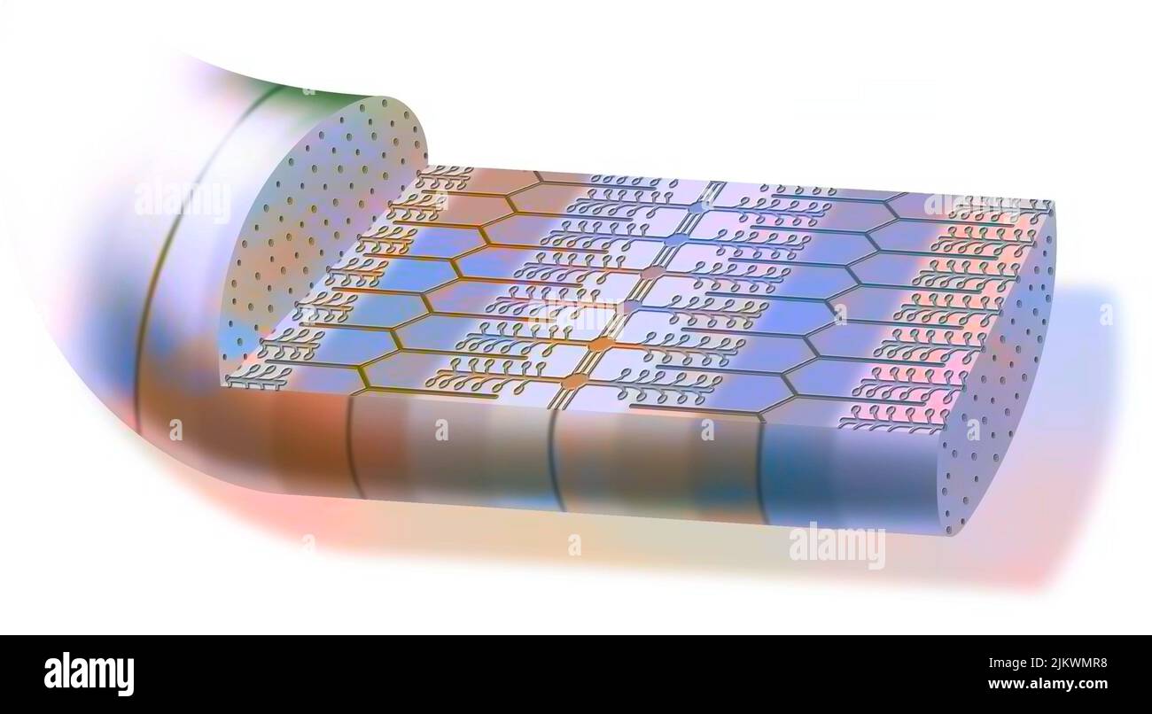 Contracted myofibril, made up of myofilaments and proteins, allowing muscle contraction. Stock Photohttps://www.alamy.com/image-license-details/?v=1https://www.alamy.com/contracted-myofibril-made-up-of-myofilaments-and-proteins-allowing-muscle-contraction-image476923532.html
RF2JKWMR8–Contracted myofibril, made up of myofilaments and proteins, allowing muscle contraction.
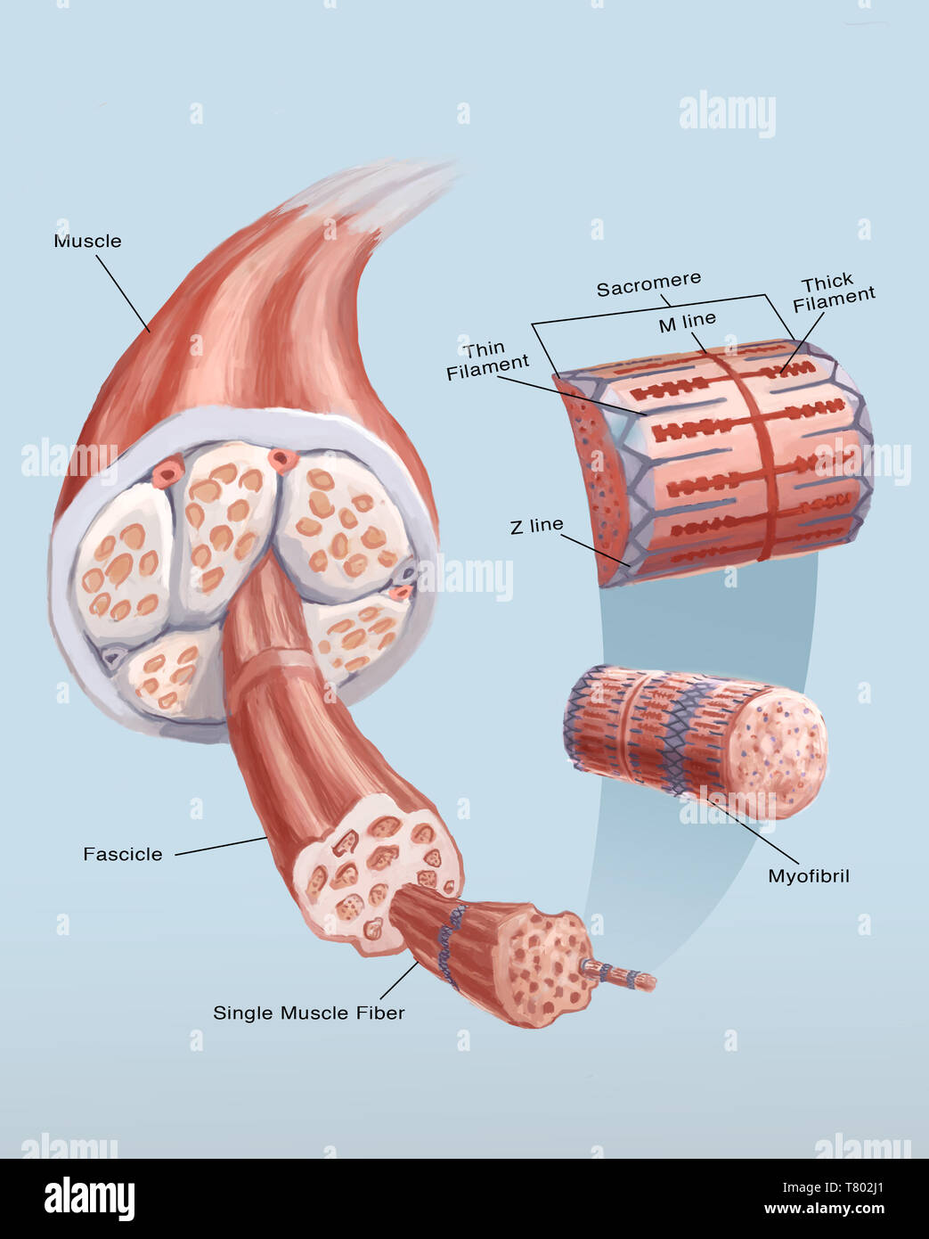 Muscle Cell, Illustration Stock Photohttps://www.alamy.com/image-license-details/?v=1https://www.alamy.com/muscle-cell-illustration-image245864473.html
RMT802J1–Muscle Cell, Illustration
 Cat Soleus Muscle, TEM Stock Photohttps://www.alamy.com/image-license-details/?v=1https://www.alamy.com/stock-photo-cat-soleus-muscle-tem-134986078.html
RMHRH43A–Cat Soleus Muscle, TEM
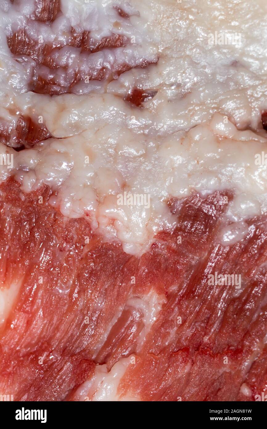 Education anatomy and Histological sample of Muscle tissue and Adipose tissue close up. Selective focus. Animal tissues Stock Photohttps://www.alamy.com/image-license-details/?v=1https://www.alamy.com/education-anatomy-and-histological-sample-of-muscle-tissue-and-adipose-tissue-close-up-selective-focus-animal-tissues-image337298805.html
RF2AGN81W–Education anatomy and Histological sample of Muscle tissue and Adipose tissue close up. Selective focus. Animal tissues
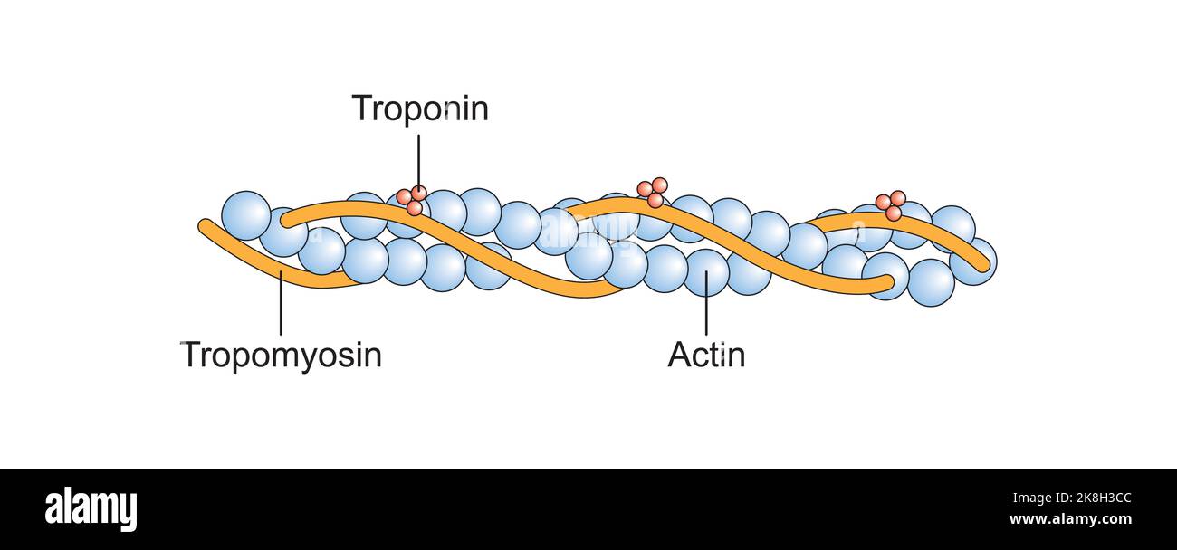 Scientific Designing of Actin Filament Structure. Colorful Symbols. Vector Illustration. Stock Vectorhttps://www.alamy.com/image-license-details/?v=1https://www.alamy.com/scientific-designing-of-actin-filament-structure-colorful-symbols-vector-illustration-image487183436.html
RF2K8H3CC–Scientific Designing of Actin Filament Structure. Colorful Symbols. Vector Illustration.
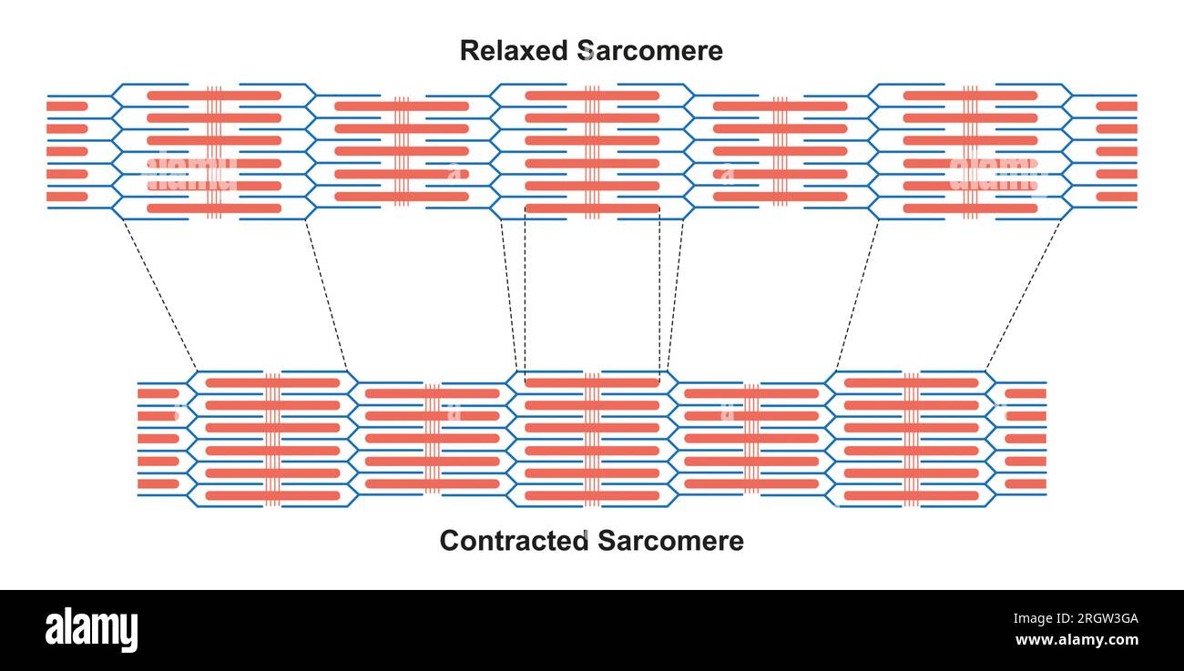 Contracted and relaxed sarcomeres, illustration Stock Photohttps://www.alamy.com/image-license-details/?v=1https://www.alamy.com/contracted-and-relaxed-sarcomeres-illustration-image561117882.html
RF2RGW3GA–Contracted and relaxed sarcomeres, illustration
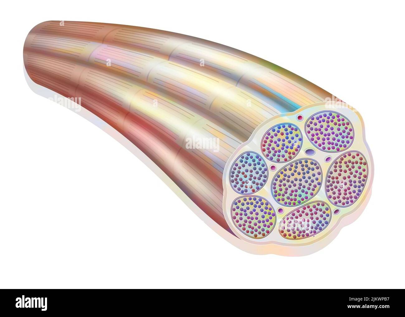 Muscle fiber showing the myofibrils made up of myofilaments (actin and myosin). Stock Photohttps://www.alamy.com/image-license-details/?v=1https://www.alamy.com/muscle-fiber-showing-the-myofibrils-made-up-of-myofilaments-actin-and-myosin-image476924763.html
RF2JKWPB7–Muscle fiber showing the myofibrils made up of myofilaments (actin and myosin).
 Cat Soleus Muscle, TEM Stock Photohttps://www.alamy.com/image-license-details/?v=1https://www.alamy.com/stock-photo-cat-soleus-muscle-tem-135013894.html
RMHRJBGP–Cat Soleus Muscle, TEM
 Scientific Designing of Actin Filament Structure. Colorful Symbols. Vector Illustration. Stock Vectorhttps://www.alamy.com/image-license-details/?v=1https://www.alamy.com/scientific-designing-of-actin-filament-structure-colorful-symbols-vector-illustration-image487183415.html
RF2K8H3BK–Scientific Designing of Actin Filament Structure. Colorful Symbols. Vector Illustration.
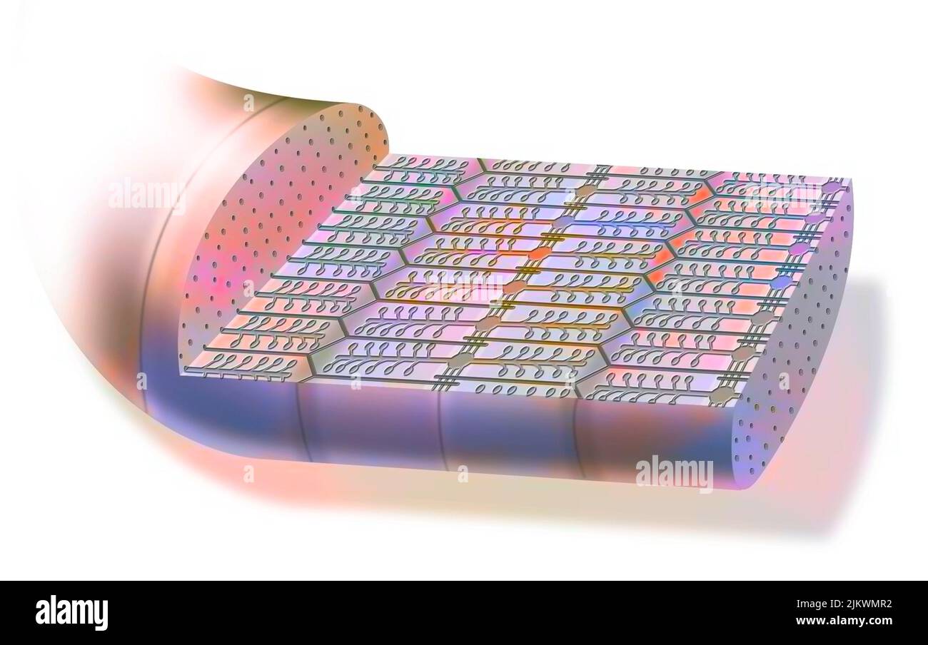 Contracted myofibril, made up of myofilaments and proteins, allowing muscle contraction. Stock Photohttps://www.alamy.com/image-license-details/?v=1https://www.alamy.com/contracted-myofibril-made-up-of-myofilaments-and-proteins-allowing-muscle-contraction-image476923526.html
RF2JKWMR2–Contracted myofibril, made up of myofilaments and proteins, allowing muscle contraction.
 Cat Soleus Muscle, TEM Stock Photohttps://www.alamy.com/image-license-details/?v=1https://www.alamy.com/stock-photo-cat-soleus-muscle-tem-135018835.html
RMHRJHW7–Cat Soleus Muscle, TEM
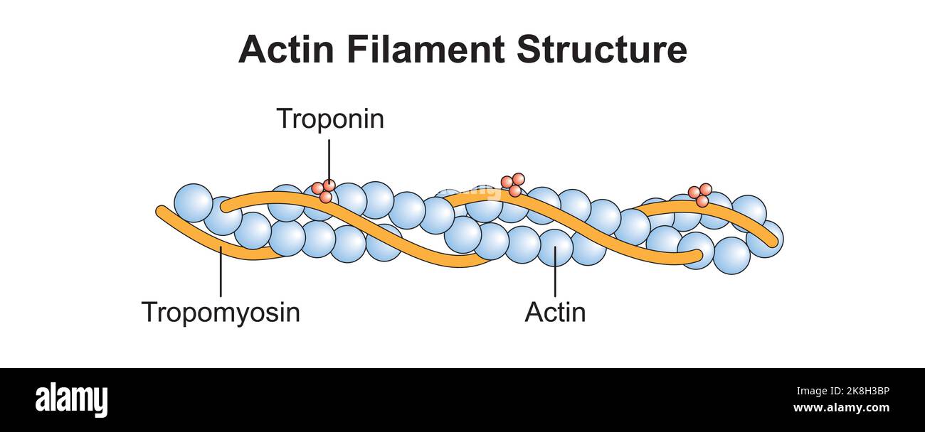 Scientific Designing of Actin Filament Structure. Colorful Symbols. Vector Illustration. Stock Vectorhttps://www.alamy.com/image-license-details/?v=1https://www.alamy.com/scientific-designing-of-actin-filament-structure-colorful-symbols-vector-illustration-image487183418.html
RF2K8H3BP–Scientific Designing of Actin Filament Structure. Colorful Symbols. Vector Illustration.
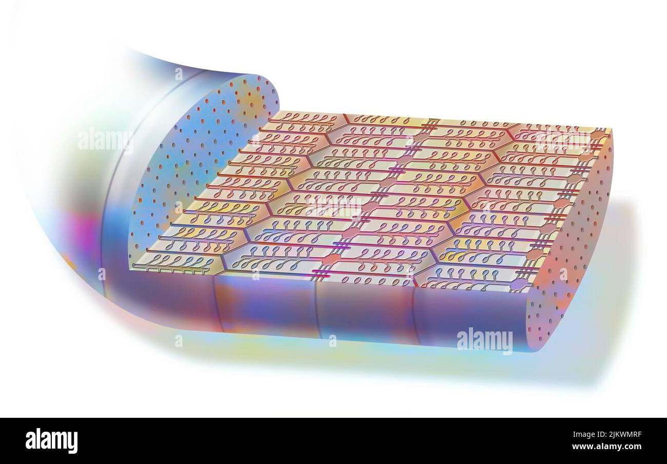 Contracted myofibril, made up of myofilaments and proteins, allowing muscle contraction. Stock Photohttps://www.alamy.com/image-license-details/?v=1https://www.alamy.com/contracted-myofibril-made-up-of-myofilaments-and-proteins-allowing-muscle-contraction-image476923539.html
RF2JKWMRF–Contracted myofibril, made up of myofilaments and proteins, allowing muscle contraction.
 Cat Soleus Muscle, TEM Stock Photohttps://www.alamy.com/image-license-details/?v=1https://www.alamy.com/stock-photo-cat-soleus-muscle-tem-135018836.html
RMHRJHW8–Cat Soleus Muscle, TEM
 Scientific Designing of Myosin Molecule Structure. Colorful Symbols. Vector Illustration. Stock Vectorhttps://www.alamy.com/image-license-details/?v=1https://www.alamy.com/scientific-designing-of-myosin-molecule-structure-colorful-symbols-vector-illustration-image487183430.html
RF2K8H3C6–Scientific Designing of Myosin Molecule Structure. Colorful Symbols. Vector Illustration.
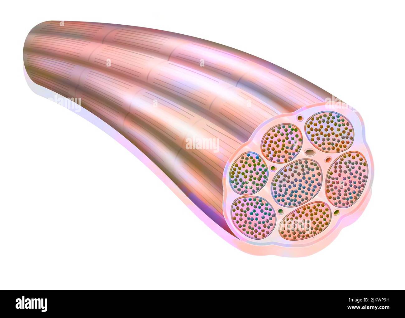 Muscle fiber showing the myofibrils made up of myofilaments (actin and myosin). Stock Photohttps://www.alamy.com/image-license-details/?v=1https://www.alamy.com/muscle-fiber-showing-the-myofibrils-made-up-of-myofilaments-actin-and-myosin-image476924717.html
RF2JKWP9H–Muscle fiber showing the myofibrils made up of myofilaments (actin and myosin).
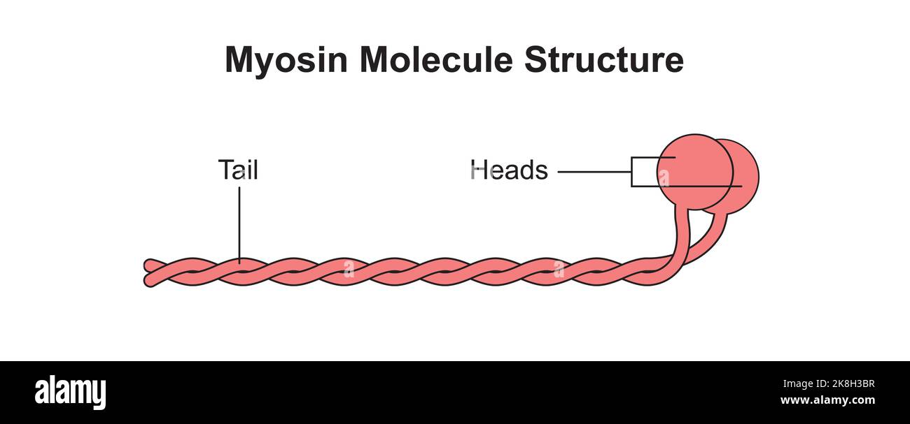 Scientific Designing of Myosin Molecule Structure. Colorful Symbols. Vector Illustration. Stock Vectorhttps://www.alamy.com/image-license-details/?v=1https://www.alamy.com/scientific-designing-of-myosin-molecule-structure-colorful-symbols-vector-illustration-image487183419.html
RF2K8H3BR–Scientific Designing of Myosin Molecule Structure. Colorful Symbols. Vector Illustration.
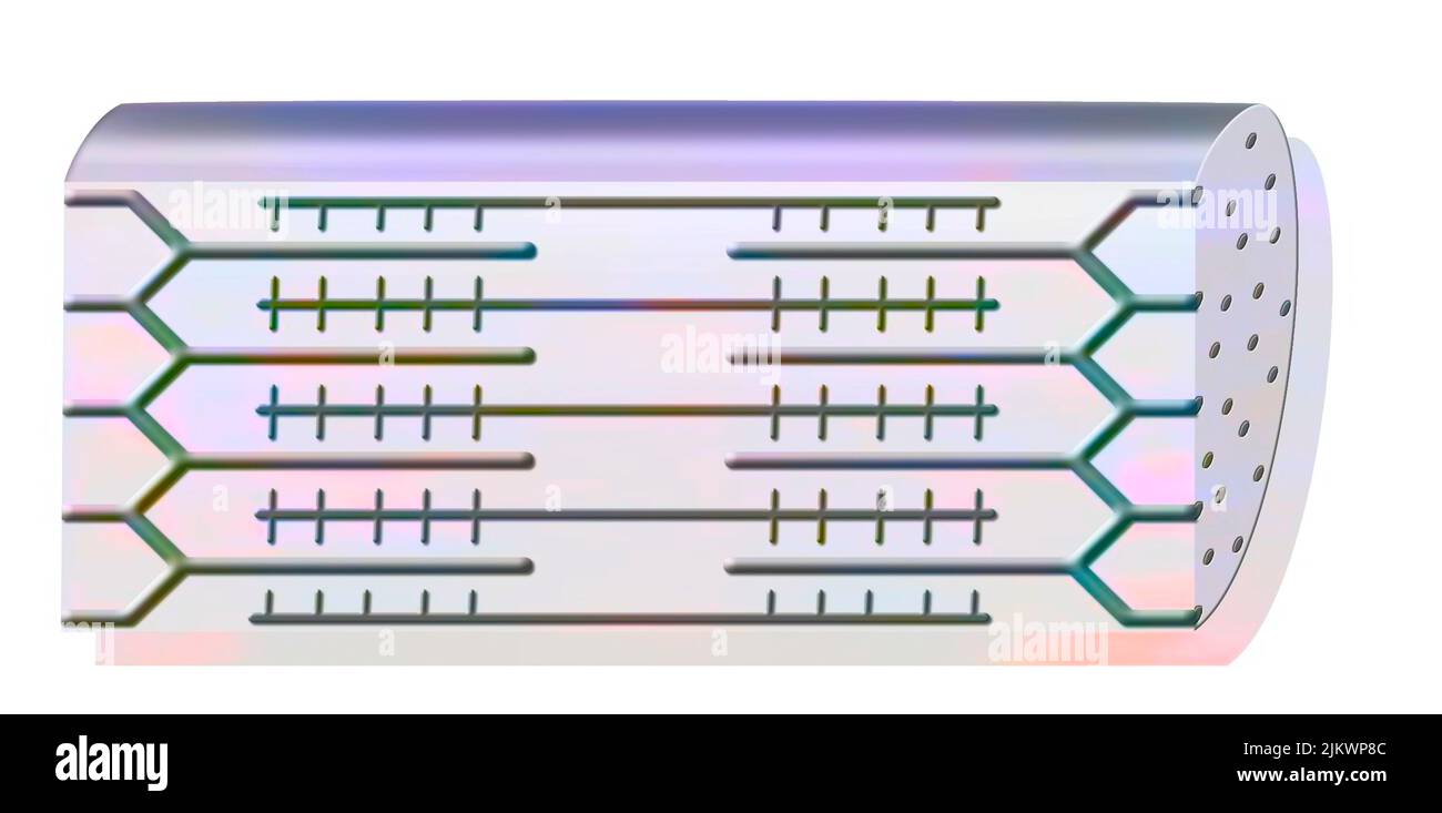 Resting myofibril made up of fine (actin) and thick (myosin) filaments. Stock Photohttps://www.alamy.com/image-license-details/?v=1https://www.alamy.com/resting-myofibril-made-up-of-fine-actin-and-thick-myosin-filaments-image476924684.html
RF2JKWP8C–Resting myofibril made up of fine (actin) and thick (myosin) filaments.
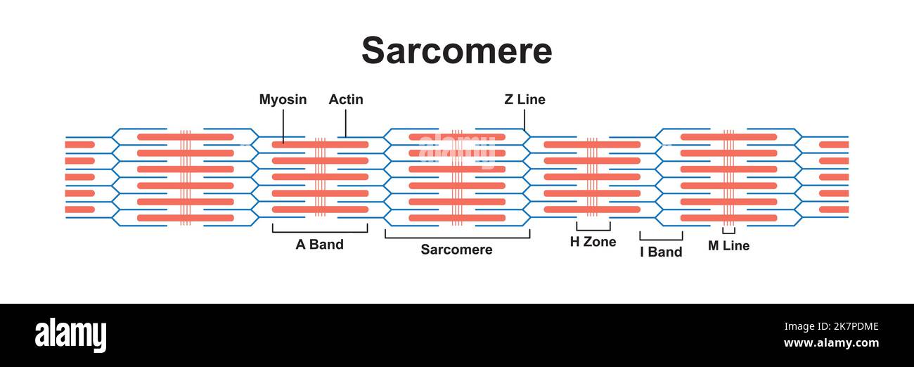 Scientific Designing of Sarcomere. Structural and Functional Unity of The Muscle. Colorful Symbols. Vector Illustration. Stock Vectorhttps://www.alamy.com/image-license-details/?v=1https://www.alamy.com/scientific-designing-of-sarcomere-structural-and-functional-unity-of-the-muscle-colorful-symbols-vector-illustration-image486686606.html
RF2K7PDME–Scientific Designing of Sarcomere. Structural and Functional Unity of The Muscle. Colorful Symbols. Vector Illustration.
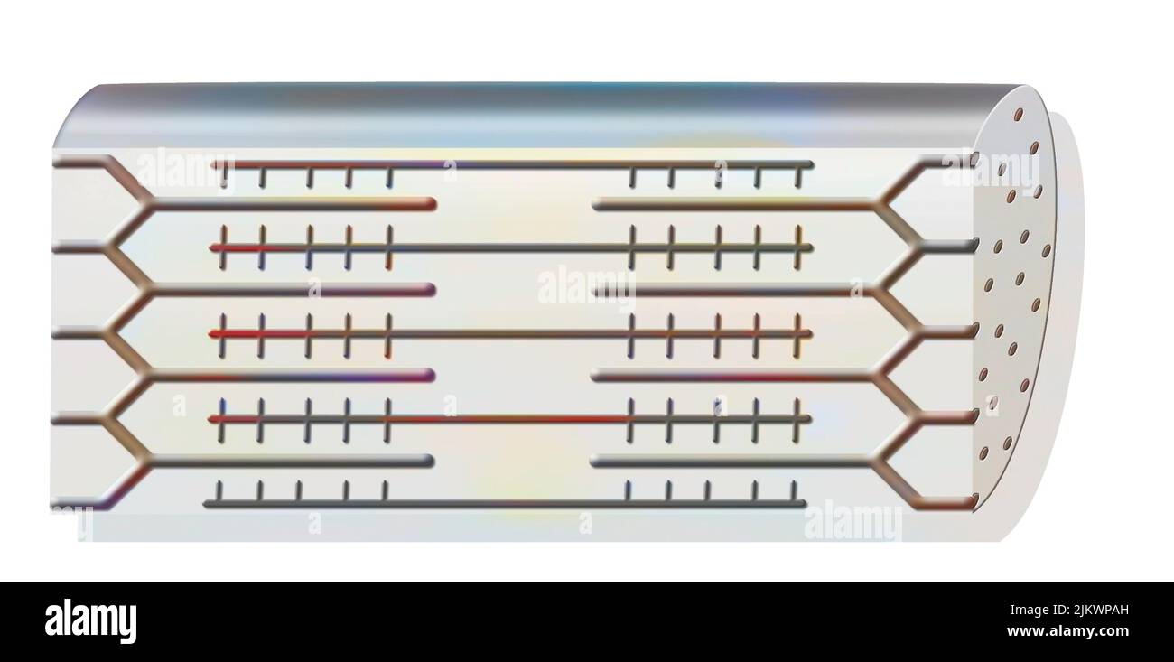 Resting myofibril made up of fine (actin) and thick (myosin) filaments. Stock Photohttps://www.alamy.com/image-license-details/?v=1https://www.alamy.com/resting-myofibril-made-up-of-fine-actin-and-thick-myosin-filaments-image476924745.html
RF2JKWPAH–Resting myofibril made up of fine (actin) and thick (myosin) filaments.
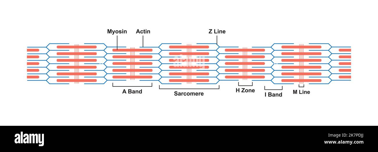 Scientific Designing of Sarcomere. Structural and Functional Unity of The Muscle. Colorful Symbols. Vector Illustration. Stock Vectorhttps://www.alamy.com/image-license-details/?v=1https://www.alamy.com/scientific-designing-of-sarcomere-structural-and-functional-unity-of-the-muscle-colorful-symbols-vector-illustration-image486686554.html
RF2K7PDJJ–Scientific Designing of Sarcomere. Structural and Functional Unity of The Muscle. Colorful Symbols. Vector Illustration.
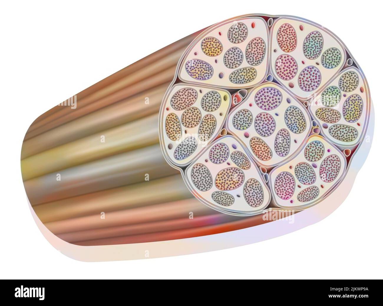 Muscle bundle made up of several largely vascularized muscle fibers. Stock Photohttps://www.alamy.com/image-license-details/?v=1https://www.alamy.com/muscle-bundle-made-up-of-several-largely-vascularized-muscle-fibers-image476924710.html
RF2JKWP9A–Muscle bundle made up of several largely vascularized muscle fibers.
 Scientific Designing of Sarcomere. Structural and Functional Unity of The Muscle. Colorful Symbols. Vector Illustration. Stock Vectorhttps://www.alamy.com/image-license-details/?v=1https://www.alamy.com/scientific-designing-of-sarcomere-structural-and-functional-unity-of-the-muscle-colorful-symbols-vector-illustration-image486686551.html
RF2K7PDJF–Scientific Designing of Sarcomere. Structural and Functional Unity of The Muscle. Colorful Symbols. Vector Illustration.
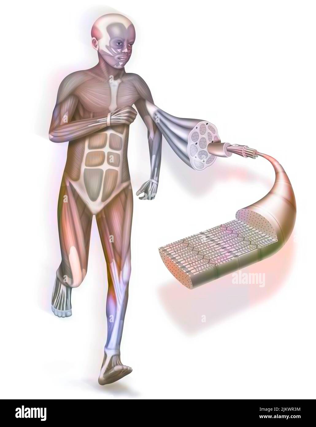 A cut away and zoom on a muscle and its structure: tendon, muscle. Stock Photohttps://www.alamy.com/image-license-details/?v=1https://www.alamy.com/a-cut-away-and-zoom-on-a-muscle-and-its-structure-tendon-muscle-image476925336.html
RF2JKWR3M–A cut away and zoom on a muscle and its structure: tendon, muscle.
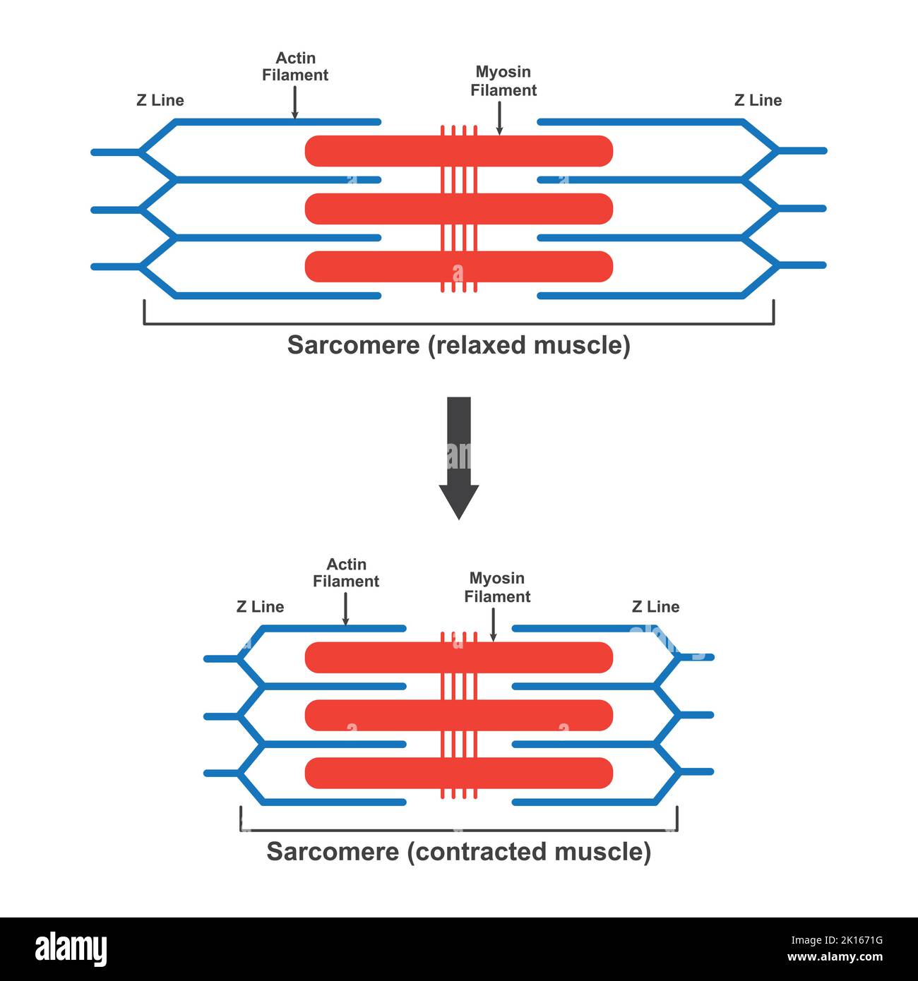 Scientific Designing of Differences Between Relaxed And Contracted Muscle (Sarcomere). Colorful Symbols. Vector Illustration. Stock Vectorhttps://www.alamy.com/image-license-details/?v=1https://www.alamy.com/scientific-designing-of-differences-between-relaxed-and-contracted-muscle-sarcomere-colorful-symbols-vector-illustration-image482642204.html
RF2K1671G–Scientific Designing of Differences Between Relaxed And Contracted Muscle (Sarcomere). Colorful Symbols. Vector Illustration.
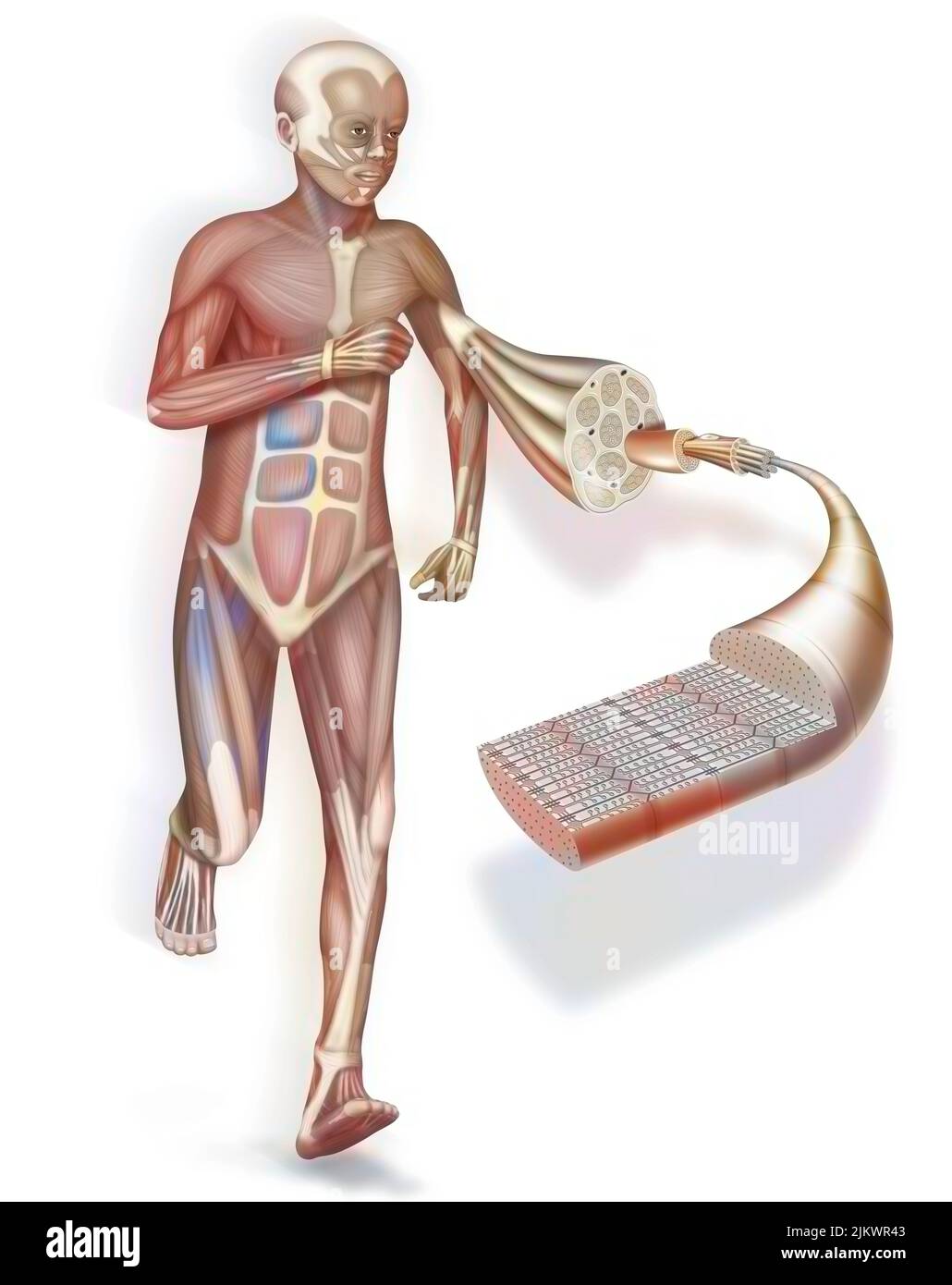 A cut away and zoom on a muscle and its structure: tendon, muscle. Stock Photohttps://www.alamy.com/image-license-details/?v=1https://www.alamy.com/a-cut-away-and-zoom-on-a-muscle-and-its-structure-tendon-muscle-image476925347.html
RF2JKWR43–A cut away and zoom on a muscle and its structure: tendon, muscle.
 Scientific Designing of Contraction and Relaxation of Muscular Sarcomere. Muscle Contraction and Muscle Relaxation Colorful Symbols. Vector Illustrati Stock Vectorhttps://www.alamy.com/image-license-details/?v=1https://www.alamy.com/scientific-designing-of-contraction-and-relaxation-of-muscular-sarcomere-muscle-contraction-and-muscle-relaxation-colorful-symbols-vector-illustrati-image486686586.html
RF2K7PDKP–Scientific Designing of Contraction and Relaxation of Muscular Sarcomere. Muscle Contraction and Muscle Relaxation Colorful Symbols. Vector Illustrati