Quick filters:
Thoracic aorta Stock Photos and Images
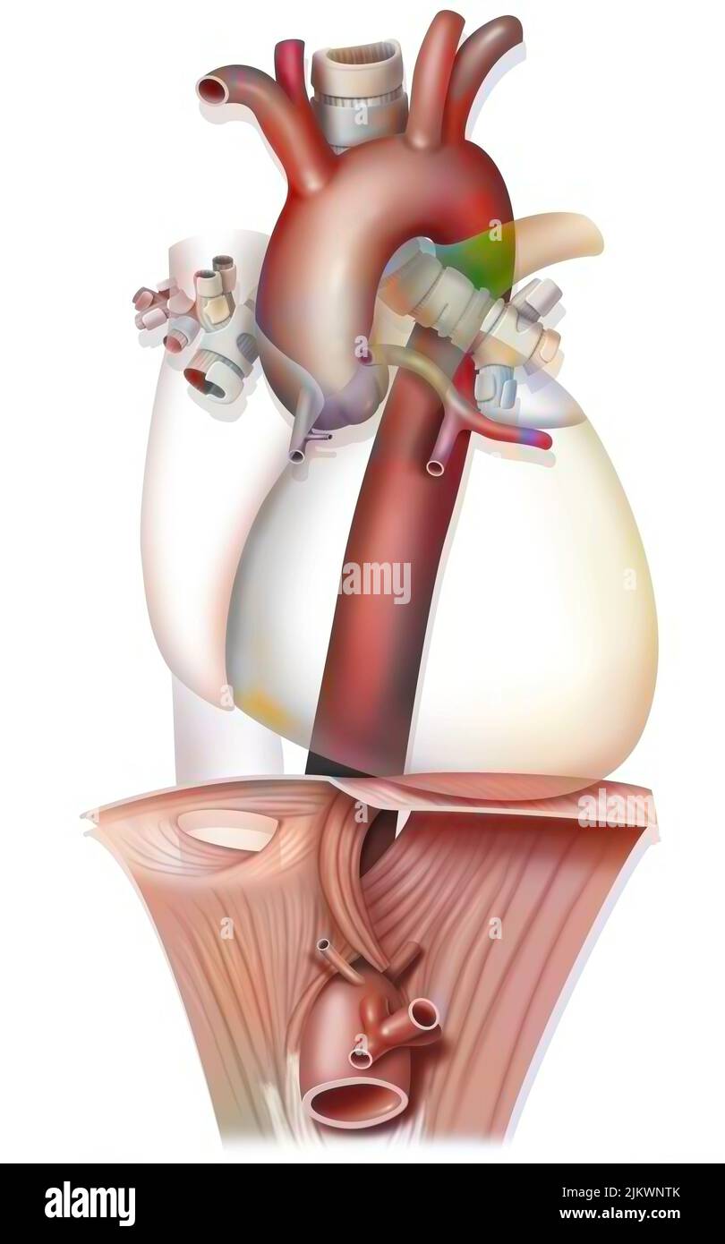 Anatomy of the thoracic aorta crossing the diaphragm. Stock Photohttps://www.alamy.com/image-license-details/?v=1https://www.alamy.com/anatomy-of-the-thoracic-aorta-crossing-the-diaphragm-image476924355.html
RF2JKWNTK–Anatomy of the thoracic aorta crossing the diaphragm.
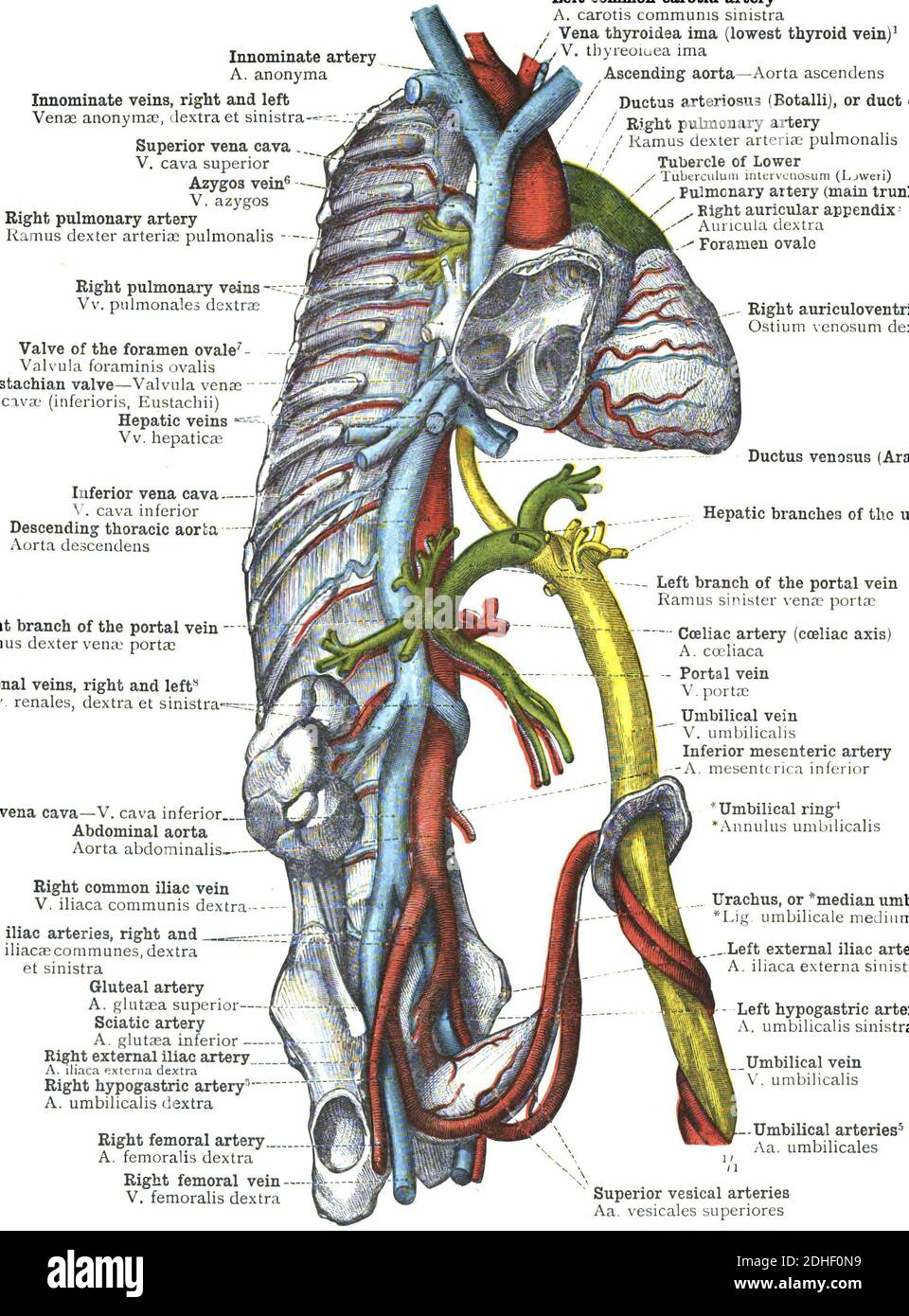 The branches of the thoracic aorta anatomy Stock Photohttps://www.alamy.com/image-license-details/?v=1https://www.alamy.com/the-branches-of-the-thoracic-aorta-anatomy-image389407125.html
RF2DHF0N9–The branches of the thoracic aorta anatomy
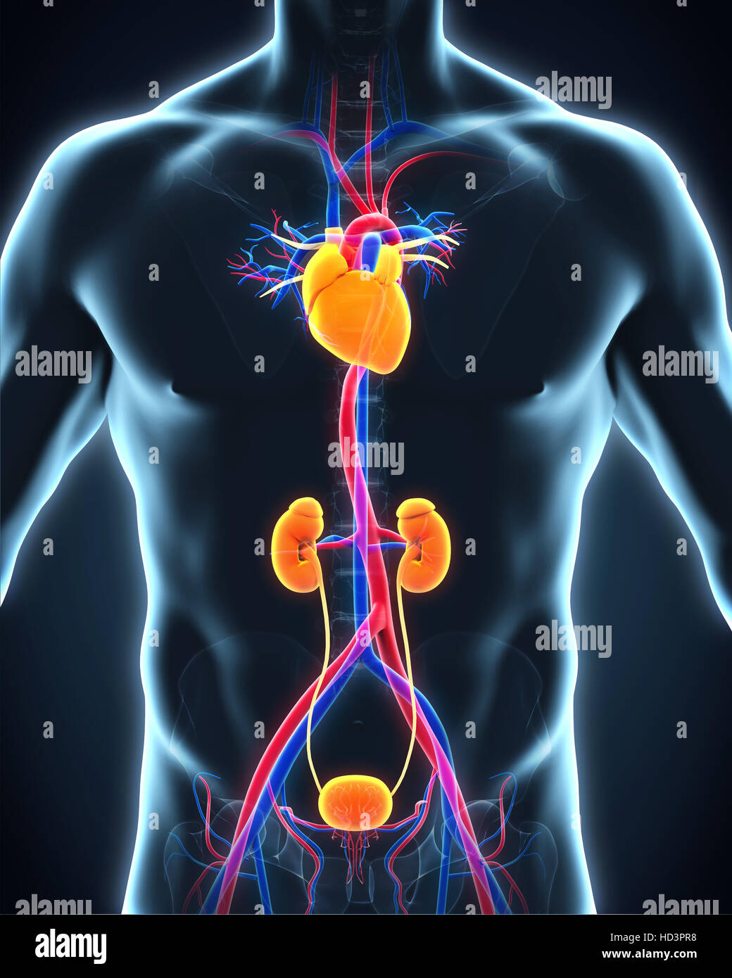 Human Thoracic Aorta Stock Photohttps://www.alamy.com/image-license-details/?v=1https://www.alamy.com/stock-photo-human-thoracic-aorta-128546860.html
RFHD3PR8–Human Thoracic Aorta
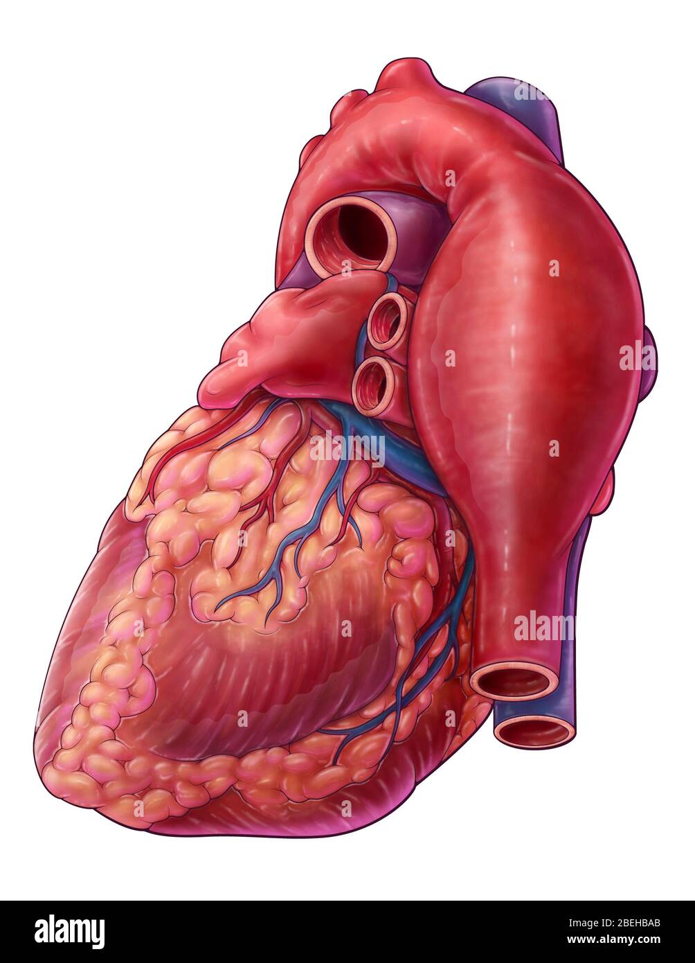 Thoracic Aortic Aneurysm, Illustration Stock Photohttps://www.alamy.com/image-license-details/?v=1https://www.alamy.com/thoracic-aortic-aneurysm-illustration-image353194643.html
RM2BEHBAB–Thoracic Aortic Aneurysm, Illustration
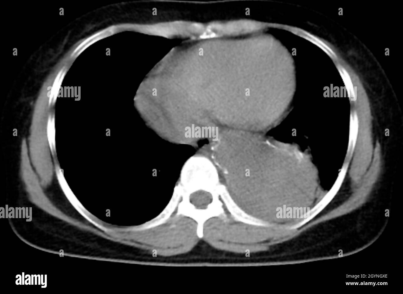 Aortic aneurysm, CT scan Stock Photohttps://www.alamy.com/image-license-details/?v=1https://www.alamy.com/aortic-aneurysm-ct-scan-image447329190.html
RF2GYNGXE–Aortic aneurysm, CT scan
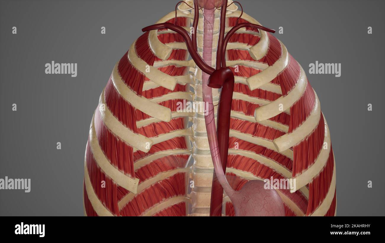 Medical Ilustration of Bood Supply of Esophagus Stock Photohttps://www.alamy.com/image-license-details/?v=1https://www.alamy.com/medical-ilustration-of-bood-supply-of-esophagus-image488428583.html
RF2KAHRHY–Medical Ilustration of Bood Supply of Esophagus
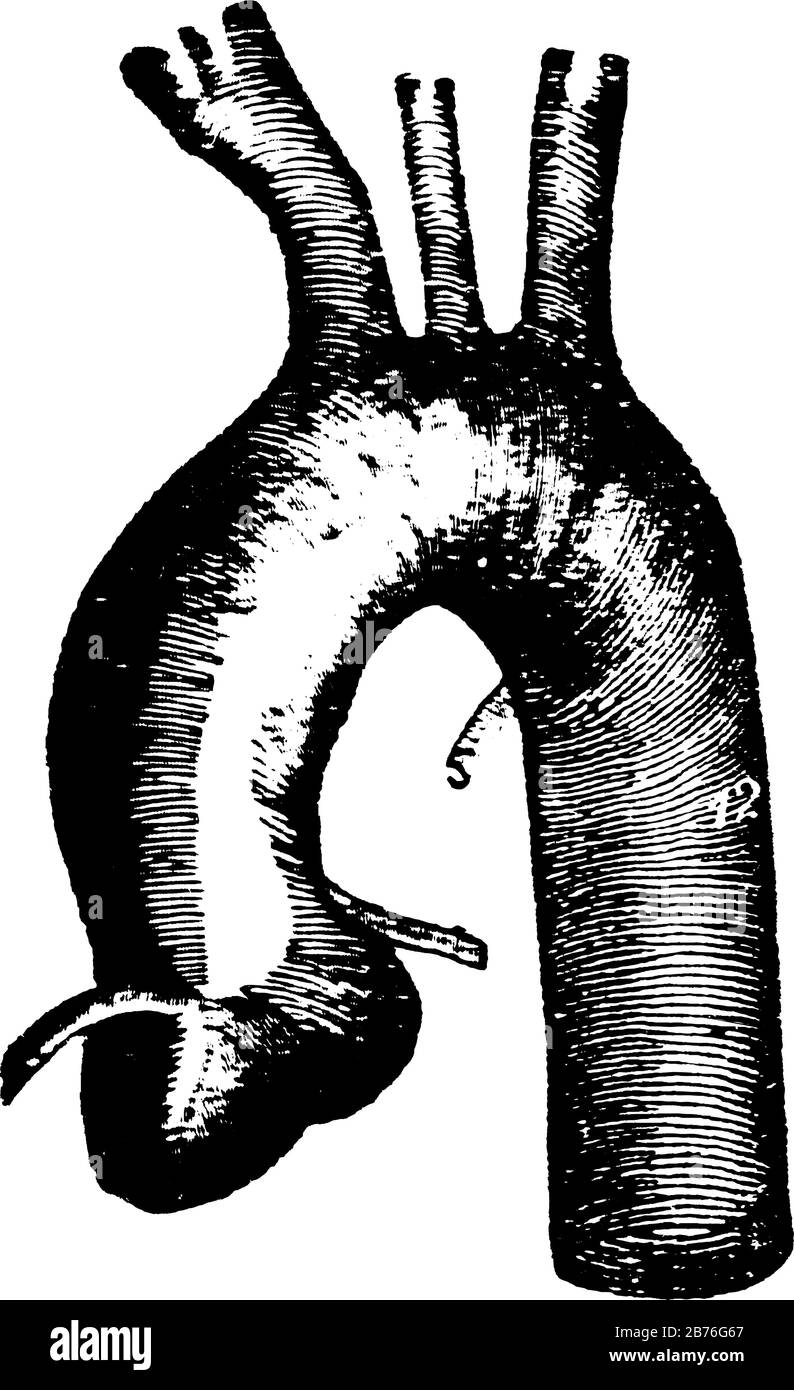 This diagram represents a Thoracic Aorta, vintage line drawing or engraving illustration. Stock Vectorhttps://www.alamy.com/image-license-details/?v=1https://www.alamy.com/this-diagram-represents-a-thoracic-aorta-vintage-line-drawing-or-engraving-illustration-image348654383.html
RF2B76G67–This diagram represents a Thoracic Aorta, vintage line drawing or engraving illustration.
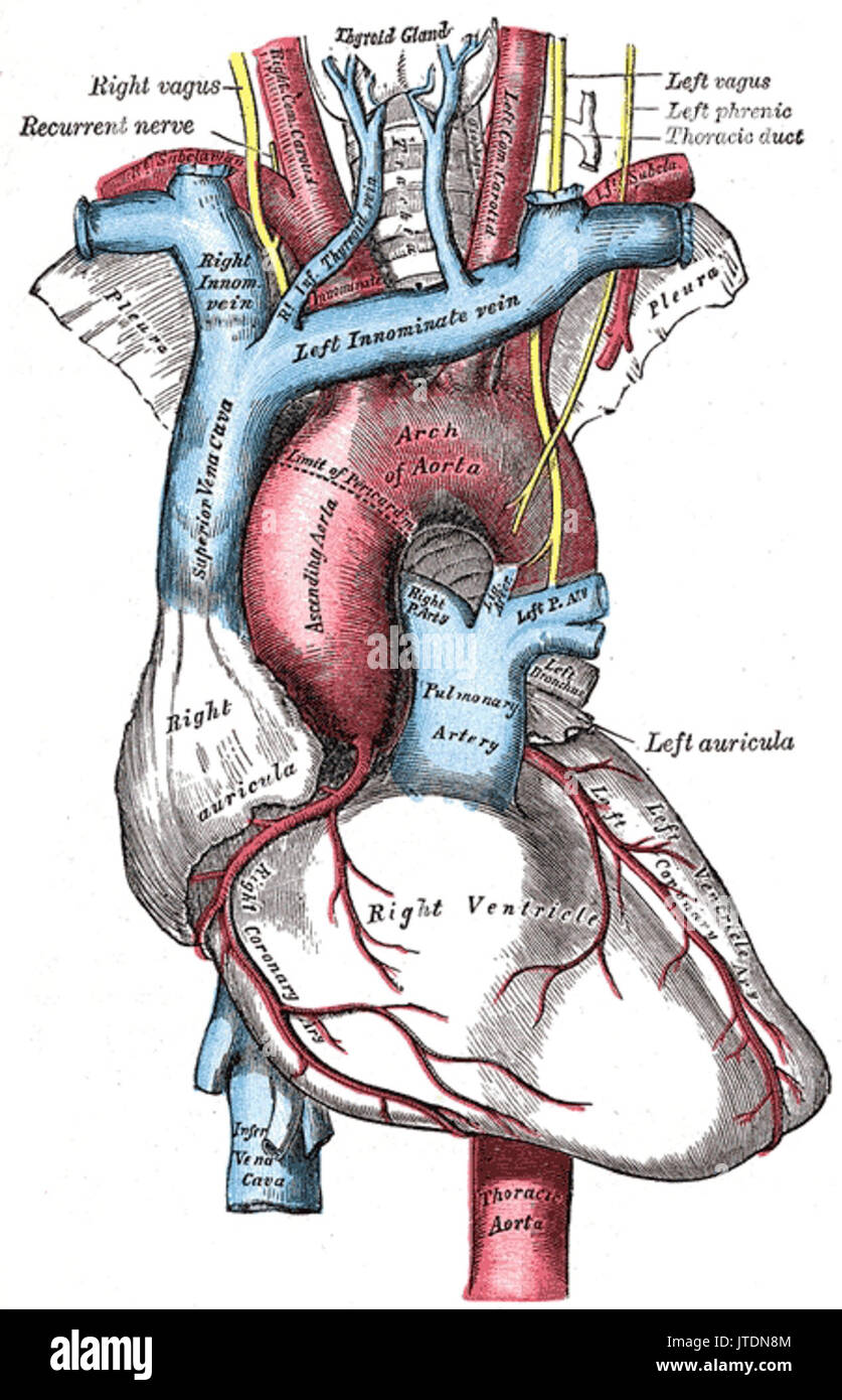 Thoracic aorta Stock Photohttps://www.alamy.com/image-license-details/?v=1https://www.alamy.com/thoracic-aorta-image152736772.html
RMJTDN8M–Thoracic aorta
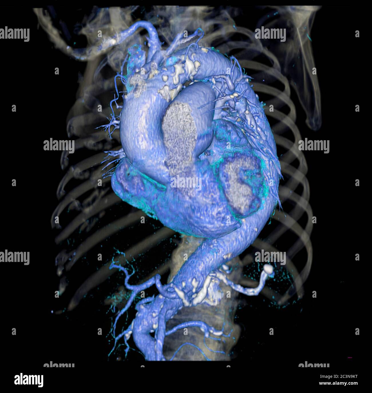 CTA thoracic aorta 3D rendering image for diagnotic abdominal aortic aneurysm or AAA and aortic dissection Stock Photohttps://www.alamy.com/image-license-details/?v=1https://www.alamy.com/cta-thoracic-aorta-3d-rendering-image-for-diagnotic-abdominal-aortic-aneurysm-or-aaa-and-aortic-dissection-image363730300.html
RF2C3N9KT–CTA thoracic aorta 3D rendering image for diagnotic abdominal aortic aneurysm or AAA and aortic dissection
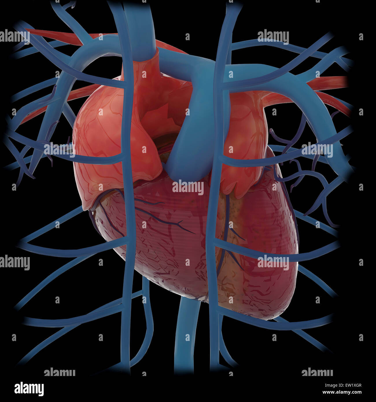 3D rendering of human heart and thoracic veins. Stock Photohttps://www.alamy.com/image-license-details/?v=1https://www.alamy.com/stock-photo-3d-rendering-of-human-heart-and-thoracic-veins-84250679.html
RFEW1XGR–3D rendering of human heart and thoracic veins.
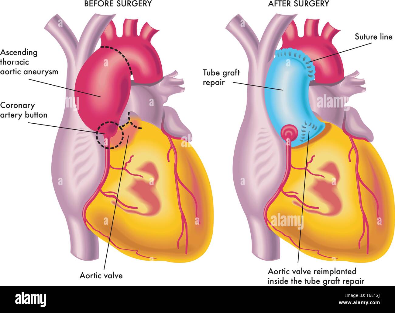 Medical illustration of a thoracic aortic aneurysm surgery Stock Vectorhttps://www.alamy.com/image-license-details/?v=1https://www.alamy.com/medical-illustration-of-a-thoracic-aortic-aneurysm-surgery-image244941274.html
RFT6E12J–Medical illustration of a thoracic aortic aneurysm surgery
 Thoracic (descending) aortic aneurysm and endovascular surgery Stock Photohttps://www.alamy.com/image-license-details/?v=1https://www.alamy.com/stock-photo-thoracic-descending-aortic-aneurysm-and-endovascular-surgery-50553039.html
RFCX6TWK–Thoracic (descending) aortic aneurysm and endovascular surgery
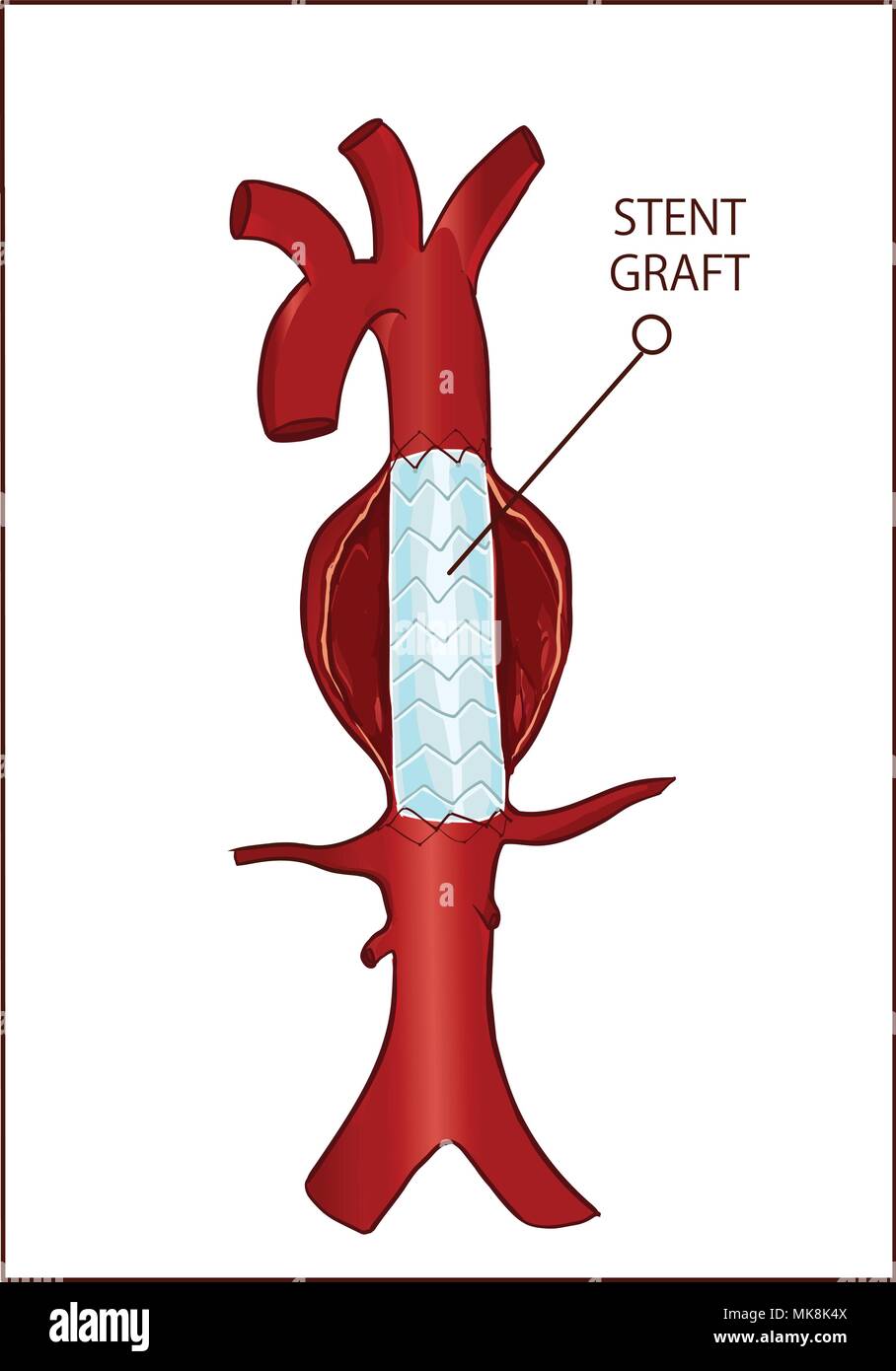 Vector - Thoracic (descending) aortic aneurysm and endovascular surgery Stock Vectorhttps://www.alamy.com/image-license-details/?v=1https://www.alamy.com/vector-thoracic-descending-aortic-aneurysm-and-endovascular-surgery-image183972794.html
RFMK8K4X–Vector - Thoracic (descending) aortic aneurysm and endovascular surgery
 Chest x-ray of an 84 year old woman with a small right pleural effusion and a moderate left pleural effusion. Stock Photohttps://www.alamy.com/image-license-details/?v=1https://www.alamy.com/stock-photo-chest-x-ray-of-an-84-year-old-woman-with-a-small-right-pleural-effusion-26899457.html
RMBFNAEW–Chest x-ray of an 84 year old woman with a small right pleural effusion and a moderate left pleural effusion.
 Aneurysm being a complex subject, related to other important topics. Stock Photohttps://www.alamy.com/image-license-details/?v=1https://www.alamy.com/aneurysm-being-a-complex-subject-related-to-other-important-topics-image619012314.html
RF2XY2CEJ–Aneurysm being a complex subject, related to other important topics.
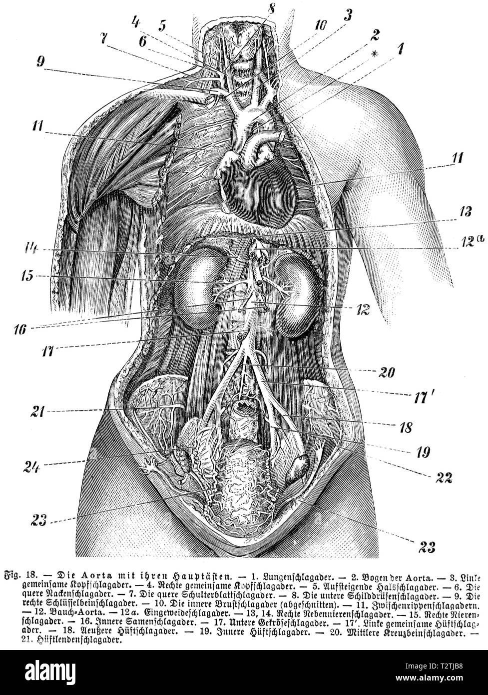 Human: aorta with its main branches, 1) pulmonary artery, 2) arch of aorta, 3) left common carotid artery, 4) right common carotid artery, 5) ascending carotid artery, 6) transverse carotid artery, 7) transverse scapula, 8) lower thyroid artery, 9 ) Right coronary artery, 10) Internal thoracic aorta (truncated), 11) Intercostal veins, 12) Abdominal aorta, 12 a) Intestinal staph, 13, 14) Right adrenal artery, 15) Right renal artery, 16) Internal seminal vein, 17) Lower mesentery artery, 17 ') Left common hip artery, 18) Outer hip artery, 19) Internal hip artery, 20) Mid-sacral artery, 21) Hip a Stock Photohttps://www.alamy.com/image-license-details/?v=1https://www.alamy.com/human-aorta-with-its-main-branches-1-pulmonary-artery-2-arch-of-aorta-3-left-common-carotid-artery-4-right-common-carotid-artery-5-ascending-carotid-artery-6-transverse-carotid-artery-7-transverse-scapula-8-lower-thyroid-artery-9-right-coronary-artery-10-internal-thoracic-aorta-truncated-11-intercostal-veins-12-abdominal-aorta-12-a-intestinal-staph-13-14-right-adrenal-artery-15-right-renal-artery-16-internal-seminal-vein-17-lower-mesentery-artery-17-left-common-hip-artery-18-outer-hip-artery-19-internal-hip-artery-20-mid-sacral-artery-21-hip-a-image242715740.html
RMT2TJB8–Human: aorta with its main branches, 1) pulmonary artery, 2) arch of aorta, 3) left common carotid artery, 4) right common carotid artery, 5) ascending carotid artery, 6) transverse carotid artery, 7) transverse scapula, 8) lower thyroid artery, 9 ) Right coronary artery, 10) Internal thoracic aorta (truncated), 11) Intercostal veins, 12) Abdominal aorta, 12 a) Intestinal staph, 13, 14) Right adrenal artery, 15) Right renal artery, 16) Internal seminal vein, 17) Lower mesentery artery, 17 ') Left common hip artery, 18) Outer hip artery, 19) Internal hip artery, 20) Mid-sacral artery, 21) Hip a
 The blood vessels of the thorax Stock Photohttps://www.alamy.com/image-license-details/?v=1https://www.alamy.com/stock-photo-the-blood-vessels-of-the-thorax-13170408.html
RFACJG8W–The blood vessels of the thorax
 Model showing thoracic anatomy. Stock Photohttps://www.alamy.com/image-license-details/?v=1https://www.alamy.com/stock-photo-model-showing-thoracic-anatomy-52089833.html
RMD0MW35–Model showing thoracic anatomy.
 Thoracic aorta and abdominal aorta Aortic Brust und Bauchaorta Object Type : photo stereo picture Item number: RP-F F26535 Manufacturer : Photographer: JF Bergman Date: 1900 - 1940 Physical features: photography on carton material: Cardboard Technique: Photography Dimensions: Secondary medium: h 75 mm × W 150 mm Stock Photohttps://www.alamy.com/image-license-details/?v=1https://www.alamy.com/thoracic-aorta-and-abdominal-aorta-aortic-brust-und-bauchaorta-object-type-photo-stereo-picture-item-number-rp-f-f26535-manufacturer-photographer-jf-bergman-date-1900-1940-physical-features-photography-on-carton-material-cardboard-technique-photography-dimensions-secondary-medium-h-75-mm-w-150-mm-image348237156.html
RM2B6FG18–Thoracic aorta and abdominal aorta Aortic Brust und Bauchaorta Object Type : photo stereo picture Item number: RP-F F26535 Manufacturer : Photographer: JF Bergman Date: 1900 - 1940 Physical features: photography on carton material: Cardboard Technique: Photography Dimensions: Secondary medium: h 75 mm × W 150 mm
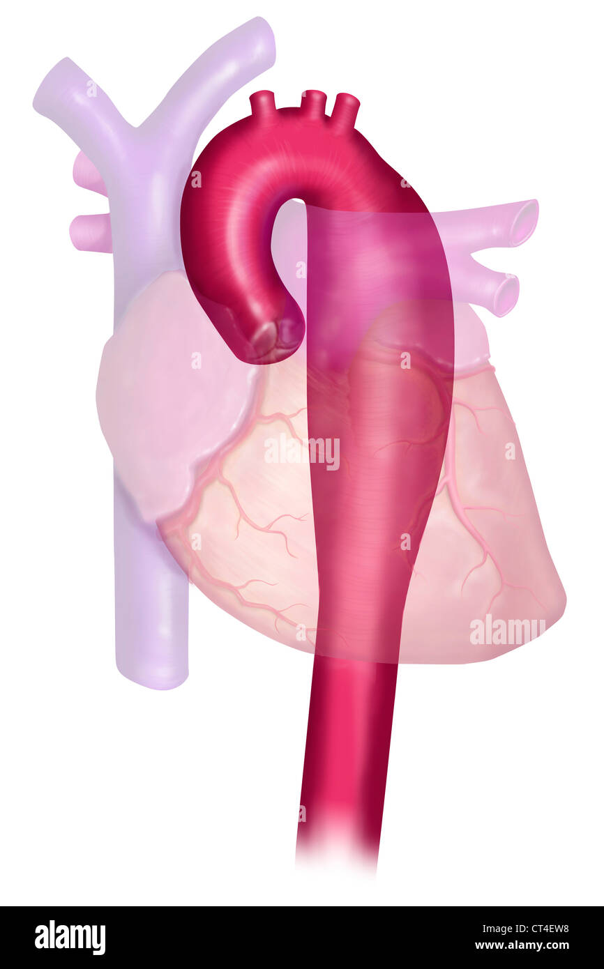 ANEURYSM OF THE THORACIC AORTA Stock Photohttps://www.alamy.com/image-license-details/?v=1https://www.alamy.com/stock-photo-aneurysm-of-the-thoracic-aorta-49271972.html
RMCT4EW8–ANEURYSM OF THE THORACIC AORTA
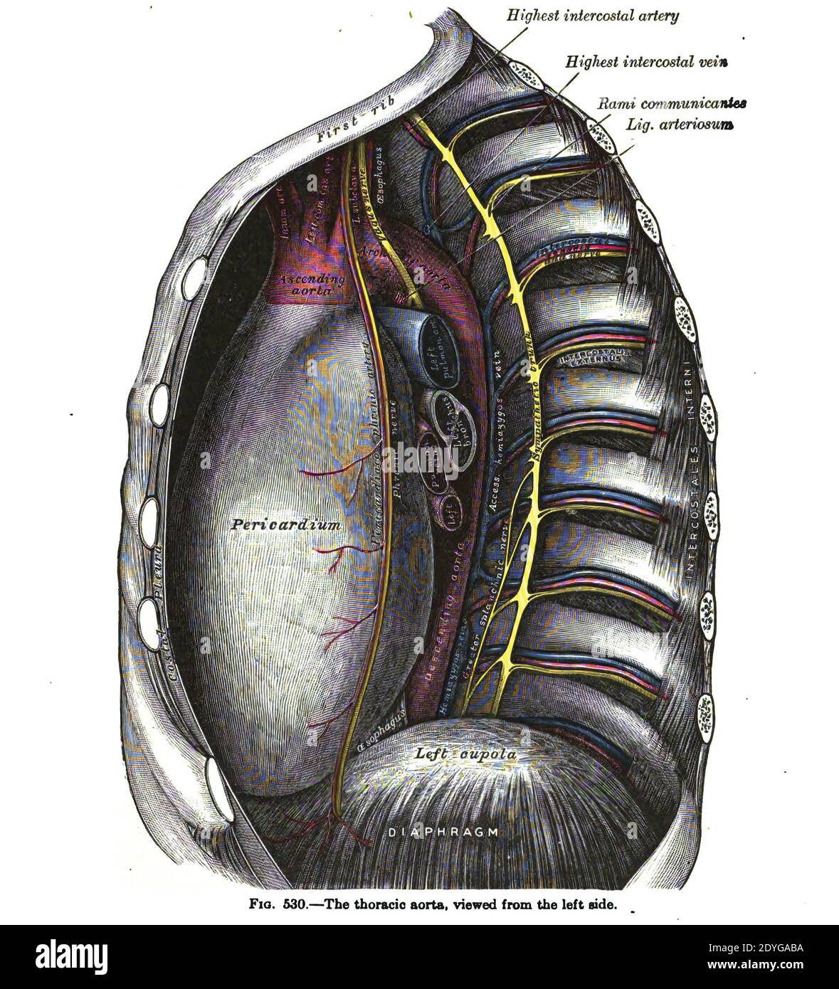 A vertical anatomy drawing and text of the thoracic aorta, from the 19th-century Stock Photohttps://www.alamy.com/image-license-details/?v=1https://www.alamy.com/a-vertical-anatomy-drawing-and-text-of-the-thoracic-aorta-from-the-19th-century-image395583198.html
RF2DYGABA–A vertical anatomy drawing and text of the thoracic aorta, from the 19th-century
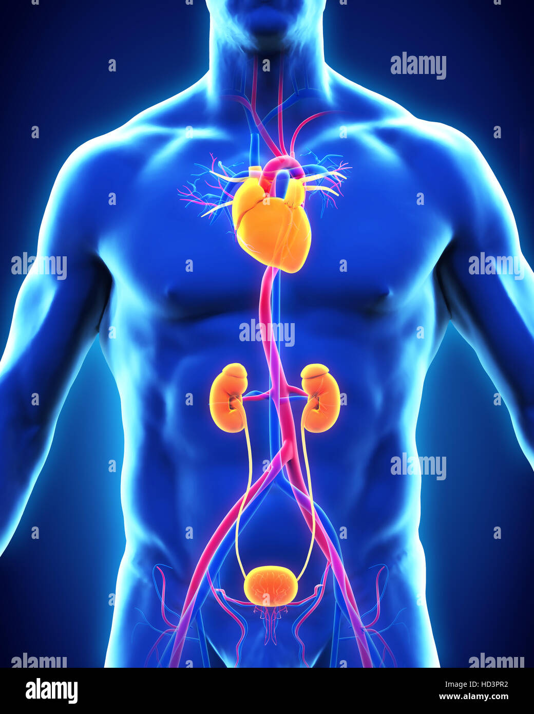 Human Thoracic Aorta Stock Photohttps://www.alamy.com/image-license-details/?v=1https://www.alamy.com/stock-photo-human-thoracic-aorta-128546854.html
RFHD3PR2–Human Thoracic Aorta
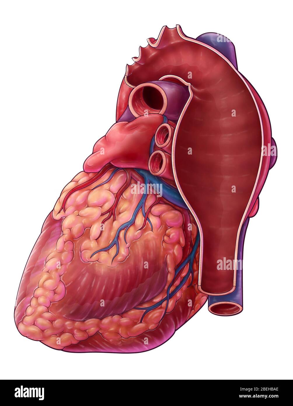 Thoracic Aortic Aneurysm, Illustration Stock Photohttps://www.alamy.com/image-license-details/?v=1https://www.alamy.com/thoracic-aortic-aneurysm-illustration-image353194646.html
RM2BEHBAE–Thoracic Aortic Aneurysm, Illustration
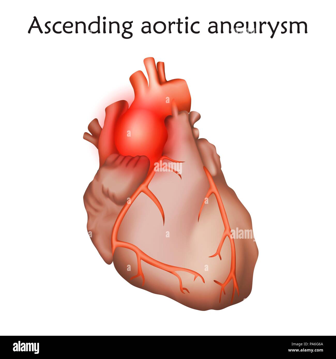 Ascending aortic aneurysm, illustration. Enlargement of a weakened area in the ascending aorta. Stock Photohttps://www.alamy.com/image-license-details/?v=1https://www.alamy.com/ascending-aortic-aneurysm-illustration-enlargement-of-a-weakened-area-in-the-ascending-aorta-image212815410.html
RFPA6G6A–Ascending aortic aneurysm, illustration. Enlargement of a weakened area in the ascending aorta.
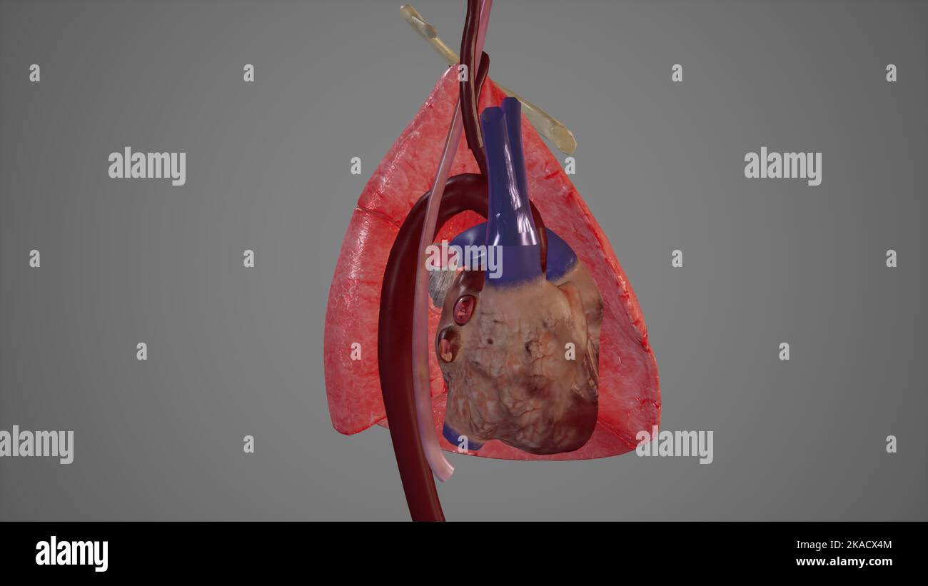 Medial View of Left Lung showing Major structures Related to It Stock Photohttps://www.alamy.com/image-license-details/?v=1https://www.alamy.com/medial-view-of-left-lung-showing-major-structures-related-to-it-image488320804.html
RF2KACX4M–Medial View of Left Lung showing Major structures Related to It
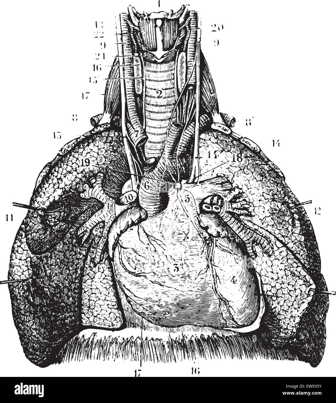 Main reports of the lungs. (Thoracic organs seen by their front face), vintage engraved illustration. Usual Medicine Dictionary Stock Vectorhttps://www.alamy.com/image-license-details/?v=1https://www.alamy.com/stock-photo-main-reports-of-the-lungs-thoracic-organs-seen-by-their-front-face-84407959.html
RFEW935Y–Main reports of the lungs. (Thoracic organs seen by their front face), vintage engraved illustration. Usual Medicine Dictionary
 The thoracic aorta originates from the left ventricle, guarded by the aortic valve 3d illustration Stock Photohttps://www.alamy.com/image-license-details/?v=1https://www.alamy.com/the-thoracic-aorta-originates-from-the-left-ventricle-guarded-by-the-aortic-valve-3d-illustration-image596593373.html
RF2WJH4X5–The thoracic aorta originates from the left ventricle, guarded by the aortic valve 3d illustration
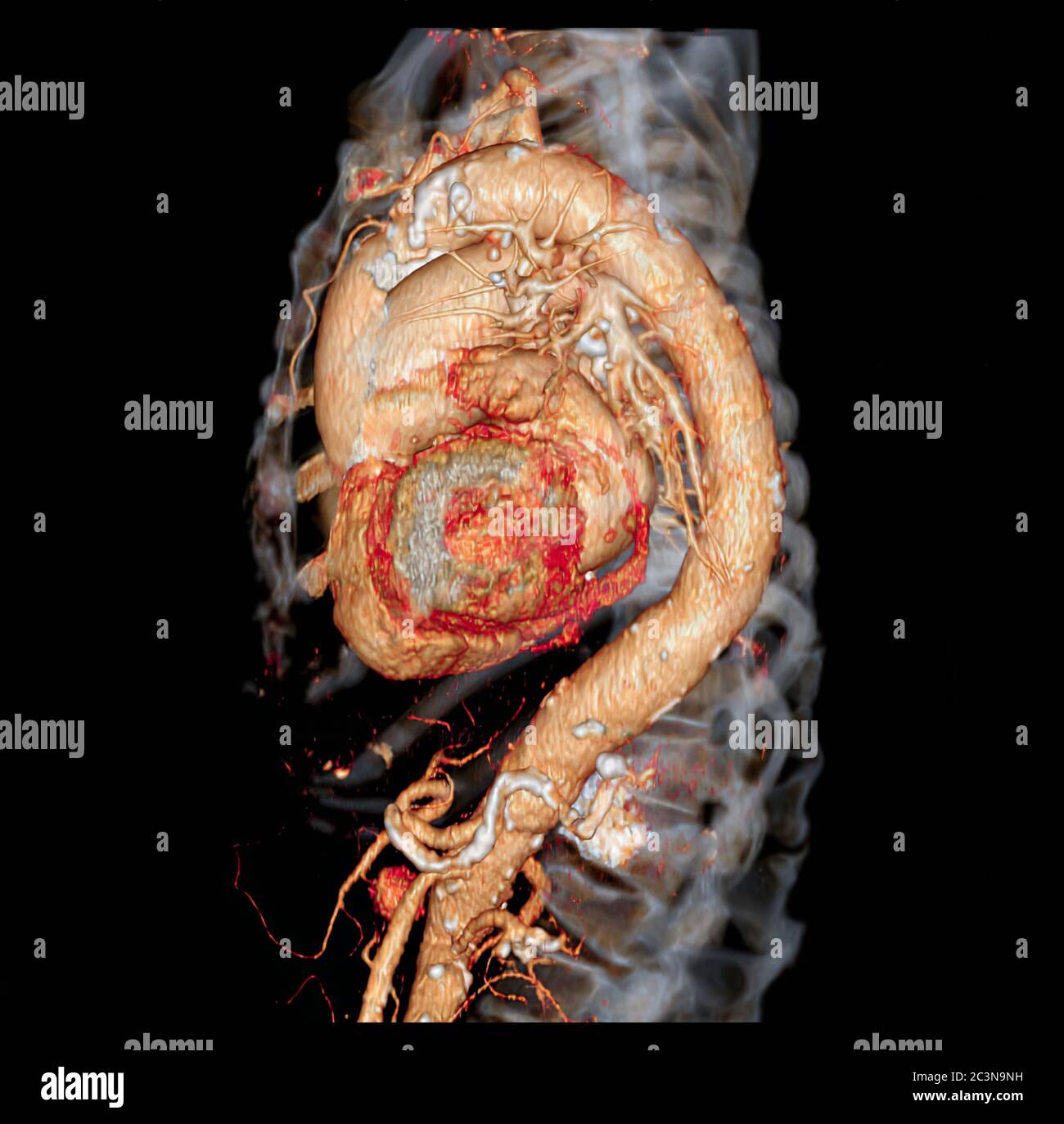 CTA thoracic aorta 3D rendering image for diagnotic abdominal aortic aneurysm or AAA and aortic dissection Stock Photohttps://www.alamy.com/image-license-details/?v=1https://www.alamy.com/cta-thoracic-aorta-3d-rendering-image-for-diagnotic-abdominal-aortic-aneurysm-or-aaa-and-aortic-dissection-image363730349.html
RF2C3N9NH–CTA thoracic aorta 3D rendering image for diagnotic abdominal aortic aneurysm or AAA and aortic dissection
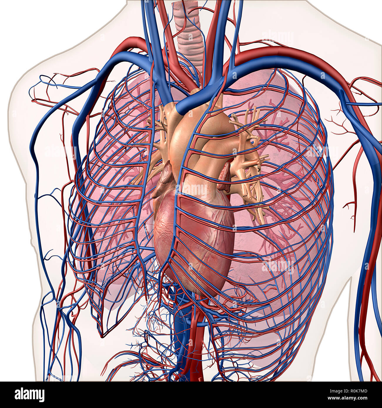 Layered view of circulatory and respiratory system. Stock Photohttps://www.alamy.com/image-license-details/?v=1https://www.alamy.com/layered-view-of-circulatory-and-respiratory-system-image224157933.html
RFR0K7MD–Layered view of circulatory and respiratory system.
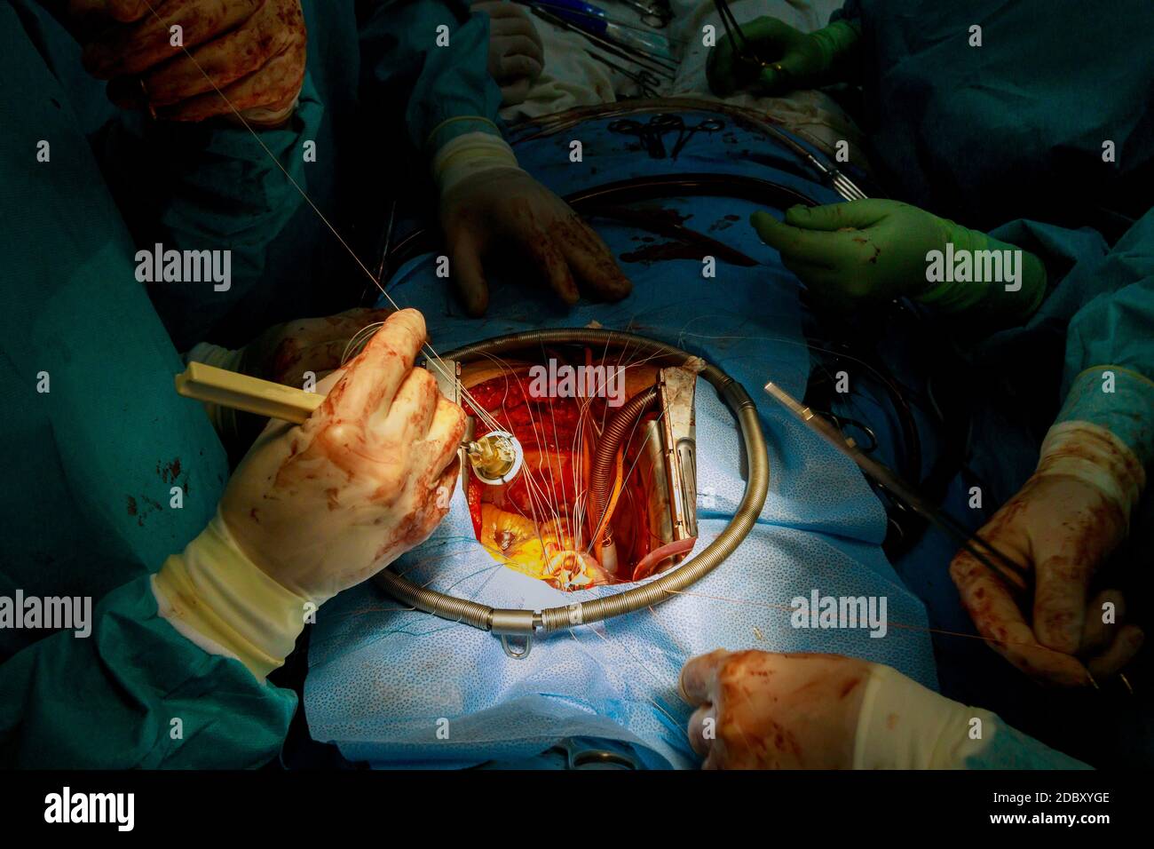 Aortic valve replacement surgery on of human heart final surgical stitches attaching prosthesis to aorta wall Stock Photohttps://www.alamy.com/image-license-details/?v=1https://www.alamy.com/aortic-valve-replacement-surgery-on-of-human-heart-final-surgical-stitches-attaching-prosthesis-to-aorta-wall-image385981694.html
RF2DBXYGE–Aortic valve replacement surgery on of human heart final surgical stitches attaching prosthesis to aorta wall
 Thoracic aorta Stock Photohttps://www.alamy.com/image-license-details/?v=1https://www.alamy.com/thoracic-aorta-image478802809.html
RM2JPY9T9–Thoracic aorta
 'Diseases of the heart and thoracic aorta' (1884) Stock Photohttps://www.alamy.com/image-license-details/?v=1https://www.alamy.com/diseases-of-the-heart-and-thoracic-aorta-1884-image557944593.html
RM2RBMG0H–'Diseases of the heart and thoracic aorta' (1884)
 Diseases of the heart and thoracic aorta (1884) Stock Photohttps://www.alamy.com/image-license-details/?v=1https://www.alamy.com/stock-photo-diseases-of-the-heart-and-thoracic-aorta-1884-101157594.html
RMFTG3F6–Diseases of the heart and thoracic aorta (1884)
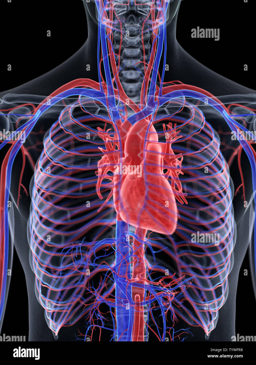 3d rendered medically accurate illustration of the human heart Stock Photohttps://www.alamy.com/image-license-details/?v=1https://www.alamy.com/3d-rendered-medically-accurate-illustration-of-the-human-heart-image258102076.html
RFTYWFR8–3d rendered medically accurate illustration of the human heart
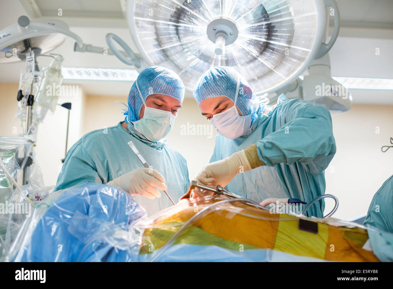 Da Vinci® Robotic Assisted Heart Surgery, double aortic coronary bypass with Da Vinci ® surgical robot. Limoges hospital, Stock Photohttps://www.alamy.com/image-license-details/?v=1https://www.alamy.com/stock-photo-da-vinci-robotic-assisted-heart-surgery-double-aortic-coronary-bypass-72441132.html
RME5RYB8–Da Vinci® Robotic Assisted Heart Surgery, double aortic coronary bypass with Da Vinci ® surgical robot. Limoges hospital,
 The blood vessels of the back Stock Photohttps://www.alamy.com/image-license-details/?v=1https://www.alamy.com/stock-photo-the-blood-vessels-of-the-back-13175555.html
RFACK3HT–The blood vessels of the back
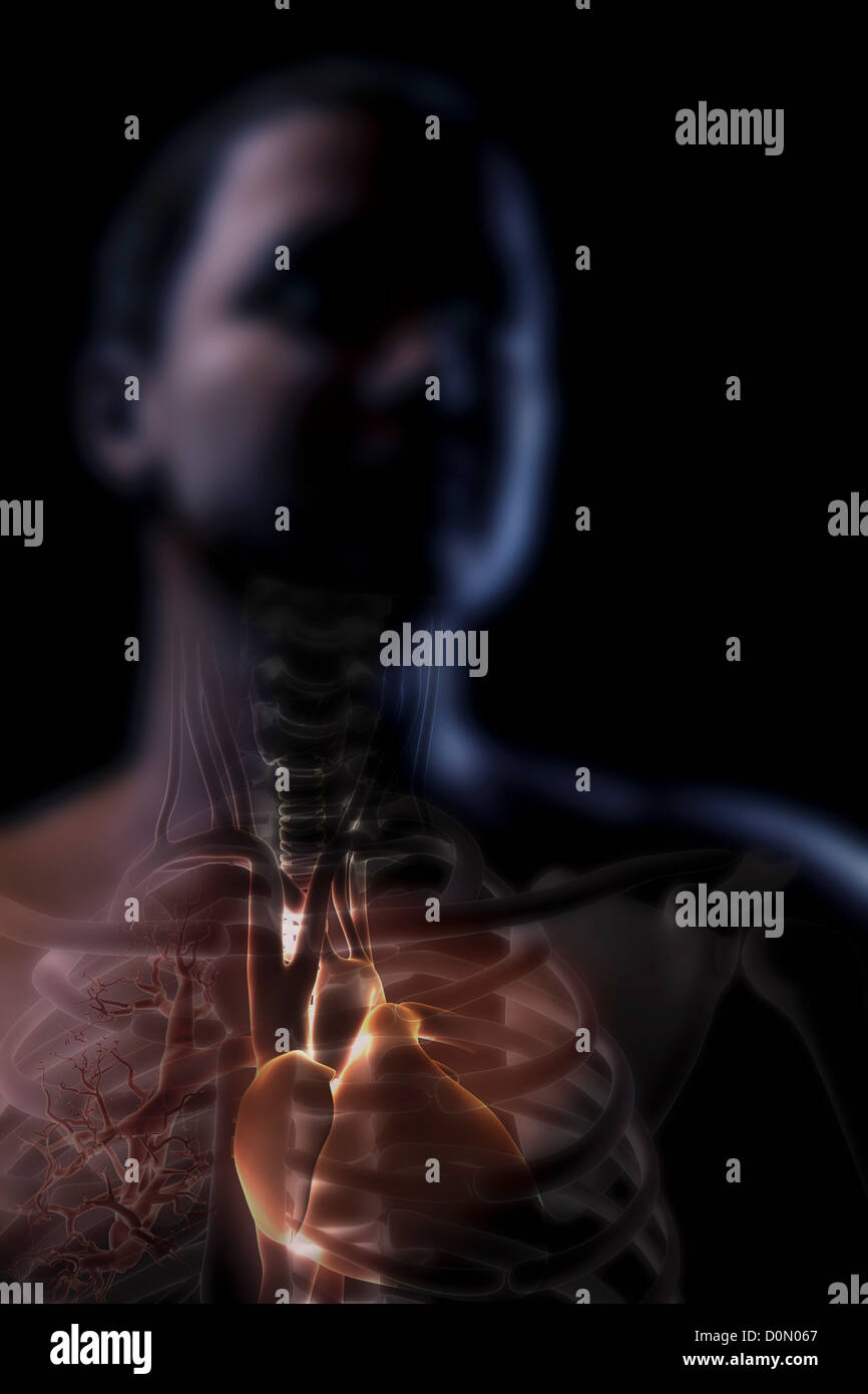 Anatomical model showing the upper thoracic organs. Stock Photohttps://www.alamy.com/image-license-details/?v=1https://www.alamy.com/stock-photo-anatomical-model-showing-the-upper-thoracic-organs-52092271.html
RMD0N067–Anatomical model showing the upper thoracic organs.
 abdominal aneurysm description medical vector illustration on white background Stock Vectorhttps://www.alamy.com/image-license-details/?v=1https://www.alamy.com/abdominal-aneurysm-description-medical-vector-illustration-on-white-background-image248875018.html
RFTCW6HE–abdominal aneurysm description medical vector illustration on white background
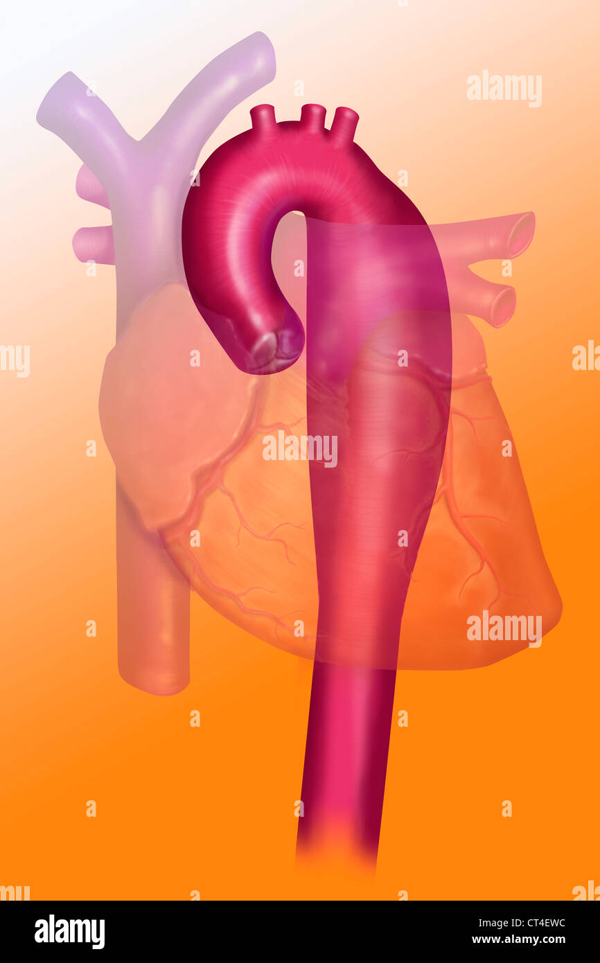 ANEURYSM OF THE THORACIC AORTA Stock Photohttps://www.alamy.com/image-license-details/?v=1https://www.alamy.com/stock-photo-aneurysm-of-the-thoracic-aorta-49271976.html
RMCT4EWC–ANEURYSM OF THE THORACIC AORTA
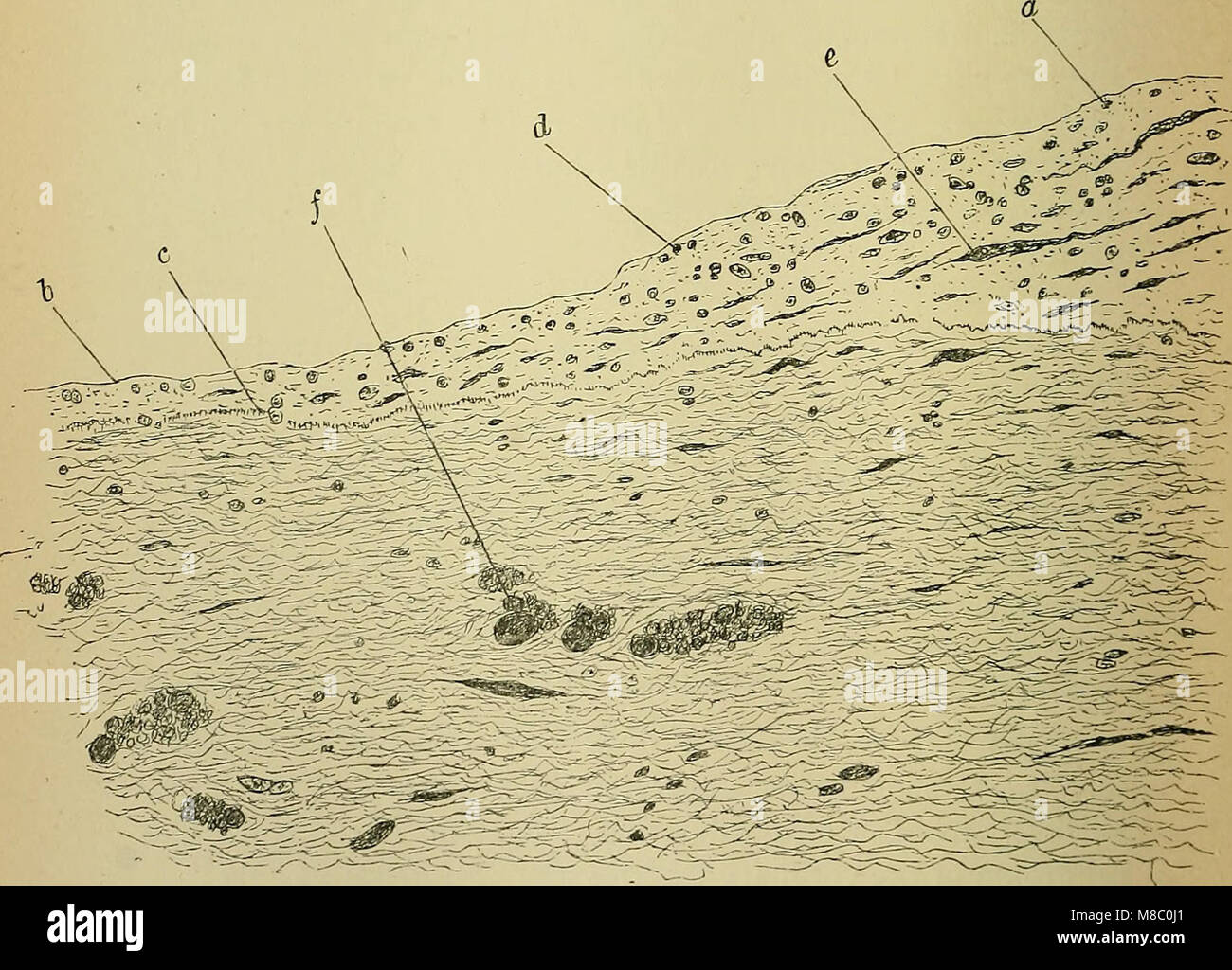 Diseases of the heart and thoracic aorta (1884) (14784549702) Stock Photohttps://www.alamy.com/image-license-details/?v=1https://www.alamy.com/stock-photo-diseases-of-the-heart-and-thoracic-aorta-1884-14784549702-177284857.html
RMM8C0J1–Diseases of the heart and thoracic aorta (1884) (14784549702)
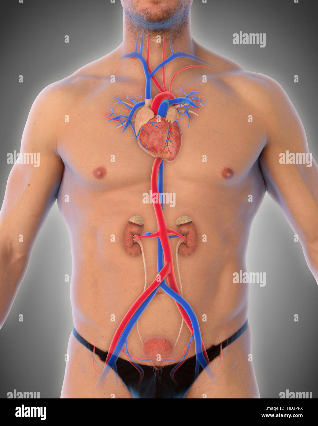 Human Thoracic Aorta Stock Photohttps://www.alamy.com/image-license-details/?v=1https://www.alamy.com/stock-photo-human-thoracic-aorta-128546850.html
RFHD3PPX–Human Thoracic Aorta
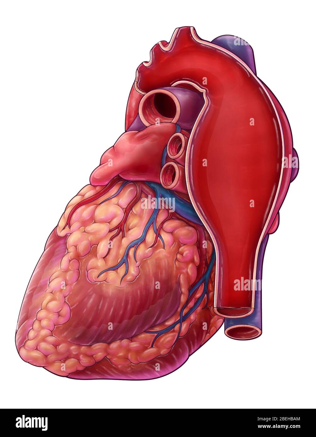 Thoracic Aortic Aneurysm, Illustration Stock Photohttps://www.alamy.com/image-license-details/?v=1https://www.alamy.com/thoracic-aortic-aneurysm-illustration-image353194652.html
RM2BEHBAM–Thoracic Aortic Aneurysm, Illustration
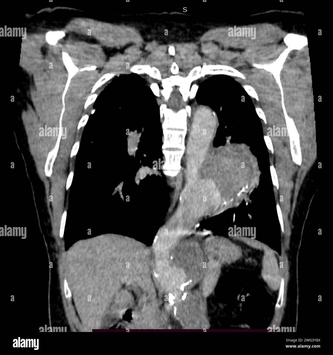 Saccular aortic aneurysm, CT scan Stock Photohttps://www.alamy.com/image-license-details/?v=1https://www.alamy.com/saccular-aortic-aneurysm-ct-scan-image595074282.html
RF2WG3Y8X–Saccular aortic aneurysm, CT scan
 Relation between esophagus, Trachea and Aortic Arch.3d rendering Stock Photohttps://www.alamy.com/image-license-details/?v=1https://www.alamy.com/relation-between-esophagus-trachea-and-aortic-arch3d-rendering-image501581036.html
RF2M40YMC–Relation between esophagus, Trachea and Aortic Arch.3d rendering
 Main reports of the lungs. (Thoracic organs seen by their front face), vintage engraved illustration. Usual Medicine Dictionary Stock Vectorhttps://www.alamy.com/image-license-details/?v=1https://www.alamy.com/stock-photo-main-reports-of-the-lungs-thoracic-organs-seen-by-their-front-face-84419982.html
RFEW9JFA–Main reports of the lungs. (Thoracic organs seen by their front face), vintage engraved illustration. Usual Medicine Dictionary
 The thoracic aorta originates from the left ventricle, guarded by the aortic valve 3d illustration Stock Photohttps://www.alamy.com/image-license-details/?v=1https://www.alamy.com/the-thoracic-aorta-originates-from-the-left-ventricle-guarded-by-the-aortic-valve-3d-illustration-image596592658.html
RF2WJH40J–The thoracic aorta originates from the left ventricle, guarded by the aortic valve 3d illustration
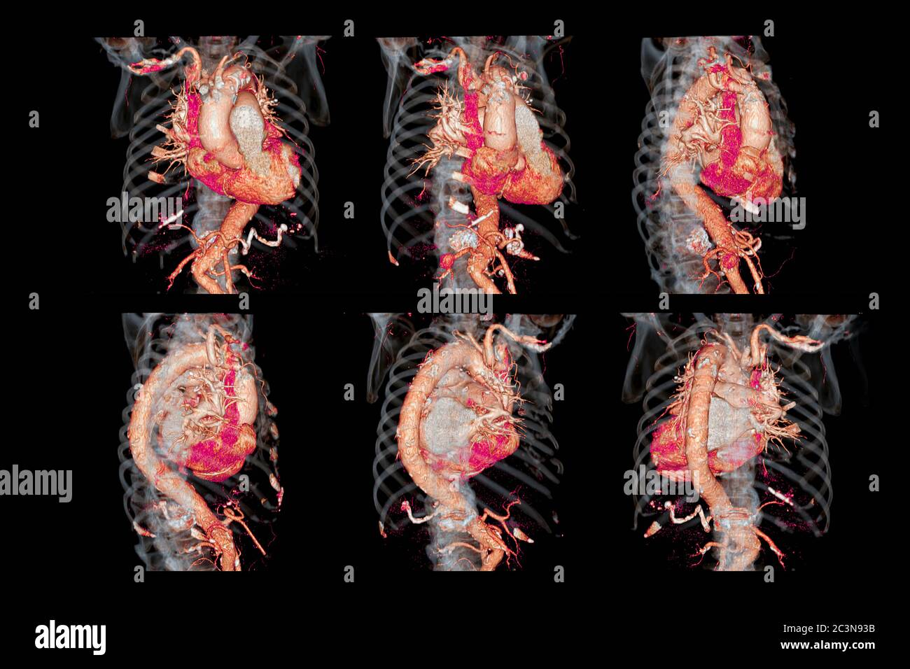 Collection of CTA thoracic aorta 3D rendering image for diagnotic abdominal aortic aneurysm or AAA and aortic dissection Stock Photohttps://www.alamy.com/image-license-details/?v=1https://www.alamy.com/collection-of-cta-thoracic-aorta-3d-rendering-image-for-diagnotic-abdominal-aortic-aneurysm-or-aaa-and-aortic-dissection-image363729839.html
RF2C3N93B–Collection of CTA thoracic aorta 3D rendering image for diagnotic abdominal aortic aneurysm or AAA and aortic dissection
 The human body with superimposed colored plates, by Julien Bougle, circa 1899. Stock Photohttps://www.alamy.com/image-license-details/?v=1https://www.alamy.com/stock-photo-the-human-body-with-superimposed-colored-plates-by-julien-bougle-circa-77722840.html
RFEECG7M–The human body with superimposed colored plates, by Julien Bougle, circa 1899.
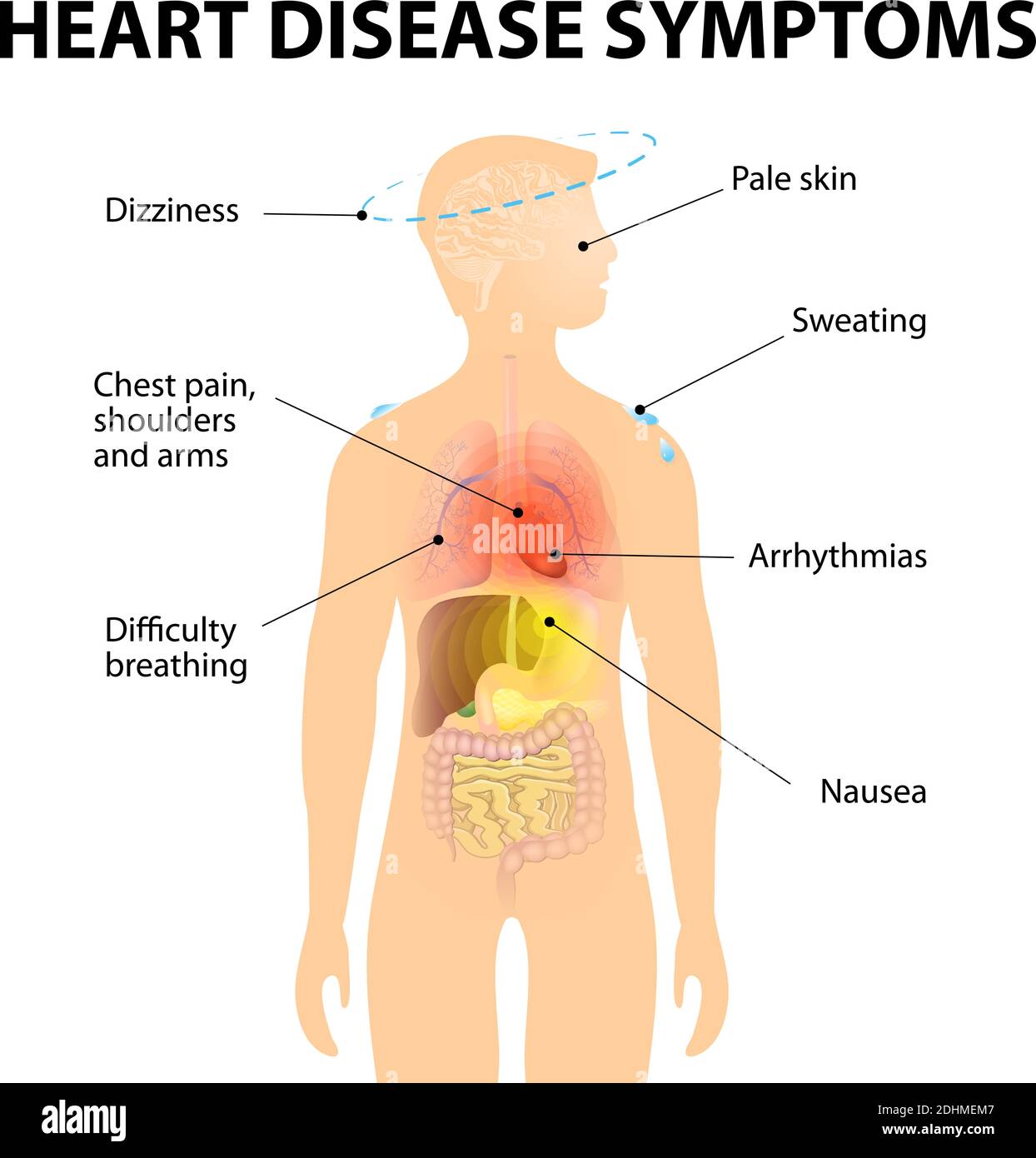 heart disease: valve disease, aneurysm, coronary artery disease, cardiac arrhythmia, heart failture, cardiomyopathy and pericarditis Stock Vectorhttps://www.alamy.com/image-license-details/?v=1https://www.alamy.com/heart-disease-valve-disease-aneurysm-coronary-artery-disease-cardiac-arrhythmia-heart-failture-cardiomyopathy-and-pericarditis-image389527831.html
RF2DHMEM7–heart disease: valve disease, aneurysm, coronary artery disease, cardiac arrhythmia, heart failture, cardiomyopathy and pericarditis
 Endovascular treatment of intracranial aneurysms. Aneurysm is a weakness in the wall of a blood vessel which causes the blood vessel to swell. Brain Stock Photohttps://www.alamy.com/image-license-details/?v=1https://www.alamy.com/endovascular-treatment-of-intracranial-aneurysms-aneurysm-is-a-weakness-in-the-wall-of-a-blood-vessel-which-causes-the-blood-vessel-to-swell-brain-image384512823.html
RF2D9G20R–Endovascular treatment of intracranial aneurysms. Aneurysm is a weakness in the wall of a blood vessel which causes the blood vessel to swell. Brain
RF2G8854P–Cardiothoracic surgery, heart surgery icon - Perfect use for designing and developing websites, printed files and presentations, Promotional Materials
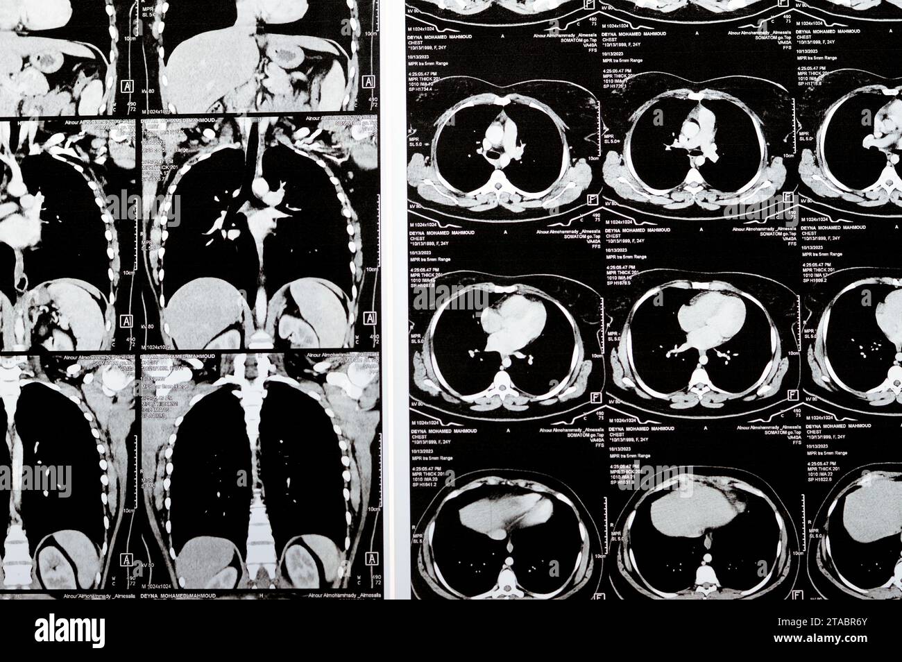 Cairo, Egypt, October 15 2023: CT scan axial slices through chest with contrast injection showing low grade of inflammatory reaction, parenchymal vess Stock Photohttps://www.alamy.com/image-license-details/?v=1https://www.alamy.com/cairo-egypt-october-15-2023-ct-scan-axial-slices-through-chest-with-contrast-injection-showing-low-grade-of-inflammatory-reaction-parenchymal-vess-image574348403.html
RF2TABR6Y–Cairo, Egypt, October 15 2023: CT scan axial slices through chest with contrast injection showing low grade of inflammatory reaction, parenchymal vess
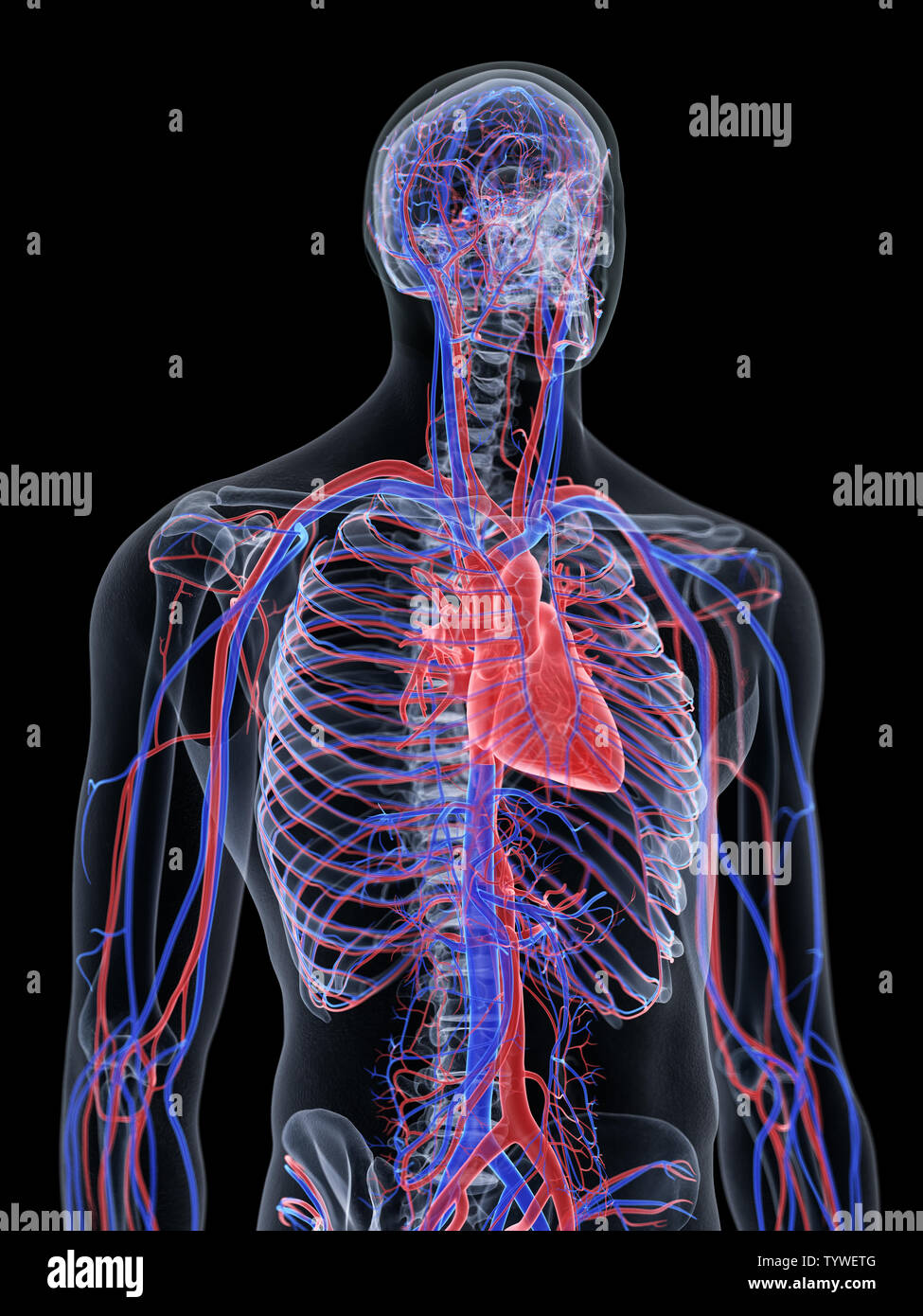 3d rendered medically accurate illustration of the heart and vascular system Stock Photohttps://www.alamy.com/image-license-details/?v=1https://www.alamy.com/3d-rendered-medically-accurate-illustration-of-the-heart-and-vascular-system-image258101328.html
RFTYWETG–3d rendered medically accurate illustration of the heart and vascular system
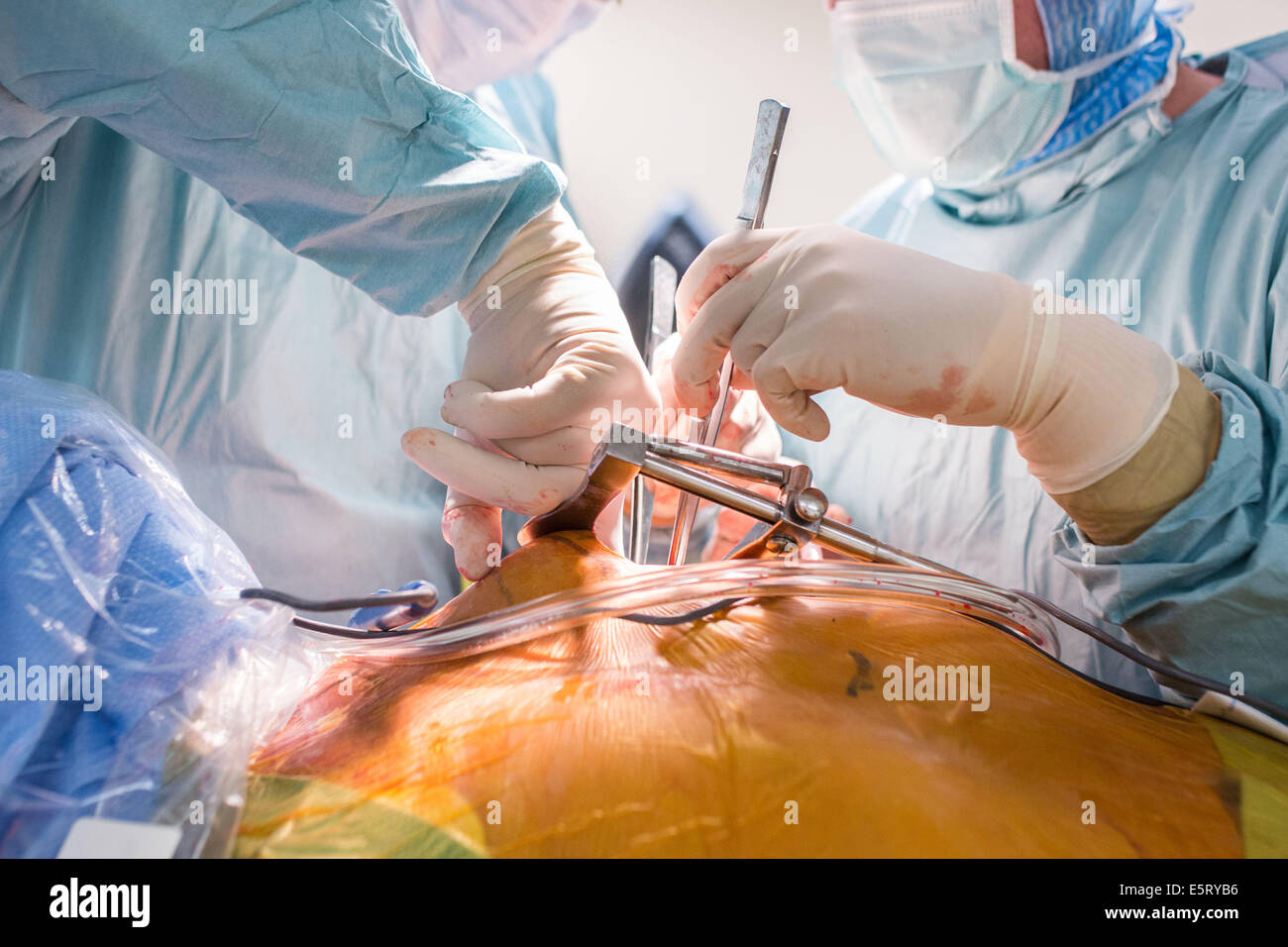 Da Vinci® Robotic Assisted Heart Surgery, double aortic coronary bypass with Da Vinci ® surgical robot. Limoges hospital, Stock Photohttps://www.alamy.com/image-license-details/?v=1https://www.alamy.com/stock-photo-da-vinci-robotic-assisted-heart-surgery-double-aortic-coronary-bypass-72441130.html
RME5RYB6–Da Vinci® Robotic Assisted Heart Surgery, double aortic coronary bypass with Da Vinci ® surgical robot. Limoges hospital,
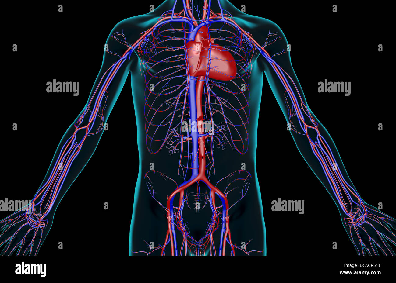 The blood supply of the trunk Stock Photohttps://www.alamy.com/image-license-details/?v=1https://www.alamy.com/stock-photo-the-blood-supply-of-the-trunk-13213667.html
RFACR51T–The blood supply of the trunk
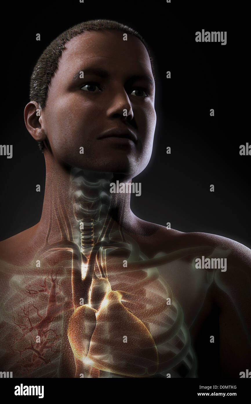 Anatomical model showing the upper thoracic organs. Stock Photohttps://www.alamy.com/image-license-details/?v=1https://www.alamy.com/stock-photo-anatomical-model-showing-the-upper-thoracic-organs-52089508.html
RMD0MTKG–Anatomical model showing the upper thoracic organs.
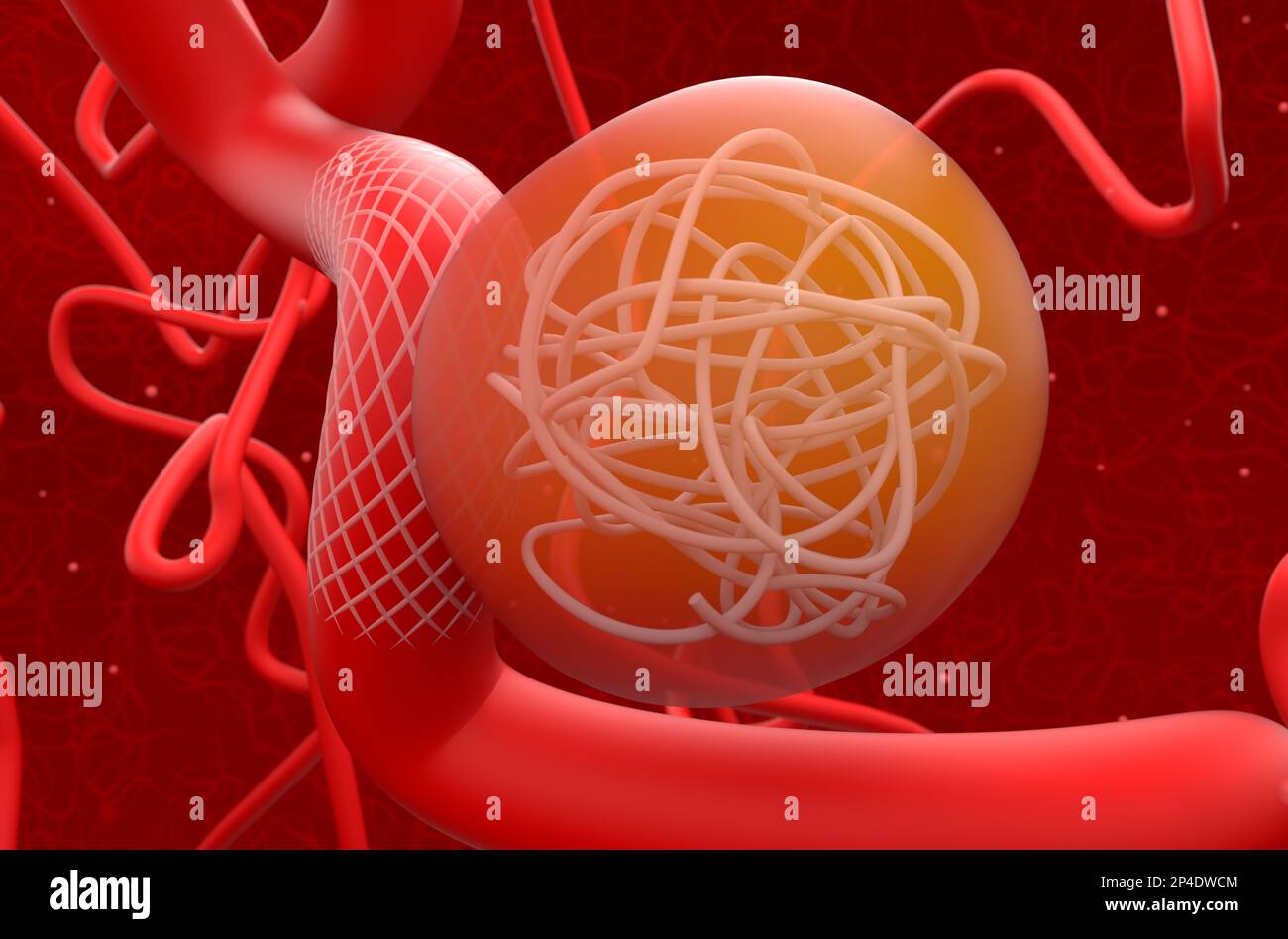 Aneurysm treat with mesh stent and coils - 3d illustration closeup view Stock Photohttps://www.alamy.com/image-license-details/?v=1https://www.alamy.com/aneurysm-treat-with-mesh-stent-and-coils-3d-illustration-closeup-view-image536285364.html
RF2P4DWCM–Aneurysm treat with mesh stent and coils - 3d illustration closeup view
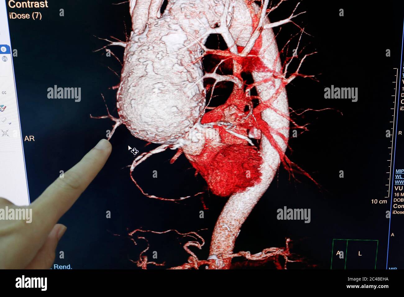 Heart cat scan result shown on monitor Stock Photohttps://www.alamy.com/image-license-details/?v=1https://www.alamy.com/heart-cat-scan-result-shown-on-monitor-image364129286.html
RM2C4BEHA–Heart cat scan result shown on monitor
 Diseases of the heart and thoracic aorta (1884) (14784520412) Stock Photohttps://www.alamy.com/image-license-details/?v=1https://www.alamy.com/stock-photo-diseases-of-the-heart-and-thoracic-aorta-1884-14784520412-177284854.html
RMM8C0HX–Diseases of the heart and thoracic aorta (1884) (14784520412)
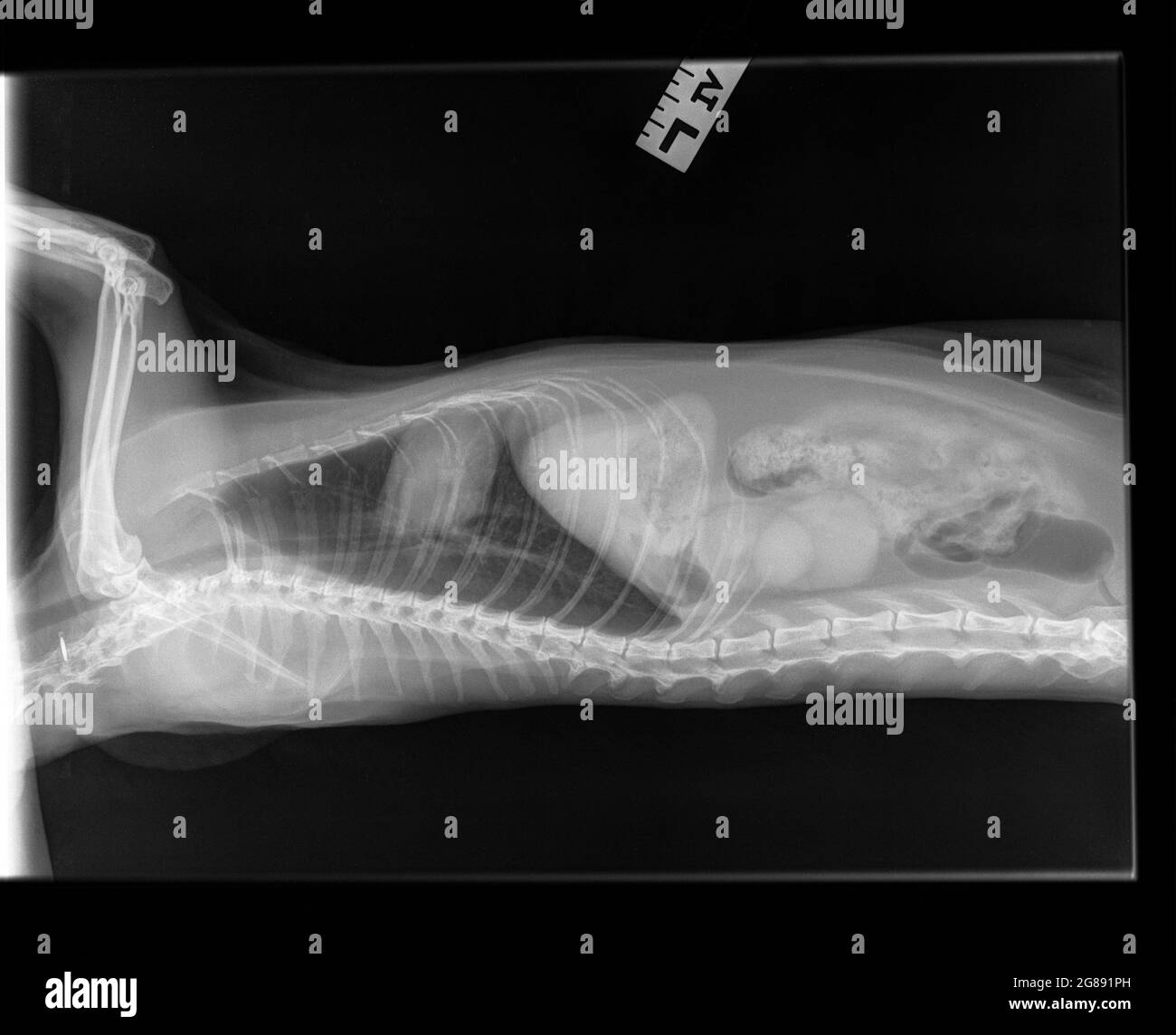 Radiograph of an old spayed female cat. Lateral view. A cat with suspected chronic renal (kidney) disease. A changed structure of the left kidney. Stock Photohttps://www.alamy.com/image-license-details/?v=1https://www.alamy.com/radiograph-of-an-old-spayed-female-cat-lateral-view-a-cat-with-suspected-chronic-renal-kidney-disease-a-changed-structure-of-the-left-kidney-image435375433.html
RF2G891PH–Radiograph of an old spayed female cat. Lateral view. A cat with suspected chronic renal (kidney) disease. A changed structure of the left kidney.
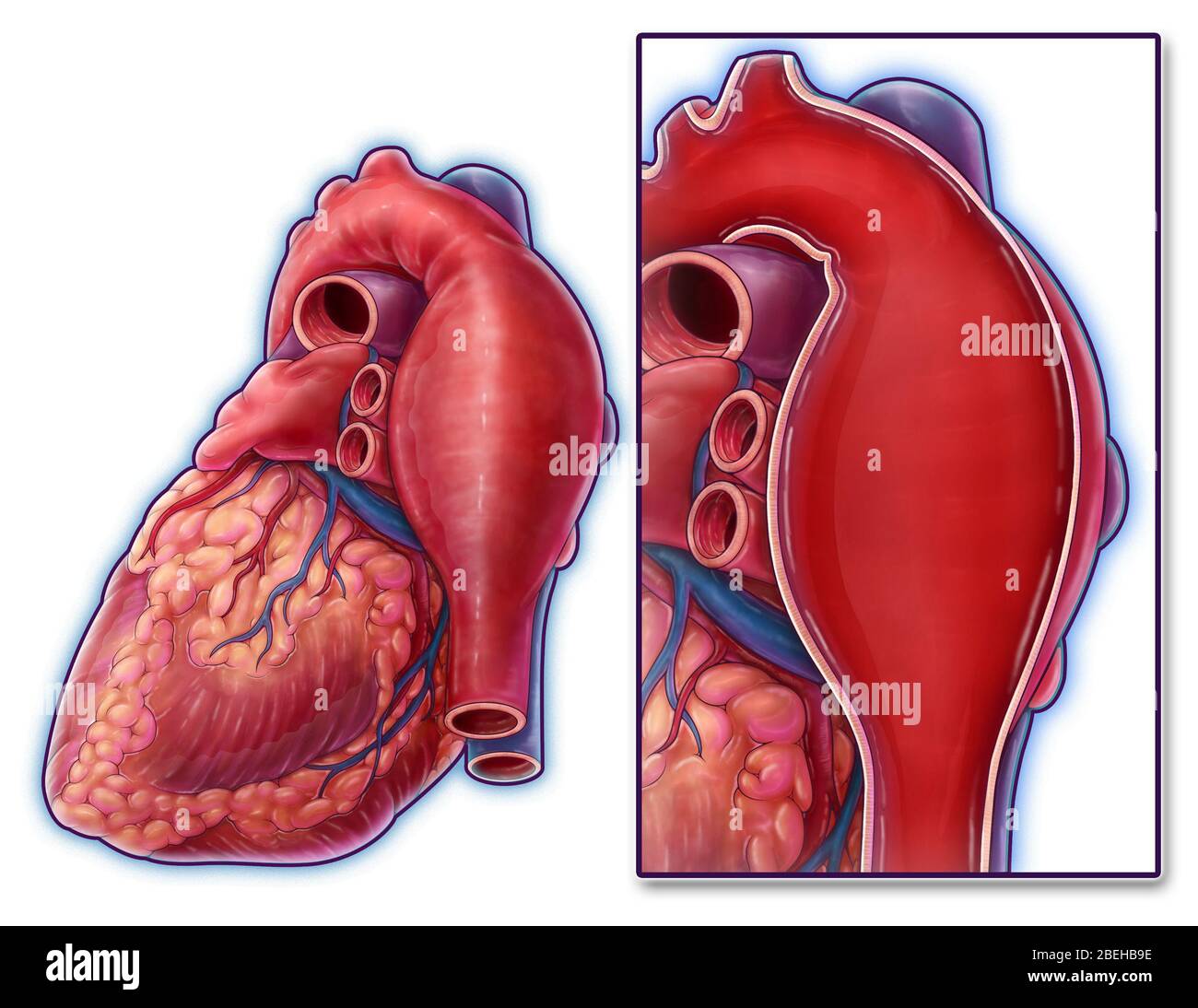 Thoracic Aortic Aneurysm, Illustration Stock Photohttps://www.alamy.com/image-license-details/?v=1https://www.alamy.com/thoracic-aortic-aneurysm-illustration-image353194618.html
RM2BEHB9E–Thoracic Aortic Aneurysm, Illustration
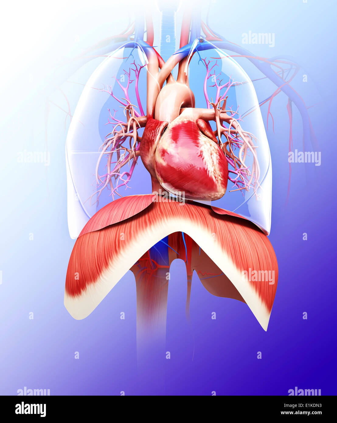 Human respiratory system computer artwork. Stock Photohttps://www.alamy.com/image-license-details/?v=1https://www.alamy.com/human-respiratory-system-computer-artwork-image69883999.html
RFE1KDN3–Human respiratory system computer artwork.
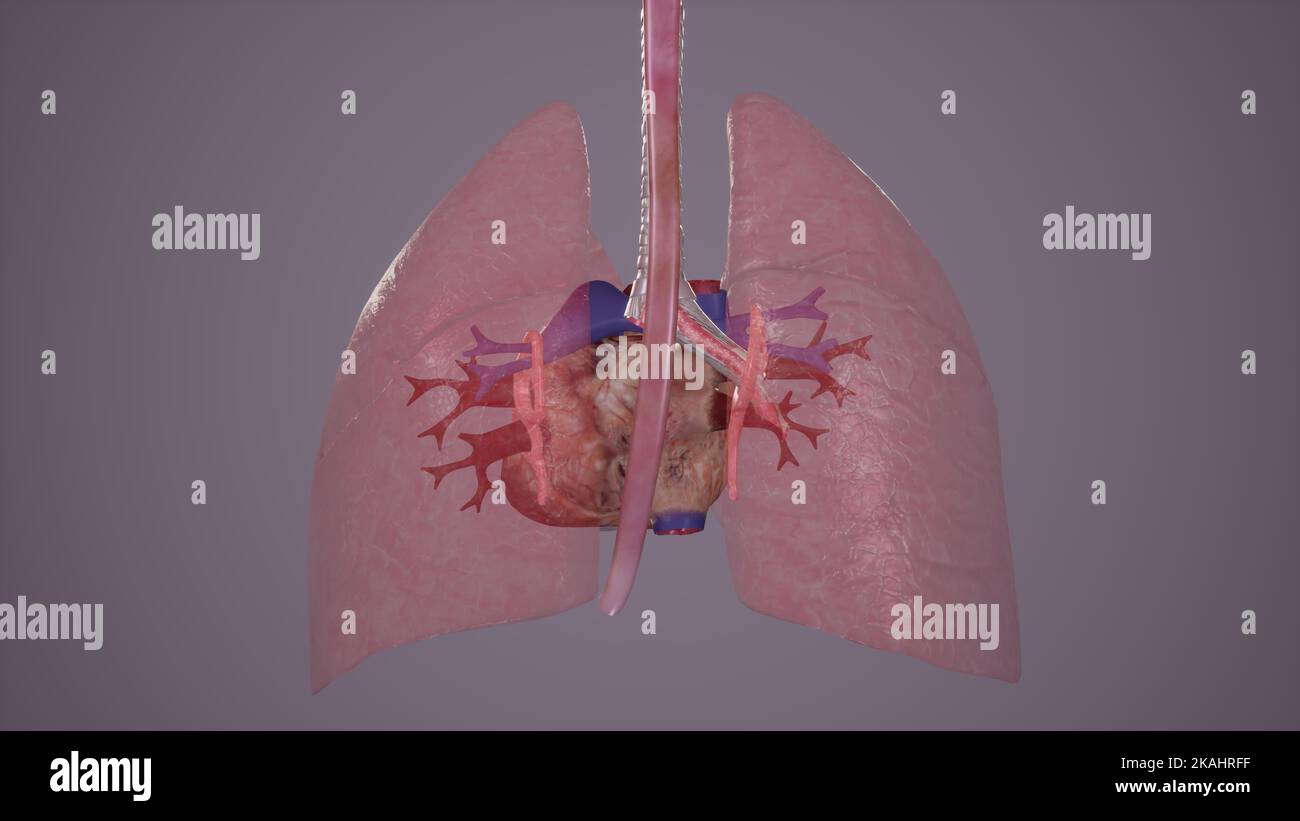 Medical Illustration of Pulmonary Vessels Stock Photohttps://www.alamy.com/image-license-details/?v=1https://www.alamy.com/medical-illustration-of-pulmonary-vessels-image488428515.html
RF2KAHRFF–Medical Illustration of Pulmonary Vessels
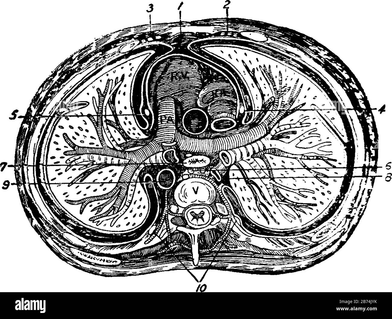 The transverse section of the thorax, vintage line drawing or engraving illustration. Stock Vectorhttps://www.alamy.com/image-license-details/?v=1https://www.alamy.com/the-transverse-section-of-the-thorax-vintage-line-drawing-or-engraving-illustration-image348612647.html
RF2B74JYK–The transverse section of the thorax, vintage line drawing or engraving illustration.
 The thoracic aorta originates from the left ventricle, guarded by the aortic valve 3d illustration Stock Photohttps://www.alamy.com/image-license-details/?v=1https://www.alamy.com/the-thoracic-aorta-originates-from-the-left-ventricle-guarded-by-the-aortic-valve-3d-illustration-image596593508.html
RF2WJH530–The thoracic aorta originates from the left ventricle, guarded by the aortic valve 3d illustration
 CTA thoracic aorta 3D rendering image posterior view or back view for diagnotic abdominal aortic aneurysm or AAA and aortic dissection Stock Photohttps://www.alamy.com/image-license-details/?v=1https://www.alamy.com/cta-thoracic-aorta-3d-rendering-image-posterior-view-or-back-view-for-diagnotic-abdominal-aortic-aneurysm-or-aaa-and-aortic-dissection-image363730340.html
RF2C3N9N8–CTA thoracic aorta 3D rendering image posterior view or back view for diagnotic abdominal aortic aneurysm or AAA and aortic dissection
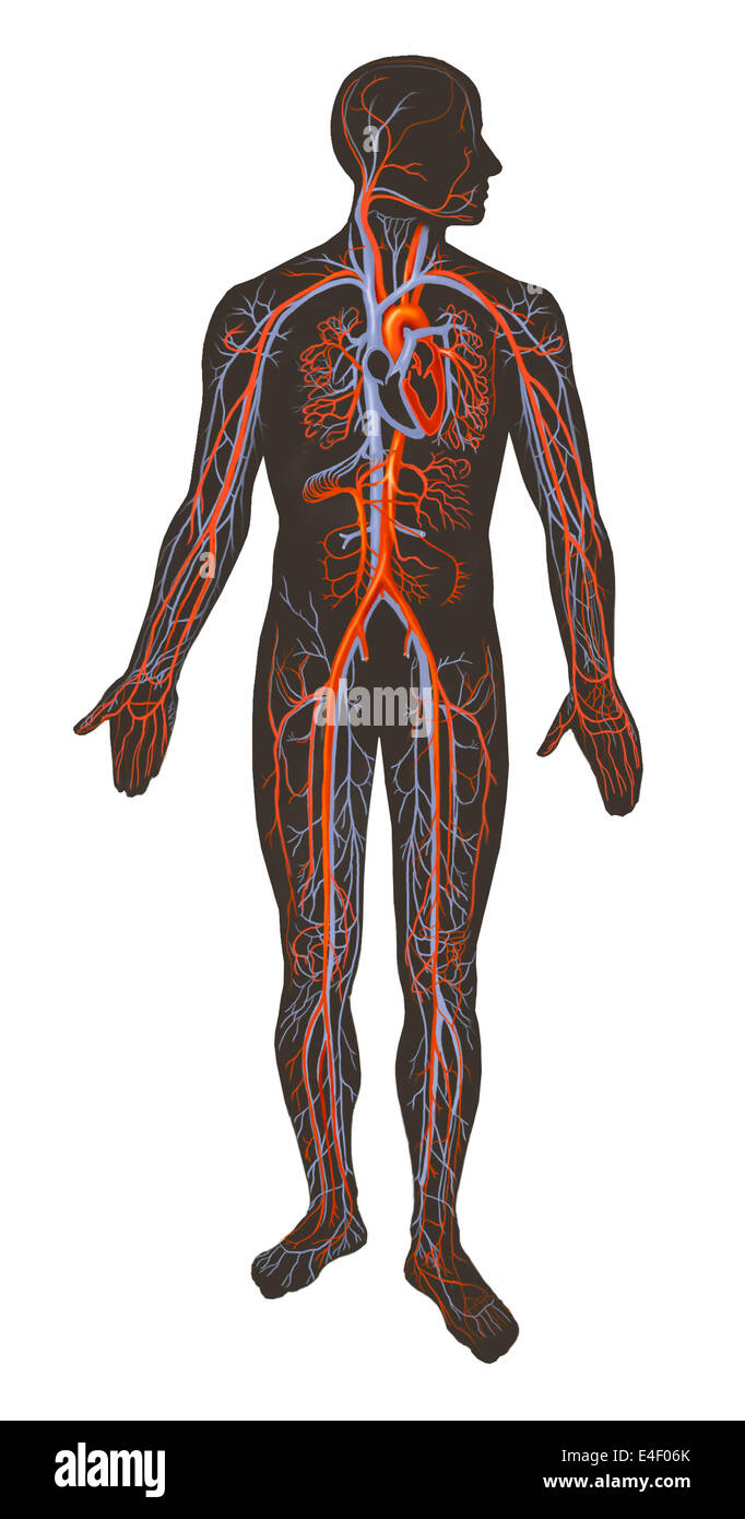 Arteries and veins of the human body. Stock Photohttps://www.alamy.com/image-license-details/?v=1https://www.alamy.com/stock-photo-arteries-and-veins-of-the-human-body-71629563.html
RME4F06K–Arteries and veins of the human body.
RFGM3NK3–Lungs sketch icon
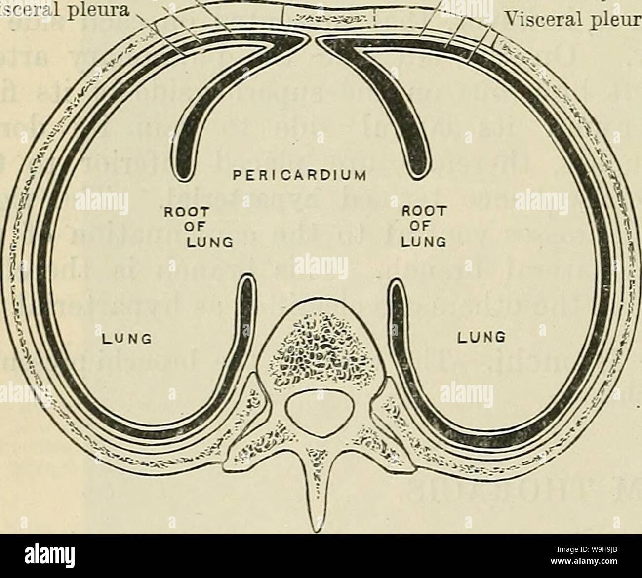 Archive image from page 1117 of Cunningham's Text-book of anatomy (1914). Cunningham's Text-book of anatomy cunninghamstextb00cunn Year: 1914 ( 1084 THE EESPIEATOEY SYSTEM. Costal part of parietal pleura Pleural cavity ,. Visceral pleura Costal part of parietal pleura Pleural cavity /•Visceral pleura marked projection of the heart to the left side, and to the position of the thoracic aorta on the left side of the median plane, the left pleural chamber, although it is deeper than the right, is greatly reduced in width. The two pleural cavities, therefore, are very far from being symmetrical in Stock Photohttps://www.alamy.com/image-license-details/?v=1https://www.alamy.com/archive-image-from-page-1117-of-cunninghams-text-book-of-anatomy-1914-cunninghams-text-book-of-anatomy-cunninghamstextb00cunn-year-1914-1084-the-eespieatoey-system-costal-part-of-parietal-pleura-pleural-cavity-visceral-pleura-costal-part-of-parietal-pleura-pleural-cavity-visceral-pleura-marked-projection-of-the-heart-to-the-left-side-and-to-the-position-of-the-thoracic-aorta-on-the-left-side-of-the-median-plane-the-left-pleural-chamber-although-it-is-deeper-than-the-right-is-greatly-reduced-in-width-the-two-pleural-cavities-therefore-are-very-far-from-being-symmetrical-in-image264068179.html
RMW9H9JB–Archive image from page 1117 of Cunningham's Text-book of anatomy (1914). Cunningham's Text-book of anatomy cunninghamstextb00cunn Year: 1914 ( 1084 THE EESPIEATOEY SYSTEM. Costal part of parietal pleura Pleural cavity ,. Visceral pleura Costal part of parietal pleura Pleural cavity /•Visceral pleura marked projection of the heart to the left side, and to the position of the thoracic aorta on the left side of the median plane, the left pleural chamber, although it is deeper than the right, is greatly reduced in width. The two pleural cavities, therefore, are very far from being symmetrical in
RF2G8857X–Cardiothoracic surgery, heart surgery icon - Perfect use for designing and developing websites, printed files and presentations, Promotional Materials
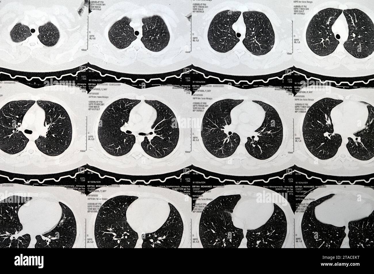 Cairo, Egypt, October 15 2023: CT scan axial slices through chest with contrast injection showing low grade of inflammatory reaction, parenchymal vess Stock Photohttps://www.alamy.com/image-license-details/?v=1https://www.alamy.com/cairo-egypt-october-15-2023-ct-scan-axial-slices-through-chest-with-contrast-injection-showing-low-grade-of-inflammatory-reaction-parenchymal-vess-image574363660.html
RF2TACEKT–Cairo, Egypt, October 15 2023: CT scan axial slices through chest with contrast injection showing low grade of inflammatory reaction, parenchymal vess
 3d rendered medically accurate illustration of the human heart Stock Photohttps://www.alamy.com/image-license-details/?v=1https://www.alamy.com/3d-rendered-medically-accurate-illustration-of-the-human-heart-image258102104.html
RFTYWFT8–3d rendered medically accurate illustration of the human heart
 Da Vinci® Robotic Assisted Heart Surgery, double aortic coronary bypass with Da Vinci ® surgical robot. Limoges hospital, Stock Photohttps://www.alamy.com/image-license-details/?v=1https://www.alamy.com/stock-photo-da-vinci-robotic-assisted-heart-surgery-double-aortic-coronary-bypass-72441131.html
RME5RYB7–Da Vinci® Robotic Assisted Heart Surgery, double aortic coronary bypass with Da Vinci ® surgical robot. Limoges hospital,
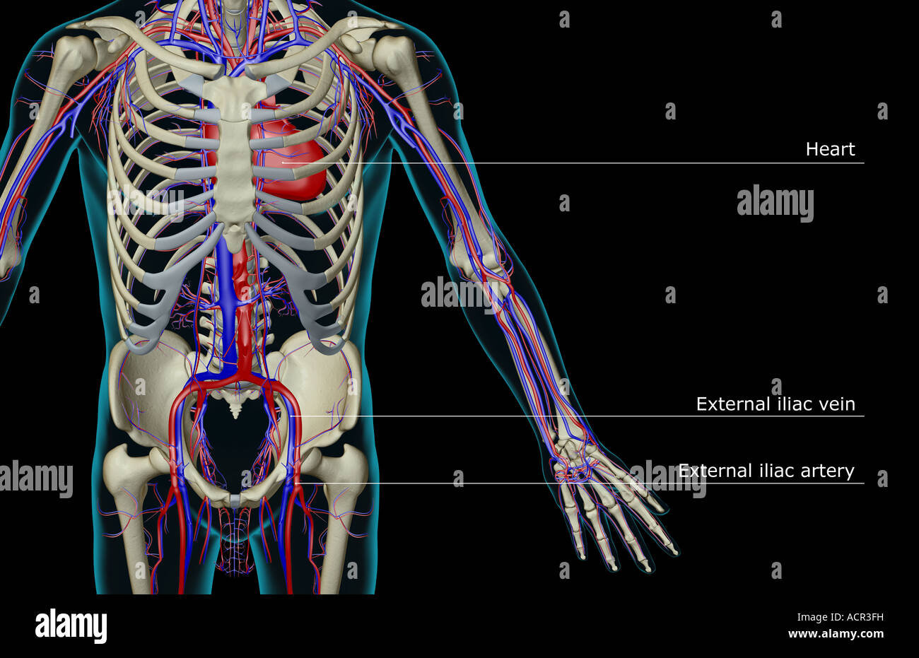 The blood supply of the trunk Stock Photohttps://www.alamy.com/image-license-details/?v=1https://www.alamy.com/stock-photo-the-blood-supply-of-the-trunk-13213156.html
RFACR3FH–The blood supply of the trunk
 Surgeons operating during cardiac operation of operation room Stock Photohttps://www.alamy.com/image-license-details/?v=1https://www.alamy.com/surgeons-operating-during-cardiac-operation-of-operation-room-image330339680.html
RF2A5C7HM–Surgeons operating during cardiac operation of operation room
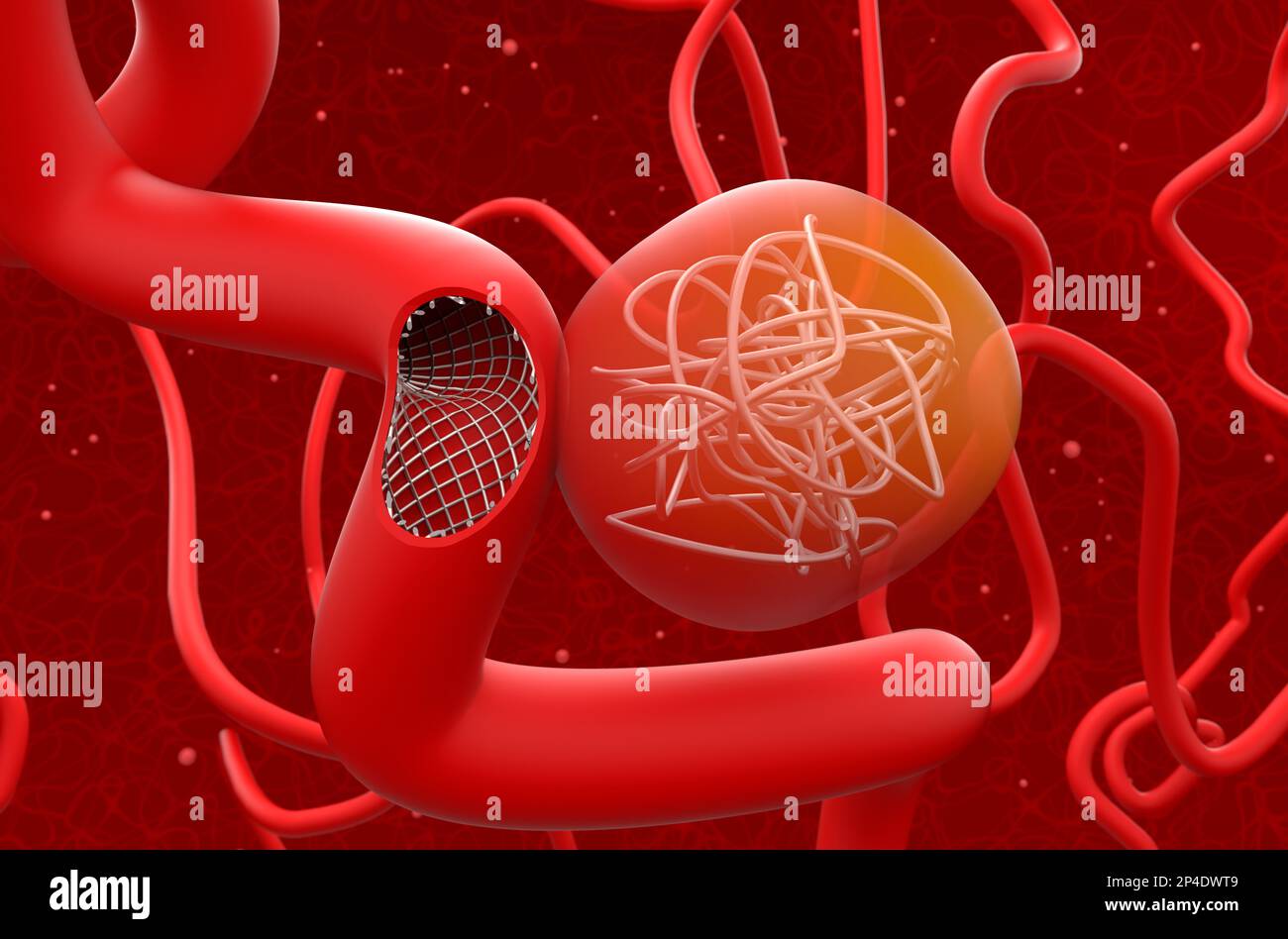 Aneurysm treat with mesh stent and coils (section) - 3d illustration closeup view Stock Photohttps://www.alamy.com/image-license-details/?v=1https://www.alamy.com/aneurysm-treat-with-mesh-stent-and-coils-section-3d-illustration-closeup-view-image536285689.html
RF2P4DWT9–Aneurysm treat with mesh stent and coils (section) - 3d illustration closeup view
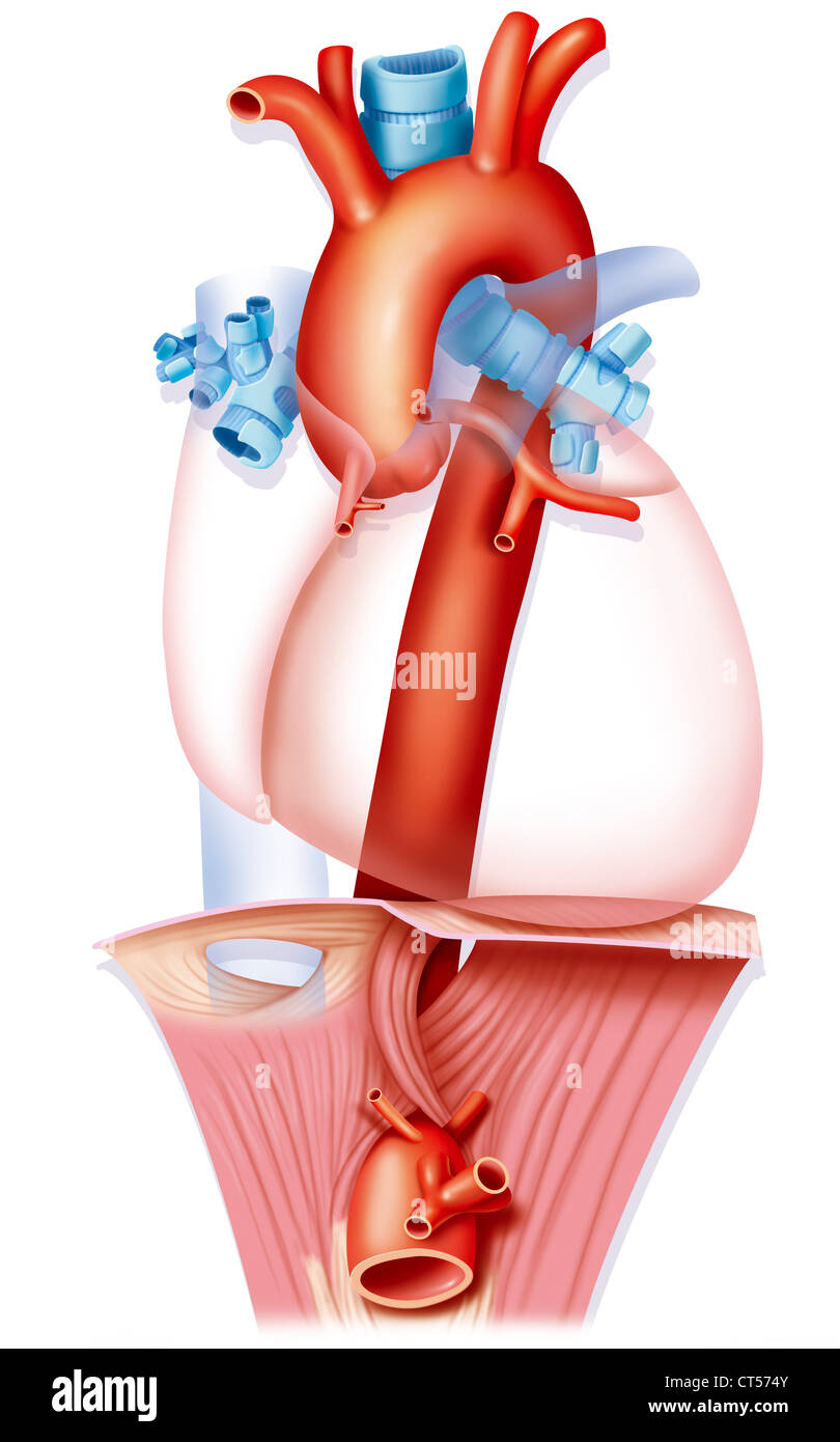 AORTA DRAWING Stock Photohttps://www.alamy.com/image-license-details/?v=1https://www.alamy.com/stock-photo-aorta-drawing-49287867.html
RMCT574Y–AORTA DRAWING
 Diseases of the heart and thoracic aorta (1884) (14598261059) Stock Photohttps://www.alamy.com/image-license-details/?v=1https://www.alamy.com/stock-photo-diseases-of-the-heart-and-thoracic-aorta-1884-14598261059-177284841.html
RMM8C0HD–Diseases of the heart and thoracic aorta (1884) (14598261059)
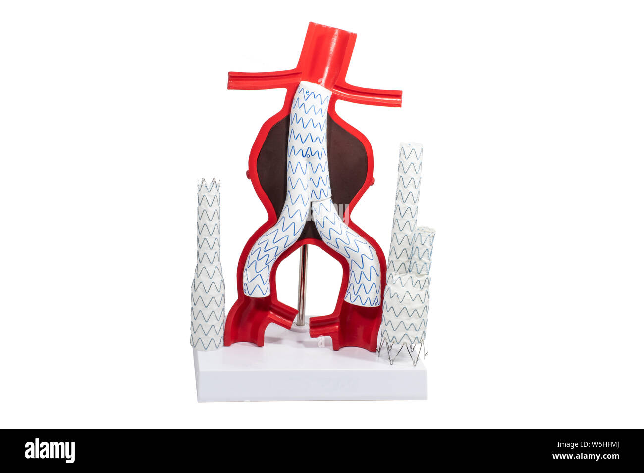 Model aneurysm treatment with endovascular fenestrated and branched stent-grafts. Stock Photohttps://www.alamy.com/image-license-details/?v=1https://www.alamy.com/model-aneurysm-treatment-with-endovascular-fenestrated-and-branched-stent-grafts-image261614322.html
RFW5HFMJ–Model aneurysm treatment with endovascular fenestrated and branched stent-grafts.
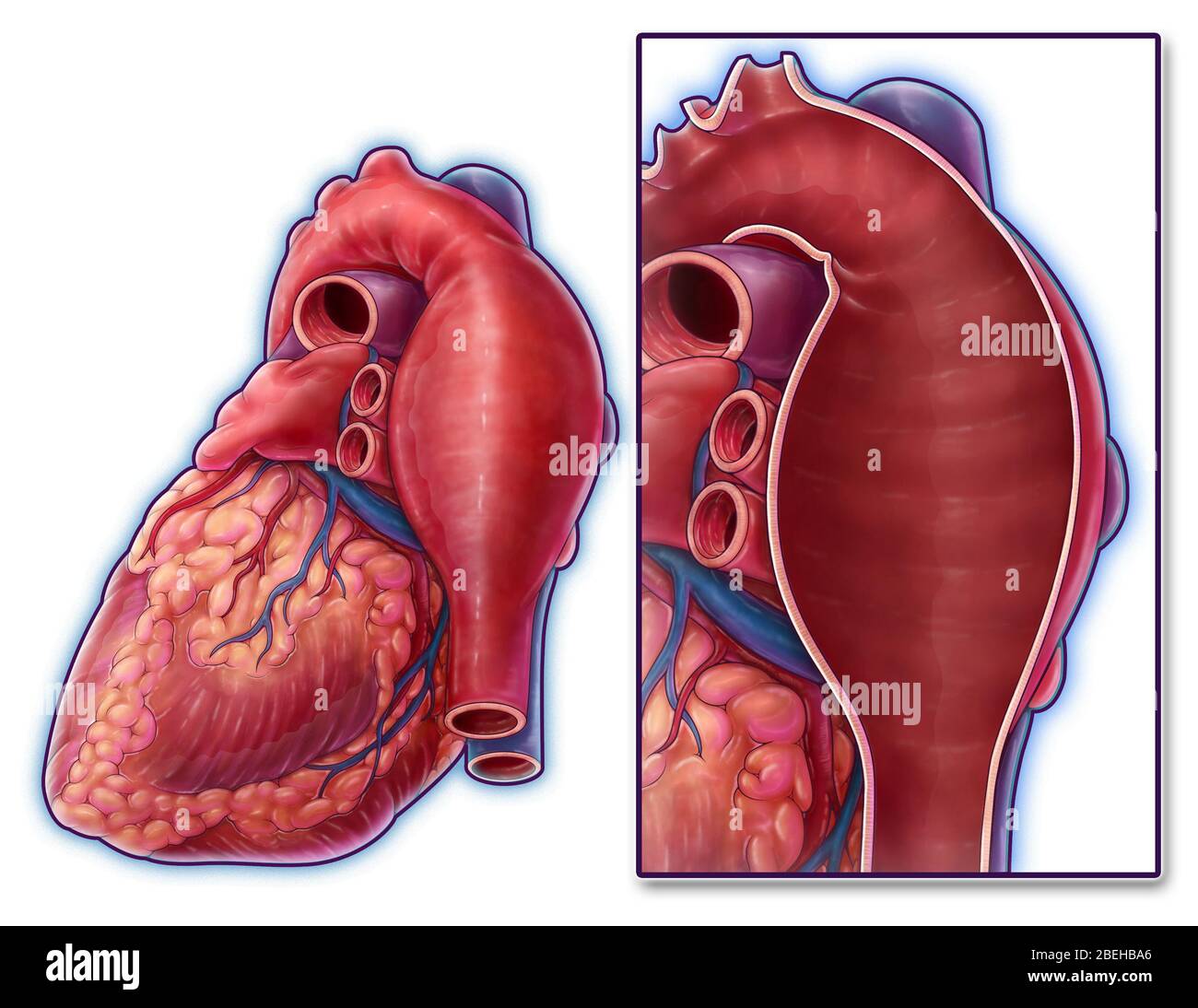 Thoracic Aortic Aneurysm, Illustration Stock Photohttps://www.alamy.com/image-license-details/?v=1https://www.alamy.com/thoracic-aortic-aneurysm-illustration-image353194638.html
RM2BEHBA6–Thoracic Aortic Aneurysm, Illustration
 Human cardiovascular system and diaphragm computer artwork. Stock Photohttps://www.alamy.com/image-license-details/?v=1https://www.alamy.com/human-cardiovascular-system-and-diaphragm-computer-artwork-image69883971.html
RFE1KDM3–Human cardiovascular system and diaphragm computer artwork.
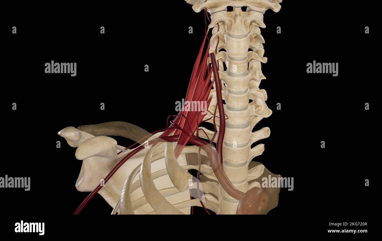 Branches of Subclavian Artery Stock Photohttps://www.alamy.com/image-license-details/?v=1https://www.alamy.com/branches-of-subclavian-artery-image491880055.html
RF2KG720R–Branches of Subclavian Artery
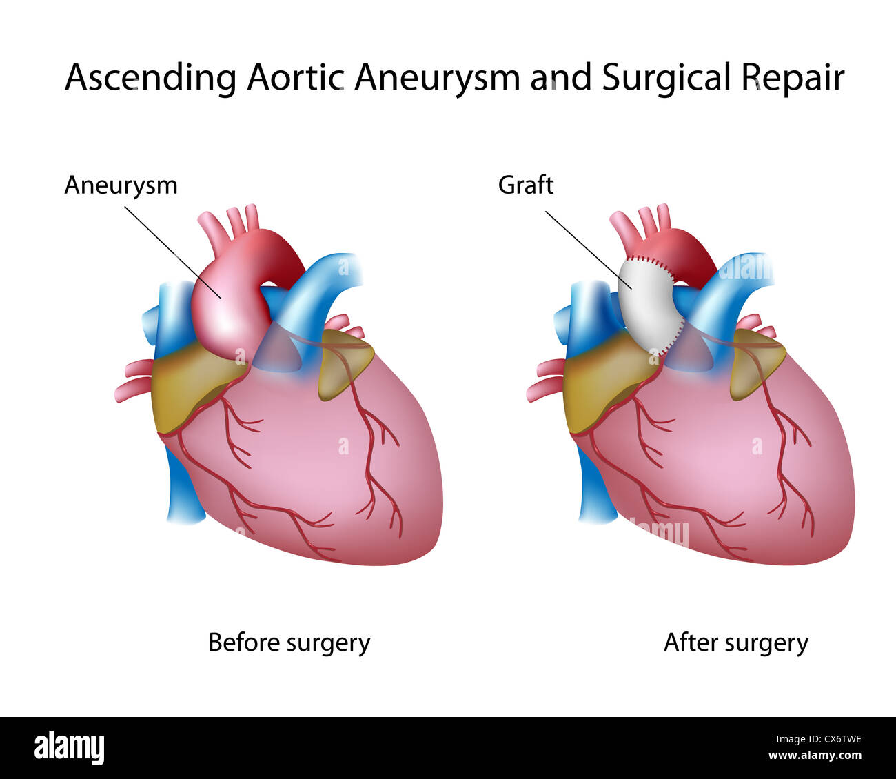 Ascending aortic aneurysm and open surgery Stock Photohttps://www.alamy.com/image-license-details/?v=1https://www.alamy.com/stock-photo-ascending-aortic-aneurysm-and-open-surgery-50553034.html
RFCX6TWE–Ascending aortic aneurysm and open surgery
 The radicular arteries arise from anterior cervical arteries, thoracic aorta, and lumbar arteries 3d illustration Stock Photohttps://www.alamy.com/image-license-details/?v=1https://www.alamy.com/the-radicular-arteries-arise-from-anterior-cervical-arteries-thoracic-aorta-and-lumbar-arteries-3d-illustration-image596586832.html
RF2WJGTGG–The radicular arteries arise from anterior cervical arteries, thoracic aorta, and lumbar arteries 3d illustration
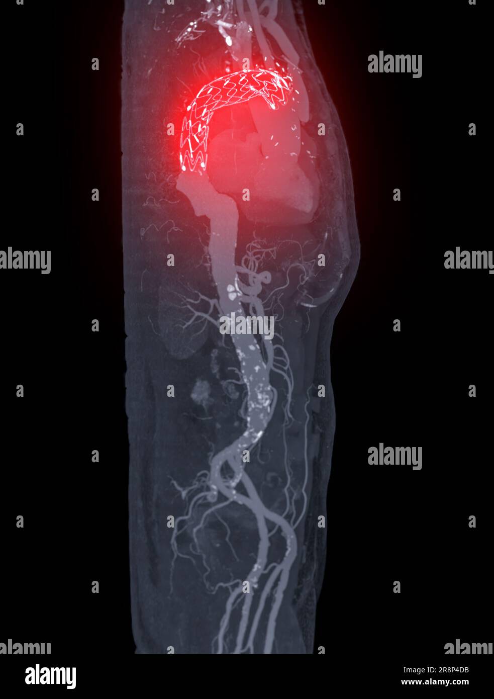 CTA whole aorta with thoracic aorta stent graft 3D rendering image in case abdominal aortic aneurysms. Stock Photohttps://www.alamy.com/image-license-details/?v=1https://www.alamy.com/cta-whole-aorta-with-thoracic-aorta-stent-graft-3d-rendering-image-in-case-abdominal-aortic-aneurysms-image556135479.html
RF2R8P4DB–CTA whole aorta with thoracic aorta stent graft 3D rendering image in case abdominal aortic aneurysms.
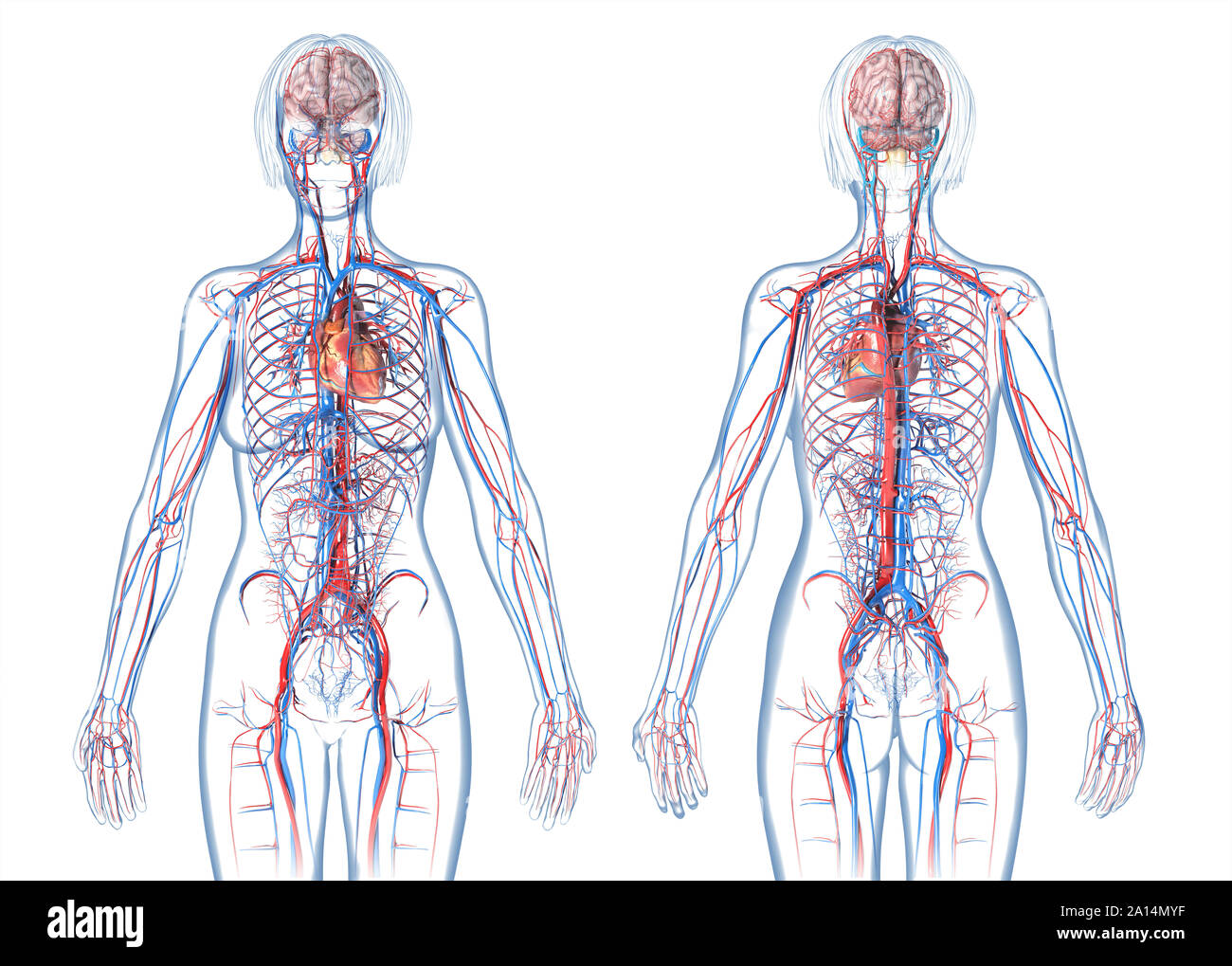 Woman cardiovascular system, rear and front views, on white background. Stock Photohttps://www.alamy.com/image-license-details/?v=1https://www.alamy.com/woman-cardiovascular-system-rear-and-front-views-on-white-background-image327715907.html
RF2A14MYF–Woman cardiovascular system, rear and front views, on white background.
RFGKTCKM–Lungs thin line icon
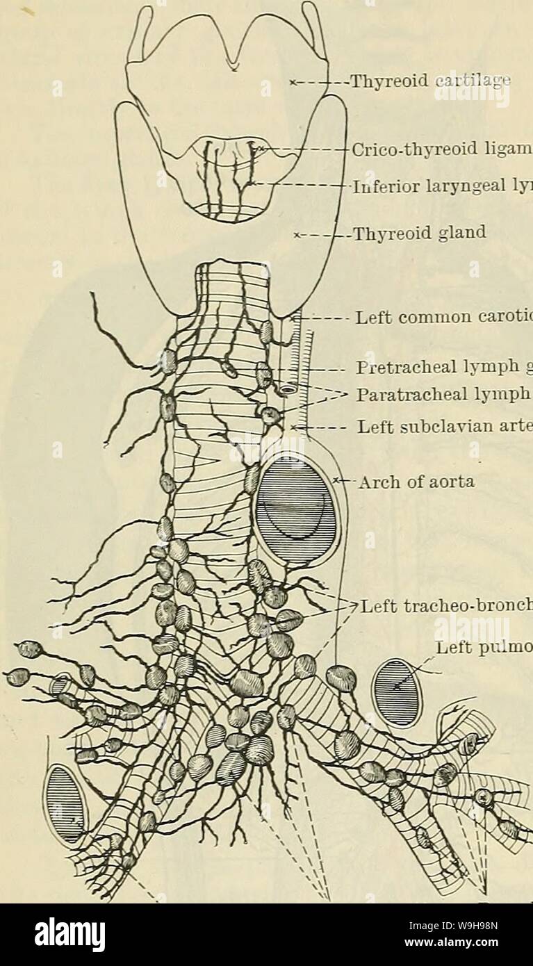 Archive image from page 1045 of Cunningham's Text-book of anatomy (1914). Cunningham's Text-book of anatomy cunninghamstextb00cunn Year: 1914 ( 1012 THE VASCULAR SYSTEM. âThyreoid cartilage Crico-thyreoid ligament â Inferior laryngeal lymph vessels -Thyreoid gland â Left common carotid artery -II Pretracheal lymph gland 'â¢--â - Paratracheal lymph glands Left subclavian artery --?Left tracheo-bronchial glands Left pulmonary artery (4) Lymphoglandulse Mediastinales Posteriores.âThe posterior mediastinal lymph glands, 8-12, lie along the descending part of the thoracic aorta and the thoracic pa Stock Photohttps://www.alamy.com/image-license-details/?v=1https://www.alamy.com/archive-image-from-page-1045-of-cunninghams-text-book-of-anatomy-1914-cunninghams-text-book-of-anatomy-cunninghamstextb00cunn-year-1914-1012-the-vascular-system-thyreoid-cartilage-crico-thyreoid-ligament-inferior-laryngeal-lymph-vessels-thyreoid-gland-left-common-carotid-artery-ii-pretracheal-lymph-gland-paratracheal-lymph-glands-left-subclavian-artery-left-tracheo-bronchial-glands-left-pulmonary-artery-4-lymphoglandulse-mediastinales-posterioresthe-posterior-mediastinal-lymph-glands-8-12-lie-along-the-descending-part-of-the-thoracic-aorta-and-the-thoracic-pa-image264067909.html
RMW9H98N–Archive image from page 1045 of Cunningham's Text-book of anatomy (1914). Cunningham's Text-book of anatomy cunninghamstextb00cunn Year: 1914 ( 1012 THE VASCULAR SYSTEM. âThyreoid cartilage Crico-thyreoid ligament â Inferior laryngeal lymph vessels -Thyreoid gland â Left common carotid artery -II Pretracheal lymph gland 'â¢--â - Paratracheal lymph glands Left subclavian artery --?Left tracheo-bronchial glands Left pulmonary artery (4) Lymphoglandulse Mediastinales Posteriores.âThe posterior mediastinal lymph glands, 8-12, lie along the descending part of the thoracic aorta and the thoracic pa
RF2G8857T–Cardiothoracic surgery, heart surgery icon - Perfect use for designing and developing websites, printed files and presentations, Promotional Materials
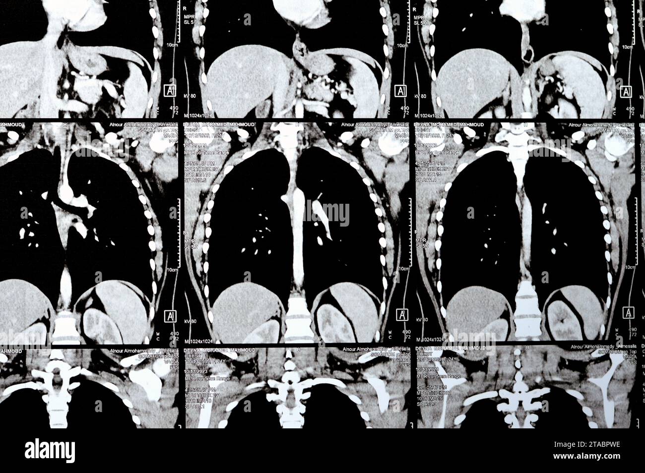 Cairo, Egypt, October 15 2023: CT scan axial slices through chest with contrast injection showing low grade of inflammatory reaction, parenchymal vess Stock Photohttps://www.alamy.com/image-license-details/?v=1https://www.alamy.com/cairo-egypt-october-15-2023-ct-scan-axial-slices-through-chest-with-contrast-injection-showing-low-grade-of-inflammatory-reaction-parenchymal-vess-image574348138.html
RF2TABPWE–Cairo, Egypt, October 15 2023: CT scan axial slices through chest with contrast injection showing low grade of inflammatory reaction, parenchymal vess
 3d rendered medically accurate illustration of the heart and vascular system Stock Photohttps://www.alamy.com/image-license-details/?v=1https://www.alamy.com/3d-rendered-medically-accurate-illustration-of-the-heart-and-vascular-system-image258101297.html
RFTYWERD–3d rendered medically accurate illustration of the heart and vascular system
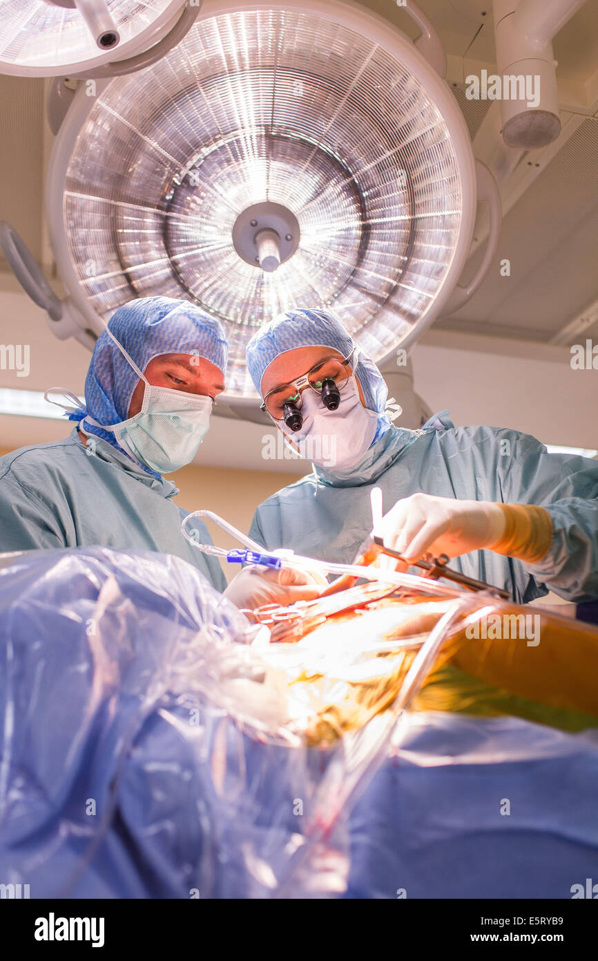 Da Vinci® Robotic Assisted Heart Surgery, double aortic coronary bypass with Da Vinci ® surgical robot, Limoges hospital, Stock Photohttps://www.alamy.com/image-license-details/?v=1https://www.alamy.com/stock-photo-da-vinci-robotic-assisted-heart-surgery-double-aortic-coronary-bypass-72441133.html
RME5RYB9–Da Vinci® Robotic Assisted Heart Surgery, double aortic coronary bypass with Da Vinci ® surgical robot, Limoges hospital,
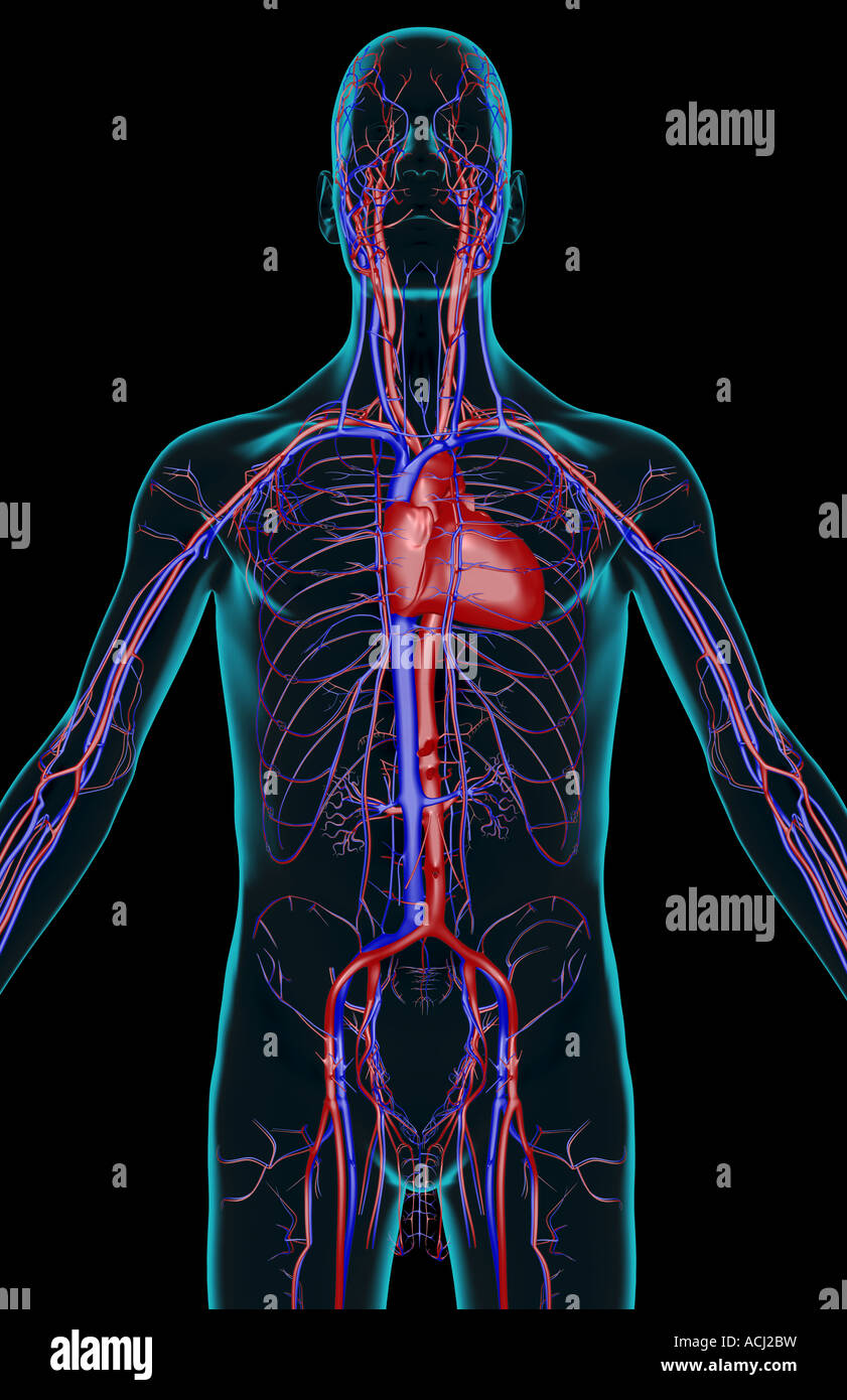 The blood supply of the upper body Stock Photohttps://www.alamy.com/image-license-details/?v=1https://www.alamy.com/stock-photo-the-blood-supply-of-the-upper-body-13165740.html
RFACJ2BW–The blood supply of the upper body
 Aorta artery, rib cage, CAT (Computerized Axial Tomography) scan, Radiology, Medical imaging for diagnosis. Hospital Policlinica Gipuzkoa, San Sebastian, Donostia, Euskadi, Spain. Stock Photohttps://www.alamy.com/image-license-details/?v=1https://www.alamy.com/aorta-artery-rib-cage-cat-computerized-axial-tomography-scan-radiology-medical-imaging-for-diagnosis-hospital-policlinica-gipuzkoa-san-sebastian-donostia-euskadi-spain-image602289430.html
RM2WYTJ8P–Aorta artery, rib cage, CAT (Computerized Axial Tomography) scan, Radiology, Medical imaging for diagnosis. Hospital Policlinica Gipuzkoa, San Sebastian, Donostia, Euskadi, Spain.
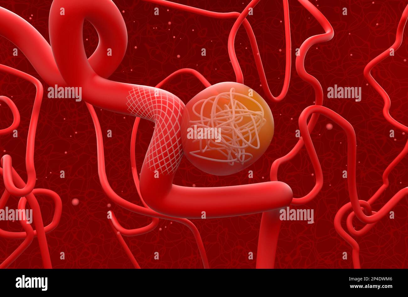 Aneurysm treat with mesh stent and coils - 3d illustration isometric view Stock Photohttps://www.alamy.com/image-license-details/?v=1https://www.alamy.com/aneurysm-treat-with-mesh-stent-and-coils-3d-illustration-isometric-view-image536285574.html
RF2P4DWM6–Aneurysm treat with mesh stent and coils - 3d illustration isometric view
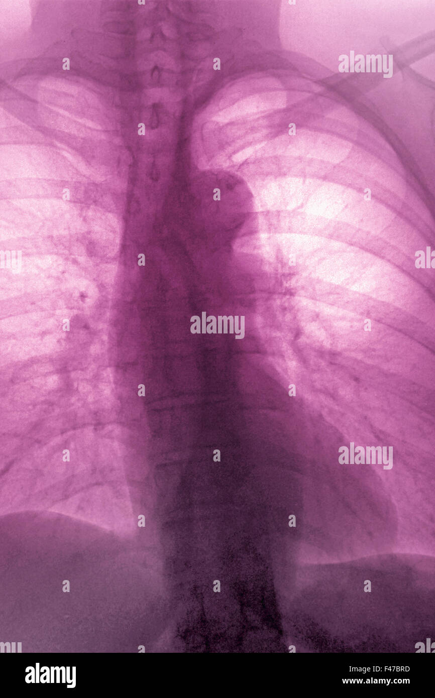 ATHEROMATOUS AORTA, X-RAY Stock Photohttps://www.alamy.com/image-license-details/?v=1https://www.alamy.com/stock-photo-atheromatous-aorta-x-ray-88673409.html
RMF47BRD–ATHEROMATOUS AORTA, X-RAY
 Diseases of the heart and thoracic aorta (1884) (14781756061) Stock Photohttps://www.alamy.com/image-license-details/?v=1https://www.alamy.com/stock-photo-diseases-of-the-heart-and-thoracic-aorta-1884-14781756061-177284849.html
RMM8C0HN–Diseases of the heart and thoracic aorta (1884) (14781756061)
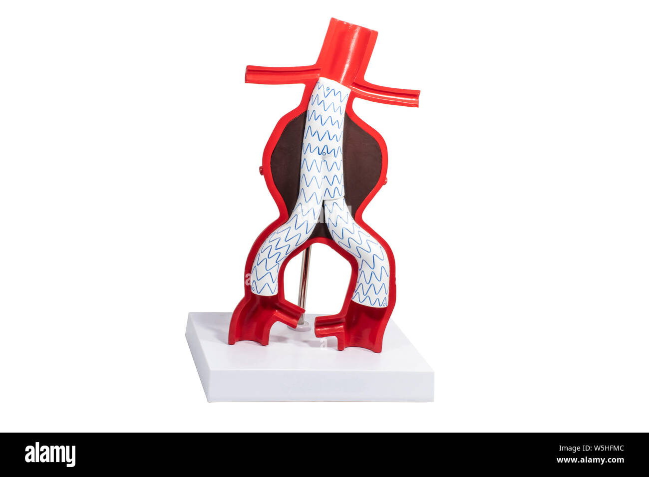 Model aneurysm treatment with endovascular fenestrated and branched stent-grafts. Stock Photohttps://www.alamy.com/image-license-details/?v=1https://www.alamy.com/model-aneurysm-treatment-with-endovascular-fenestrated-and-branched-stent-grafts-image261614316.html
RFW5HFMC–Model aneurysm treatment with endovascular fenestrated and branched stent-grafts.
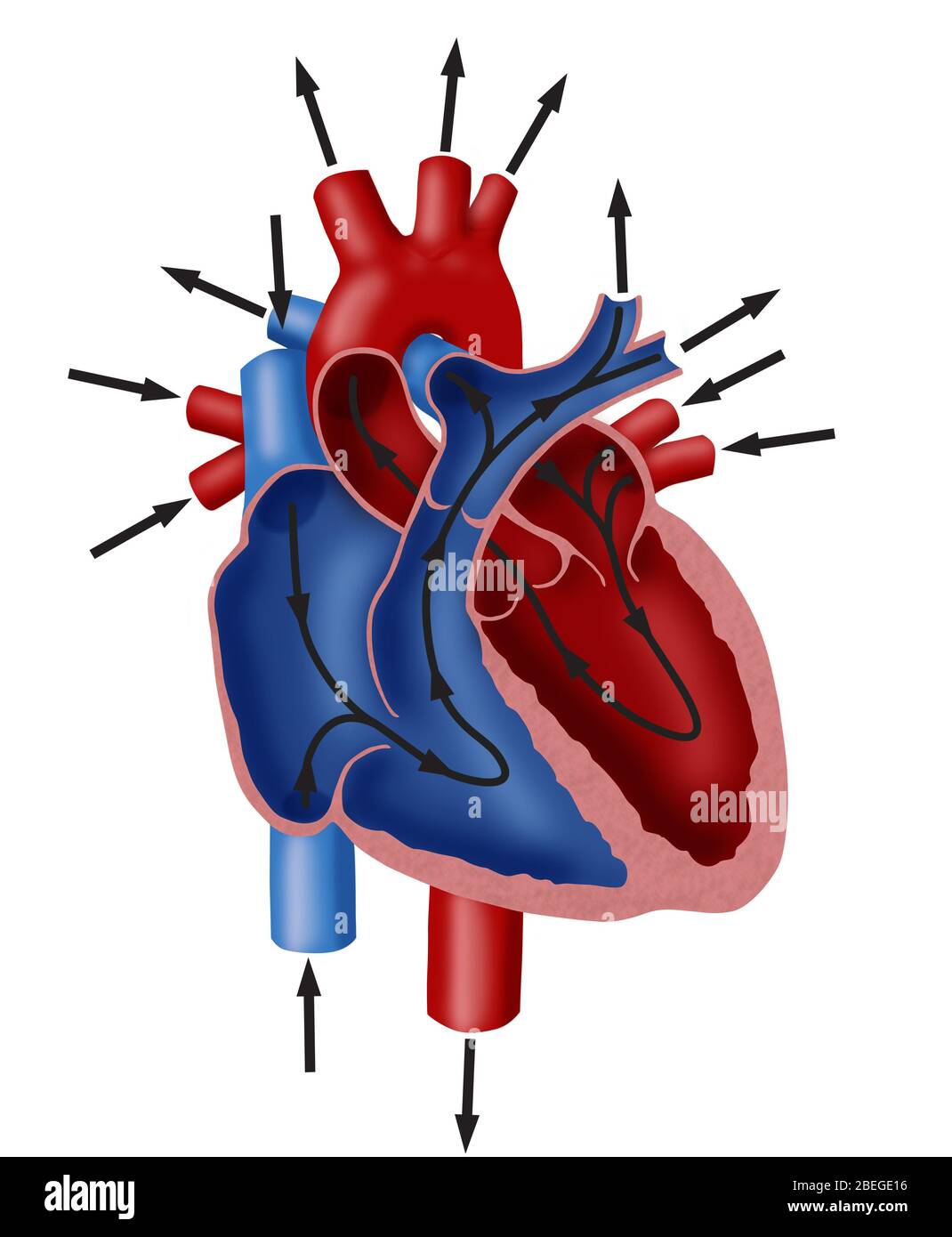 Blood Flow Diagram Stock Photohttps://www.alamy.com/image-license-details/?v=1https://www.alamy.com/blood-flow-diagram-image353174786.html
RM2BEGE16–Blood Flow Diagram
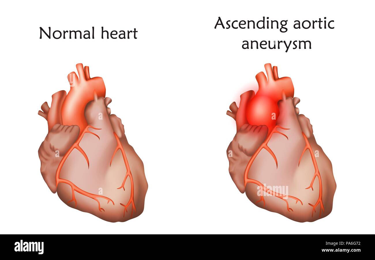 Ascending aortic aneurysm, illustration. Comparison between a damaged and normal heart. Stock Photohttps://www.alamy.com/image-license-details/?v=1https://www.alamy.com/ascending-aortic-aneurysm-illustration-comparison-between-a-damaged-and-normal-heart-image212815430.html
RFPA6G72–Ascending aortic aneurysm, illustration. Comparison between a damaged and normal heart.