Quick filters:
Trabecula Stock Photos and Images
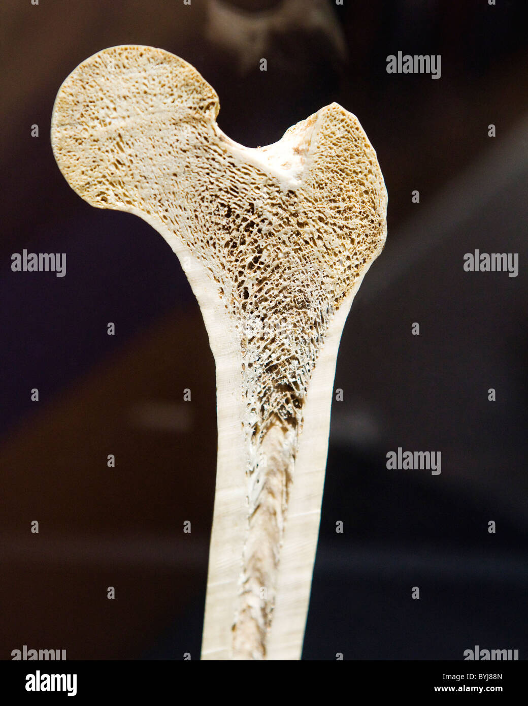 Cross section view of a human femur bone showing trabecula Stock Photohttps://www.alamy.com/image-license-details/?v=1https://www.alamy.com/stock-photo-cross-section-view-of-a-human-femur-bone-showing-trabecula-34207733.html
RMBYJ88N–Cross section view of a human femur bone showing trabecula
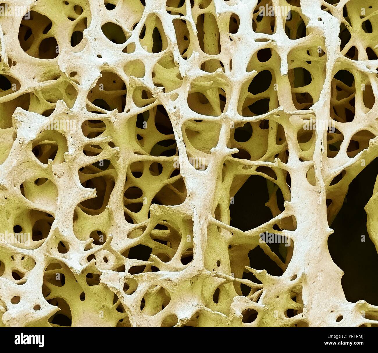 Bone tissue. Coloured scanning electron micrograph (SEM) of human cancellous (spongy) bone. Bone tissue can be either cortical (compact) or cancellous. Cortical bone usually makes up the exterior of the bone, while cancellous bone is found in the interior. Cancellous bone is characterised by a honeycomb arrangement, comprising a network of trabeculae (rod-shaped tissue). These structures provide support and strength to the bone. The spaces within this tissue contain bone marrow (not seen), a blood forming substance. Magnification: x13 when printed 10cm wide. Stock Photohttps://www.alamy.com/image-license-details/?v=1https://www.alamy.com/bone-tissue-coloured-scanning-electron-micrograph-sem-of-human-cancellous-spongy-bone-bone-tissue-can-be-either-cortical-compact-or-cancellous-cortical-bone-usually-makes-up-the-exterior-of-the-bone-while-cancellous-bone-is-found-in-the-interior-cancellous-bone-is-characterised-by-a-honeycomb-arrangement-comprising-a-network-of-trabeculae-rod-shaped-tissue-these-structures-provide-support-and-strength-to-the-bone-the-spaces-within-this-tissue-contain-bone-marrow-not-seen-a-blood-forming-substance-magnification-x13-when-printed-10cm-wide-image220702066.html
RFPR1RMJ–Bone tissue. Coloured scanning electron micrograph (SEM) of human cancellous (spongy) bone. Bone tissue can be either cortical (compact) or cancellous. Cortical bone usually makes up the exterior of the bone, while cancellous bone is found in the interior. Cancellous bone is characterised by a honeycomb arrangement, comprising a network of trabeculae (rod-shaped tissue). These structures provide support and strength to the bone. The spaces within this tissue contain bone marrow (not seen), a blood forming substance. Magnification: x13 when printed 10cm wide.
 Cross section of human long bone showing trabecula bone tissue Stock Photohttps://www.alamy.com/image-license-details/?v=1https://www.alamy.com/stock-photo-cross-section-of-human-long-bone-showing-trabecula-bone-tissue-37656425.html
RMC57B49–Cross section of human long bone showing trabecula bone tissue
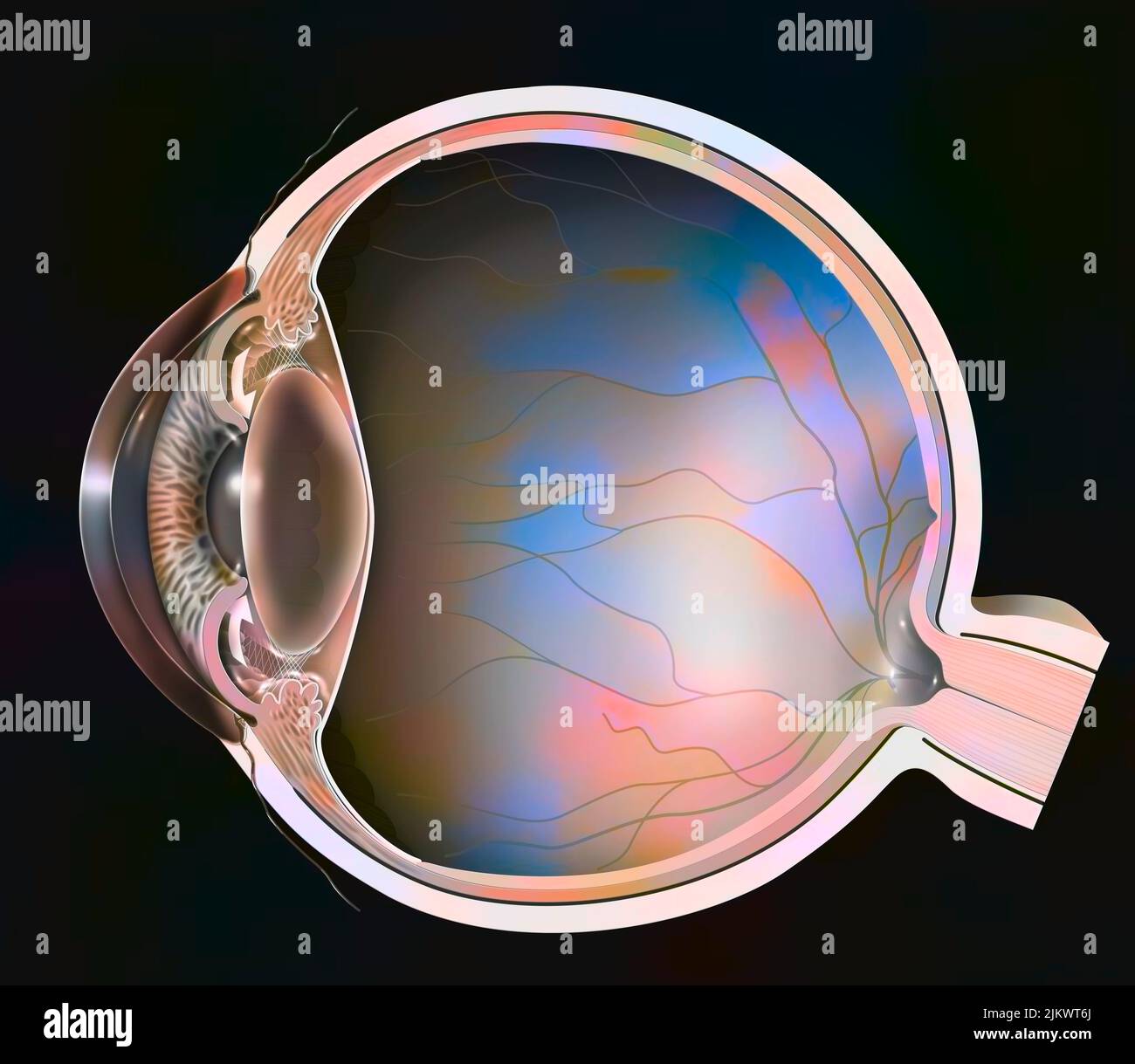 Closed-angle glaucoma: the trabecula are blocked because of the pupil. Stock Photohttps://www.alamy.com/image-license-details/?v=1https://www.alamy.com/closed-angle-glaucoma-the-trabecula-are-blocked-because-of-the-pupil-image476926202.html
RF2JKWT6J–Closed-angle glaucoma: the trabecula are blocked because of the pupil.
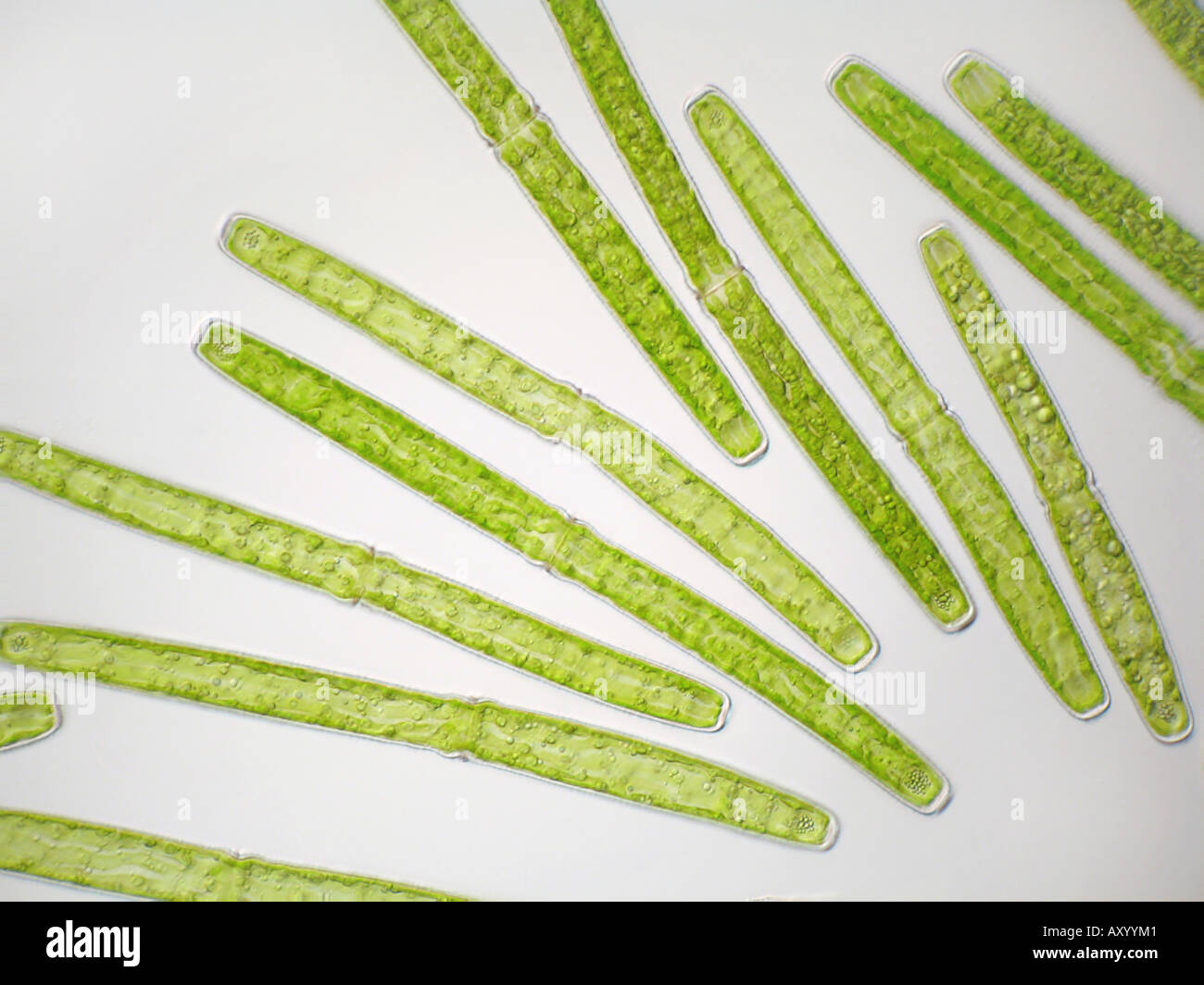 Pleurotaenium trabecula (Pleurotaenium trabecula), in shining-through light Stock Photohttps://www.alamy.com/image-license-details/?v=1https://www.alamy.com/pleurotaenium-trabecula-pleurotaenium-trabecula-in-shining-through-image9683840.html
RMAXYYM1–Pleurotaenium trabecula (Pleurotaenium trabecula), in shining-through light
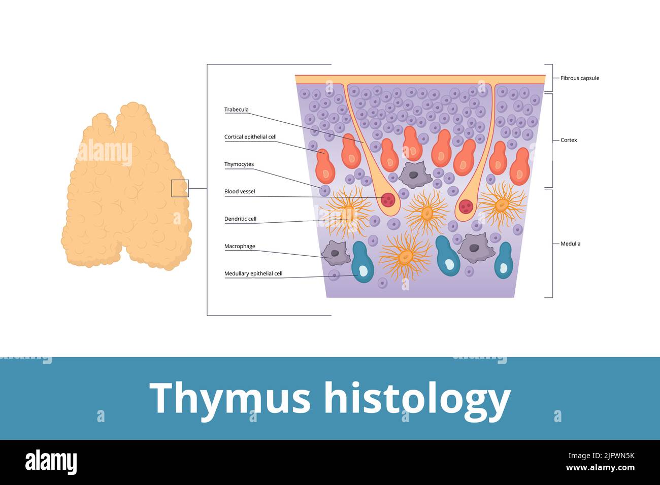 Thymus histology. Visualization of thymus tissue including thymocytes, trabecula, medulla, blood vessels, and cortical epithelial cells. Stock Vectorhttps://www.alamy.com/image-license-details/?v=1https://www.alamy.com/thymus-histology-visualization-of-thymus-tissue-including-thymocytes-trabecula-medulla-blood-vessels-and-cortical-epithelial-cells-image474465199.html
RF2JFWN5K–Thymus histology. Visualization of thymus tissue including thymocytes, trabecula, medulla, blood vessels, and cortical epithelial cells.
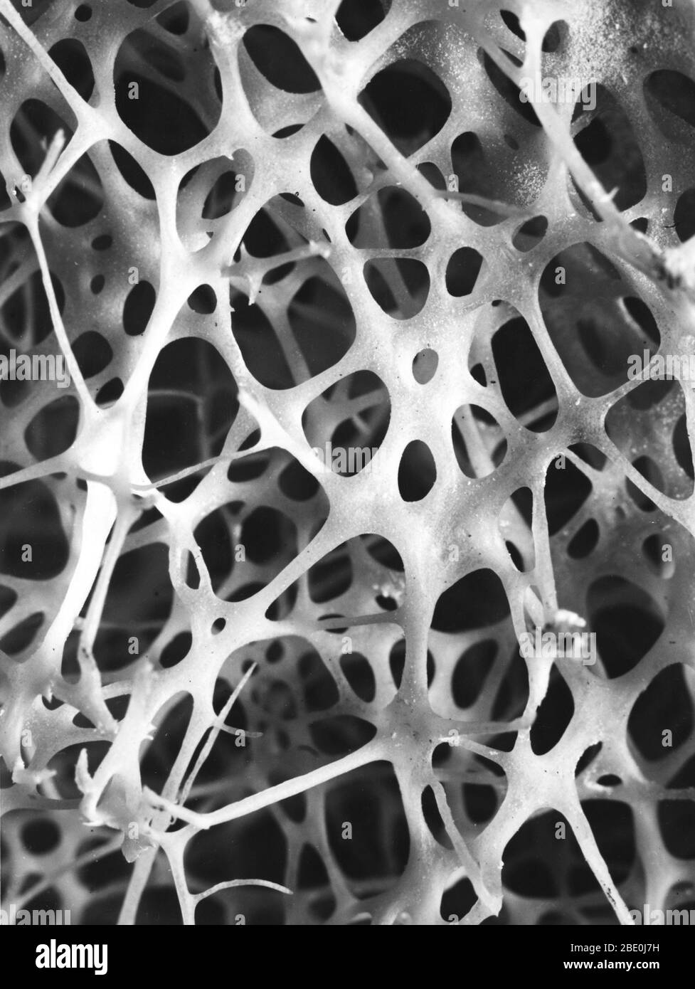 Scanning electron micrograph (SEM) of cancellous (spongy) bone of the human shin. Bone tissue is either compact or cancellous. Compact bone usually makes up the exterior of the bone, while cancellous bone is found in the interior. Cancellous bone is characterised by a honeycomb arrangement of trabeculae. These structures help to provide support and strength. The spaces within this tissue normally contain bone marrow, a blood forming substance. Stock Photohttps://www.alamy.com/image-license-details/?v=1https://www.alamy.com/scanning-electron-micrograph-sem-of-cancellous-spongy-bone-of-the-human-shin-bone-tissue-is-either-compact-or-cancellous-compact-bone-usually-makes-up-the-exterior-of-the-bone-while-cancellous-bone-is-found-in-the-interior-cancellous-bone-is-characterised-by-a-honeycomb-arrangement-of-trabeculae-these-structures-help-to-provide-support-and-strength-the-spaces-within-this-tissue-normally-contain-bone-marrow-a-blood-forming-substance-image352826869.html
RM2BE0J7H–Scanning electron micrograph (SEM) of cancellous (spongy) bone of the human shin. Bone tissue is either compact or cancellous. Compact bone usually makes up the exterior of the bone, while cancellous bone is found in the interior. Cancellous bone is characterised by a honeycomb arrangement of trabeculae. These structures help to provide support and strength. The spaces within this tissue normally contain bone marrow, a blood forming substance.
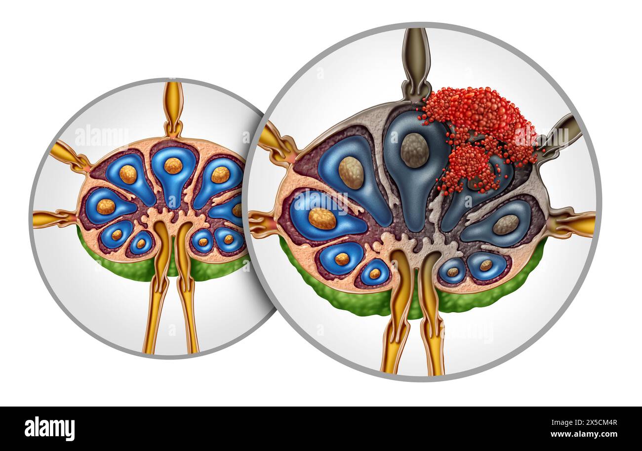 Lymphoma Lymph Node Cancer Anatomy spreading in the Lymphatic system as a tumor in nodes as a concept of the immune system or lymphoid illness. Stock Photohttps://www.alamy.com/image-license-details/?v=1https://www.alamy.com/lymphoma-lymph-node-cancer-anatomy-spreading-in-the-lymphatic-system-as-a-tumor-in-nodes-as-a-concept-of-the-immune-system-or-lymphoid-illness-image605715399.html
RF2X5CM4R–Lymphoma Lymph Node Cancer Anatomy spreading in the Lymphatic system as a tumor in nodes as a concept of the immune system or lymphoid illness.
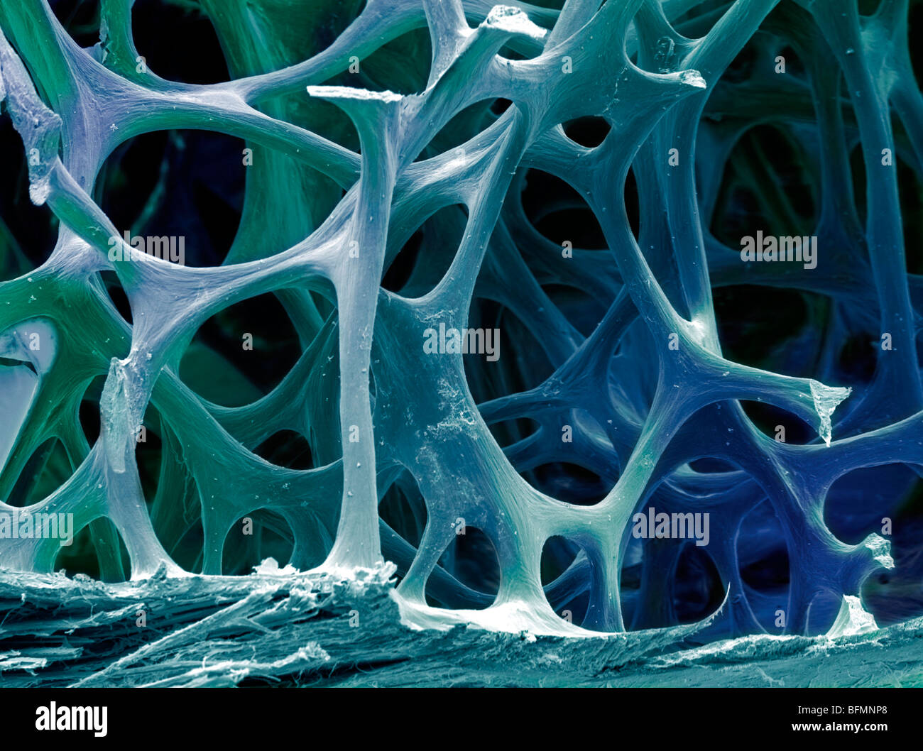 Bone tissue, SEM Stock Photohttps://www.alamy.com/image-license-details/?v=1https://www.alamy.com/stock-photo-bone-tissue-sem-26886336.html
RFBFMNP8–Bone tissue, SEM
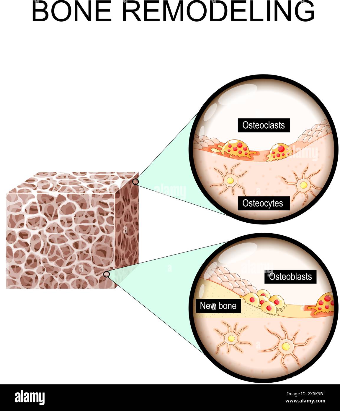 Bone remodeling from resorption to new bone formation. Close-up of Trabecular bone matrix, Osteoblasts, Osteoclasts and Osteocytes. Vector illustratio Stock Vectorhttps://www.alamy.com/image-license-details/?v=1https://www.alamy.com/bone-remodeling-from-resorption-to-new-bone-formation-close-up-of-trabecular-bone-matrix-osteoblasts-osteoclasts-and-osteocytes-vector-illustratio-image616924421.html
RF2XRK9B1–Bone remodeling from resorption to new bone formation. Close-up of Trabecular bone matrix, Osteoblasts, Osteoclasts and Osteocytes. Vector illustratio
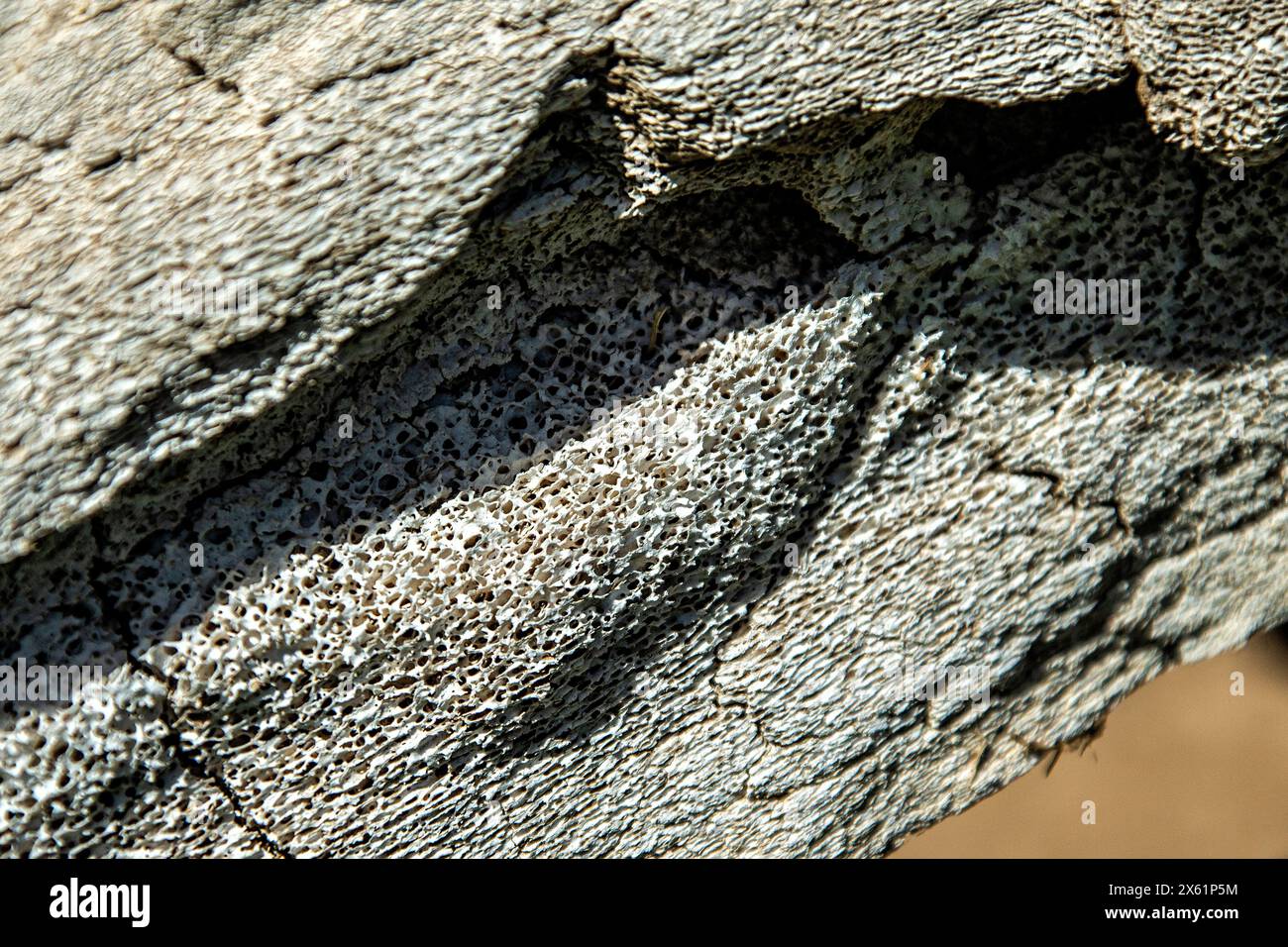 A weathered whale bone showing the sponge-like or honeycomb structure, suitable as a background. Stock Photohttps://www.alamy.com/image-license-details/?v=1https://www.alamy.com/a-weathered-whale-bone-showing-the-sponge-like-or-honeycomb-structure-suitable-as-a-background-image606090176.html
RF2X61P5M–A weathered whale bone showing the sponge-like or honeycomb structure, suitable as a background.
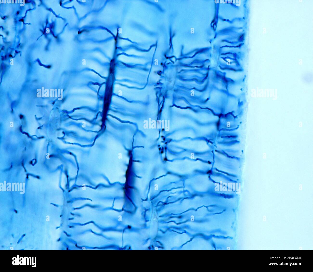 High magnification light micrograph showing osteocytes stained with the Schmorl’s technique. From its elongated cell body, many thin and long processe Stock Photohttps://www.alamy.com/image-license-details/?v=1https://www.alamy.com/high-magnification-light-micrograph-showing-osteocytes-stained-with-the-schmorls-technique-from-its-elongated-cell-body-many-thin-and-long-processe-image346977006.html
RF2B4E4KX–High magnification light micrograph showing osteocytes stained with the Schmorl’s technique. From its elongated cell body, many thin and long processe
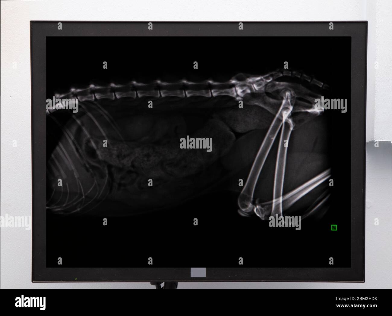 rx examination of a cat in a veterinary clinic blue mask vet Stock Photohttps://www.alamy.com/image-license-details/?v=1https://www.alamy.com/rx-examination-of-a-cat-in-a-veterinary-clinic-blue-mask-vet-image356558084.html
RF2BM2HD8–rx examination of a cat in a veterinary clinic blue mask vet
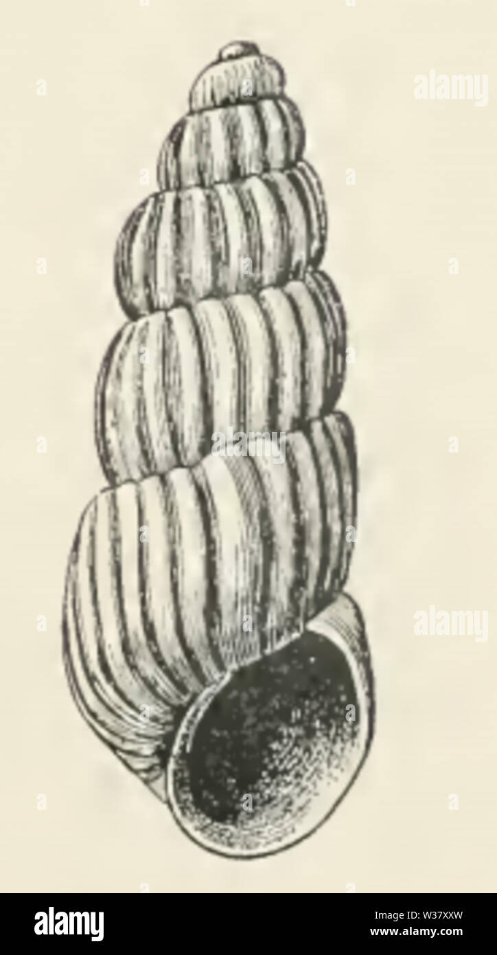 Trabecula laxa 001 Stock Photohttps://www.alamy.com/image-license-details/?v=1https://www.alamy.com/trabecula-laxa-001-image260174289.html
RMW37XXW–Trabecula laxa 001
 Archive image from page 117 of The anatomy, physiology, morphology and. The anatomy, physiology, morphology and development of the blow-fly (Calliphora erythrocephala.) A study in the comparative anatomy and morphology of insects; with plates and illustrations executed directly from the drawings of the author; CUbiodiversity4765349-9875 Year: 1890 ( 444 THE NERVOUS SYSTEM. large fasciculus of fibrils, corresponding with the peduncle and trabecula of the insect brain. This terminates in the cortex and penetrates a very remarkable group of small round cells, which I shall term the corpus fungif Stock Photohttps://www.alamy.com/image-license-details/?v=1https://www.alamy.com/archive-image-from-page-117-of-the-anatomy-physiology-morphology-and-the-anatomy-physiology-morphology-and-development-of-the-blow-fly-calliphora-erythrocephala-a-study-in-the-comparative-anatomy-and-morphology-of-insects-with-plates-and-illustrations-executed-directly-from-the-drawings-of-the-author-cubiodiversity4765349-9875-year-1890-444-the-nervous-system-large-fasciculus-of-fibrils-corresponding-with-the-peduncle-and-trabecula-of-the-insect-brain-this-terminates-in-the-cortex-and-penetrates-a-very-remarkable-group-of-small-round-cells-which-i-shall-term-the-corpus-fungif-image264043168.html
RMW9G5N4–Archive image from page 117 of The anatomy, physiology, morphology and. The anatomy, physiology, morphology and development of the blow-fly (Calliphora erythrocephala.) A study in the comparative anatomy and morphology of insects; with plates and illustrations executed directly from the drawings of the author; CUbiodiversity4765349-9875 Year: 1890 ( 444 THE NERVOUS SYSTEM. large fasciculus of fibrils, corresponding with the peduncle and trabecula of the insect brain. This terminates in the cortex and penetrates a very remarkable group of small round cells, which I shall term the corpus fungif
![Infographic of the composition of bones, their formation, growth and regeneration following a fracture. [QuarkXPress (.qxp); 6259x4015]. Stock Photo Infographic of the composition of bones, their formation, growth and regeneration following a fracture. [QuarkXPress (.qxp); 6259x4015]. Stock Photo](https://c8.alamy.com/comp/2NEBHRE/infographic-of-the-composition-of-bones-their-formation-growth-and-regeneration-following-a-fracture-quarkxpress-qxp-6259x4015-2NEBHRE.jpg) Infographic of the composition of bones, their formation, growth and regeneration following a fracture. [QuarkXPress (.qxp); 6259x4015]. Stock Photohttps://www.alamy.com/image-license-details/?v=1https://www.alamy.com/infographic-of-the-composition-of-bones-their-formation-growth-and-regeneration-following-a-fracture-quarkxpress-qxp-6259x4015-image525171682.html
Infographic of the composition of bones, their formation, growth and regeneration following a fracture. [QuarkXPress (.qxp); 6259x4015]. Stock Photohttps://www.alamy.com/image-license-details/?v=1https://www.alamy.com/infographic-of-the-composition-of-bones-their-formation-growth-and-regeneration-following-a-fracture-quarkxpress-qxp-6259x4015-image525171682.htmlRM2NEBHRE–Infographic of the composition of bones, their formation, growth and regeneration following a fracture. [QuarkXPress (.qxp); 6259x4015].
 The Journal of laboratory and clinical medicine . Fig. 16.—(Same as lig. 10.) .Malignant degen-eration of the liver-trabecula. This type of ma-lignancy was otjserved in every section. No met-astatic nodules were found. (H.R. x300 diam.). Fig. 17-A. Stock Photohttps://www.alamy.com/image-license-details/?v=1https://www.alamy.com/the-journal-of-laboratory-and-clinical-medicine-fig-16same-as-lig-10-malignant-degen-eration-of-the-liver-trabecula-this-type-of-ma-lignancy-was-otjserved-in-every-section-no-met-astatic-nodules-were-found-hr-x300-diam-fig-17-a-image340158502.html
RM2ANBFHX–The Journal of laboratory and clinical medicine . Fig. 16.—(Same as lig. 10.) .Malignant degen-eration of the liver-trabecula. This type of ma-lignancy was otjserved in every section. No met-astatic nodules were found. (H.R. x300 diam.). Fig. 17-A.
RF2PP52RG–Skeleton linear icons set. Bs, Anatomy, Skeletal, Joints, Skull, Ribcage, Spine line vector and concept signs. Osteology,X-ray,Cartilage outline
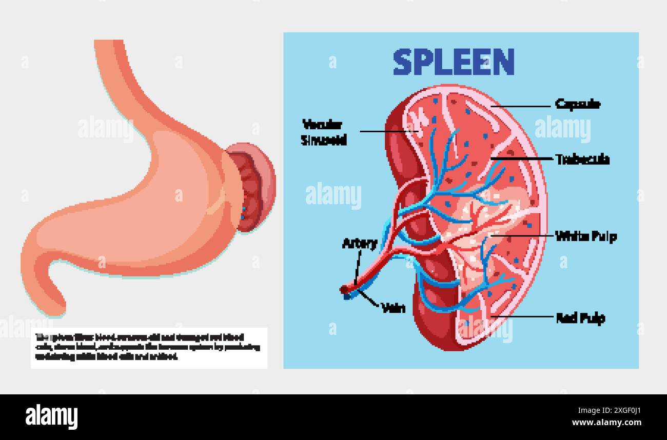 Detailed diagram of the human spleen anatomy Stock Vectorhttps://www.alamy.com/image-license-details/?v=1https://www.alamy.com/detailed-diagram-of-the-human-spleen-anatomy-image612527161.html
RF2XGF0J1–Detailed diagram of the human spleen anatomy
 . The anatomy, physiology, morphology and development of the blow-fly (Calliphora erythrocephala.) A study in the comparative anatomy and morphology of insects; with plates and illustrations executed directly from the drawings of the author;. Blowflies. 444 THE NERVOUS SYSTEM. large fasciculus of fibrils, corresponding with the peduncle and trabecula of the insect brain. This terminates in the cortex and penetrates a very remarkable group of small round cells, which I shall term the corpus fungiforme, as I regard it as the homologue of the corpora fungiformia of insects. Lateral sections (Fig. Stock Photohttps://www.alamy.com/image-license-details/?v=1https://www.alamy.com/the-anatomy-physiology-morphology-and-development-of-the-blow-fly-calliphora-erythrocephala-a-study-in-the-comparative-anatomy-and-morphology-of-insects-with-plates-and-illustrations-executed-directly-from-the-drawings-of-the-author-blowflies-444-the-nervous-system-large-fasciculus-of-fibrils-corresponding-with-the-peduncle-and-trabecula-of-the-insect-brain-this-terminates-in-the-cortex-and-penetrates-a-very-remarkable-group-of-small-round-cells-which-i-shall-term-the-corpus-fungiforme-as-i-regard-it-as-the-homologue-of-the-corpora-fungiformia-of-insects-lateral-sections-fig-image216287639.html
RMPFTN2F–. The anatomy, physiology, morphology and development of the blow-fly (Calliphora erythrocephala.) A study in the comparative anatomy and morphology of insects; with plates and illustrations executed directly from the drawings of the author;. Blowflies. 444 THE NERVOUS SYSTEM. large fasciculus of fibrils, corresponding with the peduncle and trabecula of the insect brain. This terminates in the cortex and penetrates a very remarkable group of small round cells, which I shall term the corpus fungiforme, as I regard it as the homologue of the corpora fungiformia of insects. Lateral sections (Fig.
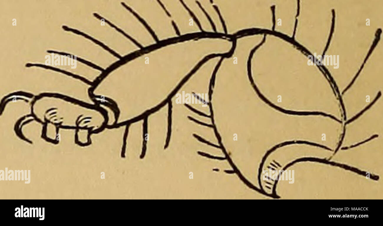 . Economic entomology . Trinoton luridum, from the duck. Anterior eg of ditto. 2 lines in length. Infests various wild ducks, besides the domestic species. Sub-family Ptilopterid^ {Buj-m.). Antennae five-jointed; mouth with strong mandibles. Genus Docophorus {Nitzsch). The most notable character by which this genus is distinguished is a small movable projection or tooth called a trabecula in front of the antennae, as shown in the woodcut. The only other genus in which this occurs is Nirmus (which has so many other points of coincidence with Docophorus that we should prefer their being consoli Stock Photohttps://www.alamy.com/image-license-details/?v=1https://www.alamy.com/economic-entomology-trinoton-luridum-from-the-duck-anterior-eg-of-ditto-2-lines-in-length-infests-various-wild-ducks-besides-the-domestic-species-sub-family-ptilopterid-buj-m-antennae-five-jointed-mouth-with-strong-mandibles-genus-docophorus-nitzsch-the-most-notable-character-by-which-this-genus-is-distinguished-is-a-small-movable-projection-or-tooth-called-a-trabecula-in-front-of-the-antennae-as-shown-in-the-woodcut-the-only-other-genus-in-which-this-occurs-is-nirmus-which-has-so-many-other-points-of-coincidence-with-docophorus-that-we-should-prefer-their-being-consoli-image178479523.html
RMMAACCK–. Economic entomology . Trinoton luridum, from the duck. Anterior eg of ditto. 2 lines in length. Infests various wild ducks, besides the domestic species. Sub-family Ptilopterid^ {Buj-m.). Antennae five-jointed; mouth with strong mandibles. Genus Docophorus {Nitzsch). The most notable character by which this genus is distinguished is a small movable projection or tooth called a trabecula in front of the antennae, as shown in the woodcut. The only other genus in which this occurs is Nirmus (which has so many other points of coincidence with Docophorus that we should prefer their being consoli
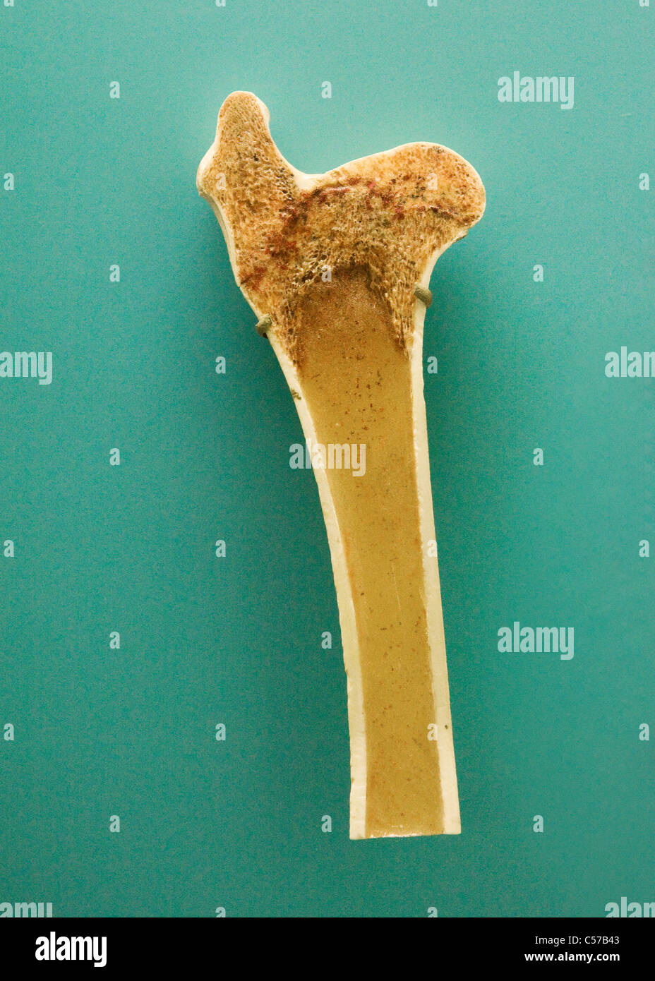 Cross section of human long bone showing trabecula bone tissue Stock Photohttps://www.alamy.com/image-license-details/?v=1https://www.alamy.com/stock-photo-cross-section-of-human-long-bone-showing-trabecula-bone-tissue-37656419.html
RMC57B43–Cross section of human long bone showing trabecula bone tissue
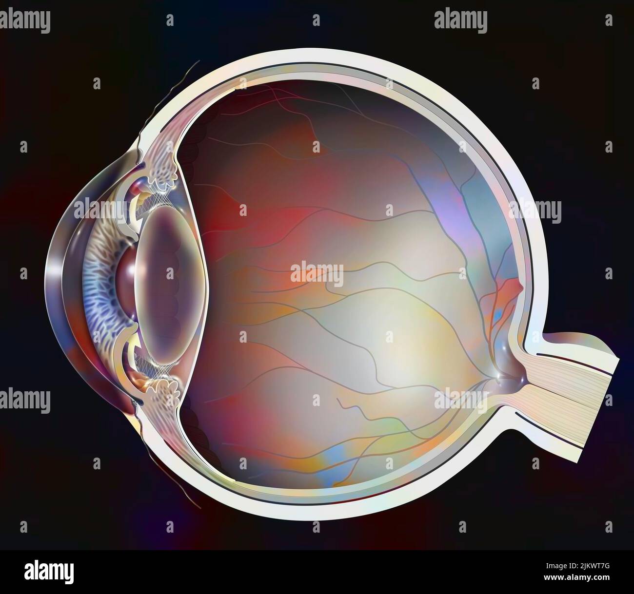 Closed-angle glaucoma: the trabecula are blocked because of the pupil. Stock Photohttps://www.alamy.com/image-license-details/?v=1https://www.alamy.com/closed-angle-glaucoma-the-trabecula-are-blocked-because-of-the-pupil-image476926228.html
RF2JKWT7G–Closed-angle glaucoma: the trabecula are blocked because of the pupil.
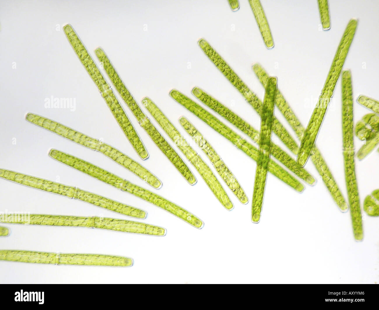 Pleurotaenium trabecula (Pleurotaenium trabecula), in shining-through light Stock Photohttps://www.alamy.com/image-license-details/?v=1https://www.alamy.com/pleurotaenium-trabecula-pleurotaenium-trabecula-in-shining-through-image9683845.html
RMAXYYM6–Pleurotaenium trabecula (Pleurotaenium trabecula), in shining-through light
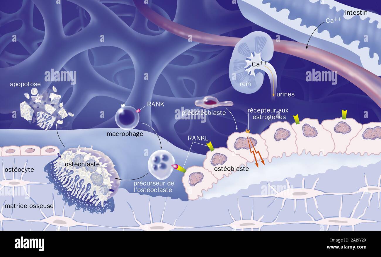 Cancellous bone, reproductive cycle of osteoblasts and osteoblasts, treatments. View of bone trabeculae of spongy bone (blue). Osteoclasts (yellow) de Stock Photohttps://www.alamy.com/image-license-details/?v=1https://www.alamy.com/cancellous-bone-reproductive-cycle-of-osteoblasts-and-osteoblasts-treatments-view-of-bone-trabeculae-of-spongy-bone-blue-osteoclasts-yellow-de-image338279618.html
RM2AJ9Y2X–Cancellous bone, reproductive cycle of osteoblasts and osteoblasts, treatments. View of bone trabeculae of spongy bone (blue). Osteoclasts (yellow) de
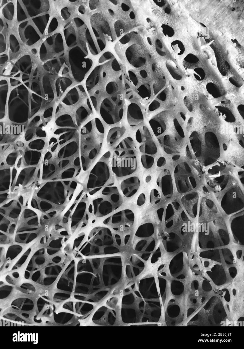 Scanning electron micrograph (SEM) of cancellous (spongy) bone of the human shin. Bone tissue is either compact or cancellous. Compact bone usually makes up the exterior of the bone, while cancellous bone is found in the interior. Cancellous bone is characterized by a honeycomb arrangement of trabeculae. These structures help to provide support and strength. The spaces within this tissue normally contain bone marrow, a blood forming substance. Stock Photohttps://www.alamy.com/image-license-details/?v=1https://www.alamy.com/scanning-electron-micrograph-sem-of-cancellous-spongy-bone-of-the-human-shin-bone-tissue-is-either-compact-or-cancellous-compact-bone-usually-makes-up-the-exterior-of-the-bone-while-cancellous-bone-is-found-in-the-interior-cancellous-bone-is-characterized-by-a-honeycomb-arrangement-of-trabeculae-these-structures-help-to-provide-support-and-strength-the-spaces-within-this-tissue-normally-contain-bone-marrow-a-blood-forming-substance-image352826904.html
RM2BE0J8T–Scanning electron micrograph (SEM) of cancellous (spongy) bone of the human shin. Bone tissue is either compact or cancellous. Compact bone usually makes up the exterior of the bone, while cancellous bone is found in the interior. Cancellous bone is characterized by a honeycomb arrangement of trabeculae. These structures help to provide support and strength. The spaces within this tissue normally contain bone marrow, a blood forming substance.
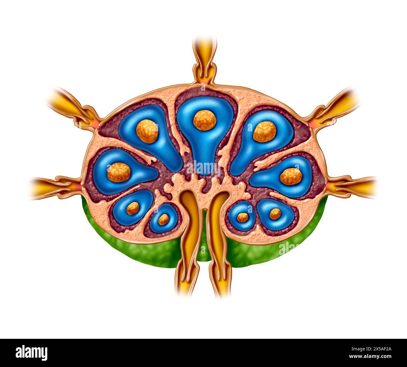 Lymph Node Anatomy as a cross section of the gland of the Lymphatic system function concept and immune system or lymphoid organ concept Stock Photohttps://www.alamy.com/image-license-details/?v=1https://www.alamy.com/lymph-node-anatomy-as-a-cross-section-of-the-gland-of-the-lymphatic-system-function-concept-and-immune-system-or-lymphoid-organ-concept-image605667506.html
RF2X5AF2A–Lymph Node Anatomy as a cross section of the gland of the Lymphatic system function concept and immune system or lymphoid organ concept
 Bone tissue. Coloured scanning electron micrograph(SEM) of cancellous (spongy) bone. Stock Photohttps://www.alamy.com/image-license-details/?v=1https://www.alamy.com/stock-photo-bone-tissue-coloured-scanning-electron-micrographsem-of-cancellous-21207758.html
RFB6E2KX–Bone tissue. Coloured scanning electron micrograph(SEM) of cancellous (spongy) bone.
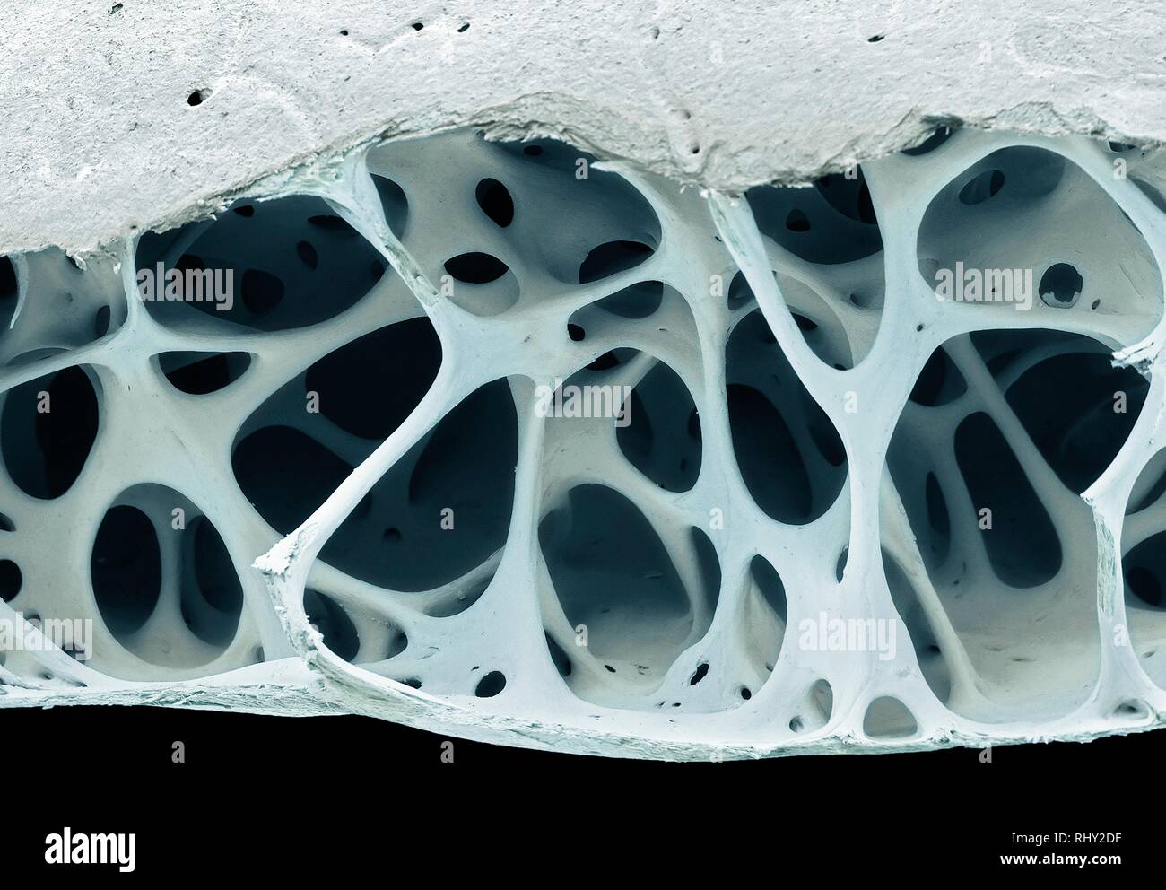 Bird bone tissue, SEM Stock Photohttps://www.alamy.com/image-license-details/?v=1https://www.alamy.com/bird-bone-tissue-sem-image234778587.html
RFRHY2DF–Bird bone tissue, SEM
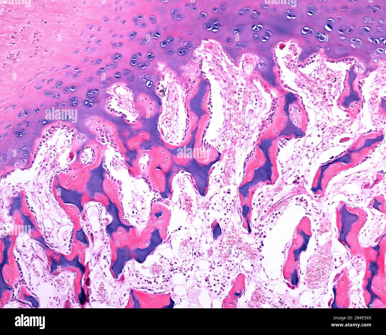 Mixed trabeculae generated in epiphyseal ossification. Unlike those formed in the epiphyseal or growth cartilage, the mixed trabeculae of the epiphyse Stock Photohttps://www.alamy.com/image-license-details/?v=1https://www.alamy.com/mixed-trabeculae-generated-in-epiphyseal-ossification-unlike-those-formed-in-the-epiphyseal-or-growth-cartilage-the-mixed-trabeculae-of-the-epiphyse-image346977426.html
RF2B4E56X–Mixed trabeculae generated in epiphyseal ossification. Unlike those formed in the epiphyseal or growth cartilage, the mixed trabeculae of the epiphyse
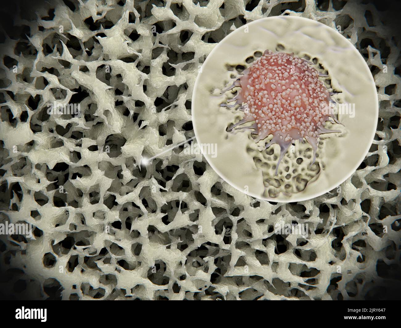 Osteoporotic bone tissue, illustration Stock Photohttps://www.alamy.com/image-license-details/?v=1https://www.alamy.com/osteoporotic-bone-tissue-illustration-image479414551.html
RF2JRY647–Osteoporotic bone tissue, illustration
 (Pleurotaenium trabecula) Stock Photohttps://www.alamy.com/image-license-details/?v=1https://www.alamy.com/pleurotaenium-trabecula-image637232348.html
RM2S0MCA4–(Pleurotaenium trabecula)
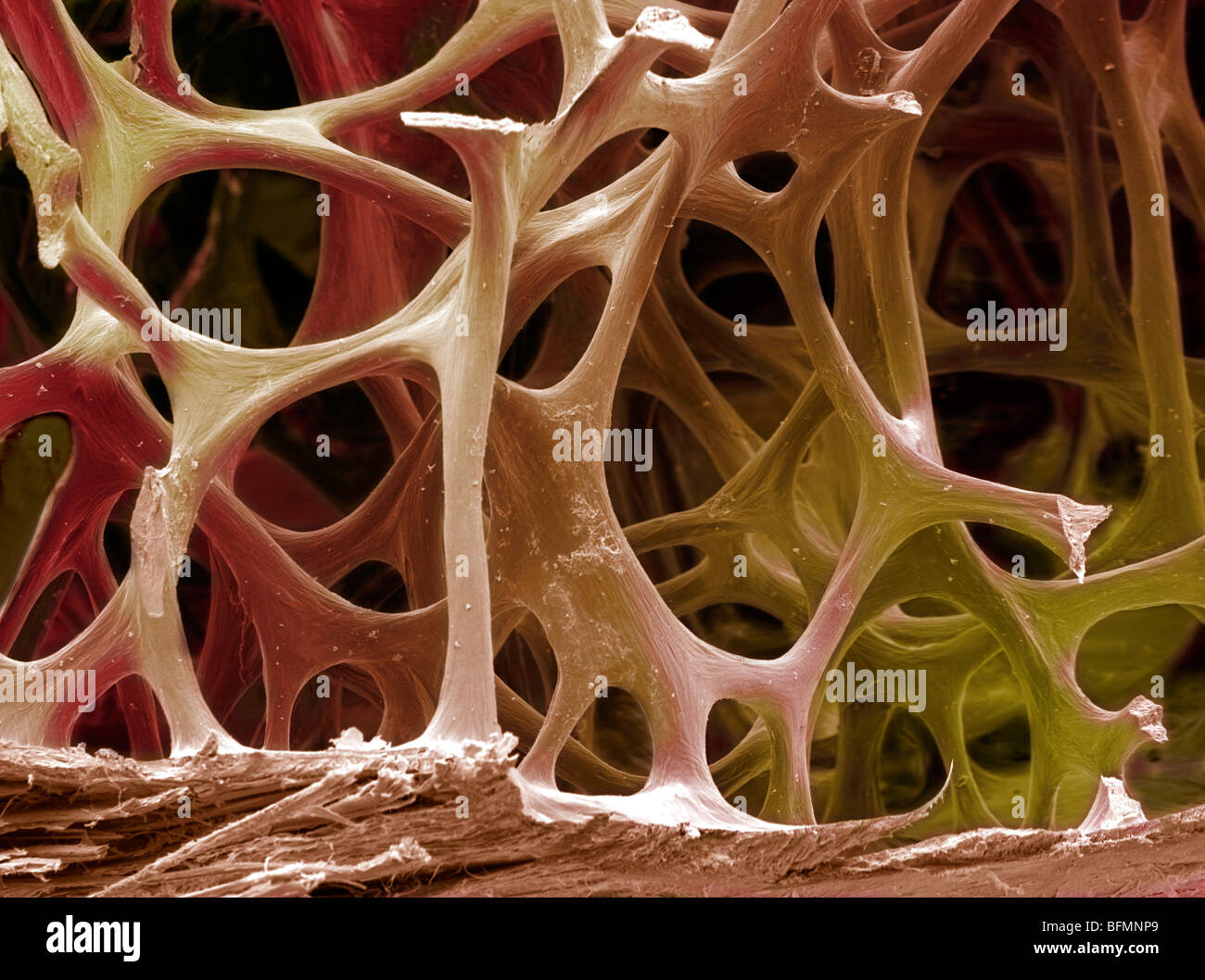 Bone tissue, SEM Stock Photohttps://www.alamy.com/image-license-details/?v=1https://www.alamy.com/stock-photo-bone-tissue-sem-26886337.html
RFBFMNP9–Bone tissue, SEM
 Elements of the comparative anatomy Elements of the comparative anatomy of vertebrates elementsofcompar00wied Year: 1886 58 COMPARATIVE AXATn.MY. Ill ni(i>t cases ;i iiii'iliaii cartilaginous liar (i n t i/rt rabeeul a is formed between tlie tralieeuhu in front, fusing with theiii. and formim: tin- ellnuu-ii;i>ul septum. It ocea.-ionally pr 'ji-cts forwards to i'orin a rostrum. We must now follow further the processes of growth, taking as a foundation the primary condition of things described above, in which the trabecula- havt- unii'-d together in the middle line. The Stock Photohttps://www.alamy.com/image-license-details/?v=1https://www.alamy.com/elements-of-the-comparative-anatomy-elements-of-the-comparative-anatomy-of-vertebrates-elementsofcompar00wied-year-1886-58-comparative-axatnmy-ill-niigtt-cases-i-iiiiiliaii-cartilaginous-liar-i-n-t-irt-rabeeul-a-is-formed-between-tlie-tralieeuhu-in-front-fusing-with-theiii-and-formim-tin-ellnuu-iiigtul-septum-it-ocea-ionally-pr-ji-cts-forwards-to-iorin-a-rostrum-we-must-now-follow-further-the-processes-of-growth-taking-as-a-foundation-the-primary-condition-of-things-described-above-in-which-the-trabecula-havt-unii-d-together-in-the-middle-line-the-image239579907.html
RMRWNPH7–Elements of the comparative anatomy Elements of the comparative anatomy of vertebrates elementsofcompar00wied Year: 1886 58 COMPARATIVE AXATn.MY. Ill ni(i>t cases ;i iiii'iliaii cartilaginous liar (i n t i/rt rabeeul a is formed between tlie tralieeuhu in front, fusing with theiii. and formim: tin- ellnuu-ii;i>ul septum. It ocea.-ionally pr 'ji-cts forwards to i'orin a rostrum. We must now follow further the processes of growth, taking as a foundation the primary condition of things described above, in which the trabecula- havt- unii'-d together in the middle line. The
 The Journal of laboratory and clinical medicine . Fig. 15.—(Same as Fig. 10.) Tumor of rightovary. 6 cm. in diameter. Two types of carcinoma.Diffuse type mixed with solid strands. (H.E. X350 diam.). Fig. 16.—(Same as lig. 10.) .Malignant degen-eration of the liver-trabecula. This type of ma-lignancy was otjserved in every section. No met-astatic nodules were found. (H.R. x300 diam.) Stock Photohttps://www.alamy.com/image-license-details/?v=1https://www.alamy.com/the-journal-of-laboratory-and-clinical-medicine-fig-15same-as-fig-10-tumor-of-rightovary-6-cm-in-diameter-two-types-of-carcinomadiffuse-type-mixed-with-solid-strands-he-x350-diam-fig-16same-as-lig-10-malignant-degen-eration-of-the-liver-trabecula-this-type-of-ma-lignancy-was-otjserved-in-every-section-no-met-astatic-nodules-were-found-hr-x300-diam-image340158963.html
RM2ANBG6B–The Journal of laboratory and clinical medicine . Fig. 15.—(Same as Fig. 10.) Tumor of rightovary. 6 cm. in diameter. Two types of carcinoma.Diffuse type mixed with solid strands. (H.E. X350 diam.). Fig. 16.—(Same as lig. 10.) .Malignant degen-eration of the liver-trabecula. This type of ma-lignancy was otjserved in every section. No met-astatic nodules were found. (H.R. x300 diam.)
RF2PP00RN–Skeleton linear icons set. Bs, Anatomy, Skeletal, Joints, Skull, Ribcage, Spine line vector and concept signs. Osteology,X-ray,Cartilage outline
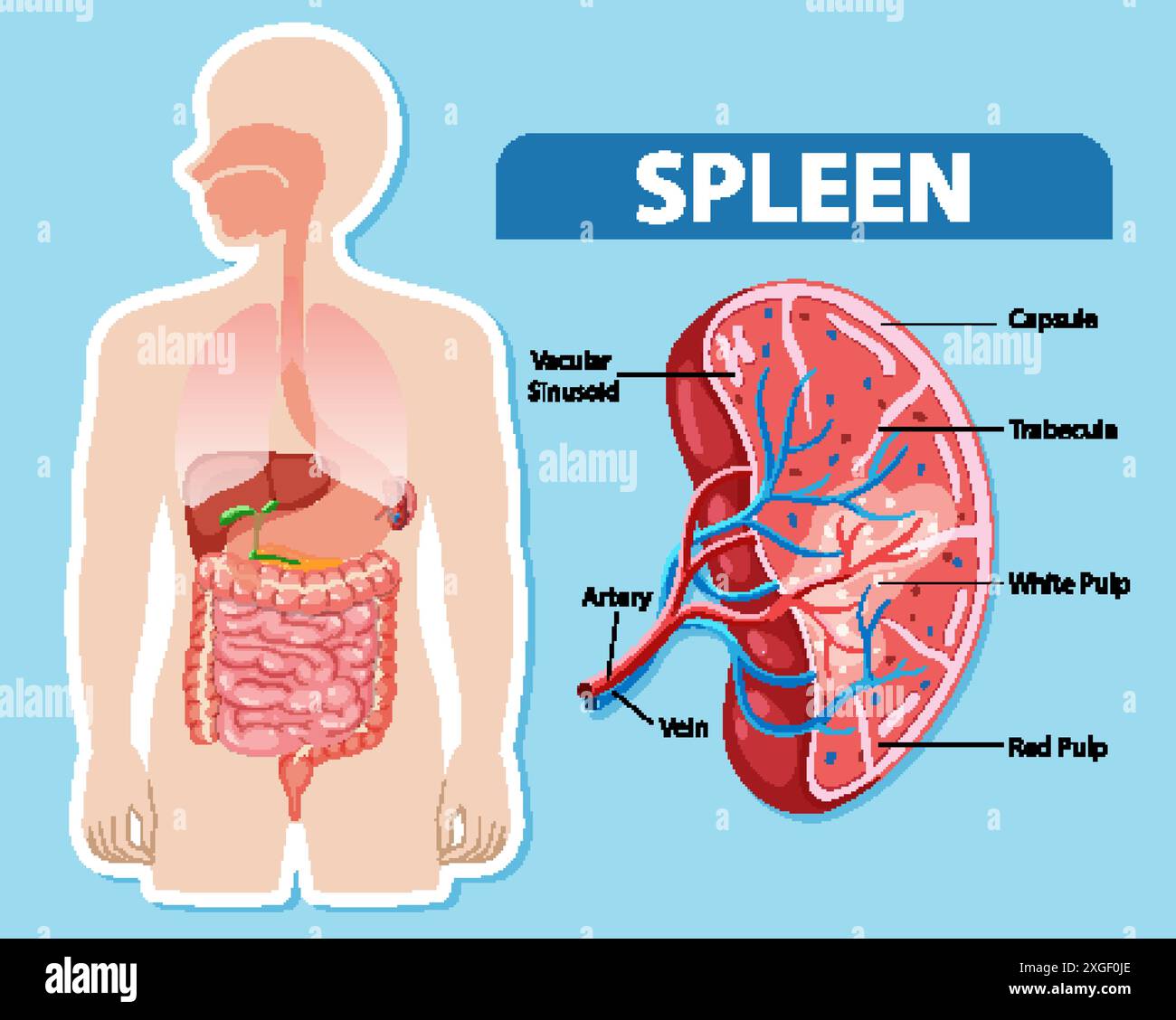 Detailed diagram of spleen and its parts Stock Vectorhttps://www.alamy.com/image-license-details/?v=1https://www.alamy.com/detailed-diagram-of-spleen-and-its-parts-image612527174.html
RF2XGF0JE–Detailed diagram of spleen and its parts
 . The anatomy, physiology, morphology and development of the blow-fly (Calliphora erythrocephala.) A study in the comparative anatomy and morphology of insects; with plates and illustrations executed directly from the drawings of the author;. Blowflies. Fig. 56. ^Frontal section of the supra-oesophageal nerve centre of the cockroach (Blalta orientalis): a, n, antsnnal (olfactory) ganglia; c c, corpus centrale ; m, mesocerehron; m e, external, and m i, internal medullary nucleus of the optic ganglion ; o .<, optic peduncle ; ir, trabecula;. In the Crickets (Gryllidas), as in the Crustacea an Stock Photohttps://www.alamy.com/image-license-details/?v=1https://www.alamy.com/the-anatomy-physiology-morphology-and-development-of-the-blow-fly-calliphora-erythrocephala-a-study-in-the-comparative-anatomy-and-morphology-of-insects-with-plates-and-illustrations-executed-directly-from-the-drawings-of-the-author-blowflies-fig-56-frontal-section-of-the-supra-oesophageal-nerve-centre-of-the-cockroach-blalta-orientalis-a-n-antsnnal-olfactory-ganglia-c-c-corpus-centrale-m-mesocerehron-m-e-external-and-m-i-internal-medullary-nucleus-of-the-optic-ganglion-o-lt-optic-peduncle-ir-trabecula-in-the-crickets-gryllidas-as-in-the-crustacea-an-image216287624.html
RMPFTN20–. The anatomy, physiology, morphology and development of the blow-fly (Calliphora erythrocephala.) A study in the comparative anatomy and morphology of insects; with plates and illustrations executed directly from the drawings of the author;. Blowflies. Fig. 56. ^Frontal section of the supra-oesophageal nerve centre of the cockroach (Blalta orientalis): a, n, antsnnal (olfactory) ganglia; c c, corpus centrale ; m, mesocerehron; m e, external, and m i, internal medullary nucleus of the optic ganglion ; o .<, optic peduncle ; ir, trabecula;. In the Crickets (Gryllidas), as in the Crustacea an
 . Abb. 99. Listropsylla dorippe R. 5- Kopf. — (Orig.) langer Sporn. Antennengruben berühren sich im Kopf nicht, Trabecula centralis fehlt. Vorderteil des Tentorium entwickelt. Das Ende der Palpi labiales spitz, asymmetrisch. Der untere Winkel des Mesopleurencollare gerade, nicht abgerundet. Collare des Mesonotum mit zahlreichen Pseudochaeten. Metanotum ohne Apikalzähnchen. 2. Abdominaltergit mit 2 Borstenreihen. Die Lateralborsten des ersten Paares am 5. Tarsenglied zusammengeschoben oder in basale Plantarborsten verwandelt. Gattung: Listropsylla Roths. 1907. Mehrere Arten Listropsylla leben i Stock Photohttps://www.alamy.com/image-license-details/?v=1https://www.alamy.com/abb-99-listropsylla-dorippe-r-5-kopf-orig-langer-sporn-antennengruben-berhren-sich-im-kopf-nicht-trabecula-centralis-fehlt-vorderteil-des-tentorium-entwickelt-das-ende-der-palpi-labiales-spitz-asymmetrisch-der-untere-winkel-des-mesopleurencollare-gerade-nicht-abgerundet-collare-des-mesonotum-mit-zahlreichen-pseudochaeten-metanotum-ohne-apikalzhnchen-2-abdominaltergit-mit-2-borstenreihen-die-lateralborsten-des-ersten-paares-am-5-tarsenglied-zusammengeschoben-oder-in-basale-plantarborsten-verwandelt-gattung-listropsylla-roths-1907-mehrere-arten-listropsylla-leben-i-image179899289.html
RMMCK3AH–. Abb. 99. Listropsylla dorippe R. 5- Kopf. — (Orig.) langer Sporn. Antennengruben berühren sich im Kopf nicht, Trabecula centralis fehlt. Vorderteil des Tentorium entwickelt. Das Ende der Palpi labiales spitz, asymmetrisch. Der untere Winkel des Mesopleurencollare gerade, nicht abgerundet. Collare des Mesonotum mit zahlreichen Pseudochaeten. Metanotum ohne Apikalzähnchen. 2. Abdominaltergit mit 2 Borstenreihen. Die Lateralborsten des ersten Paares am 5. Tarsenglied zusammengeschoben oder in basale Plantarborsten verwandelt. Gattung: Listropsylla Roths. 1907. Mehrere Arten Listropsylla leben i
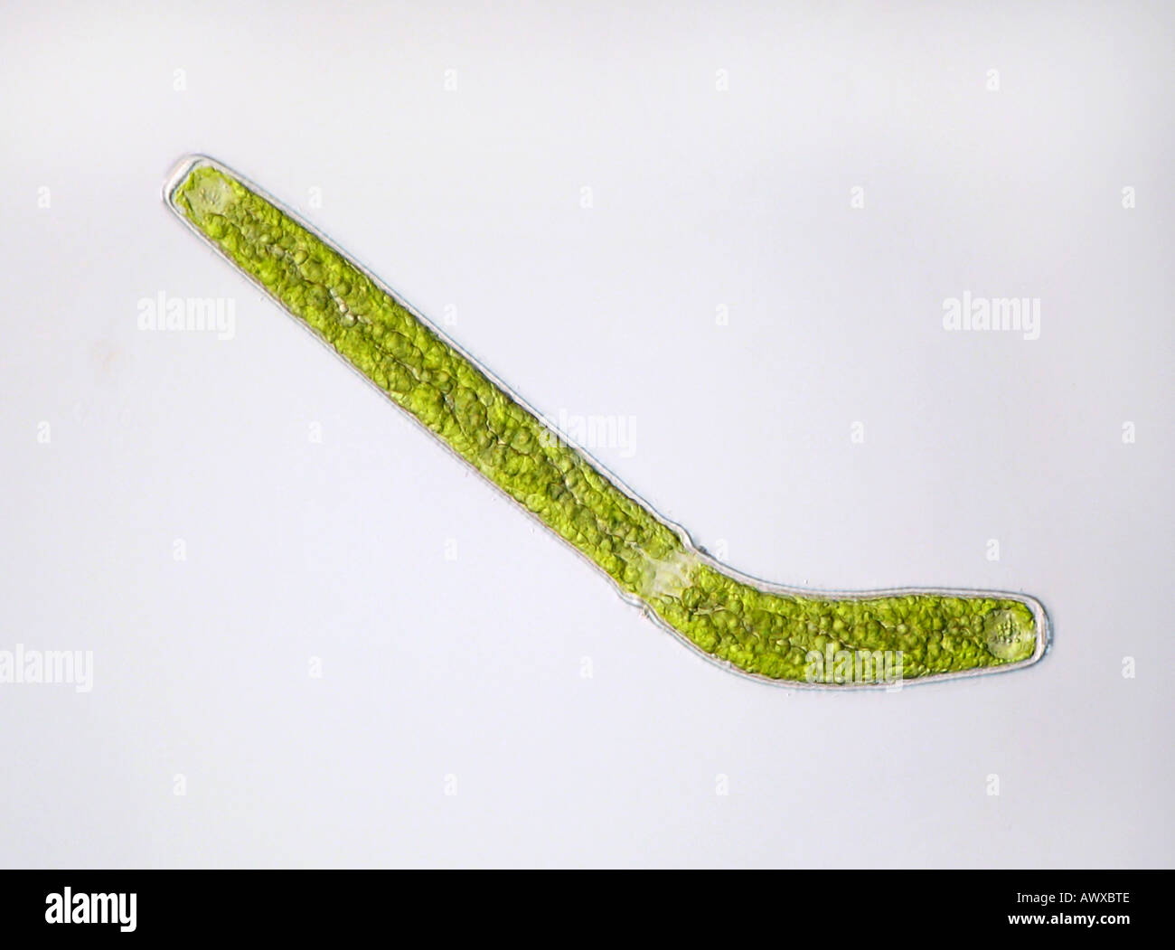 Pleurotaenium trabecula (Pleurotaenium trabecula), growth anomaly, in shining-through light Stock Photohttps://www.alamy.com/image-license-details/?v=1https://www.alamy.com/pleurotaenium-trabecula-pleurotaenium-trabecula-growth-anomaly-in-image9524941.html
RMAWXBTE–Pleurotaenium trabecula (Pleurotaenium trabecula), growth anomaly, in shining-through light
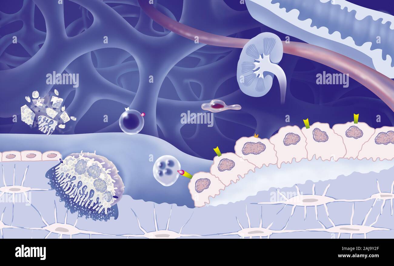 Cancellous bone, reproductive cycle of osteoblasts and osteoblasts, treatments. View of bone trabeculae of spongy bone (blue). Osteoclasts (yellow) de Stock Photohttps://www.alamy.com/image-license-details/?v=1https://www.alamy.com/cancellous-bone-reproductive-cycle-of-osteoblasts-and-osteoblasts-treatments-view-of-bone-trabeculae-of-spongy-bone-blue-osteoclasts-yellow-de-image338279607.html
RM2AJ9Y2F–Cancellous bone, reproductive cycle of osteoblasts and osteoblasts, treatments. View of bone trabeculae of spongy bone (blue). Osteoclasts (yellow) de
 (Pleurotaenium trabecula) Stock Photohttps://www.alamy.com/image-license-details/?v=1https://www.alamy.com/pleurotaenium-trabecula-image632007095.html
RM2YM6BDY–(Pleurotaenium trabecula)
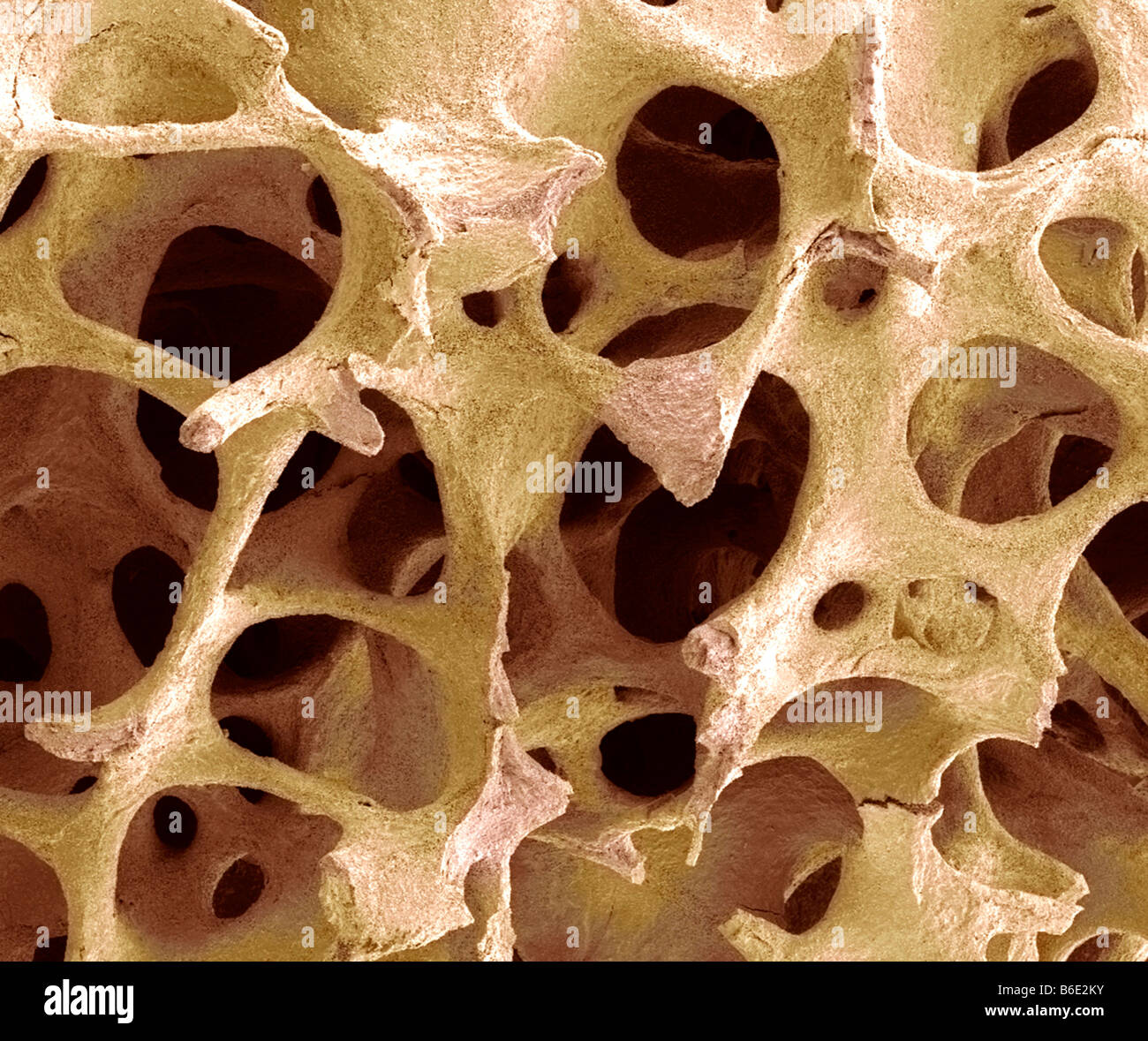 Bone tissue. Coloured scanning electron micrograph(SEM) of cancellous (spongy) bone. Stock Photohttps://www.alamy.com/image-license-details/?v=1https://www.alamy.com/stock-photo-bone-tissue-coloured-scanning-electron-micrographsem-of-cancellous-21207759.html
RFB6E2KY–Bone tissue. Coloured scanning electron micrograph(SEM) of cancellous (spongy) bone.
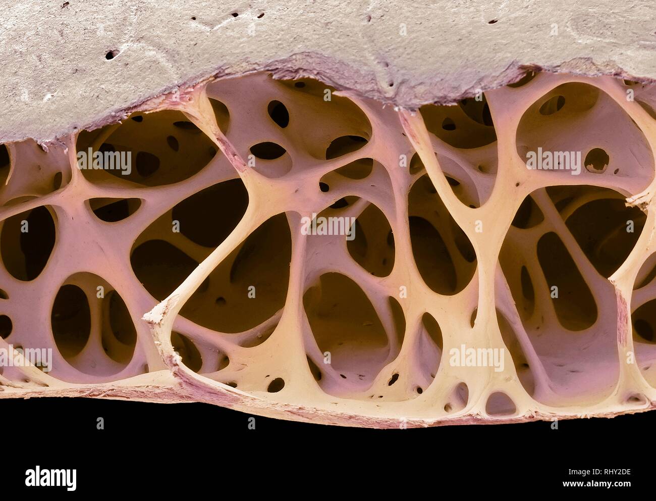 Bird bone tissue, SEM Stock Photohttps://www.alamy.com/image-license-details/?v=1https://www.alamy.com/bird-bone-tissue-sem-image234778586.html
RFRHY2DE–Bird bone tissue, SEM
 Diseases of the chest and the principles of physical diagnosis . - resembled a hernia of the lung.. Fig. 232.—Case No. 238S (Phipps Institute). Upper three-fourths of lung taken up bythree cavities with great outgrowth of fibrous tissue. Another important feature of cavities is that those of any considerablesize contain in their walls or the trabecula traversing them, blood-vessels(Fig. 233). As a rule the blood-vessels in and about the cavity becomethrombosed and obliterated. If thej- remain patulous, small aneurismaldilatations occur in their walls and the rupture of one of these will giveri Stock Photohttps://www.alamy.com/image-license-details/?v=1https://www.alamy.com/diseases-of-the-chest-and-the-principles-of-physical-diagnosis-resembled-a-hernia-of-the-lung-fig-232case-no-238s-phipps-institute-upper-three-fourths-of-lung-taken-up-bythree-cavities-with-great-outgrowth-of-fibrous-tissue-another-important-feature-of-cavities-is-that-those-of-any-considerablesize-contain-in-their-walls-or-the-trabecula-traversing-them-blood-vesselsfig-233-as-a-rule-the-blood-vessels-in-and-about-the-cavity-becomethrombosed-and-obliterated-if-thej-remain-patulous-small-aneurismaldilatations-occur-in-their-walls-and-the-rupture-of-one-of-these-will-giveri-image340250997.html
RM2ANFNH9–Diseases of the chest and the principles of physical diagnosis . - resembled a hernia of the lung.. Fig. 232.—Case No. 238S (Phipps Institute). Upper three-fourths of lung taken up bythree cavities with great outgrowth of fibrous tissue. Another important feature of cavities is that those of any considerablesize contain in their walls or the trabecula traversing them, blood-vessels(Fig. 233). As a rule the blood-vessels in and about the cavity becomethrombosed and obliterated. If thej- remain patulous, small aneurismaldilatations occur in their walls and the rupture of one of these will giveri
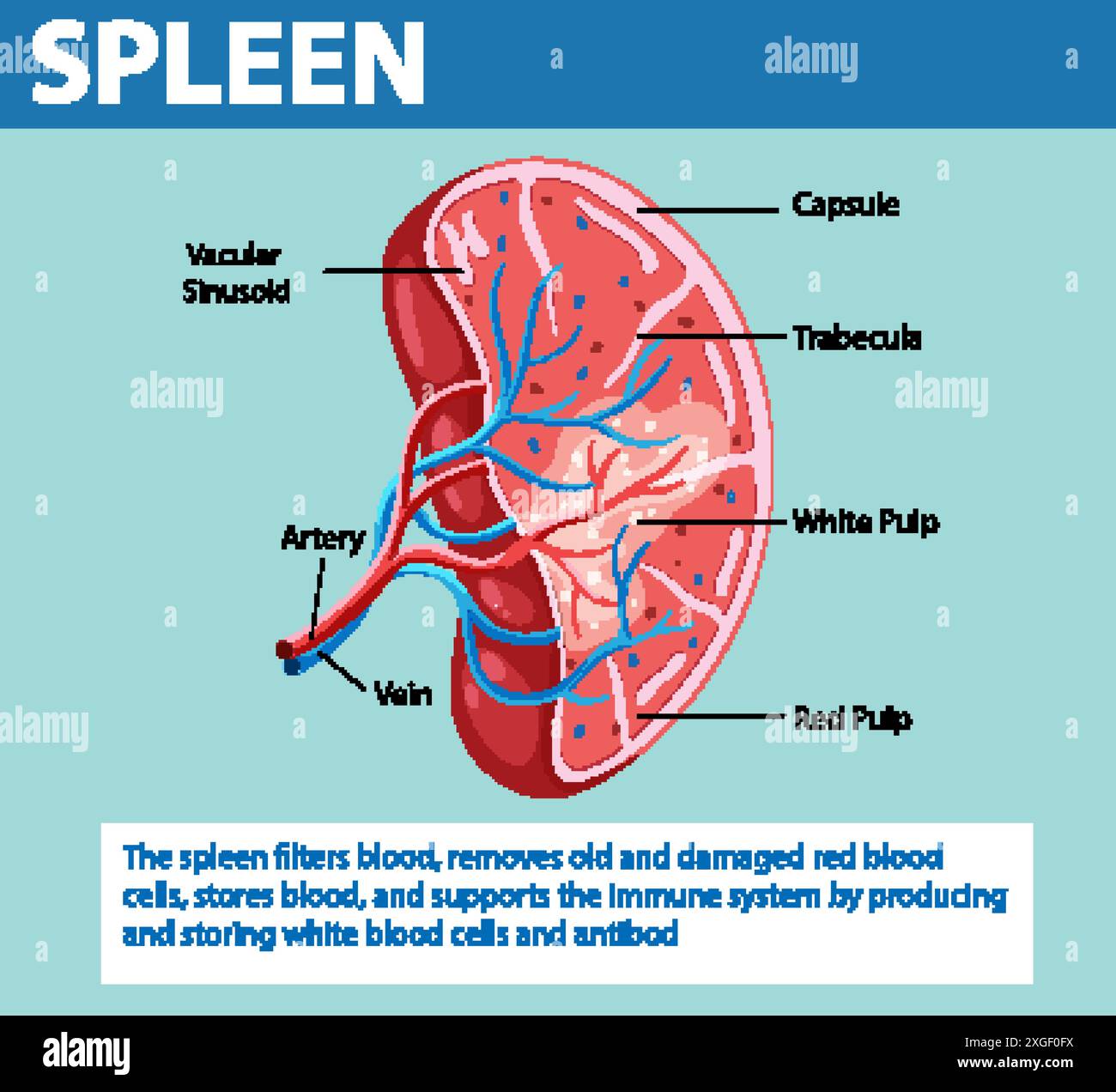 Detailed illustration of spleen structure and functions Stock Vectorhttps://www.alamy.com/image-license-details/?v=1https://www.alamy.com/detailed-illustration-of-spleen-structure-and-functions-image612527102.html
RF2XGF0FX–Detailed illustration of spleen structure and functions
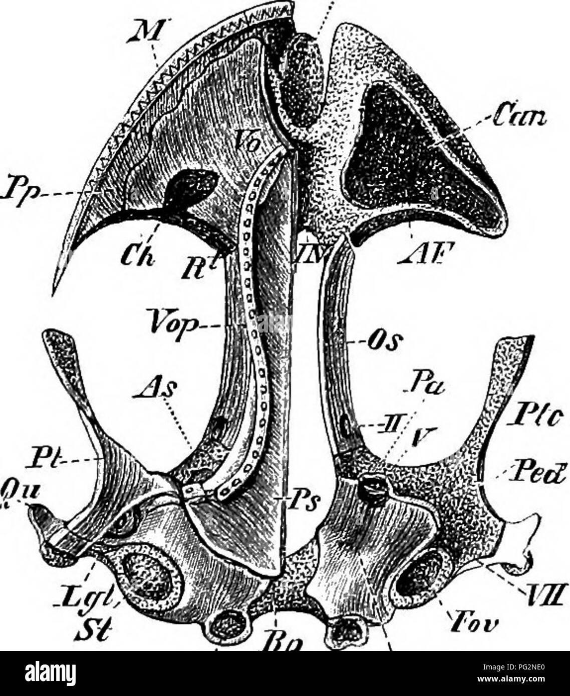 . Elements of the comparative anatomy of vertebrates. Anatomy, Comparative. Con- Fii!. 63.—SkulTj of a Young AxoLOTL. Ventral view. face Osp Fig. 64.—Skull of Salamandra atra (Adult). Dorsal view. Tn..-. Cooo 'OB Fig. 65.—Skull of Salamandra atra (Adult). Ventral vi^w. Tr, trabecula; OB, auditory capsule ; Fov, fenestra ovalis, closed on one side by the stapes [St); Lgt, ligament between the stapes and suspensorium ; Cocc, occipital condyles ; Bp, cartilaginous basilar plate between the auditory cap- sules ; Osp, dorsal tract of the occipital cartilage ; IN, internasal plate, which extends lat Stock Photohttps://www.alamy.com/image-license-details/?v=1https://www.alamy.com/elements-of-the-comparative-anatomy-of-vertebrates-anatomy-comparative-con-fii!-63skultj-of-a-young-axolotl-ventral-view-face-osp-fig-64skull-of-salamandra-atra-adult-dorsal-view-tn-cooo-ob-fig-65skull-of-salamandra-atra-adult-ventral-viw-tr-trabecula-ob-auditory-capsule-fov-fenestra-ovalis-closed-on-one-side-by-the-stapes-st-lgt-ligament-between-the-stapes-and-suspensorium-cocc-occipital-condyles-bp-cartilaginous-basilar-plate-between-the-auditory-cap-sules-osp-dorsal-tract-of-the-occipital-cartilage-in-internasal-plate-which-extends-lat-image216419672.html
RMPG2NE0–. Elements of the comparative anatomy of vertebrates. Anatomy, Comparative. Con- Fii!. 63.—SkulTj of a Young AxoLOTL. Ventral view. face Osp Fig. 64.—Skull of Salamandra atra (Adult). Dorsal view. Tn..-. Cooo 'OB Fig. 65.—Skull of Salamandra atra (Adult). Ventral vi^w. Tr, trabecula; OB, auditory capsule ; Fov, fenestra ovalis, closed on one side by the stapes [St); Lgt, ligament between the stapes and suspensorium ; Cocc, occipital condyles ; Bp, cartilaginous basilar plate between the auditory cap- sules ; Osp, dorsal tract of the occipital cartilage ; IN, internasal plate, which extends lat
 . Abb. 86. CtetiopsyUus fallux R. $. - (Orig.) ^ Chimaeropsylla lebt auf Rhynchocion (eine eigenartige Gattung der Insectivora), und CorypsyUa auf Scapanus. Ihre Eigentümlichkeiten sind: Bei ChimaeropsyUa (Abb. 85) hat das Gesicht, wie bei Stenoponia, einen vertikalen Spalt und die basale Hälfte der grobzähnigen Mandibcln (md) ist von den breiten Wangen verdeckt; vor dem Auge liegt eine scharfe Trabecula frontalis (//); der Vorderrand des ,,Metcpimerum" zieht sich vertikal nach oben, wie bei den Pulicidae. Bei CorypsyUa haben die Meso- pleuren kein stabförmiges Entoskelct (wie bei Pulicin Stock Photohttps://www.alamy.com/image-license-details/?v=1https://www.alamy.com/abb-86-ctetiopsyuus-fallux-r-orig-chimaeropsylla-lebt-auf-rhynchocion-eine-eigenartige-gattung-der-insectivora-und-corypsyua-auf-scapanus-ihre-eigentmlichkeiten-sind-bei-chimaeropsyua-abb-85-hat-das-gesicht-wie-bei-stenoponia-einen-vertikalen-spalt-und-die-basale-hlfte-der-grobzhnigen-mandibcln-md-ist-von-den-breiten-wangen-verdeckt-vor-dem-auge-liegt-eine-scharfe-trabecula-frontalis-der-vorderrand-des-metcpimerumquot-zieht-sich-vertikal-nach-oben-wie-bei-den-pulicidae-bei-corypsyua-haben-die-meso-pleuren-kein-stabfrmiges-entoskelct-wie-bei-pulicin-image179899277.html
RMMCK3A5–. Abb. 86. CtetiopsyUus fallux R. $. - (Orig.) ^ Chimaeropsylla lebt auf Rhynchocion (eine eigenartige Gattung der Insectivora), und CorypsyUa auf Scapanus. Ihre Eigentümlichkeiten sind: Bei ChimaeropsyUa (Abb. 85) hat das Gesicht, wie bei Stenoponia, einen vertikalen Spalt und die basale Hälfte der grobzähnigen Mandibcln (md) ist von den breiten Wangen verdeckt; vor dem Auge liegt eine scharfe Trabecula frontalis (//); der Vorderrand des ,,Metcpimerum" zieht sich vertikal nach oben, wie bei den Pulicidae. Bei CorypsyUa haben die Meso- pleuren kein stabförmiges Entoskelct (wie bei Pulicin
 Pleurotaenium trabecula (Pleurotaenium trabecula), in shining-through light Stock Photohttps://www.alamy.com/image-license-details/?v=1https://www.alamy.com/pleurotaenium-trabecula-pleurotaenium-trabecula-in-shining-through-image8114579.html
RMAGG2H4–Pleurotaenium trabecula (Pleurotaenium trabecula), in shining-through light
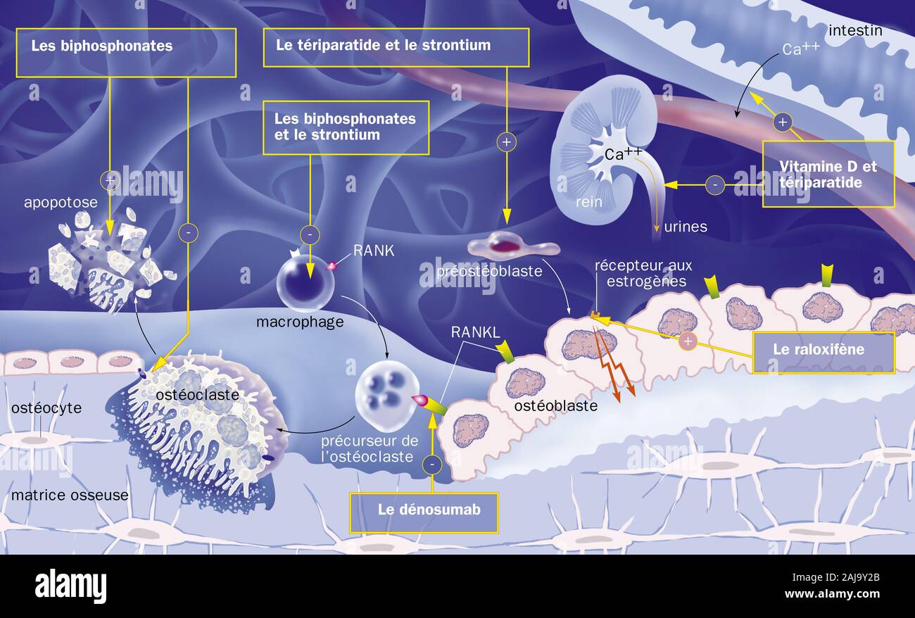 Cancellous bone, reproductive cycle of osteoblasts and osteoblasts, treatments. View of bone trabeculae of spongy bone (blue). Osteoclasts (yellow) de Stock Photohttps://www.alamy.com/image-license-details/?v=1https://www.alamy.com/cancellous-bone-reproductive-cycle-of-osteoblasts-and-osteoblasts-treatments-view-of-bone-trabeculae-of-spongy-bone-blue-osteoclasts-yellow-de-image338279603.html
RM2AJ9Y2B–Cancellous bone, reproductive cycle of osteoblasts and osteoblasts, treatments. View of bone trabeculae of spongy bone (blue). Osteoclasts (yellow) de
 (Pleurotaenium trabecula) Stock Photohttps://www.alamy.com/image-license-details/?v=1https://www.alamy.com/pleurotaenium-trabecula-image632136452.html
RM2YMC8DT–(Pleurotaenium trabecula)
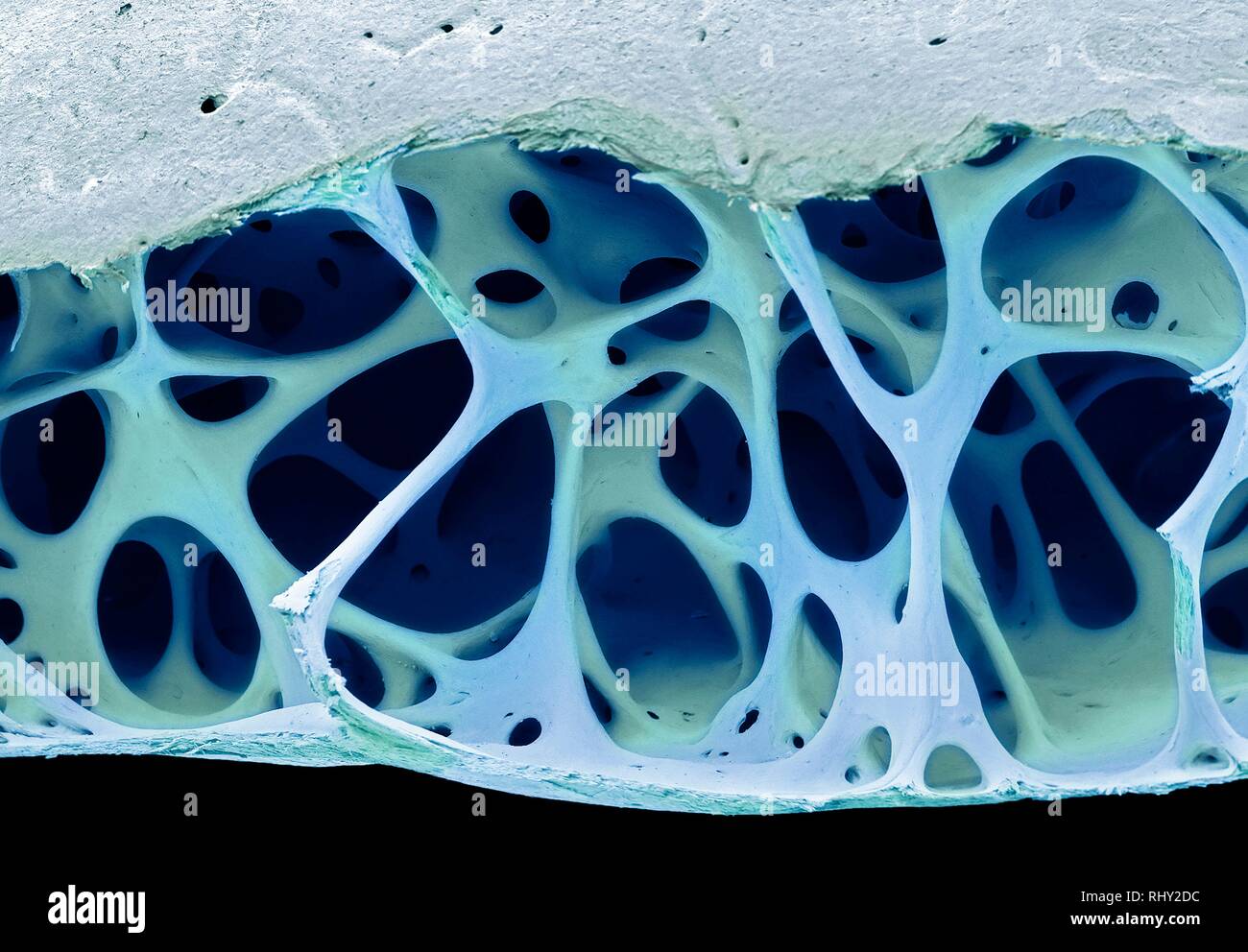 Bird bone tissue, SEM Stock Photohttps://www.alamy.com/image-license-details/?v=1https://www.alamy.com/bird-bone-tissue-sem-image234778584.html
RFRHY2DC–Bird bone tissue, SEM
 The origin of disease : especially of disease resulting from intrinsic as opposed to extrinsic causes : with chapters on diagnosis, prognosis, and treatment . ould not be recog-nizable as liver cells. Between the fibrous walls and the secreting cells there is a spacewhich contains debris and blood-corpuscles, the blood having escaped from its naturalchannels and forced its way into the trabecula to destroy the secreting cells. Fig. 73.—Nutmeg Liver, (x 280.) The region zv from Fig. 71, more highly magnified. A beautiful demonstration ofthe common appearances of nutmeg liver when the process ha Stock Photohttps://www.alamy.com/image-license-details/?v=1https://www.alamy.com/the-origin-of-disease-especially-of-disease-resulting-from-intrinsic-as-opposed-to-extrinsic-causes-with-chapters-on-diagnosis-prognosis-and-treatment-ould-not-be-recog-nizable-as-liver-cells-between-the-fibrous-walls-and-the-secreting-cells-there-is-a-spacewhich-contains-debris-and-blood-corpuscles-the-blood-having-escaped-from-its-naturalchannels-and-forced-its-way-into-the-trabecula-to-destroy-the-secreting-cells-fig-73nutmeg-liver-x-280-the-region-zv-from-fig-71-more-highly-magnified-a-beautiful-demonstration-ofthe-common-appearances-of-nutmeg-liver-when-the-process-ha-image338163138.html
RM2AJ4JEX–The origin of disease : especially of disease resulting from intrinsic as opposed to extrinsic causes : with chapters on diagnosis, prognosis, and treatment . ould not be recog-nizable as liver cells. Between the fibrous walls and the secreting cells there is a spacewhich contains debris and blood-corpuscles, the blood having escaped from its naturalchannels and forced its way into the trabecula to destroy the secreting cells. Fig. 73.—Nutmeg Liver, (x 280.) The region zv from Fig. 71, more highly magnified. A beautiful demonstration ofthe common appearances of nutmeg liver when the process ha
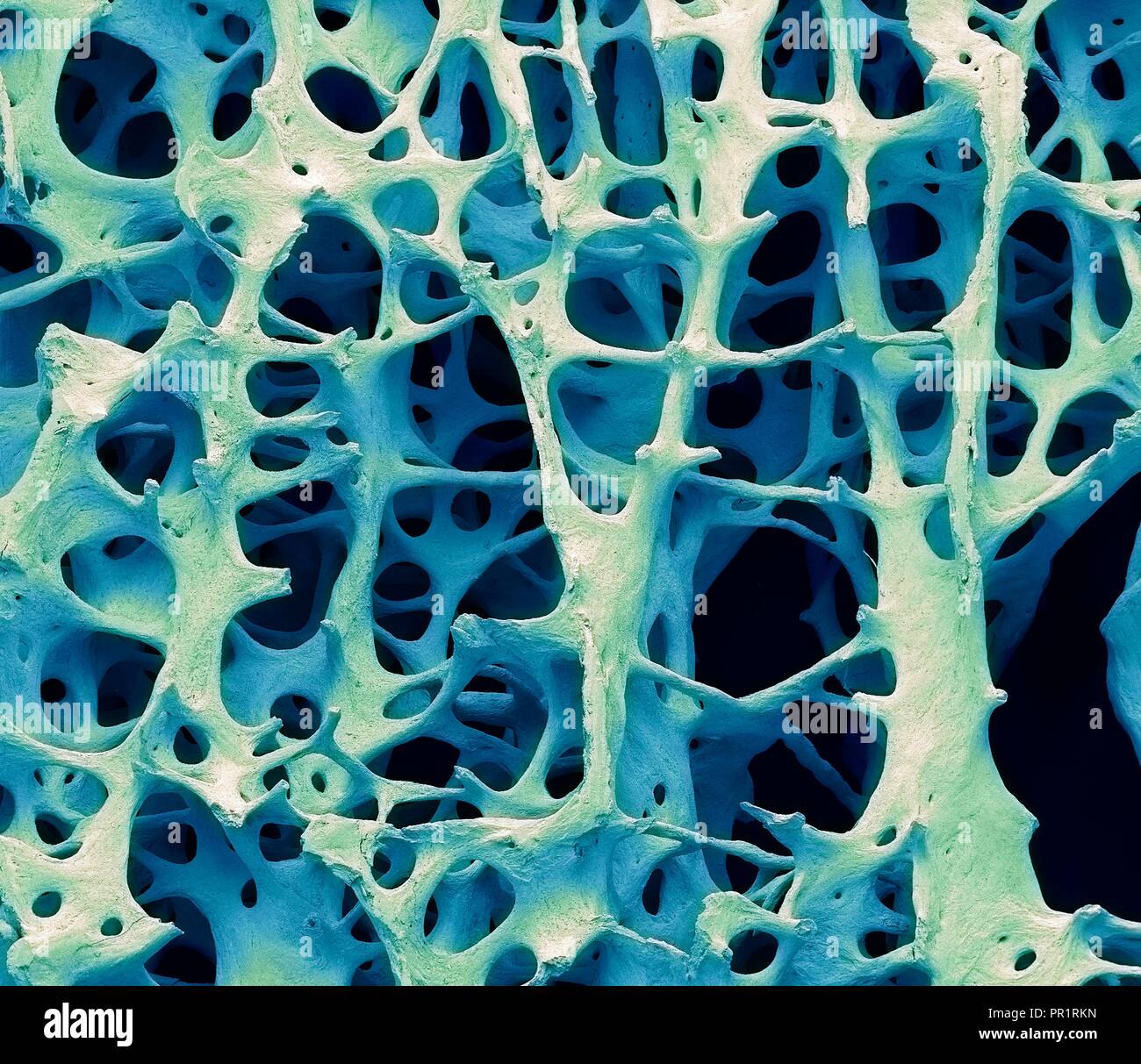 Bone tissue. Coloured scanning electron micrograph (SEM) of human cancellous (spongy) bone. Bone tissue can be either cortical (compact) or cancellous. Cortical bone usually makes up the exterior of the bone, while cancellous bone is found in the interior. Cancellous bone is characterised by a honeycomb arrangement, comprising a network of trabeculae (rod-shaped tissue). These structures provide support and strength to the bone. The spaces within this tissue contain bone marrow (not seen), a blood forming substance. Magnification: x13 when printed 10cm wide. Stock Photohttps://www.alamy.com/image-license-details/?v=1https://www.alamy.com/bone-tissue-coloured-scanning-electron-micrograph-sem-of-human-cancellous-spongy-bone-bone-tissue-can-be-either-cortical-compact-or-cancellous-cortical-bone-usually-makes-up-the-exterior-of-the-bone-while-cancellous-bone-is-found-in-the-interior-cancellous-bone-is-characterised-by-a-honeycomb-arrangement-comprising-a-network-of-trabeculae-rod-shaped-tissue-these-structures-provide-support-and-strength-to-the-bone-the-spaces-within-this-tissue-contain-bone-marrow-not-seen-a-blood-forming-substance-magnification-x13-when-printed-10cm-wide-image220702041.html
RFPR1RKN–Bone tissue. Coloured scanning electron micrograph (SEM) of human cancellous (spongy) bone. Bone tissue can be either cortical (compact) or cancellous. Cortical bone usually makes up the exterior of the bone, while cancellous bone is found in the interior. Cancellous bone is characterised by a honeycomb arrangement, comprising a network of trabeculae (rod-shaped tissue). These structures provide support and strength to the bone. The spaces within this tissue contain bone marrow (not seen), a blood forming substance. Magnification: x13 when printed 10cm wide.
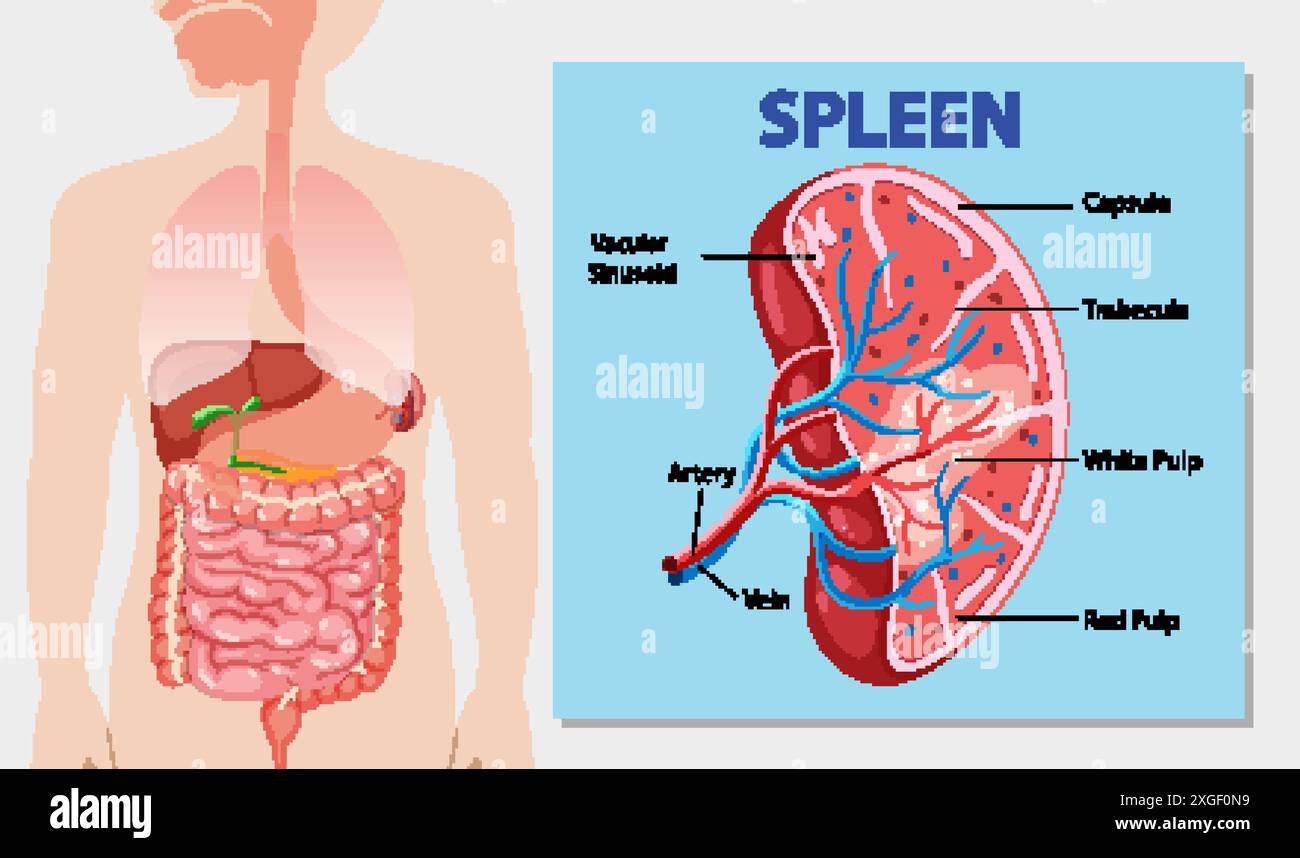 Detailed diagram of the human spleen and its parts Stock Vectorhttps://www.alamy.com/image-license-details/?v=1https://www.alamy.com/detailed-diagram-of-the-human-spleen-and-its-parts-image612527253.html
RF2XGF0N9–Detailed diagram of the human spleen and its parts
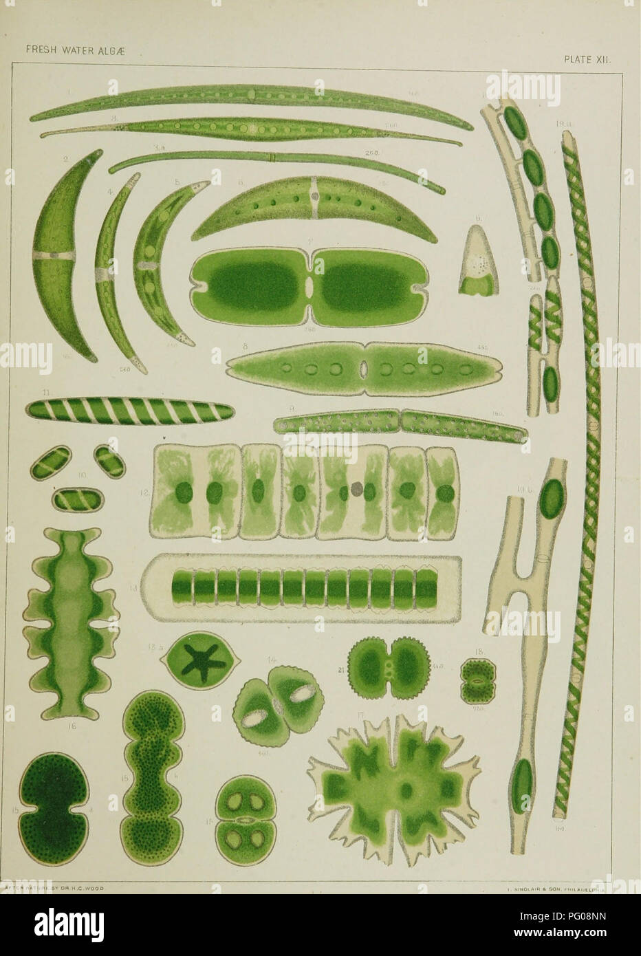 . A contribution to the history of the fresh-water algœ of North America. Botany; Algae. ArTtP (MATURE B'' DRH.C.WOQO Fig. 1. COSMARIUM LINEATUM " 2. C, EHRENBERGII. " 3. C. ROSTRATUM, " 4. C. DIAN/C, " 5. C. PARVULUM, 6. C. LEIBLEINIt. 7. TETMEMORUS GIGANTEUS. 8. TETMEMORUS GRANULATUS, 9. PLEUROT/ENIUM TRABECULA. 10. SPIROT/ENIA BRYOPHILA . 11. SPIROT/tNIA CONDENSATA 13. HYALOTHECA DISSILIENS. 13. DIDYMOPRIUM 6REVILLII. H. COSMARIUM BOTRYTIS. 15. C, CUCUMIS. Fig. 15. EUASTRUM MULTILOBATUM. " 17, MICRASTERIAS AMERICANA. " 18. COSMARIUM MENEGHENII. " 19. SPIRO Stock Photohttps://www.alamy.com/image-license-details/?v=1https://www.alamy.com/a-contribution-to-the-history-of-the-fresh-water-alg-of-north-america-botany-algae-arttp-mature-b-drhcwoqo-fig-1-cosmarium-lineatum-quot-2-c-ehrenbergii-quot-3-c-rostratum-quot-4-c-dianc-quot-5-c-parvulum-6-c-leibleinit-7-tetmemorus-giganteus-8-tetmemorus-granulatus-9-pleurotenium-trabecula-10-spirotenia-bryophila-11-spirottnia-condensata-13-hyalotheca-dissiliens-13-didymoprium-6revillii-h-cosmarium-botrytis-15-c-cucumis-fig-15-euastrum-multilobatum-quot-17-micrasterias-americana-quot-18-cosmarium-meneghenii-quot-19-spiro-image216365793.html
RMPG08NN–. A contribution to the history of the fresh-water algœ of North America. Botany; Algae. ArTtP (MATURE B'' DRH.C.WOQO Fig. 1. COSMARIUM LINEATUM " 2. C, EHRENBERGII. " 3. C. ROSTRATUM, " 4. C. DIAN/C, " 5. C. PARVULUM, 6. C. LEIBLEINIt. 7. TETMEMORUS GIGANTEUS. 8. TETMEMORUS GRANULATUS, 9. PLEUROT/ENIUM TRABECULA. 10. SPIROT/ENIA BRYOPHILA . 11. SPIROT/tNIA CONDENSATA 13. HYALOTHECA DISSILIENS. 13. DIDYMOPRIUM 6REVILLII. H. COSMARIUM BOTRYTIS. 15. C, CUCUMIS. Fig. 15. EUASTRUM MULTILOBATUM. " 17, MICRASTERIAS AMERICANA. " 18. COSMARIUM MENEGHENII. " 19. SPIRO
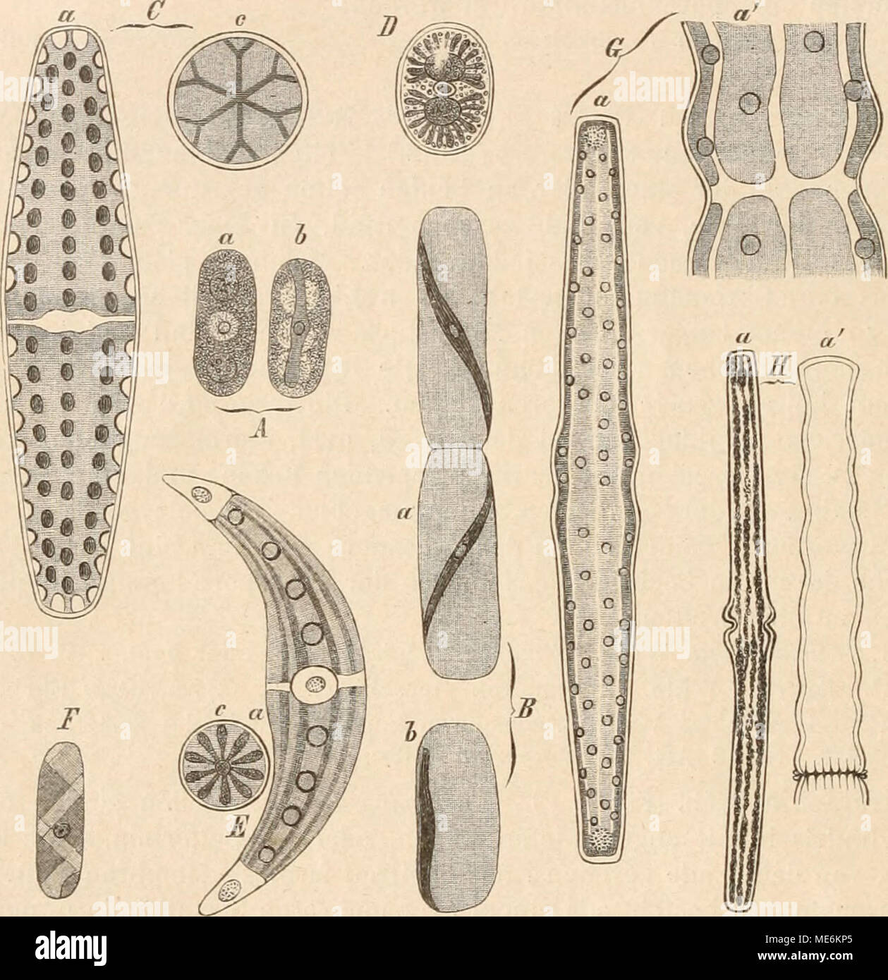 . Die Natürlichen Pflanzenfamilien nebst ihren Gattungen und wichtigeren Arten, insbesondere den Nutzpflanzen, unter Mitwirkung zahlreicher hervorragender Fachgelehrten begründet . Fig. 6. A Mesotaenium Braunii de Bary (390/1); B Ancylonema NoräenslUöldii Berggr. (650/1); 0 Peniwn Digitus Bröb. (400/1); D Cylindroci/stis crassa de Bary (390/1); E Closteriwn moniliferiim (Bory) Ehrb. (200/1); F Spiro- taenia nmscieola de Bary (750/1); 6 Pleurotaenium Trabecula (Ehrb.) Näg. [a 100/1; a' 600/1); Ea ßociditim Ba- culum Breb., a' D. dilatatum Cleve (340/1). a von oben, 6 von der Seite, c vom Ende g Stock Photohttps://www.alamy.com/image-license-details/?v=1https://www.alamy.com/die-natrlichen-pflanzenfamilien-nebst-ihren-gattungen-und-wichtigeren-arten-insbesondere-den-nutzpflanzen-unter-mitwirkung-zahlreicher-hervorragender-fachgelehrten-begrndet-fig-6-a-mesotaenium-braunii-de-bary-3901-b-ancylonema-norensluldii-berggr-6501-0-peniwn-digitus-brb-4001-d-cylindrocistis-crassa-de-bary-3901-e-closteriwn-moniliferiim-bory-ehrb-2001-f-spiro-taenia-nmscieola-de-bary-7501-6-pleurotaenium-trabecula-ehrb-ng-a-1001-a-6001-ea-ociditim-ba-culum-breb-a-d-dilatatum-cleve-3401-a-von-oben-6-von-der-seite-c-vom-ende-g-image180856093.html
RMME6KP5–. Die Natürlichen Pflanzenfamilien nebst ihren Gattungen und wichtigeren Arten, insbesondere den Nutzpflanzen, unter Mitwirkung zahlreicher hervorragender Fachgelehrten begründet . Fig. 6. A Mesotaenium Braunii de Bary (390/1); B Ancylonema NoräenslUöldii Berggr. (650/1); 0 Peniwn Digitus Bröb. (400/1); D Cylindroci/stis crassa de Bary (390/1); E Closteriwn moniliferiim (Bory) Ehrb. (200/1); F Spiro- taenia nmscieola de Bary (750/1); 6 Pleurotaenium Trabecula (Ehrb.) Näg. [a 100/1; a' 600/1); Ea ßociditim Ba- culum Breb., a' D. dilatatum Cleve (340/1). a von oben, 6 von der Seite, c vom Ende g
 (Pleurotaenium trabecula) Stock Photohttps://www.alamy.com/image-license-details/?v=1https://www.alamy.com/pleurotaenium-trabecula-image632012293.html
RM2YM6J3H–(Pleurotaenium trabecula)
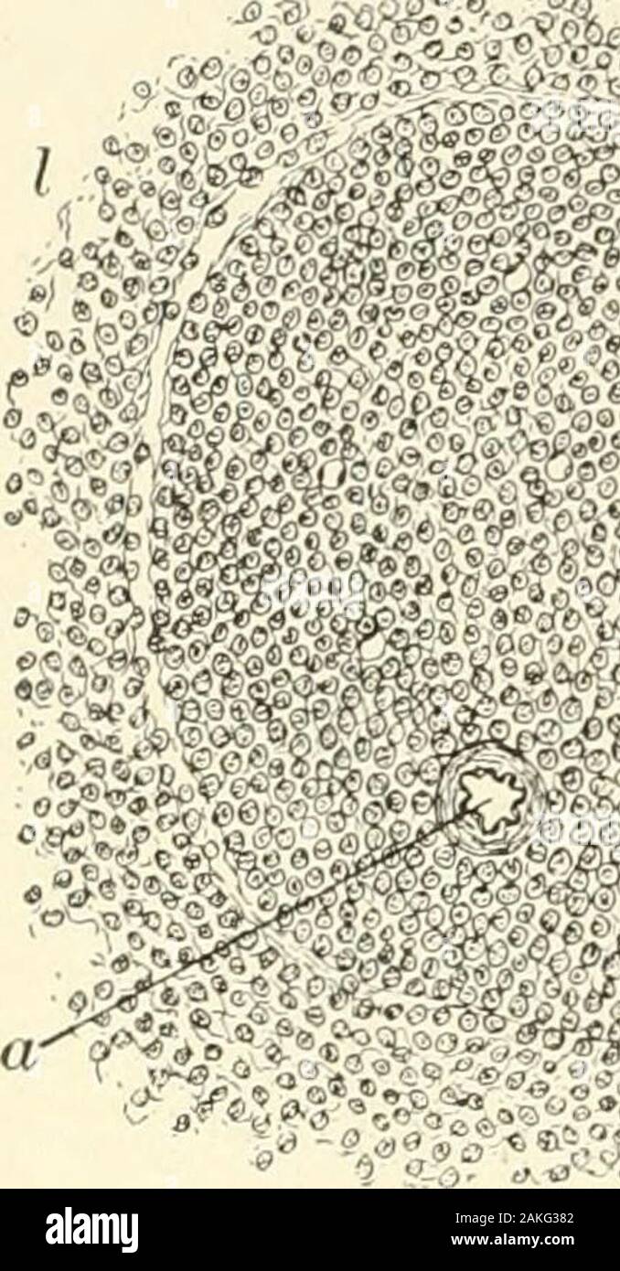 Textbook of normal histology: including an account of the development of the tissues and of the organs . Section of human spleen, showingtrabecular (it) and fibrous reticulum (6)continued into the surrounding splenicpulp ; c, lymphoid cells Transverse section of largetrabecula of human spleen : a,fibrous tissue, containing a fewgroups of plane muscle-cells (i);c, extension of trabecula intofibrous reticulum; d, lymph-corpuscles. 124 NORMAL HISTOLOGY. Fig. 151.. ?&:& *fc^ intimately related vascular channels, forms the splenic pulp, and asthe cylindrical or spherical masses of dense adenoid tis Stock Photohttps://www.alamy.com/image-license-details/?v=1https://www.alamy.com/textbook-of-normal-histology-including-an-account-of-the-development-of-the-tissues-and-of-the-organs-section-of-human-spleen-showingtrabecular-it-and-fibrous-reticulum-6continued-into-the-surrounding-splenicpulp-c-lymphoid-cells-transverse-section-of-largetrabecula-of-human-spleen-afibrous-tissue-containing-a-fewgroups-of-plane-muscle-cells-ic-extension-of-trabecula-intofibrous-reticulum-d-lymph-corpuscles-124-normal-histology-fig-151-fc-intimately-related-vascular-channels-forms-the-splenic-pulp-and-asthe-cylindrical-or-spherical-masses-of-dense-adenoid-tis-image339029266.html
RM2AKG382–Textbook of normal histology: including an account of the development of the tissues and of the organs . Section of human spleen, showingtrabecular (it) and fibrous reticulum (6)continued into the surrounding splenicpulp ; c, lymphoid cells Transverse section of largetrabecula of human spleen : a,fibrous tissue, containing a fewgroups of plane muscle-cells (i);c, extension of trabecula intofibrous reticulum; d, lymph-corpuscles. 124 NORMAL HISTOLOGY. Fig. 151.. ?&:& *fc^ intimately related vascular channels, forms the splenic pulp, and asthe cylindrical or spherical masses of dense adenoid tis
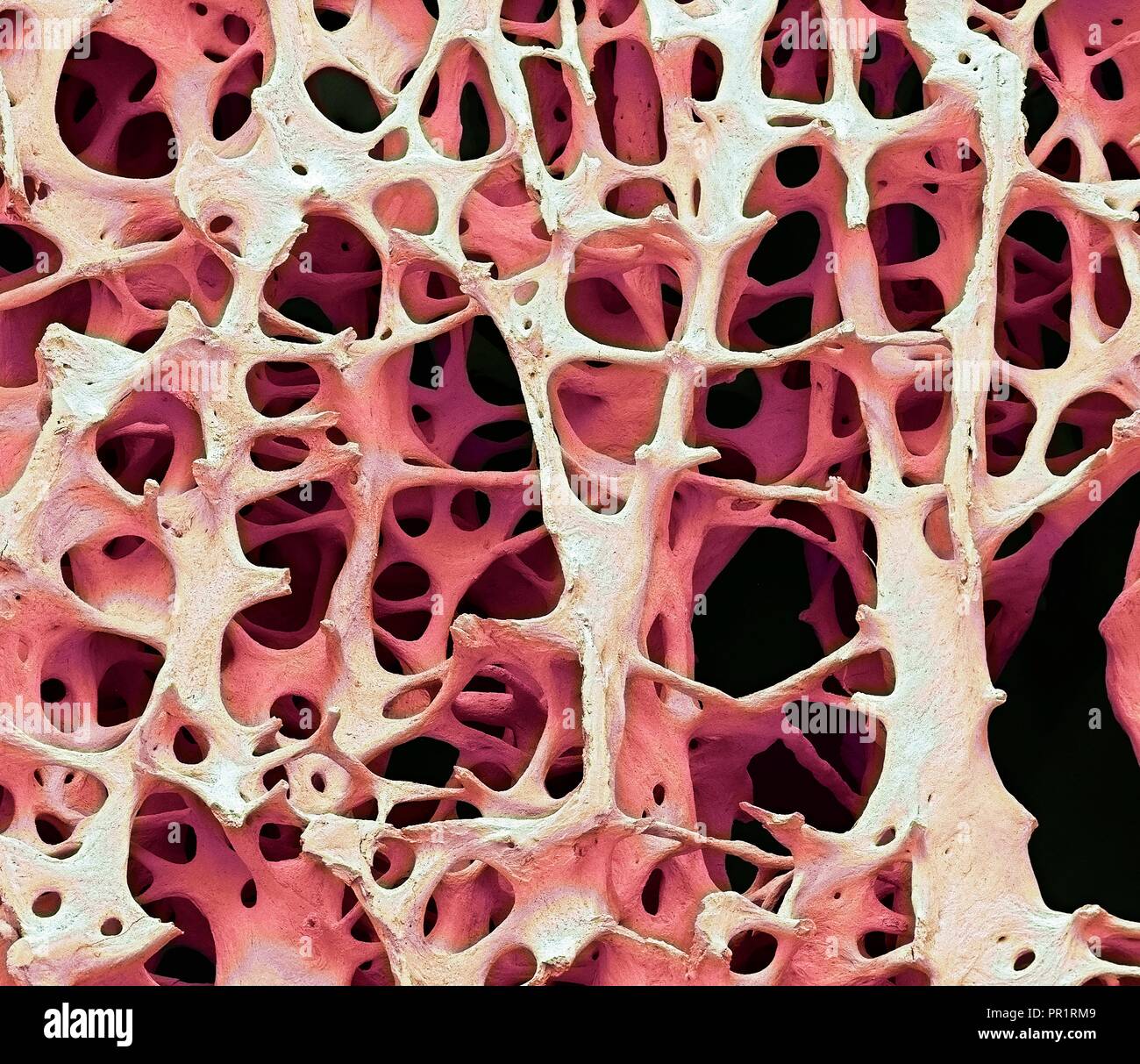 Bone tissue. Coloured scanning electron micrograph (SEM) of human cancellous (spongy) bone. Bone tissue can be either cortical (compact) or cancellous. Cortical bone usually makes up the exterior of the bone, while cancellous bone is found in the interior. Cancellous bone is characterised by a honeycomb arrangement, comprising a network of trabeculae (rod-shaped tissue). These structures provide support and strength to the bone. The spaces within this tissue contain bone marrow (not seen), a blood forming substance. Magnification: x13 when printed 10cm wide. Stock Photohttps://www.alamy.com/image-license-details/?v=1https://www.alamy.com/bone-tissue-coloured-scanning-electron-micrograph-sem-of-human-cancellous-spongy-bone-bone-tissue-can-be-either-cortical-compact-or-cancellous-cortical-bone-usually-makes-up-the-exterior-of-the-bone-while-cancellous-bone-is-found-in-the-interior-cancellous-bone-is-characterised-by-a-honeycomb-arrangement-comprising-a-network-of-trabeculae-rod-shaped-tissue-these-structures-provide-support-and-strength-to-the-bone-the-spaces-within-this-tissue-contain-bone-marrow-not-seen-a-blood-forming-substance-magnification-x13-when-printed-10cm-wide-image220702057.html
RFPR1RM9–Bone tissue. Coloured scanning electron micrograph (SEM) of human cancellous (spongy) bone. Bone tissue can be either cortical (compact) or cancellous. Cortical bone usually makes up the exterior of the bone, while cancellous bone is found in the interior. Cancellous bone is characterised by a honeycomb arrangement, comprising a network of trabeculae (rod-shaped tissue). These structures provide support and strength to the bone. The spaces within this tissue contain bone marrow (not seen), a blood forming substance. Magnification: x13 when printed 10cm wide.
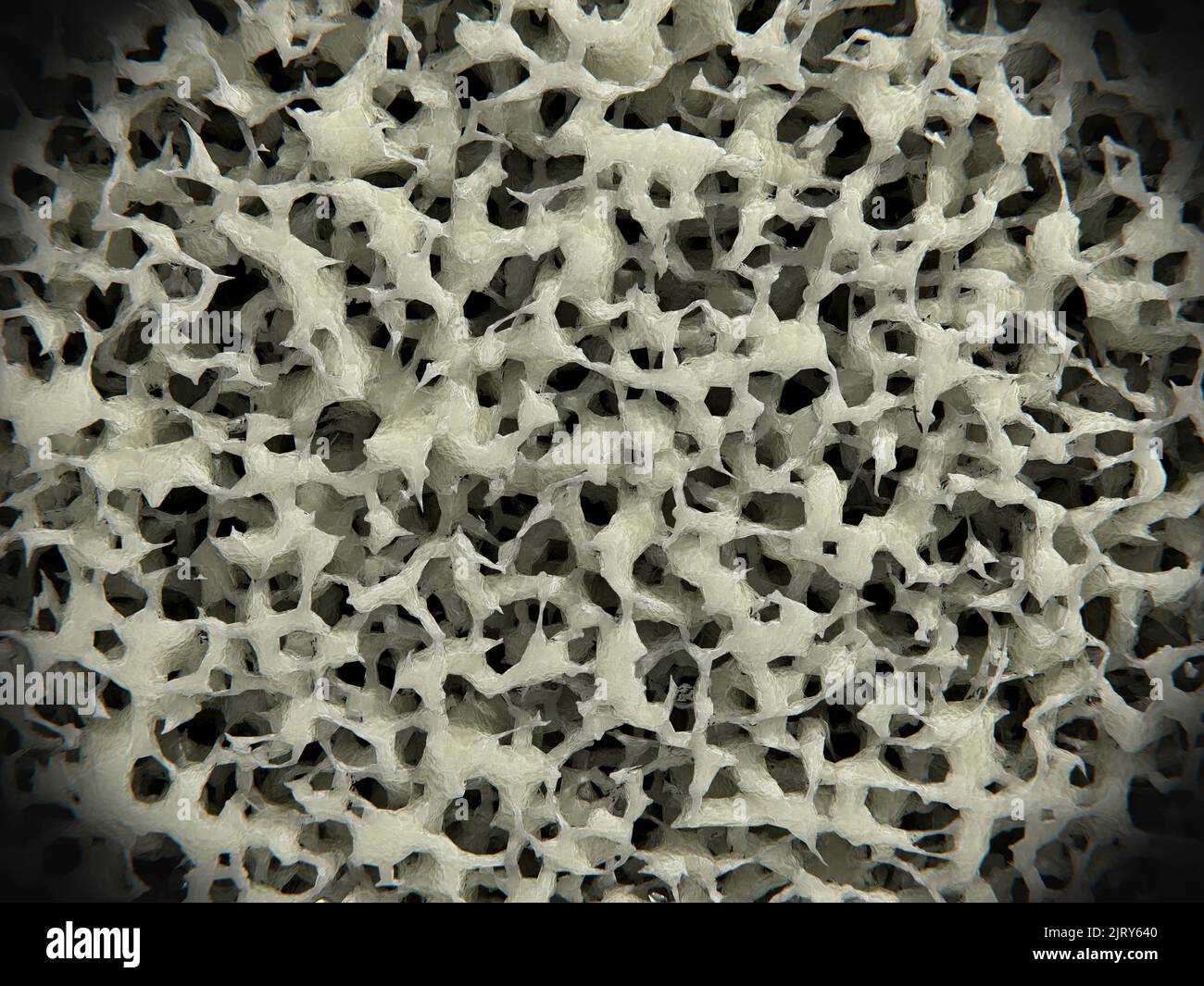 Osteoporotic bone tissue, illustration Stock Photohttps://www.alamy.com/image-license-details/?v=1https://www.alamy.com/osteoporotic-bone-tissue-illustration-image479414544.html
RF2JRY640–Osteoporotic bone tissue, illustration
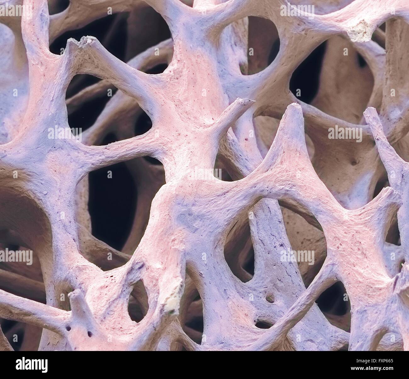 Bone tissue. Coloured scanning electron micrograph (SEM) of cancellous (spongy) bone. Bone tissue can be either cortical (compact) or cancellous. Cortical bone usually makes up the exterior of the bone, while cancellous bone is found in the interior. Cancellous bone is characterised by a honeycomb arrangement, comprising a network of trabeculae (rod-shaped tissue). These structures provide support and strength to the bone. The spaces within this tissue contain bone marrow (not seen), a blood forming substance. Magnification: x40 when printed 10cm wide. Stock Photohttps://www.alamy.com/image-license-details/?v=1https://www.alamy.com/stock-photo-bone-tissue-coloured-scanning-electron-micrograph-sem-of-cancellous-102520717.html
RFFXP665–Bone tissue. Coloured scanning electron micrograph (SEM) of cancellous (spongy) bone. Bone tissue can be either cortical (compact) or cancellous. Cortical bone usually makes up the exterior of the bone, while cancellous bone is found in the interior. Cancellous bone is characterised by a honeycomb arrangement, comprising a network of trabeculae (rod-shaped tissue). These structures provide support and strength to the bone. The spaces within this tissue contain bone marrow (not seen), a blood forming substance. Magnification: x40 when printed 10cm wide.
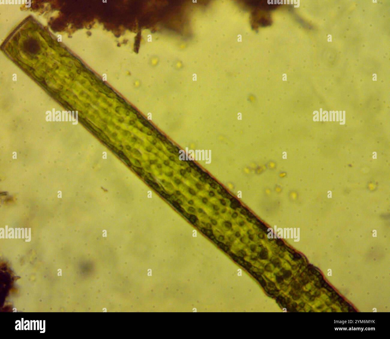 (Pleurotaenium trabecula) Stock Photohttps://www.alamy.com/image-license-details/?v=1https://www.alamy.com/pleurotaenium-trabecula-image632014535.html
RM2YM6MYK–(Pleurotaenium trabecula)
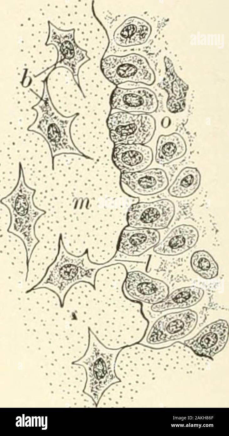 Textbook of normal histology: including an account of the development of the tissues and of the organs . Developing bone—trabecula of endochondralbone: a, the new bone; 6, bone-cells; c, stillunabsorbed remains of calcified cartilage-matrix. Developing bone—the surface of portion ofbone-trabecula, exhibiting the conversion of theosteoblasts into the bone-corpuscles : b, lacunawith young bone-cell; o, osteoblasts arranged onthe surface of the newly-formed osseous matrix(m); at / an osteoblast just being isolated. places forming an outer envelope closely embracing the central endo-chondral bone. Stock Photohttps://www.alamy.com/image-license-details/?v=1https://www.alamy.com/textbook-of-normal-histology-including-an-account-of-the-development-of-the-tissues-and-of-the-organs-developing-bonetrabecula-of-endochondralbone-a-the-new-bone-6-bone-cells-c-stillunabsorbed-remains-of-calcified-cartilage-matrix-developing-bonethe-surface-of-portion-ofbone-trabecula-exhibiting-the-conversion-of-theosteoblasts-into-the-bone-corpuscles-b-lacunawith-young-bone-cell-o-osteoblasts-arranged-onthe-surface-of-the-newly-formed-osseous-matrixm-at-an-osteoblast-just-being-isolated-places-forming-an-outer-envelope-closely-embracing-the-central-endo-chondral-bone-image339055095.html
RM2AKH86F–Textbook of normal histology: including an account of the development of the tissues and of the organs . Developing bone—trabecula of endochondralbone: a, the new bone; 6, bone-cells; c, stillunabsorbed remains of calcified cartilage-matrix. Developing bone—the surface of portion ofbone-trabecula, exhibiting the conversion of theosteoblasts into the bone-corpuscles : b, lacunawith young bone-cell; o, osteoblasts arranged onthe surface of the newly-formed osseous matrix(m); at / an osteoblast just being isolated. places forming an outer envelope closely embracing the central endo-chondral bone.
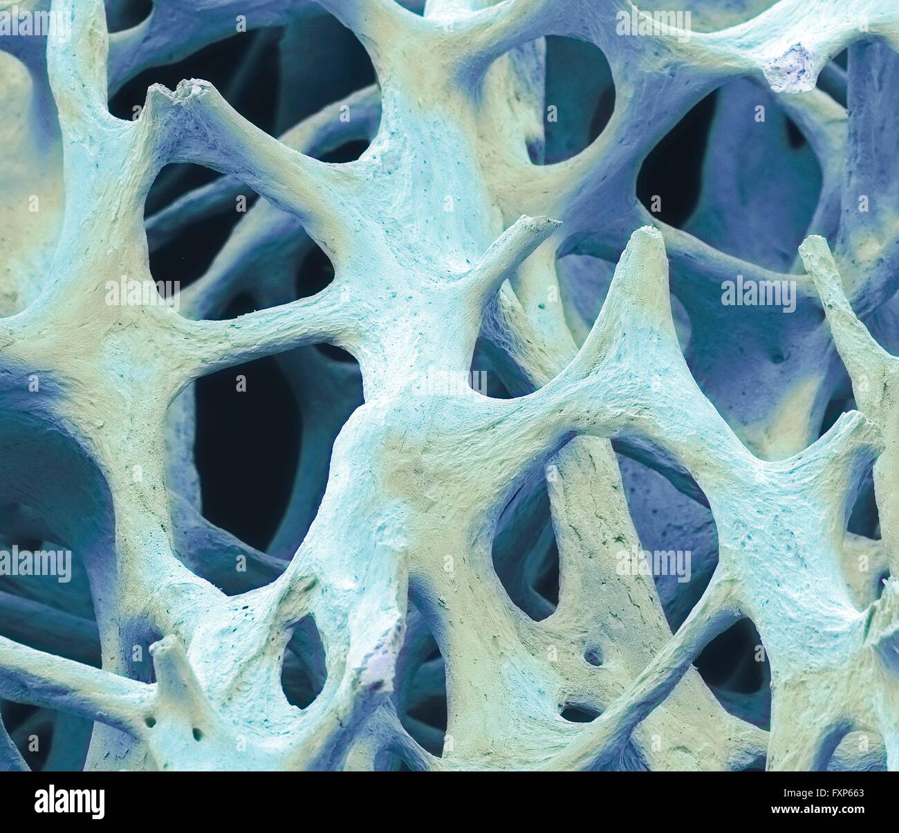 Bone tissue. Coloured scanning electron micrograph (SEM) of cancellous (spongy) bone. Bone tissue can be either cortical (compact) or cancellous. Cortical bone usually makes up the exterior of the bone, while cancellous bone is found in the interior. Cancellous bone is characterised by a honeycomb arrangement, comprising a network of trabeculae (rod-shaped tissue). These structures provide support and strength to the bone. The spaces within this tissue contain bone marrow (not seen), a blood forming substance. Magnification: x40 when printed 10cm wide. Stock Photohttps://www.alamy.com/image-license-details/?v=1https://www.alamy.com/stock-photo-bone-tissue-coloured-scanning-electron-micrograph-sem-of-cancellous-102520715.html
RFFXP663–Bone tissue. Coloured scanning electron micrograph (SEM) of cancellous (spongy) bone. Bone tissue can be either cortical (compact) or cancellous. Cortical bone usually makes up the exterior of the bone, while cancellous bone is found in the interior. Cancellous bone is characterised by a honeycomb arrangement, comprising a network of trabeculae (rod-shaped tissue). These structures provide support and strength to the bone. The spaces within this tissue contain bone marrow (not seen), a blood forming substance. Magnification: x40 when printed 10cm wide.
 Paget's disease, light micrograph Stock Photohttps://www.alamy.com/image-license-details/?v=1https://www.alamy.com/stock-photo-pagets-disease-light-micrograph-30585712.html
RFBNN8AT–Paget's disease, light micrograph
 (Pleurotaenium trabecula) Stock Photohttps://www.alamy.com/image-license-details/?v=1https://www.alamy.com/pleurotaenium-trabecula-image632021010.html
RM2YM716X–(Pleurotaenium trabecula)
 The American journal of anatomy . •:•• / • / THE AMBBICAN JOURNAL OF ANATOMY, VOL. 12, NO. 3 271 PLATE 2 EXPLANATION OF FKiURES 5 Cyst invaginated by a vascular pial trabecula, forming a glomerulus from15 cm. stage of development. X 1500. 6 Photomicrograph of peripheral portion of pineal body of 21 cm. stage, show-ing several cysts and several vascular trabeculae, and the presence of an enormousnumber of melanic granules. X 250. 7 Transverse section of a small alveolus or cyst, showing the character of thecells, the distribution of the melanic granules and the reticular (coagidated) con-tent Stock Photohttps://www.alamy.com/image-license-details/?v=1https://www.alamy.com/the-american-journal-of-anatomy-the-ambbican-journal-of-anatomy-vol-12-no-3-271-plate-2-explanation-of-fkiures-5-cyst-invaginated-by-a-vascular-pial-trabecula-forming-a-glomerulus-from15-cm-stage-of-development-x-1500-6-photomicrograph-of-peripheral-portion-of-pineal-body-of-21-cm-stage-show-ing-several-cysts-and-several-vascular-trabeculae-and-the-presence-of-an-enormousnumber-of-melanic-granules-x-250-7-transverse-section-of-a-small-alveolus-or-cyst-showing-the-character-of-thecells-the-distribution-of-the-melanic-granules-and-the-reticular-coagidated-con-tent-image339108276.html
RM2AKKM1T–The American journal of anatomy . •:•• / • / THE AMBBICAN JOURNAL OF ANATOMY, VOL. 12, NO. 3 271 PLATE 2 EXPLANATION OF FKiURES 5 Cyst invaginated by a vascular pial trabecula, forming a glomerulus from15 cm. stage of development. X 1500. 6 Photomicrograph of peripheral portion of pineal body of 21 cm. stage, show-ing several cysts and several vascular trabeculae, and the presence of an enormousnumber of melanic granules. X 250. 7 Transverse section of a small alveolus or cyst, showing the character of thecells, the distribution of the melanic granules and the reticular (coagidated) con-tent
 Osteoporotic bone, coloured scanning electronmicrograph (SEM). Stock Photohttps://www.alamy.com/image-license-details/?v=1https://www.alamy.com/stock-photo-osteoporotic-bone-coloured-scanning-electronmicrograph-sem-21207791.html
RFB6E2N3–Osteoporotic bone, coloured scanning electronmicrograph (SEM).
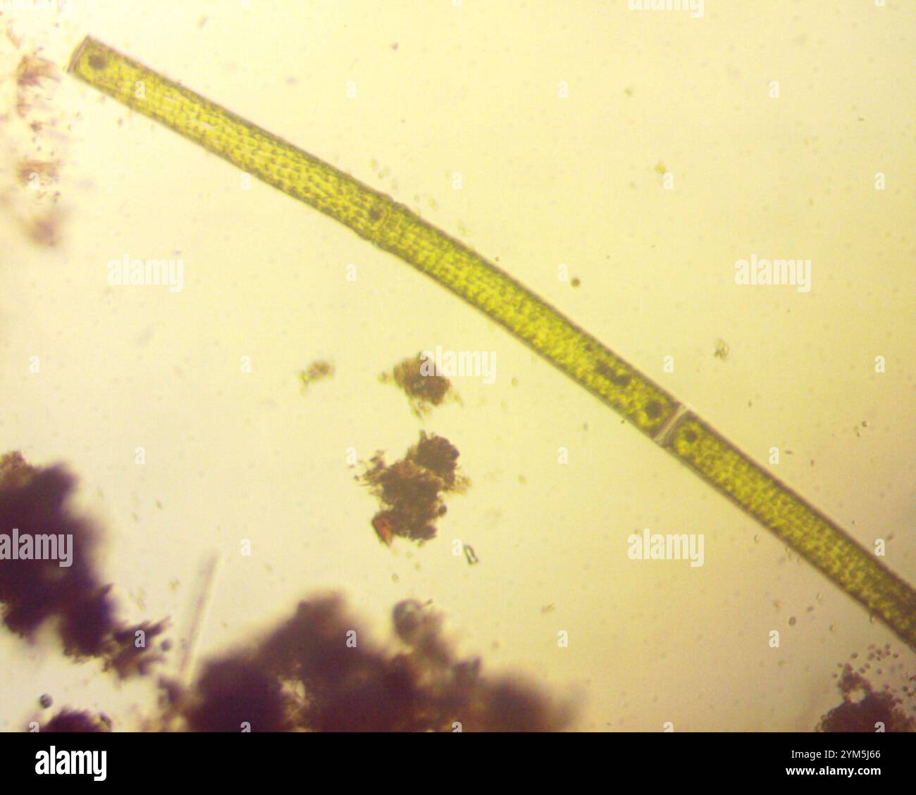 (Pleurotaenium trabecula) Stock Photohttps://www.alamy.com/image-license-details/?v=1https://www.alamy.com/pleurotaenium-trabecula-image631990414.html
RM2YM5J66–(Pleurotaenium trabecula)
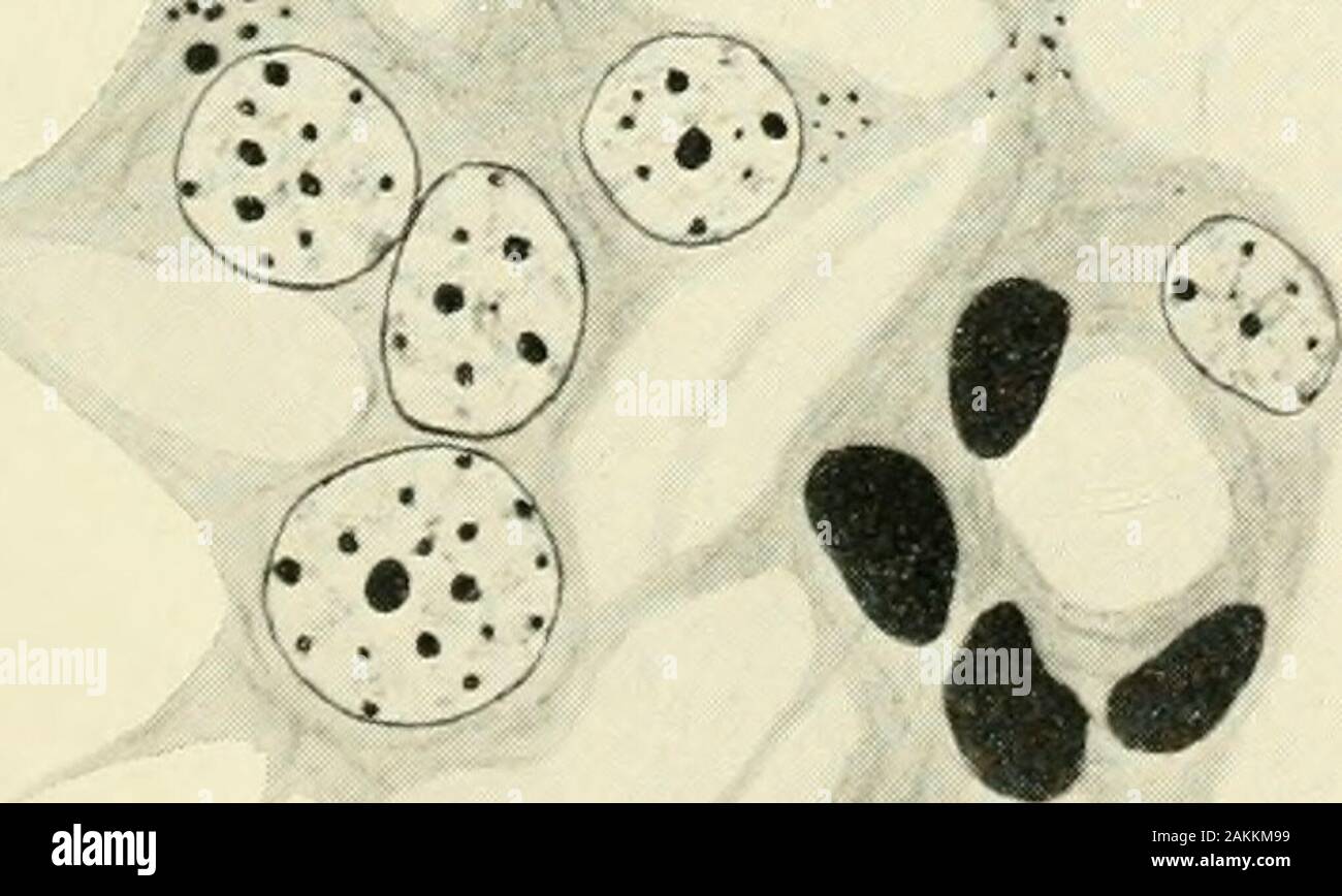 The American journal of anatomy . .V .T^-X •:^ Lumen ,•••. •:•• / • / THE AMBBICAN JOURNAL OF ANATOMY, VOL. 12, NO. 3 271 PLATE 2 EXPLANATION OF FKiURES 5 Cyst invaginated by a vascular pial trabecula, forming a glomerulus from15 cm. stage of development. X 1500. 6 Photomicrograph of peripheral portion of pineal body of 21 cm. stage, show-ing several cysts and several vascular trabeculae, and the presence of an enormousnumber of melanic granules. X 250. 7 Transverse section of a small alveolus or cyst, showing the character of thecells, the distribution of the melanic granules and the reticu Stock Photohttps://www.alamy.com/image-license-details/?v=1https://www.alamy.com/the-american-journal-of-anatomy-v-t-x-lumen-the-ambbican-journal-of-anatomy-vol-12-no-3-271-plate-2-explanation-of-fkiures-5-cyst-invaginated-by-a-vascular-pial-trabecula-forming-a-glomerulus-from15-cm-stage-of-development-x-1500-6-photomicrograph-of-peripheral-portion-of-pineal-body-of-21-cm-stage-show-ing-several-cysts-and-several-vascular-trabeculae-and-the-presence-of-an-enormousnumber-of-melanic-granules-x-250-7-transverse-section-of-a-small-alveolus-or-cyst-showing-the-character-of-thecells-the-distribution-of-the-melanic-granules-and-the-reticu-image339108485.html
RM2AKKM99–The American journal of anatomy . .V .T^-X •:^ Lumen ,•••. •:•• / • / THE AMBBICAN JOURNAL OF ANATOMY, VOL. 12, NO. 3 271 PLATE 2 EXPLANATION OF FKiURES 5 Cyst invaginated by a vascular pial trabecula, forming a glomerulus from15 cm. stage of development. X 1500. 6 Photomicrograph of peripheral portion of pineal body of 21 cm. stage, show-ing several cysts and several vascular trabeculae, and the presence of an enormousnumber of melanic granules. X 250. 7 Transverse section of a small alveolus or cyst, showing the character of thecells, the distribution of the melanic granules and the reticu
 Osteoporotic bone, coloured scanning electronmicrograph (SEM). Stock Photohttps://www.alamy.com/image-license-details/?v=1https://www.alamy.com/stock-photo-osteoporotic-bone-coloured-scanning-electronmicrograph-sem-21207789.html
RFB6E2N1–Osteoporotic bone, coloured scanning electronmicrograph (SEM).
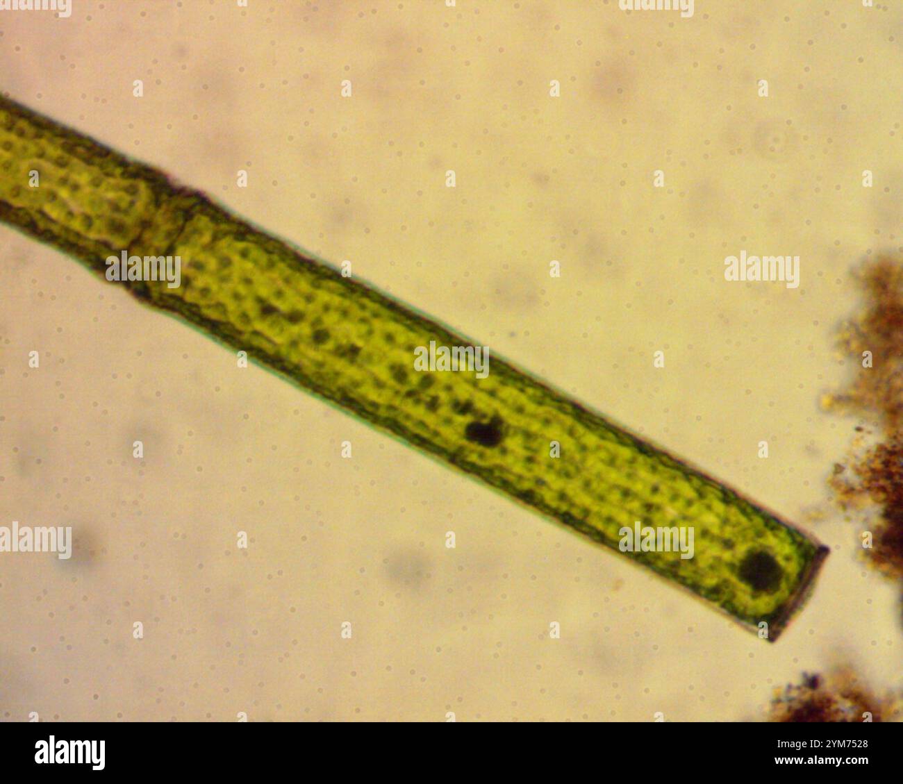 (Pleurotaenium trabecula) Stock Photohttps://www.alamy.com/image-license-details/?v=1https://www.alamy.com/pleurotaenium-trabecula-image632024016.html
RM2YM7528–(Pleurotaenium trabecula)
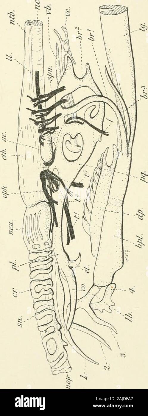 A treatise on zoology . rebrain; for, foramen ; hb, habenular ganglion ; hp,hypophysial plate ; hy, hyoid region ; g.g, gasseriau ganglion ; gl, glossopharyngeal; lb, labialcartilage ; mb, midbrain ; md, medulla ; na.c, nasal capsule ; n.c, nerve-cord ; nt, notochord; ol,olfactory lobe; ophtli, ophthalmic l)ranch of trigeminal nerve; pi, palatine, and pq, quadrate regions ; sob, suborbital nerve; sp, spinal nerve ; (i-S, branclies of trigeminal ; tr,trabecula cranii; ts, preorbital sensory bitinch ; v, vagus nerve ; v.r, ventral root of spinalnerve ; 1, 2, 3, 4, cartilages of the tentacles. Fo Stock Photohttps://www.alamy.com/image-license-details/?v=1https://www.alamy.com/a-treatise-on-zoology-rebrain-for-foramen-hb-habenular-ganglion-hphypophysial-plate-hy-hyoid-region-gg-gasseriau-ganglion-gl-glossopharyngeal-lb-labialcartilage-mb-midbrain-md-medulla-nac-nasal-capsule-nc-nerve-cord-nt-notochord-ololfactory-lobe-ophtli-ophthalmic-lranch-of-trigeminal-nerve-pi-palatine-and-pq-quadrate-regions-sob-suborbital-nerve-sp-spinal-nerve-i-s-branclies-of-trigeminal-trtrabecula-cranii-ts-preorbital-sensory-bitinch-v-vagus-nerve-vr-ventral-root-of-spinalnerve-1-2-3-4-cartilages-of-the-tentacles-fo-image338358223.html
RM2AJDFA7–A treatise on zoology . rebrain; for, foramen ; hb, habenular ganglion ; hp,hypophysial plate ; hy, hyoid region ; g.g, gasseriau ganglion ; gl, glossopharyngeal; lb, labialcartilage ; mb, midbrain ; md, medulla ; na.c, nasal capsule ; n.c, nerve-cord ; nt, notochord; ol,olfactory lobe; ophtli, ophthalmic l)ranch of trigeminal nerve; pi, palatine, and pq, quadrate regions ; sob, suborbital nerve; sp, spinal nerve ; (i-S, branclies of trigeminal ; tr,trabecula cranii; ts, preorbital sensory bitinch ; v, vagus nerve ; v.r, ventral root of spinalnerve ; 1, 2, 3, 4, cartilages of the tentacles. Fo
 (Pleurotaenium trabecula) Stock Photohttps://www.alamy.com/image-license-details/?v=1https://www.alamy.com/pleurotaenium-trabecula-image632009616.html
RM2YM6EM0–(Pleurotaenium trabecula)
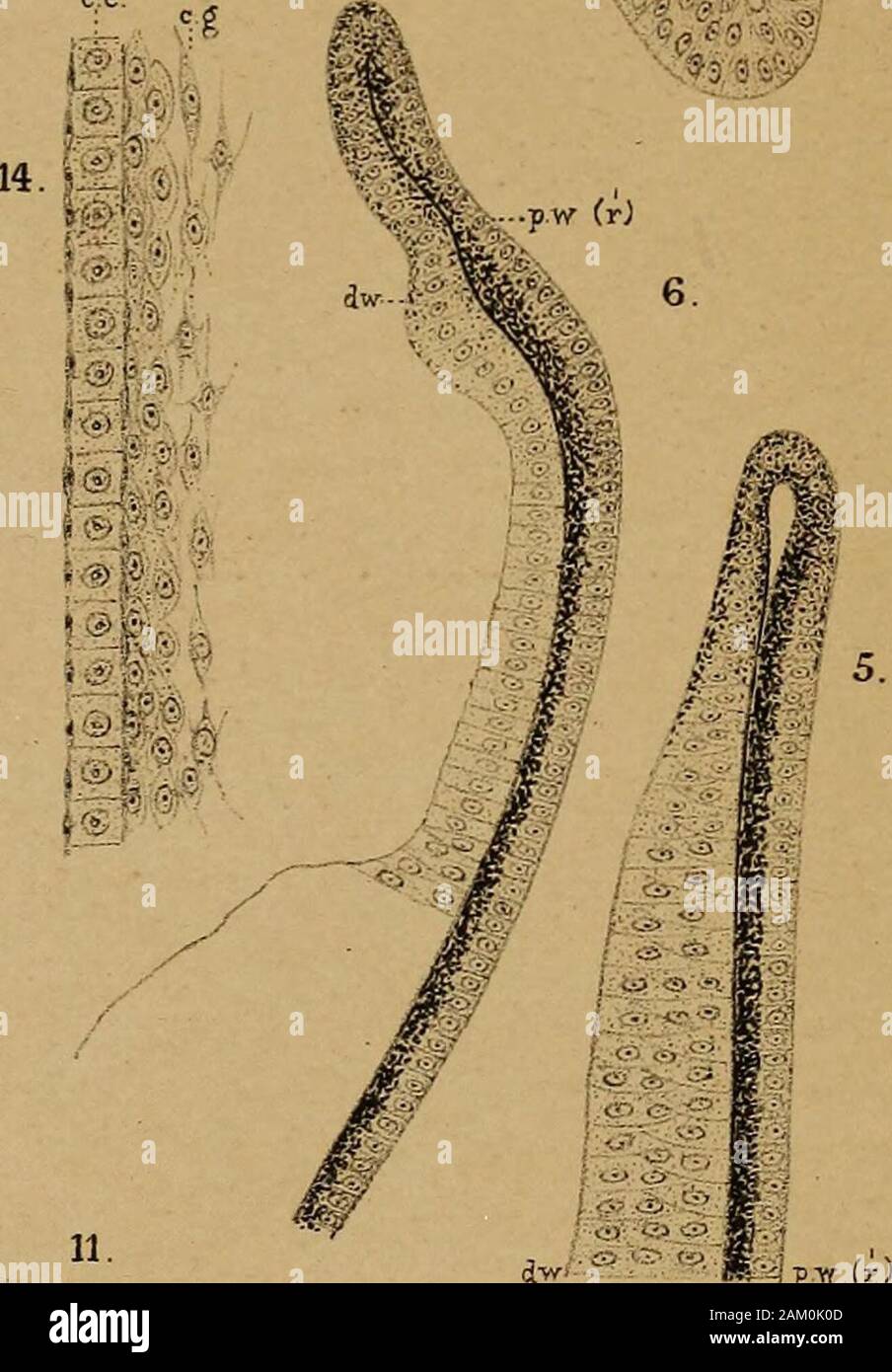 DrH.GBronn's Klassen und Ordnungen des Thier-Reichs : wissenchaftlich dargestellt in Wort und Bild . 13. >^> ^x m! 12. 15, ..-<<? ..-^ DiC.O.ael. Steendr V P.WM.Trap- A.J W. hth. Erklärung- von Tafel CLXVl. Gültige Bezeiclinung für alle Figuren. ft Articulare. all Ampulla horizontalis. h in Fig. 2, siehe S. 2012. hh Trabecula. c Cornea, ce Cornea-Epithel. eil Canalis horizontalis. cl Cochlea. cp Conjonctiva palpehrarum. CS Canalis sagittalis. et Cutis. de Ductus endolymphaticus, (j.a.f Ganglion acustico-facialis. g.h Gehörbläschen. fj.c Ganglion cochleare. g.g.f Ganglion geniculi Stock Photohttps://www.alamy.com/image-license-details/?v=1https://www.alamy.com/drhgbronns-klassen-und-ordnungen-des-thier-reichs-wissenchaftlich-dargestellt-in-wort-und-bild-13-gtgt-x-m!-12-15-ltlt-dicoael-steendr-v-pwmtrap-aj-w-hth-erklrung-von-tafel-clxvl-gltige-bezeiclinung-fr-alle-figuren-ft-articulare-all-ampulla-horizontalis-h-in-fig-2-siehe-s-2012-hh-trabecula-c-cornea-ce-cornea-epithel-eil-canalis-horizontalis-cl-cochlea-cp-conjonctiva-palpehrarum-cs-canalis-sagittalis-et-cutis-de-ductus-endolymphaticus-jaf-ganglion-acustico-facialis-gh-gehrblschen-fjc-ganglion-cochleare-ggf-ganglion-geniculi-image339305021.html
RM2AM0K0D–DrH.GBronn's Klassen und Ordnungen des Thier-Reichs : wissenchaftlich dargestellt in Wort und Bild . 13. >^> ^x m! 12. 15, ..-<<? ..-^ DiC.O.ael. Steendr V P.WM.Trap- A.J W. hth. Erklärung- von Tafel CLXVl. Gültige Bezeiclinung für alle Figuren. ft Articulare. all Ampulla horizontalis. h in Fig. 2, siehe S. 2012. hh Trabecula. c Cornea, ce Cornea-Epithel. eil Canalis horizontalis. cl Cochlea. cp Conjonctiva palpehrarum. CS Canalis sagittalis. et Cutis. de Ductus endolymphaticus, (j.a.f Ganglion acustico-facialis. g.h Gehörbläschen. fj.c Ganglion cochleare. g.g.f Ganglion geniculi
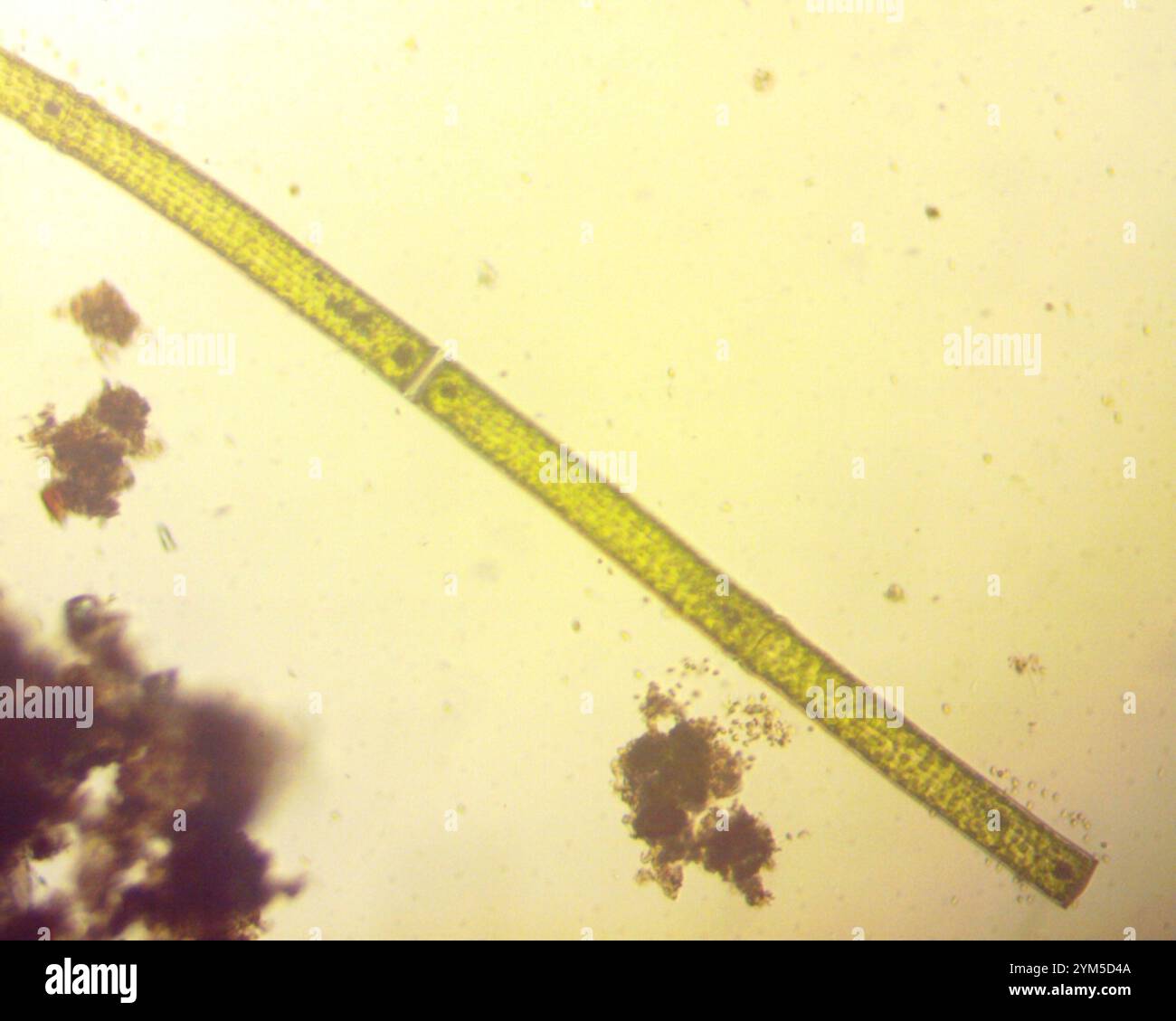 (Pleurotaenium trabecula) Stock Photohttps://www.alamy.com/image-license-details/?v=1https://www.alamy.com/pleurotaenium-trabecula-image631986442.html
RM2YM5D4A–(Pleurotaenium trabecula)
 . Transactions of the Southern Surgical and Gynecological Association. Fig. 3.—Growth of tumor along the cord. Low power: a, fibrous trabecula bearingthe essential cells; dense fibrous tissue about the tumor.. Fig. 4.—Growth of tumor alorif>; the cord. High power: a, a, delicate fibrous trabecular; b, tumorcells in columns on the trabecular; c, small bloodvessel in trabecula. Stock Photohttps://www.alamy.com/image-license-details/?v=1https://www.alamy.com/transactions-of-the-southern-surgical-and-gynecological-association-fig-3growth-of-tumor-along-the-cord-low-power-a-fibrous-trabecula-bearingthe-essential-cells-dense-fibrous-tissue-about-the-tumor-fig-4growth-of-tumor-alorifgt-the-cord-high-power-a-a-delicate-fibrous-trabecular-b-tumorcells-in-columns-on-the-trabecular-c-small-bloodvessel-in-trabecula-image370523163.html
RM2CEPP23–. Transactions of the Southern Surgical and Gynecological Association. Fig. 3.—Growth of tumor along the cord. Low power: a, fibrous trabecula bearingthe essential cells; dense fibrous tissue about the tumor.. Fig. 4.—Growth of tumor alorif>; the cord. High power: a, a, delicate fibrous trabecular; b, tumorcells in columns on the trabecular; c, small bloodvessel in trabecula.
 (Pleurotaenium trabecula) Stock Photohttps://www.alamy.com/image-license-details/?v=1https://www.alamy.com/pleurotaenium-trabecula-image632020755.html
RM2YM70WR–(Pleurotaenium trabecula)
 . Text-book of normal histology: including an account of the development of the tissues and of the organs. Section of human spleen, showingtrabeculse (a) and fibrous reticulum (b)continued into the surrounding splenicpulp ; c, lymphoid cells Transverse section of largetrabecula of human spleen : a,fibrous tissue, containing a fewgroups of plane muscle-cells {b);c, extension of trabecula intofibrous reticulum; d, lymph-corpuscles. 124 NORMAL HISTOLOGY. Stock Photohttps://www.alamy.com/image-license-details/?v=1https://www.alamy.com/text-book-of-normal-histology-including-an-account-of-the-development-of-the-tissues-and-of-the-organs-section-of-human-spleen-showingtrabeculse-a-and-fibrous-reticulum-bcontinued-into-the-surrounding-splenicpulp-c-lymphoid-cells-transverse-section-of-largetrabecula-of-human-spleen-afibrous-tissue-containing-a-fewgroups-of-plane-muscle-cells-bc-extension-of-trabecula-intofibrous-reticulum-d-lymph-corpuscles-124-normal-histology-image370392321.html
RM2CEGR55–. Text-book of normal histology: including an account of the development of the tissues and of the organs. Section of human spleen, showingtrabeculse (a) and fibrous reticulum (b)continued into the surrounding splenicpulp ; c, lymphoid cells Transverse section of largetrabecula of human spleen : a,fibrous tissue, containing a fewgroups of plane muscle-cells {b);c, extension of trabecula intofibrous reticulum; d, lymph-corpuscles. 124 NORMAL HISTOLOGY.
 . Transactions of the Southern Surgical and Gynecological Association. Fig. 4.—Growth of tumor alorif>; the cord. High power: a, a, delicate fibrous trabecular; b, tumorcells in columns on the trabecular; c, small bloodvessel in trabecula.. Fig. 5.—Section from the abdominal mass. Low power: a, a, blood spaces in themidst of the cells. Fig. 6 is from this area. Stock Photohttps://www.alamy.com/image-license-details/?v=1https://www.alamy.com/transactions-of-the-southern-surgical-and-gynecological-association-fig-4growth-of-tumor-alorifgt-the-cord-high-power-a-a-delicate-fibrous-trabecular-b-tumorcells-in-columns-on-the-trabecular-c-small-bloodvessel-in-trabecula-fig-5section-from-the-abdominal-mass-low-power-a-a-blood-spaces-in-themidst-of-the-cells-fig-6-is-from-this-area-image370522894.html
RM2CEPNME–. Transactions of the Southern Surgical and Gynecological Association. Fig. 4.—Growth of tumor alorif>; the cord. High power: a, a, delicate fibrous trabecular; b, tumorcells in columns on the trabecular; c, small bloodvessel in trabecula.. Fig. 5.—Section from the abdominal mass. Low power: a, a, blood spaces in themidst of the cells. Fig. 6 is from this area.
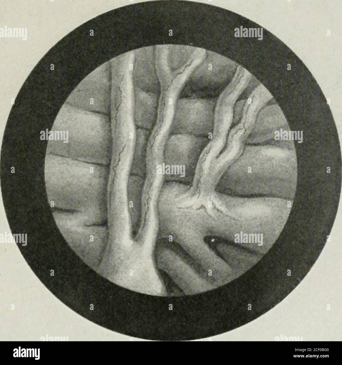 . Annals of surgery. apart from any of thegross forms of urethral obstruction with which we arefamiliar. There is a difference between this form of trabecula-tion and that which is observed in obstructive diseases. Thetrabeculation of an obstructed bladder is coarse, the muscularridges thick and irregularly branching, and the interspacesdeeply pouched; the openings of the saccules are often narrow(Fig. i). In the trabeculated bladder without gross obstruc-tion, the muscle ridges are fine and evenly set and the branch-ings regular and orderly (Fig. 2). Very- fine twigs can fre-quently be seen b Stock Photohttps://www.alamy.com/image-license-details/?v=1https://www.alamy.com/annals-of-surgery-apart-from-any-of-thegross-forms-of-urethral-obstruction-with-which-we-arefamiliar-there-is-a-difference-between-this-form-of-trabecula-tion-and-that-which-is-observed-in-obstructive-diseases-thetrabeculation-of-an-obstructed-bladder-is-coarse-the-muscularridges-thick-and-irregularly-branching-and-the-interspacesdeeply-pouched-the-openings-of-the-saccules-are-often-narrowfig-i-in-the-trabeculated-bladder-without-gross-obstruc-tion-the-muscle-ridges-are-fine-and-evenly-set-and-the-branch-ings-regular-and-orderly-fig-2-very-fine-twigs-can-fre-quently-be-seen-b-image370646640.html
RM2CF0BG0–. Annals of surgery. apart from any of thegross forms of urethral obstruction with which we arefamiliar. There is a difference between this form of trabecula-tion and that which is observed in obstructive diseases. Thetrabeculation of an obstructed bladder is coarse, the muscularridges thick and irregularly branching, and the interspacesdeeply pouched; the openings of the saccules are often narrow(Fig. i). In the trabeculated bladder without gross obstruc-tion, the muscle ridges are fine and evenly set and the branch-ings regular and orderly (Fig. 2). Very- fine twigs can fre-quently be seen b
 . Some points in the surgery of the brain and its membranes . OF THE CEREBRAL MEMBRANES 21 subdural hasmorrhage I have not previouslypublished :— C. K., female, aged twenty-six years. AdmittedDecember 21, 1904, into the National Hospital underDr. Ferrier. History (obtained from husband).—No neurosesin family. Married eighteen months. Now five. Fig. 10.—Dissection of head of turtle, with brain stem exposed. Note the trabecula of areolar tissue crossing the wide subdural space to preventdisplacement against the surrounding rigid brain case. The turtle heads were kindly supplied by Messrs. Buszar Stock Photohttps://www.alamy.com/image-license-details/?v=1https://www.alamy.com/some-points-in-the-surgery-of-the-brain-and-its-membranes-of-the-cerebral-membranes-21-subdural-hasmorrhage-i-have-not-previouslypublished-c-k-female-aged-twenty-six-years-admitteddecember-21-1904-into-the-national-hospital-underdr-ferrier-history-obtained-from-husbandno-neurosesin-family-married-eighteen-months-now-five-fig-10dissection-of-head-of-turtle-with-brain-stem-exposed-note-the-trabecula-of-areolar-tissue-crossing-the-wide-subdural-space-to-preventdisplacement-against-the-surrounding-rigid-brain-case-the-turtle-heads-were-kindly-supplied-by-messrs-buszar-image370039641.html
RM2CE0N9D–. Some points in the surgery of the brain and its membranes . OF THE CEREBRAL MEMBRANES 21 subdural hasmorrhage I have not previouslypublished :— C. K., female, aged twenty-six years. AdmittedDecember 21, 1904, into the National Hospital underDr. Ferrier. History (obtained from husband).—No neurosesin family. Married eighteen months. Now five. Fig. 10.—Dissection of head of turtle, with brain stem exposed. Note the trabecula of areolar tissue crossing the wide subdural space to preventdisplacement against the surrounding rigid brain case. The turtle heads were kindly supplied by Messrs. Buszar
![. On the anatomy of vertebrates [electronic resource] . Trabecula from tlie spleen of a Pig;magti. 350 diam. ccvm. plenic or Malpighian corpuscles, on brandiesof an arteriole: from the spleen of the Pig,magu. 10 diam. ccvm. 433 the most delicate plates of the trabecular tissue, especially inquadrupeds. The splenic corpuscles, fig. 432, c, c, are whitishspherical bodies imbedded in the (lienine ; most constant andconspicuous in ruminant, equine, and some other quadrupeds ;less conspicuous, or wanting, in adult human spleens, especiallyafter lethal disease. They are elliptical, averagingone-sixt Stock Photo . On the anatomy of vertebrates [electronic resource] . Trabecula from tlie spleen of a Pig;magti. 350 diam. ccvm. plenic or Malpighian corpuscles, on brandiesof an arteriole: from the spleen of the Pig,magu. 10 diam. ccvm. 433 the most delicate plates of the trabecular tissue, especially inquadrupeds. The splenic corpuscles, fig. 432, c, c, are whitishspherical bodies imbedded in the (lienine ; most constant andconspicuous in ruminant, equine, and some other quadrupeds ;less conspicuous, or wanting, in adult human spleens, especiallyafter lethal disease. They are elliptical, averagingone-sixt Stock Photo](https://c8.alamy.com/comp/2CP6MAF/on-the-anatomy-of-vertebrates-electronic-resource-trabecula-from-tlie-spleen-of-a-pigmagti-350-diam-ccvm-plenic-or-malpighian-corpuscles-on-brandiesof-an-arteriole-from-the-spleen-of-the-pigmagu-10-diam-ccvm-433-the-most-delicate-plates-of-the-trabecular-tissue-especially-inquadrupeds-the-splenic-corpuscles-fig-432-c-c-are-whitishspherical-bodies-imbedded-in-the-lienine-most-constant-andconspicuous-in-ruminant-equine-and-some-other-quadrupeds-less-conspicuous-or-wanting-in-adult-human-spleens-especiallyafter-lethal-disease-they-are-elliptical-averagingone-sixt-2CP6MAF.jpg) . On the anatomy of vertebrates [electronic resource] . Trabecula from tlie spleen of a Pig;magti. 350 diam. ccvm. plenic or Malpighian corpuscles, on brandiesof an arteriole: from the spleen of the Pig,magu. 10 diam. ccvm. 433 the most delicate plates of the trabecular tissue, especially inquadrupeds. The splenic corpuscles, fig. 432, c, c, are whitishspherical bodies imbedded in the (lienine ; most constant andconspicuous in ruminant, equine, and some other quadrupeds ;less conspicuous, or wanting, in adult human spleens, especiallyafter lethal disease. They are elliptical, averagingone-sixt Stock Photohttps://www.alamy.com/image-license-details/?v=1https://www.alamy.com/on-the-anatomy-of-vertebrates-electronic-resource-trabecula-from-tlie-spleen-of-a-pigmagti-350-diam-ccvm-plenic-or-malpighian-corpuscles-on-brandiesof-an-arteriole-from-the-spleen-of-the-pigmagu-10-diam-ccvm-433-the-most-delicate-plates-of-the-trabecular-tissue-especially-inquadrupeds-the-splenic-corpuscles-fig-432-c-c-are-whitishspherical-bodies-imbedded-in-the-lienine-most-constant-andconspicuous-in-ruminant-equine-and-some-other-quadrupeds-less-conspicuous-or-wanting-in-adult-human-spleens-especiallyafter-lethal-disease-they-are-elliptical-averagingone-sixt-image375087847.html
. On the anatomy of vertebrates [electronic resource] . Trabecula from tlie spleen of a Pig;magti. 350 diam. ccvm. plenic or Malpighian corpuscles, on brandiesof an arteriole: from the spleen of the Pig,magu. 10 diam. ccvm. 433 the most delicate plates of the trabecular tissue, especially inquadrupeds. The splenic corpuscles, fig. 432, c, c, are whitishspherical bodies imbedded in the (lienine ; most constant andconspicuous in ruminant, equine, and some other quadrupeds ;less conspicuous, or wanting, in adult human spleens, especiallyafter lethal disease. They are elliptical, averagingone-sixt Stock Photohttps://www.alamy.com/image-license-details/?v=1https://www.alamy.com/on-the-anatomy-of-vertebrates-electronic-resource-trabecula-from-tlie-spleen-of-a-pigmagti-350-diam-ccvm-plenic-or-malpighian-corpuscles-on-brandiesof-an-arteriole-from-the-spleen-of-the-pigmagu-10-diam-ccvm-433-the-most-delicate-plates-of-the-trabecular-tissue-especially-inquadrupeds-the-splenic-corpuscles-fig-432-c-c-are-whitishspherical-bodies-imbedded-in-the-lienine-most-constant-andconspicuous-in-ruminant-equine-and-some-other-quadrupeds-less-conspicuous-or-wanting-in-adult-human-spleens-especiallyafter-lethal-disease-they-are-elliptical-averagingone-sixt-image375087847.htmlRM2CP6MAF–. On the anatomy of vertebrates [electronic resource] . Trabecula from tlie spleen of a Pig;magti. 350 diam. ccvm. plenic or Malpighian corpuscles, on brandiesof an arteriole: from the spleen of the Pig,magu. 10 diam. ccvm. 433 the most delicate plates of the trabecular tissue, especially inquadrupeds. The splenic corpuscles, fig. 432, c, c, are whitishspherical bodies imbedded in the (lienine ; most constant andconspicuous in ruminant, equine, and some other quadrupeds ;less conspicuous, or wanting, in adult human spleens, especiallyafter lethal disease. They are elliptical, averagingone-sixt
 . Diseases of bones and joints . tainly presentin the active period. The Marrozv. Of the changes one meets in themarrow it is hard to say which are characteristic.Perhaps a fatty change is met most frequently. Thenormal marrow may be almost replaced by newMarrow fibrous tissues in whose meshes the marrow cellsChanges may be distinguished. Again, the marrow is packedwith cells; in spots it appears practically normal.The marrow spaces are less capacious than normal,on account of the production of new bone. Islandsof cartilage in the marrow probably represent theearly stages of new bone trabecula Stock Photohttps://www.alamy.com/image-license-details/?v=1https://www.alamy.com/diseases-of-bones-and-joints-tainly-presentin-the-active-period-the-marrozv-of-the-changes-one-meets-in-themarrow-it-is-hard-to-say-which-are-characteristicperhaps-a-fatty-change-is-met-most-frequently-thenormal-marrow-may-be-almost-replaced-by-newmarrow-fibrous-tissues-in-whose-meshes-the-marrow-cellschanges-may-be-distinguished-again-the-marrow-is-packedwith-cells-in-spots-it-appears-practically-normalthe-marrow-spaces-are-less-capacious-than-normalon-account-of-the-production-of-new-bone-islandsof-cartilage-in-the-marrow-probably-represent-theearly-stages-of-new-bone-trabecula-image376018232.html
RM2CRN32G–. Diseases of bones and joints . tainly presentin the active period. The Marrozv. Of the changes one meets in themarrow it is hard to say which are characteristic.Perhaps a fatty change is met most frequently. Thenormal marrow may be almost replaced by newMarrow fibrous tissues in whose meshes the marrow cellsChanges may be distinguished. Again, the marrow is packedwith cells; in spots it appears practically normal.The marrow spaces are less capacious than normal,on account of the production of new bone. Islandsof cartilage in the marrow probably represent theearly stages of new bone trabecula
 . Mosses with hand-lens and microscope : a non-technical hand-book of the more common mosses of the northeastern United States. is sectionshows clearly the method of formation of the double peristome. The teeth of the outer peristome are formedby thickenings, or plates, laid down on the inner and outer faces of the outer wall of a layer of cells extend-ing around the capsule; these thickenings are continued along the top and bottom walls of these cells to formthe transverse bars or trabecula;. The inner wall of this same layer of cells becomes thickened to form theinner peristome. 3 shows one- Stock Photohttps://www.alamy.com/image-license-details/?v=1https://www.alamy.com/mosses-with-hand-lens-and-microscope-a-non-technical-hand-book-of-the-more-common-mosses-of-the-northeastern-united-states-is-sectionshows-clearly-the-method-of-formation-of-the-double-peristome-the-teeth-of-the-outer-peristome-are-formedby-thickenings-or-plates-laid-down-on-the-inner-and-outer-faces-of-the-outer-wall-of-a-layer-of-cells-extend-ing-around-the-capsule-these-thickenings-are-continued-along-the-top-and-bottom-walls-of-these-cells-to-formthe-transverse-bars-or-trabecula-the-inner-wall-of-this-same-layer-of-cells-becomes-thickened-to-form-theinner-peristome-3-shows-one-image370421207.html
RM2CEJ40R–. Mosses with hand-lens and microscope : a non-technical hand-book of the more common mosses of the northeastern United States. is sectionshows clearly the method of formation of the double peristome. The teeth of the outer peristome are formedby thickenings, or plates, laid down on the inner and outer faces of the outer wall of a layer of cells extend-ing around the capsule; these thickenings are continued along the top and bottom walls of these cells to formthe transverse bars or trabecula;. The inner wall of this same layer of cells becomes thickened to form theinner peristome. 3 shows one-
 . Text-book of normal histology: including an account of the development of the tissues and of the organs. Developing bone—trabecula of endochondral Developing bone—the surface of portion of bone : a, the new bone ; b, bone-cells ; c, still bone-trabecula, exhibiting the conversion of the unabsorbed remains of calcified cartilage-matrix, osteoblasts into the bone-corpuscles: ^, lacuna with young bone-cell ; o, osteoblasts arranged onthe surface of the newly-formed osseous matrix(>«); at / an osteoblast just being isolated. places forming an outer envelope closely embracing the central endo- Stock Photohttps://www.alamy.com/image-license-details/?v=1https://www.alamy.com/text-book-of-normal-histology-including-an-account-of-the-development-of-the-tissues-and-of-the-organs-developing-bonetrabecula-of-endochondral-developing-bonethe-surface-of-portion-of-bone-a-the-new-bone-b-bone-cells-c-still-bone-trabecula-exhibiting-the-conversion-of-the-unabsorbed-remains-of-calcified-cartilage-matrix-osteoblasts-into-the-bone-corpuscles-lacuna-with-young-bone-cell-o-osteoblasts-arranged-onthe-surface-of-the-newly-formed-osseous-matrixgt-at-an-osteoblast-just-being-isolated-places-forming-an-outer-envelope-closely-embracing-the-central-endo-image370400940.html
RM2CEH650–. Text-book of normal histology: including an account of the development of the tissues and of the organs. Developing bone—trabecula of endochondral Developing bone—the surface of portion of bone : a, the new bone ; b, bone-cells ; c, still bone-trabecula, exhibiting the conversion of the unabsorbed remains of calcified cartilage-matrix, osteoblasts into the bone-corpuscles: ^, lacuna with young bone-cell ; o, osteoblasts arranged onthe surface of the newly-formed osseous matrix(>«); at / an osteoblast just being isolated. places forming an outer envelope closely embracing the central endo-
 . Some points in the surgery of the brain and its membranes . with brain stem exposed. Note the trabecula of areolar tissue crossing the wide subdural space to preventdisplacement against the surrounding rigid brain case. The turtle heads were kindly supplied by Messrs. Buszard of Oxford Street. months pregnant. No history of injury. A monthago her husband was leaving home in the morning forhis work when he heard a cry, and on going backfound his wife shrieking and in a demented condition.She was violent, and tossed herself about. Next daycondition much the same, but some weakness of rightarm Stock Photohttps://www.alamy.com/image-license-details/?v=1https://www.alamy.com/some-points-in-the-surgery-of-the-brain-and-its-membranes-with-brain-stem-exposed-note-the-trabecula-of-areolar-tissue-crossing-the-wide-subdural-space-to-preventdisplacement-against-the-surrounding-rigid-brain-case-the-turtle-heads-were-kindly-supplied-by-messrs-buszard-of-oxford-street-months-pregnant-no-history-of-injury-a-monthago-her-husband-was-leaving-home-in-the-morning-forhis-work-when-he-heard-a-cry-and-on-going-backfound-his-wife-shrieking-and-in-a-demented-conditionshe-was-violent-and-tossed-herself-about-next-daycondition-much-the-same-but-some-weakness-of-rightarm-image370039644.html
RM2CE0N9G–. Some points in the surgery of the brain and its membranes . with brain stem exposed. Note the trabecula of areolar tissue crossing the wide subdural space to preventdisplacement against the surrounding rigid brain case. The turtle heads were kindly supplied by Messrs. Buszard of Oxford Street. months pregnant. No history of injury. A monthago her husband was leaving home in the morning forhis work when he heard a cry, and on going backfound his wife shrieking and in a demented condition.She was violent, and tossed herself about. Next daycondition much the same, but some weakness of rightarm
 . Transactions . e are no blood-corpuscles within the lunK>n. vein is enormously thickened, not by endothelial pro-liferation, but by increase in the breadth of the connective-tissue wall (Fig. 48). Farther back collaterals may be seenentering the Iuukmi of the vein from the trabecula) of thenerve, so that tlu^ lunuMi Liradually becomes larger whilethe wall remains very thick. Finally (but this doesnot occur for a considerable distance), the vein regainsits normal size and ceases to have unduly thick walls.Shortly after this it begins to leave the artery, and soon 168 DISEASES OF THE RETINA Stock Photohttps://www.alamy.com/image-license-details/?v=1https://www.alamy.com/transactions-e-are-no-blood-corpuscles-within-the-lunkgtn-vein-is-enormously-thickened-not-by-endothelial-pro-liferation-but-by-increase-in-the-breadth-of-the-connective-tissue-wall-fig-48-farther-back-collaterals-may-be-seenentering-the-iuukmi-of-the-vein-from-the-trabecula-of-thenerve-so-that-tlu-lunumi-liradually-becomes-larger-whilethe-wall-remains-very-thick-finally-but-this-doesnot-occur-for-a-considerable-distance-the-vein-regainsits-normal-size-and-ceases-to-have-unduly-thick-wallsshortly-after-this-it-begins-to-leave-the-artery-and-soon-168-diseases-of-the-retina-image369594427.html
RM2CD8DCY–. Transactions . e are no blood-corpuscles within the lunK>n. vein is enormously thickened, not by endothelial pro-liferation, but by increase in the breadth of the connective-tissue wall (Fig. 48). Farther back collaterals may be seenentering the Iuukmi of the vein from the trabecula) of thenerve, so that tlu^ lunuMi Liradually becomes larger whilethe wall remains very thick. Finally (but this doesnot occur for a considerable distance), the vein regainsits normal size and ceases to have unduly thick walls.Shortly after this it begins to leave the artery, and soon 168 DISEASES OF THE RETINA
 . Transactions . .Soon, however, there reappears a very narrow lumencontainino- red hhjod-corpuscles, while the wall of the Fio. 47.. Vein lunicn partially occluded hy a little mass wliich hasshrunk away from one side. The mass shows some si»^nsof organising- from the surrounding connective tissues.There are no blood-corpuscles within the lunK>n. vein is enormously thickened, not by endothelial pro-liferation, but by increase in the breadth of the connective-tissue wall (Fig. 48). Farther back collaterals may be seenentering the Iuukmi of the vein from the trabecula) of thenerve, so that tl Stock Photohttps://www.alamy.com/image-license-details/?v=1https://www.alamy.com/transactions-soon-however-there-reappears-a-very-narrow-lumencontainino-red-hhjod-corpuscles-while-the-wall-of-the-fio-47-vein-lunicn-partially-occluded-hy-a-little-mass-wliich-hasshrunk-away-from-one-side-the-mass-shows-some-sinsof-organising-from-the-surrounding-connective-tissuesthere-are-no-blood-corpuscles-within-the-lunkgtn-vein-is-enormously-thickened-not-by-endothelial-pro-liferation-but-by-increase-in-the-breadth-of-the-connective-tissue-wall-fig-48-farther-back-collaterals-may-be-seenentering-the-iuukmi-of-the-vein-from-the-trabecula-of-thenerve-so-that-tl-image369594774.html
RM2CD8DWA–. Transactions . .Soon, however, there reappears a very narrow lumencontainino- red hhjod-corpuscles, while the wall of the Fio. 47.. Vein lunicn partially occluded hy a little mass wliich hasshrunk away from one side. The mass shows some si»^nsof organising- from the surrounding connective tissues.There are no blood-corpuscles within the lunK>n. vein is enormously thickened, not by endothelial pro-liferation, but by increase in the breadth of the connective-tissue wall (Fig. 48). Farther back collaterals may be seenentering the Iuukmi of the vein from the trabecula) of thenerve, so that tl
 . Bulletin de la Société botanique de Genève. Fig. 118. moins prononcée et par la forme des pointes non régulièrement arron-dies. Long. 432 a ; lat. 54 p. (Held.) 23. Closterium lineatum Ehr. Long.506 fi; lat. 29p.. (Held.) 24. Closterium striolatum Ehr. Long.297 fjt; lat. 30^. (Z.) 25. Pleurotœnium trabecula(Ehr.)Nàg.Long. 296 p lat. 24,5 a. (K. et Held.)Long. 318 a; lat. 40^. (Hiid.) Long. 414u; lat. 42^. (Z.) 26. Pleurotœnium trabecula formaclavala (Kùtz.) West. Long. 334 a; lat.?46-54//; lat. istb. 38 a. (K.) 27. Euastrum oblongum (Grév.) Rails.Très commun clans les quatre stations.A Kruz Stock Photohttps://www.alamy.com/image-license-details/?v=1https://www.alamy.com/bulletin-de-la-socit-botanique-de-genve-fig-118-moins-prononce-et-par-la-forme-des-pointes-non-rgulirement-arron-dies-long-432-a-lat-54-p-held-23-closterium-lineatum-ehr-long506-fi-lat-29p-held-24-closterium-striolatum-ehr-long297-fjt-lat-30-z-25-pleurotnium-trabeculaehrnglong-296-p-lat-245-a-k-et-heldlong-318-a-lat-40-hiid-long-414u-lat-42-z-26-pleurotnium-trabecula-formaclavala-ktz-west-long-334-a-lat46-54-lat-istb-38-a-k-27-euastrum-oblongum-grv-railstrs-commun-clans-les-quatre-stationsa-kruz-image370569286.html
RM2CETTWA–. Bulletin de la Société botanique de Genève. Fig. 118. moins prononcée et par la forme des pointes non régulièrement arron-dies. Long. 432 a ; lat. 54 p. (Held.) 23. Closterium lineatum Ehr. Long.506 fi; lat. 29p.. (Held.) 24. Closterium striolatum Ehr. Long.297 fjt; lat. 30^. (Z.) 25. Pleurotœnium trabecula(Ehr.)Nàg.Long. 296 p lat. 24,5 a. (K. et Held.)Long. 318 a; lat. 40^. (Hiid.) Long. 414u; lat. 42^. (Z.) 26. Pleurotœnium trabecula formaclavala (Kùtz.) West. Long. 334 a; lat.?46-54//; lat. istb. 38 a. (K.) 27. Euastrum oblongum (Grév.) Rails.Très commun clans les quatre stations.A Kruz
 . Chordate morphology. Morphology (Animals); Chordata. trabecula and orbital cartilage. lateral otic wall occipital arch notochord foramen apical' nasal tectum Meckel's cartilage ^^;^^ 3^eratobranchials MM copula -^cerotohyal quadrate cartilage. Please note that these images are extracted from scanned page images that may have been digitally enhanced for readability - coloration and appearance of these illustrations may not perfectly resemble the original work.. Jollie, Malcolm. New York, Reinhold Stock Photohttps://www.alamy.com/image-license-details/?v=1https://www.alamy.com/chordate-morphology-morphology-animals-chordata-trabecula-and-orbital-cartilage-lateral-otic-wall-occipital-arch-notochord-foramen-apical-nasal-tectum-meckels-cartilage-3eratobranchials-mm-copula-cerotohyal-quadrate-cartilage-please-note-that-these-images-are-extracted-from-scanned-page-images-that-may-have-been-digitally-enhanced-for-readability-coloration-and-appearance-of-these-illustrations-may-not-perfectly-resemble-the-original-work-jollie-malcolm-new-york-reinhold-image234923804.html
RMRJ5KKT–. Chordate morphology. Morphology (Animals); Chordata. trabecula and orbital cartilage. lateral otic wall occipital arch notochord foramen apical' nasal tectum Meckel's cartilage ^^;^^ 3^eratobranchials MM copula -^cerotohyal quadrate cartilage. Please note that these images are extracted from scanned page images that may have been digitally enhanced for readability - coloration and appearance of these illustrations may not perfectly resemble the original work.. Jollie, Malcolm. New York, Reinhold
![. Anatomische Hefte. Studien zur Kenntnis des Wirbeltierkopfes, :;:;:; II. Ast des Trige minus III. Ast des Tri minus M>-*' :iiü '. Kieferblastem Hyalblastem m. ''?'«i'O Trabecula baseos. Please note that these images are extracted from scanned page images that may have been digitally enhanced for readability - coloration and appearance of these illustrations may not perfectly resemble the original work.. München [etc. ] J. F. Bergmann Stock Photo . Anatomische Hefte. Studien zur Kenntnis des Wirbeltierkopfes, :;:;:; II. Ast des Trige minus III. Ast des Tri minus M>-*' :iiü '. Kieferblastem Hyalblastem m. ''?'«i'O Trabecula baseos. Please note that these images are extracted from scanned page images that may have been digitally enhanced for readability - coloration and appearance of these illustrations may not perfectly resemble the original work.. München [etc. ] J. F. Bergmann Stock Photo](https://c8.alamy.com/comp/RN98E1/anatomische-hefte-studien-zur-kenntnis-des-wirbeltierkopfes-ii-ast-des-trige-minus-iii-ast-des-tri-minus-mgt-ii-kieferblastem-hyalblastem-m-io-trabecula-baseos-please-note-that-these-images-are-extracted-from-scanned-page-images-that-may-have-been-digitally-enhanced-for-readability-coloration-and-appearance-of-these-illustrations-may-not-perfectly-resemble-the-original-work-mnchen-etc-j-f-bergmann-RN98E1.jpg) . Anatomische Hefte. Studien zur Kenntnis des Wirbeltierkopfes, :;:;:; II. Ast des Trige minus III. Ast des Tri minus M>-*' :iiü '. Kieferblastem Hyalblastem m. ''?'«i'O Trabecula baseos. Please note that these images are extracted from scanned page images that may have been digitally enhanced for readability - coloration and appearance of these illustrations may not perfectly resemble the original work.. München [etc. ] J. F. Bergmann Stock Photohttps://www.alamy.com/image-license-details/?v=1https://www.alamy.com/anatomische-hefte-studien-zur-kenntnis-des-wirbeltierkopfes-ii-ast-des-trige-minus-iii-ast-des-tri-minus-mgt-ii-kieferblastem-hyalblastem-m-io-trabecula-baseos-please-note-that-these-images-are-extracted-from-scanned-page-images-that-may-have-been-digitally-enhanced-for-readability-coloration-and-appearance-of-these-illustrations-may-not-perfectly-resemble-the-original-work-mnchen-etc-j-f-bergmann-image236846793.html
. Anatomische Hefte. Studien zur Kenntnis des Wirbeltierkopfes, :;:;:; II. Ast des Trige minus III. Ast des Tri minus M>-*' :iiü '. Kieferblastem Hyalblastem m. ''?'«i'O Trabecula baseos. Please note that these images are extracted from scanned page images that may have been digitally enhanced for readability - coloration and appearance of these illustrations may not perfectly resemble the original work.. München [etc. ] J. F. Bergmann Stock Photohttps://www.alamy.com/image-license-details/?v=1https://www.alamy.com/anatomische-hefte-studien-zur-kenntnis-des-wirbeltierkopfes-ii-ast-des-trige-minus-iii-ast-des-tri-minus-mgt-ii-kieferblastem-hyalblastem-m-io-trabecula-baseos-please-note-that-these-images-are-extracted-from-scanned-page-images-that-may-have-been-digitally-enhanced-for-readability-coloration-and-appearance-of-these-illustrations-may-not-perfectly-resemble-the-original-work-mnchen-etc-j-f-bergmann-image236846793.htmlRMRN98E1–. Anatomische Hefte. Studien zur Kenntnis des Wirbeltierkopfes, :;:;:; II. Ast des Trige minus III. Ast des Tri minus M>-*' :iiü '. Kieferblastem Hyalblastem m. ''?'«i'O Trabecula baseos. Please note that these images are extracted from scanned page images that may have been digitally enhanced for readability - coloration and appearance of these illustrations may not perfectly resemble the original work.. München [etc. ] J. F. Bergmann
 . Catalogue of the madreporarian corals in the British Museum (Natural History). Scleractinia. ANALYSIS AND DISTRIBUTION OF TYPES OF CALICLES. 273 hence we ought to find extraordinary variation in this respect. That, however, is not the case. The rule is that the composition of the wall is very uniform. We find, for instance, that while in valleys and depressions where the caUcles are squeezed together the trabeculse of the calicular skeleton may be almost indefinitely aborted, on convexities where the calicles have abundance of room it is seldom that more than one extra trabecula appears. If Stock Photohttps://www.alamy.com/image-license-details/?v=1https://www.alamy.com/catalogue-of-the-madreporarian-corals-in-the-british-museum-natural-history-scleractinia-analysis-and-distribution-of-types-of-calicles-273-hence-we-ought-to-find-extraordinary-variation-in-this-respect-that-however-is-not-the-case-the-rule-is-that-the-composition-of-the-wall-is-very-uniform-we-find-for-instance-that-while-in-valleys-and-depressions-where-the-caucles-are-squeezed-together-the-trabeculse-of-the-calicular-skeleton-may-be-almost-indefinitely-aborted-on-convexities-where-the-calicles-have-abundance-of-room-it-is-seldom-that-more-than-one-extra-trabecula-appears-if-image232967413.html
RMRF0G8N–. Catalogue of the madreporarian corals in the British Museum (Natural History). Scleractinia. ANALYSIS AND DISTRIBUTION OF TYPES OF CALICLES. 273 hence we ought to find extraordinary variation in this respect. That, however, is not the case. The rule is that the composition of the wall is very uniform. We find, for instance, that while in valleys and depressions where the caUcles are squeezed together the trabeculse of the calicular skeleton may be almost indefinitely aborted, on convexities where the calicles have abundance of room it is seldom that more than one extra trabecula appears. If
 . Catalogue of the madreporarian corals in the British Museum (Natural History). Scleractinia. ANALYSIS AND DISTRIBUTION OF TYPES OF CALICLES. 273 hence we ought to find extraordinary variation in this respect. That, however, is not the case. The rule is that the composition of the wall is very uniform. We find, for instance, that while in valleys and depressions where the caUcles are squeezed together the trabeculse of the calicular skeleton may be almost indefinitely aborted, on convexities where the calicles have abundance of room it is seldom that more than one extra trabecula appears. If Stock Photohttps://www.alamy.com/image-license-details/?v=1https://www.alamy.com/catalogue-of-the-madreporarian-corals-in-the-british-museum-natural-history-scleractinia-analysis-and-distribution-of-types-of-calicles-273-hence-we-ought-to-find-extraordinary-variation-in-this-respect-that-however-is-not-the-case-the-rule-is-that-the-composition-of-the-wall-is-very-uniform-we-find-for-instance-that-while-in-valleys-and-depressions-where-the-caucles-are-squeezed-together-the-trabeculse-of-the-calicular-skeleton-may-be-almost-indefinitely-aborted-on-convexities-where-the-calicles-have-abundance-of-room-it-is-seldom-that-more-than-one-extra-trabecula-appears-if-image233101739.html
RMRF6KJ3–. Catalogue of the madreporarian corals in the British Museum (Natural History). Scleractinia. ANALYSIS AND DISTRIBUTION OF TYPES OF CALICLES. 273 hence we ought to find extraordinary variation in this respect. That, however, is not the case. The rule is that the composition of the wall is very uniform. We find, for instance, that while in valleys and depressions where the caUcles are squeezed together the trabeculse of the calicular skeleton may be almost indefinitely aborted, on convexities where the calicles have abundance of room it is seldom that more than one extra trabecula appears. If
 . The anatomy, physiology, morphology and development of the blow-fly (Calliphora erythrocephala) A study in the comparative anatomy and morphology of insects; with plates and illustrations executed directly from the drawings of the author;. Blowflies. 444 THE NERVOUS SYSTEM. lar-^e fasciculus of fibrils, corresponding with the peduncle and trabecula of the insect brain. This terminates in the cortex and penetrates a very remarkable group of small round cells, which I shall term the corpus fungiforme, as I regard it as the homologue of the corpora fungiformia of insects. Lateral sections (Fig. Stock Photohttps://www.alamy.com/image-license-details/?v=1https://www.alamy.com/the-anatomy-physiology-morphology-and-development-of-the-blow-fly-calliphora-erythrocephala-a-study-in-the-comparative-anatomy-and-morphology-of-insects-with-plates-and-illustrations-executed-directly-from-the-drawings-of-the-author-blowflies-444-the-nervous-system-lar-e-fasciculus-of-fibrils-corresponding-with-the-peduncle-and-trabecula-of-the-insect-brain-this-terminates-in-the-cortex-and-penetrates-a-very-remarkable-group-of-small-round-cells-which-i-shall-term-the-corpus-fungiforme-as-i-regard-it-as-the-homologue-of-the-corpora-fungiformia-of-insects-lateral-sections-fig-image236801352.html
RMRN76F4–. The anatomy, physiology, morphology and development of the blow-fly (Calliphora erythrocephala) A study in the comparative anatomy and morphology of insects; with plates and illustrations executed directly from the drawings of the author;. Blowflies. 444 THE NERVOUS SYSTEM. lar-^e fasciculus of fibrils, corresponding with the peduncle and trabecula of the insect brain. This terminates in the cortex and penetrates a very remarkable group of small round cells, which I shall term the corpus fungiforme, as I regard it as the homologue of the corpora fungiformia of insects. Lateral sections (Fig.
 . Comparative embryology of the vertebrates; with 2057 drawings and photos. grouped as 380 illus. Vertebrates -- Embryology; Comparative embryology. 682 THE SKELETAL SYSTEM. TRABECULA CRANII CENTRAL STEM SPHENOLATERAL (ORBITAL) CARTILAGE HYPOPHYSIAL STALK HYPOPHYSIAL CARTILAGE-.. POLAR CARTILAGE INTERNAL CAROTID ARTERY NOTOCHORD PARACHORDAL CARTILAGE AUDITORY CAPSULE. Please note that these images are extracted from scanned page images that may have been digitally enhanced for readability - coloration and appearance of these illustrations may not perfectly resemble the original work.. Nelsen, Stock Photohttps://www.alamy.com/image-license-details/?v=1https://www.alamy.com/comparative-embryology-of-the-vertebrates-with-2057-drawings-and-photos-grouped-as-380-illus-vertebrates-embryology-comparative-embryology-682-the-skeletal-system-trabecula-cranii-central-stem-sphenolateral-orbital-cartilage-hypophysial-stalk-hypophysial-cartilage-polar-cartilage-internal-carotid-artery-notochord-parachordal-cartilage-auditory-capsule-please-note-that-these-images-are-extracted-from-scanned-page-images-that-may-have-been-digitally-enhanced-for-readability-coloration-and-appearance-of-these-illustrations-may-not-perfectly-resemble-the-original-work-nelsen-image232674430.html
RMREF6H2–. Comparative embryology of the vertebrates; with 2057 drawings and photos. grouped as 380 illus. Vertebrates -- Embryology; Comparative embryology. 682 THE SKELETAL SYSTEM. TRABECULA CRANII CENTRAL STEM SPHENOLATERAL (ORBITAL) CARTILAGE HYPOPHYSIAL STALK HYPOPHYSIAL CARTILAGE-.. POLAR CARTILAGE INTERNAL CAROTID ARTERY NOTOCHORD PARACHORDAL CARTILAGE AUDITORY CAPSULE. Please note that these images are extracted from scanned page images that may have been digitally enhanced for readability - coloration and appearance of these illustrations may not perfectly resemble the original work.. Nelsen,
 . Album général des diatomées marines, d'eau douce ou fossiles : album représentant tous les genres de diatomées et leurs principales espèces. Diatoms. ^Mi Pleupostaeniun trabecula Closterium species 3. Desmidiaceœ. Please note that these images are extracted from scanned page images that may have been digitally enhanced for readability - coloration and appearance of these illustrations may not perfectly resemble the original work.. Coupin, Henri, b. 1868. Paris : H. Coupin Stock Photohttps://www.alamy.com/image-license-details/?v=1https://www.alamy.com/album-gnral-des-diatomes-marines-deau-douce-ou-fossiles-album-reprsentant-tous-les-genres-de-diatomes-et-leurs-principales-espces-diatoms-mi-pleupostaeniun-trabecula-closterium-species-3-desmidiace-please-note-that-these-images-are-extracted-from-scanned-page-images-that-may-have-been-digitally-enhanced-for-readability-coloration-and-appearance-of-these-illustrations-may-not-perfectly-resemble-the-original-work-coupin-henri-b-1868-paris-h-coupin-image237819076.html
RMRPWGJC–. Album général des diatomées marines, d'eau douce ou fossiles : album représentant tous les genres de diatomées et leurs principales espèces. Diatoms. ^Mi Pleupostaeniun trabecula Closterium species 3. Desmidiaceœ. Please note that these images are extracted from scanned page images that may have been digitally enhanced for readability - coloration and appearance of these illustrations may not perfectly resemble the original work.. Coupin, Henri, b. 1868. Paris : H. Coupin
![. Anatomische Hefte. ''?'«i'O Trabecula baseos. Hyomandibulartasche I / Fig. 30. Crocodilus biporcatus. Embryo von 5 mm Kopflänge. Schnitt durch die Hyo- mandibulartasche. Gan?l. Trigem. V. cap. lat. Quadratum Paukenhöhle Extracolumella. Please note that these images are extracted from scanned page images that may have been digitally enhanced for readability - coloration and appearance of these illustrations may not perfectly resemble the original work.. München [etc. ] J. F. Bergmann Stock Photo . Anatomische Hefte. ''?'«i'O Trabecula baseos. Hyomandibulartasche I / Fig. 30. Crocodilus biporcatus. Embryo von 5 mm Kopflänge. Schnitt durch die Hyo- mandibulartasche. Gan?l. Trigem. V. cap. lat. Quadratum Paukenhöhle Extracolumella. Please note that these images are extracted from scanned page images that may have been digitally enhanced for readability - coloration and appearance of these illustrations may not perfectly resemble the original work.. München [etc. ] J. F. Bergmann Stock Photo](https://c8.alamy.com/comp/RN98DM/anatomische-hefte-io-trabecula-baseos-hyomandibulartasche-i-fig-30-crocodilus-biporcatus-embryo-von-5-mm-kopflnge-schnitt-durch-die-hyo-mandibulartasche-ganl-trigem-v-cap-lat-quadratum-paukenhhle-extracolumella-please-note-that-these-images-are-extracted-from-scanned-page-images-that-may-have-been-digitally-enhanced-for-readability-coloration-and-appearance-of-these-illustrations-may-not-perfectly-resemble-the-original-work-mnchen-etc-j-f-bergmann-RN98DM.jpg) . Anatomische Hefte. ''?'«i'O Trabecula baseos. Hyomandibulartasche I / Fig. 30. Crocodilus biporcatus. Embryo von 5 mm Kopflänge. Schnitt durch die Hyo- mandibulartasche. Gan?l. Trigem. V. cap. lat. Quadratum Paukenhöhle Extracolumella. Please note that these images are extracted from scanned page images that may have been digitally enhanced for readability - coloration and appearance of these illustrations may not perfectly resemble the original work.. München [etc. ] J. F. Bergmann Stock Photohttps://www.alamy.com/image-license-details/?v=1https://www.alamy.com/anatomische-hefte-io-trabecula-baseos-hyomandibulartasche-i-fig-30-crocodilus-biporcatus-embryo-von-5-mm-kopflnge-schnitt-durch-die-hyo-mandibulartasche-ganl-trigem-v-cap-lat-quadratum-paukenhhle-extracolumella-please-note-that-these-images-are-extracted-from-scanned-page-images-that-may-have-been-digitally-enhanced-for-readability-coloration-and-appearance-of-these-illustrations-may-not-perfectly-resemble-the-original-work-mnchen-etc-j-f-bergmann-image236846784.html
. Anatomische Hefte. ''?'«i'O Trabecula baseos. Hyomandibulartasche I / Fig. 30. Crocodilus biporcatus. Embryo von 5 mm Kopflänge. Schnitt durch die Hyo- mandibulartasche. Gan?l. Trigem. V. cap. lat. Quadratum Paukenhöhle Extracolumella. Please note that these images are extracted from scanned page images that may have been digitally enhanced for readability - coloration and appearance of these illustrations may not perfectly resemble the original work.. München [etc. ] J. F. Bergmann Stock Photohttps://www.alamy.com/image-license-details/?v=1https://www.alamy.com/anatomische-hefte-io-trabecula-baseos-hyomandibulartasche-i-fig-30-crocodilus-biporcatus-embryo-von-5-mm-kopflnge-schnitt-durch-die-hyo-mandibulartasche-ganl-trigem-v-cap-lat-quadratum-paukenhhle-extracolumella-please-note-that-these-images-are-extracted-from-scanned-page-images-that-may-have-been-digitally-enhanced-for-readability-coloration-and-appearance-of-these-illustrations-may-not-perfectly-resemble-the-original-work-mnchen-etc-j-f-bergmann-image236846784.htmlRMRN98DM–. Anatomische Hefte. ''?'«i'O Trabecula baseos. Hyomandibulartasche I / Fig. 30. Crocodilus biporcatus. Embryo von 5 mm Kopflänge. Schnitt durch die Hyo- mandibulartasche. Gan?l. Trigem. V. cap. lat. Quadratum Paukenhöhle Extracolumella. Please note that these images are extracted from scanned page images that may have been digitally enhanced for readability - coloration and appearance of these illustrations may not perfectly resemble the original work.. München [etc. ] J. F. Bergmann
 . The anatomy, physiology, morphology and development of the blow-fly (Calliphora erythrocephala.) A study in the comparative anatomy and morphology of insects; with plates and illustrations executed directly from the drawings of the author;. Blowflies. 444 THE NERVOUS SYSTEM. large fasciculus of fibrils, corresponding with the peduncle and trabecula of the insect brain. This terminates in the cortex and penetrates a very remarkable group of small round cells, which I shall term the corpus fungiforme, as I regard it as the homologue of the corpora fungiformia of insects. Lateral sections (Fig. Stock Photohttps://www.alamy.com/image-license-details/?v=1https://www.alamy.com/the-anatomy-physiology-morphology-and-development-of-the-blow-fly-calliphora-erythrocephala-a-study-in-the-comparative-anatomy-and-morphology-of-insects-with-plates-and-illustrations-executed-directly-from-the-drawings-of-the-author-blowflies-444-the-nervous-system-large-fasciculus-of-fibrils-corresponding-with-the-peduncle-and-trabecula-of-the-insect-brain-this-terminates-in-the-cortex-and-penetrates-a-very-remarkable-group-of-small-round-cells-which-i-shall-term-the-corpus-fungiforme-as-i-regard-it-as-the-homologue-of-the-corpora-fungiformia-of-insects-lateral-sections-fig-image231878235.html
RMRD6Y1F–. The anatomy, physiology, morphology and development of the blow-fly (Calliphora erythrocephala.) A study in the comparative anatomy and morphology of insects; with plates and illustrations executed directly from the drawings of the author;. Blowflies. 444 THE NERVOUS SYSTEM. large fasciculus of fibrils, corresponding with the peduncle and trabecula of the insect brain. This terminates in the cortex and penetrates a very remarkable group of small round cells, which I shall term the corpus fungiforme, as I regard it as the homologue of the corpora fungiformia of insects. Lateral sections (Fig.