Quick filters:
Tracheid Stock Photos and Images
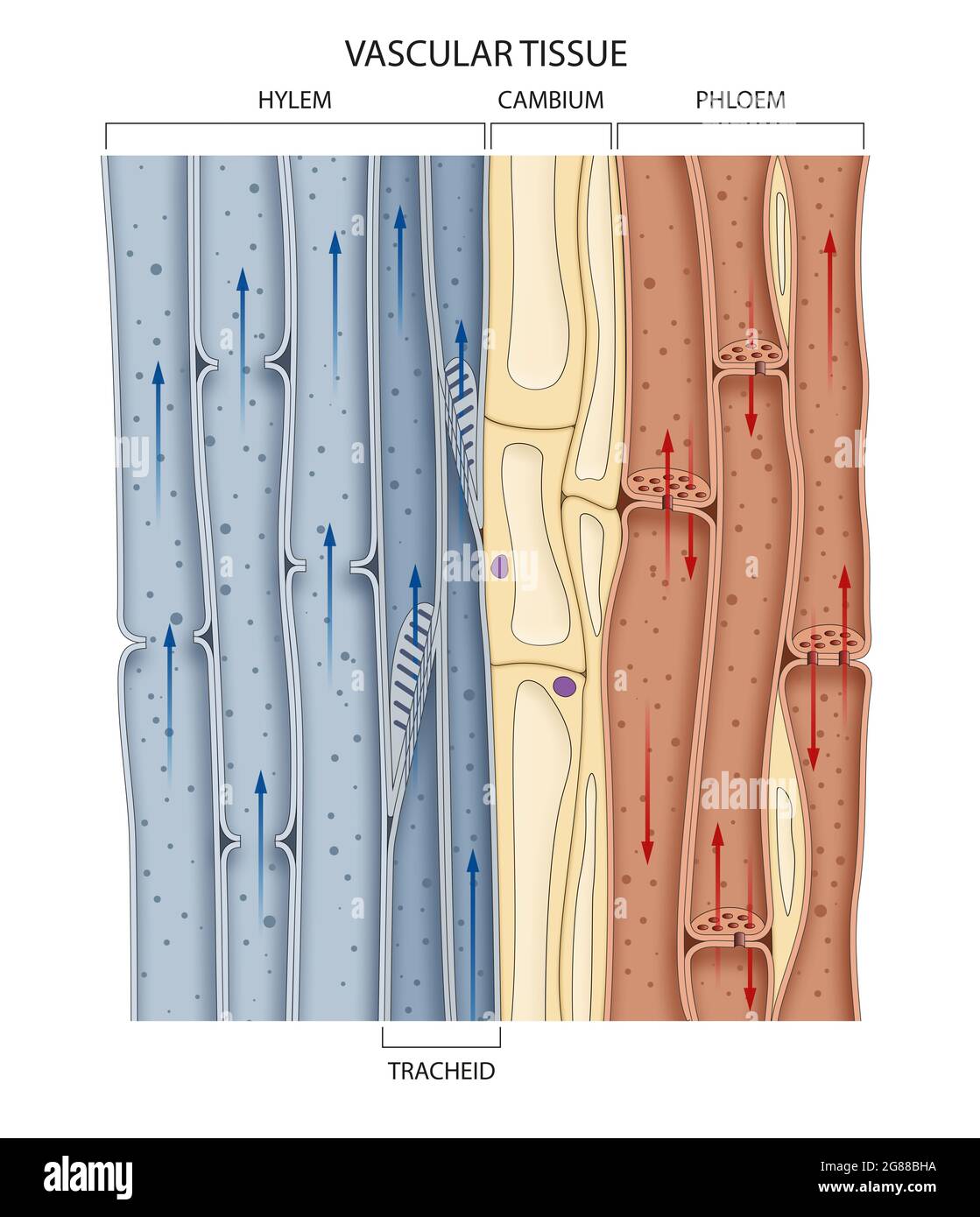 Cross section of a vascular bundle in the stem showing food-conducting phloem and water-conducting xylem Stock Photohttps://www.alamy.com/image-license-details/?v=1https://www.alamy.com/cross-section-of-a-vascular-bundle-in-the-stem-showing-food-conducting-phloem-and-water-conducting-xylem-image435361174.html
RF2G88BHA–Cross section of a vascular bundle in the stem showing food-conducting phloem and water-conducting xylem
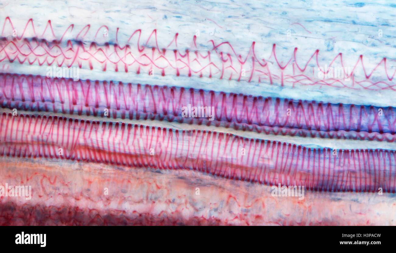 Xylem tissue. Light micrograph (LM) of a section through sunflower(helianthus annuus) tissue showing spiral tracheids, a type of xylem. Tracheids are long tubular cells with lignin, a material that provides support, in the cell walls. Spiral thickening of the cells can be seen. Tracheids conduct water from the roots of a plant along the stems to the leaves. Magnification: x210 when printed 10cm wide. Stock Photohttps://www.alamy.com/image-license-details/?v=1https://www.alamy.com/stock-photo-xylem-tissue-light-micrograph-lm-of-a-section-through-sunflowerhelianthus-122807689.html
RFH3PACW–Xylem tissue. Light micrograph (LM) of a section through sunflower(helianthus annuus) tissue showing spiral tracheids, a type of xylem. Tracheids are long tubular cells with lignin, a material that provides support, in the cell walls. Spiral thickening of the cells can be seen. Tracheids conduct water from the roots of a plant along the stems to the leaves. Magnification: x210 when printed 10cm wide.
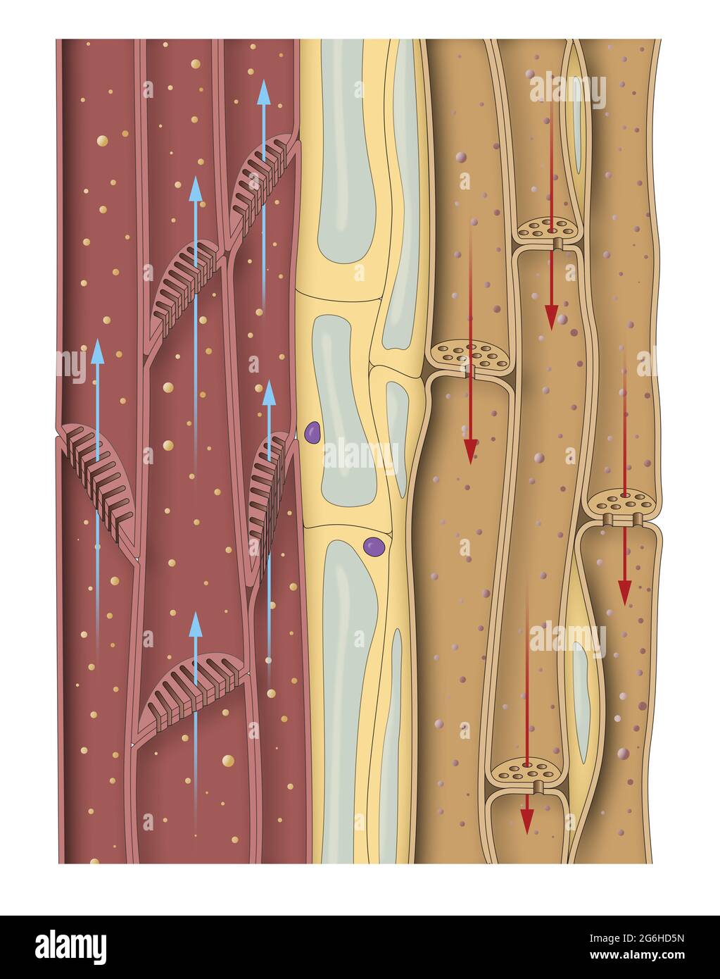 Section of the stem plant. Tracheids, phloem Stock Photohttps://www.alamy.com/image-license-details/?v=1https://www.alamy.com/section-of-the-stem-plant-tracheids-phloem-image434330673.html
RF2G6HD5N–Section of the stem plant. Tracheids, phloem
 Tracheids, a kind of xylem. Optical microscope X100. Stock Photohttps://www.alamy.com/image-license-details/?v=1https://www.alamy.com/tracheids-a-kind-of-xylem-optical-microscope-x100-image575103066.html
RF2TBJ5R6–Tracheids, a kind of xylem. Optical microscope X100.
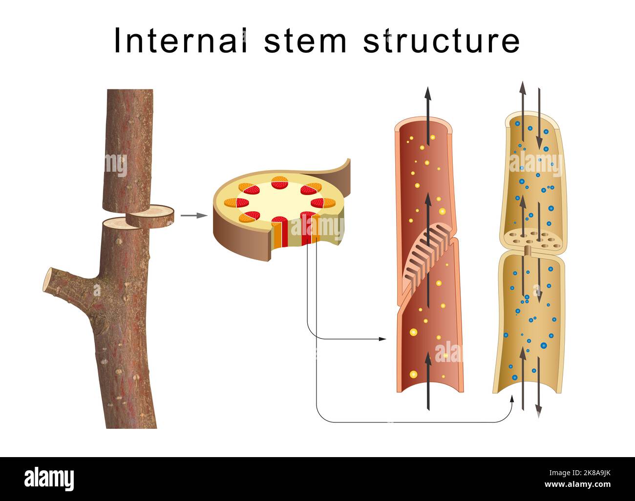 Internal anatomy of the tree stem Stock Photohttps://www.alamy.com/image-license-details/?v=1https://www.alamy.com/internal-anatomy-of-the-tree-stem-image487034651.html
RF2K8A9JK–Internal anatomy of the tree stem
 Still Life - Leaves Stock Photohttps://www.alamy.com/image-license-details/?v=1https://www.alamy.com/still-life-leaves-image180551226.html
RFMDMPX2–Still Life - Leaves
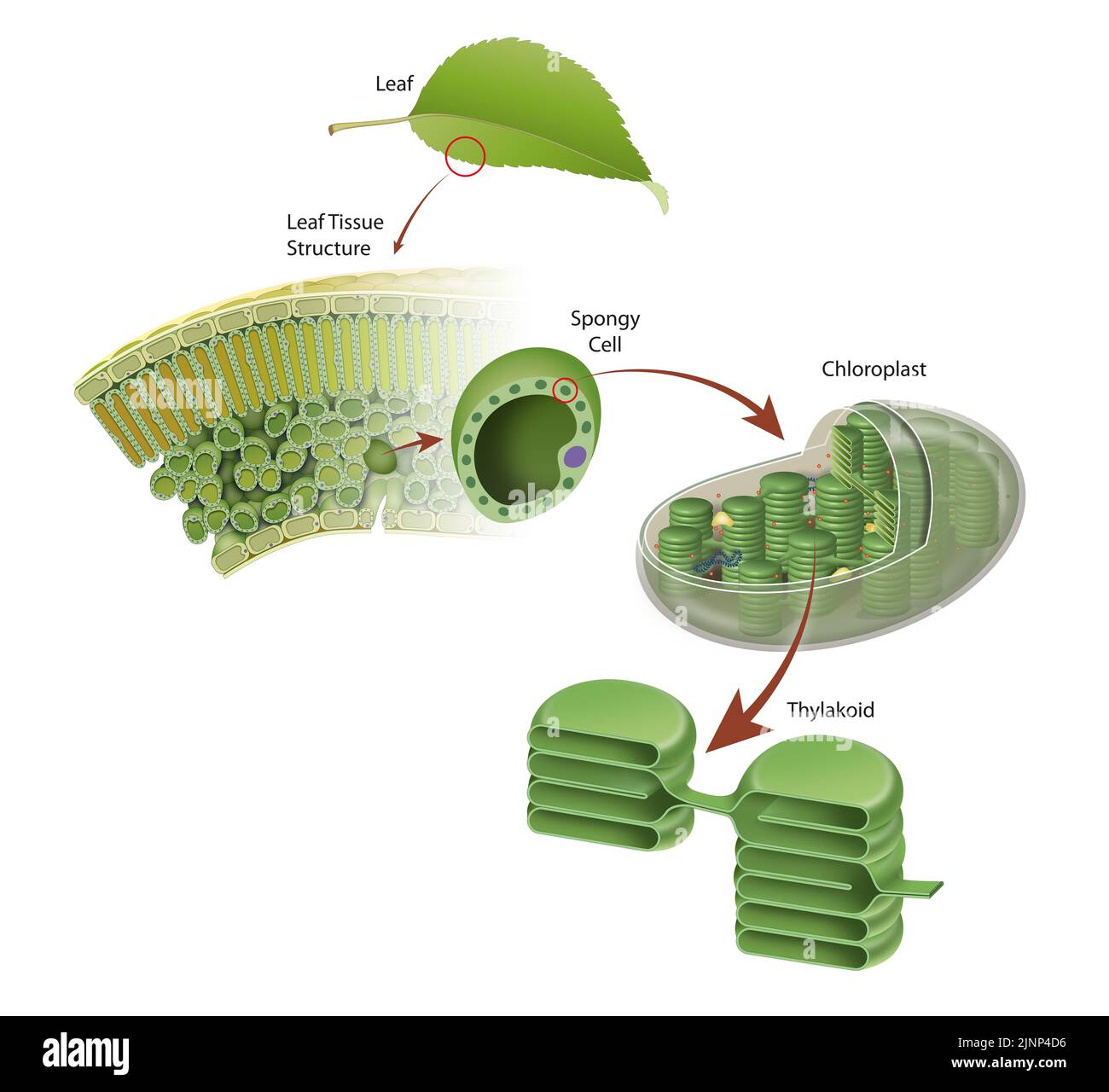 Diagram of a leaf structure Stock Photohttps://www.alamy.com/image-license-details/?v=1https://www.alamy.com/diagram-of-a-leaf-structure-image478074162.html
RF2JNP4D6–Diagram of a leaf structure
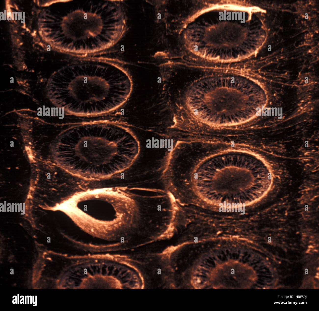 Xylem Stock Photohttps://www.alamy.com/image-license-details/?v=1https://www.alamy.com/stock-photo-xylem-134943134.html
RMHRF59J–Xylem
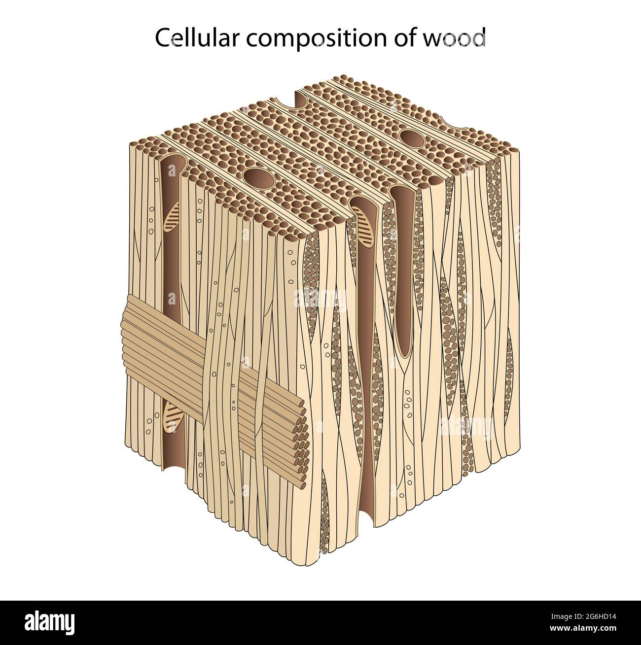 Composition and Structure of Wood Cells Stock Photohttps://www.alamy.com/image-license-details/?v=1https://www.alamy.com/composition-and-structure-of-wood-cells-image434330544.html
RF2G6HD14–Composition and Structure of Wood Cells
 . The structure and development of mosses and ferns (Archegoniatae). asm remains in them. Faint elongatedtransverse pits become evident, and the spaces between theserapidly thicken at the expense of the cell contents until all theprotoplasm is used up. The thickened bars between the pitsgive the characteristic ladder-like appearance to the older IX FILICINE^ LEPTOSPORANGIAT^ 331 tracheid (Fig. 184, B). In cross-section these bars are nearlyrhomboidal, and give the famihar beaded appearance to sectionsof the tracheid wall. Sieve-tubes of very characteristic form are found in thebundles of all t Stock Photohttps://www.alamy.com/image-license-details/?v=1https://www.alamy.com/the-structure-and-development-of-mosses-and-ferns-archegoniatae-asm-remains-in-them-faint-elongatedtransverse-pits-become-evident-and-the-spaces-between-theserapidly-thicken-at-the-expense-of-the-cell-contents-until-all-theprotoplasm-is-used-up-the-thickened-bars-between-the-pitsgive-the-characteristic-ladder-like-appearance-to-the-older-ix-filicine-leptosporangiat-331-tracheid-fig-184-b-in-cross-section-these-bars-are-nearlyrhomboidal-and-give-the-famihar-beaded-appearance-to-sectionsof-the-tracheid-wall-sieve-tubes-of-very-characteristic-form-are-found-in-thebundles-of-all-t-image336609501.html
RM2AFHTRW–. The structure and development of mosses and ferns (Archegoniatae). asm remains in them. Faint elongatedtransverse pits become evident, and the spaces between theserapidly thicken at the expense of the cell contents until all theprotoplasm is used up. The thickened bars between the pitsgive the characteristic ladder-like appearance to the older IX FILICINE^ LEPTOSPORANGIAT^ 331 tracheid (Fig. 184, B). In cross-section these bars are nearlyrhomboidal, and give the famihar beaded appearance to sectionsof the tracheid wall. Sieve-tubes of very characteristic form are found in thebundles of all t
 cross section of a portion of a plant stem. Stock Photohttps://www.alamy.com/image-license-details/?v=1https://www.alamy.com/stock-photo-cross-section-of-a-portion-of-a-plant-stem-111895272.html
RFGE17FM–cross section of a portion of a plant stem.
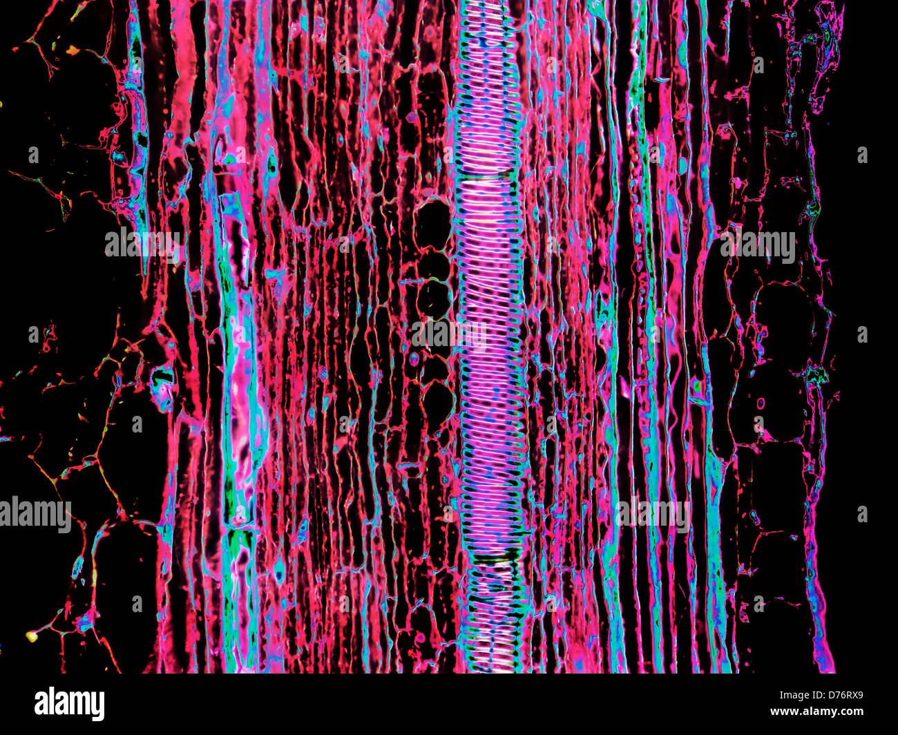 Portion longitudinal stem Cucurbita squash gourds showing spiral tracheids magnification 200x image enhanced Stock Photohttps://www.alamy.com/image-license-details/?v=1https://www.alamy.com/stock-photo-portion-longitudinal-stem-cucurbita-squash-gourds-showing-spiral-tracheids-56084177.html
RMD76RX9–Portion longitudinal stem Cucurbita squash gourds showing spiral tracheids magnification 200x image enhanced
 . The structure and development of mosses and ferns (Archegoniatae). Plant morphology; Mosses; Ferns. FILICINE^ LEPTOSPORANGIATM 331 tracheid (Fig. 184, B). In cross-section these bars are nearly rhomboidal, and give the famihar beaded appearance to sections of the tracheid wall. Sieve-tubes of very characteristic form are found in the bundles of all the Polypodiacese. In 0. strutliiopteris they occupy an irregular area at each end of the bundle. Their differentiation begins shortly after that of the large scalariform tracheids, and in some respects resembles it. The procambium cells from whic Stock Photohttps://www.alamy.com/image-license-details/?v=1https://www.alamy.com/the-structure-and-development-of-mosses-and-ferns-archegoniatae-plant-morphology-mosses-ferns-filicine-leptosporangiatm-331-tracheid-fig-184-b-in-cross-section-these-bars-are-nearly-rhomboidal-and-give-the-famihar-beaded-appearance-to-sections-of-the-tracheid-wall-sieve-tubes-of-very-characteristic-form-are-found-in-the-bundles-of-all-the-polypodiacese-in-0-strutliiopteris-they-occupy-an-irregular-area-at-each-end-of-the-bundle-their-differentiation-begins-shortly-after-that-of-the-large-scalariform-tracheids-and-in-some-respects-resembles-it-the-procambium-cells-from-whic-image216368836.html
RMPG0CJC–. The structure and development of mosses and ferns (Archegoniatae). Plant morphology; Mosses; Ferns. FILICINE^ LEPTOSPORANGIATM 331 tracheid (Fig. 184, B). In cross-section these bars are nearly rhomboidal, and give the famihar beaded appearance to sections of the tracheid wall. Sieve-tubes of very characteristic form are found in the bundles of all the Polypodiacese. In 0. strutliiopteris they occupy an irregular area at each end of the bundle. Their differentiation begins shortly after that of the large scalariform tracheids, and in some respects resembles it. The procambium cells from whic
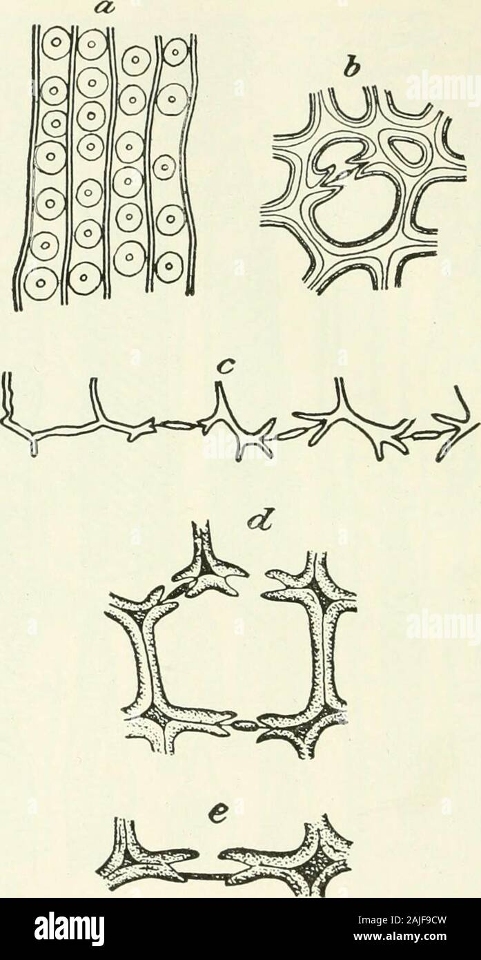
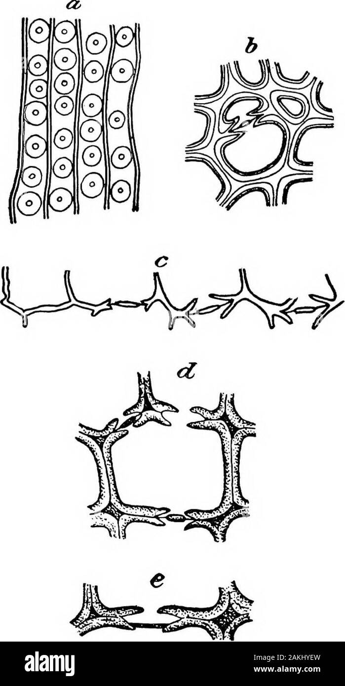
 . Fio. -280. â Tracheid of P(>w«destroyed by Polt/iiorun .lixtoti-'iiiouhf. The celbilose has been for the most part extnicted, and the waU« con- sist of lignin (wood-gum). Cracks occur in the dry secondary wall, while the wall (o, 0) remains intact. The spiral structure of the secondary wall causes cross- ing of the fissures in the walls of adjoining cells at tlie bordered pits, e, and at bore-holes, d, e ; where neither pits nor holes are. present the fissures are simple,/; (After H. Hartig.) Stock Photohttps://www.alamy.com/image-license-details/?v=1https://www.alamy.com/fio-280-tracheid-of-pgtwdestroyed-by-poltiiorun-lixtoti-iiiouhf-the-celbilose-has-been-for-the-most-part-extnicted-and-the-wau-con-sist-of-lignin-wood-gum-cracks-occur-in-the-dry-secondary-wall-while-the-wall-o-0-remains-intact-the-spiral-structure-of-the-secondary-wall-causes-cross-ing-of-the-fissures-in-the-walls-of-adjoining-cells-at-tlie-bordered-pits-e-and-at-bore-holes-d-e-where-neither-pits-nor-holes-are-present-the-fissures-are-simple-after-h-hartig-image179901362.html
RMMCK60J–. Fio. -280. â Tracheid of P(>w«destroyed by Polt/iiorun .lixtoti-'iiiouhf. The celbilose has been for the most part extnicted, and the waU« con- sist of lignin (wood-gum). Cracks occur in the dry secondary wall, while the wall (o, 0) remains intact. The spiral structure of the secondary wall causes cross- ing of the fissures in the walls of adjoining cells at tlie bordered pits, e, and at bore-holes, d, e ; where neither pits nor holes are. present the fissures are simple,/; (After H. Hartig.)
 Diseases of plants induced by Diseases of plants induced by cryptogamic parasites; introduction to the study of pathogenic Fungi, slime-Fungi, bacteria, & Algae diseasesofplant00tube Year: 1897 Fig. 13. Fig. 14. Fio. 13.—Tracheid of Pinus si/lvestris destroyed by Trametes pint. The primary cell-Wall is completely dissolved from below upwards to a, a ; 6, secondary and tertiary layers of the walls consisting in the under portion of cellulose only, in wliich granules of chalk are recoffnizable ; c, fungus-hyphae boring through the walls, leaving holes d and c. (After R. Hartig.) Fig. 14.—Trach Stock Photohttps://www.alamy.com/image-license-details/?v=1https://www.alamy.com/diseases-of-plants-induced-by-diseases-of-plants-induced-by-cryptogamic-parasites-introduction-to-the-study-of-pathogenic-fungi-slime-fungi-bacteria-algae-diseasesofplant00tube-year-1897-fig-13-fig-14-fio-13tracheid-of-pinus-silvestris-destroyed-by-trametes-pint-the-primary-cell-wall-is-completely-dissolved-from-below-upwards-to-a-a-6-secondary-and-tertiary-layers-of-the-walls-consisting-in-the-under-portion-of-cellulose-only-in-wliich-granules-of-chalk-are-recoffnizable-c-fungus-hyphae-boring-through-the-walls-leaving-holes-d-and-c-after-r-hartig-fig-14trach-image241939227.html
RMT1H7XK–Diseases of plants induced by Diseases of plants induced by cryptogamic parasites; introduction to the study of pathogenic Fungi, slime-Fungi, bacteria, & Algae diseasesofplant00tube Year: 1897 Fig. 13. Fig. 14. Fio. 13.—Tracheid of Pinus si/lvestris destroyed by Trametes pint. The primary cell-Wall is completely dissolved from below upwards to a, a ; 6, secondary and tertiary layers of the walls consisting in the under portion of cellulose only, in wliich granules of chalk are recoffnizable ; c, fungus-hyphae boring through the walls, leaving holes d and c. (After R. Hartig.) Fig. 14.—Trach
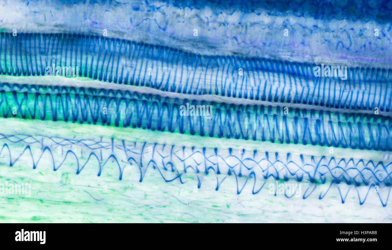 Xylem tissue. Light micrograph (LM) of a section through sunflower(helianthus annuus) tissue showing spiral tracheids, a type of xylem. Tracheids are long tubular cells with lignin, a material that provides support, in the cell walls. Spiral thickening of the cells can be seen. Tracheids conduct water from the roots of a plant along the stems to the leaves. Magnification: x210 when printed 10cm wide. Stock Photohttps://www.alamy.com/image-license-details/?v=1https://www.alamy.com/stock-photo-xylem-tissue-light-micrograph-lm-of-a-section-through-sunflowerhelianthus-122807647.html
RFH3PABB–Xylem tissue. Light micrograph (LM) of a section through sunflower(helianthus annuus) tissue showing spiral tracheids, a type of xylem. Tracheids are long tubular cells with lignin, a material that provides support, in the cell walls. Spiral thickening of the cells can be seen. Tracheids conduct water from the roots of a plant along the stems to the leaves. Magnification: x210 when printed 10cm wide.
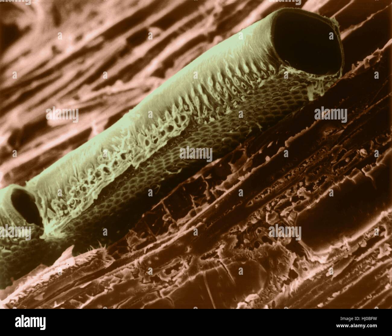 Conductive vessel element in mountain mahogany wood (Cercocarpus sp),coloured scanning electron micrograph (SEM).Note bordered pits in cellulose wall.A conductive vessel element is elongated,water-conducting cell in xylem,one of two kinds of tracheary elements.The cells die when mature,leaving only Stock Photohttps://www.alamy.com/image-license-details/?v=1https://www.alamy.com/stock-photo-conductive-vessel-element-in-mountain-mahogany-wood-cercocarpus-spcoloured-131545453.html
RFHJ0BFW–Conductive vessel element in mountain mahogany wood (Cercocarpus sp),coloured scanning electron micrograph (SEM).Note bordered pits in cellulose wall.A conductive vessel element is elongated,water-conducting cell in xylem,one of two kinds of tracheary elements.The cells die when mature,leaving only
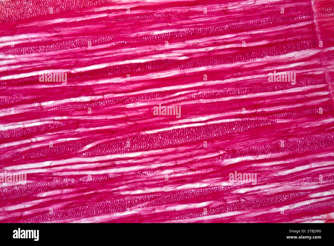 Tracheids, a kind of xylem. Optical microscope X40. Stock Photohttps://www.alamy.com/image-license-details/?v=1https://www.alamy.com/tracheids-a-kind-of-xylem-optical-microscope-x40-image575103060.html
RF2TBJ5R0–Tracheids, a kind of xylem. Optical microscope X40.
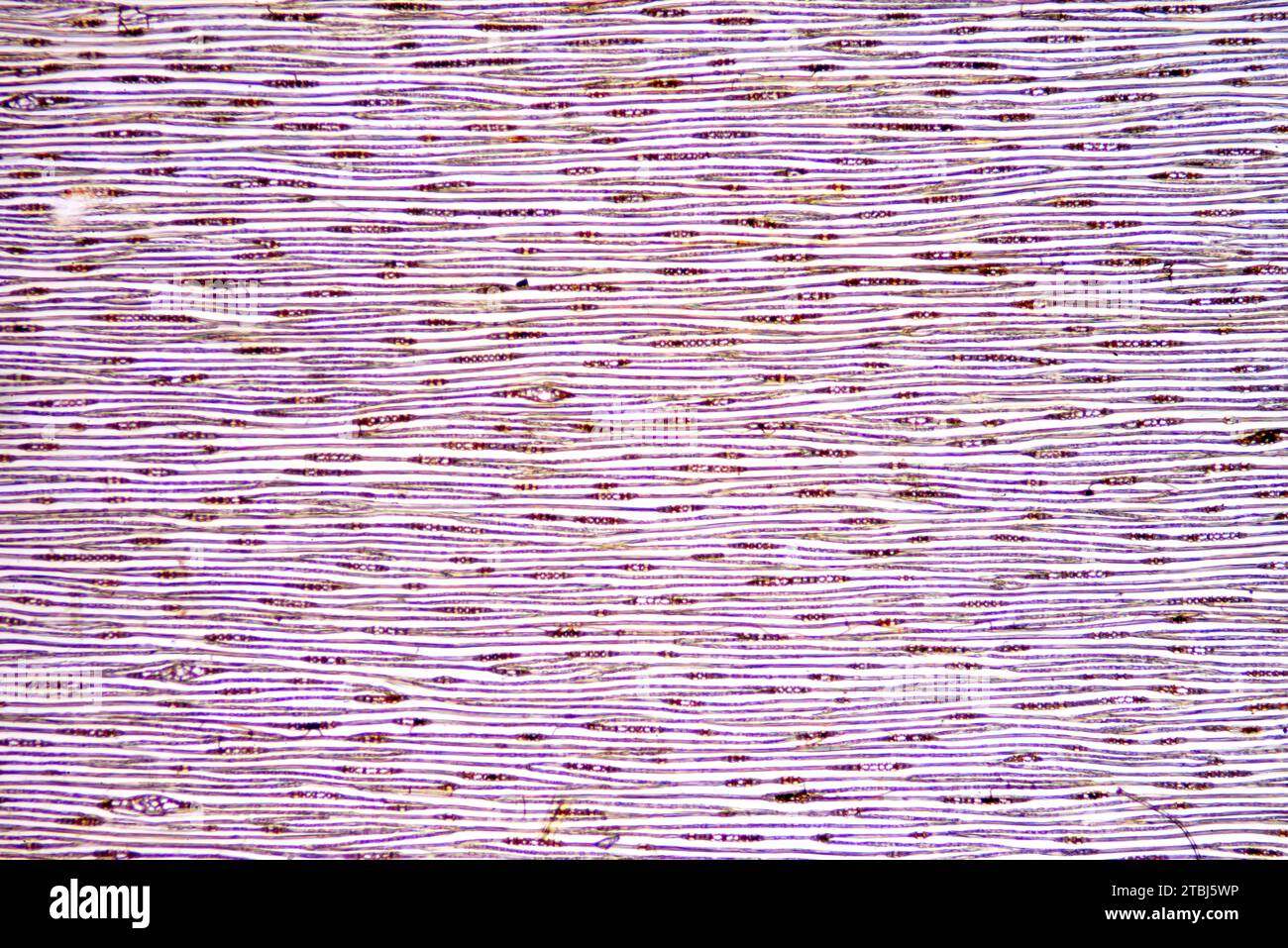 Radial section of pine trunk. Optical microscope X40. Stock Photohttps://www.alamy.com/image-license-details/?v=1https://www.alamy.com/radial-section-of-pine-trunk-optical-microscope-x40-image575103138.html
RF2TBJ5WP–Radial section of pine trunk. Optical microscope X40.
 Corn Stem, LM Stock Photohttps://www.alamy.com/image-license-details/?v=1https://www.alamy.com/stock-photo-corn-stem-lm-135018982.html
RMHRJJ2E–Corn Stem, LM
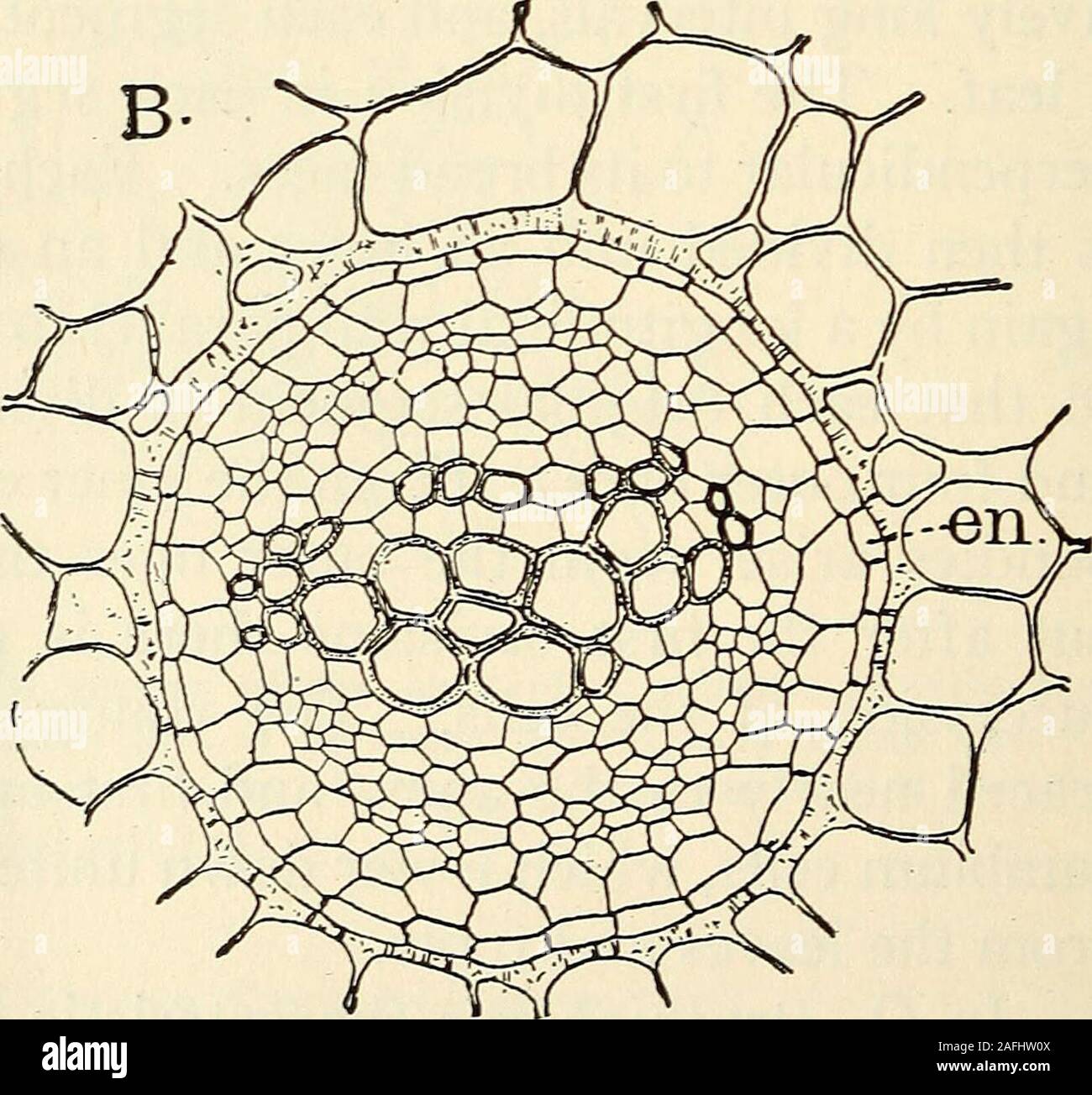 . The structure and development of mosses and ferns (Archegoniatae). Fig. 182.—Polypodium falcatum; A, Transverse section of the rhizome, X6; B, a sin-gle vascular bundle, Xi7S; en, endodermis. finally no living protoplasm remains in them. Faint elongatedtransverse pits become evident, and the spaces between theserapidly thicken at the expense of the cell contents until all theprotoplasm is used up. The thickened bars between the pitsgive the characteristic ladder-like appearance to the older IX FILICINE^ LEPTOSPORANGIAT^ 331 tracheid (Fig. 184, B). In cross-section these bars are nearlyrhombo Stock Photohttps://www.alamy.com/image-license-details/?v=1https://www.alamy.com/the-structure-and-development-of-mosses-and-ferns-archegoniatae-fig-182polypodium-falcatum-a-transverse-section-of-the-rhizome-x6-b-a-sin-gle-vascular-bundle-xi7s-en-endodermis-finally-no-living-protoplasm-remains-in-them-faint-elongatedtransverse-pits-become-evident-and-the-spaces-between-theserapidly-thicken-at-the-expense-of-the-cell-contents-until-all-theprotoplasm-is-used-up-the-thickened-bars-between-the-pitsgive-the-characteristic-ladder-like-appearance-to-the-older-ix-filicine-leptosporangiat-331-tracheid-fig-184-b-in-cross-section-these-bars-are-nearlyrhombo-image336609642.html
RM2AFHW0X–. The structure and development of mosses and ferns (Archegoniatae). Fig. 182.—Polypodium falcatum; A, Transverse section of the rhizome, X6; B, a sin-gle vascular bundle, Xi7S; en, endodermis. finally no living protoplasm remains in them. Faint elongatedtransverse pits become evident, and the spaces between theserapidly thicken at the expense of the cell contents until all theprotoplasm is used up. The thickened bars between the pitsgive the characteristic ladder-like appearance to the older IX FILICINE^ LEPTOSPORANGIAT^ 331 tracheid (Fig. 184, B). In cross-section these bars are nearlyrhombo
 cross section of a portion of a plant stem. Stock Photohttps://www.alamy.com/image-license-details/?v=1https://www.alamy.com/stock-photo-cross-section-of-a-portion-of-a-plant-stem-111895273.html
RFGE17FN–cross section of a portion of a plant stem.
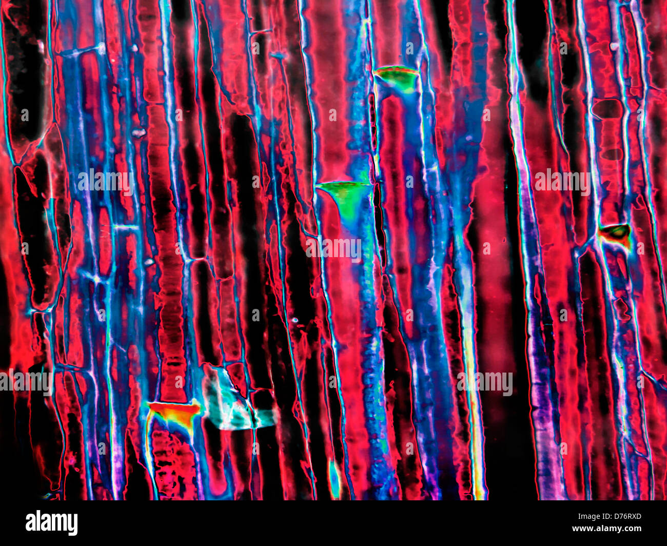 Sieve plate side view showing pores in plant stem Cucurbita gourd family magnification 200x image enhanced Stock Photohttps://www.alamy.com/image-license-details/?v=1https://www.alamy.com/stock-photo-sieve-plate-side-view-showing-pores-in-plant-stem-cucurbita-gourd-56084181.html
RMD76RXD–Sieve plate side view showing pores in plant stem Cucurbita gourd family magnification 200x image enhanced
 . A textbook of botany for colleges and universities ... Botany. 53^ ECOLOGY. Fig. 772. — A longitudiaal section through a portion of the stem of .5o/«:orttMi, showing oblique palisades (^), and also a "storage" tracheid (t); the arrow is directed toward the stem apex; jf, stoma; considerably magnified. to the leaf surface, regardless of the position of the leaf in relation to light. The position of the palisade layers seems relatively fixed in most cases (not in Lactuca and in Eucalyptus), a rudimentary palisade region often being discernible in the bud; in sub- sequent development Stock Photohttps://www.alamy.com/image-license-details/?v=1https://www.alamy.com/a-textbook-of-botany-for-colleges-and-universities-botany-53-ecology-fig-772-a-longitudiaal-section-through-a-portion-of-the-stem-of-5oorttmi-showing-oblique-palisades-and-also-a-quotstoragequot-tracheid-t-the-arrow-is-directed-toward-the-stem-apex-jf-stoma-considerably-magnified-to-the-leaf-surface-regardless-of-the-position-of-the-leaf-in-relation-to-light-the-position-of-the-palisade-layers-seems-relatively-fixed-in-most-cases-not-in-lactuca-and-in-eucalyptus-a-rudimentary-palisade-region-often-being-discernible-in-the-bud-in-sub-sequent-development-image216403770.html
RMPG2162–. A textbook of botany for colleges and universities ... Botany. 53^ ECOLOGY. Fig. 772. — A longitudiaal section through a portion of the stem of .5o/«:orttMi, showing oblique palisades (^), and also a "storage" tracheid (t); the arrow is directed toward the stem apex; jf, stoma; considerably magnified. to the leaf surface, regardless of the position of the leaf in relation to light. The position of the palisade layers seems relatively fixed in most cases (not in Lactuca and in Eucalyptus), a rudimentary palisade region often being discernible in the bud; in sub- sequent development
 . Fio. -JSO. â Tracheid of ri/iiMdestroyed by Puly/ioru* fiMtoti-niioUltf. The celhilose li:is been for the umst imrt cxtnieted, and the wiUU con- sist of ligiiin (wt)iHl-giini). Cr.icks ofc-iir in the dry secondary wall, while the wall {(I, Ij) remains intact. The spiral .structure of the secondary wall causes cross- ing; of the tissurcs in the walls of adjoiniu); cells at the liordered jiits, c. ami at Ixire-holes, J, e ; where neither jnts nor holes are present the fissures are simple,/. (After H. UartiB.) Stock Photohttps://www.alamy.com/image-license-details/?v=1https://www.alamy.com/fio-jso-tracheid-of-riiimdestroyed-by-pulyioru-fimtoti-niioultf-the-celhilose-liis-been-for-the-umst-imrt-cxtnieted-and-the-wiuu-con-sist-of-ligiiin-wtihl-giini-cricks-ofc-iir-in-the-dry-secondary-wall-while-the-wall-i-ij-remains-intact-the-spiral-structure-of-the-secondary-wall-causes-cross-ing-of-the-tissurcs-in-the-walls-of-adjoiniu-cells-at-the-liordered-jiits-c-ami-at-ixire-holes-j-e-where-neither-jnts-nor-holes-are-present-the-fissures-are-simple-after-h-uartib-image179900707.html
RMMCK557–. Fio. -JSO. â Tracheid of ri/iiMdestroyed by Puly/ioru* fiMtoti-niioUltf. The celhilose li:is been for the umst imrt cxtnieted, and the wiUU con- sist of ligiiin (wt)iHl-giini). Cr.icks ofc-iir in the dry secondary wall, while the wall {(I, Ij) remains intact. The spiral .structure of the secondary wall causes cross- ing; of the tissurcs in the walls of adjoiniu); cells at the liordered jiits, c. ami at Ixire-holes, J, e ; where neither jnts nor holes are present the fissures are simple,/. (After H. UartiB.)
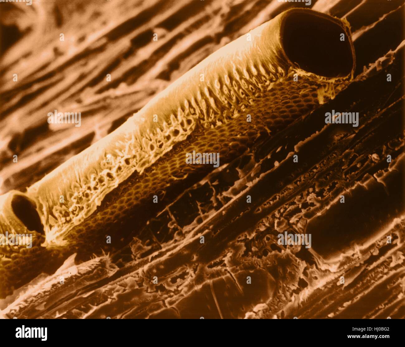 Conductive vessel element in mountain mahogany wood (Cercocarpus sp),coloured scanning electron micrograph (SEM).Note bordered pits in cellulose wall.A conductive vessel element is elongated,water-conducting cell in xylem,one of two kinds of tracheary elements.The cells die when mature,leaving only Stock Photohttps://www.alamy.com/image-license-details/?v=1https://www.alamy.com/stock-photo-conductive-vessel-element-in-mountain-mahogany-wood-cercocarpus-spcoloured-131545458.html
RFHJ0BG2–Conductive vessel element in mountain mahogany wood (Cercocarpus sp),coloured scanning electron micrograph (SEM).Note bordered pits in cellulose wall.A conductive vessel element is elongated,water-conducting cell in xylem,one of two kinds of tracheary elements.The cells die when mature,leaving only
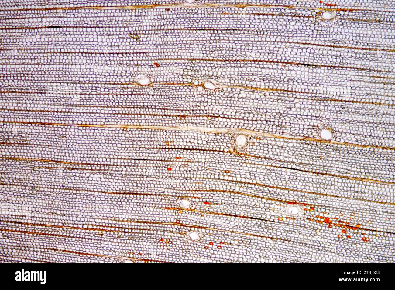 Transversal section of pine trunk. Optical microscope X40. Stock Photohttps://www.alamy.com/image-license-details/?v=1https://www.alamy.com/transversal-section-of-pine-trunk-optical-microscope-x40-image575103147.html
RF2TBJ5X3–Transversal section of pine trunk. Optical microscope X40.
 Corn Stem, LM Stock Photohttps://www.alamy.com/image-license-details/?v=1https://www.alamy.com/stock-photo-corn-stem-lm-135018981.html
RMHRJJ2D–Corn Stem, LM
 Scientific and applied pharmacognosy intended for the use of students in pharmacy, as a hand book for pharmacists, and as a reference book for food and drug analysts and pharmacologists . fibers usually have simple pores; and the medullaryray cells may become lignified. In addition, tracheid-like parenchy-matous cells are found in the wood of Opuntia and other genera. Cactus Grandiflorus, Herba Cacti Grandiflori, Night-blooming Cereus.—Cactus.—The stems and flowers of Cereus(Cactus) grandiflorus (Fam. Cactacese), a perennial, herbaceous orshrub-like plant, having thick succulent stems and prod Stock Photohttps://www.alamy.com/image-license-details/?v=1https://www.alamy.com/scientific-and-applied-pharmacognosy-intended-for-the-use-of-students-in-pharmacy-as-a-hand-book-for-pharmacists-and-as-a-reference-book-for-food-and-drug-analysts-and-pharmacologists-fibers-usually-have-simple-pores-and-the-medullaryray-cells-may-become-lignified-in-addition-tracheid-like-parenchy-matous-cells-are-found-in-the-wood-of-opuntia-and-other-genera-cactus-grandiflorus-herba-cacti-grandiflori-night-blooming-cereuscactusthe-stems-and-flowers-of-cereuscactus-grandiflorus-fam-cactacese-a-perennial-herbaceous-orshrub-like-plant-having-thick-succulent-stems-and-prod-image338203711.html
RM2AJ6E7Y–Scientific and applied pharmacognosy intended for the use of students in pharmacy, as a hand book for pharmacists, and as a reference book for food and drug analysts and pharmacologists . fibers usually have simple pores; and the medullaryray cells may become lignified. In addition, tracheid-like parenchy-matous cells are found in the wood of Opuntia and other genera. Cactus Grandiflorus, Herba Cacti Grandiflori, Night-blooming Cereus.—Cactus.—The stems and flowers of Cereus(Cactus) grandiflorus (Fam. Cactacese), a perennial, herbaceous orshrub-like plant, having thick succulent stems and prod
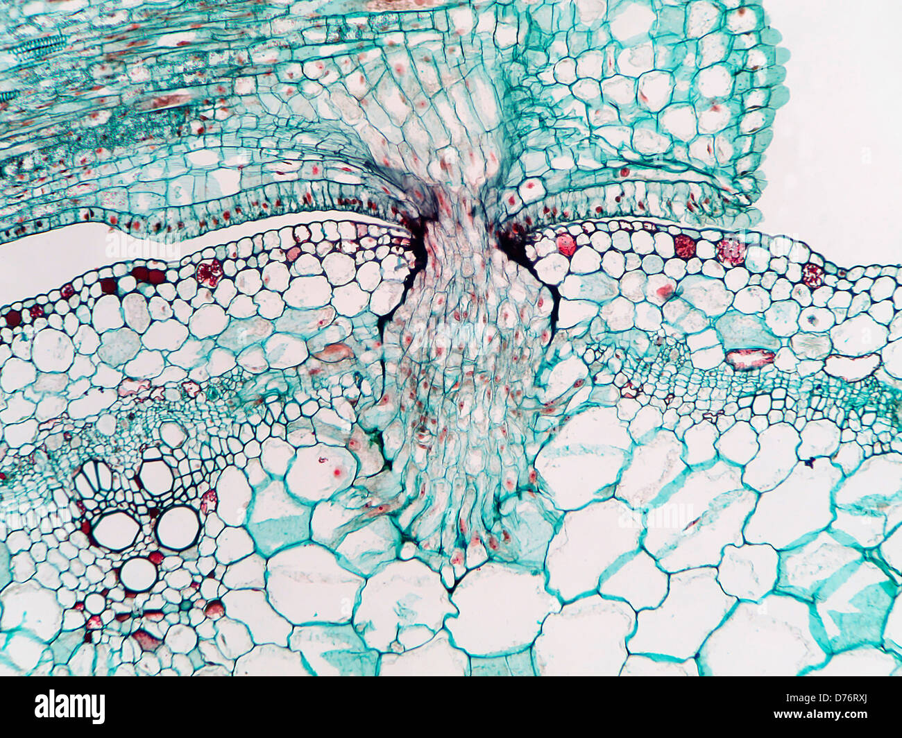 Parasitic dodder plant Cuscuta showing penetration parasitic haustoria into tissues host plant magnification 100x Stock Photohttps://www.alamy.com/image-license-details/?v=1https://www.alamy.com/stock-photo-parasitic-dodder-plant-cuscuta-showing-penetration-parasitic-haustoria-56084186.html
RMD76RXJ–Parasitic dodder plant Cuscuta showing penetration parasitic haustoria into tissues host plant magnification 100x
![. Fossil plants : for students of botany and geology . Paleobotany. XLIV] DADOXYLON 185 of growth.and with a single row of contiguous pits on the tracheid walls; Dadoxylon virginianum (Knowlton)^ from the Potomac series; Dadoxylon barremianum (Fliche)^ from the Lower Creta- ceous of France; Dadoxylon noveboracense (Holl. and Jefl.)^ from the Middle Cretaceous beds of Staten Island; Dadoxylon Zuffardii Negri * from middle Cretaceous rocks in the Gulf of Tripoli; Dado- xylon tanhoense (Stopes and Pujii)^ from Upper Cretaceous beds in Japan; Dadoxylon madagascariense (Fliche)® from Madagascar. Da Stock Photo . Fossil plants : for students of botany and geology . Paleobotany. XLIV] DADOXYLON 185 of growth.and with a single row of contiguous pits on the tracheid walls; Dadoxylon virginianum (Knowlton)^ from the Potomac series; Dadoxylon barremianum (Fliche)^ from the Lower Creta- ceous of France; Dadoxylon noveboracense (Holl. and Jefl.)^ from the Middle Cretaceous beds of Staten Island; Dadoxylon Zuffardii Negri * from middle Cretaceous rocks in the Gulf of Tripoli; Dado- xylon tanhoense (Stopes and Pujii)^ from Upper Cretaceous beds in Japan; Dadoxylon madagascariense (Fliche)® from Madagascar. Da Stock Photo](https://c8.alamy.com/comp/PG0D69/fossil-plants-for-students-of-botany-and-geology-paleobotany-xliv-dadoxylon-185-of-growthand-with-a-single-row-of-contiguous-pits-on-the-tracheid-walls-dadoxylon-virginianum-knowlton-from-the-potomac-series-dadoxylon-barremianum-fliche-from-the-lower-creta-ceous-of-france-dadoxylon-noveboracense-holl-and-jefl-from-the-middle-cretaceous-beds-of-staten-island-dadoxylon-zuffardii-negri-from-middle-cretaceous-rocks-in-the-gulf-of-tripoli-dado-xylon-tanhoense-stopes-and-pujii-from-upper-cretaceous-beds-in-japan-dadoxylon-madagascariense-fliche-from-madagascar-da-PG0D69.jpg) . Fossil plants : for students of botany and geology . Paleobotany. XLIV] DADOXYLON 185 of growth.and with a single row of contiguous pits on the tracheid walls; Dadoxylon virginianum (Knowlton)^ from the Potomac series; Dadoxylon barremianum (Fliche)^ from the Lower Creta- ceous of France; Dadoxylon noveboracense (Holl. and Jefl.)^ from the Middle Cretaceous beds of Staten Island; Dadoxylon Zuffardii Negri * from middle Cretaceous rocks in the Gulf of Tripoli; Dado- xylon tanhoense (Stopes and Pujii)^ from Upper Cretaceous beds in Japan; Dadoxylon madagascariense (Fliche)® from Madagascar. Da Stock Photohttps://www.alamy.com/image-license-details/?v=1https://www.alamy.com/fossil-plants-for-students-of-botany-and-geology-paleobotany-xliv-dadoxylon-185-of-growthand-with-a-single-row-of-contiguous-pits-on-the-tracheid-walls-dadoxylon-virginianum-knowlton-from-the-potomac-series-dadoxylon-barremianum-fliche-from-the-lower-creta-ceous-of-france-dadoxylon-noveboracense-holl-and-jefl-from-the-middle-cretaceous-beds-of-staten-island-dadoxylon-zuffardii-negri-from-middle-cretaceous-rocks-in-the-gulf-of-tripoli-dado-xylon-tanhoense-stopes-and-pujii-from-upper-cretaceous-beds-in-japan-dadoxylon-madagascariense-fliche-from-madagascar-da-image216369281.html
. Fossil plants : for students of botany and geology . Paleobotany. XLIV] DADOXYLON 185 of growth.and with a single row of contiguous pits on the tracheid walls; Dadoxylon virginianum (Knowlton)^ from the Potomac series; Dadoxylon barremianum (Fliche)^ from the Lower Creta- ceous of France; Dadoxylon noveboracense (Holl. and Jefl.)^ from the Middle Cretaceous beds of Staten Island; Dadoxylon Zuffardii Negri * from middle Cretaceous rocks in the Gulf of Tripoli; Dado- xylon tanhoense (Stopes and Pujii)^ from Upper Cretaceous beds in Japan; Dadoxylon madagascariense (Fliche)® from Madagascar. Da Stock Photohttps://www.alamy.com/image-license-details/?v=1https://www.alamy.com/fossil-plants-for-students-of-botany-and-geology-paleobotany-xliv-dadoxylon-185-of-growthand-with-a-single-row-of-contiguous-pits-on-the-tracheid-walls-dadoxylon-virginianum-knowlton-from-the-potomac-series-dadoxylon-barremianum-fliche-from-the-lower-creta-ceous-of-france-dadoxylon-noveboracense-holl-and-jefl-from-the-middle-cretaceous-beds-of-staten-island-dadoxylon-zuffardii-negri-from-middle-cretaceous-rocks-in-the-gulf-of-tripoli-dado-xylon-tanhoense-stopes-and-pujii-from-upper-cretaceous-beds-in-japan-dadoxylon-madagascariense-fliche-from-madagascar-da-image216369281.htmlRMPG0D69–. Fossil plants : for students of botany and geology . Paleobotany. XLIV] DADOXYLON 185 of growth.and with a single row of contiguous pits on the tracheid walls; Dadoxylon virginianum (Knowlton)^ from the Potomac series; Dadoxylon barremianum (Fliche)^ from the Lower Creta- ceous of France; Dadoxylon noveboracense (Holl. and Jefl.)^ from the Middle Cretaceous beds of Staten Island; Dadoxylon Zuffardii Negri * from middle Cretaceous rocks in the Gulf of Tripoli; Dado- xylon tanhoense (Stopes and Pujii)^ from Upper Cretaceous beds in Japan; Dadoxylon madagascariense (Fliche)® from Madagascar. Da
 . Electron microscopy; proceedings of the Stockholm Conference, September, 1956 . All the micrographs are of solid replicas. The scale ellipses are 25 microns in diameter. The angles of illumination and viewing are 5" and 12 respectively. Fig. 1. Reflection electron micrographic montage (considerably reduced) of a beaten spruce sulphite tracheid. Fig. 2. Reflection electron micrograph of a thin-walled spruce sulphite tracheid, heavily beaten. Fig. 3. Reflection electron micrograph of the surface of a tissue paper made from hemp and flax fibres. is not directly apparent from examination in Stock Photohttps://www.alamy.com/image-license-details/?v=1https://www.alamy.com/electron-microscopy-proceedings-of-the-stockholm-conference-september-1956-all-the-micrographs-are-of-solid-replicas-the-scale-ellipses-are-25-microns-in-diameter-the-angles-of-illumination-and-viewing-are-5quot-and-12-respectively-fig-1-reflection-electron-micrographic-montage-considerably-reduced-of-a-beaten-spruce-sulphite-tracheid-fig-2-reflection-electron-micrograph-of-a-thin-walled-spruce-sulphite-tracheid-heavily-beaten-fig-3-reflection-electron-micrograph-of-the-surface-of-a-tissue-paper-made-from-hemp-and-flax-fibres-is-not-directly-apparent-from-examination-in-image178411861.html
RMMA7A45–. Electron microscopy; proceedings of the Stockholm Conference, September, 1956 . All the micrographs are of solid replicas. The scale ellipses are 25 microns in diameter. The angles of illumination and viewing are 5" and 12 respectively. Fig. 1. Reflection electron micrographic montage (considerably reduced) of a beaten spruce sulphite tracheid. Fig. 2. Reflection electron micrograph of a thin-walled spruce sulphite tracheid, heavily beaten. Fig. 3. Reflection electron micrograph of the surface of a tissue paper made from hemp and flax fibres. is not directly apparent from examination in
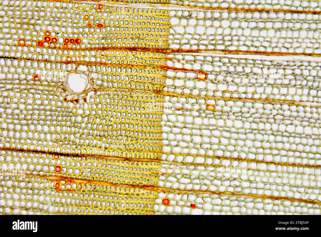 Transversal section of pine trunk. Optical microscope X100. Stock Photohttps://www.alamy.com/image-license-details/?v=1https://www.alamy.com/transversal-section-of-pine-trunk-optical-microscope-x100-image575103166.html
RF2TBJ5XP–Transversal section of pine trunk. Optical microscope X100.
 Corn Stem, LM Stock Photohttps://www.alamy.com/image-license-details/?v=1https://www.alamy.com/stock-photo-corn-stem-lm-135018980.html
RMHRJJ2C–Corn Stem, LM
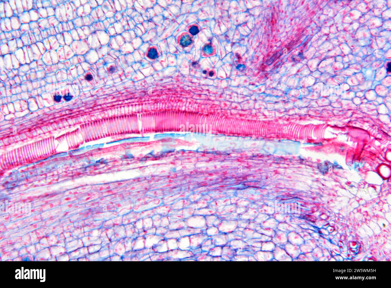 Vascular plant stem with spiral xylem. Photomicrograph X100 at 10 cm wide. Stock Photohttps://www.alamy.com/image-license-details/?v=1https://www.alamy.com/vascular-plant-stem-with-spiral-xylem-photomicrograph-x100-at-10-cm-wide-image588790429.html
RF2W5WM5H–Vascular plant stem with spiral xylem. Photomicrograph X100 at 10 cm wide.
 . The manufacture of pulp and paper : a textbook of modern pulp and paper mill practice. Fig. 15.—Cells of Wood of Broad-Leaved Trees, Greatly Enlarged. (1), wood fiber from broad-leaved tree; (2), vessel, poplar, with large, clear openings atends of segment; (3) vessel, birch, with scalariform openings at ends of segment; (4). vessel,beech, with clear or sclariform openings at ends of segments; (5) vessel, maple, with spiralthickenings; (6) vessel, chestnut, springwood; (7), vessel, chestnut, summerwood; (8),tracheid, chestnut, springwood; (9), wood parenchyma cells, which occur to a slight e Stock Photohttps://www.alamy.com/image-license-details/?v=1https://www.alamy.com/the-manufacture-of-pulp-and-paper-a-textbook-of-modern-pulp-and-paper-mill-practice-fig-15cells-of-wood-of-broad-leaved-trees-greatly-enlarged-1-wood-fiber-from-broad-leaved-tree-2-vessel-poplar-with-large-clear-openings-atends-of-segment-3-vessel-birch-with-scalariform-openings-at-ends-of-segment-4-vesselbeech-with-clear-or-sclariform-openings-at-ends-of-segments-5-vessel-maple-with-spiralthickenings-6-vessel-chestnut-springwood-7-vessel-chestnut-summerwood-8tracheid-chestnut-springwood-9-wood-parenchyma-cells-which-occur-to-a-slight-e-image336727288.html
RM2AFR72G–. The manufacture of pulp and paper : a textbook of modern pulp and paper mill practice. Fig. 15.—Cells of Wood of Broad-Leaved Trees, Greatly Enlarged. (1), wood fiber from broad-leaved tree; (2), vessel, poplar, with large, clear openings atends of segment; (3) vessel, birch, with scalariform openings at ends of segment; (4). vessel,beech, with clear or sclariform openings at ends of segments; (5) vessel, maple, with spiralthickenings; (6) vessel, chestnut, springwood; (7), vessel, chestnut, summerwood; (8),tracheid, chestnut, springwood; (9), wood parenchyma cells, which occur to a slight e
 Transversal section of pine trunk. Optical microscope X40. Stock Photohttps://www.alamy.com/image-license-details/?v=1https://www.alamy.com/transversal-section-of-pine-trunk-optical-microscope-x40-image575103158.html
RF2TBJ5XE–Transversal section of pine trunk. Optical microscope X40.
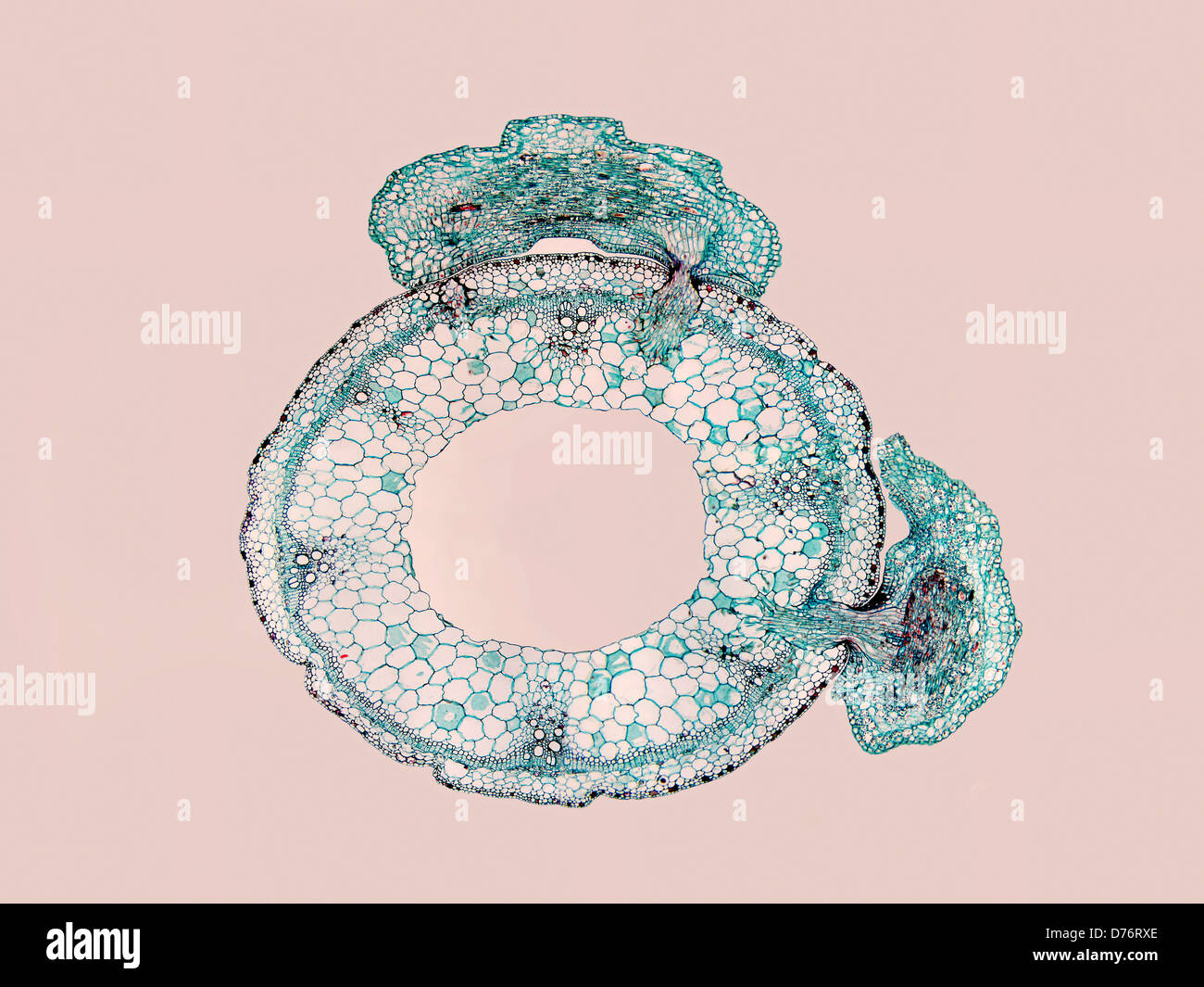 Image two parasitic dodder plants Cuscuta sp showing penetration parasitic haustoria into tissues host plant magnification 20x Stock Photohttps://www.alamy.com/image-license-details/?v=1https://www.alamy.com/stock-photo-image-two-parasitic-dodder-plants-cuscuta-sp-showing-penetration-parasitic-56084182.html
RMD76RXE–Image two parasitic dodder plants Cuscuta sp showing penetration parasitic haustoria into tissues host plant magnification 20x
 Node of a vascular plant stem with spiral xylem (transport tissue). Photomicrograph X50 at 10 cm wide. Stock Photohttps://www.alamy.com/image-license-details/?v=1https://www.alamy.com/node-of-a-vascular-plant-stem-with-spiral-xylem-transport-tissue-photomicrograph-x50-at-10-cm-wide-image588789935.html
RF2W5WKFY–Node of a vascular plant stem with spiral xylem (transport tissue). Photomicrograph X50 at 10 cm wide.
 . A textbook of botany for colleges and universities ... Botany. Fig. 772. — A longitudiaal section through a portion of the stem of .5o/«:orttMi, showing oblique palisades (^), and also a "storage" tracheid (t); the arrow is directed toward the stem apex; jf, stoma; considerably magnified. to the leaf surface, regardless of the position of the leaf in relation to light. The position of the palisade layers seems relatively fixed in most cases (not in Lactuca and in Eucalyptus), a rudimentary palisade region often being discernible in the bud; in sub- sequent development the thickness Stock Photohttps://www.alamy.com/image-license-details/?v=1https://www.alamy.com/a-textbook-of-botany-for-colleges-and-universities-botany-fig-772-a-longitudiaal-section-through-a-portion-of-the-stem-of-5oorttmi-showing-oblique-palisades-and-also-a-quotstoragequot-tracheid-t-the-arrow-is-directed-toward-the-stem-apex-jf-stoma-considerably-magnified-to-the-leaf-surface-regardless-of-the-position-of-the-leaf-in-relation-to-light-the-position-of-the-palisade-layers-seems-relatively-fixed-in-most-cases-not-in-lactuca-and-in-eucalyptus-a-rudimentary-palisade-region-often-being-discernible-in-the-bud-in-sub-sequent-development-the-thickness-image216403772.html
RMPG2164–. A textbook of botany for colleges and universities ... Botany. Fig. 772. — A longitudiaal section through a portion of the stem of .5o/«:orttMi, showing oblique palisades (^), and also a "storage" tracheid (t); the arrow is directed toward the stem apex; jf, stoma; considerably magnified. to the leaf surface, regardless of the position of the leaf in relation to light. The position of the palisade layers seems relatively fixed in most cases (not in Lactuca and in Eucalyptus), a rudimentary palisade region often being discernible in the bud; in sub- sequent development the thickness
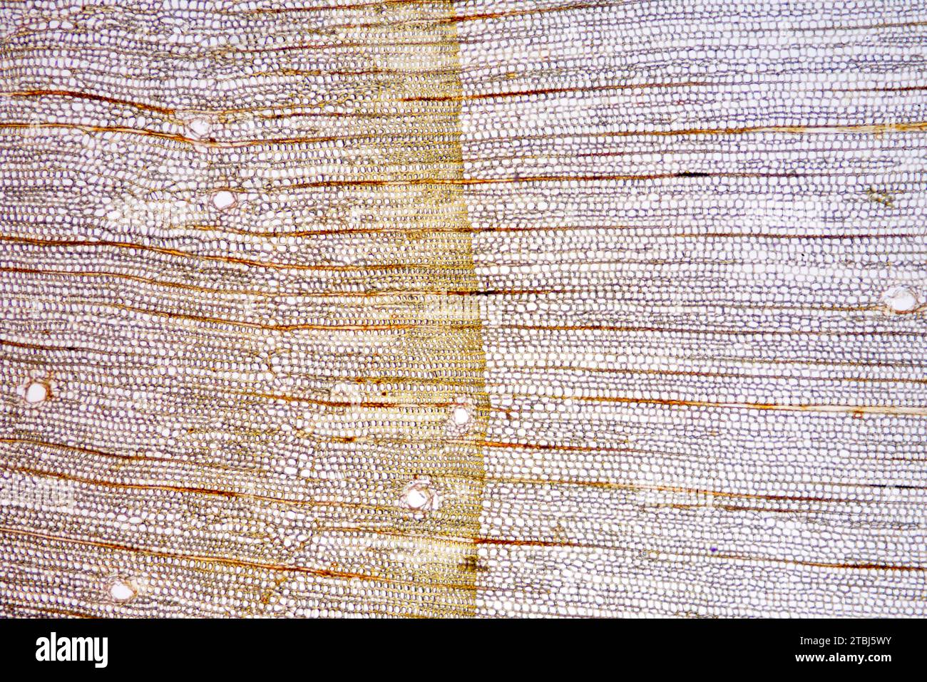 Transversal section of pine trunk. Optical microscope X40. Stock Photohttps://www.alamy.com/image-license-details/?v=1https://www.alamy.com/transversal-section-of-pine-trunk-optical-microscope-x40-image575103143.html
RF2TBJ5WY–Transversal section of pine trunk. Optical microscope X40.
 . Fig. 13. Fig. 14. Fio. 13.—Tracheid of Pinus si/lvestris destroyed by Trametes pint. The primary cell-Wall is completely dissolved from below upwards to a, a ; 6, secondary and tertiary layers of the walls consisting in the under portion of cellulose only, in wliich granules of chalk are recoffnizable ; c, fungus-hyphae boring through the walls, leaving holes d and c. (After R. Hartig.) Fig. 14.—Tracheid of Pinus destroyed by Polyporui Schweiiiitzii. The cellulose has been extracted, and the wall consists only of wood-gum. The fissures are a result of drying-up, but they do not extend into t Stock Photohttps://www.alamy.com/image-license-details/?v=1https://www.alamy.com/fig-13-fig-14-fio-13tracheid-of-pinus-silvestris-destroyed-by-trametes-pint-the-primary-cell-wall-is-completely-dissolved-from-below-upwards-to-a-a-6-secondary-and-tertiary-layers-of-the-walls-consisting-in-the-under-portion-of-cellulose-only-in-wliich-granules-of-chalk-are-recoffnizable-c-fungus-hyphae-boring-through-the-walls-leaving-holes-d-and-c-after-r-hartig-fig-14tracheid-of-pinus-destroyed-by-polyporui-schweiiiitzii-the-cellulose-has-been-extracted-and-the-wall-consists-only-of-wood-gum-the-fissures-are-a-result-of-drying-up-but-they-do-not-extend-into-t-image179902132.html
RMMCK704–. Fig. 13. Fig. 14. Fio. 13.—Tracheid of Pinus si/lvestris destroyed by Trametes pint. The primary cell-Wall is completely dissolved from below upwards to a, a ; 6, secondary and tertiary layers of the walls consisting in the under portion of cellulose only, in wliich granules of chalk are recoffnizable ; c, fungus-hyphae boring through the walls, leaving holes d and c. (After R. Hartig.) Fig. 14.—Tracheid of Pinus destroyed by Polyporui Schweiiiitzii. The cellulose has been extracted, and the wall consists only of wood-gum. The fissures are a result of drying-up, but they do not extend into t
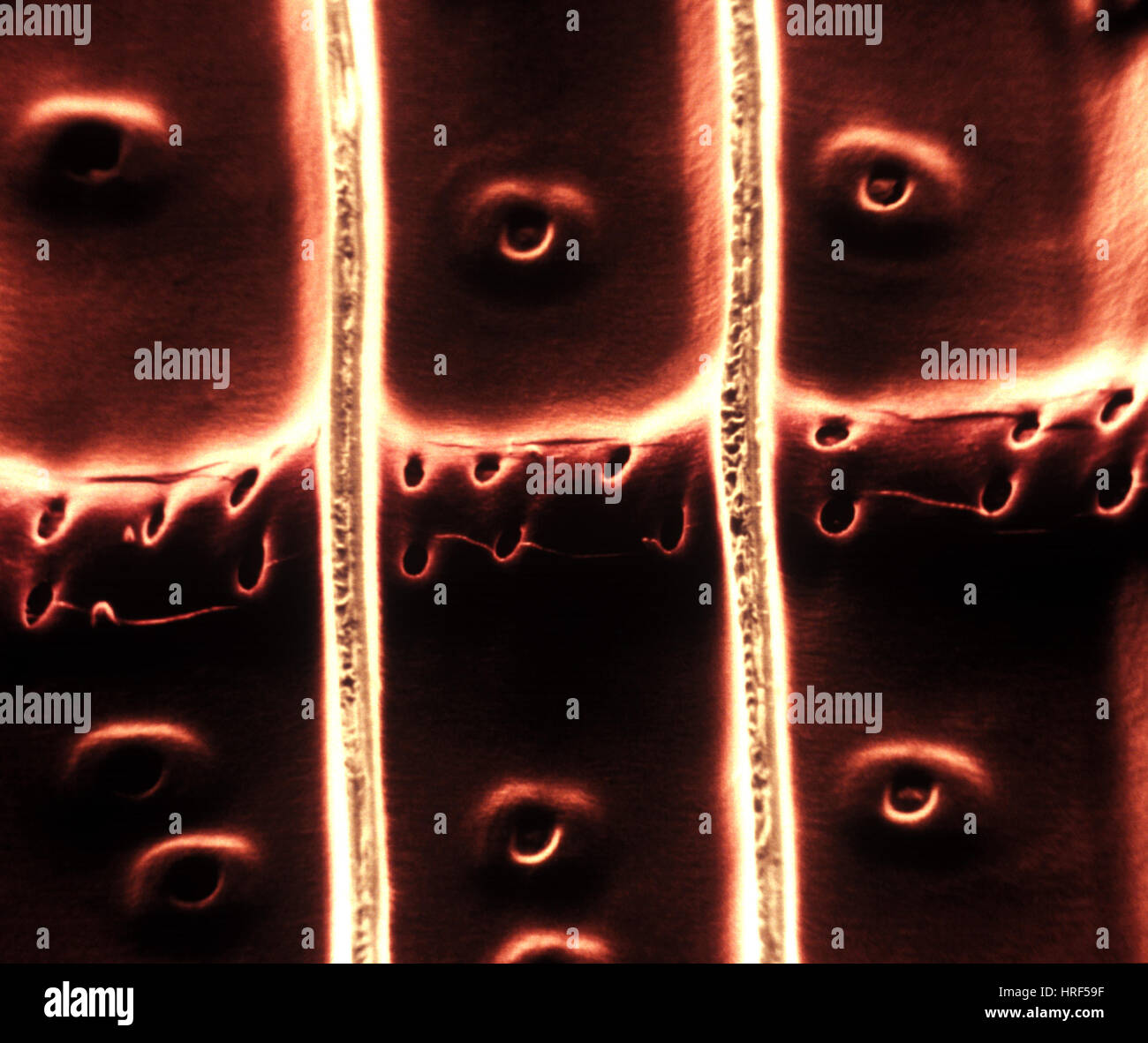 Xylem Stock Photohttps://www.alamy.com/image-license-details/?v=1https://www.alamy.com/stock-photo-xylem-134943131.html
RMHRF59F–Xylem
 Report upon the forestry investigations of the U.SDepartment of agriculture1877-1898 . o two large, simple pits to each tracheid on the radial walls of the cells of the pith ray.—Group 1. Represented in this country by F. resinosa.6. Three to sis simple pits to each tracheid on the walls of the cells of the pith ray.—Group 2. P. tccda,palustria,etc., including most of our hard and yellow pines.Section II. Walls of tracheids of pith ray smooth, without dentate projections. a. One or two large pits to each tracheid on the radial walls of each cell of the pith ray.—Group 3. P. strobus lamhertiana Stock Photohttps://www.alamy.com/image-license-details/?v=1https://www.alamy.com/report-upon-the-forestry-investigations-of-the-usdepartment-of-agriculture1877-1898-o-two-large-simple-pits-to-each-tracheid-on-the-radial-walls-of-the-cells-of-the-pith-raygroup-1-represented-in-this-country-by-f-resinosa6-three-to-sis-simple-pits-to-each-tracheid-on-the-walls-of-the-cells-of-the-pith-raygroup-2-p-tccdapalustriaetc-including-most-of-our-hard-and-yellow-pinessection-ii-walls-of-tracheids-of-pith-ray-smooth-without-dentate-projections-a-one-or-two-large-pits-to-each-tracheid-on-the-radial-walls-of-each-cell-of-the-pith-raygroup-3-p-strobus-lamhertiana-image343221057.html
RM2AXB1XW–Report upon the forestry investigations of the U.SDepartment of agriculture1877-1898 . o two large, simple pits to each tracheid on the radial walls of the cells of the pith ray.—Group 1. Represented in this country by F. resinosa.6. Three to sis simple pits to each tracheid on the walls of the cells of the pith ray.—Group 2. P. tccda,palustria,etc., including most of our hard and yellow pines.Section II. Walls of tracheids of pith ray smooth, without dentate projections. a. One or two large pits to each tracheid on the radial walls of each cell of the pith ray.—Group 3. P. strobus lamhertiana
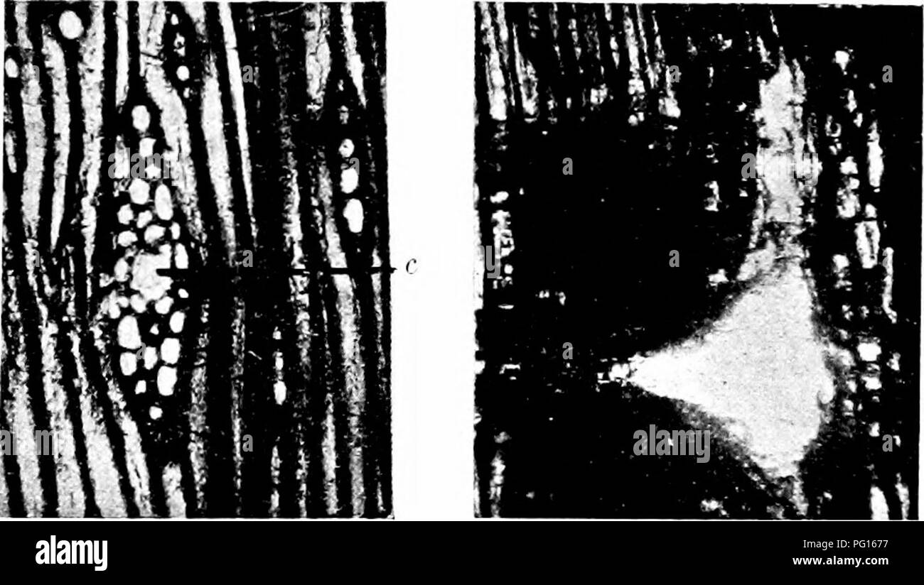 . Fossil plants : for students of botany and geology . Paleobotany. Fig. 72.5. Pityoxylon ttiggenst; c, resin-canal in a fusiform medullary ray. (British Museum, 51427, 51641, 51727.) broad; the reduction in diameter of the summer-tracheids extends over several rows, the transition being much more gradual than in some types of Coniferous wood. A characteristic feature is the occurrence of more or less circular patches where the tracheids have been destroyed with the exception of a single tracheid or a Kraus in Sclumpor (72) A. p. 378. .Schimper and Schenlv (90) A. pp. 855, 874. Schroeter (80) Stock Photohttps://www.alamy.com/image-license-details/?v=1https://www.alamy.com/fossil-plants-for-students-of-botany-and-geology-paleobotany-fig-725-pityoxylon-ttiggenst-c-resin-canal-in-a-fusiform-medullary-ray-british-museum-51427-51641-51727-broad-the-reduction-in-diameter-of-the-summer-tracheids-extends-over-several-rows-the-transition-being-much-more-gradual-than-in-some-types-of-coniferous-wood-a-characteristic-feature-is-the-occurrence-of-more-or-less-circular-patches-where-the-tracheids-have-been-destroyed-with-the-exception-of-a-single-tracheid-or-a-kraus-in-sclumpor-72-a-p-378-schimper-and-schenlv-90-a-pp-855-874-schroeter-80-image216385771.html
RMPG1677–. Fossil plants : for students of botany and geology . Paleobotany. Fig. 72.5. Pityoxylon ttiggenst; c, resin-canal in a fusiform medullary ray. (British Museum, 51427, 51641, 51727.) broad; the reduction in diameter of the summer-tracheids extends over several rows, the transition being much more gradual than in some types of Coniferous wood. A characteristic feature is the occurrence of more or less circular patches where the tracheids have been destroyed with the exception of a single tracheid or a Kraus in Sclumpor (72) A. p. 378. .Schimper and Schenlv (90) A. pp. 855, 874. Schroeter (80)
 . Electron microscopy; proceedings of the Stockholm Conference, September, 1956 . Fig. 3. Carbon replica, metal-shadowed, of a beaten spruce tracheid and its associated fibrillation. Magnification 1500, inset 13,000. Fig. 4. Carbon replica, metal-shadowed, of a surface of wood tissue. The wood—Scots pine—was cut in a radial plane with a chisel prior to replication. Magnification < 1700. remain embedded in the methacrylate ic). The stripping of the fibres is sometimes dil'Hcult, cellulose tape being by no means always effective. A more successful method employs a 10 "„ solution of poly- Stock Photohttps://www.alamy.com/image-license-details/?v=1https://www.alamy.com/electron-microscopy-proceedings-of-the-stockholm-conference-september-1956-fig-3-carbon-replica-metal-shadowed-of-a-beaten-spruce-tracheid-and-its-associated-fibrillation-magnification-1500-inset-13000-fig-4-carbon-replica-metal-shadowed-of-a-surface-of-wood-tissue-the-woodscots-pinewas-cut-in-a-radial-plane-with-a-chisel-prior-to-replication-magnification-lt-1700-remain-embedded-in-the-methacrylate-ic-the-stripping-of-the-fibres-is-sometimes-dilhcult-cellulose-tape-being-by-no-means-always-effective-a-more-successful-method-employs-a-10-quot-solution-of-poly-image178411873.html
RMMA7A4H–. Electron microscopy; proceedings of the Stockholm Conference, September, 1956 . Fig. 3. Carbon replica, metal-shadowed, of a beaten spruce tracheid and its associated fibrillation. Magnification 1500, inset 13,000. Fig. 4. Carbon replica, metal-shadowed, of a surface of wood tissue. The wood—Scots pine—was cut in a radial plane with a chisel prior to replication. Magnification < 1700. remain embedded in the methacrylate ic). The stripping of the fibres is sometimes dil'Hcult, cellulose tape being by no means always effective. A more successful method employs a 10 "„ solution of poly-
 Chemistry of pulp and paper making . Fig. 2. Drawings of Wood-elements 19. Tracheid fromTectona grandis. 20-23. Porlieria hygrometrica. 20. Con-jugate substitute fibres in cross section. 21. Ordinary substitute fibre aftermaceration. 22,23. Conjugate substitute fibres after maceration. 24-27. Cy-tisus laburnum; elements after maceration. 24. Wood-parenchyma fibre. 25.Tracheid. 26. Substitute fibre. 27. Simple libriform fibre. 28. Cross-sectionthrough cambium and youngest wood of Cytisus laburnum. 29, 30. Mahoniaaquifolium; ducts. 29. After maceration. 30. Longitudinal section. 31-36.Extremetie Stock Photohttps://www.alamy.com/image-license-details/?v=1https://www.alamy.com/chemistry-of-pulp-and-paper-making-fig-2-drawings-of-wood-elements-19-tracheid-fromtectona-grandis-20-23-porlieria-hygrometrica-20-con-jugate-substitute-fibres-in-cross-section-21-ordinary-substitute-fibre-aftermaceration-2223-conjugate-substitute-fibres-after-maceration-24-27-cy-tisus-laburnum-elements-after-maceration-24-wood-parenchyma-fibre-25tracheid-26-substitute-fibre-27-simple-libriform-fibre-28-cross-sectionthrough-cambium-and-youngest-wood-of-cytisus-laburnum-29-30-mahoniaaquifolium-ducts-29-after-maceration-30-longitudinal-section-31-36extremetie-image338397497.html
RM2AJF9CW–Chemistry of pulp and paper making . Fig. 2. Drawings of Wood-elements 19. Tracheid fromTectona grandis. 20-23. Porlieria hygrometrica. 20. Con-jugate substitute fibres in cross section. 21. Ordinary substitute fibre aftermaceration. 22,23. Conjugate substitute fibres after maceration. 24-27. Cy-tisus laburnum; elements after maceration. 24. Wood-parenchyma fibre. 25.Tracheid. 26. Substitute fibre. 27. Simple libriform fibre. 28. Cross-sectionthrough cambium and youngest wood of Cytisus laburnum. 29, 30. Mahoniaaquifolium; ducts. 29. After maceration. 30. Longitudinal section. 31-36.Extremetie
 . Diseases of plants induced by cryptogamic parasites : introduction to the study of pathogenic Fungi, slime-Fungi, bacteria, & Algae . Plant diseases; Parasitic plants; Fungi. Fio. 14. Fig. 13.—Tracheid of Pinus sytvestris destroyed by Travutes pini. The primary cell-WHll is completely dissolved from below upwards to a^ a; 6, secondary and tertiary layers of the walls consisting in the under portion of cellulose only, in which gi'anules of chalk are recognizable ; c, fungus-hyphae boring through the walls, leaving holes d and e. (After R. Hartig.) Fic. 14.—Tracheid of Pinus destroyed by P Stock Photohttps://www.alamy.com/image-license-details/?v=1https://www.alamy.com/diseases-of-plants-induced-by-cryptogamic-parasites-introduction-to-the-study-of-pathogenic-fungi-slime-fungi-bacteria-amp-algae-plant-diseases-parasitic-plants-fungi-fio-14-fig-13tracheid-of-pinus-sytvestris-destroyed-by-travutes-pini-the-primary-cell-whll-is-completely-dissolved-from-below-upwards-to-a-a-6-secondary-and-tertiary-layers-of-the-walls-consisting-in-the-under-portion-of-cellulose-only-in-which-gianules-of-chalk-are-recognizable-c-fungus-hyphae-boring-through-the-walls-leaving-holes-d-and-e-after-r-hartig-fic-14tracheid-of-pinus-destroyed-by-p-image216449055.html
RMPG42YB–. Diseases of plants induced by cryptogamic parasites : introduction to the study of pathogenic Fungi, slime-Fungi, bacteria, & Algae . Plant diseases; Parasitic plants; Fungi. Fio. 14. Fig. 13.—Tracheid of Pinus sytvestris destroyed by Travutes pini. The primary cell-WHll is completely dissolved from below upwards to a^ a; 6, secondary and tertiary layers of the walls consisting in the under portion of cellulose only, in which gi'anules of chalk are recognizable ; c, fungus-hyphae boring through the walls, leaving holes d and e. (After R. Hartig.) Fic. 14.—Tracheid of Pinus destroyed by P
 . Fio. 13.—Tracheid of Finns sylcestris destroyed by Tmmetes jiini. The primary cell-Wall is completely dissolved from below upwards to n, a ; b, secondary and tertiary layers of the walls consisting in the under portion of cellulose only, in wliich granules of chalk are recognizable ; c, fungus-hyphae boring through the walls, leaving holes d and e. (After R. Hartig.) Fi(i. 14.—Tracheid of Pinu-t destroyed by Poh/poms Schweinitzii. The cellulose has been extracted, and the wall consists only of wood-gum. The fissures are a result of drying-up, but they do not extend into the primary wall a, h Stock Photohttps://www.alamy.com/image-license-details/?v=1https://www.alamy.com/fio-13tracheid-of-finns-sylcestris-destroyed-by-tmmetes-jiini-the-primary-cell-wall-is-completely-dissolved-from-below-upwards-to-n-a-b-secondary-and-tertiary-layers-of-the-walls-consisting-in-the-under-portion-of-cellulose-only-in-wliich-granules-of-chalk-are-recognizable-c-fungus-hyphae-boring-through-the-walls-leaving-holes-d-and-e-after-r-hartig-fii-14tracheid-of-pinu-t-destroyed-by-pohpoms-schweinitzii-the-cellulose-has-been-extracted-and-the-wall-consists-only-of-wood-gum-the-fissures-are-a-result-of-drying-up-but-they-do-not-extend-into-the-primary-wall-a-h-image179901932.html
RMMCK6N0–. Fio. 13.—Tracheid of Finns sylcestris destroyed by Tmmetes jiini. The primary cell-Wall is completely dissolved from below upwards to n, a ; b, secondary and tertiary layers of the walls consisting in the under portion of cellulose only, in wliich granules of chalk are recognizable ; c, fungus-hyphae boring through the walls, leaving holes d and e. (After R. Hartig.) Fi(i. 14.—Tracheid of Pinu-t destroyed by Poh/poms Schweinitzii. The cellulose has been extracted, and the wall consists only of wood-gum. The fissures are a result of drying-up, but they do not extend into the primary wall a, h
 Chemistry of pulp and paper making . Fig. 2. Drawings of Wood-elements 19. TracheidfromTectonagrandis. 20-23. Porlieria hygrometrica. 20. Con-jugate substitute fibres in cross section. 21. Ordinary substitute fibre aftermaceration. 22, 23. Conjugate substitute fibres after maceration. 24-27. Cy-tisus laburnum; elements after maceration. 24. Wood-parenchyma fibre. 25.Tracheid. 26. Substitute fibre. 27. Simple libriform fibre. 28. Cross-sectionthrough cambium and youngest wood of Cytisus laburnum. 29, 30. Mahoniaaquifolium; ducts. 29. After maceration. 30. Longitudinal section. 31-36.Extremeties Stock Photohttps://www.alamy.com/image-license-details/?v=1https://www.alamy.com/chemistry-of-pulp-and-paper-making-fig-2-drawings-of-wood-elements-19-tracheidfromtectonagrandis-20-23-porlieria-hygrometrica-20-con-jugate-substitute-fibres-in-cross-section-21-ordinary-substitute-fibre-aftermaceration-22-23-conjugate-substitute-fibres-after-maceration-24-27-cy-tisus-laburnum-elements-after-maceration-24-wood-parenchyma-fibre-25tracheid-26-substitute-fibre-27-simple-libriform-fibre-28-cross-sectionthrough-cambium-and-youngest-wood-of-cytisus-laburnum-29-30-mahoniaaquifolium-ducts-29-after-maceration-30-longitudinal-section-31-36extremeties-image339070225.html
RM2AKHYEW–Chemistry of pulp and paper making . Fig. 2. Drawings of Wood-elements 19. TracheidfromTectonagrandis. 20-23. Porlieria hygrometrica. 20. Con-jugate substitute fibres in cross section. 21. Ordinary substitute fibre aftermaceration. 22, 23. Conjugate substitute fibres after maceration. 24-27. Cy-tisus laburnum; elements after maceration. 24. Wood-parenchyma fibre. 25.Tracheid. 26. Substitute fibre. 27. Simple libriform fibre. 28. Cross-sectionthrough cambium and youngest wood of Cytisus laburnum. 29, 30. Mahoniaaquifolium; ducts. 29. After maceration. 30. Longitudinal section. 31-36.Extremeties
 . Elements of applied microscopy. A text-book for beginners. Microscopy. MICROSCOPY OF PAPER. lOI , or oval scar-like pits arranged in regular rows over their surface. In the case of pine, lattice-like areas with ob- long openings appear at intervals; but the presence of long cells mainly of a single type and with discoid mark- ings is the characteristic of the Conifers in general. 7. The Structure of the Angiosperms.—Pulp made from the commoner Angiospermous trees shows two distinct elements, long narrow fibres and short broad. Fig, 40.—^Tracheid of Birch. (After Herzberg.) 240 diameters. cel Stock Photohttps://www.alamy.com/image-license-details/?v=1https://www.alamy.com/elements-of-applied-microscopy-a-text-book-for-beginners-microscopy-microscopy-of-paper-loi-or-oval-scar-like-pits-arranged-in-regular-rows-over-their-surface-in-the-case-of-pine-lattice-like-areas-with-ob-long-openings-appear-at-intervals-but-the-presence-of-long-cells-mainly-of-a-single-type-and-with-discoid-mark-ings-is-the-characteristic-of-the-conifers-in-general-7-the-structure-of-the-angiospermspulp-made-from-the-commoner-angiospermous-trees-shows-two-distinct-elements-long-narrow-fibres-and-short-broad-fig-40tracheid-of-birch-after-herzberg-240-diameters-cel-image216447786.html
RMPG41A2–. Elements of applied microscopy. A text-book for beginners. Microscopy. MICROSCOPY OF PAPER. lOI , or oval scar-like pits arranged in regular rows over their surface. In the case of pine, lattice-like areas with ob- long openings appear at intervals; but the presence of long cells mainly of a single type and with discoid mark- ings is the characteristic of the Conifers in general. 7. The Structure of the Angiosperms.—Pulp made from the commoner Angiospermous trees shows two distinct elements, long narrow fibres and short broad. Fig, 40.—^Tracheid of Birch. (After Herzberg.) 240 diameters. cel
![. M Fig. 44. Baclialschnitts-AnsicM aus dem Holze eines alten Stammes der Sumpf-Cypresse, Taxodiwn lUstichum L. (300/1). Links Früh-, rechts Spätholz, in diesem Strangparenchym [p]. M ein vierreihiger, durchaus parenchymatischer Markstrahl. (Nach der Natur gezeichnet von Wilhelm.) Mikroskopischer Charakter im Wesentlichen der des Holzes echter cypressenartiger Nadelbäume. Spätholzzonen auch gegen das (mitunter nur eine einzige Tracheidenschicht breite) Frühholz des näm- lichen Jahrganges sehr scharf abgesetzt, auch auf den tangentialen Längs- wänden seiner vorwiegend sehr dickwandigen Tracheid Stock Photo . M Fig. 44. Baclialschnitts-AnsicM aus dem Holze eines alten Stammes der Sumpf-Cypresse, Taxodiwn lUstichum L. (300/1). Links Früh-, rechts Spätholz, in diesem Strangparenchym [p]. M ein vierreihiger, durchaus parenchymatischer Markstrahl. (Nach der Natur gezeichnet von Wilhelm.) Mikroskopischer Charakter im Wesentlichen der des Holzes echter cypressenartiger Nadelbäume. Spätholzzonen auch gegen das (mitunter nur eine einzige Tracheidenschicht breite) Frühholz des näm- lichen Jahrganges sehr scharf abgesetzt, auch auf den tangentialen Längs- wänden seiner vorwiegend sehr dickwandigen Tracheid Stock Photo](https://c8.alamy.com/comp/MCPKJJ/m-fig-44-baclialschnitts-ansicm-aus-dem-holze-eines-alten-stammes-der-sumpf-cypresse-taxodiwn-lustichum-l-3001-links-frh-rechts-sptholz-in-diesem-strangparenchym-p-m-ein-vierreihiger-durchaus-parenchymatischer-markstrahl-nach-der-natur-gezeichnet-von-wilhelm-mikroskopischer-charakter-im-wesentlichen-der-des-holzes-echter-cypressenartiger-nadelbume-sptholzzonen-auch-gegen-das-mitunter-nur-eine-einzige-tracheidenschicht-breite-frhholz-des-nm-lichen-jahrganges-sehr-scharf-abgesetzt-auch-auf-den-tangentialen-lngs-wnden-seiner-vorwiegend-sehr-dickwandigen-tracheid-MCPKJJ.jpg) . M Fig. 44. Baclialschnitts-AnsicM aus dem Holze eines alten Stammes der Sumpf-Cypresse, Taxodiwn lUstichum L. (300/1). Links Früh-, rechts Spätholz, in diesem Strangparenchym [p]. M ein vierreihiger, durchaus parenchymatischer Markstrahl. (Nach der Natur gezeichnet von Wilhelm.) Mikroskopischer Charakter im Wesentlichen der des Holzes echter cypressenartiger Nadelbäume. Spätholzzonen auch gegen das (mitunter nur eine einzige Tracheidenschicht breite) Frühholz des näm- lichen Jahrganges sehr scharf abgesetzt, auch auf den tangentialen Längs- wänden seiner vorwiegend sehr dickwandigen Tracheid Stock Photohttps://www.alamy.com/image-license-details/?v=1https://www.alamy.com/m-fig-44-baclialschnitts-ansicm-aus-dem-holze-eines-alten-stammes-der-sumpf-cypresse-taxodiwn-lustichum-l-3001-links-frh-rechts-sptholz-in-diesem-strangparenchym-p-m-ein-vierreihiger-durchaus-parenchymatischer-markstrahl-nach-der-natur-gezeichnet-von-wilhelm-mikroskopischer-charakter-im-wesentlichen-der-des-holzes-echter-cypressenartiger-nadelbume-sptholzzonen-auch-gegen-das-mitunter-nur-eine-einzige-tracheidenschicht-breite-frhholz-des-nm-lichen-jahrganges-sehr-scharf-abgesetzt-auch-auf-den-tangentialen-lngs-wnden-seiner-vorwiegend-sehr-dickwandigen-tracheid-image179977914.html
. M Fig. 44. Baclialschnitts-AnsicM aus dem Holze eines alten Stammes der Sumpf-Cypresse, Taxodiwn lUstichum L. (300/1). Links Früh-, rechts Spätholz, in diesem Strangparenchym [p]. M ein vierreihiger, durchaus parenchymatischer Markstrahl. (Nach der Natur gezeichnet von Wilhelm.) Mikroskopischer Charakter im Wesentlichen der des Holzes echter cypressenartiger Nadelbäume. Spätholzzonen auch gegen das (mitunter nur eine einzige Tracheidenschicht breite) Frühholz des näm- lichen Jahrganges sehr scharf abgesetzt, auch auf den tangentialen Längs- wänden seiner vorwiegend sehr dickwandigen Tracheid Stock Photohttps://www.alamy.com/image-license-details/?v=1https://www.alamy.com/m-fig-44-baclialschnitts-ansicm-aus-dem-holze-eines-alten-stammes-der-sumpf-cypresse-taxodiwn-lustichum-l-3001-links-frh-rechts-sptholz-in-diesem-strangparenchym-p-m-ein-vierreihiger-durchaus-parenchymatischer-markstrahl-nach-der-natur-gezeichnet-von-wilhelm-mikroskopischer-charakter-im-wesentlichen-der-des-holzes-echter-cypressenartiger-nadelbume-sptholzzonen-auch-gegen-das-mitunter-nur-eine-einzige-tracheidenschicht-breite-frhholz-des-nm-lichen-jahrganges-sehr-scharf-abgesetzt-auch-auf-den-tangentialen-lngs-wnden-seiner-vorwiegend-sehr-dickwandigen-tracheid-image179977914.htmlRMMCPKJJ–. M Fig. 44. Baclialschnitts-AnsicM aus dem Holze eines alten Stammes der Sumpf-Cypresse, Taxodiwn lUstichum L. (300/1). Links Früh-, rechts Spätholz, in diesem Strangparenchym [p]. M ein vierreihiger, durchaus parenchymatischer Markstrahl. (Nach der Natur gezeichnet von Wilhelm.) Mikroskopischer Charakter im Wesentlichen der des Holzes echter cypressenartiger Nadelbäume. Spätholzzonen auch gegen das (mitunter nur eine einzige Tracheidenschicht breite) Frühholz des näm- lichen Jahrganges sehr scharf abgesetzt, auch auf den tangentialen Längs- wänden seiner vorwiegend sehr dickwandigen Tracheid
 Scientific and applied pharmacognosy intended for the use of students in pharmacy, as a hand book for pharmacists, and as a reference book for food and drug analysts and pharmacologists . Fig. 187.—Althaea Radix: A, transverse section of portion of the bark; m,medullary rays; sch, mucilage cells; p, parenchyma; s, leptome plates; sc,groups of bast fibers; o, rosette aggregates of calcium oxalate. B, trans-verse section through a portion of the wood; G, trachsea, having porouswalls; T, tracheid-like wood fibers; sc, wood fibers; p, wood parenchyma;m, medullary rays; sch, mucilage cells; 0, rose Stock Photohttps://www.alamy.com/image-license-details/?v=1https://www.alamy.com/scientific-and-applied-pharmacognosy-intended-for-the-use-of-students-in-pharmacy-as-a-hand-book-for-pharmacists-and-as-a-reference-book-for-food-and-drug-analysts-and-pharmacologists-fig-187althaea-radix-a-transverse-section-of-portion-of-the-bark-mmedullary-rays-sch-mucilage-cells-p-parenchyma-s-leptome-plates-scgroups-of-bast-fibers-o-rosette-aggregates-of-calcium-oxalate-b-trans-verse-section-through-a-portion-of-the-wood-g-trachsea-having-porouswalls-t-tracheid-like-wood-fibers-sc-wood-fibers-p-wood-parenchymam-medullary-rays-sch-mucilage-cells-0-rose-image338206471.html
RM2AJ6HPF–Scientific and applied pharmacognosy intended for the use of students in pharmacy, as a hand book for pharmacists, and as a reference book for food and drug analysts and pharmacologists . Fig. 187.—Althaea Radix: A, transverse section of portion of the bark; m,medullary rays; sch, mucilage cells; p, parenchyma; s, leptome plates; sc,groups of bast fibers; o, rosette aggregates of calcium oxalate. B, trans-verse section through a portion of the wood; G, trachsea, having porouswalls; T, tracheid-like wood fibers; sc, wood fibers; p, wood parenchyma;m, medullary rays; sch, mucilage cells; 0, rose
 . Wood; a manual of the natural history and industrial applications of the timbers of commerce. Wood; Timber. 22 OF WOOD IN GENERAL become pits. It has, in fact, become a water-and-air-conducting tracheid. A cambium cell in the same radial row as a pith-ray undergoes transverse division into 8-10 superposed cells which elongate radially and retain protoplasmic contents, thus continu- ing the pith-ray (Eig. 17). In spring, when there is httle heat, light, or activity of root and leaf to supply material, and when the bark, spHt by winter, may exert but Httle pressure, tracheids are produced with Stock Photohttps://www.alamy.com/image-license-details/?v=1https://www.alamy.com/wood-a-manual-of-the-natural-history-and-industrial-applications-of-the-timbers-of-commerce-wood-timber-22-of-wood-in-general-become-pits-it-has-in-fact-become-a-water-and-air-conducting-tracheid-a-cambium-cell-in-the-same-radial-row-as-a-pith-ray-undergoes-transverse-division-into-8-10-superposed-cells-which-elongate-radially-and-retain-protoplasmic-contents-thus-continu-ing-the-pith-ray-eig-17-in-spring-when-there-is-httle-heat-light-or-activity-of-root-and-leaf-to-supply-material-and-when-the-bark-spht-by-winter-may-exert-but-httle-pressure-tracheids-are-produced-with-image216289683.html
RMPFTRKF–. Wood; a manual of the natural history and industrial applications of the timbers of commerce. Wood; Timber. 22 OF WOOD IN GENERAL become pits. It has, in fact, become a water-and-air-conducting tracheid. A cambium cell in the same radial row as a pith-ray undergoes transverse division into 8-10 superposed cells which elongate radially and retain protoplasmic contents, thus continu- ing the pith-ray (Eig. 17). In spring, when there is httle heat, light, or activity of root and leaf to supply material, and when the bark, spHt by winter, may exert but Httle pressure, tracheids are produced with
![. Fir,. 234.—A few leaves enlarged from Fig. 233. Tlie leaf t« left han.l bears pycnidia on red ti>otti on the upper surface of the Iciif; the remaining Icjives boar aecidia on raised portions of their surface. Several aecidia still further enlarged show the peridia dehiscing by longitudinal slits, (v. Tul>euf del.) ti.ssues occur in tlie year-rings as already described fur (;. dacariarfonne, viz. thickened twisted tracheid.s, locsely connected together and with fissure-like ])its; medullary rays more numerous and Ijroader; the hmits of the year-ring diflicult to distinguisli: and a yel Stock Photo . Fir,. 234.—A few leaves enlarged from Fig. 233. Tlie leaf t« left han.l bears pycnidia on red ti>otti on the upper surface of the Iciif; the remaining Icjives boar aecidia on raised portions of their surface. Several aecidia still further enlarged show the peridia dehiscing by longitudinal slits, (v. Tul>euf del.) ti.ssues occur in tlie year-rings as already described fur (;. dacariarfonne, viz. thickened twisted tracheid.s, locsely connected together and with fissure-like ])its; medullary rays more numerous and Ijroader; the hmits of the year-ring diflicult to distinguisli: and a yel Stock Photo](https://c8.alamy.com/comp/MCK593/fir-234a-few-leaves-enlarged-from-fig-233-tlie-leaf-t-left-hanl-bears-pycnidia-on-red-tigtotti-on-the-upper-surface-of-the-iciif-the-remaining-icjives-boar-aecidia-on-raised-portions-of-their-surface-several-aecidia-still-further-enlarged-show-the-peridia-dehiscing-by-longitudinal-slits-v-tulgteuf-del-tissues-occur-in-tlie-year-rings-as-already-described-fur-dacariarfonne-viz-thickened-twisted-tracheids-locsely-connected-together-and-with-fissure-like-its-medullary-rays-more-numerous-and-ijroader-the-hmits-of-the-year-ring-diflicult-to-distinguisli-and-a-yel-MCK593.jpg) . Fir,. 234.—A few leaves enlarged from Fig. 233. Tlie leaf t« left han.l bears pycnidia on red ti>otti on the upper surface of the Iciif; the remaining Icjives boar aecidia on raised portions of their surface. Several aecidia still further enlarged show the peridia dehiscing by longitudinal slits, (v. Tul>euf del.) ti.ssues occur in tlie year-rings as already described fur (;. dacariarfonne, viz. thickened twisted tracheid.s, locsely connected together and with fissure-like ])its; medullary rays more numerous and Ijroader; the hmits of the year-ring diflicult to distinguisli: and a yel Stock Photohttps://www.alamy.com/image-license-details/?v=1https://www.alamy.com/fir-234a-few-leaves-enlarged-from-fig-233-tlie-leaf-t-left-hanl-bears-pycnidia-on-red-tigtotti-on-the-upper-surface-of-the-iciif-the-remaining-icjives-boar-aecidia-on-raised-portions-of-their-surface-several-aecidia-still-further-enlarged-show-the-peridia-dehiscing-by-longitudinal-slits-v-tulgteuf-del-tissues-occur-in-tlie-year-rings-as-already-described-fur-dacariarfonne-viz-thickened-twisted-tracheids-locsely-connected-together-and-with-fissure-like-its-medullary-rays-more-numerous-and-ijroader-the-hmits-of-the-year-ring-diflicult-to-distinguisli-and-a-yel-image179900815.html
. Fir,. 234.—A few leaves enlarged from Fig. 233. Tlie leaf t« left han.l bears pycnidia on red ti>otti on the upper surface of the Iciif; the remaining Icjives boar aecidia on raised portions of their surface. Several aecidia still further enlarged show the peridia dehiscing by longitudinal slits, (v. Tul>euf del.) ti.ssues occur in tlie year-rings as already described fur (;. dacariarfonne, viz. thickened twisted tracheid.s, locsely connected together and with fissure-like ])its; medullary rays more numerous and Ijroader; the hmits of the year-ring diflicult to distinguisli: and a yel Stock Photohttps://www.alamy.com/image-license-details/?v=1https://www.alamy.com/fir-234a-few-leaves-enlarged-from-fig-233-tlie-leaf-t-left-hanl-bears-pycnidia-on-red-tigtotti-on-the-upper-surface-of-the-iciif-the-remaining-icjives-boar-aecidia-on-raised-portions-of-their-surface-several-aecidia-still-further-enlarged-show-the-peridia-dehiscing-by-longitudinal-slits-v-tulgteuf-del-tissues-occur-in-tlie-year-rings-as-already-described-fur-dacariarfonne-viz-thickened-twisted-tracheids-locsely-connected-together-and-with-fissure-like-its-medullary-rays-more-numerous-and-ijroader-the-hmits-of-the-year-ring-diflicult-to-distinguisli-and-a-yel-image179900815.htmlRMMCK593–. Fir,. 234.—A few leaves enlarged from Fig. 233. Tlie leaf t« left han.l bears pycnidia on red ti>otti on the upper surface of the Iciif; the remaining Icjives boar aecidia on raised portions of their surface. Several aecidia still further enlarged show the peridia dehiscing by longitudinal slits, (v. Tul>euf del.) ti.ssues occur in tlie year-rings as already described fur (;. dacariarfonne, viz. thickened twisted tracheid.s, locsely connected together and with fissure-like ])its; medullary rays more numerous and Ijroader; the hmits of the year-ring diflicult to distinguisli: and a yel
 A practical course in botany : with especial reference to its bearings on agriculture, economics, and sanitation . Fig. 115.—Transverse section throughthe fibrovascular bundle of a cornstalk :. Fig. 116. — Vertical section of the same ; a, annular trachcid ; sp, spiral tracheid ; a and a, rings of a decomposed annular ni and m, ducts ; /, air space ; v, sievetubes ; n, companion cells ; vg, strength-ening fibers ; cp, bast; /, /, parenchyma. trachcid; v, sieve tubes; s, companioncells ; cp, bast; /, air space ; vg, strength-ening tissue ; sp, spiral duct. the complex group of cells shown in Fi Stock Photohttps://www.alamy.com/image-license-details/?v=1https://www.alamy.com/a-practical-course-in-botany-with-especial-reference-to-its-bearings-on-agriculture-economics-and-sanitation-fig-115transverse-section-throughthe-fibrovascular-bundle-of-a-cornstalk-fig-116-vertical-section-of-the-same-a-annular-trachcid-sp-spiral-tracheid-a-and-a-rings-of-a-decomposed-annular-ni-and-m-ducts-air-space-v-sievetubes-n-companion-cells-vg-strength-ening-fibers-cp-bast-parenchyma-trachcid-v-sieve-tubes-s-companioncells-cp-bast-air-space-vg-strength-ening-tissue-sp-spiral-duct-the-complex-group-of-cells-shown-in-fi-image342840395.html
RM2AWNMBR–A practical course in botany : with especial reference to its bearings on agriculture, economics, and sanitation . Fig. 115.—Transverse section throughthe fibrovascular bundle of a cornstalk :. Fig. 116. — Vertical section of the same ; a, annular trachcid ; sp, spiral tracheid ; a and a, rings of a decomposed annular ni and m, ducts ; /, air space ; v, sievetubes ; n, companion cells ; vg, strength-ening fibers ; cp, bast; /, /, parenchyma. trachcid; v, sieve tubes; s, companioncells ; cp, bast; /, air space ; vg, strength-ening tissue ; sp, spiral duct. the complex group of cells shown in Fi
 . The fungi which cause plant disease . Plant diseases; Fungi. 422 THE FUNGI WHICH CAUSE PLANT DISEASE zonate, shining, 3-10 mm. thick; tubes slender, concolorous with the context, about 1 cm. long, mouths regular, angular, 2-3 to a mm., glistening, whitish-isabelline to dark-fulvous, edges thin,. Fig. 302.— Decomposition of spruce-timber by Polyporus borealis. a, a tracheid containing a strong mycelial growth and a brownish yellow fluid which has originated in a medullary ray; at b and c the mycelium is still brownish. At d and e the walls have become attenuated and perforated, the filaments Stock Photohttps://www.alamy.com/image-license-details/?v=1https://www.alamy.com/the-fungi-which-cause-plant-disease-plant-diseases-fungi-422-the-fungi-which-cause-plant-disease-zonate-shining-3-10-mm-thick-tubes-slender-concolorous-with-the-context-about-1-cm-long-mouths-regular-angular-2-3-to-a-mm-glistening-whitish-isabelline-to-dark-fulvous-edges-thin-fig-302-decomposition-of-spruce-timber-by-polyporus-borealis-a-a-tracheid-containing-a-strong-mycelial-growth-and-a-brownish-yellow-fluid-which-has-originated-in-a-medullary-ray-at-b-and-c-the-mycelium-is-still-brownish-at-d-and-e-the-walls-have-become-attenuated-and-perforated-the-filaments-image216451316.html
RMPG45T4–. The fungi which cause plant disease . Plant diseases; Fungi. 422 THE FUNGI WHICH CAUSE PLANT DISEASE zonate, shining, 3-10 mm. thick; tubes slender, concolorous with the context, about 1 cm. long, mouths regular, angular, 2-3 to a mm., glistening, whitish-isabelline to dark-fulvous, edges thin,. Fig. 302.— Decomposition of spruce-timber by Polyporus borealis. a, a tracheid containing a strong mycelial growth and a brownish yellow fluid which has originated in a medullary ray; at b and c the mycelium is still brownish. At d and e the walls have become attenuated and perforated, the filaments
 . The manufacture of pulp and paper : a textbook of modern pulp and paper mill practice. Fin. 18.—Hemlock Fibers from Mature Wood. X 20.(Prepared by the Forest Products Laboratories of Canada.) 26 PROPERTIES OF PULP WOOD §1. (a) Part of a tracheid (fiber) of jack pine showing type of pitting M in fiber wall where itadjoins a medullary ray; also bordered pits BP, which occur in the fiber wall where it adjoinsanother fiber. X 150. (6) Part of a tracheid (fiber) of balsam fir showing piciform pitting M in fiber wall where itadjoins a medullary ray. X 150. (r) Part of a tracheid (fiber) of Douglas Stock Photohttps://www.alamy.com/image-license-details/?v=1https://www.alamy.com/the-manufacture-of-pulp-and-paper-a-textbook-of-modern-pulp-and-paper-mill-practice-fin-18hemlock-fibers-from-mature-wood-x-20prepared-by-the-forest-products-laboratories-of-canada-26-properties-of-pulp-wood-1-a-part-of-a-tracheid-fiber-of-jack-pine-showing-type-of-pitting-m-in-fiber-wall-where-itadjoins-a-medullary-ray-also-bordered-pits-bp-which-occur-in-the-fiber-wall-where-it-adjoinsanother-fiber-x-150-6-part-of-a-tracheid-fiber-of-balsam-fir-showing-piciform-pitting-m-in-fiber-wall-where-itadjoins-a-medullary-ray-x-150-r-part-of-a-tracheid-fiber-of-douglas-image336726427.html
RM2AFR5YR–. The manufacture of pulp and paper : a textbook of modern pulp and paper mill practice. Fin. 18.—Hemlock Fibers from Mature Wood. X 20.(Prepared by the Forest Products Laboratories of Canada.) 26 PROPERTIES OF PULP WOOD §1. (a) Part of a tracheid (fiber) of jack pine showing type of pitting M in fiber wall where itadjoins a medullary ray; also bordered pits BP, which occur in the fiber wall where it adjoinsanother fiber. X 150. (6) Part of a tracheid (fiber) of balsam fir showing piciform pitting M in fiber wall where itadjoins a medullary ray. X 150. (r) Part of a tracheid (fiber) of Douglas
![. Fossil plants : for students of botany and geology . Paleobotany. X] ARTHROBENDRON. 327 form of the principal medullary rays is seen in fig 83, G, and the rebiculate pitting on the radial wall of a tracheid is shown. Pig. 83. Calamites (Arthrodendron). A. Tangential section (diagrammatic) showing the course of the vas- cular strands, also leaf-traces and infranodal canals. B. Radial face of a tracheid. C. Prosenchymatous elements of a principal medullary ray. D. Transverse section of the wood. (After Williamson.) No. 36 in the Williamson Collection. in fig. 83, B. Fig. 83, A illustrates the Stock Photo . Fossil plants : for students of botany and geology . Paleobotany. X] ARTHROBENDRON. 327 form of the principal medullary rays is seen in fig 83, G, and the rebiculate pitting on the radial wall of a tracheid is shown. Pig. 83. Calamites (Arthrodendron). A. Tangential section (diagrammatic) showing the course of the vas- cular strands, also leaf-traces and infranodal canals. B. Radial face of a tracheid. C. Prosenchymatous elements of a principal medullary ray. D. Transverse section of the wood. (After Williamson.) No. 36 in the Williamson Collection. in fig. 83, B. Fig. 83, A illustrates the Stock Photo](https://c8.alamy.com/comp/PG0DCE/fossil-plants-for-students-of-botany-and-geology-paleobotany-x-arthrobendron-327-form-of-the-principal-medullary-rays-is-seen-in-fig-83-g-and-the-rebiculate-pitting-on-the-radial-wall-of-a-tracheid-is-shown-pig-83-calamites-arthrodendron-a-tangential-section-diagrammatic-showing-the-course-of-the-vas-cular-strands-also-leaf-traces-and-infranodal-canals-b-radial-face-of-a-tracheid-c-prosenchymatous-elements-of-a-principal-medullary-ray-d-transverse-section-of-the-wood-after-williamson-no-36-in-the-williamson-collection-in-fig-83-b-fig-83-a-illustrates-the-PG0DCE.jpg) . Fossil plants : for students of botany and geology . Paleobotany. X] ARTHROBENDRON. 327 form of the principal medullary rays is seen in fig 83, G, and the rebiculate pitting on the radial wall of a tracheid is shown. Pig. 83. Calamites (Arthrodendron). A. Tangential section (diagrammatic) showing the course of the vas- cular strands, also leaf-traces and infranodal canals. B. Radial face of a tracheid. C. Prosenchymatous elements of a principal medullary ray. D. Transverse section of the wood. (After Williamson.) No. 36 in the Williamson Collection. in fig. 83, B. Fig. 83, A illustrates the Stock Photohttps://www.alamy.com/image-license-details/?v=1https://www.alamy.com/fossil-plants-for-students-of-botany-and-geology-paleobotany-x-arthrobendron-327-form-of-the-principal-medullary-rays-is-seen-in-fig-83-g-and-the-rebiculate-pitting-on-the-radial-wall-of-a-tracheid-is-shown-pig-83-calamites-arthrodendron-a-tangential-section-diagrammatic-showing-the-course-of-the-vas-cular-strands-also-leaf-traces-and-infranodal-canals-b-radial-face-of-a-tracheid-c-prosenchymatous-elements-of-a-principal-medullary-ray-d-transverse-section-of-the-wood-after-williamson-no-36-in-the-williamson-collection-in-fig-83-b-fig-83-a-illustrates-the-image216369454.html
. Fossil plants : for students of botany and geology . Paleobotany. X] ARTHROBENDRON. 327 form of the principal medullary rays is seen in fig 83, G, and the rebiculate pitting on the radial wall of a tracheid is shown. Pig. 83. Calamites (Arthrodendron). A. Tangential section (diagrammatic) showing the course of the vas- cular strands, also leaf-traces and infranodal canals. B. Radial face of a tracheid. C. Prosenchymatous elements of a principal medullary ray. D. Transverse section of the wood. (After Williamson.) No. 36 in the Williamson Collection. in fig. 83, B. Fig. 83, A illustrates the Stock Photohttps://www.alamy.com/image-license-details/?v=1https://www.alamy.com/fossil-plants-for-students-of-botany-and-geology-paleobotany-x-arthrobendron-327-form-of-the-principal-medullary-rays-is-seen-in-fig-83-g-and-the-rebiculate-pitting-on-the-radial-wall-of-a-tracheid-is-shown-pig-83-calamites-arthrodendron-a-tangential-section-diagrammatic-showing-the-course-of-the-vas-cular-strands-also-leaf-traces-and-infranodal-canals-b-radial-face-of-a-tracheid-c-prosenchymatous-elements-of-a-principal-medullary-ray-d-transverse-section-of-the-wood-after-williamson-no-36-in-the-williamson-collection-in-fig-83-b-fig-83-a-illustrates-the-image216369454.htmlRMPG0DCE–. Fossil plants : for students of botany and geology . Paleobotany. X] ARTHROBENDRON. 327 form of the principal medullary rays is seen in fig 83, G, and the rebiculate pitting on the radial wall of a tracheid is shown. Pig. 83. Calamites (Arthrodendron). A. Tangential section (diagrammatic) showing the course of the vas- cular strands, also leaf-traces and infranodal canals. B. Radial face of a tracheid. C. Prosenchymatous elements of a principal medullary ray. D. Transverse section of the wood. (After Williamson.) No. 36 in the Williamson Collection. in fig. 83, B. Fig. 83, A illustrates the
 . The manufacture of pulp and paper : a textbook of modern pulp and paper mill practice. (a) Part of a tracheid (fiber) of jack pine showing type of pitting M in fiber wall where itadjoins a medullary ray; also bordered pits BP, which occur in the fiber wall where it adjoinsanother fiber. X 150. (6) Part of a tracheid (fiber) of balsam fir showing piciform pitting M in fiber wall where itadjoins a medullary ray. X 150. (r) Part of a tracheid (fiber) of Douglas fir showing piciform pitting M in fiber wall whereit adjoins a medullary ray; also spiral thickenings S, which occur in the fiber wall. Stock Photohttps://www.alamy.com/image-license-details/?v=1https://www.alamy.com/the-manufacture-of-pulp-and-paper-a-textbook-of-modern-pulp-and-paper-mill-practice-a-part-of-a-tracheid-fiber-of-jack-pine-showing-type-of-pitting-m-in-fiber-wall-where-itadjoins-a-medullary-ray-also-bordered-pits-bp-which-occur-in-the-fiber-wall-where-it-adjoinsanother-fiber-x-150-6-part-of-a-tracheid-fiber-of-balsam-fir-showing-piciform-pitting-m-in-fiber-wall-where-itadjoins-a-medullary-ray-x-150-r-part-of-a-tracheid-fiber-of-douglas-fir-showing-piciform-pitting-m-in-fiber-wall-whereit-adjoins-a-medullary-ray-also-spiral-thickenings-s-which-occur-in-the-fiber-wall-image336726198.html
RM2AFR5KJ–. The manufacture of pulp and paper : a textbook of modern pulp and paper mill practice. (a) Part of a tracheid (fiber) of jack pine showing type of pitting M in fiber wall where itadjoins a medullary ray; also bordered pits BP, which occur in the fiber wall where it adjoinsanother fiber. X 150. (6) Part of a tracheid (fiber) of balsam fir showing piciform pitting M in fiber wall where itadjoins a medullary ray. X 150. (r) Part of a tracheid (fiber) of Douglas fir showing piciform pitting M in fiber wall whereit adjoins a medullary ray; also spiral thickenings S, which occur in the fiber wall.
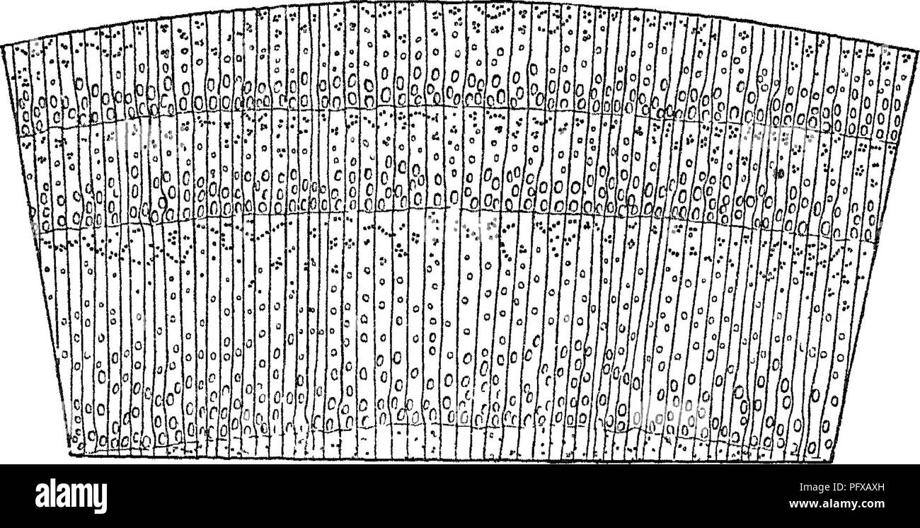 . Report upon the forestry investigations of the U. S. Department of agriculture. 1877-1898. Forests and forestry. 102 FORESTRY INVESTIGATIONS U. S. DEPARTMENT OF AGRICULTURE. Section II. Walls of tracheitis of pith ray smooth, without dentate projections. a. One or two large pits to each tracheid on the radial walls of each cell of the pith ray.—Group 3. P. strobus, lambertiana, and other true white pines. b. Three to six small pits on the radial walls of each cell of the pith ray.—Group 4. P. parryana, and other nut pines, including also 7*. balfouriana. II, Ring-porous Woods. [Some of Group Stock Photohttps://www.alamy.com/image-license-details/?v=1https://www.alamy.com/report-upon-the-forestry-investigations-of-the-u-s-department-of-agriculture-1877-1898-forests-and-forestry-102-forestry-investigations-u-s-department-of-agriculture-section-ii-walls-of-tracheitis-of-pith-ray-smooth-without-dentate-projections-a-one-or-two-large-pits-to-each-tracheid-on-the-radial-walls-of-each-cell-of-the-pith-raygroup-3-p-strobus-lambertiana-and-other-true-white-pines-b-three-to-six-small-pits-on-the-radial-walls-of-each-cell-of-the-pith-raygroup-4-p-parryana-and-other-nut-pines-including-also-7-balfouriana-ii-ring-porous-woods-some-of-group-image216323593.html
RMPFXAXH–. Report upon the forestry investigations of the U. S. Department of agriculture. 1877-1898. Forests and forestry. 102 FORESTRY INVESTIGATIONS U. S. DEPARTMENT OF AGRICULTURE. Section II. Walls of tracheitis of pith ray smooth, without dentate projections. a. One or two large pits to each tracheid on the radial walls of each cell of the pith ray.—Group 3. P. strobus, lambertiana, and other true white pines. b. Three to six small pits on the radial walls of each cell of the pith ray.—Group 4. P. parryana, and other nut pines, including also 7*. balfouriana. II, Ring-porous Woods. [Some of Group
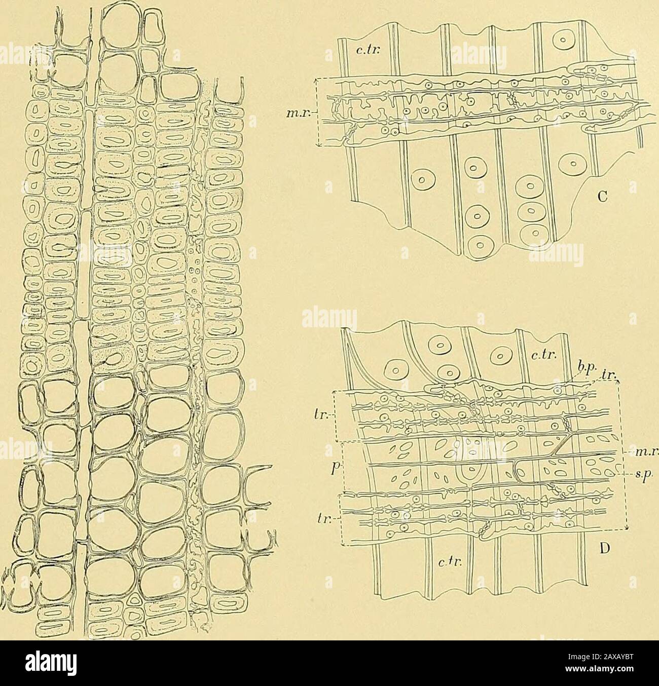 Report upon the forestry investigations of the U.SDepartment of agriculture1877-1898 . Typical Cross Sections of Pinus t/eda, heterophylla, and glabra. )•. d., resiD ducts; s. c, secreting cells; m. r., medullaiy rays; sp. w., spring wood; sii. ;c,,sumnieL- wood.4 Piws T.EDA ? a-b. transverse traoheids; c, simple pits; rf-e, row of tracheids; /, flattened terminal of tracheid.b Pinus heterophylla : a-b, row of tracheids; c, terminal of tracheid; d-e, bordered pits.c, Pinus glabra ; a-6, single row of tracheids; c-b, same row doubled. rt. |^j^^a^ j p. ^rn^S 0 ^ 1° J Ip R k) ^ r ® :^ I °J 0 Stock Photohttps://www.alamy.com/image-license-details/?v=1https://www.alamy.com/report-upon-the-forestry-investigations-of-the-usdepartment-of-agriculture1877-1898-typical-cross-sections-of-pinus-teda-heterophylla-and-glabra-d-resid-ducts-s-c-secreting-cells-m-r-medullaiy-rays-sp-w-spring-wood-sii-csumniel-wood4-piws-teda-a-b-transverse-traoheids-c-simple-pits-rf-e-row-of-tracheids-flattened-terminal-of-tracheidb-pinus-heterophylla-a-b-row-of-tracheids-c-terminal-of-tracheid-d-e-bordered-pitsc-pinus-glabra-a-6-single-row-of-tracheids-c-b-same-row-doubled-rt-ja-j-p-rns-0-1-j-ip-r-k-r-i-j-0-image343219068.html
RM2AXAYBT–Report upon the forestry investigations of the U.SDepartment of agriculture1877-1898 . Typical Cross Sections of Pinus t/eda, heterophylla, and glabra. )•. d., resiD ducts; s. c, secreting cells; m. r., medullaiy rays; sp. w., spring wood; sii. ;c,,sumnieL- wood.4 Piws T.EDA ? a-b. transverse traoheids; c, simple pits; rf-e, row of tracheids; /, flattened terminal of tracheid.b Pinus heterophylla : a-b, row of tracheids; c, terminal of tracheid; d-e, bordered pits.c, Pinus glabra ; a-6, single row of tracheids; c-b, same row doubled. rt. |^j^^a^ j p. ^rn^S 0 ^ 1° J Ip R k) ^ r ® :^ I °J 0
![. Fossil plants : for students of botany and geology . Paleobotany. X] LEAVES OF CALAMITES. 381 epidermal layer with a limiting cuticle. Internal to this we have radially elongated parenchymatous cells forming a loose or spongy tissue, the cells being often separated by fairly large spaces. Pig. 86. A leaf of Calamites. 1. Transverse section; t, vascular bundle; a;, aheath of cells, x 35. 2. Vascular bundle consisting of a few small tracheids, t. 3. A tracheid and a few parenebymatous cells, the latter with nuclei. 4. A stoma; s, s, guard-cells. 5. PaUisade cells and intercellular spaces. From Stock Photo . Fossil plants : for students of botany and geology . Paleobotany. X] LEAVES OF CALAMITES. 381 epidermal layer with a limiting cuticle. Internal to this we have radially elongated parenchymatous cells forming a loose or spongy tissue, the cells being often separated by fairly large spaces. Pig. 86. A leaf of Calamites. 1. Transverse section; t, vascular bundle; a;, aheath of cells, x 35. 2. Vascular bundle consisting of a few small tracheids, t. 3. A tracheid and a few parenebymatous cells, the latter with nuclei. 4. A stoma; s, s, guard-cells. 5. PaUisade cells and intercellular spaces. From Stock Photo](https://c8.alamy.com/comp/PG0DBR/fossil-plants-for-students-of-botany-and-geology-paleobotany-x-leaves-of-calamites-381-epidermal-layer-with-a-limiting-cuticle-internal-to-this-we-have-radially-elongated-parenchymatous-cells-forming-a-loose-or-spongy-tissue-the-cells-being-often-separated-by-fairly-large-spaces-pig-86-a-leaf-of-calamites-1-transverse-section-t-vascular-bundle-a-aheath-of-cells-x-35-2-vascular-bundle-consisting-of-a-few-small-tracheids-t-3-a-tracheid-and-a-few-parenebymatous-cells-the-latter-with-nuclei-4-a-stoma-s-s-guard-cells-5-pauisade-cells-and-intercellular-spaces-from-PG0DBR.jpg) . Fossil plants : for students of botany and geology . Paleobotany. X] LEAVES OF CALAMITES. 381 epidermal layer with a limiting cuticle. Internal to this we have radially elongated parenchymatous cells forming a loose or spongy tissue, the cells being often separated by fairly large spaces. Pig. 86. A leaf of Calamites. 1. Transverse section; t, vascular bundle; a;, aheath of cells, x 35. 2. Vascular bundle consisting of a few small tracheids, t. 3. A tracheid and a few parenebymatous cells, the latter with nuclei. 4. A stoma; s, s, guard-cells. 5. PaUisade cells and intercellular spaces. From Stock Photohttps://www.alamy.com/image-license-details/?v=1https://www.alamy.com/fossil-plants-for-students-of-botany-and-geology-paleobotany-x-leaves-of-calamites-381-epidermal-layer-with-a-limiting-cuticle-internal-to-this-we-have-radially-elongated-parenchymatous-cells-forming-a-loose-or-spongy-tissue-the-cells-being-often-separated-by-fairly-large-spaces-pig-86-a-leaf-of-calamites-1-transverse-section-t-vascular-bundle-a-aheath-of-cells-x-35-2-vascular-bundle-consisting-of-a-few-small-tracheids-t-3-a-tracheid-and-a-few-parenebymatous-cells-the-latter-with-nuclei-4-a-stoma-s-s-guard-cells-5-pauisade-cells-and-intercellular-spaces-from-image216369435.html
. Fossil plants : for students of botany and geology . Paleobotany. X] LEAVES OF CALAMITES. 381 epidermal layer with a limiting cuticle. Internal to this we have radially elongated parenchymatous cells forming a loose or spongy tissue, the cells being often separated by fairly large spaces. Pig. 86. A leaf of Calamites. 1. Transverse section; t, vascular bundle; a;, aheath of cells, x 35. 2. Vascular bundle consisting of a few small tracheids, t. 3. A tracheid and a few parenebymatous cells, the latter with nuclei. 4. A stoma; s, s, guard-cells. 5. PaUisade cells and intercellular spaces. From Stock Photohttps://www.alamy.com/image-license-details/?v=1https://www.alamy.com/fossil-plants-for-students-of-botany-and-geology-paleobotany-x-leaves-of-calamites-381-epidermal-layer-with-a-limiting-cuticle-internal-to-this-we-have-radially-elongated-parenchymatous-cells-forming-a-loose-or-spongy-tissue-the-cells-being-often-separated-by-fairly-large-spaces-pig-86-a-leaf-of-calamites-1-transverse-section-t-vascular-bundle-a-aheath-of-cells-x-35-2-vascular-bundle-consisting-of-a-few-small-tracheids-t-3-a-tracheid-and-a-few-parenebymatous-cells-the-latter-with-nuclei-4-a-stoma-s-s-guard-cells-5-pauisade-cells-and-intercellular-spaces-from-image216369435.htmlRMPG0DBR–. Fossil plants : for students of botany and geology . Paleobotany. X] LEAVES OF CALAMITES. 381 epidermal layer with a limiting cuticle. Internal to this we have radially elongated parenchymatous cells forming a loose or spongy tissue, the cells being often separated by fairly large spaces. Pig. 86. A leaf of Calamites. 1. Transverse section; t, vascular bundle; a;, aheath of cells, x 35. 2. Vascular bundle consisting of a few small tracheids, t. 3. A tracheid and a few parenebymatous cells, the latter with nuclei. 4. A stoma; s, s, guard-cells. 5. PaUisade cells and intercellular spaces. From
![. Botanical gazette. Plants. igig] BELYE A—SEQUOIA 469. Fig. I.—Sequoia washingtoniana (virgin growth): radial section showing ordinary typt of isolated detached ray tracheid.. Please note that these images are extracted from scanned page images that may have been digitally enhanced for readability - coloration and appearance of these illustrations may not perfectly resemble the original work.. Hanover, Ind. : J. M. Coulter Stock Photo . Botanical gazette. Plants. igig] BELYE A—SEQUOIA 469. Fig. I.—Sequoia washingtoniana (virgin growth): radial section showing ordinary typt of isolated detached ray tracheid.. Please note that these images are extracted from scanned page images that may have been digitally enhanced for readability - coloration and appearance of these illustrations may not perfectly resemble the original work.. Hanover, Ind. : J. M. Coulter Stock Photo](https://c8.alamy.com/comp/RHB23X/botanical-gazette-plants-igig-belye-asequoia-469-fig-isequoia-washingtoniana-virgin-growth-radial-section-showing-ordinary-typt-of-isolated-detached-ray-tracheid-please-note-that-these-images-are-extracted-from-scanned-page-images-that-may-have-been-digitally-enhanced-for-readability-coloration-and-appearance-of-these-illustrations-may-not-perfectly-resemble-the-original-work-hanover-ind-j-m-coulter-RHB23X.jpg) . Botanical gazette. Plants. igig] BELYE A—SEQUOIA 469. Fig. I.—Sequoia washingtoniana (virgin growth): radial section showing ordinary typt of isolated detached ray tracheid.. Please note that these images are extracted from scanned page images that may have been digitally enhanced for readability - coloration and appearance of these illustrations may not perfectly resemble the original work.. Hanover, Ind. : J. M. Coulter Stock Photohttps://www.alamy.com/image-license-details/?v=1https://www.alamy.com/botanical-gazette-plants-igig-belye-asequoia-469-fig-isequoia-washingtoniana-virgin-growth-radial-section-showing-ordinary-typt-of-isolated-detached-ray-tracheid-please-note-that-these-images-are-extracted-from-scanned-page-images-that-may-have-been-digitally-enhanced-for-readability-coloration-and-appearance-of-these-illustrations-may-not-perfectly-resemble-the-original-work-hanover-ind-j-m-coulter-image234427086.html
. Botanical gazette. Plants. igig] BELYE A—SEQUOIA 469. Fig. I.—Sequoia washingtoniana (virgin growth): radial section showing ordinary typt of isolated detached ray tracheid.. Please note that these images are extracted from scanned page images that may have been digitally enhanced for readability - coloration and appearance of these illustrations may not perfectly resemble the original work.. Hanover, Ind. : J. M. Coulter Stock Photohttps://www.alamy.com/image-license-details/?v=1https://www.alamy.com/botanical-gazette-plants-igig-belye-asequoia-469-fig-isequoia-washingtoniana-virgin-growth-radial-section-showing-ordinary-typt-of-isolated-detached-ray-tracheid-please-note-that-these-images-are-extracted-from-scanned-page-images-that-may-have-been-digitally-enhanced-for-readability-coloration-and-appearance-of-these-illustrations-may-not-perfectly-resemble-the-original-work-hanover-ind-j-m-coulter-image234427086.htmlRMRHB23X–. Botanical gazette. Plants. igig] BELYE A—SEQUOIA 469. Fig. I.—Sequoia washingtoniana (virgin growth): radial section showing ordinary typt of isolated detached ray tracheid.. Please note that these images are extracted from scanned page images that may have been digitally enhanced for readability - coloration and appearance of these illustrations may not perfectly resemble the original work.. Hanover, Ind. : J. M. Coulter
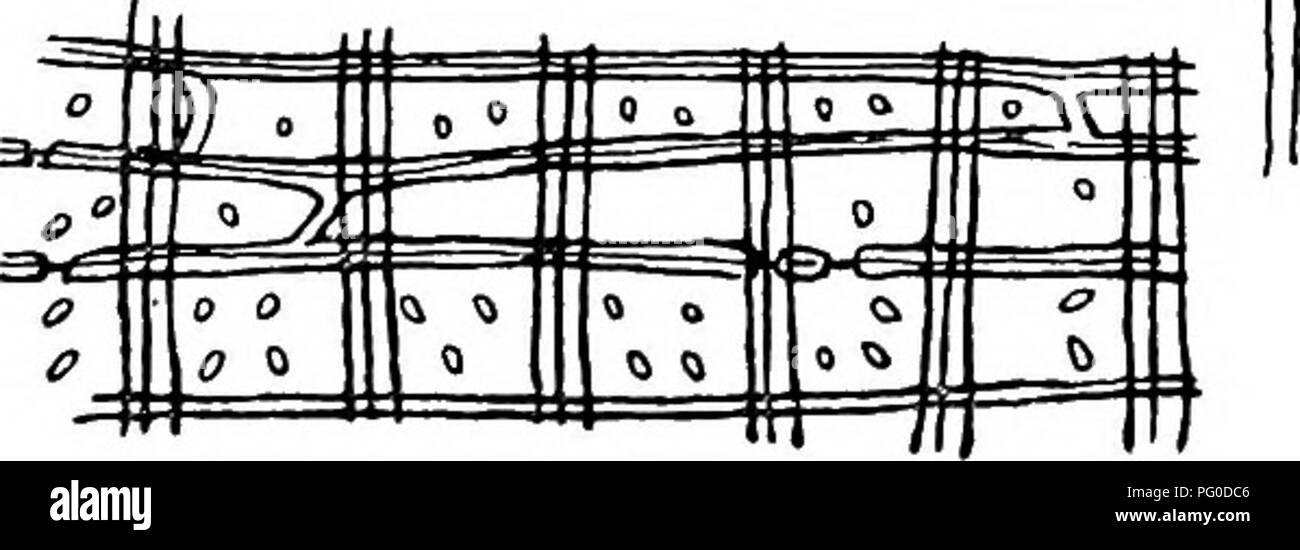 . Fossil plants : for students of botany and geology . Paleobotany. M' O Fig. 693. A, Abies Veitchii, medullary ray. B, Pinus silvestris, medullary-ray tracheid. C, Abies balsamea, pits and Sanio's rims. T>, E, Pits in medullary-ray cells in Abies homolepis. F, Pits in tangential wall of ray cell of Juniperus virginiana. G, H, K, L, M, N, 0, Pits in medullary-ray cells (radial view) in Cedrus atlantica (G); Taxodium distickum (H); Podocarpas andina (K); P. salici- folia (L); Olyptostrobus (M); Sciadopitys (N); sp, spring wood; s, summer wood; Thuya gigantea (0). I, Bars, b, in tracheids of Stock Photohttps://www.alamy.com/image-license-details/?v=1https://www.alamy.com/fossil-plants-for-students-of-botany-and-geology-paleobotany-m-o-fig-693-a-abies-veitchii-medullary-ray-b-pinus-silvestris-medullary-ray-tracheid-c-abies-balsamea-pits-and-sanios-rims-tgt-e-pits-in-medullary-ray-cells-in-abies-homolepis-f-pits-in-tangential-wall-of-ray-cell-of-juniperus-virginiana-g-h-k-l-m-n-0-pits-in-medullary-ray-cells-radial-view-in-cedrus-atlantica-g-taxodium-distickum-h-podocarpas-andina-k-p-salici-folia-l-olyptostrobus-m-sciadopitys-n-sp-spring-wood-s-summer-wood-thuya-gigantea-0-i-bars-b-in-tracheids-of-image216369446.html
RMPG0DC6–. Fossil plants : for students of botany and geology . Paleobotany. M' O Fig. 693. A, Abies Veitchii, medullary ray. B, Pinus silvestris, medullary-ray tracheid. C, Abies balsamea, pits and Sanio's rims. T>, E, Pits in medullary-ray cells in Abies homolepis. F, Pits in tangential wall of ray cell of Juniperus virginiana. G, H, K, L, M, N, 0, Pits in medullary-ray cells (radial view) in Cedrus atlantica (G); Taxodium distickum (H); Podocarpas andina (K); P. salici- folia (L); Olyptostrobus (M); Sciadopitys (N); sp, spring wood; s, summer wood; Thuya gigantea (0). I, Bars, b, in tracheids of
 . The structure and development of mosses and ferns (Archegoniatae). Plant morphology; Mosses; Ferns. FILICINE^ LEPTOSPORANGIATM 331 tracheid (Fig. 184, B). In cross-section these bars are nearly rhomboidal, and give the famihar beaded appearance to sections of the tracheid wall. Sieve-tubes of very characteristic form are found in the bundles of all the Polypodiacese. In 0. strutliiopteris they occupy an irregular area at each end of the bundle. Their differentiation begins shortly after that of the large scalariform tracheids, and in some respects resembles it. The procambium cells from whic Stock Photohttps://www.alamy.com/image-license-details/?v=1https://www.alamy.com/the-structure-and-development-of-mosses-and-ferns-archegoniatae-plant-morphology-mosses-ferns-filicine-leptosporangiatm-331-tracheid-fig-184-b-in-cross-section-these-bars-are-nearly-rhomboidal-and-give-the-famihar-beaded-appearance-to-sections-of-the-tracheid-wall-sieve-tubes-of-very-characteristic-form-are-found-in-the-bundles-of-all-the-polypodiacese-in-0-strutliiopteris-they-occupy-an-irregular-area-at-each-end-of-the-bundle-their-differentiation-begins-shortly-after-that-of-the-large-scalariform-tracheids-and-in-some-respects-resembles-it-the-procambium-cells-from-whic-image232064095.html
RMRDFC3B–. The structure and development of mosses and ferns (Archegoniatae). Plant morphology; Mosses; Ferns. FILICINE^ LEPTOSPORANGIATM 331 tracheid (Fig. 184, B). In cross-section these bars are nearly rhomboidal, and give the famihar beaded appearance to sections of the tracheid wall. Sieve-tubes of very characteristic form are found in the bundles of all the Polypodiacese. In 0. strutliiopteris they occupy an irregular area at each end of the bundle. Their differentiation begins shortly after that of the large scalariform tracheids, and in some respects resembles it. The procambium cells from whic
 . The anatomy of woody plants. Botany -- Anatomy. 66 THE ANATOMY OF WOODY PLANTS according to common consent, find their successors in the conifers of the present age. Since it is highly probable that the Cordaitales gave origin to the Coniferales, and since, as a consequence, they present us with. FIG. 48.—View of cordaitean wood in three dimensions. Explanation in the text. the starting-point of the radial parenchyma of the latter, it will be well to consider the organization of this group in respect to the structure of the rays. Fig. 48 shows the structure of a cordaitean wood. The tracheid Stock Photohttps://www.alamy.com/image-license-details/?v=1https://www.alamy.com/the-anatomy-of-woody-plants-botany-anatomy-66-the-anatomy-of-woody-plants-according-to-common-consent-find-their-successors-in-the-conifers-of-the-present-age-since-it-is-highly-probable-that-the-cordaitales-gave-origin-to-the-coniferales-and-since-as-a-consequence-they-present-us-with-fig-48view-of-cordaitean-wood-in-three-dimensions-explanation-in-the-text-the-starting-point-of-the-radial-parenchyma-of-the-latter-it-will-be-well-to-consider-the-organization-of-this-group-in-respect-to-the-structure-of-the-rays-fig-48-shows-the-structure-of-a-cordaitean-wood-the-tracheid-image236799590.html
RMRN7486–. The anatomy of woody plants. Botany -- Anatomy. 66 THE ANATOMY OF WOODY PLANTS according to common consent, find their successors in the conifers of the present age. Since it is highly probable that the Cordaitales gave origin to the Coniferales, and since, as a consequence, they present us with. FIG. 48.—View of cordaitean wood in three dimensions. Explanation in the text. the starting-point of the radial parenchyma of the latter, it will be well to consider the organization of this group in respect to the structure of the rays. Fig. 48 shows the structure of a cordaitean wood. The tracheid
![. A manual of the North American gymnosperms [microform] : exclusive of the cycadales but together with certain exotic species. Bois; Trees; Gymnosperms; Gymnospermes; Arbres; Wood. 188 ANATOMY OF THE GYMNOSPERMS free growth afforded by the long cavity of the tracheid or resin passage may serve somewhat to influence the direction of growth as the fungus searches for food, although, as in the previous case, it cannot be regarded as a determining factor of primary importance, inasmuch as there is a constant tendency to the formation of branches which traverse the wood at right angles to the wall Stock Photo . A manual of the North American gymnosperms [microform] : exclusive of the cycadales but together with certain exotic species. Bois; Trees; Gymnosperms; Gymnospermes; Arbres; Wood. 188 ANATOMY OF THE GYMNOSPERMS free growth afforded by the long cavity of the tracheid or resin passage may serve somewhat to influence the direction of growth as the fungus searches for food, although, as in the previous case, it cannot be regarded as a determining factor of primary importance, inasmuch as there is a constant tendency to the formation of branches which traverse the wood at right angles to the wall Stock Photo](https://c8.alamy.com/comp/REN2WP/a-manual-of-the-north-american-gymnosperms-microform-exclusive-of-the-cycadales-but-together-with-certain-exotic-species-bois-trees-gymnosperms-gymnospermes-arbres-wood-188-anatomy-of-the-gymnosperms-free-growth-afforded-by-the-long-cavity-of-the-tracheid-or-resin-passage-may-serve-somewhat-to-influence-the-direction-of-growth-as-the-fungus-searches-for-food-although-as-in-the-previous-case-it-cannot-be-regarded-as-a-determining-factor-of-primary-importance-inasmuch-as-there-is-a-constant-tendency-to-the-formation-of-branches-which-traverse-the-wood-at-right-angles-to-the-wall-REN2WP.jpg) . A manual of the North American gymnosperms [microform] : exclusive of the cycadales but together with certain exotic species. Bois; Trees; Gymnosperms; Gymnospermes; Arbres; Wood. 188 ANATOMY OF THE GYMNOSPERMS free growth afforded by the long cavity of the tracheid or resin passage may serve somewhat to influence the direction of growth as the fungus searches for food, although, as in the previous case, it cannot be regarded as a determining factor of primary importance, inasmuch as there is a constant tendency to the formation of branches which traverse the wood at right angles to the wall Stock Photohttps://www.alamy.com/image-license-details/?v=1https://www.alamy.com/a-manual-of-the-north-american-gymnosperms-microform-exclusive-of-the-cycadales-but-together-with-certain-exotic-species-bois-trees-gymnosperms-gymnospermes-arbres-wood-188-anatomy-of-the-gymnosperms-free-growth-afforded-by-the-long-cavity-of-the-tracheid-or-resin-passage-may-serve-somewhat-to-influence-the-direction-of-growth-as-the-fungus-searches-for-food-although-as-in-the-previous-case-it-cannot-be-regarded-as-a-determining-factor-of-primary-importance-inasmuch-as-there-is-a-constant-tendency-to-the-formation-of-branches-which-traverse-the-wood-at-right-angles-to-the-wall-image232803250.html
. A manual of the North American gymnosperms [microform] : exclusive of the cycadales but together with certain exotic species. Bois; Trees; Gymnosperms; Gymnospermes; Arbres; Wood. 188 ANATOMY OF THE GYMNOSPERMS free growth afforded by the long cavity of the tracheid or resin passage may serve somewhat to influence the direction of growth as the fungus searches for food, although, as in the previous case, it cannot be regarded as a determining factor of primary importance, inasmuch as there is a constant tendency to the formation of branches which traverse the wood at right angles to the wall Stock Photohttps://www.alamy.com/image-license-details/?v=1https://www.alamy.com/a-manual-of-the-north-american-gymnosperms-microform-exclusive-of-the-cycadales-but-together-with-certain-exotic-species-bois-trees-gymnosperms-gymnospermes-arbres-wood-188-anatomy-of-the-gymnosperms-free-growth-afforded-by-the-long-cavity-of-the-tracheid-or-resin-passage-may-serve-somewhat-to-influence-the-direction-of-growth-as-the-fungus-searches-for-food-although-as-in-the-previous-case-it-cannot-be-regarded-as-a-determining-factor-of-primary-importance-inasmuch-as-there-is-a-constant-tendency-to-the-formation-of-branches-which-traverse-the-wood-at-right-angles-to-the-wall-image232803250.htmlRMREN2WP–. A manual of the North American gymnosperms [microform] : exclusive of the cycadales but together with certain exotic species. Bois; Trees; Gymnosperms; Gymnospermes; Arbres; Wood. 188 ANATOMY OF THE GYMNOSPERMS free growth afforded by the long cavity of the tracheid or resin passage may serve somewhat to influence the direction of growth as the fungus searches for food, although, as in the previous case, it cannot be regarded as a determining factor of primary importance, inasmuch as there is a constant tendency to the formation of branches which traverse the wood at right angles to the wall
 . Botanical gazette. Plants. 470 BOTANICAL GAZETTE [December elongation is variable, as is also the height and the depth of the cell, which latter, however, approximates that of the ray with which it is associated. The pitting on the walls is very characteristic, espe- cially in horizontal contact with the parenchyma cell of the ray. Second, the interspersed type of ray tracheid, as shown in fig. 2, where the tracheary cells occur in the midst of the radial prolonga- tion of rays one cell high. They may be found singly, or in groups separated from other cells or groups of cells of similar stru Stock Photohttps://www.alamy.com/image-license-details/?v=1https://www.alamy.com/botanical-gazette-plants-470-botanical-gazette-december-elongation-is-variable-as-is-also-the-height-and-the-depth-of-the-cell-which-latter-however-approximates-that-of-the-ray-with-which-it-is-associated-the-pitting-on-the-walls-is-very-characteristic-espe-cially-in-horizontal-contact-with-the-parenchyma-cell-of-the-ray-second-the-interspersed-type-of-ray-tracheid-as-shown-in-fig-2-where-the-tracheary-cells-occur-in-the-midst-of-the-radial-prolonga-tion-of-rays-one-cell-high-they-may-be-found-singly-or-in-groups-separated-from-other-cells-or-groups-of-cells-of-similar-stru-image234427068.html
RMRHB238–. Botanical gazette. Plants. 470 BOTANICAL GAZETTE [December elongation is variable, as is also the height and the depth of the cell, which latter, however, approximates that of the ray with which it is associated. The pitting on the walls is very characteristic, espe- cially in horizontal contact with the parenchyma cell of the ray. Second, the interspersed type of ray tracheid, as shown in fig. 2, where the tracheary cells occur in the midst of the radial prolonga- tion of rays one cell high. They may be found singly, or in groups separated from other cells or groups of cells of similar stru
 . The anatomy of woody plants. Botany -- Anatomy. FIBROVASCULAR TISSUES: VESSELS 95 tracheid. It is distinguished by the fact that its end walls do not make a definite angle with the lateral ones but taper grad- ually. It is further peculiar in the circumstance that its terminal pits are imperforate with one exception and have their mem- branes marked by the presence of a very distinct torus. In c is figured a tangential view of a vessel in Ephedra, showing the enlarged terminal pores in profile and making it clear that in this case, as in Pteris aquilina, the vessels become patent by the dis- Stock Photohttps://www.alamy.com/image-license-details/?v=1https://www.alamy.com/the-anatomy-of-woody-plants-botany-anatomy-fibrovascular-tissues-vessels-95-tracheid-it-is-distinguished-by-the-fact-that-its-end-walls-do-not-make-a-definite-angle-with-the-lateral-ones-but-taper-grad-ually-it-is-further-peculiar-in-the-circumstance-that-its-terminal-pits-are-imperforate-with-one-exception-and-have-their-mem-branes-marked-by-the-presence-of-a-very-distinct-torus-in-c-is-figured-a-tangential-view-of-a-vessel-in-ephedra-showing-the-enlarged-terminal-pores-in-profile-and-making-it-clear-that-in-this-case-as-in-pteris-aquilina-the-vessels-become-patent-by-the-dis-image236799166.html
RMRN73N2–. The anatomy of woody plants. Botany -- Anatomy. FIBROVASCULAR TISSUES: VESSELS 95 tracheid. It is distinguished by the fact that its end walls do not make a definite angle with the lateral ones but taper grad- ually. It is further peculiar in the circumstance that its terminal pits are imperforate with one exception and have their mem- branes marked by the presence of a very distinct torus. In c is figured a tangential view of a vessel in Ephedra, showing the enlarged terminal pores in profile and making it clear that in this case, as in Pteris aquilina, the vessels become patent by the dis-
 . Botany of the living plant. Botany. THE TISSUES OF THE STEM 45 each of the largest strands on entering the stem from the leaf slants sharply inwards, but short of the centre it curves again outwards, and gradually approaches the periphery. There it fuses with other strands. As the strand is thickest in the middle of this course, the consequence is that the strands are fewest and largest at the centre, and smaller but more numerous near to the periphery {Fig. 31).. Fig. 32. Transverse section of a vascular strand from the internode of Zea.Mais. o=annular tracheid. s^ = spiral tracheid. w, w' Stock Photohttps://www.alamy.com/image-license-details/?v=1https://www.alamy.com/botany-of-the-living-plant-botany-the-tissues-of-the-stem-45-each-of-the-largest-strands-on-entering-the-stem-from-the-leaf-slants-sharply-inwards-but-short-of-the-centre-it-curves-again-outwards-and-gradually-approaches-the-periphery-there-it-fuses-with-other-strands-as-the-strand-is-thickest-in-the-middle-of-this-course-the-consequence-is-that-the-strands-are-fewest-and-largest-at-the-centre-and-smaller-but-more-numerous-near-to-the-periphery-fig-31-fig-32-transverse-section-of-a-vascular-strand-from-the-internode-of-zeamais-o=annular-tracheid-s-=-spiral-tracheid-w-w-image234383581.html
RMRH92J5–. Botany of the living plant. Botany. THE TISSUES OF THE STEM 45 each of the largest strands on entering the stem from the leaf slants sharply inwards, but short of the centre it curves again outwards, and gradually approaches the periphery. There it fuses with other strands. As the strand is thickest in the middle of this course, the consequence is that the strands are fewest and largest at the centre, and smaller but more numerous near to the periphery {Fig. 31).. Fig. 32. Transverse section of a vascular strand from the internode of Zea.Mais. o=annular tracheid. s^ = spiral tracheid. w, w'
 . The origin of a land flora, a theory based upon the facts of alternation. Plant morphology. Fig. 55. Casuarina Rumphiana, Mig. Median section of the nucellus of an ovule, with the group of sporogenous cells shaded. X285. (After Treub.). Fig. 56. Casuarina. glauca, Sieb. Median section of nucellus of an ovule showing the ceils of the sporogenous group differentiated: some are becoming elongated in the direction of the chalaza: one long cell has divided by six swollen walls: another has developed as a tracheid. X 285. (After Treub.) individual cells, which are normally vegetative, to the sporo Stock Photohttps://www.alamy.com/image-license-details/?v=1https://www.alamy.com/the-origin-of-a-land-flora-a-theory-based-upon-the-facts-of-alternation-plant-morphology-fig-55-casuarina-rumphiana-mig-median-section-of-the-nucellus-of-an-ovule-with-the-group-of-sporogenous-cells-shaded-x285-after-treub-fig-56-casuarina-glauca-sieb-median-section-of-nucellus-of-an-ovule-showing-the-ceils-of-the-sporogenous-group-differentiated-some-are-becoming-elongated-in-the-direction-of-the-chalaza-one-long-cell-has-divided-by-six-swollen-walls-another-has-developed-as-a-tracheid-x-285-after-treub-individual-cells-which-are-normally-vegetative-to-the-sporo-image232309166.html
RMRDXGKX–. The origin of a land flora, a theory based upon the facts of alternation. Plant morphology. Fig. 55. Casuarina Rumphiana, Mig. Median section of the nucellus of an ovule, with the group of sporogenous cells shaded. X285. (After Treub.). Fig. 56. Casuarina. glauca, Sieb. Median section of nucellus of an ovule showing the ceils of the sporogenous group differentiated: some are becoming elongated in the direction of the chalaza: one long cell has divided by six swollen walls: another has developed as a tracheid. X 285. (After Treub.) individual cells, which are normally vegetative, to the sporo
 . The anatomy of woody plants. Botany -- Anatomy. 44 THE ANATOMY OF WOODY PLANTS tangential walls, a feature commonly found in the case of fibrous elements in the terminal region of the annual ring in the conifers and allied groups. The septate tracheid is the most striking feature of the diagram. In this element numerous transverse walls interrupt the continuity of the central lumen. Certain of these transverse partitions are characterized by the pres- ence of bordered pits, while others offer to the eye pits of the simple type. In a third condition bordered and simple pits confront one anoth Stock Photohttps://www.alamy.com/image-license-details/?v=1https://www.alamy.com/the-anatomy-of-woody-plants-botany-anatomy-44-the-anatomy-of-woody-plants-tangential-walls-a-feature-commonly-found-in-the-case-of-fibrous-elements-in-the-terminal-region-of-the-annual-ring-in-the-conifers-and-allied-groups-the-septate-tracheid-is-the-most-striking-feature-of-the-diagram-in-this-element-numerous-transverse-walls-interrupt-the-continuity-of-the-central-lumen-certain-of-these-transverse-partitions-are-characterized-by-the-pres-ence-of-bordered-pits-while-others-offer-to-the-eye-pits-of-the-simple-type-in-a-third-condition-bordered-and-simple-pits-confront-one-anoth-image236799825.html
RMRN74GH–. The anatomy of woody plants. Botany -- Anatomy. 44 THE ANATOMY OF WOODY PLANTS tangential walls, a feature commonly found in the case of fibrous elements in the terminal region of the annual ring in the conifers and allied groups. The septate tracheid is the most striking feature of the diagram. In this element numerous transverse walls interrupt the continuity of the central lumen. Certain of these transverse partitions are characterized by the pres- ence of bordered pits, while others offer to the eye pits of the simple type. In a third condition bordered and simple pits confront one anoth
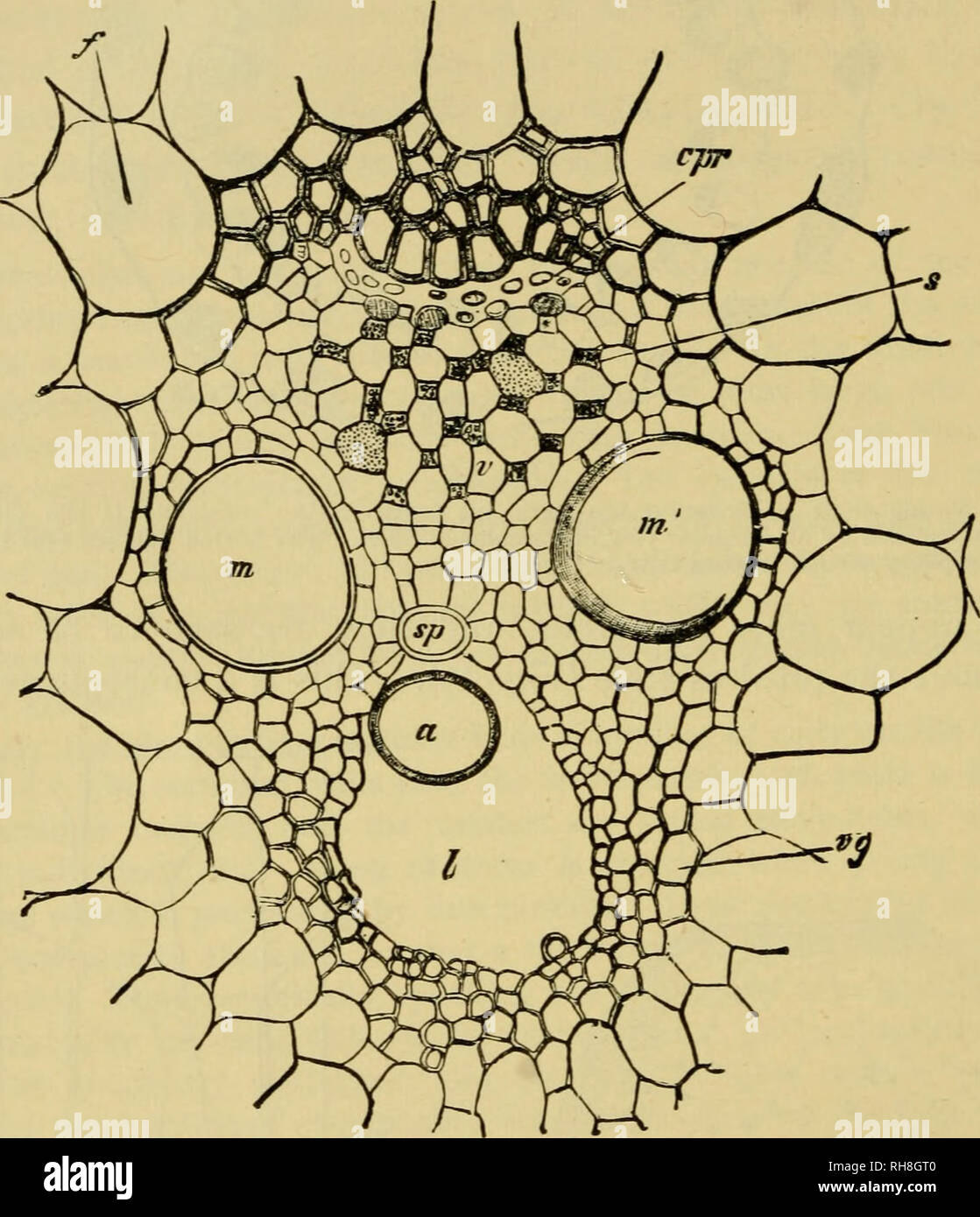 . Botany of the living plant. Botany; Plants. 52 BOTANY OF THE LIVING PLANT fact that each of the largest strands on entering the stem from the leaf slants sharply inwards, but short of the centre it curves again outwards, and gradually approaches the periphery. There it fuses with other strands. As the strand is thickest in the middle of this course, the consequence is that the strands appear fewest and largest at the centre, and smaller but more numerous near to the periphery. Fig. 31. Transverse section of a vascular strand from the internode of Zea Mais, a = annular tracheid. sp = spiral t Stock Photohttps://www.alamy.com/image-license-details/?v=1https://www.alamy.com/botany-of-the-living-plant-botany-plants-52-botany-of-the-living-plant-fact-that-each-of-the-largest-strands-on-entering-the-stem-from-the-leaf-slants-sharply-inwards-but-short-of-the-centre-it-curves-again-outwards-and-gradually-approaches-the-periphery-there-it-fuses-with-other-strands-as-the-strand-is-thickest-in-the-middle-of-this-course-the-consequence-is-that-the-strands-appear-fewest-and-largest-at-the-centre-and-smaller-but-more-numerous-near-to-the-periphery-fig-31-transverse-section-of-a-vascular-strand-from-the-internode-of-zea-mais-a-=-annular-tracheid-sp-=-spiral-t-image234372768.html
RMRH8GT0–. Botany of the living plant. Botany; Plants. 52 BOTANY OF THE LIVING PLANT fact that each of the largest strands on entering the stem from the leaf slants sharply inwards, but short of the centre it curves again outwards, and gradually approaches the periphery. There it fuses with other strands. As the strand is thickest in the middle of this course, the consequence is that the strands appear fewest and largest at the centre, and smaller but more numerous near to the periphery. Fig. 31. Transverse section of a vascular strand from the internode of Zea Mais, a = annular tracheid. sp = spiral t
 . Botany of the living plant. Botany. THE TISSUES OF THE STEM 45 each of the largest strands on entering the stem from the leaf slants sharply inwards, but short of the centre it curves again outwards, and gradually approaches the periphery. Theie it fuses with other strands. As the strand is thickest in the middle of this course, the consequence is that the strands are fewest and largest at the centre, and smaller but more numerous near to the periphery (Fig. 31).. Fig. 32. Transverse section of a vascular strand from the internode of Zea.Mais. a = annular tracheid. s^ = spiral tracheid. m, m Stock Photohttps://www.alamy.com/image-license-details/?v=1https://www.alamy.com/botany-of-the-living-plant-botany-the-tissues-of-the-stem-45-each-of-the-largest-strands-on-entering-the-stem-from-the-leaf-slants-sharply-inwards-but-short-of-the-centre-it-curves-again-outwards-and-gradually-approaches-the-periphery-theie-it-fuses-with-other-strands-as-the-strand-is-thickest-in-the-middle-of-this-course-the-consequence-is-that-the-strands-are-fewest-and-largest-at-the-centre-and-smaller-but-more-numerous-near-to-the-periphery-fig-31-fig-32-transverse-section-of-a-vascular-strand-from-the-internode-of-zeamais-a-=-annular-tracheid-s-=-spiral-tracheid-m-m-image232325467.html
RMRDY9E3–. Botany of the living plant. Botany. THE TISSUES OF THE STEM 45 each of the largest strands on entering the stem from the leaf slants sharply inwards, but short of the centre it curves again outwards, and gradually approaches the periphery. Theie it fuses with other strands. As the strand is thickest in the middle of this course, the consequence is that the strands are fewest and largest at the centre, and smaller but more numerous near to the periphery (Fig. 31).. Fig. 32. Transverse section of a vascular strand from the internode of Zea.Mais. a = annular tracheid. s^ = spiral tracheid. m, m
 . A compendium of general botany. Plants. THE CELL. 37 SOW, Steasbueger, and others). Upon these discoveries future investigators may base important conclusions of a widely diflPerent nature; as, for example, the transmission of irritability. This will not, however, be further mentioned at this point; we will main- tain that pores facilitate the interchange of fluids from cell to cell, as well as between cell and vessel (tracheid). These pores or thin cell-wall areas are often of circular or oval form, again linear or simply fissure-like. The accompanying figure represents various forms of ord Stock Photohttps://www.alamy.com/image-license-details/?v=1https://www.alamy.com/a-compendium-of-general-botany-plants-the-cell-37-sow-steasbueger-and-others-upon-these-discoveries-future-investigators-may-base-important-conclusions-of-a-widely-diflperent-nature-as-for-example-the-transmission-of-irritability-this-will-not-however-be-further-mentioned-at-this-point-we-will-main-tain-that-pores-facilitate-the-interchange-of-fluids-from-cell-to-cell-as-well-as-between-cell-and-vessel-tracheid-these-pores-or-thin-cell-wall-areas-are-often-of-circular-or-oval-form-again-linear-or-simply-fissure-like-the-accompanying-figure-represents-various-forms-of-ord-image232672960.html
RMREF4MG–. A compendium of general botany. Plants. THE CELL. 37 SOW, Steasbueger, and others). Upon these discoveries future investigators may base important conclusions of a widely diflPerent nature; as, for example, the transmission of irritability. This will not, however, be further mentioned at this point; we will main- tain that pores facilitate the interchange of fluids from cell to cell, as well as between cell and vessel (tracheid). These pores or thin cell-wall areas are often of circular or oval form, again linear or simply fissure-like. The accompanying figure represents various forms of ord
 . Identification of the economic woods of the United States : including a discussion of the structural and physical properties of wood . Wood; Trees. ECONOMIC WOODS OF THE UNITED STATES 33 bordered in the vessel or tracheid, and simple in the adjacent parenchyma cells (Fig. 11, F). Pits in typical wood fibres are simple and slit-like, and usually in oblique position (Figs. 11, K; 2,B). In many cases, however, where the fibres resemble tracheids their pits are more or less bordered. The fibres of the bast have only simple pits. The shape of the border is commonly circular, but may be oval, lent Stock Photohttps://www.alamy.com/image-license-details/?v=1https://www.alamy.com/identification-of-the-economic-woods-of-the-united-states-including-a-discussion-of-the-structural-and-physical-properties-of-wood-wood-trees-economic-woods-of-the-united-states-33-bordered-in-the-vessel-or-tracheid-and-simple-in-the-adjacent-parenchyma-cells-fig-11-f-pits-in-typical-wood-fibres-are-simple-and-slit-like-and-usually-in-oblique-position-figs-11-k-2b-in-many-cases-however-where-the-fibres-resemble-tracheids-their-pits-are-more-or-less-bordered-the-fibres-of-the-bast-have-only-simple-pits-the-shape-of-the-border-is-commonly-circular-but-may-be-oval-lent-image232202278.html
RMRDNMAE–. Identification of the economic woods of the United States : including a discussion of the structural and physical properties of wood . Wood; Trees. ECONOMIC WOODS OF THE UNITED STATES 33 bordered in the vessel or tracheid, and simple in the adjacent parenchyma cells (Fig. 11, F). Pits in typical wood fibres are simple and slit-like, and usually in oblique position (Figs. 11, K; 2,B). In many cases, however, where the fibres resemble tracheids their pits are more or less bordered. The fibres of the bast have only simple pits. The shape of the border is commonly circular, but may be oval, lent
 . A textbook of botany for colleges and universities ... Botany. 53^ ECOLOGY. Fig. 772. — A longitudiaal section through a portion of the stem of .5o/«:orttMi, showing oblique palisades (^), and also a "storage" tracheid (t); the arrow is directed toward the stem apex; jf, stoma; considerably magnified. to the leaf surface, regardless of the position of the leaf in relation to light. The position of the palisade layers seems relatively fixed in most cases (not in Lactuca and in Eucalyptus), a rudimentary palisade region often being discernible in the bud; in sub- sequent development Stock Photohttps://www.alamy.com/image-license-details/?v=1https://www.alamy.com/a-textbook-of-botany-for-colleges-and-universities-botany-53-ecology-fig-772-a-longitudiaal-section-through-a-portion-of-the-stem-of-5oorttmi-showing-oblique-palisades-and-also-a-quotstoragequot-tracheid-t-the-arrow-is-directed-toward-the-stem-apex-jf-stoma-considerably-magnified-to-the-leaf-surface-regardless-of-the-position-of-the-leaf-in-relation-to-light-the-position-of-the-palisade-layers-seems-relatively-fixed-in-most-cases-not-in-lactuca-and-in-eucalyptus-a-rudimentary-palisade-region-often-being-discernible-in-the-bud-in-sub-sequent-development-image232099131.html
RMRDH0PK–. A textbook of botany for colleges and universities ... Botany. 53^ ECOLOGY. Fig. 772. — A longitudiaal section through a portion of the stem of .5o/«:orttMi, showing oblique palisades (^), and also a "storage" tracheid (t); the arrow is directed toward the stem apex; jf, stoma; considerably magnified. to the leaf surface, regardless of the position of the leaf in relation to light. The position of the palisade layers seems relatively fixed in most cases (not in Lactuca and in Eucalyptus), a rudimentary palisade region often being discernible in the bud; in sub- sequent development
 . The anatomy of woody plants. Botany -- Anatomy. 12 THE ANATOMY OF WOODY PLANTS than in living pines. The fibrovascular strand is sharply bounded in the median region by the endodermis, a layer circular in con- figuration and well developed in Pinus, though often absent in the higher conifers. The organization of the fibrovascular strand need not particularly occupy our attention at this stage, as it will be considered in detail more appropriately in the sequel. It is enough to note that its upper or woody part is composed of empty cells often showing bordered pits—in other words, of tracheid Stock Photohttps://www.alamy.com/image-license-details/?v=1https://www.alamy.com/the-anatomy-of-woody-plants-botany-anatomy-12-the-anatomy-of-woody-plants-than-in-living-pines-the-fibrovascular-strand-is-sharply-bounded-in-the-median-region-by-the-endodermis-a-layer-circular-in-con-figuration-and-well-developed-in-pinus-though-often-absent-in-the-higher-conifers-the-organization-of-the-fibrovascular-strand-need-not-particularly-occupy-our-attention-at-this-stage-as-it-will-be-considered-in-detail-more-appropriately-in-the-sequel-it-is-enough-to-note-that-its-upper-or-woody-part-is-composed-of-empty-cells-often-showing-bordered-pitsin-other-words-of-tracheid-image236800142.html
RMRN74YX–. The anatomy of woody plants. Botany -- Anatomy. 12 THE ANATOMY OF WOODY PLANTS than in living pines. The fibrovascular strand is sharply bounded in the median region by the endodermis, a layer circular in con- figuration and well developed in Pinus, though often absent in the higher conifers. The organization of the fibrovascular strand need not particularly occupy our attention at this stage, as it will be considered in detail more appropriately in the sequel. It is enough to note that its upper or woody part is composed of empty cells often showing bordered pits—in other words, of tracheid
 . A manual of botany. Botany. 318 MANUAL OF BOTANY thickened in the same way as the lateral ones (fig. 683). The individual cells are then known as trache'ids. Tracheids occur in other forms than as the cells of a column ; thus they constitute a somewhat parenchymatous-looking tissue in the sheath of the aerial roots of certain orchids. They are always lignifled and usually pitted. There is no very sharp distinction between a fibre and a tracheid when the latter does not appear as a segment of a vessel. Thus the peculiar fibres with bordered pits, occurring in the secondary wood of the Coni- f Stock Photohttps://www.alamy.com/image-license-details/?v=1https://www.alamy.com/a-manual-of-botany-botany-318-manual-of-botany-thickened-in-the-same-way-as-the-lateral-ones-fig-683-the-individual-cells-are-then-known-as-tracheids-tracheids-occur-in-other-forms-than-as-the-cells-of-a-column-thus-they-constitute-a-somewhat-parenchymatous-looking-tissue-in-the-sheath-of-the-aerial-roots-of-certain-orchids-they-are-always-lignifled-and-usually-pitted-there-is-no-very-sharp-distinction-between-a-fibre-and-a-tracheid-when-the-latter-does-not-appear-as-a-segment-of-a-vessel-thus-the-peculiar-fibres-with-bordered-pits-occurring-in-the-secondary-wood-of-the-coni-f-image232376377.html
RMRE1JC9–. A manual of botany. Botany. 318 MANUAL OF BOTANY thickened in the same way as the lateral ones (fig. 683). The individual cells are then known as trache'ids. Tracheids occur in other forms than as the cells of a column ; thus they constitute a somewhat parenchymatous-looking tissue in the sheath of the aerial roots of certain orchids. They are always lignifled and usually pitted. There is no very sharp distinction between a fibre and a tracheid when the latter does not appear as a segment of a vessel. Thus the peculiar fibres with bordered pits, occurring in the secondary wood of the Coni- f
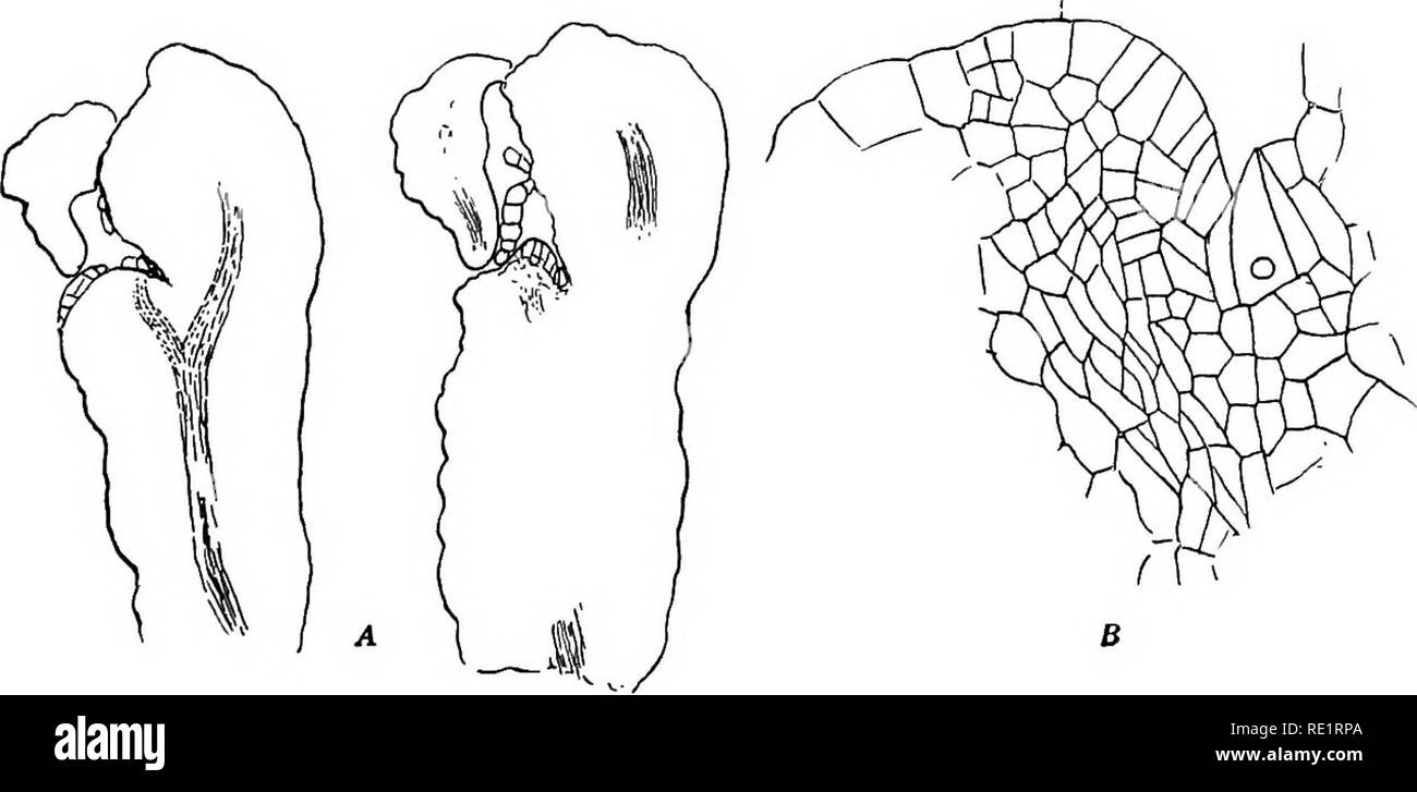 . The Eusporangiatae; the comparative morphology of the Ophioglossaceae and Marattiaceae. Ophioglossaceae; Marattiaceae. Fig. 131. A. Section of petiole of cotyledon of Z)a«£Cd e////jr;c^. X75. B. The vascular bundle. X200. C. Section of a peltate scale. X75. The section of the petiole of the cotyledon in Danaa (fig. 131) is nearly cir- cular, and the wings found in the other forms are almost entirely obhterated. The ground tissue is composed of undifferentiated parenchyma, with no trace of tan- nin cells. The vascular bundle is circular in outline, with a small group of three or four tracheid Stock Photohttps://www.alamy.com/image-license-details/?v=1https://www.alamy.com/the-eusporangiatae-the-comparative-morphology-of-the-ophioglossaceae-and-marattiaceae-ophioglossaceae-marattiaceae-fig-131-a-section-of-petiole-of-cotyledon-of-zacd-ejrc-x75-b-the-vascular-bundle-x200-c-section-of-a-peltate-scale-x75-the-section-of-the-petiole-of-the-cotyledon-in-danaa-fig-131-is-nearly-cir-cular-and-the-wings-found-in-the-other-forms-are-almost-entirely-obhterated-the-ground-tissue-is-composed-of-undifferentiated-parenchyma-with-no-trace-of-tan-nin-cells-the-vascular-bundle-is-circular-in-outline-with-a-small-group-of-three-or-four-tracheid-image232380578.html
RMRE1RPA–. The Eusporangiatae; the comparative morphology of the Ophioglossaceae and Marattiaceae. Ophioglossaceae; Marattiaceae. Fig. 131. A. Section of petiole of cotyledon of Z)a«£Cd e////jr;c^. X75. B. The vascular bundle. X200. C. Section of a peltate scale. X75. The section of the petiole of the cotyledon in Danaa (fig. 131) is nearly cir- cular, and the wings found in the other forms are almost entirely obhterated. The ground tissue is composed of undifferentiated parenchyma, with no trace of tan- nin cells. The vascular bundle is circular in outline, with a small group of three or four tracheid
 . Botany; principles and problems. Botany. 100 BOTANY: PRINCIPLES AND PROBLEMS occur in the phloem, and some parenchyma is usually present there also. Wood.—The inner portion of the fibro-vascular cylinder consists of the wood or xyleyn, which provides mechanical rigidity for the stem and transports the stream of water and dissolved substances from root to leaf. As essential elements in the xylem. Fig. 52.—Types of cells found in wood. A, fiber. B, tracheid. C, vessel- cell, with ladder-like end wall. D, vessel-cell, with completely open or porous end wall. E, very short, broad, vessel-cell. F Stock Photohttps://www.alamy.com/image-license-details/?v=1https://www.alamy.com/botany-principles-and-problems-botany-100-botany-principles-and-problems-occur-in-the-phloem-and-some-parenchyma-is-usually-present-there-also-woodthe-inner-portion-of-the-fibro-vascular-cylinder-consists-of-the-wood-or-xyleyn-which-provides-mechanical-rigidity-for-the-stem-and-transports-the-stream-of-water-and-dissolved-substances-from-root-to-leaf-as-essential-elements-in-the-xylem-fig-52types-of-cells-found-in-wood-a-fiber-b-tracheid-c-vessel-cell-with-ladder-like-end-wall-d-vessel-cell-with-completely-open-or-porous-end-wall-e-very-short-broad-vessel-cell-f-image234373310.html
RMRH8HFA–. Botany; principles and problems. Botany. 100 BOTANY: PRINCIPLES AND PROBLEMS occur in the phloem, and some parenchyma is usually present there also. Wood.—The inner portion of the fibro-vascular cylinder consists of the wood or xyleyn, which provides mechanical rigidity for the stem and transports the stream of water and dissolved substances from root to leaf. As essential elements in the xylem. Fig. 52.—Types of cells found in wood. A, fiber. B, tracheid. C, vessel- cell, with ladder-like end wall. D, vessel-cell, with completely open or porous end wall. E, very short, broad, vessel-cell. F
 . Botanical gazette. Plants. Fig. I.—Sequoia washingtoniana (virgin growth): radial section showing ordinary typt of isolated detached ray tracheid.. Fig. 2.—Radial section (mature wood) showing ray with two tracheid cells inter- spersed between ray parenchyma cells; tracheids not marginal, but components of a ray one cell high.. Please note that these images are extracted from scanned page images that may have been digitally enhanced for readability - coloration and appearance of these illustrations may not perfectly resemble the original work.. Hanover, Ind. : J. M. Coulter Stock Photohttps://www.alamy.com/image-license-details/?v=1https://www.alamy.com/botanical-gazette-plants-fig-isequoia-washingtoniana-virgin-growth-radial-section-showing-ordinary-typt-of-isolated-detached-ray-tracheid-fig-2radial-section-mature-wood-showing-ray-with-two-tracheid-cells-inter-spersed-between-ray-parenchyma-cells-tracheids-not-marginal-but-components-of-a-ray-one-cell-high-please-note-that-these-images-are-extracted-from-scanned-page-images-that-may-have-been-digitally-enhanced-for-readability-coloration-and-appearance-of-these-illustrations-may-not-perfectly-resemble-the-original-work-hanover-ind-j-m-coulter-image234427076.html
RMRHB23G–. Botanical gazette. Plants. Fig. I.—Sequoia washingtoniana (virgin growth): radial section showing ordinary typt of isolated detached ray tracheid.. Fig. 2.—Radial section (mature wood) showing ray with two tracheid cells inter- spersed between ray parenchyma cells; tracheids not marginal, but components of a ray one cell high.. Please note that these images are extracted from scanned page images that may have been digitally enhanced for readability - coloration and appearance of these illustrations may not perfectly resemble the original work.. Hanover, Ind. : J. M. Coulter
 . An introduction to the structure and reproduction of plants. Plant anatomy; Plants. SECONDARY WOOD 121 can be related to the peculiar form of the cambial segments from vhich the vessels are derived. The perforations are varied (cf. p. 35 and Fig. 17, D-F), but the type in which a number of cross-bars remain is commoner than in the primarj' wood. Tracheids (cf. p. 37) differ from vessels in being derived from single segments of the cambium, which show no open perforations in the end-walls ; they are usually much shorter than the vessels, but of about the same idth. The vessels and tracheid Stock Photohttps://www.alamy.com/image-license-details/?v=1https://www.alamy.com/an-introduction-to-the-structure-and-reproduction-of-plants-plant-anatomy-plants-secondary-wood-121-can-be-related-to-the-peculiar-form-of-the-cambial-segments-from-vhich-the-vessels-are-derived-the-perforations-are-varied-cf-p-35-and-fig-17-d-f-but-the-type-in-which-a-number-of-cross-bars-remain-is-commoner-than-in-the-primarj-wood-tracheids-cf-p-37-differ-from-vessels-in-being-derived-from-single-segments-of-the-cambium-which-show-no-open-perforations-in-the-end-walls-they-are-usually-much-shorter-than-the-vessels-but-of-about-the-same-idth-the-vessels-and-tracheid-image232292443.html
RMRDWRAK–. An introduction to the structure and reproduction of plants. Plant anatomy; Plants. SECONDARY WOOD 121 can be related to the peculiar form of the cambial segments from vhich the vessels are derived. The perforations are varied (cf. p. 35 and Fig. 17, D-F), but the type in which a number of cross-bars remain is commoner than in the primarj' wood. Tracheids (cf. p. 37) differ from vessels in being derived from single segments of the cambium, which show no open perforations in the end-walls ; they are usually much shorter than the vessels, but of about the same idth. The vessels and tracheid
 . Electron microscopy; proceedings of the Stockholm Conference, September, 1956. Electron microscopy. 288 H. W. EMERTON, D. H. PAGE AND J. WATTS. All the micrographs are of solid replicas. The scale ellipses are 25 microns in diameter. The angles of illumination and viewing are 5" and 12 respectively. Fig. 1. Reflection electron micrographic montage (considerably reduced) of a beaten spruce sulphite tracheid. Fig. 2. Reflection electron micrograph of a thin-walled spruce sulphite tracheid, heavily beaten. Fig. 3. Reflection electron micrograph of the surface of a tissue paper made from he Stock Photohttps://www.alamy.com/image-license-details/?v=1https://www.alamy.com/electron-microscopy-proceedings-of-the-stockholm-conference-september-1956-electron-microscopy-288-h-w-emerton-d-h-page-and-j-watts-all-the-micrographs-are-of-solid-replicas-the-scale-ellipses-are-25-microns-in-diameter-the-angles-of-illumination-and-viewing-are-5quot-and-12-respectively-fig-1-reflection-electron-micrographic-montage-considerably-reduced-of-a-beaten-spruce-sulphite-tracheid-fig-2-reflection-electron-micrograph-of-a-thin-walled-spruce-sulphite-tracheid-heavily-beaten-fig-3-reflection-electron-micrograph-of-the-surface-of-a-tissue-paper-made-from-he-image231867453.html
RMRD6D8D–. Electron microscopy; proceedings of the Stockholm Conference, September, 1956. Electron microscopy. 288 H. W. EMERTON, D. H. PAGE AND J. WATTS. All the micrographs are of solid replicas. The scale ellipses are 25 microns in diameter. The angles of illumination and viewing are 5" and 12 respectively. Fig. 1. Reflection electron micrographic montage (considerably reduced) of a beaten spruce sulphite tracheid. Fig. 2. Reflection electron micrograph of a thin-walled spruce sulphite tracheid, heavily beaten. Fig. 3. Reflection electron micrograph of the surface of a tissue paper made from he
![. Fossil plants : for students of botany and geology . Paleobotany. XLIV] DADOXYLON 185 of growth.and with a single row of contiguous pits on the tracheid walls; Dadoxylon virginianum (Knowlton)^ from the Potomac series; Dadoxylon barremianum (Fliche)^ from the Lower Creta- ceous of France; Dadoxylon noveboracense (Holl. and Jefl.)^ from the Middle Cretaceous beds of Staten Island; Dadoxylon Zuffardii Negri * from middle Cretaceous rocks in the Gulf of Tripoli; Dado- xylon tanhoense (Stopes and Pujii)^ from Upper Cretaceous beds in Japan; Dadoxylon madagascariense (Fliche)® from Madagascar. Da Stock Photo . Fossil plants : for students of botany and geology . Paleobotany. XLIV] DADOXYLON 185 of growth.and with a single row of contiguous pits on the tracheid walls; Dadoxylon virginianum (Knowlton)^ from the Potomac series; Dadoxylon barremianum (Fliche)^ from the Lower Creta- ceous of France; Dadoxylon noveboracense (Holl. and Jefl.)^ from the Middle Cretaceous beds of Staten Island; Dadoxylon Zuffardii Negri * from middle Cretaceous rocks in the Gulf of Tripoli; Dado- xylon tanhoense (Stopes and Pujii)^ from Upper Cretaceous beds in Japan; Dadoxylon madagascariense (Fliche)® from Madagascar. Da Stock Photo](https://c8.alamy.com/comp/RDBC5N/fossil-plants-for-students-of-botany-and-geology-paleobotany-xliv-dadoxylon-185-of-growthand-with-a-single-row-of-contiguous-pits-on-the-tracheid-walls-dadoxylon-virginianum-knowlton-from-the-potomac-series-dadoxylon-barremianum-fliche-from-the-lower-creta-ceous-of-france-dadoxylon-noveboracense-holl-and-jefl-from-the-middle-cretaceous-beds-of-staten-island-dadoxylon-zuffardii-negri-from-middle-cretaceous-rocks-in-the-gulf-of-tripoli-dado-xylon-tanhoense-stopes-and-pujii-from-upper-cretaceous-beds-in-japan-dadoxylon-madagascariense-fliche-from-madagascar-da-RDBC5N.jpg) . Fossil plants : for students of botany and geology . Paleobotany. XLIV] DADOXYLON 185 of growth.and with a single row of contiguous pits on the tracheid walls; Dadoxylon virginianum (Knowlton)^ from the Potomac series; Dadoxylon barremianum (Fliche)^ from the Lower Creta- ceous of France; Dadoxylon noveboracense (Holl. and Jefl.)^ from the Middle Cretaceous beds of Staten Island; Dadoxylon Zuffardii Negri * from middle Cretaceous rocks in the Gulf of Tripoli; Dado- xylon tanhoense (Stopes and Pujii)^ from Upper Cretaceous beds in Japan; Dadoxylon madagascariense (Fliche)® from Madagascar. Da Stock Photohttps://www.alamy.com/image-license-details/?v=1https://www.alamy.com/fossil-plants-for-students-of-botany-and-geology-paleobotany-xliv-dadoxylon-185-of-growthand-with-a-single-row-of-contiguous-pits-on-the-tracheid-walls-dadoxylon-virginianum-knowlton-from-the-potomac-series-dadoxylon-barremianum-fliche-from-the-lower-creta-ceous-of-france-dadoxylon-noveboracense-holl-and-jefl-from-the-middle-cretaceous-beds-of-staten-island-dadoxylon-zuffardii-negri-from-middle-cretaceous-rocks-in-the-gulf-of-tripoli-dado-xylon-tanhoense-stopes-and-pujii-from-upper-cretaceous-beds-in-japan-dadoxylon-madagascariense-fliche-from-madagascar-da-image231976353.html
. Fossil plants : for students of botany and geology . Paleobotany. XLIV] DADOXYLON 185 of growth.and with a single row of contiguous pits on the tracheid walls; Dadoxylon virginianum (Knowlton)^ from the Potomac series; Dadoxylon barremianum (Fliche)^ from the Lower Creta- ceous of France; Dadoxylon noveboracense (Holl. and Jefl.)^ from the Middle Cretaceous beds of Staten Island; Dadoxylon Zuffardii Negri * from middle Cretaceous rocks in the Gulf of Tripoli; Dado- xylon tanhoense (Stopes and Pujii)^ from Upper Cretaceous beds in Japan; Dadoxylon madagascariense (Fliche)® from Madagascar. Da Stock Photohttps://www.alamy.com/image-license-details/?v=1https://www.alamy.com/fossil-plants-for-students-of-botany-and-geology-paleobotany-xliv-dadoxylon-185-of-growthand-with-a-single-row-of-contiguous-pits-on-the-tracheid-walls-dadoxylon-virginianum-knowlton-from-the-potomac-series-dadoxylon-barremianum-fliche-from-the-lower-creta-ceous-of-france-dadoxylon-noveboracense-holl-and-jefl-from-the-middle-cretaceous-beds-of-staten-island-dadoxylon-zuffardii-negri-from-middle-cretaceous-rocks-in-the-gulf-of-tripoli-dado-xylon-tanhoense-stopes-and-pujii-from-upper-cretaceous-beds-in-japan-dadoxylon-madagascariense-fliche-from-madagascar-da-image231976353.htmlRMRDBC5N–. Fossil plants : for students of botany and geology . Paleobotany. XLIV] DADOXYLON 185 of growth.and with a single row of contiguous pits on the tracheid walls; Dadoxylon virginianum (Knowlton)^ from the Potomac series; Dadoxylon barremianum (Fliche)^ from the Lower Creta- ceous of France; Dadoxylon noveboracense (Holl. and Jefl.)^ from the Middle Cretaceous beds of Staten Island; Dadoxylon Zuffardii Negri * from middle Cretaceous rocks in the Gulf of Tripoli; Dado- xylon tanhoense (Stopes and Pujii)^ from Upper Cretaceous beds in Japan; Dadoxylon madagascariense (Fliche)® from Madagascar. Da
 . A textbook of botany for colleges and universities ... Botany. Fig. 772. — A longitudiaal section through a portion of the stem of .5o/«:orttMi, showing oblique palisades (^), and also a "storage" tracheid (t); the arrow is directed toward the stem apex; jf, stoma; considerably magnified. to the leaf surface, regardless of the position of the leaf in relation to light. The position of the palisade layers seems relatively fixed in most cases (not in Lactuca and in Eucalyptus), a rudimentary palisade region often being discernible in the bud; in sub- sequent development the thickness Stock Photohttps://www.alamy.com/image-license-details/?v=1https://www.alamy.com/a-textbook-of-botany-for-colleges-and-universities-botany-fig-772-a-longitudiaal-section-through-a-portion-of-the-stem-of-5oorttmi-showing-oblique-palisades-and-also-a-quotstoragequot-tracheid-t-the-arrow-is-directed-toward-the-stem-apex-jf-stoma-considerably-magnified-to-the-leaf-surface-regardless-of-the-position-of-the-leaf-in-relation-to-light-the-position-of-the-palisade-layers-seems-relatively-fixed-in-most-cases-not-in-lactuca-and-in-eucalyptus-a-rudimentary-palisade-region-often-being-discernible-in-the-bud-in-sub-sequent-development-the-thickness-image232099128.html
RMRDH0PG–. A textbook of botany for colleges and universities ... Botany. Fig. 772. — A longitudiaal section through a portion of the stem of .5o/«:orttMi, showing oblique palisades (^), and also a "storage" tracheid (t); the arrow is directed toward the stem apex; jf, stoma; considerably magnified. to the leaf surface, regardless of the position of the leaf in relation to light. The position of the palisade layers seems relatively fixed in most cases (not in Lactuca and in Eucalyptus), a rudimentary palisade region often being discernible in the bud; in sub- sequent development the thickness
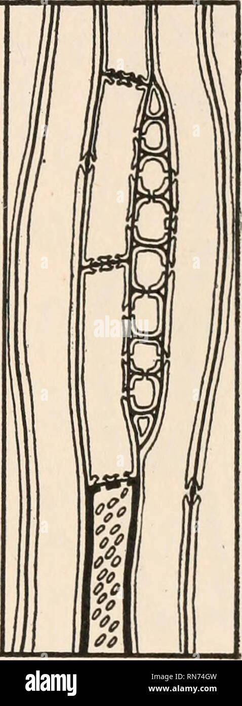 . The anatomy of woody plants. Botany -- Anatomy. FIG. 33.—Longitudinal section of the wood of the root in Picea canadcnsis. Explanation in the text. The general situation in regard to the mode of origin of longi- tudinal storage elements or wood parenchyma in the gymnosperms can perhaps be more readily illustrated by means of a diagram. Fig. 34 represents a ray, ordinary tracheids, and a septate or divided tracheid in relation to one another in the longitudinal aspect. The ray need not be specially considered, as the subject of the origin and organization of rays is to be discussed in the fol Stock Photohttps://www.alamy.com/image-license-details/?v=1https://www.alamy.com/the-anatomy-of-woody-plants-botany-anatomy-fig-33longitudinal-section-of-the-wood-of-the-root-in-picea-canadcnsis-explanation-in-the-text-the-general-situation-in-regard-to-the-mode-of-origin-of-longi-tudinal-storage-elements-or-wood-parenchyma-in-the-gymnosperms-can-perhaps-be-more-readily-illustrated-by-means-of-a-diagram-fig-34-represents-a-ray-ordinary-tracheids-and-a-septate-or-divided-tracheid-in-relation-to-one-another-in-the-longitudinal-aspect-the-ray-need-not-be-specially-considered-as-the-subject-of-the-origin-and-organization-of-rays-is-to-be-discussed-in-the-fol-image236799833.html
RMRN74GW–. The anatomy of woody plants. Botany -- Anatomy. FIG. 33.—Longitudinal section of the wood of the root in Picea canadcnsis. Explanation in the text. The general situation in regard to the mode of origin of longi- tudinal storage elements or wood parenchyma in the gymnosperms can perhaps be more readily illustrated by means of a diagram. Fig. 34 represents a ray, ordinary tracheids, and a septate or divided tracheid in relation to one another in the longitudinal aspect. The ray need not be specially considered, as the subject of the origin and organization of rays is to be discussed in the fol
 . The biology of insects. Insects -- Biology. Fig. 82.—Sections through Potato-leaves pierced by Hemiptera. a, piercing stylets (/)) of Leaf-hopper (Eupteryx) penetrating tissues, s, sheath of hardened saliva, t, tracheid ; b, puncture by Eupteryx at lower surface, saliva surrounding track {tr) of piercers; c, piercers of Mealy-bug (Dactylopius) with branching tracks in phloem shown by saliva. X 600. After K. M, Smith {A?7n. Appl. Biol, xiii, 1926). follows from alternative modes of nuclear behaviour, and it is possible that the germinal modifications for such changes. Please note that these i Stock Photohttps://www.alamy.com/image-license-details/?v=1https://www.alamy.com/the-biology-of-insects-insects-biology-fig-82sections-through-potato-leaves-pierced-by-hemiptera-a-piercing-stylets-of-leaf-hopper-eupteryx-penetrating-tissues-s-sheath-of-hardened-saliva-t-tracheid-b-puncture-by-eupteryx-at-lower-surface-saliva-surrounding-track-tr-of-piercers-c-piercers-of-mealy-bug-dactylopius-with-branching-tracks-in-phloem-shown-by-saliva-x-600-after-k-m-smith-a7n-appl-biol-xiii-1926-follows-from-alternative-modes-of-nuclear-behaviour-and-it-is-possible-that-the-germinal-modifications-for-such-changes-please-note-that-these-i-image234604896.html
RMRHK4X8–. The biology of insects. Insects -- Biology. Fig. 82.—Sections through Potato-leaves pierced by Hemiptera. a, piercing stylets (/)) of Leaf-hopper (Eupteryx) penetrating tissues, s, sheath of hardened saliva, t, tracheid ; b, puncture by Eupteryx at lower surface, saliva surrounding track {tr) of piercers; c, piercers of Mealy-bug (Dactylopius) with branching tracks in phloem shown by saliva. X 600. After K. M, Smith {A?7n. Appl. Biol, xiii, 1926). follows from alternative modes of nuclear behaviour, and it is possible that the germinal modifications for such changes. Please note that these i
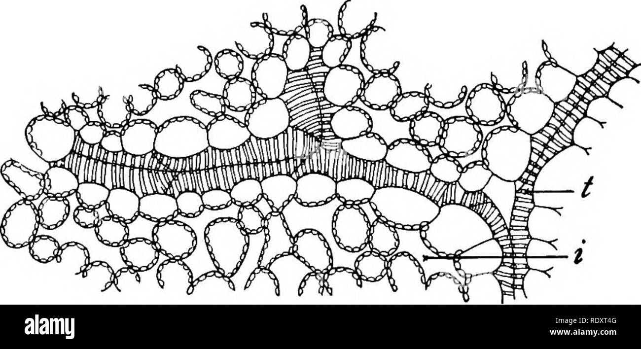 . A textbook of botany for colleges and universities ... Botany. THE MATERIAL OUTGO OF PLANTS 343 connected scries, extending from the root-hair region to the mesophyll of the leaves, among which they branch so extensively that there is scarcely a cell which is separated from a strand by more than a half dozen of its neighbors. Here the first branches end blindly (fig. 638) or join their fellows. A section of the root in the root- hair region shows likewise pm g^g—Ending of a, xylem strand among the cells of the that only a few mesophyll in a leaf of lilac {Syringa vulgaris) : t, tracheid ; i, Stock Photohttps://www.alamy.com/image-license-details/?v=1https://www.alamy.com/a-textbook-of-botany-for-colleges-and-universities-botany-the-material-outgo-of-plants-343-connected-scries-extending-from-the-root-hair-region-to-the-mesophyll-of-the-leaves-among-which-they-branch-so-extensively-that-there-is-scarcely-a-cell-which-is-separated-from-a-strand-by-more-than-a-half-dozen-of-its-neighbors-here-the-first-branches-end-blindly-fig-638-or-join-their-fellows-a-section-of-the-root-in-the-root-hair-region-shows-likewise-pm-ggending-of-a-xylem-strand-among-the-cells-of-the-that-only-a-few-mesophyll-in-a-leaf-of-lilac-syringa-vulgaris-t-tracheid-i-image232315008.html
RMRDXT4G–. A textbook of botany for colleges and universities ... Botany. THE MATERIAL OUTGO OF PLANTS 343 connected scries, extending from the root-hair region to the mesophyll of the leaves, among which they branch so extensively that there is scarcely a cell which is separated from a strand by more than a half dozen of its neighbors. Here the first branches end blindly (fig. 638) or join their fellows. A section of the root in the root- hair region shows likewise pm g^g—Ending of a, xylem strand among the cells of the that only a few mesophyll in a leaf of lilac {Syringa vulgaris) : t, tracheid ; i,
 . Elements of applied microscopy. A text-book for beginners. Microscopy. MICROSCOPY OF PAPER. lot or oval scar-like pits arranged in regular rows over their surface. In the case of pine, lattice-like areas with ob- long openings appear at intervals; but the presence of long cells mainly of a single type and with discoid mark- ings is the characteristic of the Conifers in general. 7. The Structure of the Angiosperms.—Pulp made from the commoner Angiospermous trees shows two distinct elements, long narrow fibres and short broad. Fig. 40.—^Tracheid of Birch. (After Herzberg.) 240 diameters. cells Stock Photohttps://www.alamy.com/image-license-details/?v=1https://www.alamy.com/elements-of-applied-microscopy-a-text-book-for-beginners-microscopy-microscopy-of-paper-lot-or-oval-scar-like-pits-arranged-in-regular-rows-over-their-surface-in-the-case-of-pine-lattice-like-areas-with-ob-long-openings-appear-at-intervals-but-the-presence-of-long-cells-mainly-of-a-single-type-and-with-discoid-mark-ings-is-the-characteristic-of-the-conifers-in-general-7-the-structure-of-the-angiospermspulp-made-from-the-commoner-angiospermous-trees-shows-two-distinct-elements-long-narrow-fibres-and-short-broad-fig-40tracheid-of-birch-after-herzberg-240-diameters-cells-image232251499.html
RMRDRY4B–. Elements of applied microscopy. A text-book for beginners. Microscopy. MICROSCOPY OF PAPER. lot or oval scar-like pits arranged in regular rows over their surface. In the case of pine, lattice-like areas with ob- long openings appear at intervals; but the presence of long cells mainly of a single type and with discoid mark- ings is the characteristic of the Conifers in general. 7. The Structure of the Angiosperms.—Pulp made from the commoner Angiospermous trees shows two distinct elements, long narrow fibres and short broad. Fig. 40.—^Tracheid of Birch. (After Herzberg.) 240 diameters. cells
 . The fungi which cause plant disease . Plant diseases; Fungi. 422 THE FUNGI WHICH CAUSE PLANT DISEASE zonate, shining, 3-10 mm. thick; tubes slender, concolorous with the context, about 1 cm. long, mouths regular, angular, 2-3 to a mm., glistening, whitish-isabelline to dark-fulvous, edges thin,. Fig. 302.— Decomposition of spruce-timber by Polyporus borealis. a, a tracheid containing a strong mycelial growth and a brownish yellow fluid which has originated in a medullary ray; at b and c the mycelium is still brownish. At d and e the walls have become attenuated and perforated, the filaments Stock Photohttps://www.alamy.com/image-license-details/?v=1https://www.alamy.com/the-fungi-which-cause-plant-disease-plant-diseases-fungi-422-the-fungi-which-cause-plant-disease-zonate-shining-3-10-mm-thick-tubes-slender-concolorous-with-the-context-about-1-cm-long-mouths-regular-angular-2-3-to-a-mm-glistening-whitish-isabelline-to-dark-fulvous-edges-thin-fig-302-decomposition-of-spruce-timber-by-polyporus-borealis-a-a-tracheid-containing-a-strong-mycelial-growth-and-a-brownish-yellow-fluid-which-has-originated-in-a-medullary-ray-at-b-and-c-the-mycelium-is-still-brownish-at-d-and-e-the-walls-have-become-attenuated-and-perforated-the-filaments-image232037160.html
RMRDE5NC–. The fungi which cause plant disease . Plant diseases; Fungi. 422 THE FUNGI WHICH CAUSE PLANT DISEASE zonate, shining, 3-10 mm. thick; tubes slender, concolorous with the context, about 1 cm. long, mouths regular, angular, 2-3 to a mm., glistening, whitish-isabelline to dark-fulvous, edges thin,. Fig. 302.— Decomposition of spruce-timber by Polyporus borealis. a, a tracheid containing a strong mycelial growth and a brownish yellow fluid which has originated in a medullary ray; at b and c the mycelium is still brownish. At d and e the walls have become attenuated and perforated, the filaments
![. A manual of the North American gymnosperms [microform] : exclusive of the cycadales but together with certain exotic species. Bois; Trees; Gymnosperms; Gymnospermes; Arbres; Wood. Fig. 24. PiNUS refi.exa. Medullary ray showing („) the form and disposition of the pits on the lateral walls; {/>) the ray tracheids. x 280 or p. reflexa (fig. 24), they attain to maximum 3ize and occupy nearly the entire surface of the wall within the limits of a wood tracheid, thereby becoming few in number. In Sequoia (fig. 25) or Taxodium (fig. 20) they are typically oval, in Pinus cubensis. Fig. 25. Sequoia Stock Photo . A manual of the North American gymnosperms [microform] : exclusive of the cycadales but together with certain exotic species. Bois; Trees; Gymnosperms; Gymnospermes; Arbres; Wood. Fig. 24. PiNUS refi.exa. Medullary ray showing („) the form and disposition of the pits on the lateral walls; {/>) the ray tracheids. x 280 or p. reflexa (fig. 24), they attain to maximum 3ize and occupy nearly the entire surface of the wall within the limits of a wood tracheid, thereby becoming few in number. In Sequoia (fig. 25) or Taxodium (fig. 20) they are typically oval, in Pinus cubensis. Fig. 25. Sequoia Stock Photo](https://c8.alamy.com/comp/REM7XH/a-manual-of-the-north-american-gymnosperms-microform-exclusive-of-the-cycadales-but-together-with-certain-exotic-species-bois-trees-gymnosperms-gymnospermes-arbres-wood-fig-24-pinus-refiexa-medullary-ray-showing-the-form-and-disposition-of-the-pits-on-the-lateral-walls-gt-the-ray-tracheids-x-280-or-p-reflexa-fig-24-they-attain-to-maximum-3ize-and-occupy-nearly-the-entire-surface-of-the-wall-within-the-limits-of-a-wood-tracheid-thereby-becoming-few-in-number-in-sequoia-fig-25-or-taxodium-fig-20-they-are-typically-oval-in-pinus-cubensis-fig-25-sequoia-REM7XH.jpg) . A manual of the North American gymnosperms [microform] : exclusive of the cycadales but together with certain exotic species. Bois; Trees; Gymnosperms; Gymnospermes; Arbres; Wood. Fig. 24. PiNUS refi.exa. Medullary ray showing („) the form and disposition of the pits on the lateral walls; {/>) the ray tracheids. x 280 or p. reflexa (fig. 24), they attain to maximum 3ize and occupy nearly the entire surface of the wall within the limits of a wood tracheid, thereby becoming few in number. In Sequoia (fig. 25) or Taxodium (fig. 20) they are typically oval, in Pinus cubensis. Fig. 25. Sequoia Stock Photohttps://www.alamy.com/image-license-details/?v=1https://www.alamy.com/a-manual-of-the-north-american-gymnosperms-microform-exclusive-of-the-cycadales-but-together-with-certain-exotic-species-bois-trees-gymnosperms-gymnospermes-arbres-wood-fig-24-pinus-refiexa-medullary-ray-showing-the-form-and-disposition-of-the-pits-on-the-lateral-walls-gt-the-ray-tracheids-x-280-or-p-reflexa-fig-24-they-attain-to-maximum-3ize-and-occupy-nearly-the-entire-surface-of-the-wall-within-the-limits-of-a-wood-tracheid-thereby-becoming-few-in-number-in-sequoia-fig-25-or-taxodium-fig-20-they-are-typically-oval-in-pinus-cubensis-fig-25-sequoia-image232785241.html
. A manual of the North American gymnosperms [microform] : exclusive of the cycadales but together with certain exotic species. Bois; Trees; Gymnosperms; Gymnospermes; Arbres; Wood. Fig. 24. PiNUS refi.exa. Medullary ray showing („) the form and disposition of the pits on the lateral walls; {/>) the ray tracheids. x 280 or p. reflexa (fig. 24), they attain to maximum 3ize and occupy nearly the entire surface of the wall within the limits of a wood tracheid, thereby becoming few in number. In Sequoia (fig. 25) or Taxodium (fig. 20) they are typically oval, in Pinus cubensis. Fig. 25. Sequoia Stock Photohttps://www.alamy.com/image-license-details/?v=1https://www.alamy.com/a-manual-of-the-north-american-gymnosperms-microform-exclusive-of-the-cycadales-but-together-with-certain-exotic-species-bois-trees-gymnosperms-gymnospermes-arbres-wood-fig-24-pinus-refiexa-medullary-ray-showing-the-form-and-disposition-of-the-pits-on-the-lateral-walls-gt-the-ray-tracheids-x-280-or-p-reflexa-fig-24-they-attain-to-maximum-3ize-and-occupy-nearly-the-entire-surface-of-the-wall-within-the-limits-of-a-wood-tracheid-thereby-becoming-few-in-number-in-sequoia-fig-25-or-taxodium-fig-20-they-are-typically-oval-in-pinus-cubensis-fig-25-sequoia-image232785241.htmlRMREM7XH–. A manual of the North American gymnosperms [microform] : exclusive of the cycadales but together with certain exotic species. Bois; Trees; Gymnosperms; Gymnospermes; Arbres; Wood. Fig. 24. PiNUS refi.exa. Medullary ray showing („) the form and disposition of the pits on the lateral walls; {/>) the ray tracheids. x 280 or p. reflexa (fig. 24), they attain to maximum 3ize and occupy nearly the entire surface of the wall within the limits of a wood tracheid, thereby becoming few in number. In Sequoia (fig. 25) or Taxodium (fig. 20) they are typically oval, in Pinus cubensis. Fig. 25. Sequoia
 . Experiments with plants. Botany. cri/shed bast. 134. Cross-section of a bundle of Corn: id) dnct, (a) annular tracheid (i. e., with ring- shaped thickenings), {sp) spiral tracheid (i. e. with spiral thickenings), iicp) wood parenchyma, {st) sieve-tube, {cc) companion cell, {str) strengthening fibers.. Please note that these images are extracted from scanned page images that may have been digitally enhanced for readability - coloration and appearance of these illustrations may not perfectly resemble the original work.. Osterhout, Winthrop John Van Leuven, 1871-. New York, The Macmillan compan Stock Photohttps://www.alamy.com/image-license-details/?v=1https://www.alamy.com/experiments-with-plants-botany-crished-bast-134-cross-section-of-a-bundle-of-corn-id-dnct-a-annular-tracheid-i-e-with-ring-shaped-thickenings-sp-spiral-tracheid-i-e-with-spiral-thickenings-iicp-wood-parenchyma-st-sieve-tube-cc-companion-cell-str-strengthening-fibers-please-note-that-these-images-are-extracted-from-scanned-page-images-that-may-have-been-digitally-enhanced-for-readability-coloration-and-appearance-of-these-illustrations-may-not-perfectly-resemble-the-original-work-osterhout-winthrop-john-van-leuven-1871-new-york-the-macmillan-compan-image232375987.html
RMRE1HXB–. Experiments with plants. Botany. cri/shed bast. 134. Cross-section of a bundle of Corn: id) dnct, (a) annular tracheid (i. e., with ring- shaped thickenings), {sp) spiral tracheid (i. e. with spiral thickenings), iicp) wood parenchyma, {st) sieve-tube, {cc) companion cell, {str) strengthening fibers.. Please note that these images are extracted from scanned page images that may have been digitally enhanced for readability - coloration and appearance of these illustrations may not perfectly resemble the original work.. Osterhout, Winthrop John Van Leuven, 1871-. New York, The Macmillan compan
 . Elements of applied microscopy. A text-book for beginners. Microscopy. MICROSCOPY OF PAPER. lOI , or oval scar-like pits arranged in regular rows over their surface. In the case of pine, lattice-like areas with ob- long openings appear at intervals; but the presence of long cells mainly of a single type and with discoid mark- ings is the characteristic of the Conifers in general. 7. The Structure of the Angiosperms.—Pulp made from the commoner Angiospermous trees shows two distinct elements, long narrow fibres and short broad. Fig, 40.—^Tracheid of Birch. (After Herzberg.) 240 diameters. cel Stock Photohttps://www.alamy.com/image-license-details/?v=1https://www.alamy.com/elements-of-applied-microscopy-a-text-book-for-beginners-microscopy-microscopy-of-paper-loi-or-oval-scar-like-pits-arranged-in-regular-rows-over-their-surface-in-the-case-of-pine-lattice-like-areas-with-ob-long-openings-appear-at-intervals-but-the-presence-of-long-cells-mainly-of-a-single-type-and-with-discoid-mark-ings-is-the-characteristic-of-the-conifers-in-general-7-the-structure-of-the-angiospermspulp-made-from-the-commoner-angiospermous-trees-shows-two-distinct-elements-long-narrow-fibres-and-short-broad-fig-40tracheid-of-birch-after-herzberg-240-diameters-cel-image232133824.html
RMRDJH1M–. Elements of applied microscopy. A text-book for beginners. Microscopy. MICROSCOPY OF PAPER. lOI , or oval scar-like pits arranged in regular rows over their surface. In the case of pine, lattice-like areas with ob- long openings appear at intervals; but the presence of long cells mainly of a single type and with discoid mark- ings is the characteristic of the Conifers in general. 7. The Structure of the Angiosperms.—Pulp made from the commoner Angiospermous trees shows two distinct elements, long narrow fibres and short broad. Fig, 40.—^Tracheid of Birch. (After Herzberg.) 240 diameters. cel
 . Bulletin of the British Museum (Natural History), Geology. . Figs. 5, 6. Fig. 5, Transverse section of part of the stem stele near its apex showing the incompletely differentiated metaxylem and an isolated mesarch protoxylem tracheid. B.M. (N.H.) Oliver Coll. 1247. x 80. Fig. 6, A small group of dark sclerized cells from the inner cortex of the stem. Gordon Coll., King's College, x 80.. Please note that these images are extracted from scanned page images that may have been digitally enhanced for readability - coloration and appearance of these illustrations may not perfectly resemble the ori Stock Photohttps://www.alamy.com/image-license-details/?v=1https://www.alamy.com/bulletin-of-the-british-museum-natural-history-geology-figs-5-6-fig-5-transverse-section-of-part-of-the-stem-stele-near-its-apex-showing-the-incompletely-differentiated-metaxylem-and-an-isolated-mesarch-protoxylem-tracheid-bm-nh-oliver-coll-1247-x-80-fig-6-a-small-group-of-dark-sclerized-cells-from-the-inner-cortex-of-the-stem-gordon-coll-kings-college-x-80-please-note-that-these-images-are-extracted-from-scanned-page-images-that-may-have-been-digitally-enhanced-for-readability-coloration-and-appearance-of-these-illustrations-may-not-perfectly-resemble-the-ori-image233977483.html
RMRGJGJK–. Bulletin of the British Museum (Natural History), Geology. . Figs. 5, 6. Fig. 5, Transverse section of part of the stem stele near its apex showing the incompletely differentiated metaxylem and an isolated mesarch protoxylem tracheid. B.M. (N.H.) Oliver Coll. 1247. x 80. Fig. 6, A small group of dark sclerized cells from the inner cortex of the stem. Gordon Coll., King's College, x 80.. Please note that these images are extracted from scanned page images that may have been digitally enhanced for readability - coloration and appearance of these illustrations may not perfectly resemble the ori
![. A manual of the North American gymnosperms [microform] : exclusive of the cycadales but together with certain exotic species. Bois; Trees; Gymnosperms; Gymnospermes; Arbres; Wood. 230 ANATOMY OF THE GYMNOSPERMS Tracheids more or less conspicuously rounded throughout. Ray cells (radial) somewhat contracted at the ends, strongly resinous. Pits on the lateral walls of the ray cells 1-2, rarely 4, per tracheid. 6. C. macrocarpa. Tracheids barely if at all rounded. Ray cells (radial) contracted at the ends, more or less resinous. Pits on the lateral walls of the ray cells 1-2, rarely 3, per trach Stock Photo . A manual of the North American gymnosperms [microform] : exclusive of the cycadales but together with certain exotic species. Bois; Trees; Gymnosperms; Gymnospermes; Arbres; Wood. 230 ANATOMY OF THE GYMNOSPERMS Tracheids more or less conspicuously rounded throughout. Ray cells (radial) somewhat contracted at the ends, strongly resinous. Pits on the lateral walls of the ray cells 1-2, rarely 4, per tracheid. 6. C. macrocarpa. Tracheids barely if at all rounded. Ray cells (radial) contracted at the ends, more or less resinous. Pits on the lateral walls of the ray cells 1-2, rarely 3, per trach Stock Photo](https://c8.alamy.com/comp/REN2TM/a-manual-of-the-north-american-gymnosperms-microform-exclusive-of-the-cycadales-but-together-with-certain-exotic-species-bois-trees-gymnosperms-gymnospermes-arbres-wood-230-anatomy-of-the-gymnosperms-tracheids-more-or-less-conspicuously-rounded-throughout-ray-cells-radial-somewhat-contracted-at-the-ends-strongly-resinous-pits-on-the-lateral-walls-of-the-ray-cells-1-2-rarely-4-per-tracheid-6-c-macrocarpa-tracheids-barely-if-at-all-rounded-ray-cells-radial-contracted-at-the-ends-more-or-less-resinous-pits-on-the-lateral-walls-of-the-ray-cells-1-2-rarely-3-per-trach-REN2TM.jpg) . A manual of the North American gymnosperms [microform] : exclusive of the cycadales but together with certain exotic species. Bois; Trees; Gymnosperms; Gymnospermes; Arbres; Wood. 230 ANATOMY OF THE GYMNOSPERMS Tracheids more or less conspicuously rounded throughout. Ray cells (radial) somewhat contracted at the ends, strongly resinous. Pits on the lateral walls of the ray cells 1-2, rarely 4, per tracheid. 6. C. macrocarpa. Tracheids barely if at all rounded. Ray cells (radial) contracted at the ends, more or less resinous. Pits on the lateral walls of the ray cells 1-2, rarely 3, per trach Stock Photohttps://www.alamy.com/image-license-details/?v=1https://www.alamy.com/a-manual-of-the-north-american-gymnosperms-microform-exclusive-of-the-cycadales-but-together-with-certain-exotic-species-bois-trees-gymnosperms-gymnospermes-arbres-wood-230-anatomy-of-the-gymnosperms-tracheids-more-or-less-conspicuously-rounded-throughout-ray-cells-radial-somewhat-contracted-at-the-ends-strongly-resinous-pits-on-the-lateral-walls-of-the-ray-cells-1-2-rarely-4-per-tracheid-6-c-macrocarpa-tracheids-barely-if-at-all-rounded-ray-cells-radial-contracted-at-the-ends-more-or-less-resinous-pits-on-the-lateral-walls-of-the-ray-cells-1-2-rarely-3-per-trach-image232803220.html
. A manual of the North American gymnosperms [microform] : exclusive of the cycadales but together with certain exotic species. Bois; Trees; Gymnosperms; Gymnospermes; Arbres; Wood. 230 ANATOMY OF THE GYMNOSPERMS Tracheids more or less conspicuously rounded throughout. Ray cells (radial) somewhat contracted at the ends, strongly resinous. Pits on the lateral walls of the ray cells 1-2, rarely 4, per tracheid. 6. C. macrocarpa. Tracheids barely if at all rounded. Ray cells (radial) contracted at the ends, more or less resinous. Pits on the lateral walls of the ray cells 1-2, rarely 3, per trach Stock Photohttps://www.alamy.com/image-license-details/?v=1https://www.alamy.com/a-manual-of-the-north-american-gymnosperms-microform-exclusive-of-the-cycadales-but-together-with-certain-exotic-species-bois-trees-gymnosperms-gymnospermes-arbres-wood-230-anatomy-of-the-gymnosperms-tracheids-more-or-less-conspicuously-rounded-throughout-ray-cells-radial-somewhat-contracted-at-the-ends-strongly-resinous-pits-on-the-lateral-walls-of-the-ray-cells-1-2-rarely-4-per-tracheid-6-c-macrocarpa-tracheids-barely-if-at-all-rounded-ray-cells-radial-contracted-at-the-ends-more-or-less-resinous-pits-on-the-lateral-walls-of-the-ray-cells-1-2-rarely-3-per-trach-image232803220.htmlRMREN2TM–. A manual of the North American gymnosperms [microform] : exclusive of the cycadales but together with certain exotic species. Bois; Trees; Gymnosperms; Gymnospermes; Arbres; Wood. 230 ANATOMY OF THE GYMNOSPERMS Tracheids more or less conspicuously rounded throughout. Ray cells (radial) somewhat contracted at the ends, strongly resinous. Pits on the lateral walls of the ray cells 1-2, rarely 4, per tracheid. 6. C. macrocarpa. Tracheids barely if at all rounded. Ray cells (radial) contracted at the ends, more or less resinous. Pits on the lateral walls of the ray cells 1-2, rarely 3, per trach
 . Diseases of plants induced by cryptogamic parasites : introduction to the study of pathogenic Fungi, slime-Fungi, bacteria, & Algae . Plant diseases; Parasitic plants; Fungi. Fio. 14. Fig. 13.—Tracheid of Pinus sytvestris destroyed by Travutes pini. The primary cell-WHll is completely dissolved from below upwards to a^ a; 6, secondary and tertiary layers of the walls consisting in the under portion of cellulose only, in which gi'anules of chalk are recognizable ; c, fungus-hyphae boring through the walls, leaving holes d and e. (After R. Hartig.) Fic. 14.—Tracheid of Pinus destroyed by P Stock Photohttps://www.alamy.com/image-license-details/?v=1https://www.alamy.com/diseases-of-plants-induced-by-cryptogamic-parasites-introduction-to-the-study-of-pathogenic-fungi-slime-fungi-bacteria-amp-algae-plant-diseases-parasitic-plants-fungi-fio-14-fig-13tracheid-of-pinus-sytvestris-destroyed-by-travutes-pini-the-primary-cell-whll-is-completely-dissolved-from-below-upwards-to-a-a-6-secondary-and-tertiary-layers-of-the-walls-consisting-in-the-under-portion-of-cellulose-only-in-which-gianules-of-chalk-are-recognizable-c-fungus-hyphae-boring-through-the-walls-leaving-holes-d-and-e-after-r-hartig-fic-14tracheid-of-pinus-destroyed-by-p-image232030994.html
RMRDDWW6–. Diseases of plants induced by cryptogamic parasites : introduction to the study of pathogenic Fungi, slime-Fungi, bacteria, & Algae . Plant diseases; Parasitic plants; Fungi. Fio. 14. Fig. 13.—Tracheid of Pinus sytvestris destroyed by Travutes pini. The primary cell-WHll is completely dissolved from below upwards to a^ a; 6, secondary and tertiary layers of the walls consisting in the under portion of cellulose only, in which gi'anules of chalk are recognizable ; c, fungus-hyphae boring through the walls, leaving holes d and e. (After R. Hartig.) Fic. 14.—Tracheid of Pinus destroyed by P
![. Fossil plants : for students of botany and geology . Paleobotany. X] ARTHROBENDRON. 327 form of the principal medullary rays is seen in fig 83, G, and the rebiculate pitting on the radial wall of a tracheid is shown. Pig. 83. Calamites (Arthrodendron). A. Tangential section (diagrammatic) showing the course of the vas- cular strands, also leaf-traces and infranodal canals. B. Radial face of a tracheid. C. Prosenchymatous elements of a principal medullary ray. D. Transverse section of the wood. (After Williamson.) No. 36 in the Williamson Collection. in fig. 83, B. Fig. 83, A illustrates the Stock Photo . Fossil plants : for students of botany and geology . Paleobotany. X] ARTHROBENDRON. 327 form of the principal medullary rays is seen in fig 83, G, and the rebiculate pitting on the radial wall of a tracheid is shown. Pig. 83. Calamites (Arthrodendron). A. Tangential section (diagrammatic) showing the course of the vas- cular strands, also leaf-traces and infranodal canals. B. Radial face of a tracheid. C. Prosenchymatous elements of a principal medullary ray. D. Transverse section of the wood. (After Williamson.) No. 36 in the Williamson Collection. in fig. 83, B. Fig. 83, A illustrates the Stock Photo](https://c8.alamy.com/comp/RDBCB8/fossil-plants-for-students-of-botany-and-geology-paleobotany-x-arthrobendron-327-form-of-the-principal-medullary-rays-is-seen-in-fig-83-g-and-the-rebiculate-pitting-on-the-radial-wall-of-a-tracheid-is-shown-pig-83-calamites-arthrodendron-a-tangential-section-diagrammatic-showing-the-course-of-the-vas-cular-strands-also-leaf-traces-and-infranodal-canals-b-radial-face-of-a-tracheid-c-prosenchymatous-elements-of-a-principal-medullary-ray-d-transverse-section-of-the-wood-after-williamson-no-36-in-the-williamson-collection-in-fig-83-b-fig-83-a-illustrates-the-RDBCB8.jpg) . Fossil plants : for students of botany and geology . Paleobotany. X] ARTHROBENDRON. 327 form of the principal medullary rays is seen in fig 83, G, and the rebiculate pitting on the radial wall of a tracheid is shown. Pig. 83. Calamites (Arthrodendron). A. Tangential section (diagrammatic) showing the course of the vas- cular strands, also leaf-traces and infranodal canals. B. Radial face of a tracheid. C. Prosenchymatous elements of a principal medullary ray. D. Transverse section of the wood. (After Williamson.) No. 36 in the Williamson Collection. in fig. 83, B. Fig. 83, A illustrates the Stock Photohttps://www.alamy.com/image-license-details/?v=1https://www.alamy.com/fossil-plants-for-students-of-botany-and-geology-paleobotany-x-arthrobendron-327-form-of-the-principal-medullary-rays-is-seen-in-fig-83-g-and-the-rebiculate-pitting-on-the-radial-wall-of-a-tracheid-is-shown-pig-83-calamites-arthrodendron-a-tangential-section-diagrammatic-showing-the-course-of-the-vas-cular-strands-also-leaf-traces-and-infranodal-canals-b-radial-face-of-a-tracheid-c-prosenchymatous-elements-of-a-principal-medullary-ray-d-transverse-section-of-the-wood-after-williamson-no-36-in-the-williamson-collection-in-fig-83-b-fig-83-a-illustrates-the-image231976508.html
. Fossil plants : for students of botany and geology . Paleobotany. X] ARTHROBENDRON. 327 form of the principal medullary rays is seen in fig 83, G, and the rebiculate pitting on the radial wall of a tracheid is shown. Pig. 83. Calamites (Arthrodendron). A. Tangential section (diagrammatic) showing the course of the vas- cular strands, also leaf-traces and infranodal canals. B. Radial face of a tracheid. C. Prosenchymatous elements of a principal medullary ray. D. Transverse section of the wood. (After Williamson.) No. 36 in the Williamson Collection. in fig. 83, B. Fig. 83, A illustrates the Stock Photohttps://www.alamy.com/image-license-details/?v=1https://www.alamy.com/fossil-plants-for-students-of-botany-and-geology-paleobotany-x-arthrobendron-327-form-of-the-principal-medullary-rays-is-seen-in-fig-83-g-and-the-rebiculate-pitting-on-the-radial-wall-of-a-tracheid-is-shown-pig-83-calamites-arthrodendron-a-tangential-section-diagrammatic-showing-the-course-of-the-vas-cular-strands-also-leaf-traces-and-infranodal-canals-b-radial-face-of-a-tracheid-c-prosenchymatous-elements-of-a-principal-medullary-ray-d-transverse-section-of-the-wood-after-williamson-no-36-in-the-williamson-collection-in-fig-83-b-fig-83-a-illustrates-the-image231976508.htmlRMRDBCB8–. Fossil plants : for students of botany and geology . Paleobotany. X] ARTHROBENDRON. 327 form of the principal medullary rays is seen in fig 83, G, and the rebiculate pitting on the radial wall of a tracheid is shown. Pig. 83. Calamites (Arthrodendron). A. Tangential section (diagrammatic) showing the course of the vas- cular strands, also leaf-traces and infranodal canals. B. Radial face of a tracheid. C. Prosenchymatous elements of a principal medullary ray. D. Transverse section of the wood. (After Williamson.) No. 36 in the Williamson Collection. in fig. 83, B. Fig. 83, A illustrates the
 . Wood; a manual of the natural history and industrial applications of the timbers of commerce. Wood; Timber. 22 OF WOOD IN GENERAL become pits. It has, in fact, become a water-and-air-conducting tracheid. A cambium cell in the same radial row as a pith-ray undergoes transverse division into 8-10 superposed cells which elongate radially and retain protoplasmic contents, thus continu- ing the pith-ray (Eig. 17). In spring, when there is httle heat, light, or activity of root and leaf to supply material, and when the bark, spHt by winter, may exert but Httle pressure, tracheids are produced with Stock Photohttps://www.alamy.com/image-license-details/?v=1https://www.alamy.com/wood-a-manual-of-the-natural-history-and-industrial-applications-of-the-timbers-of-commerce-wood-timber-22-of-wood-in-general-become-pits-it-has-in-fact-become-a-water-and-air-conducting-tracheid-a-cambium-cell-in-the-same-radial-row-as-a-pith-ray-undergoes-transverse-division-into-8-10-superposed-cells-which-elongate-radially-and-retain-protoplasmic-contents-thus-continu-ing-the-pith-ray-eig-17-in-spring-when-there-is-httle-heat-light-or-activity-of-root-and-leaf-to-supply-material-and-when-the-bark-spht-by-winter-may-exert-but-httle-pressure-tracheids-are-produced-with-image231922031.html
RMRD8XWK–. Wood; a manual of the natural history and industrial applications of the timbers of commerce. Wood; Timber. 22 OF WOOD IN GENERAL become pits. It has, in fact, become a water-and-air-conducting tracheid. A cambium cell in the same radial row as a pith-ray undergoes transverse division into 8-10 superposed cells which elongate radially and retain protoplasmic contents, thus continu- ing the pith-ray (Eig. 17). In spring, when there is httle heat, light, or activity of root and leaf to supply material, and when the bark, spHt by winter, may exert but Httle pressure, tracheids are produced with