Quick filters:
Tunica albuginea Stock Photos and Images
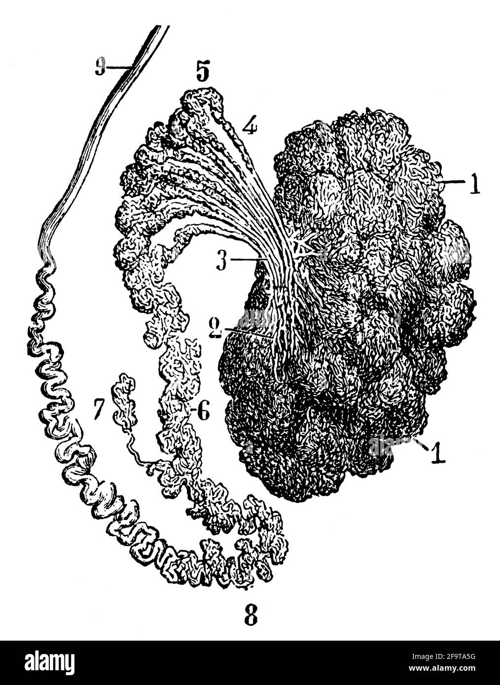 Tunica albuginea of testis. Illustration of the 19th century. Germany. White background. Stock Photohttps://www.alamy.com/image-license-details/?v=1https://www.alamy.com/tunica-albuginea-of-testis-illustration-of-the-19th-century-germany-white-background-image419115580.html
RF2F9TA5G–Tunica albuginea of testis. Illustration of the 19th century. Germany. White background.
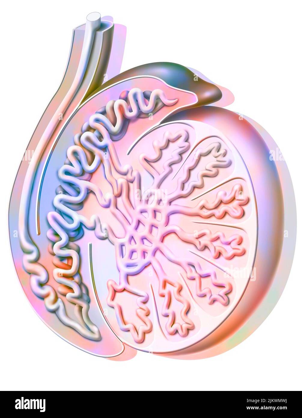 Sagittal section of a testicle showing the seminiferous tube. Stock Photohttps://www.alamy.com/image-license-details/?v=1https://www.alamy.com/sagittal-section-of-a-testicle-showing-the-seminiferous-tube-image476923598.html
RF2JKWMWJ–Sagittal section of a testicle showing the seminiferous tube.
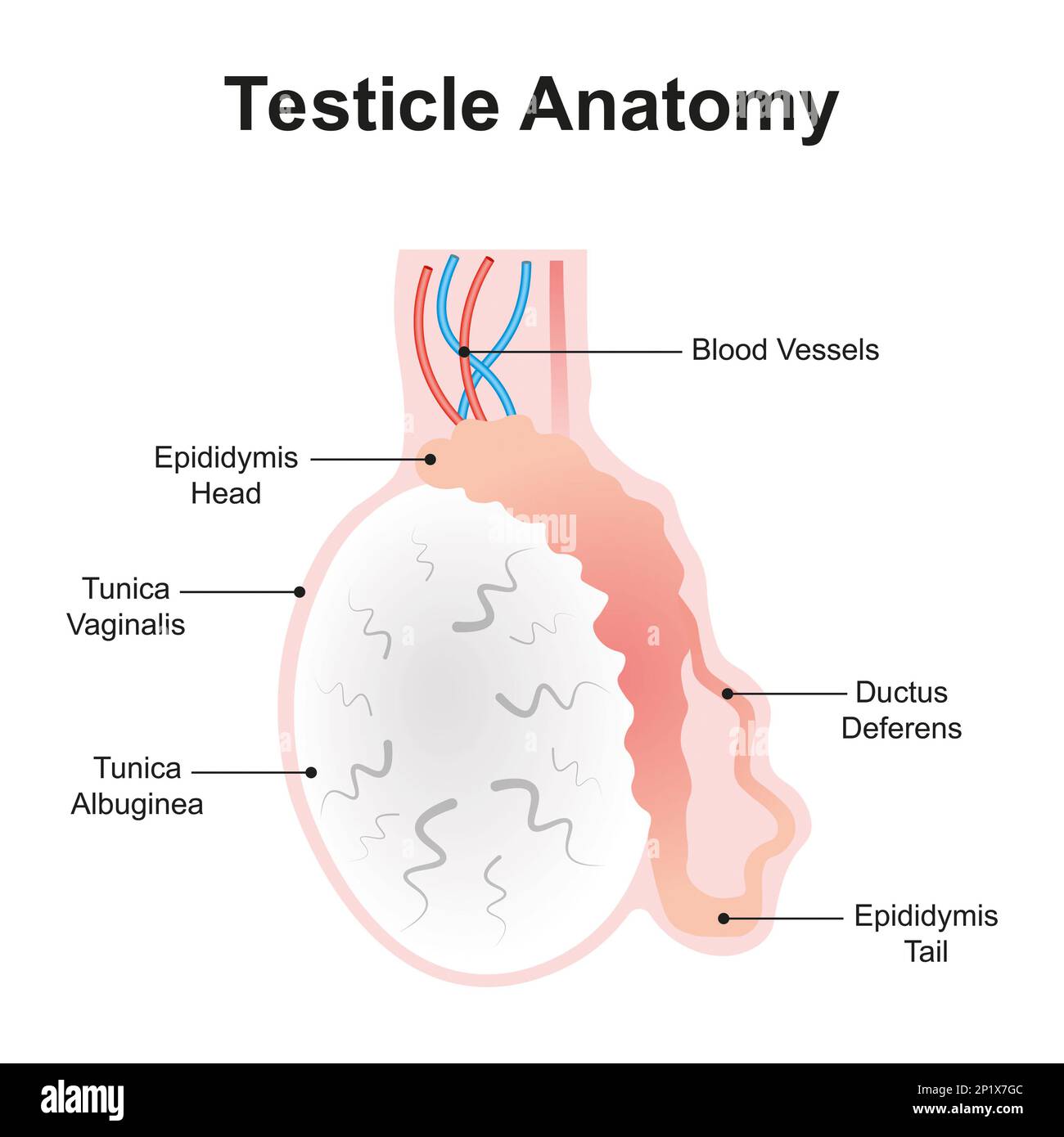 Testicle anatomy, illustration Stock Photohttps://www.alamy.com/image-license-details/?v=1https://www.alamy.com/testicle-anatomy-illustration-image534712764.html
RF2P1X7GC–Testicle anatomy, illustration
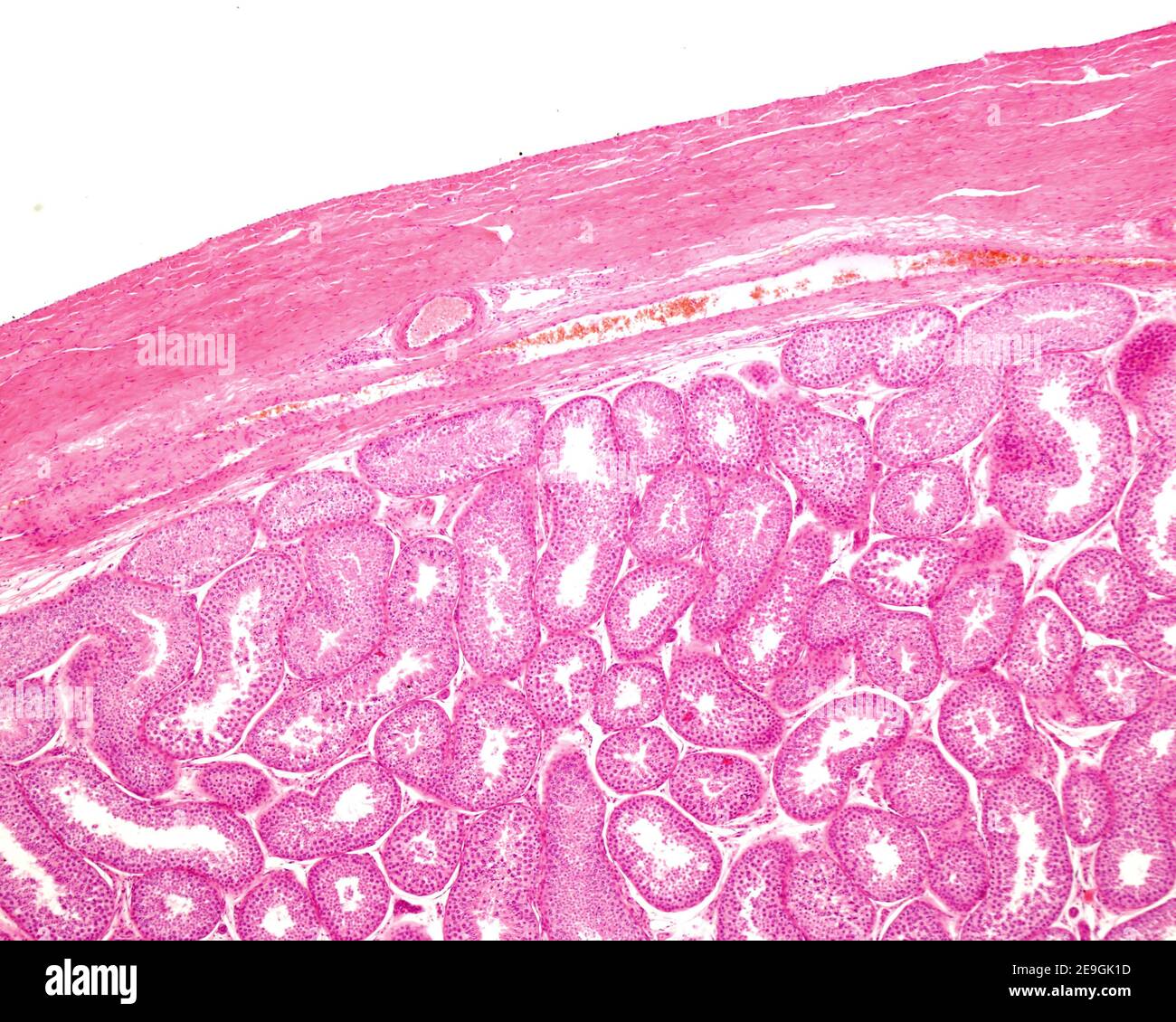 Light microscope micrograph showing the tunica albuginea of a human testicle. Below the external fibrous layer is the tunica vasculosa and the seminif Stock Photohttps://www.alamy.com/image-license-details/?v=1https://www.alamy.com/light-microscope-micrograph-showing-the-tunica-albuginea-of-a-human-testicle-below-the-external-fibrous-layer-is-the-tunica-vasculosa-and-the-seminif-image401736537.html
RF2E9GK1D–Light microscope micrograph showing the tunica albuginea of a human testicle. Below the external fibrous layer is the tunica vasculosa and the seminif
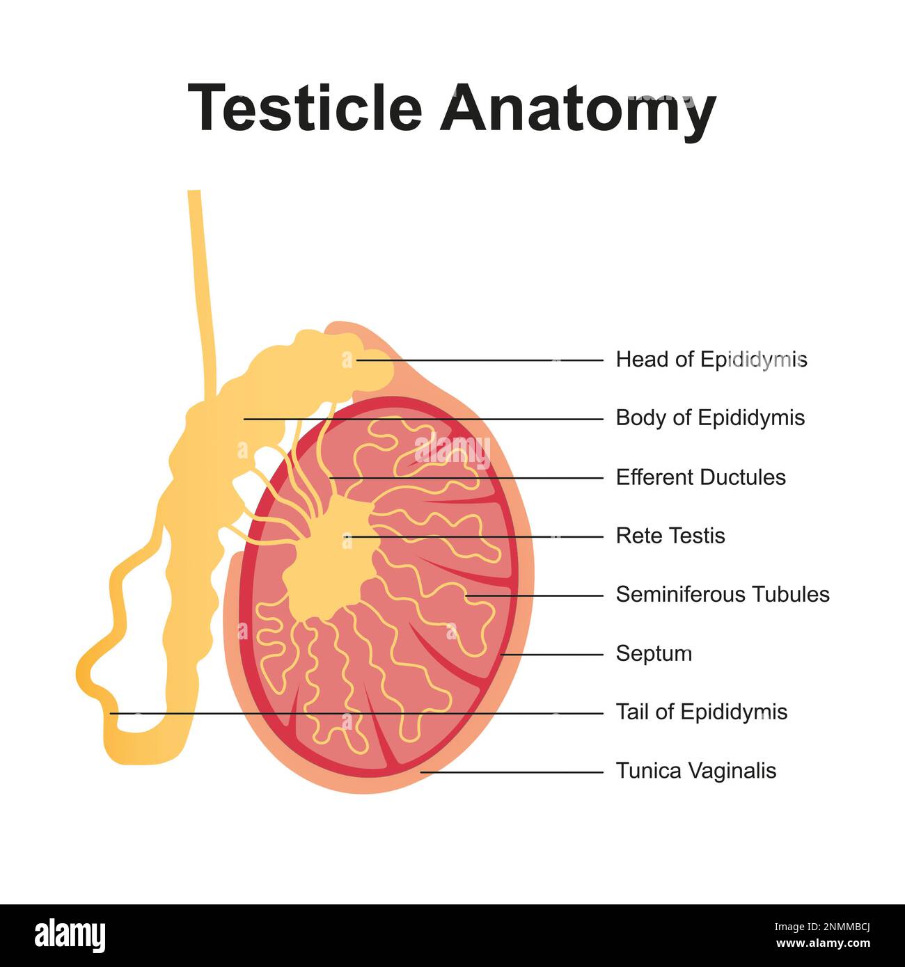 Testicle anatomy, illustration Stock Photohttps://www.alamy.com/image-license-details/?v=1https://www.alamy.com/testicle-anatomy-illustration-image529052178.html
RF2NMMBCJ–Testicle anatomy, illustration
 Manual of human histology . .The testicles, testes, are a couple of true glands, containingwithin a special tunic, the tunica albuginea s. fibrosa, thesecreting elements, in the form of complexly convolutedtubules, the spermatic tubes or tubuli seminiferi. The tunic isa white, dense and thick membrane, corresponding in structure,in all respects, with other fibrous membranes (the dura materespecially), and everywhere surrounding the parenchyma of thetestis as a closed capsule. Its external surface, except Avherethe epididymis is attached to the testis, is rendered smooth andglistening by a spec Stock Photohttps://www.alamy.com/image-license-details/?v=1https://www.alamy.com/manual-of-human-histology-the-testicles-testes-are-a-couple-of-true-glands-containingwithin-a-special-tunic-the-tunica-albuginea-s-fibrosa-thesecreting-elements-in-the-form-of-complexly-convolutedtubules-the-spermatic-tubes-or-tubuli-seminiferi-the-tunic-isa-white-dense-and-thick-membrane-corresponding-in-structurein-all-respects-with-other-fibrous-membranes-the-dura-materespecially-and-everywhere-surrounding-the-parenchyma-of-thetestis-as-a-closed-capsule-its-external-surface-except-avherethe-epididymis-is-attached-to-the-testis-is-rendered-smooth-andglistening-by-a-spec-image338226156.html
RM2AJ7EWG–Manual of human histology . .The testicles, testes, are a couple of true glands, containingwithin a special tunic, the tunica albuginea s. fibrosa, thesecreting elements, in the form of complexly convolutedtubules, the spermatic tubes or tubuli seminiferi. The tunic isa white, dense and thick membrane, corresponding in structure,in all respects, with other fibrous membranes (the dura materespecially), and everywhere surrounding the parenchyma of thetestis as a closed capsule. Its external surface, except Avherethe epididymis is attached to the testis, is rendered smooth andglistening by a spec
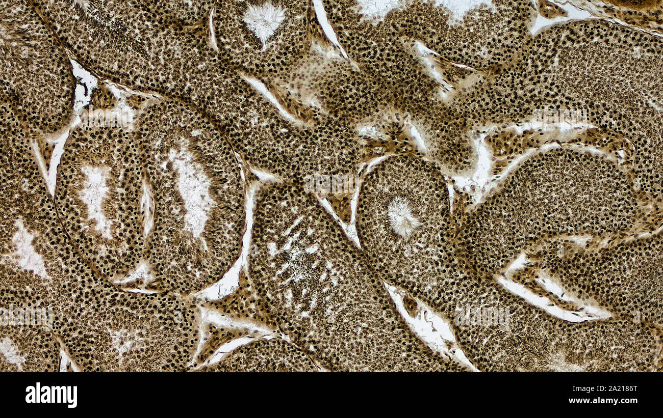 Histologically stained cross-section ofa rabbit testicle. Stock Photohttps://www.alamy.com/image-license-details/?v=1https://www.alamy.com/histologically-stained-cross-section-ofa-rabbit-testicle-image328254720.html
RM2A2186T–Histologically stained cross-section ofa rabbit testicle.
 Human testicle computer artwork. Stock Photohttps://www.alamy.com/image-license-details/?v=1https://www.alamy.com/human-testicle-computer-artwork-image69884353.html
RFE1KE5N–Human testicle computer artwork.
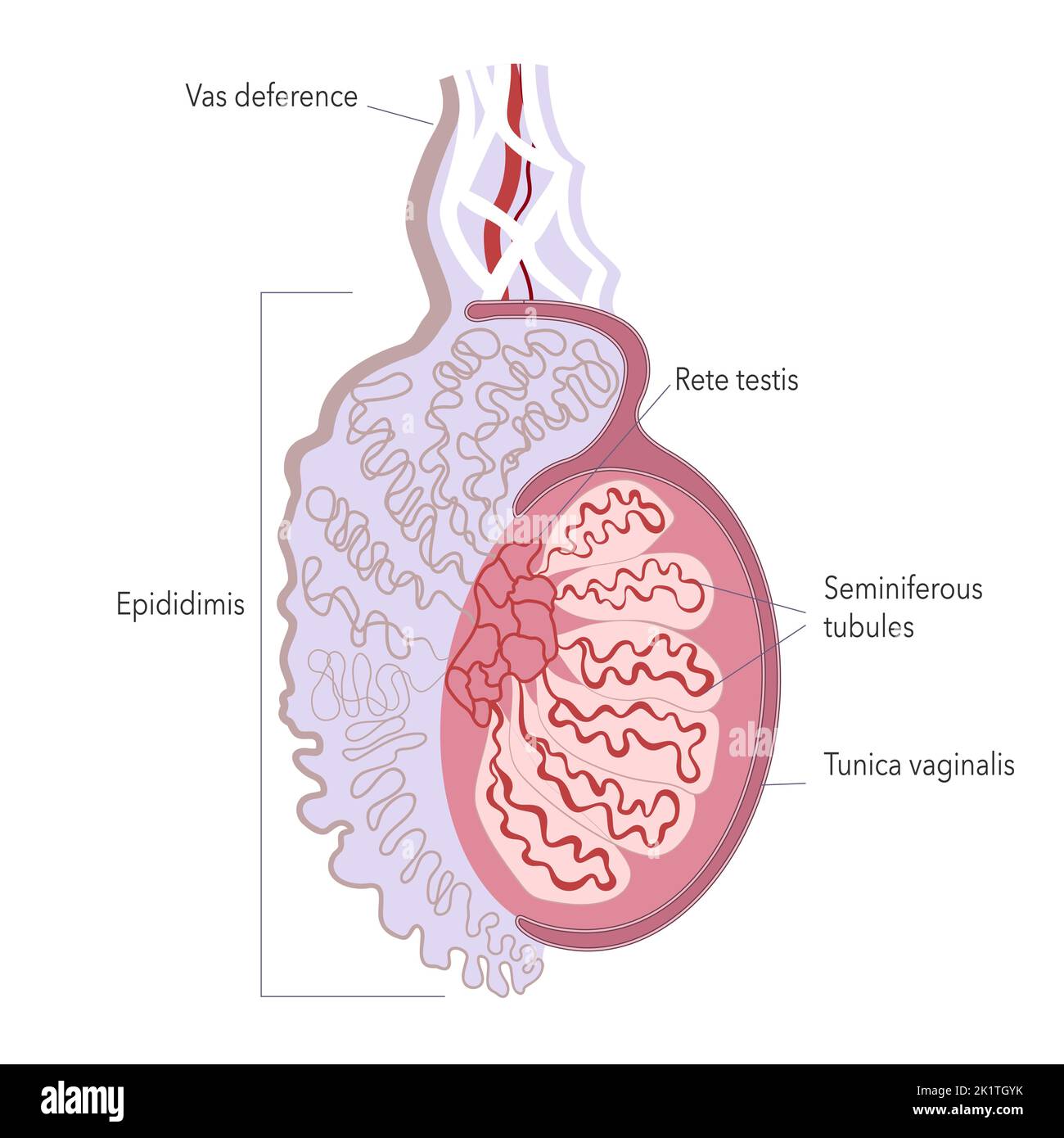 Fine structure of human testicle. Anatomy illustration of male gonad for atlas or infographics. Stock Vectorhttps://www.alamy.com/image-license-details/?v=1https://www.alamy.com/fine-structure-of-human-testicle-anatomy-illustration-of-male-gonad-for-atlas-or-infographics-image483045127.html
RF2K1TGYK–Fine structure of human testicle. Anatomy illustration of male gonad for atlas or infographics.
 . The cyclopædia of anatomy and physiology. Anatomy; Physiology; Zoology. 1008 TESTICLE (ABNORMAL ANATOMY). siderable firmness and consistency ; the tunica albuginea is thickened, and in some places as dense and indurated as cartilage ; and the surfaces of the tunica vaginalis are closely connected by old adhesions. The glandular structure is partly displaced and atrophied by the pressure of the yellow matter ; and it often happens after some time, that both undergo a slow process of wasting, so that an enlarged and indurated gland is progressively reduced, until scarcely any thing remains bey Stock Photohttps://www.alamy.com/image-license-details/?v=1https://www.alamy.com/the-cyclopdia-of-anatomy-and-physiology-anatomy-physiology-zoology-1008-testicle-abnormal-anatomy-siderable-firmness-and-consistency-the-tunica-albuginea-is-thickened-and-in-some-places-as-dense-and-indurated-as-cartilage-and-the-surfaces-of-the-tunica-vaginalis-are-closely-connected-by-old-adhesions-the-glandular-structure-is-partly-displaced-and-atrophied-by-the-pressure-of-the-yellow-matter-and-it-often-happens-after-some-time-that-both-undergo-a-slow-process-of-wasting-so-that-an-enlarged-and-indurated-gland-is-progressively-reduced-until-scarcely-any-thing-remains-bey-image216208673.html
RMPFN4A9–. The cyclopædia of anatomy and physiology. Anatomy; Physiology; Zoology. 1008 TESTICLE (ABNORMAL ANATOMY). siderable firmness and consistency ; the tunica albuginea is thickened, and in some places as dense and indurated as cartilage ; and the surfaces of the tunica vaginalis are closely connected by old adhesions. The glandular structure is partly displaced and atrophied by the pressure of the yellow matter ; and it often happens after some time, that both undergo a slow process of wasting, so that an enlarged and indurated gland is progressively reduced, until scarcely any thing remains bey
 Cross section of the rod, vintage engraved illustration. Usual Medicine Dictionary by Dr Labarthe - 1885. Stock Vectorhttps://www.alamy.com/image-license-details/?v=1https://www.alamy.com/stock-photo-cross-section-of-the-rod-vintage-engraved-illustration-usual-medicine-84407684.html
RFEW92T4–Cross section of the rod, vintage engraved illustration. Usual Medicine Dictionary by Dr Labarthe - 1885.
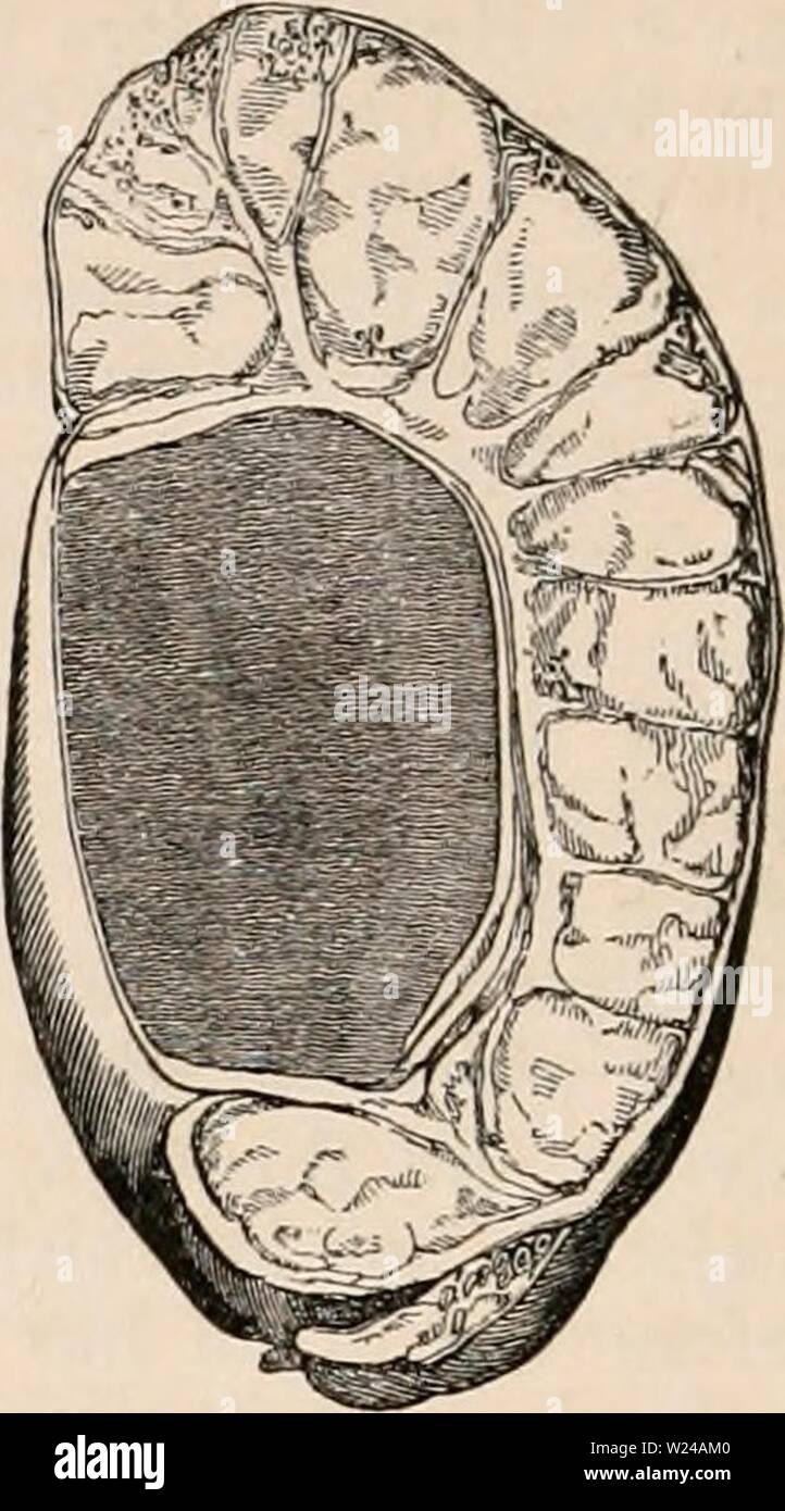 Archive image from page 223 of The cyclopædia of anatomy and. The cyclopædia of anatomy and physiology cyclopdiaofana0402todd Year: 1849 1008 TESTICLE (ABNORMAL ANATOMY). siderable firmness and consistency ; the tunica albuginea is thickened, and in some places as dense and indurated as cartilage ; and the surfaces of the tunica vaginalis are closely connected by old adhesions. The glandular structure is partly displaced and atrophied by the pressure of the yellow matter ; and it often happens after some time, that both undergo a slow process of wasting, so that an enlarged and indurated gla Stock Photohttps://www.alamy.com/image-license-details/?v=1https://www.alamy.com/archive-image-from-page-223-of-the-cyclopdia-of-anatomy-and-the-cyclopdia-of-anatomy-and-physiology-cyclopdiaofana0402todd-year-1849-1008-testicle-abnormal-anatomy-siderable-firmness-and-consistency-the-tunica-albuginea-is-thickened-and-in-some-places-as-dense-and-indurated-as-cartilage-and-the-surfaces-of-the-tunica-vaginalis-are-closely-connected-by-old-adhesions-the-glandular-structure-is-partly-displaced-and-atrophied-by-the-pressure-of-the-yellow-matter-and-it-often-happens-after-some-time-that-both-undergo-a-slow-process-of-wasting-so-that-an-enlarged-and-indurated-gla-image259481040.html
RMW24AM0–Archive image from page 223 of The cyclopædia of anatomy and. The cyclopædia of anatomy and physiology cyclopdiaofana0402todd Year: 1849 1008 TESTICLE (ABNORMAL ANATOMY). siderable firmness and consistency ; the tunica albuginea is thickened, and in some places as dense and indurated as cartilage ; and the surfaces of the tunica vaginalis are closely connected by old adhesions. The glandular structure is partly displaced and atrophied by the pressure of the yellow matter ; and it often happens after some time, that both undergo a slow process of wasting, so that an enlarged and indurated gla
 . Fig. 229—Cystic Ovary, Reduced Nymphomania. A, Normal ovary ; B, cystic gland. of quite large cysts that they project conspicuously beyond the general surface of the gland. In some cases, the ovarian tissue proper vanishes almost completely under the pressure of large cysts, firmly compressed within the enveloping tunica albuginea. In other extremely bad cases of nymphomania there are found small, atrophied, fibrous ovaries, very hard and dense, like fibro-cartilage. The examination of the ovaries of the mare is to be made upon the standing animal, in essentially the same manner as that desc Stock Photohttps://www.alamy.com/image-license-details/?v=1https://www.alamy.com/fig-229cystic-ovary-reduced-nymphomania-a-normal-ovary-b-cystic-gland-of-quite-large-cysts-that-they-project-conspicuously-beyond-the-general-surface-of-the-gland-in-some-cases-the-ovarian-tissue-proper-vanishes-almost-completely-under-the-pressure-of-large-cysts-firmly-compressed-within-the-enveloping-tunica-albuginea-in-other-extremely-bad-cases-of-nymphomania-there-are-found-small-atrophied-fibrous-ovaries-very-hard-and-dense-like-fibro-cartilage-the-examination-of-the-ovaries-of-the-mare-is-to-be-made-upon-the-standing-animal-in-essentially-the-same-manner-as-that-desc-image179903557.html
RMMCK8R1–. Fig. 229—Cystic Ovary, Reduced Nymphomania. A, Normal ovary ; B, cystic gland. of quite large cysts that they project conspicuously beyond the general surface of the gland. In some cases, the ovarian tissue proper vanishes almost completely under the pressure of large cysts, firmly compressed within the enveloping tunica albuginea. In other extremely bad cases of nymphomania there are found small, atrophied, fibrous ovaries, very hard and dense, like fibro-cartilage. The examination of the ovaries of the mare is to be made upon the standing animal, in essentially the same manner as that desc
 Semi-schematic representation of the male urinary and reproductive organs. Illustration of the 19th century. Germany. White background. Stock Photohttps://www.alamy.com/image-license-details/?v=1https://www.alamy.com/semi-schematic-representation-of-the-male-urinary-and-reproductive-organs-illustration-of-the-19th-century-germany-white-background-image419115594.html
RF2F9TA62–Semi-schematic representation of the male urinary and reproductive organs. Illustration of the 19th century. Germany. White background.
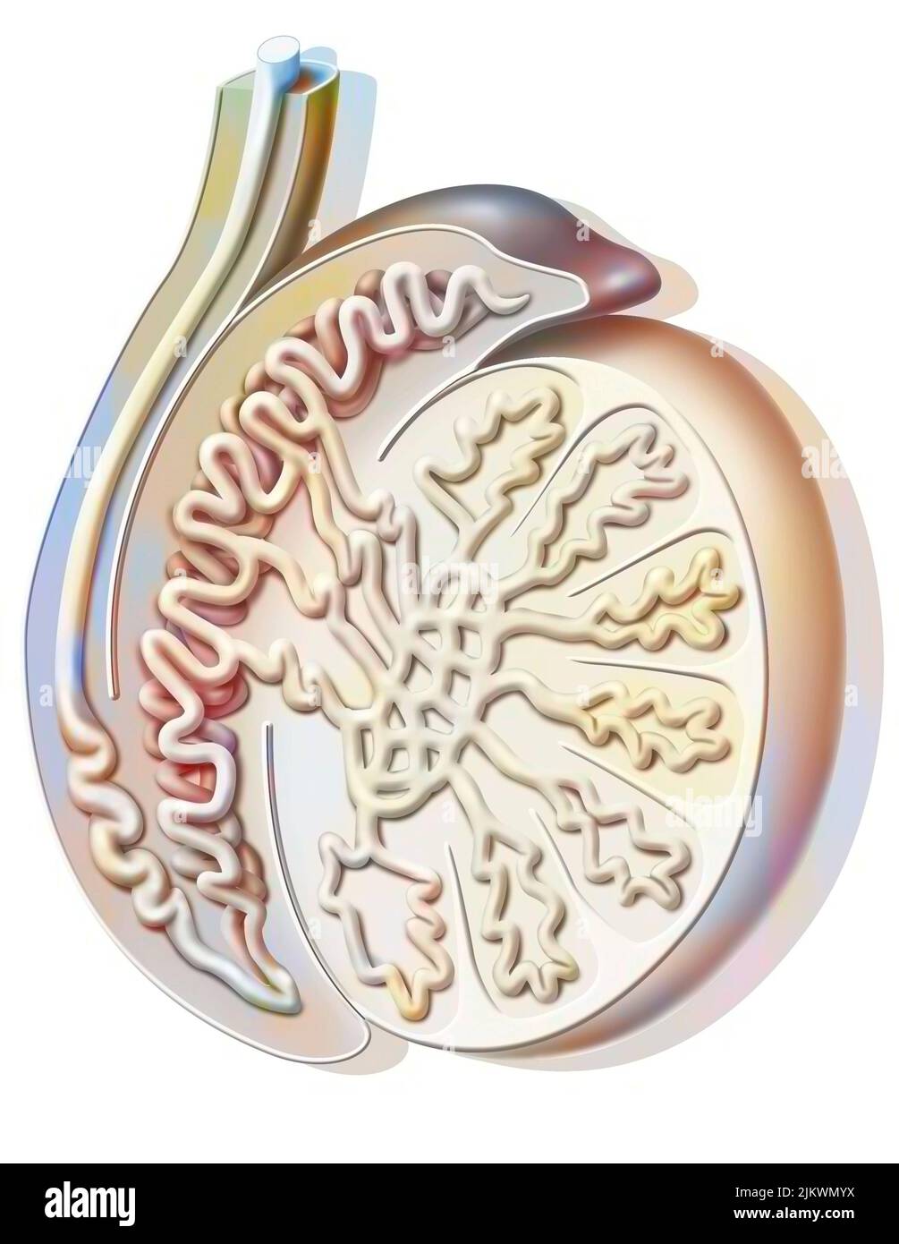 Sagittal section of a testicle showing the seminiferous tube. Stock Photohttps://www.alamy.com/image-license-details/?v=1https://www.alamy.com/sagittal-section-of-a-testicle-showing-the-seminiferous-tube-image476923662.html
RF2JKWMYX–Sagittal section of a testicle showing the seminiferous tube.
 Quain's elements of anatomy . ction. It follows from this that the pos-terior or attached border is turned upwards and inwards, and the outerflattened face slightly backwards. Fig. 592. Fig. 592.—Transverse section throtjgh the RIGHT testicle AND THE TUNICA VAGINALIS (frOm Kolliker). a, connective tissue enveloping the parietal layerof the tunica vaginalis; h, this layer itself ; c, cavityof the tunica vaginalis ; d, reflected or visceral layeradhering to e, the tunica albuginea ; /, covering ofepididymis (g) on the right or outer side ; h, medias-tinum testis; i, branches of the spermatic art Stock Photohttps://www.alamy.com/image-license-details/?v=1https://www.alamy.com/quains-elements-of-anatomy-ction-it-follows-from-this-that-the-pos-terior-or-attached-border-is-turned-upwards-and-inwards-and-the-outerflattened-face-slightly-backwards-fig-592-fig-592transverse-section-throtjgh-the-right-testicle-and-the-tunica-vaginalis-from-kolliker-a-connective-tissue-enveloping-the-parietal-layerof-the-tunica-vaginalis-h-this-layer-itself-c-cavityof-the-tunica-vaginalis-d-reflected-or-visceral-layeradhering-to-e-the-tunica-albuginea-covering-ofepididymis-g-on-the-right-or-outer-side-h-medias-tinum-testis-i-branches-of-the-spermatic-art-image340295817.html
RM2ANHPP1–Quain's elements of anatomy . ction. It follows from this that the pos-terior or attached border is turned upwards and inwards, and the outerflattened face slightly backwards. Fig. 592. Fig. 592.—Transverse section throtjgh the RIGHT testicle AND THE TUNICA VAGINALIS (frOm Kolliker). a, connective tissue enveloping the parietal layerof the tunica vaginalis; h, this layer itself ; c, cavityof the tunica vaginalis ; d, reflected or visceral layeradhering to e, the tunica albuginea ; /, covering ofepididymis (g) on the right or outer side ; h, medias-tinum testis; i, branches of the spermatic art
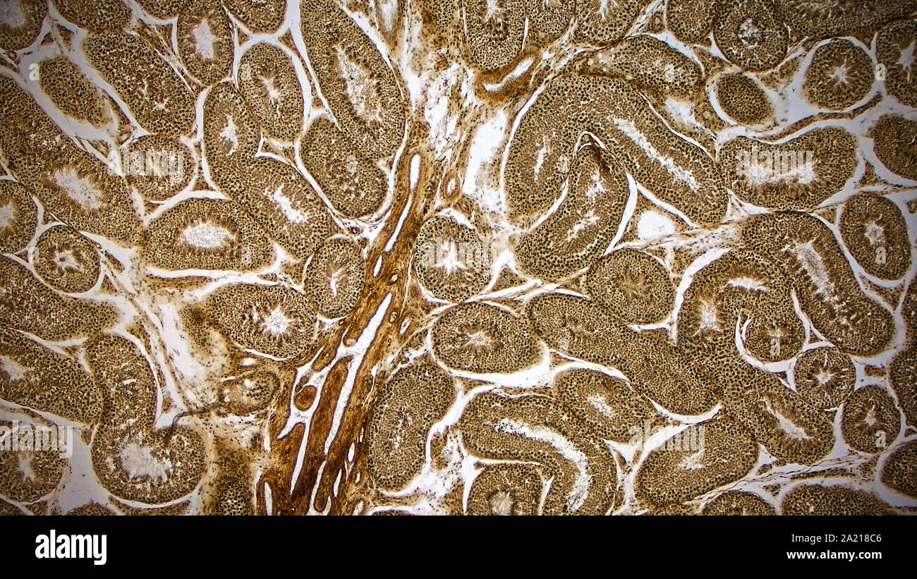 Histologically stained cross-section ofa rabbit testicle. Stock Photohttps://www.alamy.com/image-license-details/?v=1https://www.alamy.com/histologically-stained-cross-section-ofa-rabbit-testicle-image328254870.html
RM2A218C6–Histologically stained cross-section ofa rabbit testicle.
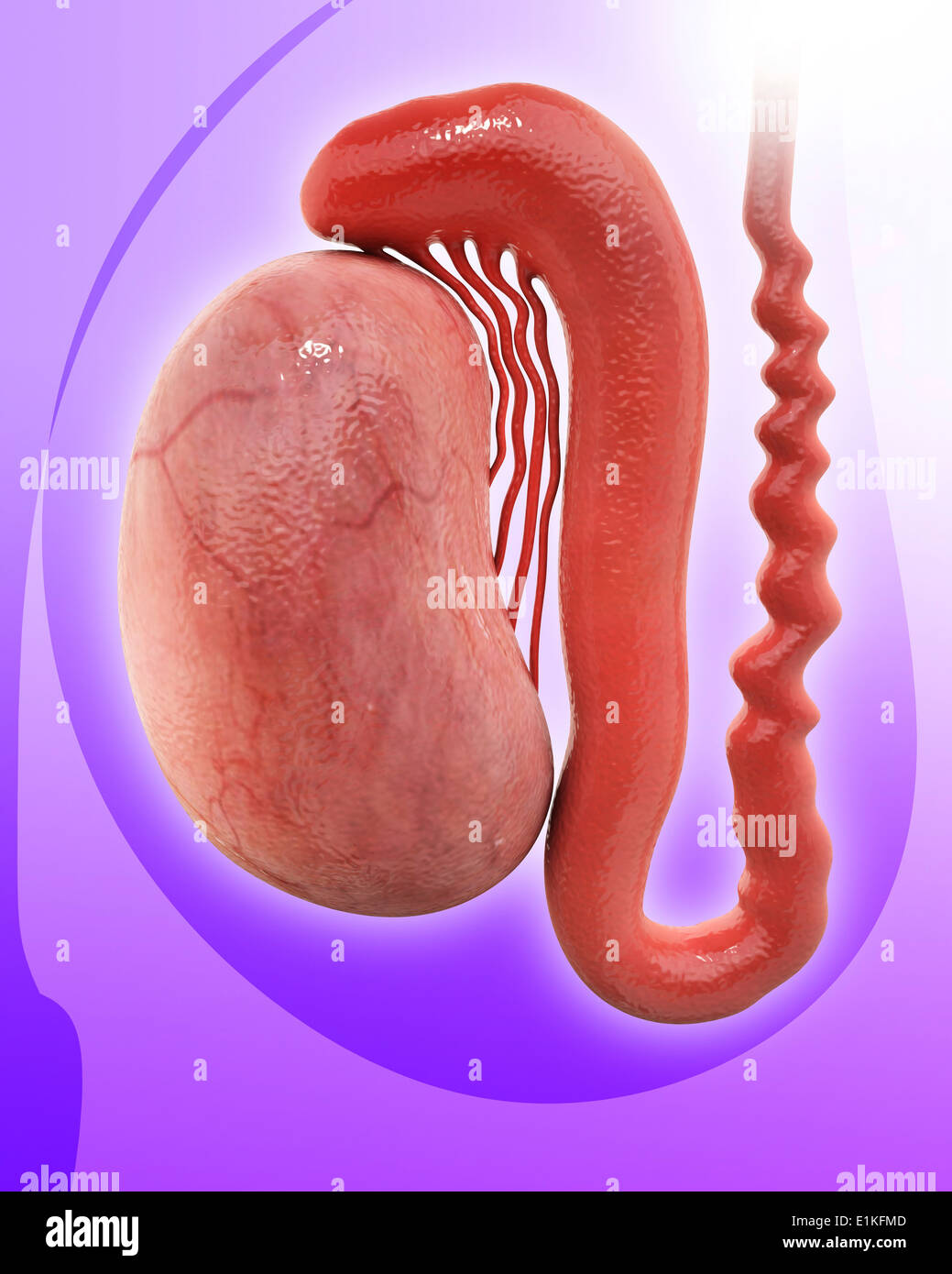 Human testicle computer artwork. Stock Photohttps://www.alamy.com/image-license-details/?v=1https://www.alamy.com/human-testicle-computer-artwork-image69885549.html
RFE1KFMD–Human testicle computer artwork.
 . The cyclopædia of anatomy and physiology. Anatomy; Physiology; Zoology. 978 TESTICLE (NORMAL ANATOMY). mentous cords from the inner surface of the tunica albuginea. I have not been able to make out any such ligamentous processes passing into the substance of the testis, as are represented in Sir A. Cooper's work (part i. pi. 2. fig. 3), which I have no doubt is an ex- aggerated view of the preparation from which it was taken. The cords described appear to me to consist chiefly of blood-vessels sup- ported by slight fibrous processes from the tunica albuginea and areolar tissue. In a well- in Stock Photohttps://www.alamy.com/image-license-details/?v=1https://www.alamy.com/the-cyclopdia-of-anatomy-and-physiology-anatomy-physiology-zoology-978-testicle-normal-anatomy-mentous-cords-from-the-inner-surface-of-the-tunica-albuginea-i-have-not-been-able-to-make-out-any-such-ligamentous-processes-passing-into-the-substance-of-the-testis-as-are-represented-in-sir-a-coopers-work-part-i-pi-2-fig-3-which-i-have-no-doubt-is-an-ex-aggerated-view-of-the-preparation-from-which-it-was-taken-the-cords-described-appear-to-me-to-consist-chiefly-of-blood-vessels-sup-ported-by-slight-fibrous-processes-from-the-tunica-albuginea-and-areolar-tissue-in-a-well-in-image216208700.html
RMPFN4B8–. The cyclopædia of anatomy and physiology. Anatomy; Physiology; Zoology. 978 TESTICLE (NORMAL ANATOMY). mentous cords from the inner surface of the tunica albuginea. I have not been able to make out any such ligamentous processes passing into the substance of the testis, as are represented in Sir A. Cooper's work (part i. pi. 2. fig. 3), which I have no doubt is an ex- aggerated view of the preparation from which it was taken. The cords described appear to me to consist chiefly of blood-vessels sup- ported by slight fibrous processes from the tunica albuginea and areolar tissue. In a well- in
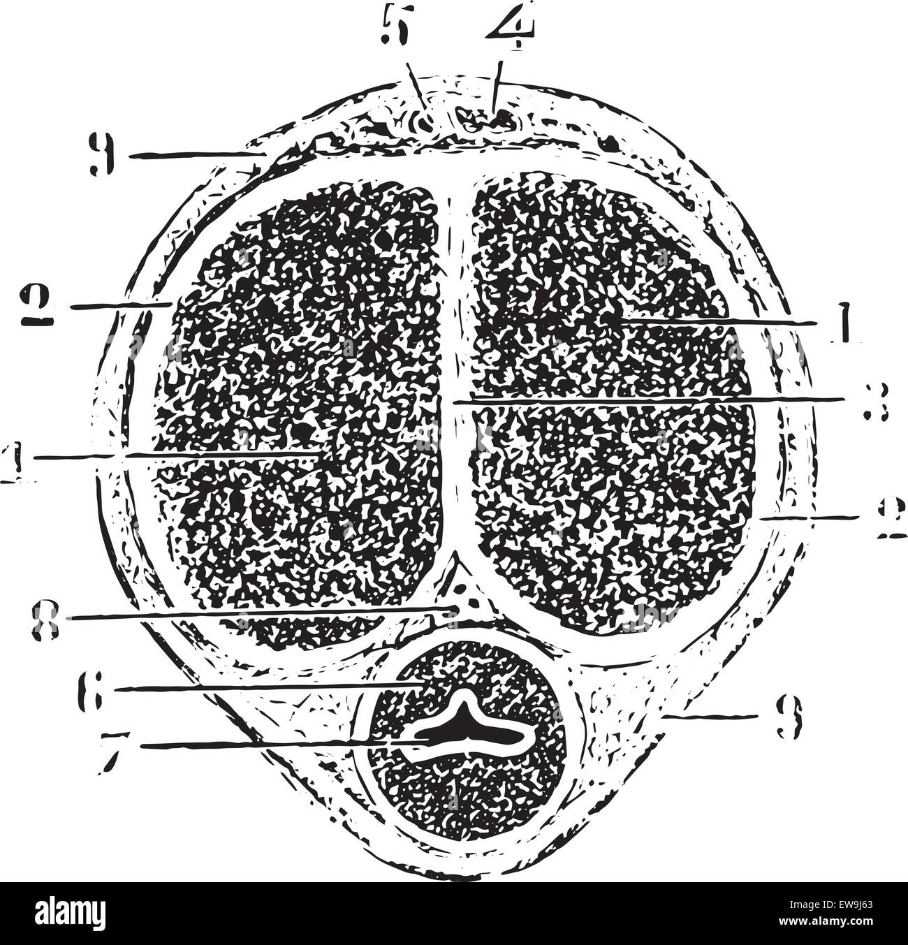 Cross section of the rod, vintage engraved illustration. Usual Medicine Dictionary by Dr Labarthe - 1885. Stock Vectorhttps://www.alamy.com/image-license-details/?v=1https://www.alamy.com/stock-photo-cross-section-of-the-rod-vintage-engraved-illustration-usual-medicine-84419723.html
RFEW9J63–Cross section of the rod, vintage engraved illustration. Usual Medicine Dictionary by Dr Labarthe - 1885.
 Archive image from page 210 of The cyclopædia of anatomy and. The cyclopædia of anatomy and physiology cyclopdiaofana0402todd Year: 1849 TESTICLE (ABNORMAL ANATOMY). 995 the inflammatory action ; but when the con- tiguous organ or subjacent part is of a dif- Fig. 638. ferent structure from that of the cellular tissue, the extension of inflammation inwards is checked. Thus, in the case of the inflamed tunica vaginalis, the cellular tissue readily transmitted the morbid action to the epididy- mis, but the tunica albuginea arrested its progress to the body of the testicle ; and this explains Stock Photohttps://www.alamy.com/image-license-details/?v=1https://www.alamy.com/archive-image-from-page-210-of-the-cyclopdia-of-anatomy-and-the-cyclopdia-of-anatomy-and-physiology-cyclopdiaofana0402todd-year-1849-testicle-abnormal-anatomy-995-the-inflammatory-action-but-when-the-con-tiguous-organ-or-subjacent-part-is-of-a-dif-fig-638-ferent-structure-from-that-of-the-cellular-tissue-the-extension-of-inflammation-inwards-is-checked-thus-in-the-case-of-the-inflamed-tunica-vaginalis-the-cellular-tissue-readily-transmitted-the-morbid-action-to-the-epididy-mis-but-the-tunica-albuginea-arrested-its-progress-to-the-body-of-the-testicle-and-this-explains-image259477959.html
RMW246NY–Archive image from page 210 of The cyclopædia of anatomy and. The cyclopædia of anatomy and physiology cyclopdiaofana0402todd Year: 1849 TESTICLE (ABNORMAL ANATOMY). 995 the inflammatory action ; but when the con- tiguous organ or subjacent part is of a dif- Fig. 638. ferent structure from that of the cellular tissue, the extension of inflammation inwards is checked. Thus, in the case of the inflamed tunica vaginalis, the cellular tissue readily transmitted the morbid action to the epididy- mis, but the tunica albuginea arrested its progress to the body of the testicle ; and this explains
 . The eggs of mammals . Fig. 2. A late anaphase in the germinal epithelium of the mouse. The plane of division is nearly parallel to the surface of the ovary. (From the American Journal of Anatomy.) According to Allen the tunica albuginea forms ^'from con- nective tissue ingrowth during the absence of ovogenetic proliferation of the germinal epithelium." Allen notes a relatively intact tunica in animals that have had a long period of dioestrus and also a complete or an almost complete absence of young follicles. Cowperthwaite (1925) has criticized Allen's data On the grounds that he gives Stock Photohttps://www.alamy.com/image-license-details/?v=1https://www.alamy.com/the-eggs-of-mammals-fig-2-a-late-anaphase-in-the-germinal-epithelium-of-the-mouse-the-plane-of-division-is-nearly-parallel-to-the-surface-of-the-ovary-from-the-american-journal-of-anatomy-according-to-allen-the-tunica-albuginea-forms-from-con-nective-tissue-ingrowth-during-the-absence-of-ovogenetic-proliferation-of-the-germinal-epitheliumquot-allen-notes-a-relatively-intact-tunica-in-animals-that-have-had-a-long-period-of-dioestrus-and-also-a-complete-or-an-almost-complete-absence-of-young-follicles-cowperthwaite-1925-has-criticized-allens-data-on-the-grounds-that-he-gives-image178417716.html
RMMA7HH8–. The eggs of mammals . Fig. 2. A late anaphase in the germinal epithelium of the mouse. The plane of division is nearly parallel to the surface of the ovary. (From the American Journal of Anatomy.) According to Allen the tunica albuginea forms ^'from con- nective tissue ingrowth during the absence of ovogenetic proliferation of the germinal epithelium." Allen notes a relatively intact tunica in animals that have had a long period of dioestrus and also a complete or an almost complete absence of young follicles. Cowperthwaite (1925) has criticized Allen's data On the grounds that he gives
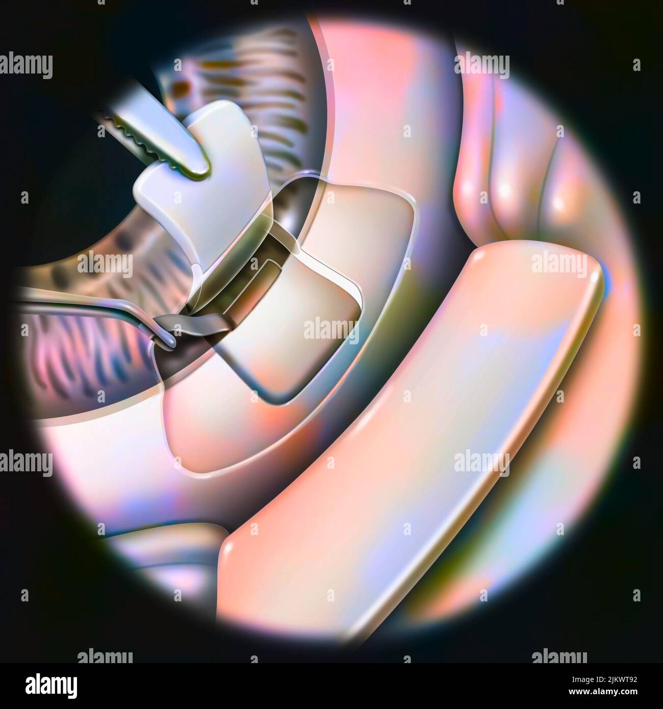 Eye (glaucoma): sclerectomy with removal of a small part of the trabeculum. Stock Photohttps://www.alamy.com/image-license-details/?v=1https://www.alamy.com/eye-glaucoma-sclerectomy-with-removal-of-a-small-part-of-the-trabeculum-image476926270.html
RF2JKWT92–Eye (glaucoma): sclerectomy with removal of a small part of the trabeculum.
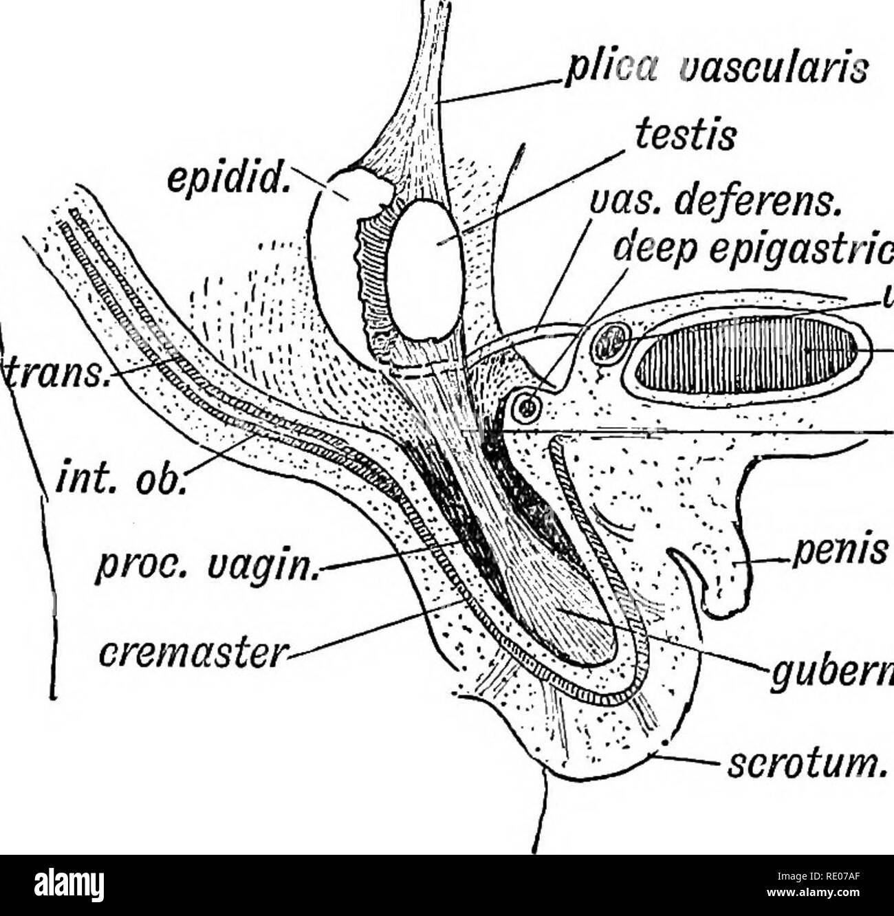 . Human embryology and morphology. Embryology, Human; Morphology. THE URO-GENITAL SYSTEM. 129 of enclosing cells and these produce the spermatoblasts. The tunica albuginea is formed from the mesoblastic covering of the genital ridge. The visceral layer of the tunica vaginalis on the testicle is the covering of germinal epithelium which remains after the ingrowth of the genital cells. The vasa efferentia and coni vasculosi are formed from the genital Wolffian tubules. The tubuli recti' and rete testis are new formations. The epididymis is the elongated upper segment of the Wolffian duct (Figs. Stock Photohttps://www.alamy.com/image-license-details/?v=1https://www.alamy.com/human-embryology-and-morphology-embryology-human-morphology-the-uro-genital-system-129-of-enclosing-cells-and-these-produce-the-spermatoblasts-the-tunica-albuginea-is-formed-from-the-mesoblastic-covering-of-the-genital-ridge-the-visceral-layer-of-the-tunica-vaginalis-on-the-testicle-is-the-covering-of-germinal-epithelium-which-remains-after-the-ingrowth-of-the-genital-cells-the-vasa-efferentia-and-coni-vasculosi-are-formed-from-the-genital-wolffian-tubules-the-tubuli-recti-and-rete-testis-are-new-formations-the-epididymis-is-the-elongated-upper-segment-of-the-wolffian-duct-figs-image232345751.html
RMRE07AF–. Human embryology and morphology. Embryology, Human; Morphology. THE URO-GENITAL SYSTEM. 129 of enclosing cells and these produce the spermatoblasts. The tunica albuginea is formed from the mesoblastic covering of the genital ridge. The visceral layer of the tunica vaginalis on the testicle is the covering of germinal epithelium which remains after the ingrowth of the genital cells. The vasa efferentia and coni vasculosi are formed from the genital Wolffian tubules. The tubuli recti' and rete testis are new formations. The epididymis is the elongated upper segment of the Wolffian duct (Figs.
 Histologically stained cross-section ofa rabbit testicle. Stock Photohttps://www.alamy.com/image-license-details/?v=1https://www.alamy.com/histologically-stained-cross-section-ofa-rabbit-testicle-image328254723.html
RM2A2186Y–Histologically stained cross-section ofa rabbit testicle.
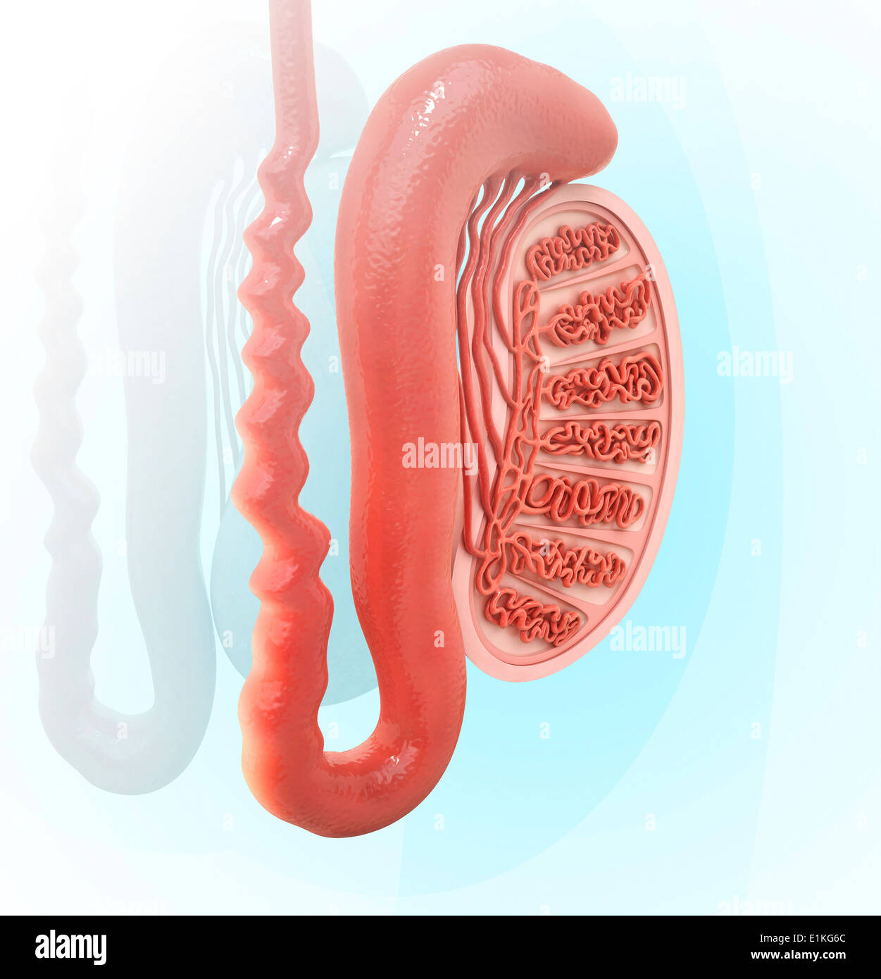 Human testicle computer artwork. Stock Photohttps://www.alamy.com/image-license-details/?v=1https://www.alamy.com/human-testicle-computer-artwork-image69885940.html
RFE1KG6C–Human testicle computer artwork.
 . The cyclopædia of anatomy and physiology. Anatomy; Physiology; Zoology. TESTICLE (ABNORMAL ANATOMY). 995 the inflammatory action ; but when the con- tiguous organ or subjacent part is of a dif- Fig. 638.. ferent structure from that of the cellular tissue, the extension of inflammation inwards is checked. Thus, in the case of the inflamed tunica vaginalis, the cellular tissue readily transmitted the morbid action to the epididy- mis, but the tunica albuginea arrested its progress to the body of the testicle ; and this explains the fact that after inflammation of the tunica vaginalis, excited Stock Photohttps://www.alamy.com/image-license-details/?v=1https://www.alamy.com/the-cyclopdia-of-anatomy-and-physiology-anatomy-physiology-zoology-testicle-abnormal-anatomy-995-the-inflammatory-action-but-when-the-con-tiguous-organ-or-subjacent-part-is-of-a-dif-fig-638-ferent-structure-from-that-of-the-cellular-tissue-the-extension-of-inflammation-inwards-is-checked-thus-in-the-case-of-the-inflamed-tunica-vaginalis-the-cellular-tissue-readily-transmitted-the-morbid-action-to-the-epididy-mis-but-the-tunica-albuginea-arrested-its-progress-to-the-body-of-the-testicle-and-this-explains-the-fact-that-after-inflammation-of-the-tunica-vaginalis-excited-image216208690.html
RMPFN4AX–. The cyclopædia of anatomy and physiology. Anatomy; Physiology; Zoology. TESTICLE (ABNORMAL ANATOMY). 995 the inflammatory action ; but when the con- tiguous organ or subjacent part is of a dif- Fig. 638.. ferent structure from that of the cellular tissue, the extension of inflammation inwards is checked. Thus, in the case of the inflamed tunica vaginalis, the cellular tissue readily transmitted the morbid action to the epididy- mis, but the tunica albuginea arrested its progress to the body of the testicle ; and this explains the fact that after inflammation of the tunica vaginalis, excited
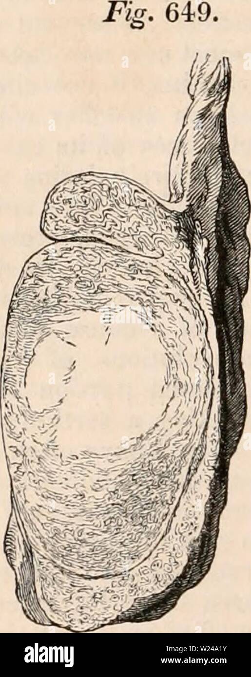 Archive image from page 221 of The cyclopædia of anatomy and. The cyclopædia of anatomy and physiology cyclopdiaofana0402todd Year: 1849 TESTICLE (ABNORMAL ANATOMY). 1006 epididymis, appears to prevent the oblitera- tion of the duct of which it is composed, and thus accounts for atrophy occurring much more rarely after consecutive orchids than after inflammation originating in the body of the gland, where the delicate seminal tubes are enclosed in the firm unyielding tunica albuginea. Chronic orcftitis. — The testicle is liable to a form of inflammatory swelling of a distinct and chronic cha Stock Photohttps://www.alamy.com/image-license-details/?v=1https://www.alamy.com/archive-image-from-page-221-of-the-cyclopdia-of-anatomy-and-the-cyclopdia-of-anatomy-and-physiology-cyclopdiaofana0402todd-year-1849-testicle-abnormal-anatomy-1006-epididymis-appears-to-prevent-the-oblitera-tion-of-the-duct-of-which-it-is-composed-and-thus-accounts-for-atrophy-occurring-much-more-rarely-after-consecutive-orchids-than-after-inflammation-originating-in-the-body-of-the-gland-where-the-delicate-seminal-tubes-are-enclosed-in-the-firm-unyielding-tunica-albuginea-chronic-orcftitis-the-testicle-is-liable-to-a-form-of-inflammatory-swelling-of-a-distinct-and-chronic-cha-image259480535.html
RMW24A1Y–Archive image from page 221 of The cyclopædia of anatomy and. The cyclopædia of anatomy and physiology cyclopdiaofana0402todd Year: 1849 TESTICLE (ABNORMAL ANATOMY). 1006 epididymis, appears to prevent the oblitera- tion of the duct of which it is composed, and thus accounts for atrophy occurring much more rarely after consecutive orchids than after inflammation originating in the body of the gland, where the delicate seminal tubes are enclosed in the firm unyielding tunica albuginea. Chronic orcftitis. — The testicle is liable to a form of inflammatory swelling of a distinct and chronic cha
 . Fig. 229—Cystic Ovary, Reduced. Nymphomania. A, Normal ovary ; B, cystic gland. of quite large cysts that they project conspicuously beyond the general surface of the gland. In some cases, the ovarian tissue proper vanishes almost completely under the pressure of large cysts, firmly compressed within the enveloping tunica albuginea. In other extremely bad cases of nymphomania there are found small, atrophied, fibrous ovaries, very hard and dense, like fibro-cartilage. The examination of the ovaries of the mare is to be made upon the standing animal, in essentially the same manner as that des Stock Photohttps://www.alamy.com/image-license-details/?v=1https://www.alamy.com/fig-229cystic-ovary-reduced-nymphomania-a-normal-ovary-b-cystic-gland-of-quite-large-cysts-that-they-project-conspicuously-beyond-the-general-surface-of-the-gland-in-some-cases-the-ovarian-tissue-proper-vanishes-almost-completely-under-the-pressure-of-large-cysts-firmly-compressed-within-the-enveloping-tunica-albuginea-in-other-extremely-bad-cases-of-nymphomania-there-are-found-small-atrophied-fibrous-ovaries-very-hard-and-dense-like-fibro-cartilage-the-examination-of-the-ovaries-of-the-mare-is-to-be-made-upon-the-standing-animal-in-essentially-the-same-manner-as-that-des-image179902914.html
RMMCK802–. Fig. 229—Cystic Ovary, Reduced. Nymphomania. A, Normal ovary ; B, cystic gland. of quite large cysts that they project conspicuously beyond the general surface of the gland. In some cases, the ovarian tissue proper vanishes almost completely under the pressure of large cysts, firmly compressed within the enveloping tunica albuginea. In other extremely bad cases of nymphomania there are found small, atrophied, fibrous ovaries, very hard and dense, like fibro-cartilage. The examination of the ovaries of the mare is to be made upon the standing animal, in essentially the same manner as that des
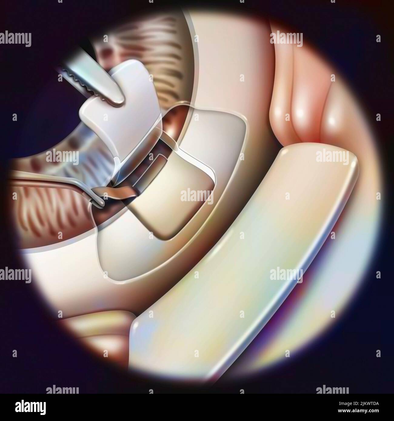 Eye (glaucoma): sclerectomy with removal of a small part of the trabeculum. Stock Photohttps://www.alamy.com/image-license-details/?v=1https://www.alamy.com/eye-glaucoma-sclerectomy-with-removal-of-a-small-part-of-the-trabeculum-image476926390.html
RF2JKWTDA–Eye (glaucoma): sclerectomy with removal of a small part of the trabeculum.
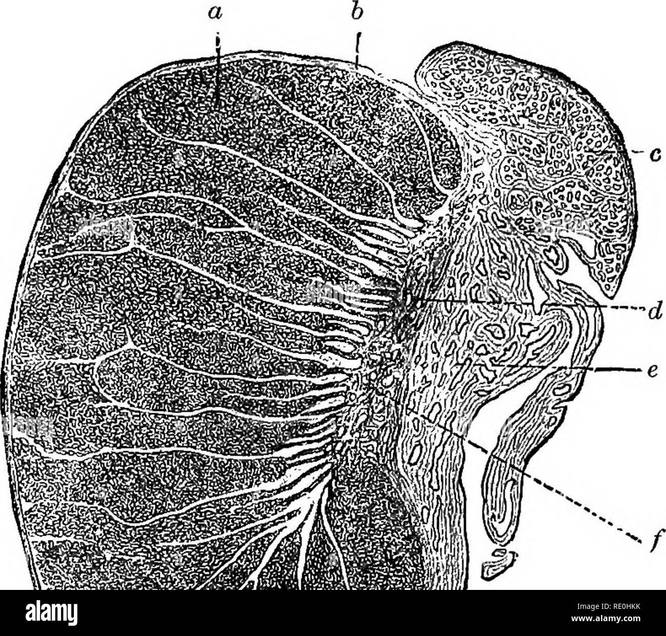 . The physiology of reproduction. Reproduction. 166 THE PHYSIOLOGY OF REPRODUCTION This organ is enclosed within a fibrous capsule, the tunica albuginea, which is very rich in lymphatics. It is covered by a layer of serous epithelium reflected from the tunica vaginalis. Posteriorly the capsule is prolonged into the interior of the testis in the form of a mass of fibrous tissue (the mediastinum testis). Certain other fibrous processes or trabeculse also pro-. Fig. 40.—Section through human testis and epididymis. (After Bohm and von Davidoff, from Schafer.) glandular substance divided into lob Stock Photohttps://www.alamy.com/image-license-details/?v=1https://www.alamy.com/the-physiology-of-reproduction-reproduction-166-the-physiology-of-reproduction-this-organ-is-enclosed-within-a-fibrous-capsule-the-tunica-albuginea-which-is-very-rich-in-lymphatics-it-is-covered-by-a-layer-of-serous-epithelium-reflected-from-the-tunica-vaginalis-posteriorly-the-capsule-is-prolonged-into-the-interior-of-the-testis-in-the-form-of-a-mass-of-fibrous-tissue-the-mediastinum-testis-certain-other-fibrous-processes-or-trabeculse-also-pro-fig-40section-through-human-testis-and-epididymis-after-bohm-and-von-davidoff-from-schafer-glandular-substance-divided-into-lob-image232353847.html
RMRE0HKK–. The physiology of reproduction. Reproduction. 166 THE PHYSIOLOGY OF REPRODUCTION This organ is enclosed within a fibrous capsule, the tunica albuginea, which is very rich in lymphatics. It is covered by a layer of serous epithelium reflected from the tunica vaginalis. Posteriorly the capsule is prolonged into the interior of the testis in the form of a mass of fibrous tissue (the mediastinum testis). Certain other fibrous processes or trabeculse also pro-. Fig. 40.—Section through human testis and epididymis. (After Bohm and von Davidoff, from Schafer.) glandular substance divided into lob
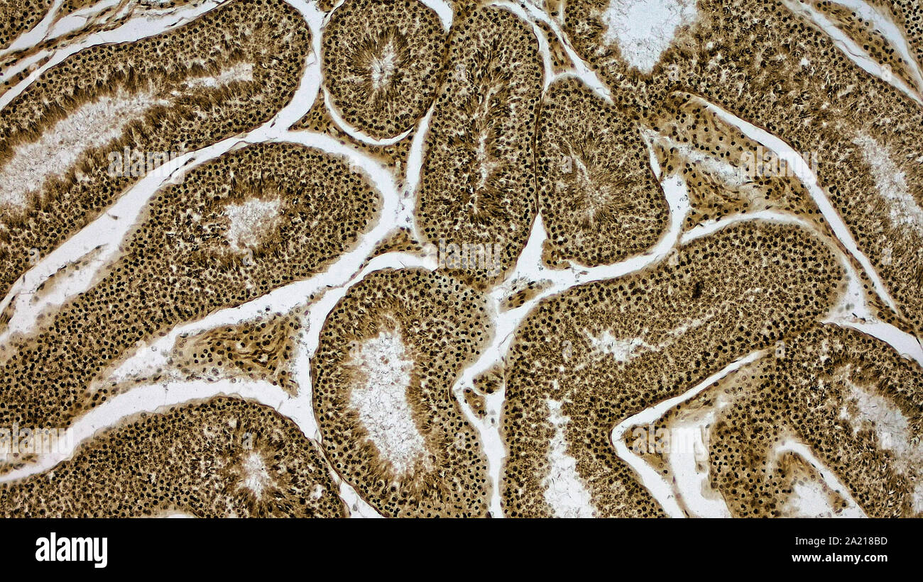 Histologically stained cross-section ofa rabbit testicle. Stock Photohttps://www.alamy.com/image-license-details/?v=1https://www.alamy.com/histologically-stained-cross-section-ofa-rabbit-testicle-image328254849.html
RM2A218BD–Histologically stained cross-section ofa rabbit testicle.
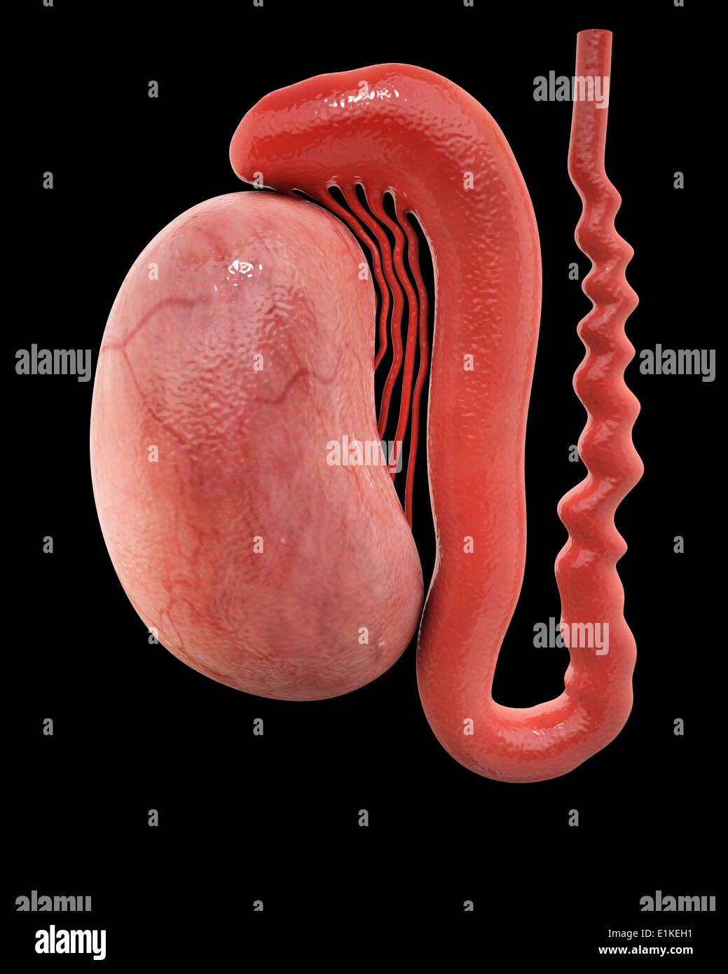 Human testicle computer artwork. Stock Photohttps://www.alamy.com/image-license-details/?v=1https://www.alamy.com/human-testicle-computer-artwork-image69884669.html
RFE1KEH1–Human testicle computer artwork.
 . The cyclopædia of anatomy and physiology. Anatomy; Physiology; Zoology. TESTICLE (ABNORMAL ANATOMY). 1006 epididymis, appears to prevent the oblitera- tion of the duct of which it is composed, and thus accounts for atrophy occurring much more rarely after consecutive orchids than after inflammation originating in the body of the gland, where the delicate seminal tubes are enclosed in the firm unyielding tunica albuginea. Chronic orcftitis. — The testicle is liable to a form of inflammatory swelling of a distinct and chronic character, which occasionally succeeds acute orchitis but far more c Stock Photohttps://www.alamy.com/image-license-details/?v=1https://www.alamy.com/the-cyclopdia-of-anatomy-and-physiology-anatomy-physiology-zoology-testicle-abnormal-anatomy-1006-epididymis-appears-to-prevent-the-oblitera-tion-of-the-duct-of-which-it-is-composed-and-thus-accounts-for-atrophy-occurring-much-more-rarely-after-consecutive-orchids-than-after-inflammation-originating-in-the-body-of-the-gland-where-the-delicate-seminal-tubes-are-enclosed-in-the-firm-unyielding-tunica-albuginea-chronic-orcftitis-the-testicle-is-liable-to-a-form-of-inflammatory-swelling-of-a-distinct-and-chronic-character-which-occasionally-succeeds-acute-orchitis-but-far-more-c-image216208674.html
RMPFN4AA–. The cyclopædia of anatomy and physiology. Anatomy; Physiology; Zoology. TESTICLE (ABNORMAL ANATOMY). 1006 epididymis, appears to prevent the oblitera- tion of the duct of which it is composed, and thus accounts for atrophy occurring much more rarely after consecutive orchids than after inflammation originating in the body of the gland, where the delicate seminal tubes are enclosed in the firm unyielding tunica albuginea. Chronic orcftitis. — The testicle is liable to a form of inflammatory swelling of a distinct and chronic character, which occasionally succeeds acute orchitis but far more c
 Archive image from page 193 of The cyclopædia of anatomy and. The cyclopædia of anatomy and physiology cyclopdiaofana0402todd Year: 1849 978 TESTICLE (NORMAL ANATOMY). mentous cords from the inner surface of the tunica albuginea. I have not been able to make out any such ligamentous processes passing into the substance of the testis, as are represented in Sir A. Cooper's work (part i. pi. 2. fig. 3), which I have no doubt is an ex- aggerated view of the preparation from which it was taken. The cords described appear to me to consist chiefly of blood-vessels sup- ported by slight fibrous proc Stock Photohttps://www.alamy.com/image-license-details/?v=1https://www.alamy.com/archive-image-from-page-193-of-the-cyclopdia-of-anatomy-and-the-cyclopdia-of-anatomy-and-physiology-cyclopdiaofana0402todd-year-1849-978-testicle-normal-anatomy-mentous-cords-from-the-inner-surface-of-the-tunica-albuginea-i-have-not-been-able-to-make-out-any-such-ligamentous-processes-passing-into-the-substance-of-the-testis-as-are-represented-in-sir-a-coopers-work-part-i-pi-2-fig-3-which-i-have-no-doubt-is-an-ex-aggerated-view-of-the-preparation-from-which-it-was-taken-the-cords-described-appear-to-me-to-consist-chiefly-of-blood-vessels-sup-ported-by-slight-fibrous-proc-image259472978.html
RMW240C2–Archive image from page 193 of The cyclopædia of anatomy and. The cyclopædia of anatomy and physiology cyclopdiaofana0402todd Year: 1849 978 TESTICLE (NORMAL ANATOMY). mentous cords from the inner surface of the tunica albuginea. I have not been able to make out any such ligamentous processes passing into the substance of the testis, as are represented in Sir A. Cooper's work (part i. pi. 2. fig. 3), which I have no doubt is an ex- aggerated view of the preparation from which it was taken. The cords described appear to me to consist chiefly of blood-vessels sup- ported by slight fibrous proc
 . Fig. 229—Cystic Ovai-y, Reduced Nymphomania. A, Normal ovary ; />', cystic gland. of quite large cysts that they project conspicuously beyond the general surface of the gland. In some cases, the ovarian tissue proper vanishes almost completely under the pressure of large cysts, firmly compressed within the enveloping tunica albuginea. In other extremely bad cases of nymphomania there are found small, atrophied, fibrous ovaries, very hard and dense, like fibro-cartilage. The examination of the ovaries of the mare is to be made upon the standing animal, in essentially the same manner as tha Stock Photohttps://www.alamy.com/image-license-details/?v=1https://www.alamy.com/fig-229cystic-ovai-y-reduced-nymphomania-a-normal-ovary-gt-cystic-gland-of-quite-large-cysts-that-they-project-conspicuously-beyond-the-general-surface-of-the-gland-in-some-cases-the-ovarian-tissue-proper-vanishes-almost-completely-under-the-pressure-of-large-cysts-firmly-compressed-within-the-enveloping-tunica-albuginea-in-other-extremely-bad-cases-of-nymphomania-there-are-found-small-atrophied-fibrous-ovaries-very-hard-and-dense-like-fibro-cartilage-the-examination-of-the-ovaries-of-the-mare-is-to-be-made-upon-the-standing-animal-in-essentially-the-same-manner-as-tha-image179903293.html
RMMCK8DH–. Fig. 229—Cystic Ovai-y, Reduced Nymphomania. A, Normal ovary ; />', cystic gland. of quite large cysts that they project conspicuously beyond the general surface of the gland. In some cases, the ovarian tissue proper vanishes almost completely under the pressure of large cysts, firmly compressed within the enveloping tunica albuginea. In other extremely bad cases of nymphomania there are found small, atrophied, fibrous ovaries, very hard and dense, like fibro-cartilage. The examination of the ovaries of the mare is to be made upon the standing animal, in essentially the same manner as tha
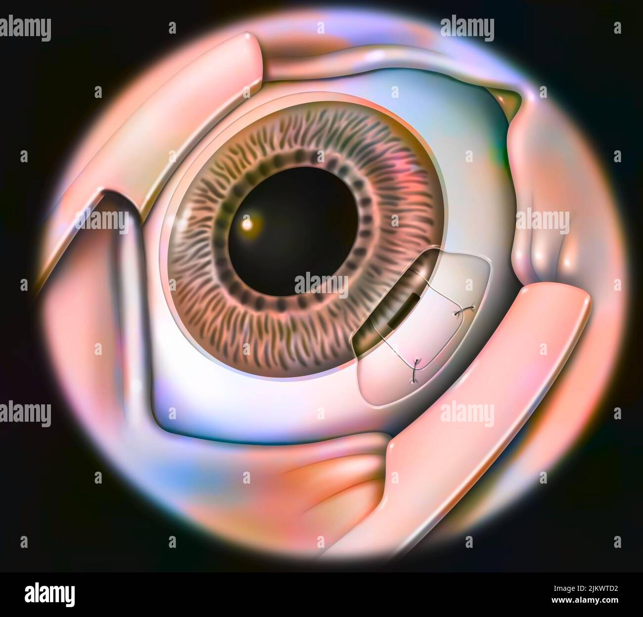 Eye, glaucoma: sclerectomy with sclera suture. Stock Photohttps://www.alamy.com/image-license-details/?v=1https://www.alamy.com/eye-glaucoma-sclerectomy-with-sclera-suture-image476926382.html
RF2JKWTD2–Eye, glaucoma: sclerectomy with sclera suture.
 . The American journal of anatomy. Fig. , C. M. Jackson 75 extending in from the tunica albuginea (Fig. 4). The appearance ofthe corpora cavernosa in this region is in striking contrast to that ofthe corpus spongiosum, especially in injected specimens. Still morestriking is the appearance of a specimen stained with osmic acid, inwhich the corpora cavernosa are stained jet black. Passing from thedistal extremity, the corpora cavernosa gradually enlarge, and becomemore fibrous and vascular. The vessels are irregular venous channels,best developed in the center of each corpus, so that the adipose Stock Photohttps://www.alamy.com/image-license-details/?v=1https://www.alamy.com/the-american-journal-of-anatomy-fig-c-m-jackson-75-extending-in-from-the-tunica-albuginea-fig-4-the-appearance-ofthe-corpora-cavernosa-in-this-region-is-in-striking-contrast-to-that-ofthe-corpus-spongiosum-especially-in-injected-specimens-still-morestriking-is-the-appearance-of-a-specimen-stained-with-osmic-acid-inwhich-the-corpora-cavernosa-are-stained-jet-black-passing-from-thedistal-extremity-the-corpora-cavernosa-gradually-enlarge-and-becomemore-fibrous-and-vascular-the-vessels-are-irregular-venous-channelsbest-developed-in-the-center-of-each-corpus-so-that-the-adipose-image336928393.html
RM2AG4BGW–. The American journal of anatomy. Fig. , C. M. Jackson 75 extending in from the tunica albuginea (Fig. 4). The appearance ofthe corpora cavernosa in this region is in striking contrast to that ofthe corpus spongiosum, especially in injected specimens. Still morestriking is the appearance of a specimen stained with osmic acid, inwhich the corpora cavernosa are stained jet black. Passing from thedistal extremity, the corpora cavernosa gradually enlarge, and becomemore fibrous and vascular. The vessels are irregular venous channels,best developed in the center of each corpus, so that the adipose
 Histologically stained cross-section ofa rabbit testicle. Stock Photohttps://www.alamy.com/image-license-details/?v=1https://www.alamy.com/histologically-stained-cross-section-ofa-rabbit-testicle-image328254854.html
RM2A218BJ–Histologically stained cross-section ofa rabbit testicle.
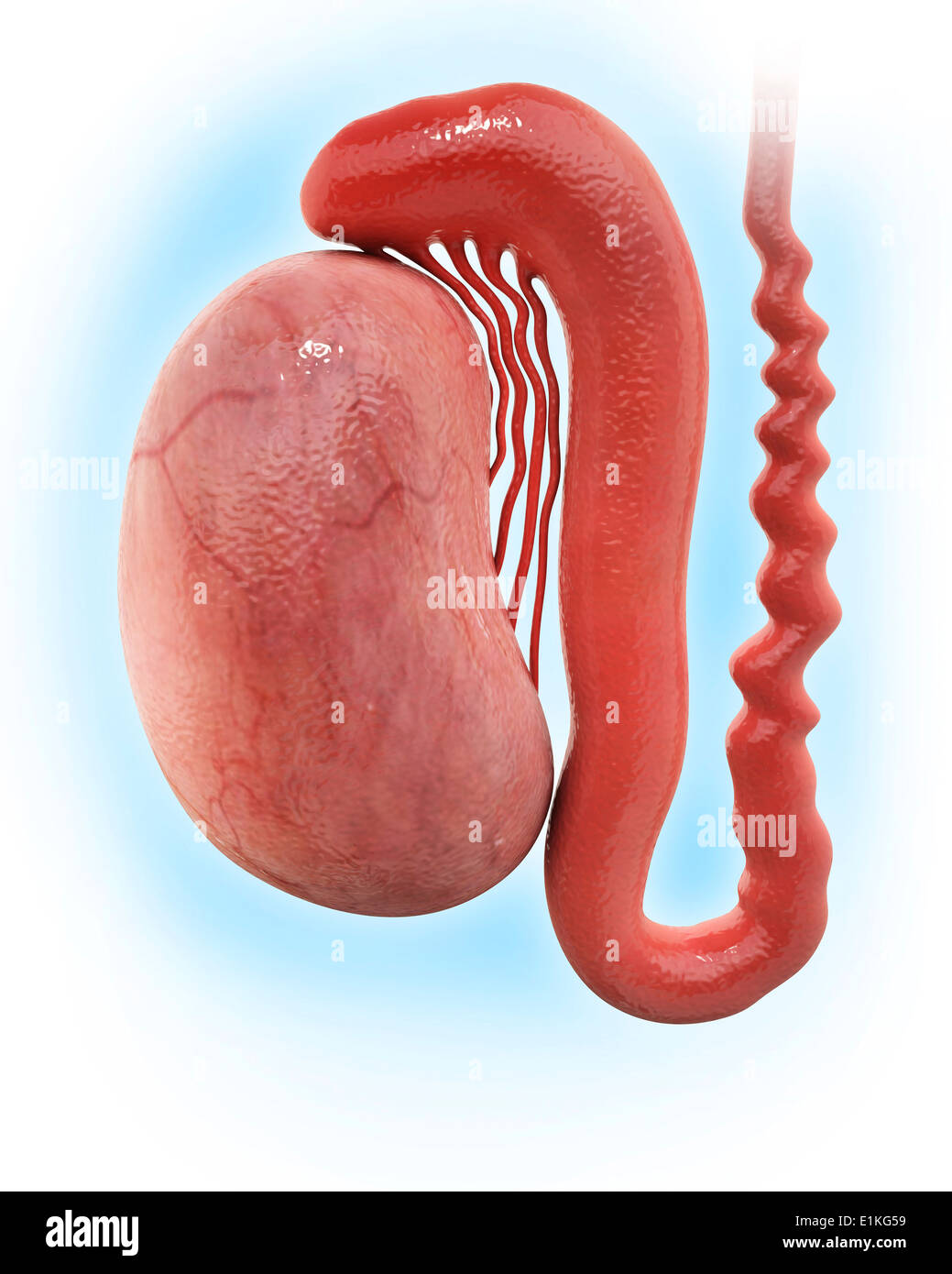 Human testicle computer artwork. Stock Photohttps://www.alamy.com/image-license-details/?v=1https://www.alamy.com/human-testicle-computer-artwork-image69885909.html
RFE1KG59–Human testicle computer artwork.
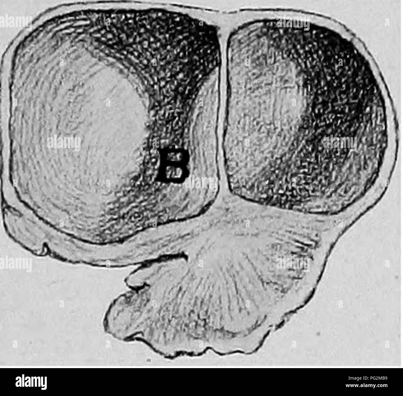 . The diseases of the genital organs of domestic animals. Veterinary medicine. Fig. 229—Cystic Ovary, Reduced Nymphomania. A, Normal ovary ; B, cystic gland. of quite large cysts that they project conspicuously beyond the general surface of the gland. In some cases, the ovarian tissue proper vanishes almost completely under the pressure of large cysts, firmly compressed within the enveloping tunica albuginea. In other extremely bad cases of nymphomania there are found small, atrophied, fibrous ovaries, very hard and dense, like fibro-cartilage. The examination of the ovaries of the mare is to Stock Photohttps://www.alamy.com/image-license-details/?v=1https://www.alamy.com/the-diseases-of-the-genital-organs-of-domestic-animals-veterinary-medicine-fig-229cystic-ovary-reduced-nymphomania-a-normal-ovary-b-cystic-gland-of-quite-large-cysts-that-they-project-conspicuously-beyond-the-general-surface-of-the-gland-in-some-cases-the-ovarian-tissue-proper-vanishes-almost-completely-under-the-pressure-of-large-cysts-firmly-compressed-within-the-enveloping-tunica-albuginea-in-other-extremely-bad-cases-of-nymphomania-there-are-found-small-atrophied-fibrous-ovaries-very-hard-and-dense-like-fibro-cartilage-the-examination-of-the-ovaries-of-the-mare-is-to-image216418813.html
RMPG2MB9–. The diseases of the genital organs of domestic animals. Veterinary medicine. Fig. 229—Cystic Ovary, Reduced Nymphomania. A, Normal ovary ; B, cystic gland. of quite large cysts that they project conspicuously beyond the general surface of the gland. In some cases, the ovarian tissue proper vanishes almost completely under the pressure of large cysts, firmly compressed within the enveloping tunica albuginea. In other extremely bad cases of nymphomania there are found small, atrophied, fibrous ovaries, very hard and dense, like fibro-cartilage. The examination of the ovaries of the mare is to
 Archive image from page 624 of The cyclopædia of anatomy and. The cyclopædia of anatomy and physiology cyclopdiaofana05todd Year: 1859 OVARY—(NORMAL ANATOMY). 549 explained by the much smaller number of ovary. It lies immediately beneath the tunica albuginea, and fdls up the whole of the inter- mediate space between the ovisacs, to which it acts as a germ bed, protecting the ova from injury, and serving for the conveyance of blood- vessels to the ovisacs. This tissue is some- times of a pale-pink, but more often of a bright-red colour, from the large number of blood-vessels which it contains, Stock Photohttps://www.alamy.com/image-license-details/?v=1https://www.alamy.com/archive-image-from-page-624-of-the-cyclopdia-of-anatomy-and-the-cyclopdia-of-anatomy-and-physiology-cyclopdiaofana05todd-year-1859-ovarynormal-anatomy-549-explained-by-the-much-smaller-number-of-ovary-it-lies-immediately-beneath-the-tunica-albuginea-and-fdls-up-the-whole-of-the-inter-mediate-space-between-the-ovisacs-to-which-it-acts-as-a-germ-bed-protecting-the-ova-from-injury-and-serving-for-the-conveyance-of-blood-vessels-to-the-ovisacs-this-tissue-is-some-times-of-a-pale-pink-but-more-often-of-a-bright-red-colour-from-the-large-number-of-blood-vessels-which-it-contains-image259326339.html
RMW1W9AY–Archive image from page 624 of The cyclopædia of anatomy and. The cyclopædia of anatomy and physiology cyclopdiaofana05todd Year: 1859 OVARY—(NORMAL ANATOMY). 549 explained by the much smaller number of ovary. It lies immediately beneath the tunica albuginea, and fdls up the whole of the inter- mediate space between the ovisacs, to which it acts as a germ bed, protecting the ova from injury, and serving for the conveyance of blood- vessels to the ovisacs. This tissue is some- times of a pale-pink, but more often of a bright-red colour, from the large number of blood-vessels which it contains,
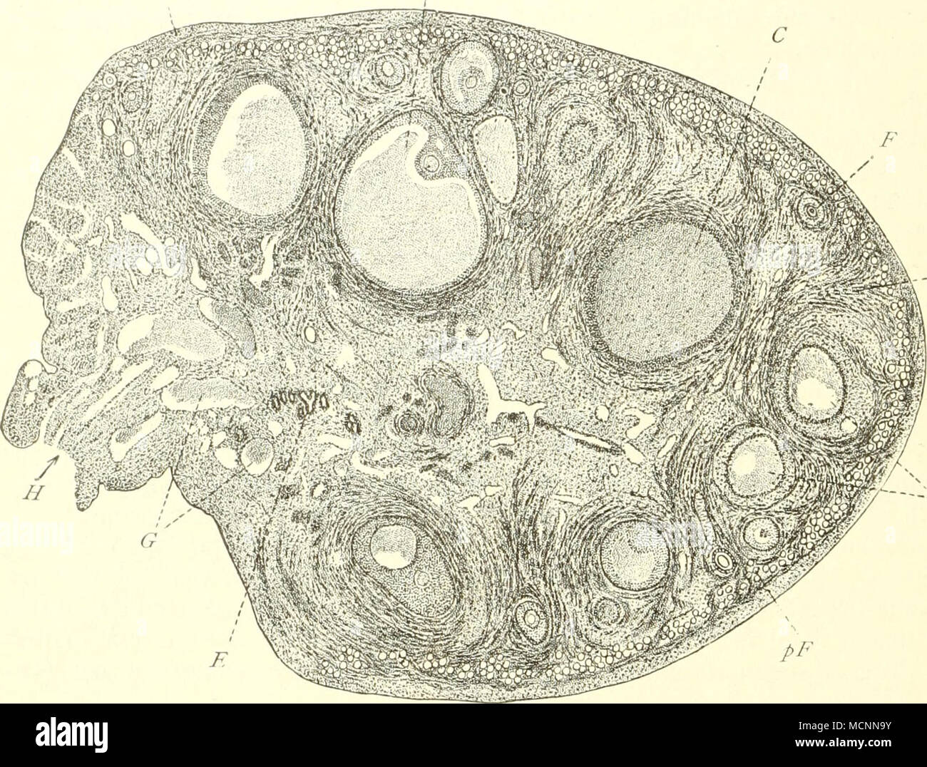 . R 1- Fd Fig-. 193. Schnitt durch ein Ovarium von Fehs domestica, nach K. C. Schneider. 7" Tunica albuginea; R Bindegewebe der Rinde; G Gefäße der Marlisubstanz; //Hihis ovarii; E Epoophoron; pI" Priniärfollikel; Fv Sekundärfollikel mit Liquor gefüllt, mit hügelförmig vorspringendem Cumulus oophorus, der das Ei enthält; /^(/degenerierende, F sich entwickelnder Follikel; C Corpus luteum. Fig. 194. Testikel, umgeben durch die Tuiiica albuginea A die durch bindegewebige Septula s, den Hoden in Lappen verteilt. In diesen liegen die TnbuH contorti tc, die in die Tubuli recti /;• übergehe Stock Photohttps://www.alamy.com/image-license-details/?v=1https://www.alamy.com/r-1-fd-fig-193-schnitt-durch-ein-ovarium-von-fehs-domestica-nach-k-c-schneider-7quot-tunica-albuginea-r-bindegewebe-der-rinde-g-gefe-der-marlisubstanz-hihis-ovarii-e-epoophoron-piquot-prinirfollikel-fv-sekundrfollikel-mit-liquor-gefllt-mit-hgelfrmig-vorspringendem-cumulus-oophorus-der-das-ei-enthlt-degenerierende-f-sich-entwickelnder-follikel-c-corpus-luteum-fig-194-testikel-umgeben-durch-die-tuiiica-albuginea-a-die-durch-bindegewebige-septula-s-den-hoden-in-lappen-verteilt-in-diesen-liegen-die-tnbuh-contorti-tc-die-in-die-tubuli-recti-bergehe-image179957287.html
RMMCNN9Y–. R 1- Fd Fig-. 193. Schnitt durch ein Ovarium von Fehs domestica, nach K. C. Schneider. 7" Tunica albuginea; R Bindegewebe der Rinde; G Gefäße der Marlisubstanz; //Hihis ovarii; E Epoophoron; pI" Priniärfollikel; Fv Sekundärfollikel mit Liquor gefüllt, mit hügelförmig vorspringendem Cumulus oophorus, der das Ei enthält; /^(/degenerierende, F sich entwickelnder Follikel; C Corpus luteum. Fig. 194. Testikel, umgeben durch die Tuiiica albuginea A die durch bindegewebige Septula s, den Hoden in Lappen verteilt. In diesen liegen die TnbuH contorti tc, die in die Tubuli recti /;• übergehe
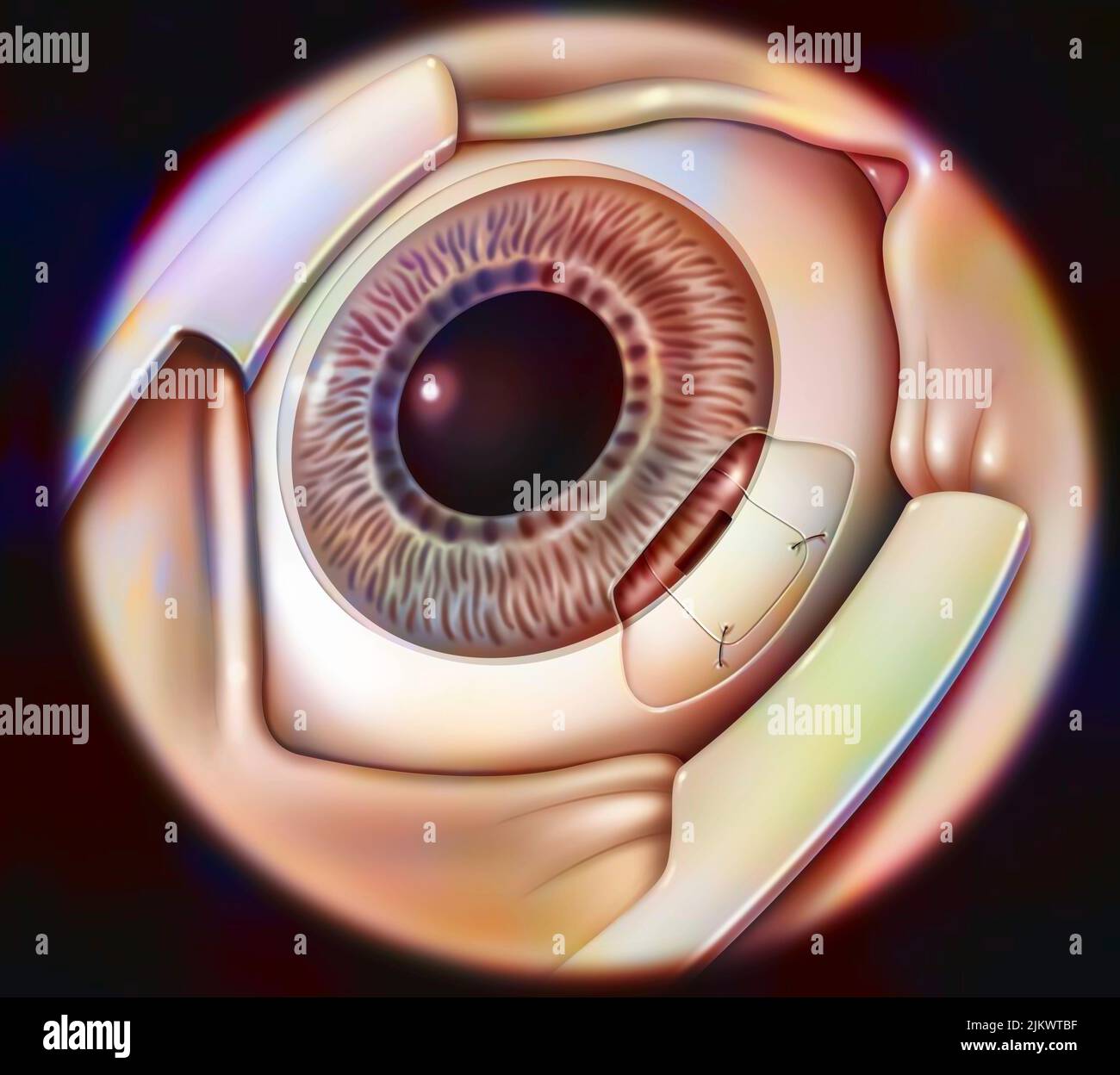 Eye, glaucoma: sclerectomy with sclera suture. Stock Photohttps://www.alamy.com/image-license-details/?v=1https://www.alamy.com/eye-glaucoma-sclerectomy-with-sclera-suture-image476926339.html
RF2JKWTBF–Eye, glaucoma: sclerectomy with sclera suture.
 Die männlichen Geschlechtsorgane . Urethra :^^ .cSI ,id- ^ — - Glandulaebulboupethrales ¥ Glandulae bulbo=urethrales. Corpus cavennofum bulh ? Septum Tunica albuginea Stock Photohttps://www.alamy.com/image-license-details/?v=1https://www.alamy.com/die-mnnlichen-geschlechtsorgane-urethra-csi-id-glandulaebulboupethrales-glandulae-bulbo=urethrales-corpus-cavennofum-bulh-septum-tunica-albuginea-image338423405.html
RM2AJGEE5–Die männlichen Geschlechtsorgane . Urethra :^^ .cSI ,id- ^ — - Glandulaebulboupethrales ¥ Glandulae bulbo=urethrales. Corpus cavennofum bulh ? Septum Tunica albuginea
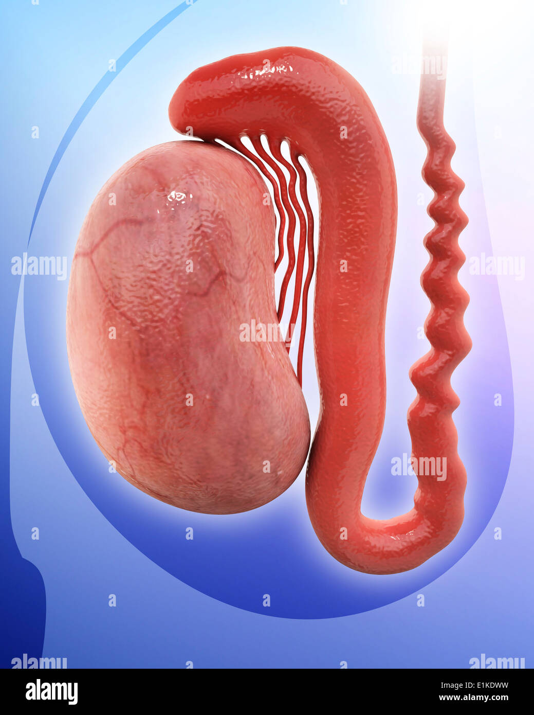 Human testicle computer artwork. Stock Photohttps://www.alamy.com/image-license-details/?v=1https://www.alamy.com/human-testicle-computer-artwork-image69884133.html
RFE1KDWW–Human testicle computer artwork.
 . The cyclopædia of anatomy and physiology. Anatomy; Physiology; Zoology. OVARY—(NORMAL ANATOMY). 549 explained by the much smaller number of ovary. It lies immediately beneath the tunica albuginea, and fdls up the whole of the inter- mediate space between the ovisacs, to which it acts as a germ bed, protecting the ova from injury, and serving for the conveyance of blood- vessels to the ovisacs. This tissue is some- times of a pale-pink, but more often of a bright-red colour, from the large number of blood-vessels which it contains, whose ar- rangement proceeding from within, and radi- ating o Stock Photohttps://www.alamy.com/image-license-details/?v=1https://www.alamy.com/the-cyclopdia-of-anatomy-and-physiology-anatomy-physiology-zoology-ovarynormal-anatomy-549-explained-by-the-much-smaller-number-of-ovary-it-lies-immediately-beneath-the-tunica-albuginea-and-fdls-up-the-whole-of-the-inter-mediate-space-between-the-ovisacs-to-which-it-acts-as-a-germ-bed-protecting-the-ova-from-injury-and-serving-for-the-conveyance-of-blood-vessels-to-the-ovisacs-this-tissue-is-some-times-of-a-pale-pink-but-more-often-of-a-bright-red-colour-from-the-large-number-of-blood-vessels-which-it-contains-whose-ar-rangement-proceeding-from-within-and-radi-ating-o-image216209273.html
RMPFN53N–. The cyclopædia of anatomy and physiology. Anatomy; Physiology; Zoology. OVARY—(NORMAL ANATOMY). 549 explained by the much smaller number of ovary. It lies immediately beneath the tunica albuginea, and fdls up the whole of the inter- mediate space between the ovisacs, to which it acts as a germ bed, protecting the ova from injury, and serving for the conveyance of blood- vessels to the ovisacs. This tissue is some- times of a pale-pink, but more often of a bright-red colour, from the large number of blood-vessels which it contains, whose ar- rangement proceeding from within, and radi- ating o
 The diseases of the genital The diseases of the genital organs of domestic animals diseasesofgenita00willrich Year: 1921 Fig. 229—Cystic Ovai-y, Reduced Nymphomania. A, Normal ovary ; />', cystic gland. of quite large cysts that they project conspicuously beyond the general surface of the gland. In some cases, the ovarian tissue proper vanishes almost completely under the pressure of large cysts, firmly compressed within the enveloping tunica albuginea. In other extremely bad cases of nymphomania there are found small, atrophied, fibrous ovaries, very hard and dense, like fibro-cartilage. Stock Photohttps://www.alamy.com/image-license-details/?v=1https://www.alamy.com/the-diseases-of-the-genital-the-diseases-of-the-genital-organs-of-domestic-animals-diseasesofgenita00willrich-year-1921-fig-229cystic-ovai-y-reduced-nymphomania-a-normal-ovary-gt-cystic-gland-of-quite-large-cysts-that-they-project-conspicuously-beyond-the-general-surface-of-the-gland-in-some-cases-the-ovarian-tissue-proper-vanishes-almost-completely-under-the-pressure-of-large-cysts-firmly-compressed-within-the-enveloping-tunica-albuginea-in-other-extremely-bad-cases-of-nymphomania-there-are-found-small-atrophied-fibrous-ovaries-very-hard-and-dense-like-fibro-cartilage-image241986847.html
RMT1KCKB–The diseases of the genital The diseases of the genital organs of domestic animals diseasesofgenita00willrich Year: 1921 Fig. 229—Cystic Ovai-y, Reduced Nymphomania. A, Normal ovary ; />', cystic gland. of quite large cysts that they project conspicuously beyond the general surface of the gland. In some cases, the ovarian tissue proper vanishes almost completely under the pressure of large cysts, firmly compressed within the enveloping tunica albuginea. In other extremely bad cases of nymphomania there are found small, atrophied, fibrous ovaries, very hard and dense, like fibro-cartilage.
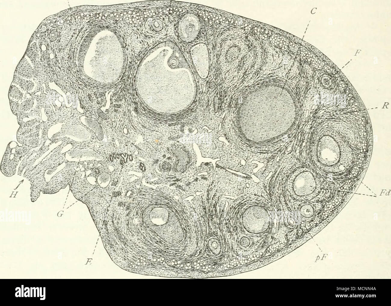 . Fig. 193. Schnitt durch ein Ovarium von Felis domestica, nach K. C. Schneider. T'Tunica albuginea; R Bindegewebe der Rinde; G GefäÃe der Marksubstanz; Ã^Hiius ovarü; E Epoophoron; pP Frimärfollikel; Fv Sekundärfollikel mit Liquor gefüllt, mit hügelförmig vorspringendem Cumulus oophorus, der das Ei enthält; i^c/degenerierende, F sich entwickelnder Follikel; C Corpus luteum. Fig. 194. Testikel, umgeben durch die Tunica albuginea t, die durch bindegewebige Septula s, den Hoden in Lappen verteilt. In diesen liegen die Tubuli contorti tc. die in die Tubuli recti //â übergehen, den Highmo Stock Photohttps://www.alamy.com/image-license-details/?v=1https://www.alamy.com/fig-193-schnitt-durch-ein-ovarium-von-felis-domestica-nach-k-c-schneider-ttunica-albuginea-r-bindegewebe-der-rinde-g-gefe-der-marksubstanz-hiius-ovar-e-epoophoron-pp-frimrfollikel-fv-sekundrfollikel-mit-liquor-gefllt-mit-hgelfrmig-vorspringendem-cumulus-oophorus-der-das-ei-enthlt-icdegenerierende-f-sich-entwickelnder-follikel-c-corpus-luteum-fig-194-testikel-umgeben-durch-die-tunica-albuginea-t-die-durch-bindegewebige-septula-s-den-hoden-in-lappen-verteilt-in-diesen-liegen-die-tubuli-contorti-tc-die-in-die-tubuli-recti-bergehen-den-highmo-image179957130.html
RMMCNN4A–. Fig. 193. Schnitt durch ein Ovarium von Felis domestica, nach K. C. Schneider. T'Tunica albuginea; R Bindegewebe der Rinde; G GefäÃe der Marksubstanz; Ã^Hiius ovarü; E Epoophoron; pP Frimärfollikel; Fv Sekundärfollikel mit Liquor gefüllt, mit hügelförmig vorspringendem Cumulus oophorus, der das Ei enthält; i^c/degenerierende, F sich entwickelnder Follikel; C Corpus luteum. Fig. 194. Testikel, umgeben durch die Tunica albuginea t, die durch bindegewebige Septula s, den Hoden in Lappen verteilt. In diesen liegen die Tubuli contorti tc. die in die Tubuli recti //â übergehen, den Highmo
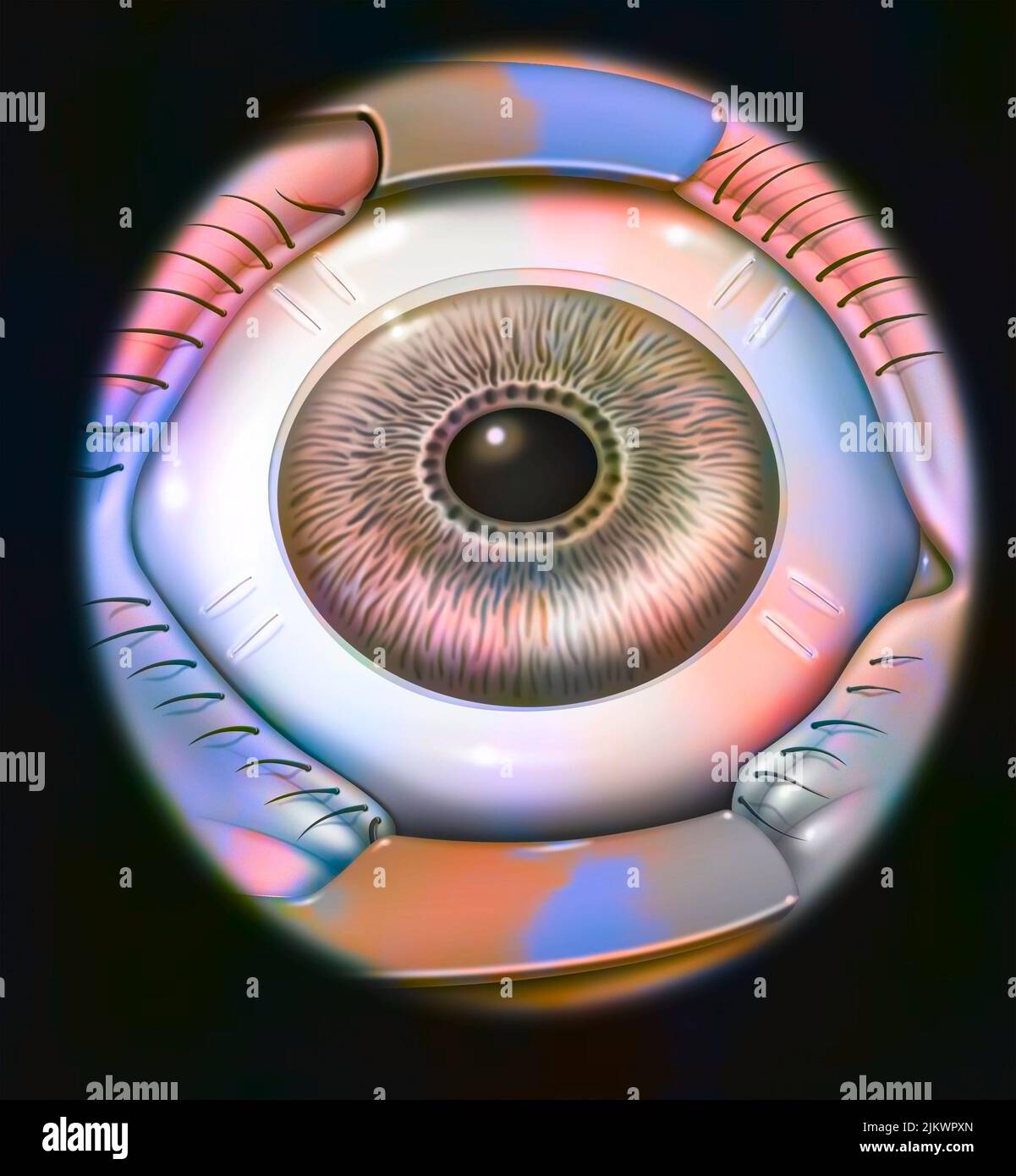 Treatment of presbyopia by scleral expansion (refractive surgery): scleral incisions. Stock Photohttps://www.alamy.com/image-license-details/?v=1https://www.alamy.com/treatment-of-presbyopia-by-scleral-expansion-refractive-surgery-scleral-incisions-image476925197.html
RF2JKWPXN–Treatment of presbyopia by scleral expansion (refractive surgery): scleral incisions.
 The Hahnemannian monthly . Thickened tunica albuginea.Multiplied 200 times. Pig. 5.. Inflammatory deposit on surface of ovaryMultiplied 200 times. Fig. 7. Stock Photohttps://www.alamy.com/image-license-details/?v=1https://www.alamy.com/the-hahnemannian-monthly-thickened-tunica-albugineamultiplied-200-times-pig-5-inflammatory-deposit-on-surface-of-ovarymultiplied-200-times-fig-7-image338178117.html
RM2AJ59HW–The Hahnemannian monthly . Thickened tunica albuginea.Multiplied 200 times. Pig. 5.. Inflammatory deposit on surface of ovaryMultiplied 200 times. Fig. 7.
 Human testicle computer artwork. Stock Photohttps://www.alamy.com/image-license-details/?v=1https://www.alamy.com/human-testicle-computer-artwork-image69885528.html
RFE1KFKM–Human testicle computer artwork.
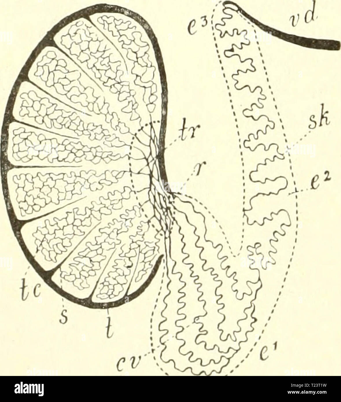 Archive image from page 257 of Die säugetiere Einführung in die Die säugetiere. Einführung in die anatomie und systematik der recenten und fossilen Mammalia diesugetiereei00webe Year: 1904 . R 1- Fd Fig-. 193. Schnitt durch ein Ovarium von Fehs domestica, nach K. C. Schneider. 7' Tunica albuginea; R Bindegewebe der Rinde; G Gefäße der Marlisubstanz; //Hihis ovarii; E Epoophoron; pI' Priniärfollikel; Fv Sekundärfollikel mit Liquor gefüllt, mit hügelförmig vorspringendem Cumulus oophorus, der das Ei enthält; /(/degenerierende, F sich entwickelnder Follikel; C Corpus luteum. Fig. 194. Testikel, Stock Photohttps://www.alamy.com/image-license-details/?v=1https://www.alamy.com/archive-image-from-page-257-of-die-sugetiere-einfhrung-in-die-die-sugetiere-einfhrung-in-die-anatomie-und-systematik-der-recenten-und-fossilen-mammalia-diesugetiereei00webe-year-1904-r-1-fd-fig-193-schnitt-durch-ein-ovarium-von-fehs-domestica-nach-k-c-schneider-7-tunica-albuginea-r-bindegewebe-der-rinde-g-gefe-der-marlisubstanz-hihis-ovarii-e-epoophoron-pi-prinirfollikel-fv-sekundrfollikel-mit-liquor-gefllt-mit-hgelfrmig-vorspringendem-cumulus-oophorus-der-das-ei-enthlt-degenerierende-f-sich-entwickelnder-follikel-c-corpus-luteum-fig-194-testikel-image242259189.html
RMT23T1W–Archive image from page 257 of Die säugetiere Einführung in die Die säugetiere. Einführung in die anatomie und systematik der recenten und fossilen Mammalia diesugetiereei00webe Year: 1904 . R 1- Fd Fig-. 193. Schnitt durch ein Ovarium von Fehs domestica, nach K. C. Schneider. 7' Tunica albuginea; R Bindegewebe der Rinde; G Gefäße der Marlisubstanz; //Hihis ovarii; E Epoophoron; pI' Priniärfollikel; Fv Sekundärfollikel mit Liquor gefüllt, mit hügelförmig vorspringendem Cumulus oophorus, der das Ei enthält; /(/degenerierende, F sich entwickelnder Follikel; C Corpus luteum. Fig. 194. Testikel,
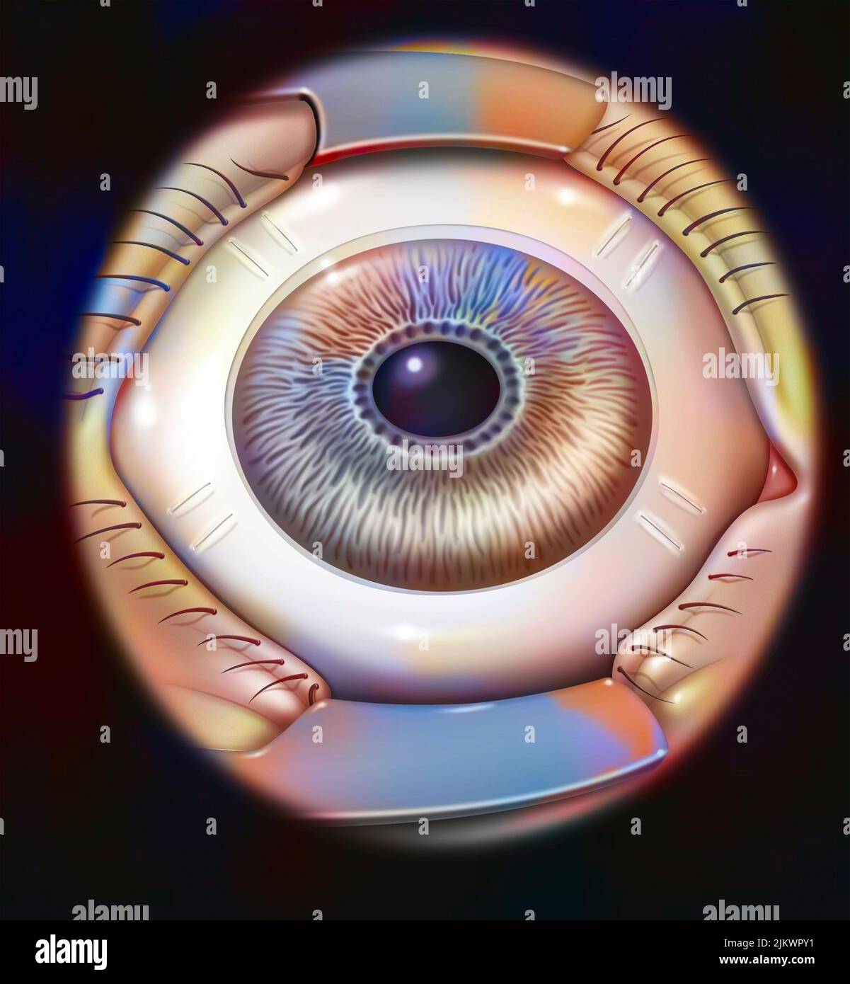 Treatment of presbyopia by scleral expansion (refractive surgery): scleral incisions. Stock Photohttps://www.alamy.com/image-license-details/?v=1https://www.alamy.com/treatment-of-presbyopia-by-scleral-expansion-refractive-surgery-scleral-incisions-image476925205.html
RF2JKWPY1–Treatment of presbyopia by scleral expansion (refractive surgery): scleral incisions.
 Transactions of the Homoeopathic Medical Society of the State of Pennsylvania . Thickened tunica albuginea. Multiplied 200 diameters. Fig. 5.. Inflammatory deposit on surface of ovary.Multiplied 200 diameters. Fig. 7. Stock Photohttps://www.alamy.com/image-license-details/?v=1https://www.alamy.com/transactions-of-the-homoeopathic-medical-society-of-the-state-of-pennsylvania-thickened-tunica-albuginea-multiplied-200-diameters-fig-5-inflammatory-deposit-on-surface-of-ovarymultiplied-200-diameters-fig-7-image343245163.html
RM2AXC4KR–Transactions of the Homoeopathic Medical Society of the State of Pennsylvania . Thickened tunica albuginea. Multiplied 200 diameters. Fig. 5.. Inflammatory deposit on surface of ovary.Multiplied 200 diameters. Fig. 7.
 Human testicle computer artwork. Stock Photohttps://www.alamy.com/image-license-details/?v=1https://www.alamy.com/human-testicle-computer-artwork-image69885504.html
RFE1KFJT–Human testicle computer artwork.
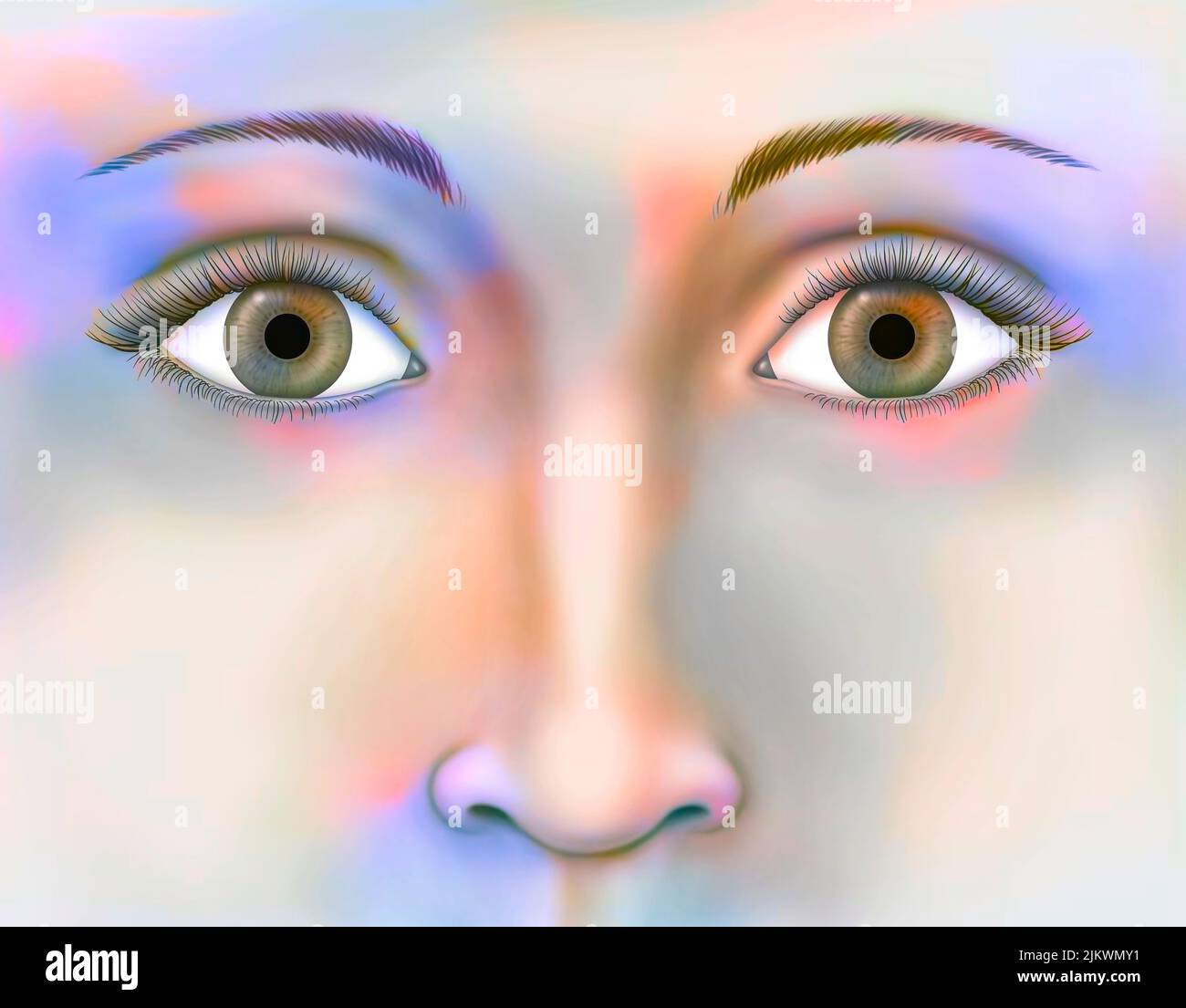 Woman's face seen from the front centered on the eyes. Stock Photohttps://www.alamy.com/image-license-details/?v=1https://www.alamy.com/womans-face-seen-from-the-front-centered-on-the-eyes-image476923637.html
RF2JKWMY1–Woman's face seen from the front centered on the eyes.
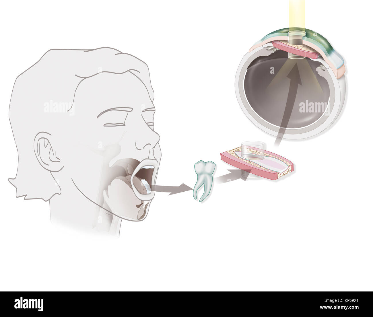 OSTEO-ODONTO-KERATOPROSTHESIS Stock Photohttps://www.alamy.com/image-license-details/?v=1https://www.alamy.com/stock-image-osteo-odonto-keratoprosthesis-168555241.html
RMKP69X1–OSTEO-ODONTO-KERATOPROSTHESIS
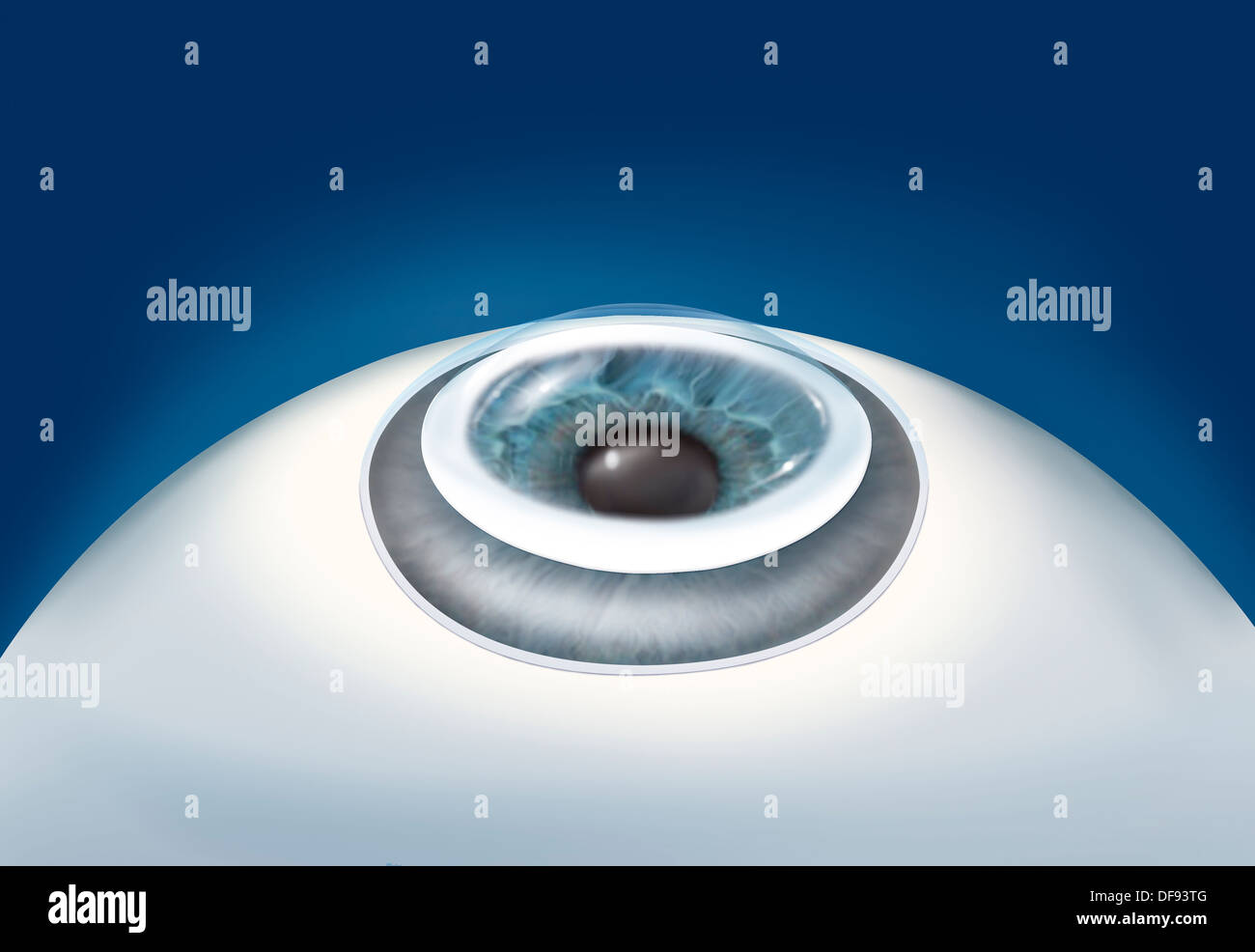 CORNEAL TRANSPLANT, DRAWING Stock Photohttps://www.alamy.com/image-license-details/?v=1https://www.alamy.com/corneal-transplant-drawing-image61051552.html
RMDF93TG–CORNEAL TRANSPLANT, DRAWING
 The Hahnemannian monthly . Thick layer of glandular cells on walls of a cystic Graafian follicle.Multiplied 200 times. Fig. 4.. Thickened tunica albuginea.Multiplied 200 times. Pig. 5. Stock Photohttps://www.alamy.com/image-license-details/?v=1https://www.alamy.com/the-hahnemannian-monthly-thick-layer-of-glandular-cells-on-walls-of-a-cystic-graafian-folliclemultiplied-200-times-fig-4-thickened-tunica-albugineamultiplied-200-times-pig-5-image338178406.html
RM2AJ5A06–The Hahnemannian monthly . Thick layer of glandular cells on walls of a cystic Graafian follicle.Multiplied 200 times. Fig. 4.. Thickened tunica albuginea.Multiplied 200 times. Pig. 5.
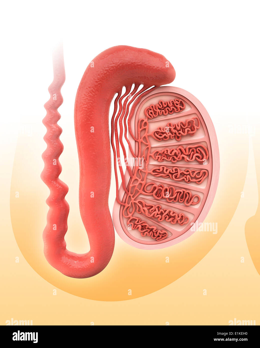 Human testicle computer artwork. Stock Photohttps://www.alamy.com/image-license-details/?v=1https://www.alamy.com/human-testicle-computer-artwork-image69884668.html
RFE1KEH0–Human testicle computer artwork.
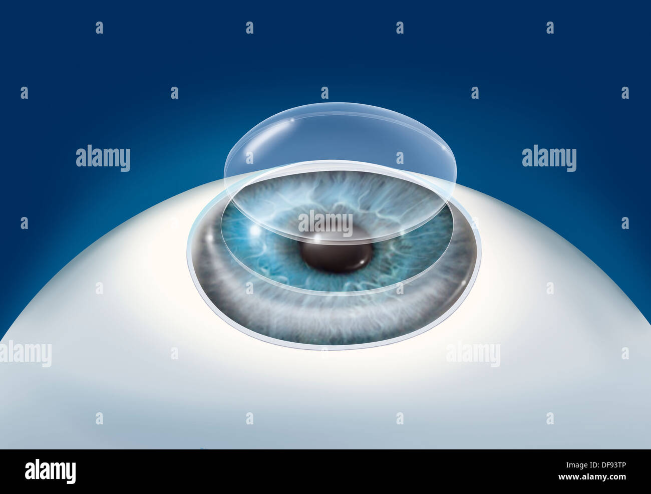 CORNEAL TRANSPLANT, DRAWING Stock Photohttps://www.alamy.com/image-license-details/?v=1https://www.alamy.com/corneal-transplant-drawing-image61051558.html
RMDF93TP–CORNEAL TRANSPLANT, DRAWING
 The principles and practice of obstetrics . broad ligament. The inner extremity of eachovary is attached to the side of the uterus by a solidfibrous cord, termed the ligament of the ovary. Under the peritoneal coat is the tunica albuginea, afibrous envelope so dense as to give form to the ovary.From the inner surface of this coat there are numerousprolongations of fibrous bands, in the interstices ofwhich will be found a large number of small bodies,which give a soft spongy character to the whole tissueduring childhood. These are the rudiments of theGraafian vesicles or ovisacs. Toward the age Stock Photohttps://www.alamy.com/image-license-details/?v=1https://www.alamy.com/the-principles-and-practice-of-obstetrics-broad-ligament-the-inner-extremity-of-eachovary-is-attached-to-the-side-of-the-uterus-by-a-solidfibrous-cord-termed-the-ligament-of-the-ovary-under-the-peritoneal-coat-is-the-tunica-albuginea-afibrous-envelope-so-dense-as-to-give-form-to-the-ovaryfrom-the-inner-surface-of-this-coat-there-are-numerousprolongations-of-fibrous-bands-in-the-interstices-ofwhich-will-be-found-a-large-number-of-small-bodieswhich-give-a-soft-spongy-character-to-the-whole-tissueduring-childhood-these-are-the-rudiments-of-thegraafian-vesicles-or-ovisacs-toward-the-age-image338443443.html
RM2AJHC1R–The principles and practice of obstetrics . broad ligament. The inner extremity of eachovary is attached to the side of the uterus by a solidfibrous cord, termed the ligament of the ovary. Under the peritoneal coat is the tunica albuginea, afibrous envelope so dense as to give form to the ovary.From the inner surface of this coat there are numerousprolongations of fibrous bands, in the interstices ofwhich will be found a large number of small bodies,which give a soft spongy character to the whole tissueduring childhood. These are the rudiments of theGraafian vesicles or ovisacs. Toward the age
 Human testicle computer artwork. Stock Photohttps://www.alamy.com/image-license-details/?v=1https://www.alamy.com/human-testicle-computer-artwork-image69884887.html
RFE1KETR–Human testicle computer artwork.
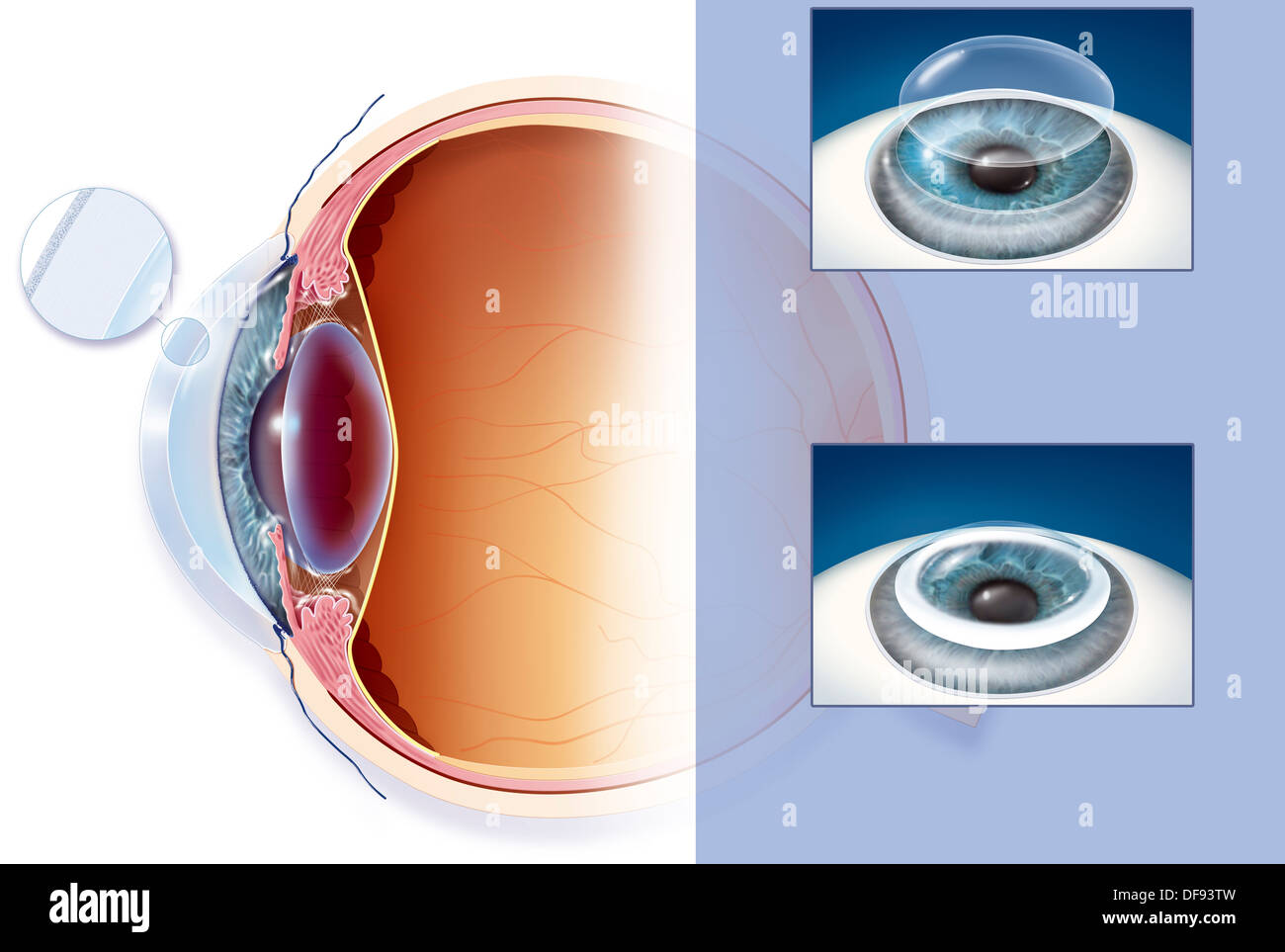 CORNEAL TRANSPLANT, DRAWING Stock Photohttps://www.alamy.com/image-license-details/?v=1https://www.alamy.com/corneal-transplant-drawing-image61051561.html
RMDF93TW–CORNEAL TRANSPLANT, DRAWING
 Diseases of the ovaries : their diagnosis and treatment . me of them,however, were three-eighths of an inch broad, and one inchor more long. The walls of these were considerably (perhapsfour times) thicker than the others ; they could be dissectedfree, and were found to be continuous with and to branch fromthe tunica albuginea. One thing is worthy of notice—thelarger cysts were not spherical, but elliptical. The tumour in Case No. 113 weighed from fifteen to twentypounds ; its texture was soft and friable, so that in handling,it tore by its own weight. On what had originally been itsinferior a Stock Photohttps://www.alamy.com/image-license-details/?v=1https://www.alamy.com/diseases-of-the-ovaries-their-diagnosis-and-treatment-me-of-themhowever-were-three-eighths-of-an-inch-broad-and-one-inchor-more-long-the-walls-of-these-were-considerably-perhapsfour-times-thicker-than-the-others-they-could-be-dissectedfree-and-were-found-to-be-continuous-with-and-to-branch-fromthe-tunica-albuginea-one-thing-is-worthy-of-noticethelarger-cysts-were-not-spherical-but-elliptical-the-tumour-in-case-no-113-weighed-from-fifteen-to-twentypounds-its-texture-was-soft-and-friable-so-that-in-handlingit-tore-by-its-own-weight-on-what-had-originally-been-itsinferior-a-image339451721.html
RM2AM7A3N–Diseases of the ovaries : their diagnosis and treatment . me of them,however, were three-eighths of an inch broad, and one inchor more long. The walls of these were considerably (perhapsfour times) thicker than the others ; they could be dissectedfree, and were found to be continuous with and to branch fromthe tunica albuginea. One thing is worthy of notice—thelarger cysts were not spherical, but elliptical. The tumour in Case No. 113 weighed from fifteen to twentypounds ; its texture was soft and friable, so that in handling,it tore by its own weight. On what had originally been itsinferior a
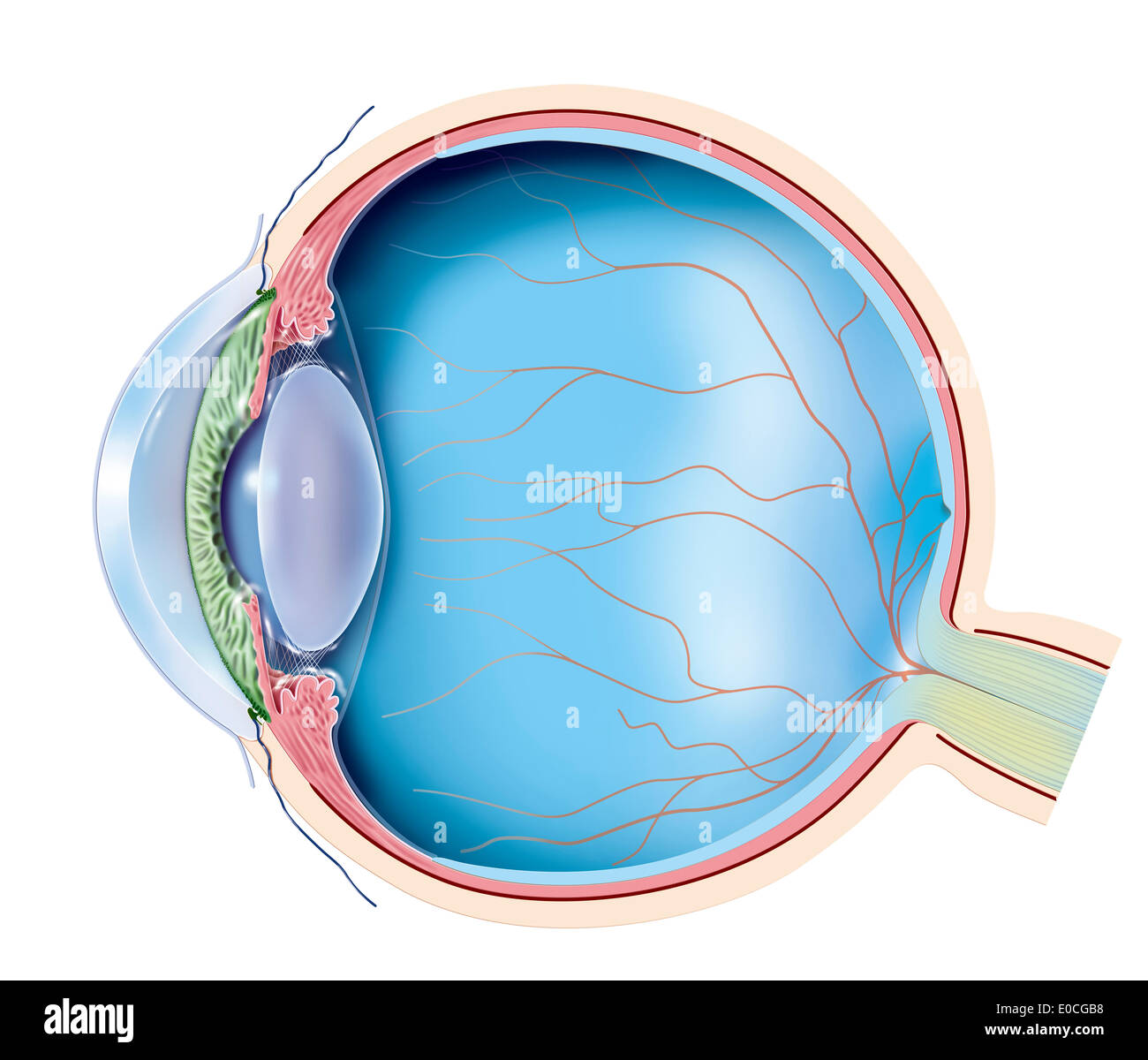 Anatomy, eye Stock Photohttps://www.alamy.com/image-license-details/?v=1https://www.alamy.com/anatomy-eye-image69117756.html
RME0CGB8–Anatomy, eye
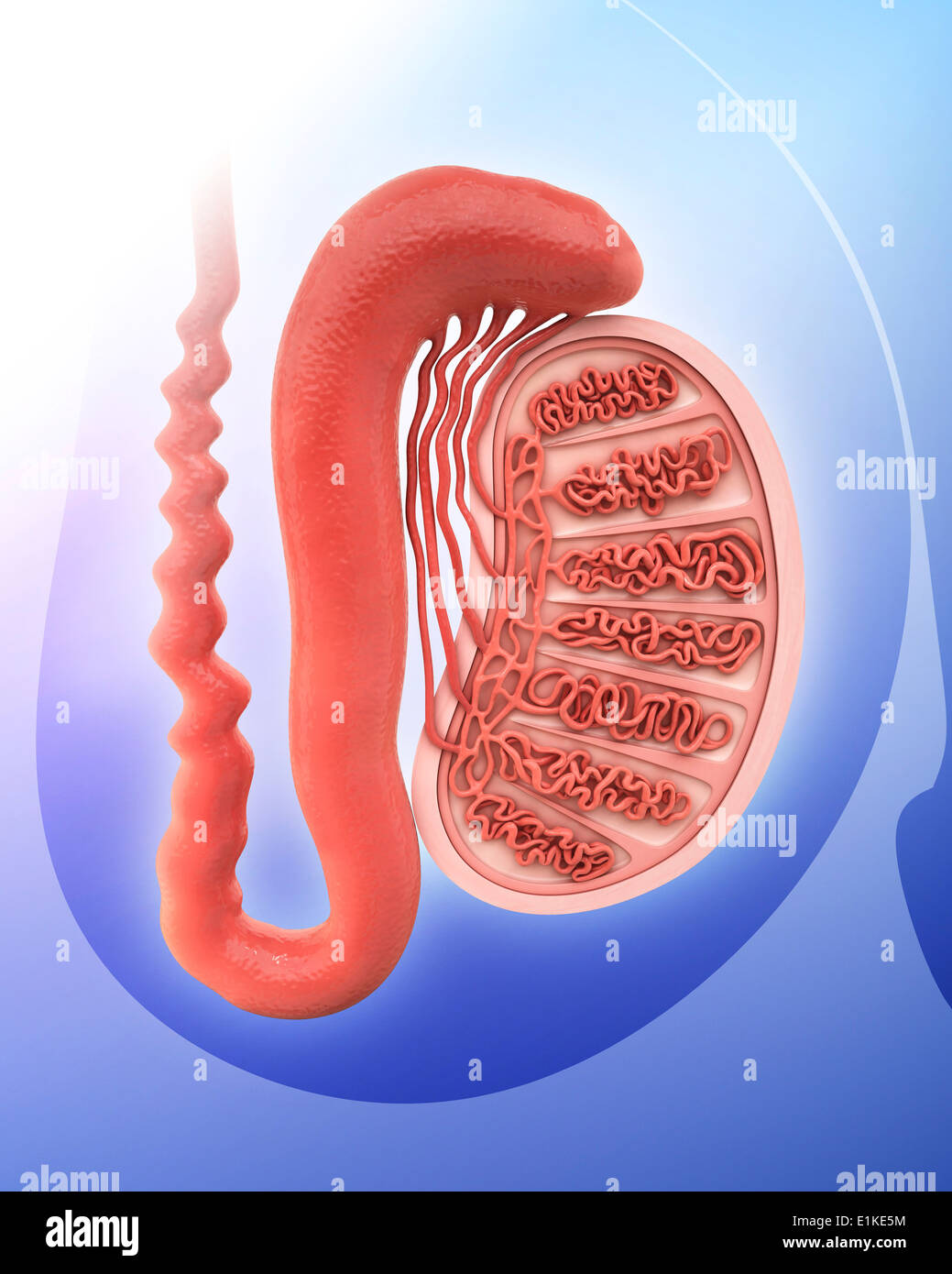 Human testicle computer artwork. Stock Photohttps://www.alamy.com/image-license-details/?v=1https://www.alamy.com/human-testicle-computer-artwork-image69884352.html
RFE1KE5M–Human testicle computer artwork.
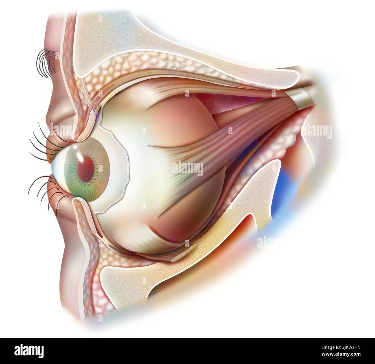 Anatomy of the eye and eyelid (viewed from 3/4) with iris, pupil. Stock Photohttps://www.alamy.com/image-license-details/?v=1https://www.alamy.com/anatomy-of-the-eye-and-eyelid-viewed-from-34-with-iris-pupil-image476926513.html
RF2JKWTHN–Anatomy of the eye and eyelid (viewed from 3/4) with iris, pupil.
 Manual of human histology . isdivisible into a smaller, anterior, transparent portion—thecornea; and a larger, opaque, posterior part—the sclerotic;but, as shown by its development and more intimate structure,is to be regarded as a membrane continuous throughout. The sclerotica, also termed the tunica albuginea, is a white,very tough and strong, fibrous membrane, which graduallydiminishes in thickness as it advances forwards from the pos-terior part of the eye, where it is directly connected with thesheath of the optic nerve, although it is again strengthened,in front, by the expanded tendons Stock Photohttps://www.alamy.com/image-license-details/?v=1https://www.alamy.com/manual-of-human-histology-isdivisible-into-a-smaller-anterior-transparent-portionthecornea-and-a-larger-opaque-posterior-partthe-scleroticbut-as-shown-by-its-development-and-more-intimate-structureis-to-be-regarded-as-a-membrane-continuous-throughout-the-sclerotica-also-termed-the-tunica-albuginea-is-a-whitevery-tough-and-strong-fibrous-membrane-which-graduallydiminishes-in-thickness-as-it-advances-forwards-from-the-pos-terior-part-of-the-eye-where-it-is-directly-connected-with-thesheath-of-the-optic-nerve-although-it-is-again-strengthenedin-front-by-the-expanded-tendons-image338220019.html
RM2AJ772B–Manual of human histology . isdivisible into a smaller, anterior, transparent portion—thecornea; and a larger, opaque, posterior part—the sclerotic;but, as shown by its development and more intimate structure,is to be regarded as a membrane continuous throughout. The sclerotica, also termed the tunica albuginea, is a white,very tough and strong, fibrous membrane, which graduallydiminishes in thickness as it advances forwards from the pos-terior part of the eye, where it is directly connected with thesheath of the optic nerve, although it is again strengthened,in front, by the expanded tendons
 Human testicle computer artwork. Stock Photohttps://www.alamy.com/image-license-details/?v=1https://www.alamy.com/human-testicle-computer-artwork-image69885050.html
RFE1KF2J–Human testicle computer artwork.
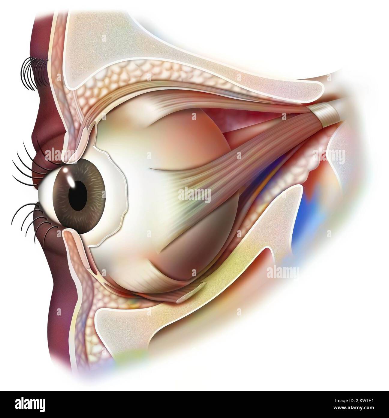 Anatomy of the eye and eyelid (viewed from 3/4) with iris, pupil. Stock Photohttps://www.alamy.com/image-license-details/?v=1https://www.alamy.com/anatomy-of-the-eye-and-eyelid-viewed-from-34-with-iris-pupil-image476926493.html
RF2JKWTH1–Anatomy of the eye and eyelid (viewed from 3/4) with iris, pupil.
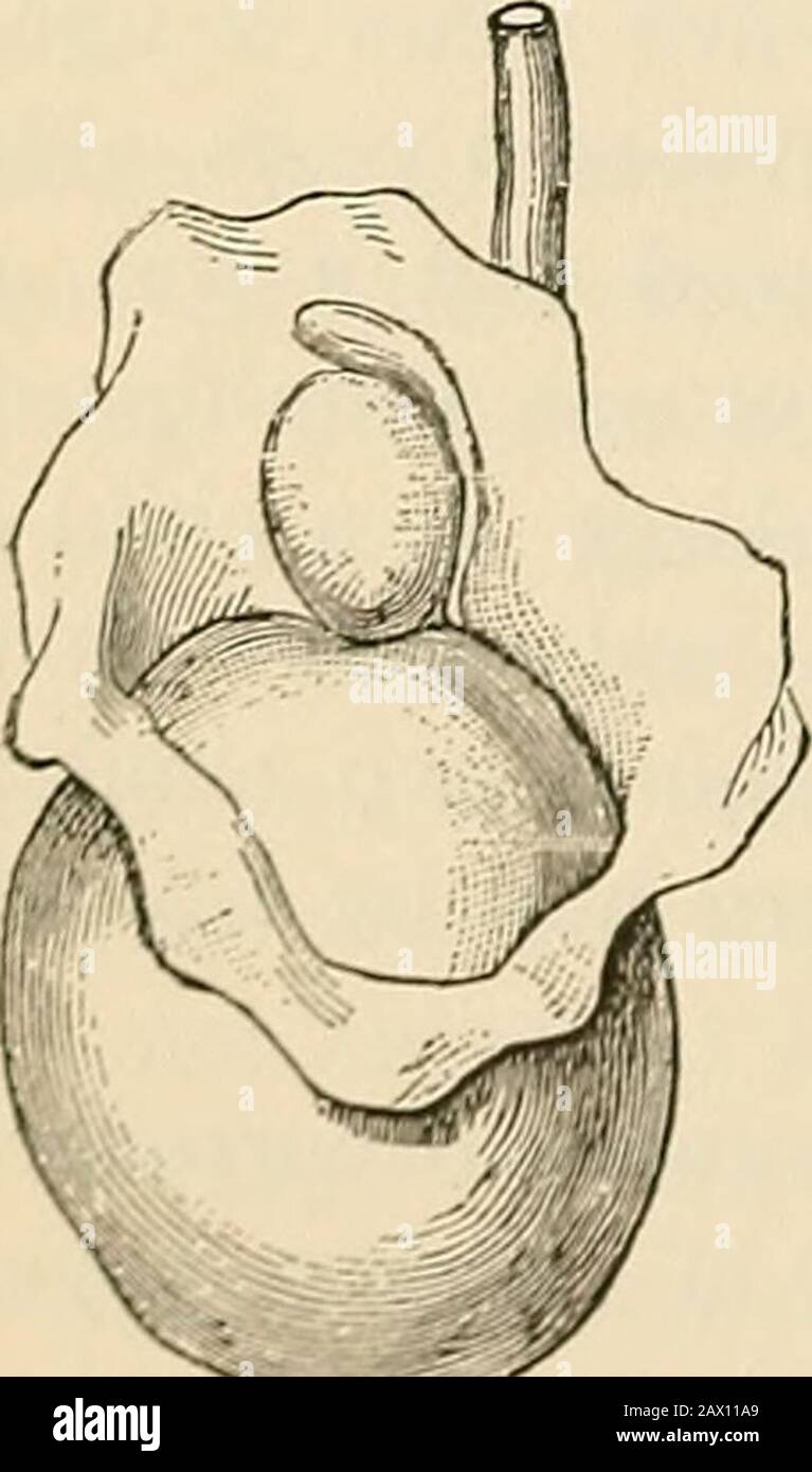 The pathology and surgical treatment of tumors . ground-substance. (Magnification same as that of Figure 434.) tion of the tunica albuginea has taken place, the tumor grows veryrapidly. Extension along the spermatic cord results in speedy andextensive regional infection. Metastasis fre-quently precedes the fatal termination. Veryoften the same affection appears in the oppositetesticle. In the differential diagnosis of sarcoma of thetesticle it is important to exclude carcinoma, tuber-culosis, gumma, and hematocele. Figure 436represents a sarcoma of the testicle that occurredin a child three an Stock Photohttps://www.alamy.com/image-license-details/?v=1https://www.alamy.com/the-pathology-and-surgical-treatment-of-tumors-ground-substance-magnification-same-as-that-of-figure-434-tion-of-the-tunica-albuginea-has-taken-place-the-tumor-grows-veryrapidly-extension-along-the-spermatic-cord-results-in-speedy-andextensive-regional-infection-metastasis-fre-quently-precedes-the-fatal-termination-veryoften-the-same-affection-appears-in-the-oppositetesticle-in-the-differential-diagnosis-of-sarcoma-of-thetesticle-it-is-important-to-exclude-carcinoma-tuber-culosis-gumma-and-hematocele-figure-436represents-a-sarcoma-of-the-testicle-that-occurredin-a-child-three-an-image343001073.html
RM2AX11A9–The pathology and surgical treatment of tumors . ground-substance. (Magnification same as that of Figure 434.) tion of the tunica albuginea has taken place, the tumor grows veryrapidly. Extension along the spermatic cord results in speedy andextensive regional infection. Metastasis fre-quently precedes the fatal termination. Veryoften the same affection appears in the oppositetesticle. In the differential diagnosis of sarcoma of thetesticle it is important to exclude carcinoma, tuber-culosis, gumma, and hematocele. Figure 436represents a sarcoma of the testicle that occurredin a child three an
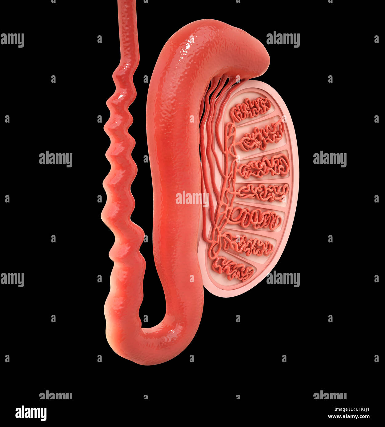 Human testicle computer artwork. Stock Photohttps://www.alamy.com/image-license-details/?v=1https://www.alamy.com/human-testicle-computer-artwork-image69885481.html
RFE1KFJ1–Human testicle computer artwork.
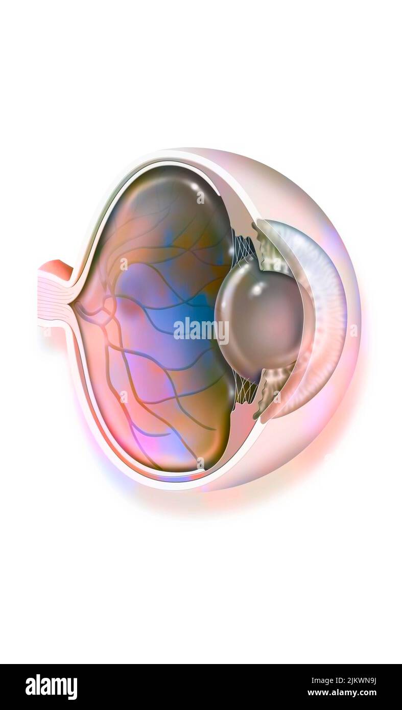 Anatomy of the eye with lens, retinal veins and arteries. Stock Photohttps://www.alamy.com/image-license-details/?v=1https://www.alamy.com/anatomy-of-the-eye-with-lens-retinal-veins-and-arteries-image476923934.html
RF2JKWN9J–Anatomy of the eye with lens, retinal veins and arteries.
 The anatomist's vade mecum : a system of human anatomy . The globusmajor is situated against the upperend of the testicle, to which it isclosely adherent; the globus minoris placed at its lower end, is attachedto the testis by cellular tissue, andcurves upwards, to become conti- * A transverse section of the testicle. 1. The cavity of the tunica vaginalis;the most external layer is the tunica vaginalis reflexa; and that in contactwith the organ, the tunica vaginalis propria. 2. The tunica albuginea. 3. Themediastinum testis giving off numerous fibrous cords in a radiated directionto the intern Stock Photohttps://www.alamy.com/image-license-details/?v=1https://www.alamy.com/the-anatomists-vade-mecum-a-system-of-human-anatomy-the-globusmajor-is-situated-against-the-upperend-of-the-testicle-to-which-it-isclosely-adherent-the-globus-minoris-placed-at-its-lower-end-is-attachedto-the-testis-by-cellular-tissue-andcurves-upwards-to-become-conti-a-transverse-section-of-the-testicle-1-the-cavity-of-the-tunica-vaginalisthe-most-external-layer-is-the-tunica-vaginalis-reflexa-and-that-in-contactwith-the-organ-the-tunica-vaginalis-propria-2-the-tunica-albuginea-3-themediastinum-testis-giving-off-numerous-fibrous-cords-in-a-radiated-directionto-the-intern-image342722727.html
RM2AWGA9B–The anatomist's vade mecum : a system of human anatomy . The globusmajor is situated against the upperend of the testicle, to which it isclosely adherent; the globus minoris placed at its lower end, is attachedto the testis by cellular tissue, andcurves upwards, to become conti- * A transverse section of the testicle. 1. The cavity of the tunica vaginalis;the most external layer is the tunica vaginalis reflexa; and that in contactwith the organ, the tunica vaginalis propria. 2. The tunica albuginea. 3. Themediastinum testis giving off numerous fibrous cords in a radiated directionto the intern
 Human testicle computer artwork. Stock Photohttps://www.alamy.com/image-license-details/?v=1https://www.alamy.com/human-testicle-computer-artwork-image69885368.html
RFE1KFE0–Human testicle computer artwork.
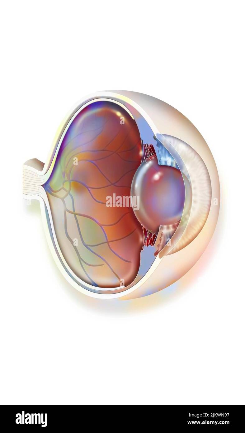 Anatomy of the eye with lens, retinal veins and arteries. Stock Photohttps://www.alamy.com/image-license-details/?v=1https://www.alamy.com/anatomy-of-the-eye-with-lens-retinal-veins-and-arteries-image476923923.html
RF2JKWN97–Anatomy of the eye with lens, retinal veins and arteries.
 Gynaecology for students and practitioners . s not yet differentiated as ovary or testis ; thisdifferentiation is first seen in embryos 14 mm. long (fifth to sixth week),when a testis begins to show a tunica albuginea and a marked de-velopment of genital cords. Absence of these structures at thisstage marks the gland as an ovary. At this stage the ovary showsa development of connective tissue which grows from the depths intothe germinal layer and divides the germ-epithelial cells and genitalcells into groups {egg-follicles). In each such follicle we have a genital DEVELOPMENT OF GENITO-URINARY Stock Photohttps://www.alamy.com/image-license-details/?v=1https://www.alamy.com/gynaecology-for-students-and-practitioners-s-not-yet-differentiated-as-ovary-or-testis-thisdifferentiation-is-first-seen-in-embryos-14-mm-long-fifth-to-sixth-weekwhen-a-testis-begins-to-show-a-tunica-albuginea-and-a-marked-de-velopment-of-genital-cords-absence-of-these-structures-at-thisstage-marks-the-gland-as-an-ovary-at-this-stage-the-ovary-showsa-development-of-connective-tissue-which-grows-from-the-depths-intothe-germinal-layer-and-divides-the-germ-epithelial-cells-and-genitalcells-into-groups-egg-follicles-in-each-such-follicle-we-have-a-genital-development-of-genito-urinary-image340084015.html
RM2AN84HK–Gynaecology for students and practitioners . s not yet differentiated as ovary or testis ; thisdifferentiation is first seen in embryos 14 mm. long (fifth to sixth week),when a testis begins to show a tunica albuginea and a marked de-velopment of genital cords. Absence of these structures at thisstage marks the gland as an ovary. At this stage the ovary showsa development of connective tissue which grows from the depths intothe germinal layer and divides the germ-epithelial cells and genitalcells into groups {egg-follicles). In each such follicle we have a genital DEVELOPMENT OF GENITO-URINARY
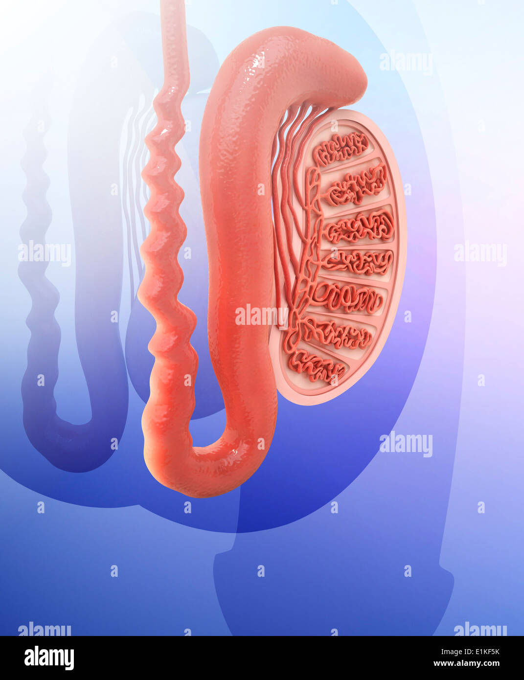 Human testicle computer artwork. Stock Photohttps://www.alamy.com/image-license-details/?v=1https://www.alamy.com/human-testicle-computer-artwork-image69885135.html
RFE1KF5K–Human testicle computer artwork.
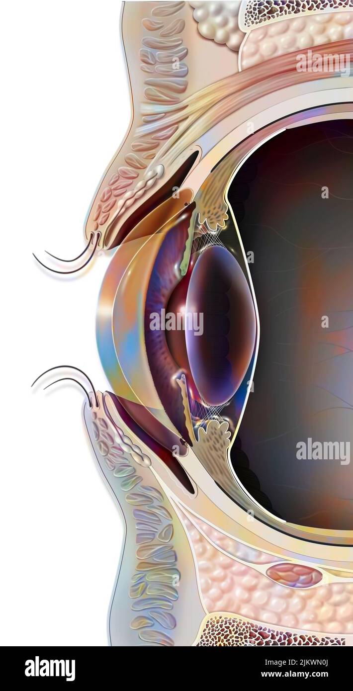 Median sagittal section of the eye and eyelid. Stock Photohttps://www.alamy.com/image-license-details/?v=1https://www.alamy.com/median-sagittal-section-of-the-eye-and-eyelid-image476923682.html
RF2JKWN0J–Median sagittal section of the eye and eyelid.
 A system of obstetrics . , and does not form a distinct sep-arable coat comparable to the tunica albuginea of the testis. Along the hilus, blood-vessels, lymphatics, and nerves enter and leaveih ovary: they branch through the stroma, forming part of it. Thearteries and veins are comparatively large at the hilus and in the centreof the ovary, but divide into minute twigs near the surface. A certainamount of plain muscular tissue lies near the arteries. The Graafian FoUides9OT egg-chambers, are numerous. In the ova-ries of a newborn female infant there are about seventy thousand ofthem, and prob Stock Photohttps://www.alamy.com/image-license-details/?v=1https://www.alamy.com/a-system-of-obstetrics-and-does-not-form-a-distinct-sep-arable-coat-comparable-to-the-tunica-albuginea-of-the-testis-along-the-hilus-blood-vessels-lymphatics-and-nerves-enter-and-leaveih-ovary-they-branch-through-the-stroma-forming-part-of-it-thearteries-and-veins-are-comparatively-large-at-the-hilus-and-in-the-centreof-the-ovary-but-divide-into-minute-twigs-near-the-surface-a-certainamount-of-plain-muscular-tissue-lies-near-the-arteries-the-graafian-fouides9ot-egg-chambers-are-numerous-in-the-ova-ries-of-a-newborn-female-infant-there-are-about-seventy-thousand-ofthem-and-prob-image342828607.html
RM2AWN5AR–A system of obstetrics . , and does not form a distinct sep-arable coat comparable to the tunica albuginea of the testis. Along the hilus, blood-vessels, lymphatics, and nerves enter and leaveih ovary: they branch through the stroma, forming part of it. Thearteries and veins are comparatively large at the hilus and in the centreof the ovary, but divide into minute twigs near the surface. A certainamount of plain muscular tissue lies near the arteries. The Graafian FoUides9OT egg-chambers, are numerous. In the ova-ries of a newborn female infant there are about seventy thousand ofthem, and prob
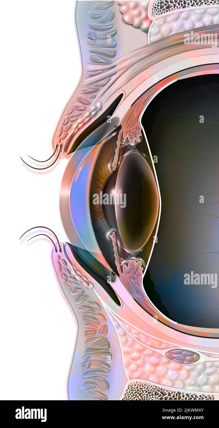 Median sagittal section of the eye and eyelid. Stock Photohttps://www.alamy.com/image-license-details/?v=1https://www.alamy.com/median-sagittal-section-of-the-eye-and-eyelid-image476923635.html
RF2JKWMXY–Median sagittal section of the eye and eyelid.
 Veterinary obstetrics, including the diseases of breeding animals and of the new-born . become materially changed, to cow?Xw.^ permanentova. In the process of development the connective tissue stromathrows out a thin layer, the tunica albuginea, parallel to the sur- Maturation of the Graafian Follicle 45 face of the ovary and serving to divide the germinal epitheliuminto a superficial, columnar lajer and a deeper one broken upinto irregular columns or clumps of spherical or polygonal cells.lu these cell masses .q. perniaiient ova, developed from the prim-itive ova, become much larger, wh Stock Photohttps://www.alamy.com/image-license-details/?v=1https://www.alamy.com/veterinary-obstetrics-including-the-diseases-of-breeding-animals-and-of-the-new-born-become-materially-changed-to-cowxw-permanentova-in-the-process-of-development-the-connective-tissue-stromathrows-out-a-thin-layer-the-tunica-albuginea-parallel-to-the-sur-maturation-of-the-graafian-follicle-45-face-of-the-ovary-and-serving-to-divide-the-germinal-epitheliuminto-a-superficial-columnar-lajer-and-a-deeper-one-broken-upinto-irregular-columns-or-clumps-of-spherical-or-polygonal-cellslu-these-cell-masses-q-perniaiient-ova-developed-from-the-prim-itive-ova-become-much-larger-wh-image342673431.html
RM2AWE3CR–Veterinary obstetrics, including the diseases of breeding animals and of the new-born . become materially changed, to cow?Xw.^ permanentova. In the process of development the connective tissue stromathrows out a thin layer, the tunica albuginea, parallel to the sur- Maturation of the Graafian Follicle 45 face of the ovary and serving to divide the germinal epitheliuminto a superficial, columnar lajer and a deeper one broken upinto irregular columns or clumps of spherical or polygonal cells.lu these cell masses .q. perniaiient ova, developed from the prim-itive ova, become much larger, wh
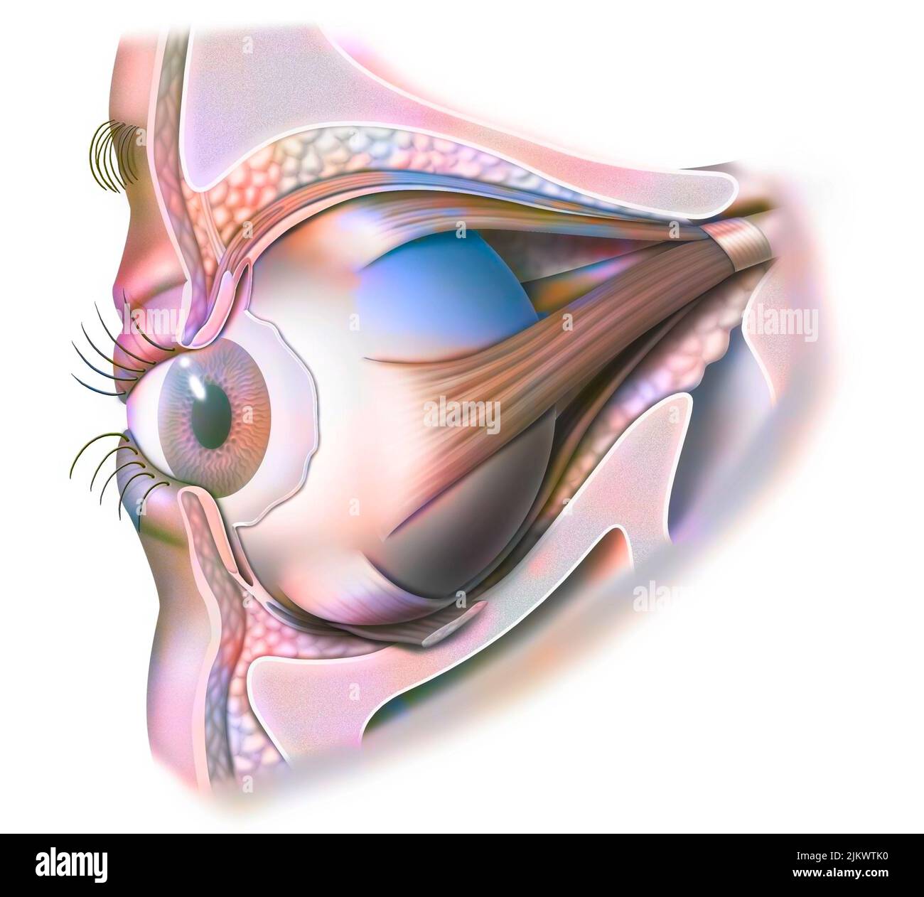 Anatomy of the eye and eyelid (viewed from 3/4) with iris, pupil. Stock Photohttps://www.alamy.com/image-license-details/?v=1https://www.alamy.com/anatomy-of-the-eye-and-eyelid-viewed-from-34-with-iris-pupil-image476926548.html
RF2JKWTK0–Anatomy of the eye and eyelid (viewed from 3/4) with iris, pupil.
 Gynecological diagnosis and pathology . of the tunica albuginea is met with in cases of chronic ovaritis.The previous acute inflammation on the surface of the ovary results in acicatricial thickening sufficient to prevent rupture. In other cases thetunica albuginea may be denser than normal, without any sign ofprevious inflammation being present. (2) In some cases it would appearthat the follicle itself is at fault. The ripening process goes on apparentlynormally up to a certain stage and then ceases, and the ovum dies anddisappears. THE CYSTIC OVARY 57 In some eases the formation of lutein al Stock Photohttps://www.alamy.com/image-license-details/?v=1https://www.alamy.com/gynecological-diagnosis-and-pathology-of-the-tunica-albuginea-is-met-with-in-cases-of-chronic-ovaritisthe-previous-acute-inflammation-on-the-surface-of-the-ovary-results-in-acicatricial-thickening-sufficient-to-prevent-rupture-in-other-cases-thetunica-albuginea-may-be-denser-than-normal-without-any-sign-ofprevious-inflammation-being-present-2-in-some-cases-it-would-appearthat-the-follicle-itself-is-at-fault-the-ripening-process-goes-on-apparentlynormally-up-to-a-certain-stage-and-then-ceases-and-the-ovum-dies-anddisappears-the-cystic-ovary-57-in-some-eases-the-formation-of-lutein-al-image338507477.html
RM2AJM9MN–Gynecological diagnosis and pathology . of the tunica albuginea is met with in cases of chronic ovaritis.The previous acute inflammation on the surface of the ovary results in acicatricial thickening sufficient to prevent rupture. In other cases thetunica albuginea may be denser than normal, without any sign ofprevious inflammation being present. (2) In some cases it would appearthat the follicle itself is at fault. The ripening process goes on apparentlynormally up to a certain stage and then ceases, and the ovum dies anddisappears. THE CYSTIC OVARY 57 In some eases the formation of lutein al
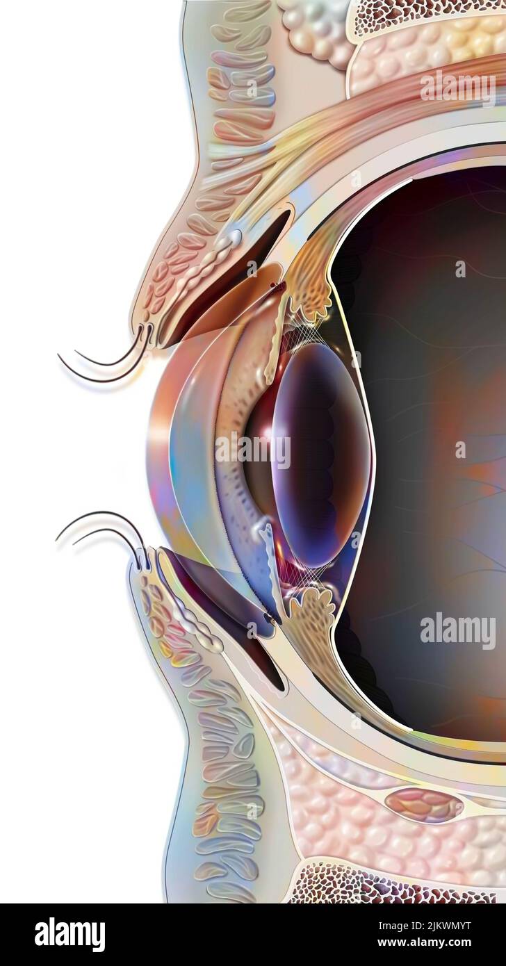 Median sagittal section of the eye and eyelid. Stock Photohttps://www.alamy.com/image-license-details/?v=1https://www.alamy.com/median-sagittal-section-of-the-eye-and-eyelid-image476923660.html
RF2JKWMYT–Median sagittal section of the eye and eyelid.
 An atlas of human anatomy for students and physicians . Gastrosplenic omentumor ligament Ligamentum gastrolienale InferiorextremityExtremitas inferior Hilum of the spleen Accessory spleen (var.)Lien accestorius (var.) Fig. 747.—The Spleen, with the Gastro-splenic Omentum left attached, seenFROM Before. Lien Accessorius, anAccessory Spleen. Pulp of the spleenPulpa lienis , Malpighian corpuscles, or lym-)ihoid follicles of the spleen N(«lii!i IvmphaticiliLiiales (Malpighii) I. Fibrous coat or capsule (tunica propria) Tunica albuginea Briiiich ot tue splenic arteryRamus arteria; lienalis Fig. 748 Stock Photohttps://www.alamy.com/image-license-details/?v=1https://www.alamy.com/an-atlas-of-human-anatomy-for-students-and-physicians-gastrosplenic-omentumor-ligament-ligamentum-gastrolienale-inferiorextremityextremitas-inferior-hilum-of-the-spleen-accessory-spleen-varlien-accestorius-var-fig-747the-spleen-with-the-gastro-splenic-omentum-left-attached-seenfrom-before-lien-accessorius-anaccessory-spleen-pulp-of-the-spleenpulpa-lienis-malpighian-corpuscles-or-lym-ihoid-follicles-of-the-spleen-nlii!i-ivmphaticililiiales-malpighii-i-fibrous-coat-or-capsule-tunica-propria-tunica-albuginea-briiiich-ot-tue-splenic-arteryramus-arteria-lienalis-fig-748-image338332261.html
RM2AJCA71–An atlas of human anatomy for students and physicians . Gastrosplenic omentumor ligament Ligamentum gastrolienale InferiorextremityExtremitas inferior Hilum of the spleen Accessory spleen (var.)Lien accestorius (var.) Fig. 747.—The Spleen, with the Gastro-splenic Omentum left attached, seenFROM Before. Lien Accessorius, anAccessory Spleen. Pulp of the spleenPulpa lienis , Malpighian corpuscles, or lym-)ihoid follicles of the spleen N(«lii!i IvmphaticiliLiiales (Malpighii) I. Fibrous coat or capsule (tunica propria) Tunica albuginea Briiiich ot tue splenic arteryRamus arteria; lienalis Fig. 748
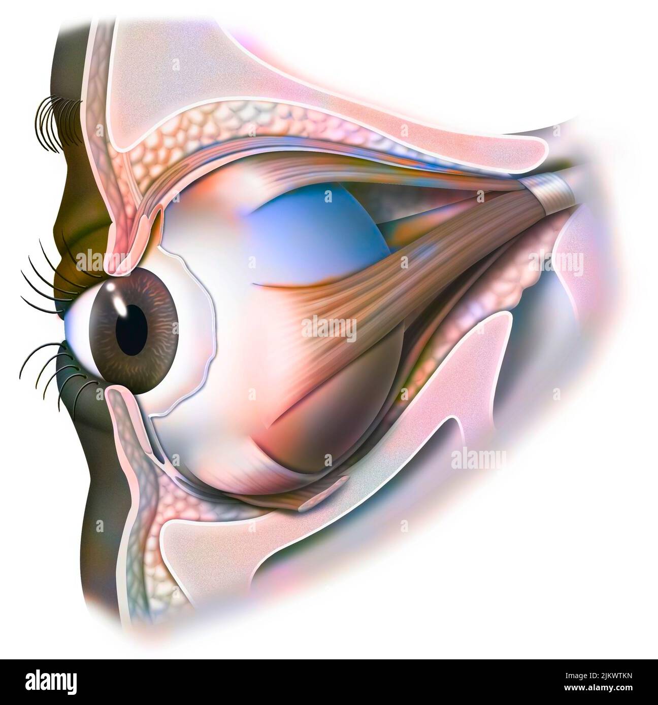 Anatomy of the eye and eyelid (viewed from 3/4) with iris, pupil. Stock Photohttps://www.alamy.com/image-license-details/?v=1https://www.alamy.com/anatomy-of-the-eye-and-eyelid-viewed-from-34-with-iris-pupil-image476926569.html
RF2JKWTKN–Anatomy of the eye and eyelid (viewed from 3/4) with iris, pupil.
 An atlas of human anatomy for students and physicians . Base of the pyramidBasis pyiamidis Pyramid of Malpighi Py.inlir ml CM.ilpighii Renal papilla Papilla renahs Arterial archA. arciformis Renal papilla , Papilla renalis! Medulla /ISubstantia meduUarisCortexSubstantia corticalis Infundibulum Cal)x major Capsule, fibrous coat, or tunica/ albuginea of the kidney Tunica fibrosa Calices Calyces minores. Fig. 825.—Coronal Section through the RightKidney and the Renal Pelvis. Substantia Cor-ticalis, the Cortex ; Substantia Medullaris,the Medulla. ^ See Appendb I Base of the pyramidBasis pyramidis Stock Photohttps://www.alamy.com/image-license-details/?v=1https://www.alamy.com/an-atlas-of-human-anatomy-for-students-and-physicians-base-of-the-pyramidbasis-pyiamidis-pyramid-of-malpighi-pyinlir-ml-cmilpighii-renal-papilla-papilla-renahs-arterial-archa-arciformis-renal-papilla-papilla-renalis!-medulla-isubstantia-meduuariscortexsubstantia-corticalis-infundibulum-calx-major-capsule-fibrous-coat-or-tunica-albuginea-of-the-kidney-tunica-fibrosa-calices-calyces-minores-fig-825coronal-section-through-the-rightkidney-and-the-renal-pelvis-substantia-cor-ticalis-the-cortex-substantia-medullaristhe-medulla-see-appendb-i-base-of-the-pyramidbasis-pyramidis-image338304745.html
RM2AJB349–An atlas of human anatomy for students and physicians . Base of the pyramidBasis pyiamidis Pyramid of Malpighi Py.inlir ml CM.ilpighii Renal papilla Papilla renahs Arterial archA. arciformis Renal papilla , Papilla renalis! Medulla /ISubstantia meduUarisCortexSubstantia corticalis Infundibulum Cal)x major Capsule, fibrous coat, or tunica/ albuginea of the kidney Tunica fibrosa Calices Calyces minores. Fig. 825.—Coronal Section through the RightKidney and the Renal Pelvis. Substantia Cor-ticalis, the Cortex ; Substantia Medullaris,the Medulla. ^ See Appendb I Base of the pyramidBasis pyramidis
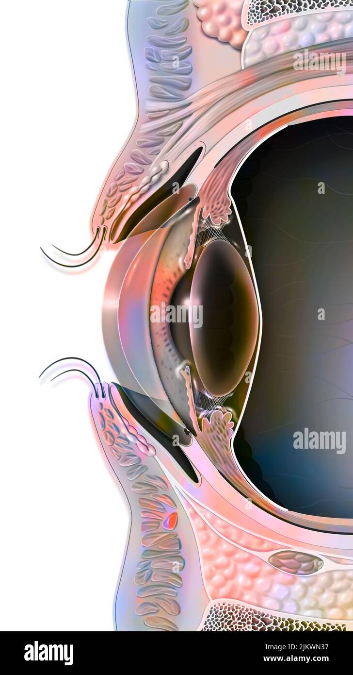 Median sagittal section of the eye and eyelid. Stock Photohttps://www.alamy.com/image-license-details/?v=1https://www.alamy.com/median-sagittal-section-of-the-eye-and-eyelid-image476923755.html
RF2JKWN37–Median sagittal section of the eye and eyelid.
 The pathology and treatment of diseases of the ovaries : (being the Hastings essay for 1873).. . Fig. 7 (after Balfour).—o, e, frerminal epithelium; t,trabecwloB ; A, hilum, with canal. ANATOMY AND PHYSIOLOGY OP THE OVAKY. 17. Pig. 8 (after Balfour).—p, o, primi-tive ova ; t a, tunica albuginea ; c, e,central epithelium. the Malpighian bodies of the Wolffian structures, and are meresurvivals. The germinal epithelium grows rapidly in thickness by thedivision of its cells, and the vascularstroma greatly increases in quantity, sothat the epithelial tissue is honeycombedby the vascular trabeculse, Stock Photohttps://www.alamy.com/image-license-details/?v=1https://www.alamy.com/the-pathology-and-treatment-of-diseases-of-the-ovaries-being-the-hastings-essay-for-1873-fig-7-after-balfouro-e-frerminal-epithelium-ttrabecwlob-a-hilum-with-canal-anatomy-and-physiology-op-the-ovaky-17-pig-8-after-balfourp-o-primi-tive-ova-t-a-tunica-albuginea-c-ecentral-epithelium-the-malpighian-bodies-of-the-wolffian-structures-and-are-meresurvivals-the-germinal-epithelium-grows-rapidly-in-thickness-by-thedivision-of-its-cells-and-the-vascularstroma-greatly-increases-in-quantity-sothat-the-epithelial-tissue-is-honeycombedby-the-vascular-trabeculse-image338085833.html
RM2AJ13X1–The pathology and treatment of diseases of the ovaries : (being the Hastings essay for 1873).. . Fig. 7 (after Balfour).—o, e, frerminal epithelium; t,trabecwloB ; A, hilum, with canal. ANATOMY AND PHYSIOLOGY OP THE OVAKY. 17. Pig. 8 (after Balfour).—p, o, primi-tive ova ; t a, tunica albuginea ; c, e,central epithelium. the Malpighian bodies of the Wolffian structures, and are meresurvivals. The germinal epithelium grows rapidly in thickness by thedivision of its cells, and the vascularstroma greatly increases in quantity, sothat the epithelial tissue is honeycombedby the vascular trabeculse,
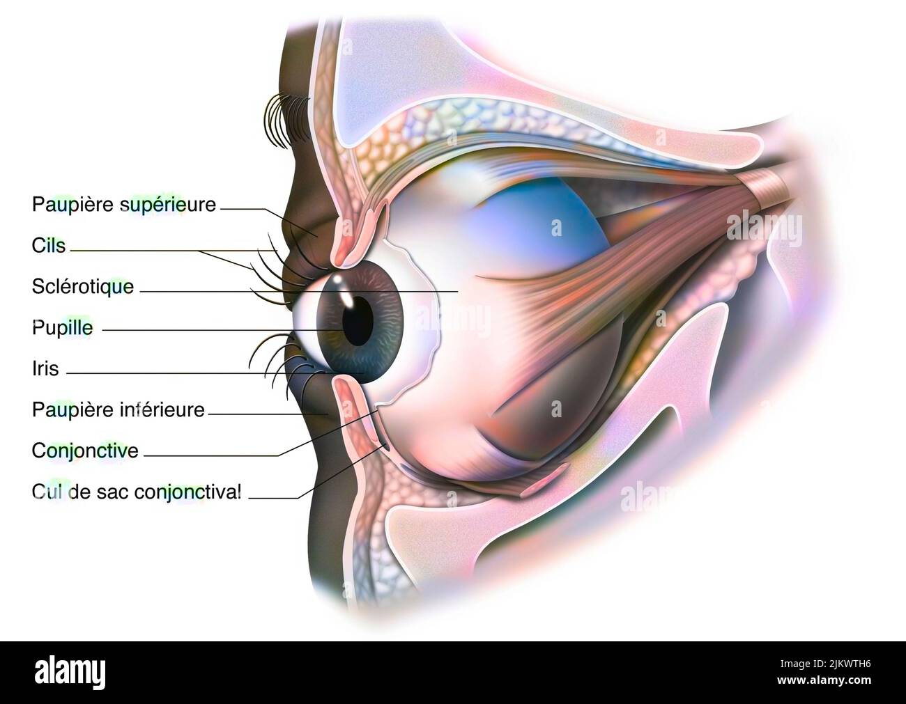 Anatomy of the eye and eyelid (viewed from 3/4) with iris, pupil. Stock Photohttps://www.alamy.com/image-license-details/?v=1https://www.alamy.com/anatomy-of-the-eye-and-eyelid-viewed-from-34-with-iris-pupil-image476926498.html
RF2JKWTH6–Anatomy of the eye and eyelid (viewed from 3/4) with iris, pupil.
 EYE, DRAWING Stock Photohttps://www.alamy.com/image-license-details/?v=1https://www.alamy.com/stock-photo-eye-drawing-75741306.html
RMEB68PJ–EYE, DRAWING
 Textbook of normal histology: including an account of the development of the tissues and of the organs . progressively tortu-ous continuations of the vasa efferentia terminating in a mass, the Fig. 250. p; Section of human testicle, including portion of tunica albuginea, ex-hibiting general arrangement and structure of tubules : a, tunica albu-ginea ; b, seminiferous tubules cut in various directions ; c, basement-membrane; d, secreting cells ; e, groups of interstitial cells ;/, intertubularconnective tissue. globus major, which represents the sum of the tortuous coni vas-culosi. These last-n Stock Photohttps://www.alamy.com/image-license-details/?v=1https://www.alamy.com/textbook-of-normal-histology-including-an-account-of-the-development-of-the-tissues-and-of-the-organs-progressively-tortu-ous-continuations-of-the-vasa-efferentia-terminating-in-a-mass-the-fig-250-p-section-of-human-testicle-including-portion-of-tunica-albuginea-ex-hibiting-general-arrangement-and-structure-of-tubules-a-tunica-albu-ginea-b-seminiferous-tubules-cut-in-various-directions-c-basement-membrane-d-secreting-cells-e-groups-of-interstitial-cells-intertubularconnective-tissue-globus-major-which-represents-the-sum-of-the-tortuous-coni-vas-culosi-these-last-n-image338949405.html
RM2AKCDBW–Textbook of normal histology: including an account of the development of the tissues and of the organs . progressively tortu-ous continuations of the vasa efferentia terminating in a mass, the Fig. 250. p; Section of human testicle, including portion of tunica albuginea, ex-hibiting general arrangement and structure of tubules : a, tunica albu-ginea ; b, seminiferous tubules cut in various directions ; c, basement-membrane; d, secreting cells ; e, groups of interstitial cells ;/, intertubularconnective tissue. globus major, which represents the sum of the tortuous coni vas-culosi. These last-n
 EYE DISORDER, DRAWING Stock Photohttps://www.alamy.com/image-license-details/?v=1https://www.alamy.com/stock-photo-eye-disorder-drawing-75741302.html
RMEB68PE–EYE DISORDER, DRAWING
 Ovarian cycle, drawing Stock Photohttps://www.alamy.com/image-license-details/?v=1https://www.alamy.com/ovarian-cycle-drawing-image67527503.html
RMDWT40F–Ovarian cycle, drawing
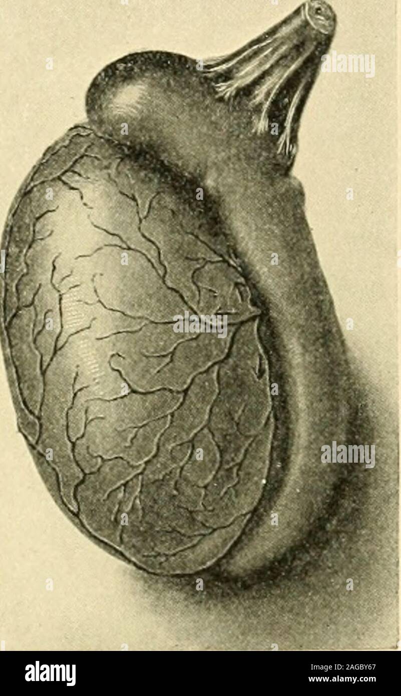 . The American journal of anatomy. ult Testis of the Pig.—The capsularartery gives off on the internal surface ofthe Tunica albuginea at rather regular in-tervals tortuous rib-like branches whichnearly encircle the gland. These branchespenetrate the substance of the gland follow-ing the septa and entering perpendicularlytill they reach the mediastinum. Except ina very few instances no branches aregiven off from these perpendicular arteriesuntil after the abrupt retro-flexion occursnear the center of the gland. After this sudden backward bending,many branches are given off which, coursing towar Stock Photohttps://www.alamy.com/image-license-details/?v=1https://www.alamy.com/the-american-journal-of-anatomy-ult-testis-of-the-pigthe-capsularartery-gives-off-on-the-internal-surface-ofthe-tunica-albuginea-at-rather-regular-in-tervals-tortuous-rib-like-branches-whichnearly-encircle-the-gland-these-branchespenetrate-the-substance-of-the-gland-follow-ing-the-septa-and-entering-perpendicularlytill-they-reach-the-mediastinum-except-ina-very-few-instances-no-branches-aregiven-off-from-these-perpendicular-arteriesuntil-after-the-abrupt-retro-flexion-occursnear-the-center-of-the-gland-after-this-sudden-backward-bendingmany-branches-are-given-off-which-coursing-towar-image337094303.html
RM2AGBY67–. The American journal of anatomy. ult Testis of the Pig.—The capsularartery gives off on the internal surface ofthe Tunica albuginea at rather regular in-tervals tortuous rib-like branches whichnearly encircle the gland. These branchespenetrate the substance of the gland follow-ing the septa and entering perpendicularlytill they reach the mediastinum. Except ina very few instances no branches aregiven off from these perpendicular arteriesuntil after the abrupt retro-flexion occursnear the center of the gland. After this sudden backward bending,many branches are given off which, coursing towar
