Typical plant cell Cut Out Stock Images
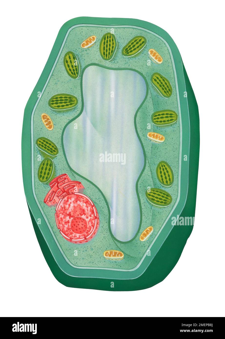 Typical plant cell Stock Photohttps://www.alamy.com/image-license-details/?v=1https://www.alamy.com/typical-plant-cell-image508197666.html
RM2MEPB8J–Typical plant cell
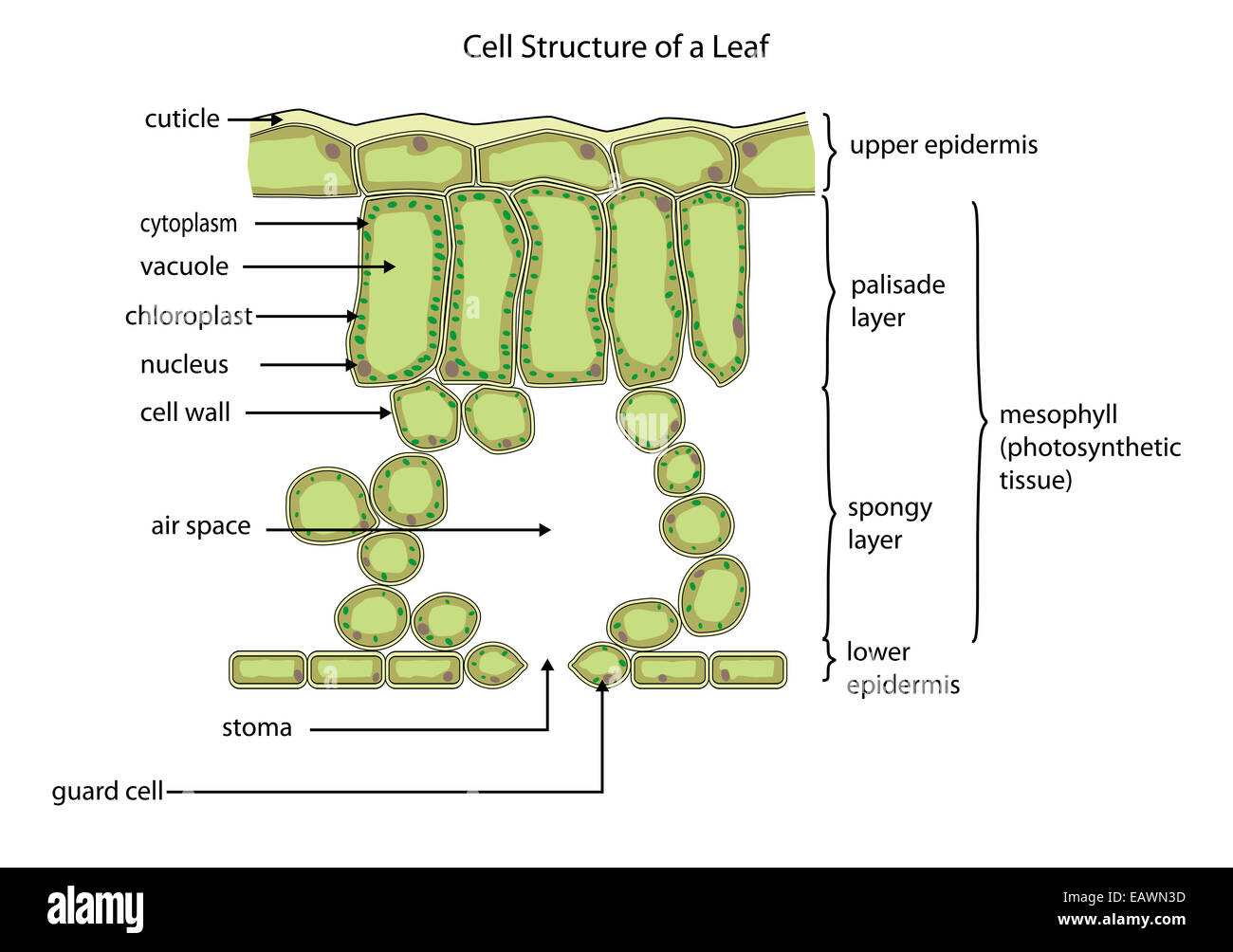 Section through a typical leaf showing the cell structure Stock Photohttps://www.alamy.com/image-license-details/?v=1https://www.alamy.com/stock-photo-section-through-a-typical-leaf-showing-the-cell-structure-75553393.html
RFEAWN3D–Section through a typical leaf showing the cell structure
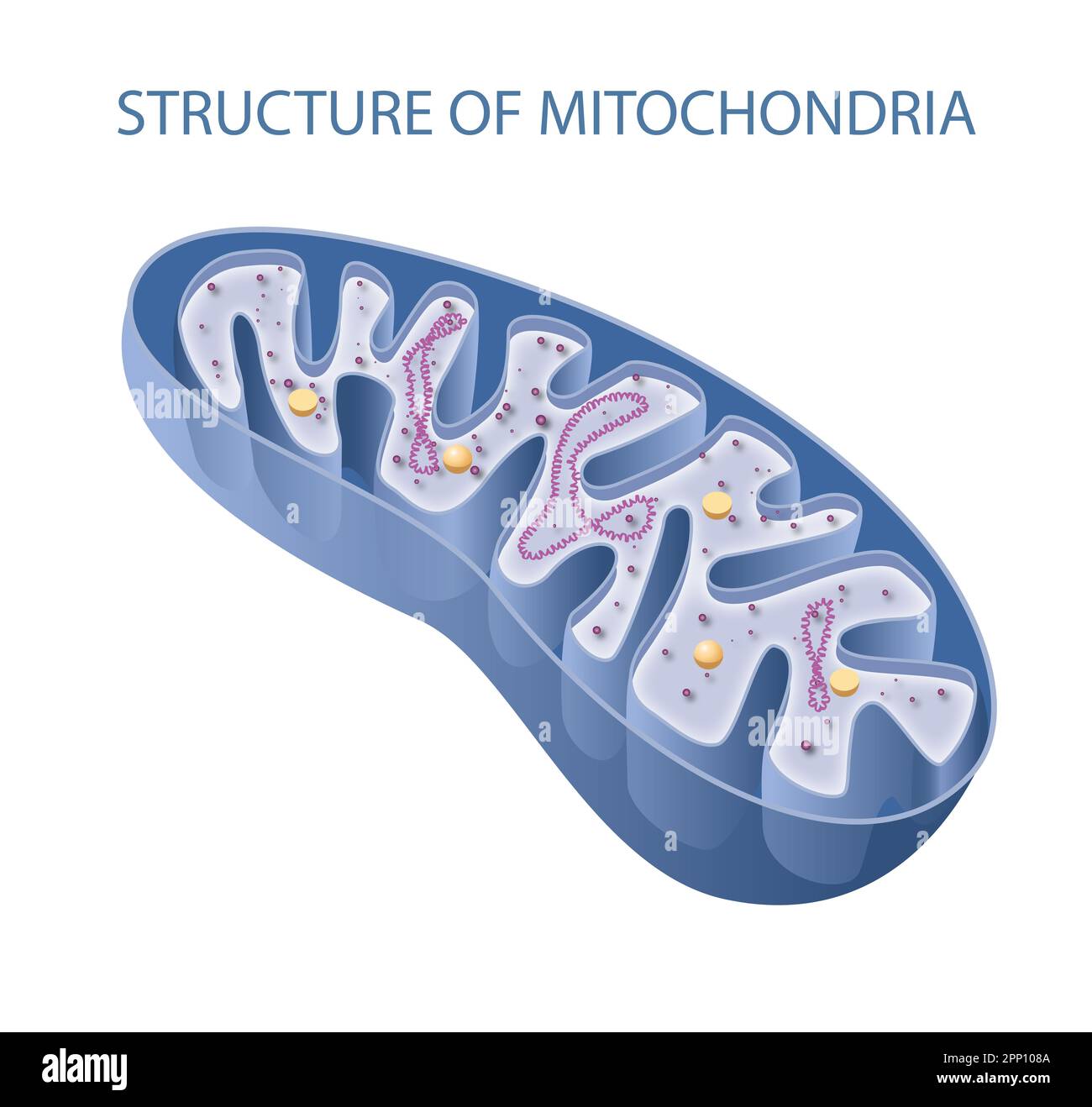 Components of a typical mitochondrion Stock Photohttps://www.alamy.com/image-license-details/?v=1https://www.alamy.com/components-of-a-typical-mitochondrion-image547066026.html
RF2PP108A–Components of a typical mitochondrion
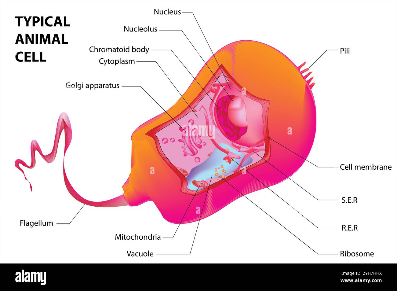 Structure of a typical animal cell. Vector Stock Vectorhttps://www.alamy.com/image-license-details/?v=1https://www.alamy.com/structure-of-a-typical-animal-cell-vector-image630189894.html
RF2YH7HHX–Structure of a typical animal cell. Vector
![Elementary anatomy, physiology and hygiene for higher grammar grades . Fio. 4. — A typical plant cell. [Bastin.] Pr. shows the protoplasm, with its graoules,but the network is not shown. The cell sap(c.«.) occupies a very large part of the cell.The cell food (c/.) makes a prominent addi-tion to the flgures. N, the nucleus, containsthe true nucleus {t.n.). This cell had sevenboundary cells. CELLS, TISSUES, AND ORGANS 31 tant ways: (1) they have a very thin membranous wallor perhaps no wall at all; (2) they have one or two smallcollections of cell sap, or more frequently no sap at all.Study Figu Stock Photo Elementary anatomy, physiology and hygiene for higher grammar grades . Fio. 4. — A typical plant cell. [Bastin.] Pr. shows the protoplasm, with its graoules,but the network is not shown. The cell sap(c.«.) occupies a very large part of the cell.The cell food (c/.) makes a prominent addi-tion to the flgures. N, the nucleus, containsthe true nucleus {t.n.). This cell had sevenboundary cells. CELLS, TISSUES, AND ORGANS 31 tant ways: (1) they have a very thin membranous wallor perhaps no wall at all; (2) they have one or two smallcollections of cell sap, or more frequently no sap at all.Study Figu Stock Photo](https://c8.alamy.com/comp/2AM8K7X/elementary-anatomy-physiology-and-hygiene-for-higher-grammar-grades-fio-4-a-typical-plant-cell-bastin-pr-shows-the-protoplasm-with-its-graoulesbut-the-network-is-not-shown-the-cell-sapc-occupies-a-very-large-part-of-the-cellthe-cell-food-c-makes-a-prominent-addi-tion-to-the-flgures-n-the-nucleus-containsthe-true-nucleus-tn-this-cell-had-sevenboundary-cells-cells-tissues-and-organs-31-tant-ways-1-they-have-a-very-thin-membranous-wallor-perhaps-no-wall-at-all-2-they-have-one-or-two-smallcollections-of-cell-sap-or-more-frequently-no-sap-at-allstudy-figu-2AM8K7X.jpg) Elementary anatomy, physiology and hygiene for higher grammar grades . Fio. 4. — A typical plant cell. [Bastin.] Pr. shows the protoplasm, with its graoules,but the network is not shown. The cell sap(c.«.) occupies a very large part of the cell.The cell food (c/.) makes a prominent addi-tion to the flgures. N, the nucleus, containsthe true nucleus {t.n.). This cell had sevenboundary cells. CELLS, TISSUES, AND ORGANS 31 tant ways: (1) they have a very thin membranous wallor perhaps no wall at all; (2) they have one or two smallcollections of cell sap, or more frequently no sap at all.Study Figu Stock Photohttps://www.alamy.com/image-license-details/?v=1https://www.alamy.com/elementary-anatomy-physiology-and-hygiene-for-higher-grammar-grades-fio-4-a-typical-plant-cell-bastin-pr-shows-the-protoplasm-with-its-graoulesbut-the-network-is-not-shown-the-cell-sapc-occupies-a-very-large-part-of-the-cellthe-cell-food-c-makes-a-prominent-addi-tion-to-the-flgures-n-the-nucleus-containsthe-true-nucleus-tn-this-cell-had-sevenboundary-cells-cells-tissues-and-organs-31-tant-ways-1-they-have-a-very-thin-membranous-wallor-perhaps-no-wall-at-all-2-they-have-one-or-two-smallcollections-of-cell-sap-or-more-frequently-no-sap-at-allstudy-figu-image339480846.html
Elementary anatomy, physiology and hygiene for higher grammar grades . Fio. 4. — A typical plant cell. [Bastin.] Pr. shows the protoplasm, with its graoules,but the network is not shown. The cell sap(c.«.) occupies a very large part of the cell.The cell food (c/.) makes a prominent addi-tion to the flgures. N, the nucleus, containsthe true nucleus {t.n.). This cell had sevenboundary cells. CELLS, TISSUES, AND ORGANS 31 tant ways: (1) they have a very thin membranous wallor perhaps no wall at all; (2) they have one or two smallcollections of cell sap, or more frequently no sap at all.Study Figu Stock Photohttps://www.alamy.com/image-license-details/?v=1https://www.alamy.com/elementary-anatomy-physiology-and-hygiene-for-higher-grammar-grades-fio-4-a-typical-plant-cell-bastin-pr-shows-the-protoplasm-with-its-graoulesbut-the-network-is-not-shown-the-cell-sapc-occupies-a-very-large-part-of-the-cellthe-cell-food-c-makes-a-prominent-addi-tion-to-the-flgures-n-the-nucleus-containsthe-true-nucleus-tn-this-cell-had-sevenboundary-cells-cells-tissues-and-organs-31-tant-ways-1-they-have-a-very-thin-membranous-wallor-perhaps-no-wall-at-all-2-they-have-one-or-two-smallcollections-of-cell-sap-or-more-frequently-no-sap-at-allstudy-figu-image339480846.htmlRM2AM8K7X–Elementary anatomy, physiology and hygiene for higher grammar grades . Fio. 4. — A typical plant cell. [Bastin.] Pr. shows the protoplasm, with its graoules,but the network is not shown. The cell sap(c.«.) occupies a very large part of the cell.The cell food (c/.) makes a prominent addi-tion to the flgures. N, the nucleus, containsthe true nucleus {t.n.). This cell had sevenboundary cells. CELLS, TISSUES, AND ORGANS 31 tant ways: (1) they have a very thin membranous wallor perhaps no wall at all; (2) they have one or two smallcollections of cell sap, or more frequently no sap at all.Study Figu
 A typical vegatable cell in a plant, vintage line drawing or engraving illustration. Stock Vectorhttps://www.alamy.com/image-license-details/?v=1https://www.alamy.com/a-typical-vegatable-cell-in-a-plant-vintage-line-drawing-or-engraving-illustration-image348617804.html
RF2B74WFT–A typical vegatable cell in a plant, vintage line drawing or engraving illustration.
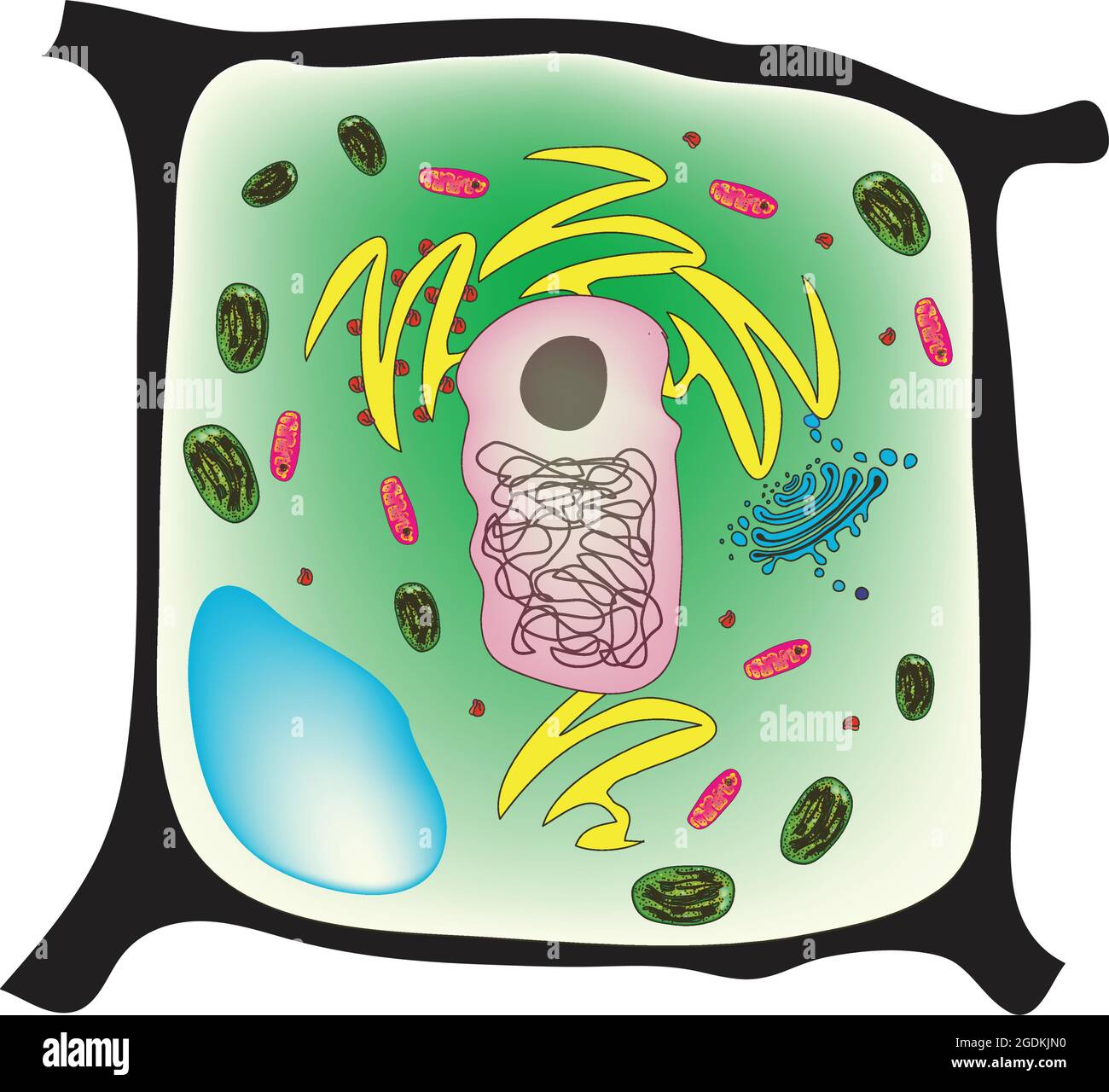 Plant cell, the basic unit of all plants. Plant cells, like animal cells, are eukaryotic, meaning they have a membrane-bound nucleus and organelles. Stock Vectorhttps://www.alamy.com/image-license-details/?v=1https://www.alamy.com/plant-cell-the-basic-unit-of-all-plants-plant-cells-like-animal-cells-are-eukaryotic-meaning-they-have-a-membrane-bound-nucleus-and-organelles-image438681516.html
RF2GDKJN0–Plant cell, the basic unit of all plants. Plant cells, like animal cells, are eukaryotic, meaning they have a membrane-bound nucleus and organelles.
 chrysanthemum green white background Stock Photohttps://www.alamy.com/image-license-details/?v=1https://www.alamy.com/chrysanthemum-green-white-background-image3345345.html
RFA1FRC2–chrysanthemum green white background
 . The structure and development of mosses and ferns (Archegoniatae). Plant morphology; Mosses; Ferns. THE HOMOSPOROUS LEPTOSPORANGIAT^ 363 subject of numerous investigations, but there still is a good deal of diversity of opinion as to their exact method of growth. Bowser ((11), p. 310) states that in 0. regalis there may be a single apical cell, such as exists in the first root of 0. Claytoni- ana and O. cinnamomea, but that it never shows the regular segmentation of the typical leptosporangiate root, and it may be replaced by two or three similar initials. In Todea barbara he found four simi Stock Photohttps://www.alamy.com/image-license-details/?v=1https://www.alamy.com/the-structure-and-development-of-mosses-and-ferns-archegoniatae-plant-morphology-mosses-ferns-the-homosporous-leptosporangiat-363-subject-of-numerous-investigations-but-there-still-is-a-good-deal-of-diversity-of-opinion-as-to-their-exact-method-of-growth-bowser-11-p-310-states-that-in-0-regalis-there-may-be-a-single-apical-cell-such-as-exists-in-the-first-root-of-0-claytoni-ana-and-o-cinnamomea-but-that-it-never-shows-the-regular-segmentation-of-the-typical-leptosporangiate-root-and-it-may-be-replaced-by-two-or-three-similar-initials-in-todea-barbara-he-found-four-simi-image216368725.html
RMPG0CED–. The structure and development of mosses and ferns (Archegoniatae). Plant morphology; Mosses; Ferns. THE HOMOSPOROUS LEPTOSPORANGIAT^ 363 subject of numerous investigations, but there still is a good deal of diversity of opinion as to their exact method of growth. Bowser ((11), p. 310) states that in 0. regalis there may be a single apical cell, such as exists in the first root of 0. Claytoni- ana and O. cinnamomea, but that it never shows the regular segmentation of the typical leptosporangiate root, and it may be replaced by two or three similar initials. In Todea barbara he found four simi
 . The structure and development of mosses and ferns (Archegoniatae). Plant morphology; Mosses; Ferns. THE HOMOSPOROUS LEPTOSPORANGIAT^ 363 subject of numerous investigations, but there still is a good deal of diversity of opinion as to their exact method of growth. Bowser ((11), p. 310) states that in 0. regalis there may be a single apical cell, such as exists in the first root of 0. Claytoni- ana and O. cinnamomea, but that it never shows the regular segmentation of the typical leptosporangiate root, and it may be replaced by two or three similar initials. In Todea barbara he found four simi Stock Photohttps://www.alamy.com/image-license-details/?v=1https://www.alamy.com/the-structure-and-development-of-mosses-and-ferns-archegoniatae-plant-morphology-mosses-ferns-the-homosporous-leptosporangiat-363-subject-of-numerous-investigations-but-there-still-is-a-good-deal-of-diversity-of-opinion-as-to-their-exact-method-of-growth-bowser-11-p-310-states-that-in-0-regalis-there-may-be-a-single-apical-cell-such-as-exists-in-the-first-root-of-0-claytoni-ana-and-o-cinnamomea-but-that-it-never-shows-the-regular-segmentation-of-the-typical-leptosporangiate-root-and-it-may-be-replaced-by-two-or-three-similar-initials-in-todea-barbara-he-found-four-simi-image232063903.html
RMRDFBTF–. The structure and development of mosses and ferns (Archegoniatae). Plant morphology; Mosses; Ferns. THE HOMOSPOROUS LEPTOSPORANGIAT^ 363 subject of numerous investigations, but there still is a good deal of diversity of opinion as to their exact method of growth. Bowser ((11), p. 310) states that in 0. regalis there may be a single apical cell, such as exists in the first root of 0. Claytoni- ana and O. cinnamomea, but that it never shows the regular segmentation of the typical leptosporangiate root, and it may be replaced by two or three similar initials. In Todea barbara he found four simi
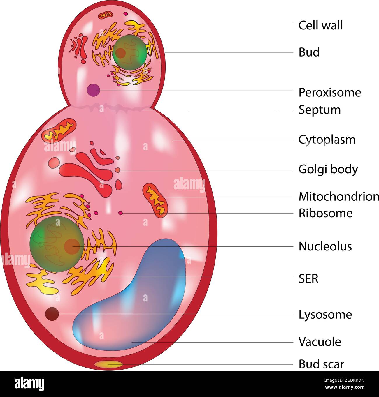 Budding fungus cell structure, Anatomy of fungal cell, typical labeled and detailed diagram of fungus cell in kingdom fungi Stock Vectorhttps://www.alamy.com/image-license-details/?v=1https://www.alamy.com/budding-fungus-cell-structure-anatomy-of-fungal-cell-typical-labeled-and-detailed-diagram-of-fungus-cell-in-kingdom-fungi-image438685233.html
RF2GDKRDN–Budding fungus cell structure, Anatomy of fungal cell, typical labeled and detailed diagram of fungus cell in kingdom fungi
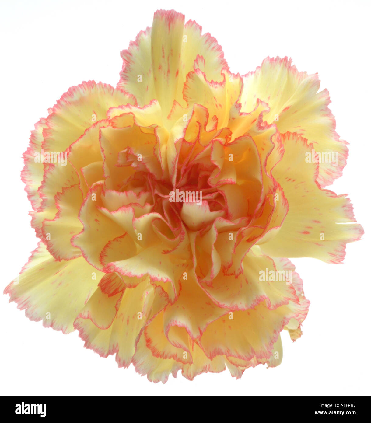 carnation pink yellow white background Stock Photohttps://www.alamy.com/image-license-details/?v=1https://www.alamy.com/carnation-pink-yellow-white-background-image3345334.html
RFA1FRB7–carnation pink yellow white background
 . The origin of a land flora, a theory based upon the facts of alternation. Plant morphology. MARATTIACEAE 517 At maturity the more or less indurated superficial layer of cells of the sporangial wall is the most conspicuous part, but the thinner-walled cells lying, within, though they may shrink, do not entirely disappear. The â essential parts of the sporangium of Angiopteris, and especially the archesporium, are thus seen to be referable in typical cases to a single parent cell : this also is the case typically for all the other genera.. Fig. 285. Marattiafraxinea, Smith. A =section transver Stock Photohttps://www.alamy.com/image-license-details/?v=1https://www.alamy.com/the-origin-of-a-land-flora-a-theory-based-upon-the-facts-of-alternation-plant-morphology-marattiaceae-517-at-maturity-the-more-or-less-indurated-superficial-layer-of-cells-of-the-sporangial-wall-is-the-most-conspicuous-part-but-the-thinner-walled-cells-lying-within-though-they-may-shrink-do-not-entirely-disappear-the-essential-parts-of-the-sporangium-of-angiopteris-and-especially-the-archesporium-are-thus-seen-to-be-referable-in-typical-cases-to-a-single-parent-cell-this-also-is-the-case-typically-for-all-the-other-genera-fig-285-marattiafraxinea-smith-a-=section-transver-image232307984.html
RMRDXF5M–. The origin of a land flora, a theory based upon the facts of alternation. Plant morphology. MARATTIACEAE 517 At maturity the more or less indurated superficial layer of cells of the sporangial wall is the most conspicuous part, but the thinner-walled cells lying, within, though they may shrink, do not entirely disappear. The â essential parts of the sporangium of Angiopteris, and especially the archesporium, are thus seen to be referable in typical cases to a single parent cell : this also is the case typically for all the other genera.. Fig. 285. Marattiafraxinea, Smith. A =section transver
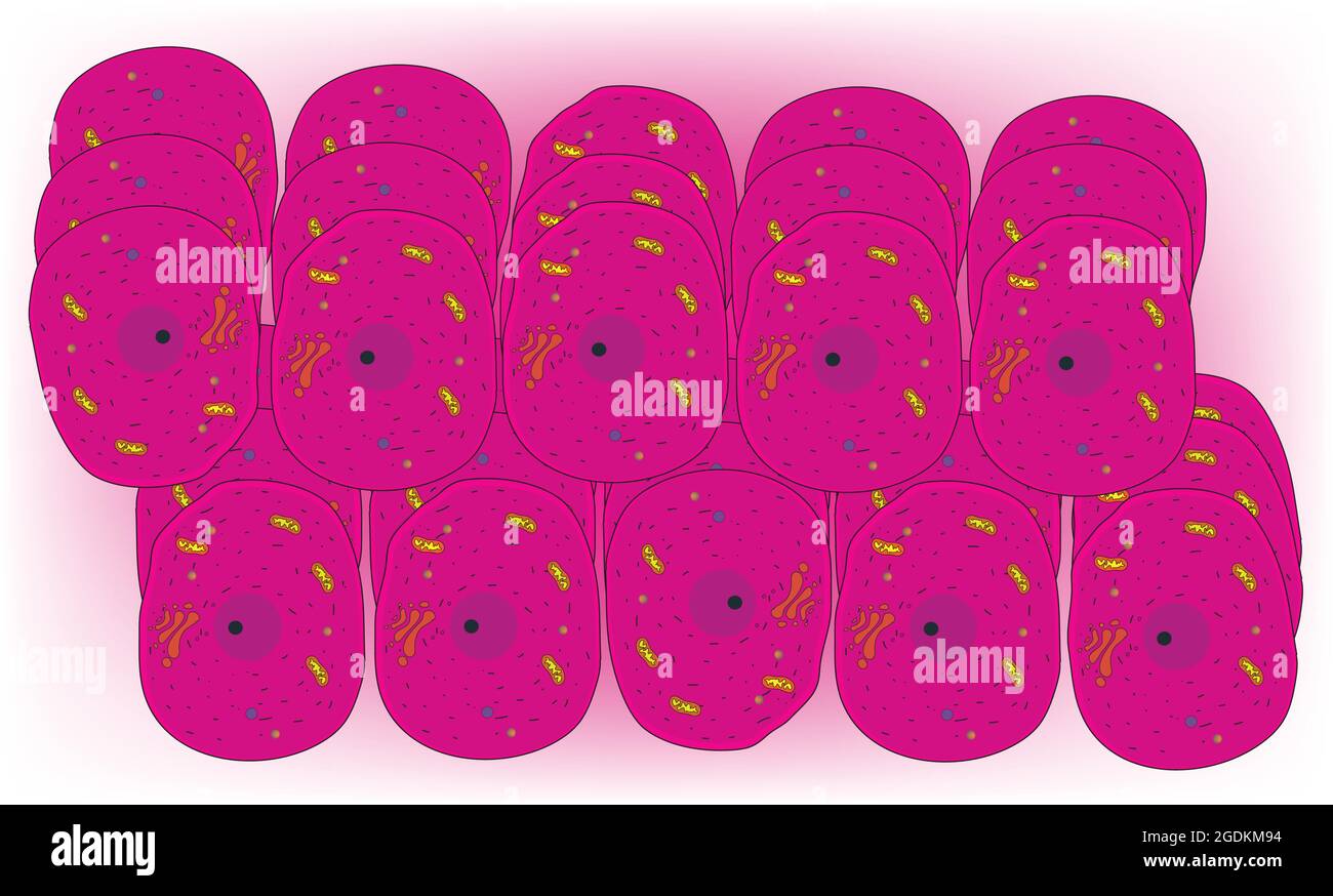 Typical animal tissue, typical of the eukaryotic cell, enclosed by a plasma membrane and containing a membrane-bound nucleus and organelles. Stock Vectorhttps://www.alamy.com/image-license-details/?v=1https://www.alamy.com/typical-animal-tissue-typical-of-the-eukaryotic-cell-enclosed-by-a-plasma-membrane-and-containing-a-membrane-bound-nucleus-and-organelles-image438682752.html
RF2GDKM94–Typical animal tissue, typical of the eukaryotic cell, enclosed by a plasma membrane and containing a membrane-bound nucleus and organelles.
 chrysanthemum pink bunch white background Stock Photohttps://www.alamy.com/image-license-details/?v=1https://www.alamy.com/chrysanthemum-pink-bunch-white-background-image3345352.html
RFA1FRC9–chrysanthemum pink bunch white background
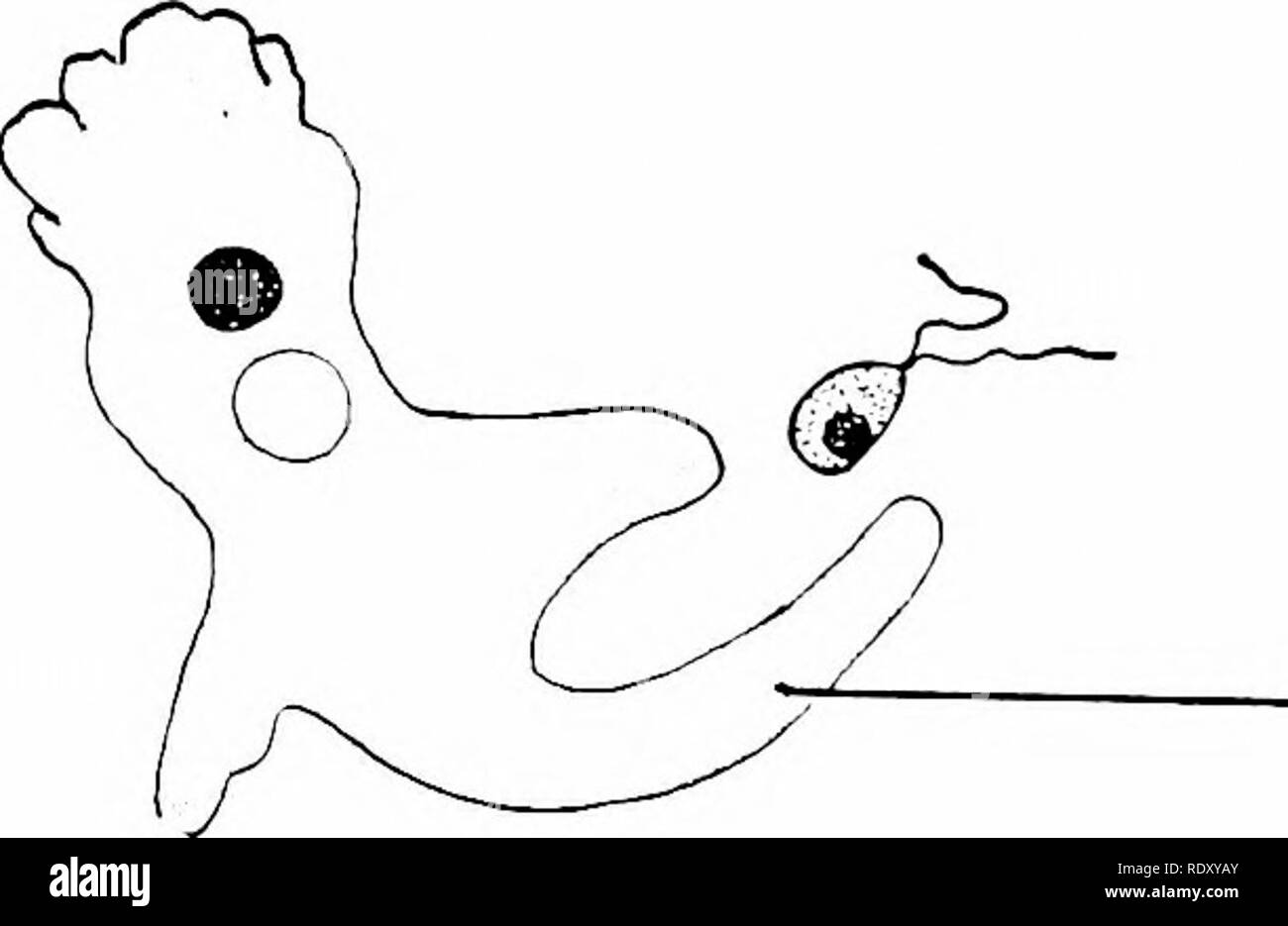 . Principles of modern biology. Biology. 128 - The Cell synthesis of the various organic components of its protoplasm, and the typical plant can live and grow indefinitely so long as these simple foods are available. But an animal cannot do this; an animal's food require- ments are on a higher level of complexity. Compared to plant cells, animal cells have more limited powers of synthesis, and animal metabolism cannot be maintained in the ab- sence of organic foods stub as preformed proteins (or amino acids), carbohydrates, lipids, and vitamins (Table 7-1). Animals generally are cjuite similar Stock Photohttps://www.alamy.com/image-license-details/?v=1https://www.alamy.com/principles-of-modern-biology-biology-128-the-cell-synthesis-of-the-various-organic-components-of-its-protoplasm-and-the-typical-plant-can-live-and-grow-indefinitely-so-long-as-these-simple-foods-are-available-but-an-animal-cannot-do-this-an-animals-food-require-ments-are-on-a-higher-level-of-complexity-compared-to-plant-cells-animal-cells-have-more-limited-powers-of-synthesis-and-animal-metabolism-cannot-be-maintained-in-the-ab-sence-of-organic-foods-stub-as-preformed-proteins-or-amino-acids-carbohydrates-lipids-and-vitamins-table-7-1-animals-generally-are-cjuite-similar-image232317539.html
RMRDXYAY–. Principles of modern biology. Biology. 128 - The Cell synthesis of the various organic components of its protoplasm, and the typical plant can live and grow indefinitely so long as these simple foods are available. But an animal cannot do this; an animal's food require- ments are on a higher level of complexity. Compared to plant cells, animal cells have more limited powers of synthesis, and animal metabolism cannot be maintained in the ab- sence of organic foods stub as preformed proteins (or amino acids), carbohydrates, lipids, and vitamins (Table 7-1). Animals generally are cjuite similar
 carnation white white background Stock Photohttps://www.alamy.com/image-license-details/?v=1https://www.alamy.com/carnation-white-white-background-image3345339.html
RFA1FRBC–carnation white white background
 . The origin of a land flora, a theory based upon the facts of alternation. Plant morphology. 454 OPHIOGLOSSALES corresponding in every other respect, do not assume the dense protoplasm of sporogenous cells. These more bulky sporangia lead on to such as that shown in Fig. 253 e, in which it is possible that the whole sporogenous group is referable to a single parent cell, though the proportions of the whole group are quite different from those of the typical sporangia; the sporogenous cells appear, however, to form two groups, and probably originated from two similar cells side by side. The in Stock Photohttps://www.alamy.com/image-license-details/?v=1https://www.alamy.com/the-origin-of-a-land-flora-a-theory-based-upon-the-facts-of-alternation-plant-morphology-454-ophioglossales-corresponding-in-every-other-respect-do-not-assume-the-dense-protoplasm-of-sporogenous-cells-these-more-bulky-sporangia-lead-on-to-such-as-that-shown-in-fig-253-e-in-which-it-is-possible-that-the-whole-sporogenous-group-is-referable-to-a-single-parent-cell-though-the-proportions-of-the-whole-group-are-quite-different-from-those-of-the-typical-sporangia-the-sporogenous-cells-appear-however-to-form-two-groups-and-probably-originated-from-two-similar-cells-side-by-side-the-in-image232308109.html
RMRDXFA5–. The origin of a land flora, a theory based upon the facts of alternation. Plant morphology. 454 OPHIOGLOSSALES corresponding in every other respect, do not assume the dense protoplasm of sporogenous cells. These more bulky sporangia lead on to such as that shown in Fig. 253 e, in which it is possible that the whole sporogenous group is referable to a single parent cell, though the proportions of the whole group are quite different from those of the typical sporangia; the sporogenous cells appear, however, to form two groups, and probably originated from two similar cells side by side. The in
 chrysanthemum white background Stock Photohttps://www.alamy.com/image-license-details/?v=1https://www.alamy.com/chrysanthemum-white-background-image3345363.html
RFA1FRD4–chrysanthemum white background
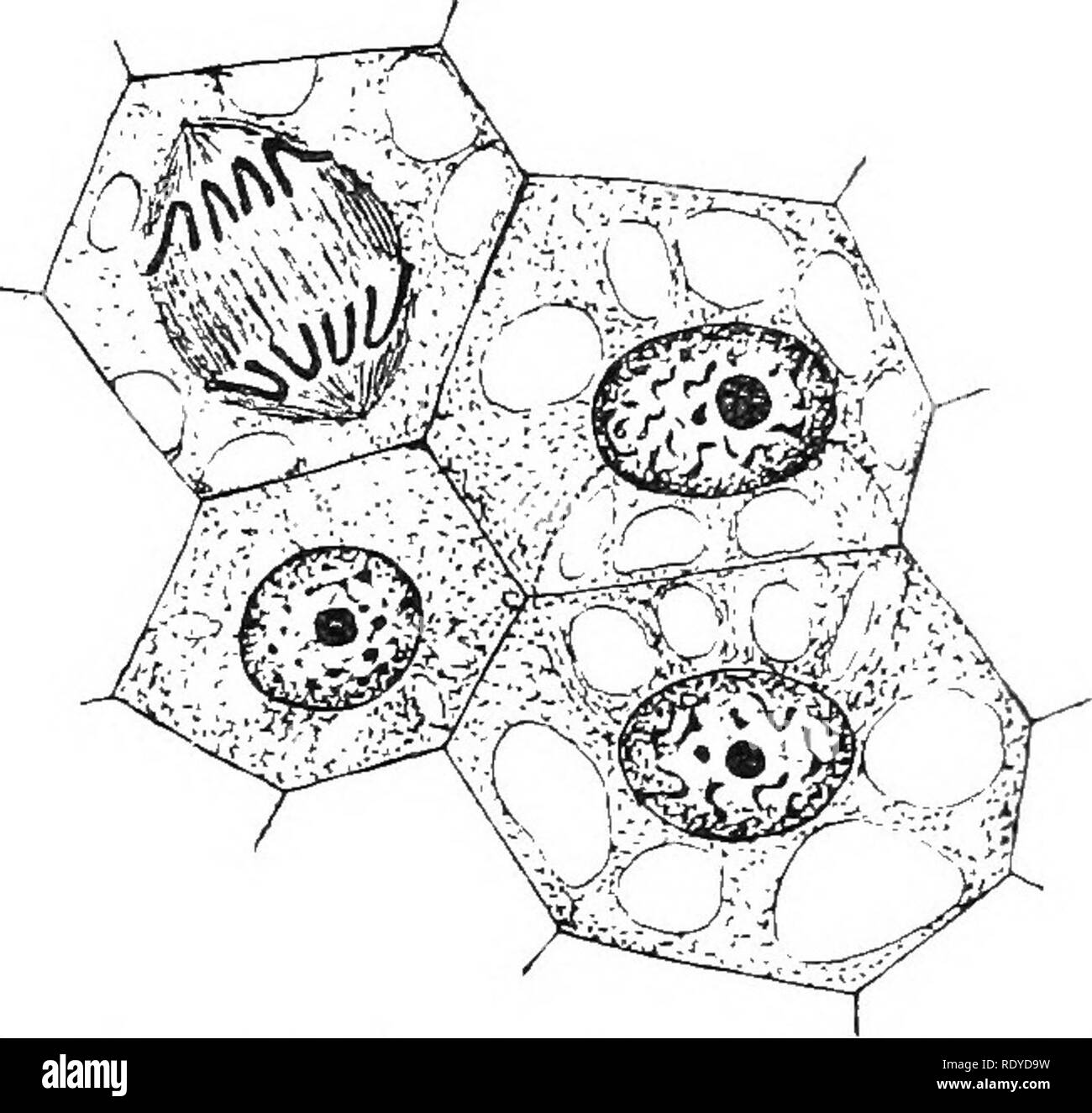 . The plant cell, its modifications and vital processes; a manual for students. Plant physiology; Plant anatomy; Plant cells and tissues. MliRISTEM. 43 it is possible to detect here and there cells in -which typical division- figures (mitosis) can be made out, especially where a thin section is cut and stained as directed in the note at the end of Chapter ii. (see Fig. 28). In these young cells the cell-walls are very thin, and on account of turgiditj- are a good deal on the stretch; the polyhedral shape so often observed in the cells of young tissues is due partly to mutual cohesion and press Stock Photohttps://www.alamy.com/image-license-details/?v=1https://www.alamy.com/the-plant-cell-its-modifications-and-vital-processes-a-manual-for-students-plant-physiology-plant-anatomy-plant-cells-and-tissues-mliristem-43-it-is-possible-to-detect-here-and-there-cells-in-which-typical-division-figures-mitosis-can-be-made-out-especially-where-a-thin-section-is-cut-and-stained-as-directed-in-the-note-at-the-end-of-chapter-ii-see-fig-28-in-these-young-cells-the-cell-walls-are-very-thin-and-on-account-of-turgiditj-are-a-good-deal-on-the-stretch-the-polyhedral-shape-so-often-observed-in-the-cells-of-young-tissues-is-due-partly-to-mutual-cohesion-and-press-image232328485.html
RMRDYD9W–. The plant cell, its modifications and vital processes; a manual for students. Plant physiology; Plant anatomy; Plant cells and tissues. MliRISTEM. 43 it is possible to detect here and there cells in -which typical division- figures (mitosis) can be made out, especially where a thin section is cut and stained as directed in the note at the end of Chapter ii. (see Fig. 28). In these young cells the cell-walls are very thin, and on account of turgiditj- are a good deal on the stretch; the polyhedral shape so often observed in the cells of young tissues is due partly to mutual cohesion and press
 chrysanthemum salmon background Stock Photohttps://www.alamy.com/image-license-details/?v=1https://www.alamy.com/chrysanthemum-salmon-background-image3345358.html
RFA1FRCF–chrysanthemum salmon background
 . Elements of plant biology. Plant physiology. LIVING PARENCHYMA 271 become chloroplasts. Certain adult living cells, how- ever, differ in one way or another from this typical structure.. Fig. 42.—Development of adult liying cells (parenchyma) from embryonic cells. A, embryonic (m^eristematic) cells. B, begin- ning of vacuolation. C, fully grown cells (here much elongated) with characteristic large central vacuole; c.w., cell wall; cyt., cytoplasm; n, nucleus; no., nucleolus; v, vacuole.. Please note that these images are extracted from scanned page images that may have been digitally enhanced Stock Photohttps://www.alamy.com/image-license-details/?v=1https://www.alamy.com/elements-of-plant-biology-plant-physiology-living-parenchyma-271-become-chloroplasts-certain-adult-living-cells-how-ever-differ-in-one-way-or-another-from-this-typical-structure-fig-42development-of-adult-liying-cells-parenchyma-from-embryonic-cells-a-embryonic-meristematic-cells-b-begin-ning-of-vacuolation-c-fully-grown-cells-here-much-elongated-with-characteristic-large-central-vacuole-cw-cell-wall-cyt-cytoplasm-n-nucleus-no-nucleolus-v-vacuole-please-note-that-these-images-are-extracted-from-scanned-page-images-that-may-have-been-digitally-enhanced-image232115623.html
RMRDHNRK–. Elements of plant biology. Plant physiology. LIVING PARENCHYMA 271 become chloroplasts. Certain adult living cells, how- ever, differ in one way or another from this typical structure.. Fig. 42.—Development of adult liying cells (parenchyma) from embryonic cells. A, embryonic (m^eristematic) cells. B, begin- ning of vacuolation. C, fully grown cells (here much elongated) with characteristic large central vacuole; c.w., cell wall; cyt., cytoplasm; n, nucleus; no., nucleolus; v, vacuole.. Please note that these images are extracted from scanned page images that may have been digitally enhanced
 chrysanthemum pink single background Stock Photohttps://www.alamy.com/image-license-details/?v=1https://www.alamy.com/chrysanthemum-pink-single-background-image3345355.html
RFA1FRCC–chrysanthemum pink single background
![. South African botany. Botany. CHAPTEE II. THE CELL. 10. The Cell.—The whole of the tissues of a plant are composed of units called Cells (fig. 10). These re- semble the cells of animal tissue in many respects, but n-. FiG. 10.—Typical Cells. n. Nucleus, v. Vacuole, cxj. Cytoplasm. differ from them in that they are surrounded by a firm wall made of a carbohydrate called Cellulose. A typical eel] is found to contain a mass of Cytoplasm, a fluid-like substance consisting of a clear ground sub- 18. Please note that these images are extracted from scanned page images that may have been digitally Stock Photo . South African botany. Botany. CHAPTEE II. THE CELL. 10. The Cell.—The whole of the tissues of a plant are composed of units called Cells (fig. 10). These re- semble the cells of animal tissue in many respects, but n-. FiG. 10.—Typical Cells. n. Nucleus, v. Vacuole, cxj. Cytoplasm. differ from them in that they are surrounded by a firm wall made of a carbohydrate called Cellulose. A typical eel] is found to contain a mass of Cytoplasm, a fluid-like substance consisting of a clear ground sub- 18. Please note that these images are extracted from scanned page images that may have been digitally Stock Photo](https://c8.alamy.com/comp/RE1KMT/south-african-botany-botany-chaptee-ii-the-cell-10-the-cellthe-whole-of-the-tissues-of-a-plant-are-composed-of-units-called-cells-fig-10-these-re-semble-the-cells-of-animal-tissue-in-many-respects-but-n-fig-10typical-cells-n-nucleus-v-vacuole-cxj-cytoplasm-differ-from-them-in-that-they-are-surrounded-by-a-firm-wall-made-of-a-carbohydrate-called-cellulose-a-typical-eel-is-found-to-contain-a-mass-of-cytoplasm-a-fluid-like-substance-consisting-of-a-clear-ground-sub-18-please-note-that-these-images-are-extracted-from-scanned-page-images-that-may-have-been-digitally-RE1KMT.jpg) . South African botany. Botany. CHAPTEE II. THE CELL. 10. The Cell.—The whole of the tissues of a plant are composed of units called Cells (fig. 10). These re- semble the cells of animal tissue in many respects, but n-. FiG. 10.—Typical Cells. n. Nucleus, v. Vacuole, cxj. Cytoplasm. differ from them in that they are surrounded by a firm wall made of a carbohydrate called Cellulose. A typical eel] is found to contain a mass of Cytoplasm, a fluid-like substance consisting of a clear ground sub- 18. Please note that these images are extracted from scanned page images that may have been digitally Stock Photohttps://www.alamy.com/image-license-details/?v=1https://www.alamy.com/south-african-botany-botany-chaptee-ii-the-cell-10-the-cellthe-whole-of-the-tissues-of-a-plant-are-composed-of-units-called-cells-fig-10-these-re-semble-the-cells-of-animal-tissue-in-many-respects-but-n-fig-10typical-cells-n-nucleus-v-vacuole-cxj-cytoplasm-differ-from-them-in-that-they-are-surrounded-by-a-firm-wall-made-of-a-carbohydrate-called-cellulose-a-typical-eel-is-found-to-contain-a-mass-of-cytoplasm-a-fluid-like-substance-consisting-of-a-clear-ground-sub-18-please-note-that-these-images-are-extracted-from-scanned-page-images-that-may-have-been-digitally-image232377400.html
. South African botany. Botany. CHAPTEE II. THE CELL. 10. The Cell.—The whole of the tissues of a plant are composed of units called Cells (fig. 10). These re- semble the cells of animal tissue in many respects, but n-. FiG. 10.—Typical Cells. n. Nucleus, v. Vacuole, cxj. Cytoplasm. differ from them in that they are surrounded by a firm wall made of a carbohydrate called Cellulose. A typical eel] is found to contain a mass of Cytoplasm, a fluid-like substance consisting of a clear ground sub- 18. Please note that these images are extracted from scanned page images that may have been digitally Stock Photohttps://www.alamy.com/image-license-details/?v=1https://www.alamy.com/south-african-botany-botany-chaptee-ii-the-cell-10-the-cellthe-whole-of-the-tissues-of-a-plant-are-composed-of-units-called-cells-fig-10-these-re-semble-the-cells-of-animal-tissue-in-many-respects-but-n-fig-10typical-cells-n-nucleus-v-vacuole-cxj-cytoplasm-differ-from-them-in-that-they-are-surrounded-by-a-firm-wall-made-of-a-carbohydrate-called-cellulose-a-typical-eel-is-found-to-contain-a-mass-of-cytoplasm-a-fluid-like-substance-consisting-of-a-clear-ground-sub-18-please-note-that-these-images-are-extracted-from-scanned-page-images-that-may-have-been-digitally-image232377400.htmlRMRE1KMT–. South African botany. Botany. CHAPTEE II. THE CELL. 10. The Cell.—The whole of the tissues of a plant are composed of units called Cells (fig. 10). These re- semble the cells of animal tissue in many respects, but n-. FiG. 10.—Typical Cells. n. Nucleus, v. Vacuole, cxj. Cytoplasm. differ from them in that they are surrounded by a firm wall made of a carbohydrate called Cellulose. A typical eel] is found to contain a mass of Cytoplasm, a fluid-like substance consisting of a clear ground sub- 18. Please note that these images are extracted from scanned page images that may have been digitally