Quick filters:
Vastus intermedius Stock Photos and Images
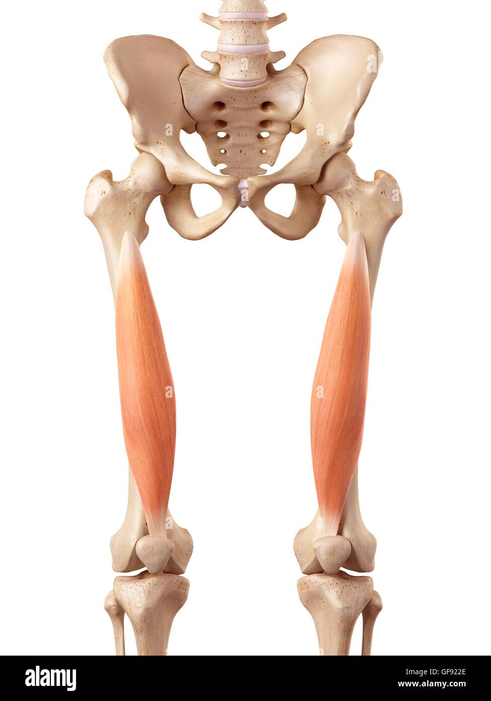
 medical accurate illustration of the vastus intermedius Stock Photohttps://www.alamy.com/image-license-details/?v=1https://www.alamy.com/stock-photo-medical-accurate-illustration-of-the-vastus-intermedius-89743102.html
RFF6046P–medical accurate illustration of the vastus intermedius
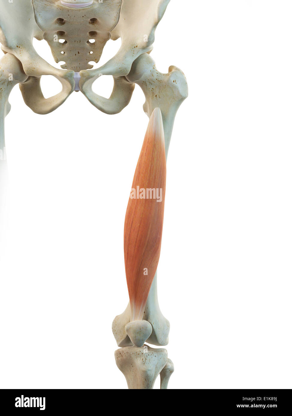 Human vastus intermedius muscle computer artwork. Stock Photohttps://www.alamy.com/image-license-details/?v=1https://www.alamy.com/human-vastus-intermedius-muscle-computer-artwork-image69879758.html
RFE1K89J–Human vastus intermedius muscle computer artwork.
 medically accurate illustration of the vastus intermedius Stock Photohttps://www.alamy.com/image-license-details/?v=1https://www.alamy.com/stock-photo-medically-accurate-illustration-of-the-vastus-intermedius-89761842.html
RFF61042–medically accurate illustration of the vastus intermedius
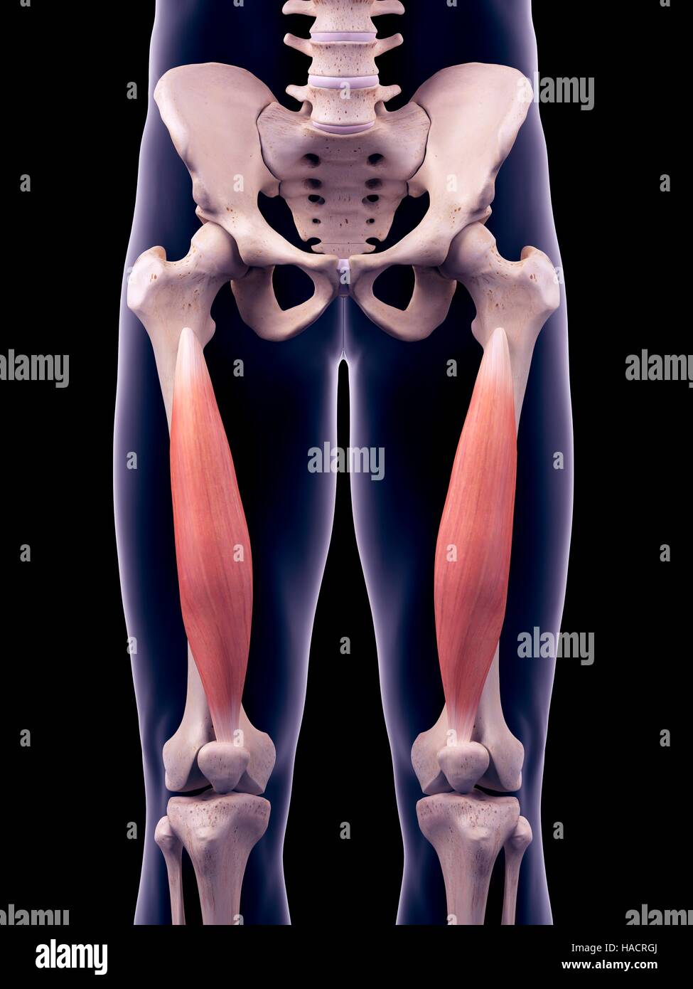 Illustration of the vastus intermedius muscles. Stock Photohttps://www.alamy.com/image-license-details/?v=1https://www.alamy.com/stock-photo-illustration-of-the-vastus-intermedius-muscles-126901058.html
RFHACRGJ–Illustration of the vastus intermedius muscles.
 medically accurate illustration of the vastus intermedius Stock Photohttps://www.alamy.com/image-license-details/?v=1https://www.alamy.com/stock-photo-medically-accurate-illustration-of-the-vastus-intermedius-89761840.html
RFF61040–medically accurate illustration of the vastus intermedius
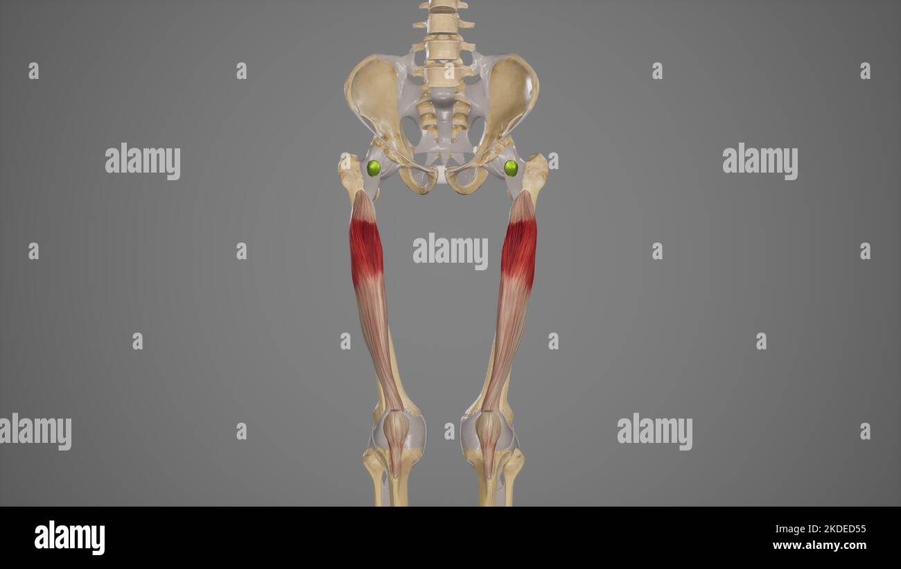 Medical Illustration of Vastus Intermedius Muscle Stock Photohttps://www.alamy.com/image-license-details/?v=1https://www.alamy.com/medical-illustration-of-vastus-intermedius-muscle-image490198497.html
RF2KDED55–Medical Illustration of Vastus Intermedius Muscle
 3d rendered medically accurate illustration of a womans Vastus Intermedius Stock Photohttps://www.alamy.com/image-license-details/?v=1https://www.alamy.com/3d-rendered-medically-accurate-illustration-of-a-womans-vastus-intermedius-image258131135.html
RFTYXTW3–3d rendered medically accurate illustration of a womans Vastus Intermedius
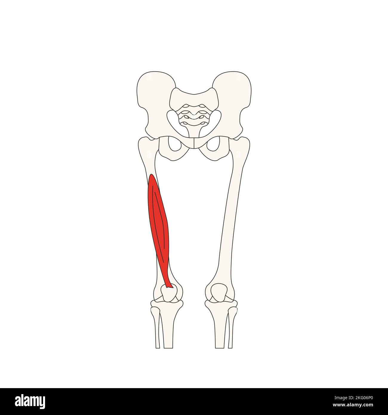 human anatomy drawing vastus intermedius Stock Photohttps://www.alamy.com/image-license-details/?v=1https://www.alamy.com/human-anatomy-drawing-vastus-intermedius-image491730120.html
RF2KG06P0–human anatomy drawing vastus intermedius
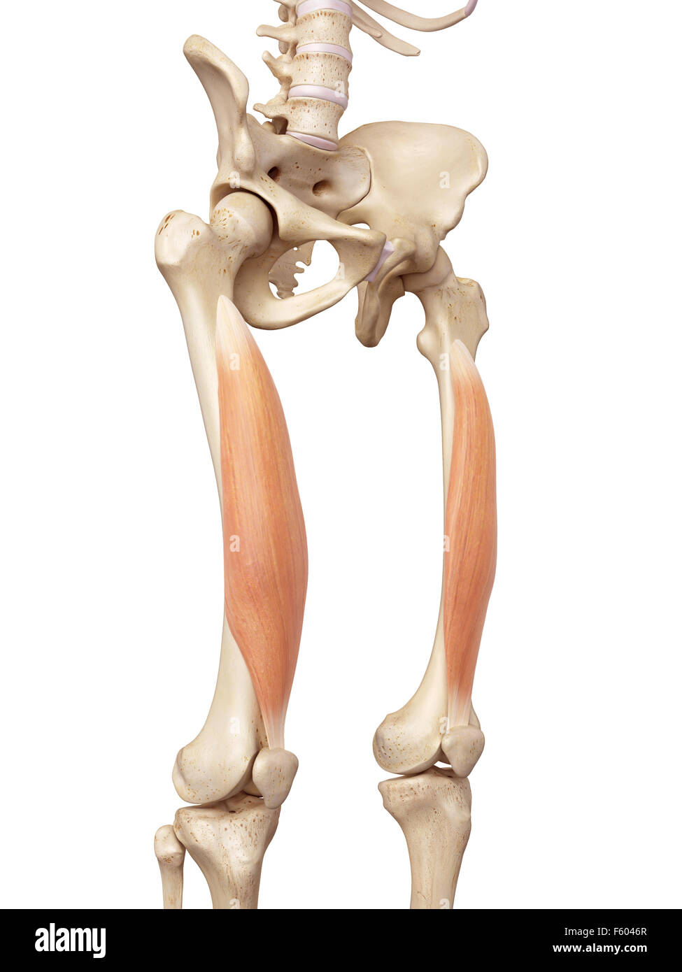 medical accurate illustration of the vastus intermedius Stock Photohttps://www.alamy.com/image-license-details/?v=1https://www.alamy.com/stock-photo-medical-accurate-illustration-of-the-vastus-intermedius-89743103.html
RFF6046R–medical accurate illustration of the vastus intermedius
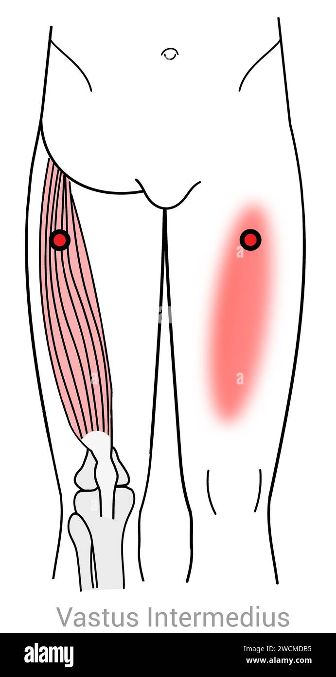 Vastus Intermedius: Myofascial trigger points and associated pain locations Stock Photohttps://www.alamy.com/image-license-details/?v=1https://www.alamy.com/vastus-intermedius-myofascial-trigger-points-and-associated-pain-locations-image592977929.html
RF2WCMDB5–Vastus Intermedius: Myofascial trigger points and associated pain locations
 Vastus intermedius muscle Dog muscle Anatomy For Medical Concept 3D Illustration Stock Photohttps://www.alamy.com/image-license-details/?v=1https://www.alamy.com/vastus-intermedius-muscle-dog-muscle-anatomy-for-medical-concept-3d-illustration-image439390859.html
RF2GERYEK–Vastus intermedius muscle Dog muscle Anatomy For Medical Concept 3D Illustration
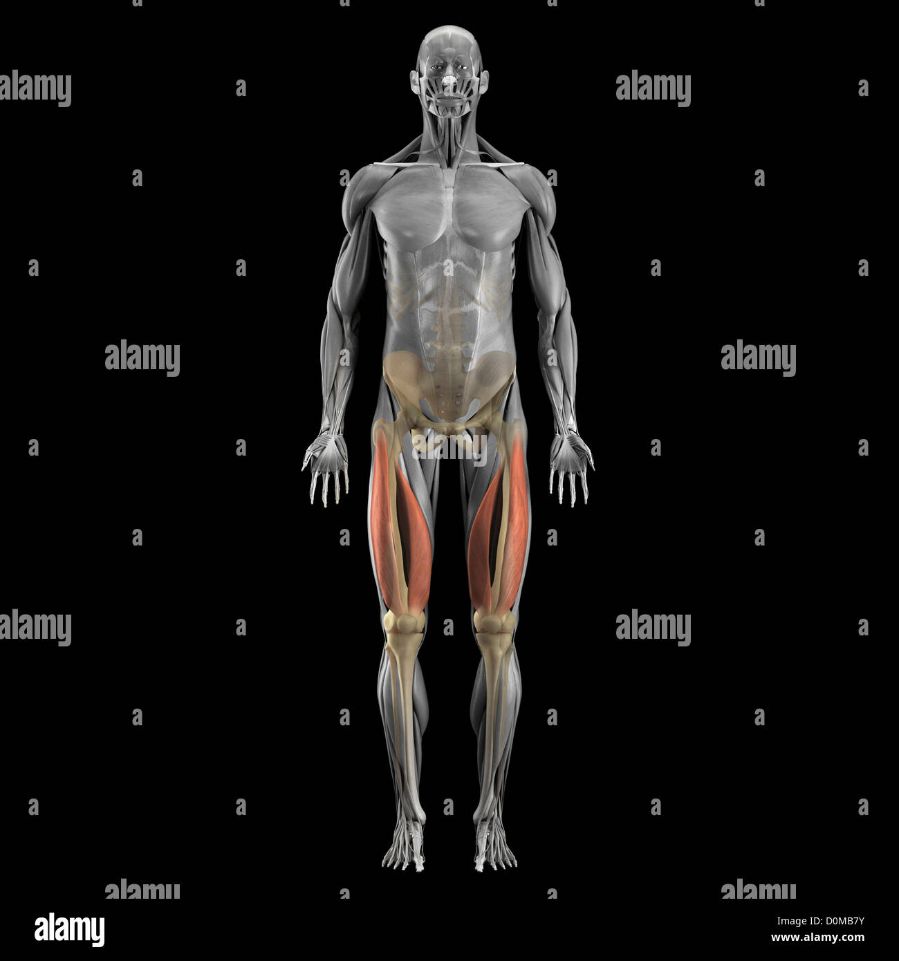 A human model showing the vastus muscles. Stock Photohttps://www.alamy.com/image-license-details/?v=1https://www.alamy.com/stock-photo-a-human-model-showing-the-vastus-muscles-52078991.html
RMD0MB7Y–A human model showing the vastus muscles.
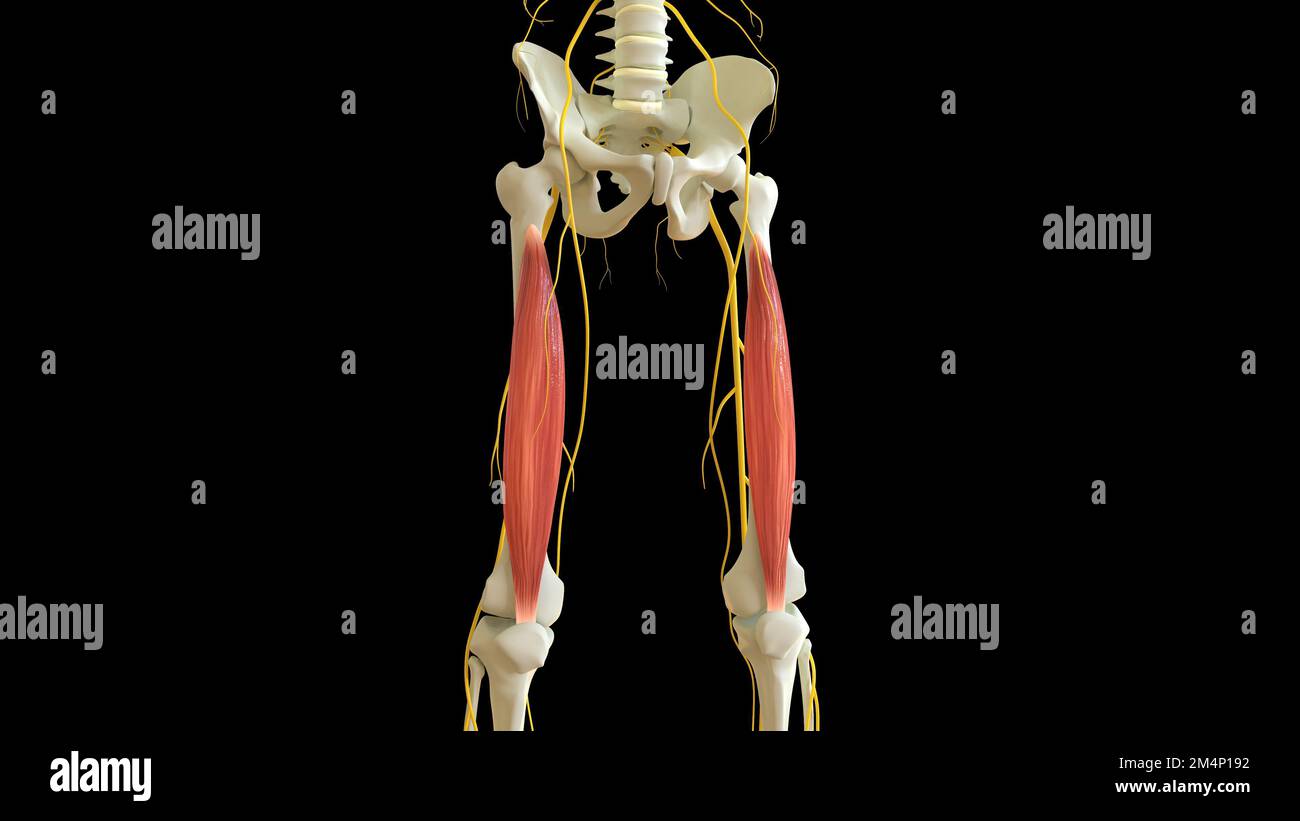 Vastus Intermedius Muscle anatomy for medical concept 3D illustration Stock Photohttps://www.alamy.com/image-license-details/?v=1https://www.alamy.com/vastus-intermedius-muscle-anatomy-for-medical-concept-3d-illustration-image502043278.html
RF2M4P192–Vastus Intermedius Muscle anatomy for medical concept 3D illustration
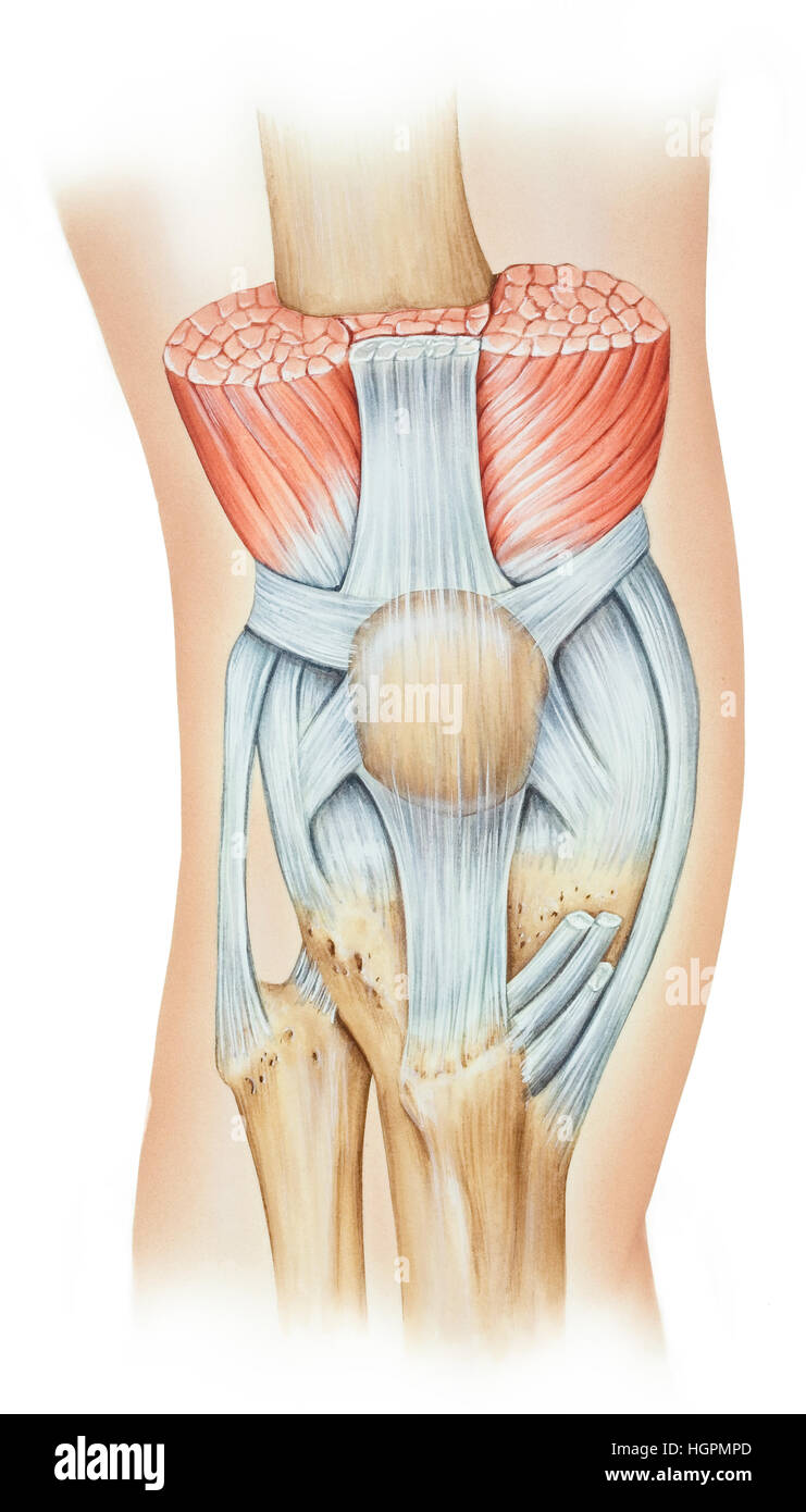 The knee extensor mechanism, which consists of the quadriceps muscle group (rectus femoris, vastus intermedius, and vastus medialis), patella, and pat Stock Photohttps://www.alamy.com/image-license-details/?v=1https://www.alamy.com/stock-photo-the-knee-extensor-mechanism-which-consists-of-the-quadriceps-muscle-130806325.html
RFHGPMPD–The knee extensor mechanism, which consists of the quadriceps muscle group (rectus femoris, vastus intermedius, and vastus medialis), patella, and pat
 Runner warming up stretching quadriceps. Stock Photohttps://www.alamy.com/image-license-details/?v=1https://www.alamy.com/stock-photo-runner-warming-up-stretching-quadriceps-149387881.html
RMJK15ND–Runner warming up stretching quadriceps.
 young sporty girl doing exercise with fitness ball, one person, natural hair, black model, close up, background, african american,lifting weight ball Stock Photohttps://www.alamy.com/image-license-details/?v=1https://www.alamy.com/young-sporty-girl-doing-exercise-with-fitness-ball-one-person-natural-hair-black-model-close-up-background-african-americanlifting-weight-ball-image429096662.html
RF2FX314P–young sporty girl doing exercise with fitness ball, one person, natural hair, black model, close up, background, african american,lifting weight ball
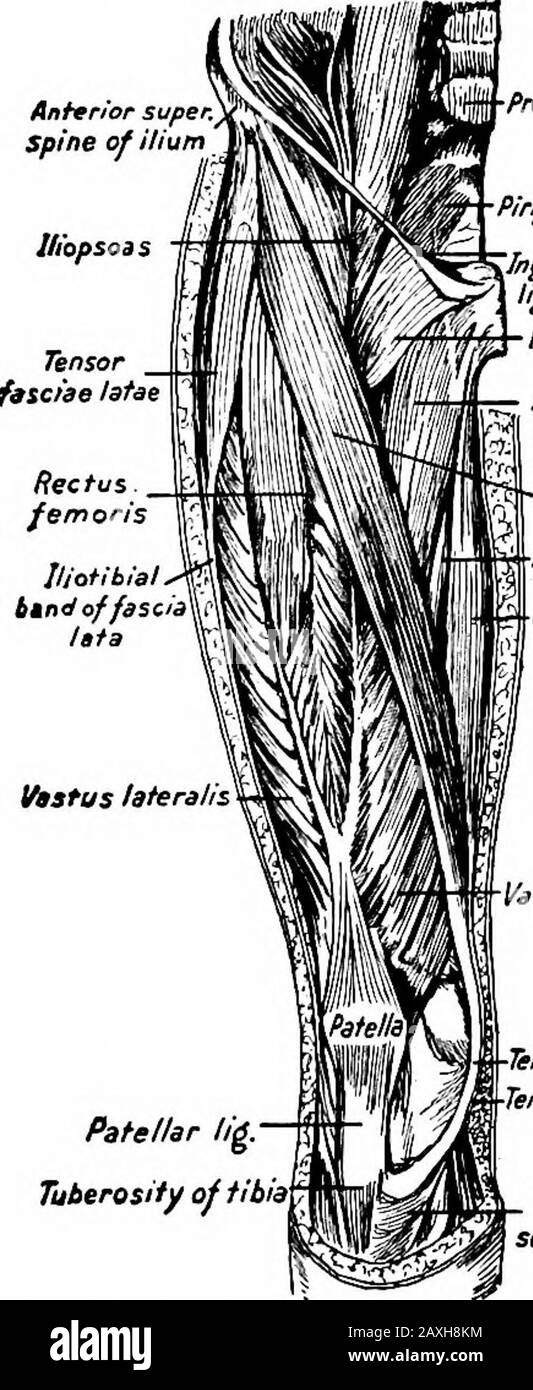 A manual of anatomy . m the tendon of the adductor magnus.It is inserted into the medial margin of the patella and the rectusfemoris tendon and the capsular and lateral Ugaments of the kneejoint. The m. vastus intermedius, or crureus, arises from the ventral andlateral surfaces (proximal two-thirds) of the femur, the intermuscularseptum and from the end of the hnea aspera and hne leading to thelateral condyle. It is inserted into the deep surfaces of the precedingthree tendons. 196 MYOLOGY The m. articularis genu, or subcrureus, arises from the distal por-tion of the ventral surface of the fem Stock Photohttps://www.alamy.com/image-license-details/?v=1https://www.alamy.com/a-manual-of-anatomy-m-the-tendon-of-the-adductor-magnusit-is-inserted-into-the-medial-margin-of-the-patella-and-the-rectusfemoris-tendon-and-the-capsular-and-lateral-ugaments-of-the-kneejoint-the-m-vastus-intermedius-or-crureus-arises-from-the-ventral-andlateral-surfaces-proximal-two-thirds-of-the-femur-the-intermuscularseptum-and-from-the-end-of-the-hnea-aspera-and-hne-leading-to-thelateral-condyle-it-is-inserted-into-the-deep-surfaces-of-the-precedingthree-tendons-196-myology-the-m-articularis-genu-or-subcrureus-arises-from-the-distal-por-tion-of-the-ventral-surface-of-the-fem-image343358056.html
RM2AXH8KM–A manual of anatomy . m the tendon of the adductor magnus.It is inserted into the medial margin of the patella and the rectusfemoris tendon and the capsular and lateral Ugaments of the kneejoint. The m. vastus intermedius, or crureus, arises from the ventral andlateral surfaces (proximal two-thirds) of the femur, the intermuscularseptum and from the end of the hnea aspera and hne leading to thelateral condyle. It is inserted into the deep surfaces of the precedingthree tendons. 196 MYOLOGY The m. articularis genu, or subcrureus, arises from the distal por-tion of the ventral surface of the fem
 . Cunningham's Text-book of anatomy. Anatomy. terior surface of the proximal part^of the Left Femur. Vastus mediai Saphenous nerve^ Femoral vessel: Sartorius Adductor lokgus Adductor magnds Gracilis. Rectus femoris Vastus lateralis Vastus intermedius Femur Femoris (short head) SEMIMEMBRANOSUS' Biceps Femoris (long head) Semitendinostj Sciatic nerve Fig. 362.—Transverse Section of the Thigh (Hunter's Adductor Canal). M. Vastus Lateralis.—The vastus lateralis has an origin, partly fleshy, partly membranous, from (1) the capsule of the hip-joint) (2) the tubercle of the femur, (3) a concave surfa Stock Photohttps://www.alamy.com/image-license-details/?v=1https://www.alamy.com/cunninghams-text-book-of-anatomy-anatomy-terior-surface-of-the-proximal-partof-the-left-femur-vastus-mediai-saphenous-nerve-femoral-vessel-sartorius-adductor-lokgus-adductor-magnds-gracilis-rectus-femoris-vastus-lateralis-vastus-intermedius-femur-femoris-short-head-semimembranosus-biceps-femoris-long-head-semitendinostj-sciatic-nerve-fig-362transverse-section-of-the-thigh-hunters-adductor-canal-m-vastus-lateralisthe-vastus-lateralis-has-an-origin-partly-fleshy-partly-membranous-from-1-the-capsule-of-the-hip-joint-2-the-tubercle-of-the-femur-3-a-concave-surfa-image216340965.html
RMPFY531–. Cunningham's Text-book of anatomy. Anatomy. terior surface of the proximal part^of the Left Femur. Vastus mediai Saphenous nerve^ Femoral vessel: Sartorius Adductor lokgus Adductor magnds Gracilis. Rectus femoris Vastus lateralis Vastus intermedius Femur Femoris (short head) SEMIMEMBRANOSUS' Biceps Femoris (long head) Semitendinostj Sciatic nerve Fig. 362.—Transverse Section of the Thigh (Hunter's Adductor Canal). M. Vastus Lateralis.—The vastus lateralis has an origin, partly fleshy, partly membranous, from (1) the capsule of the hip-joint) (2) the tubercle of the femur, (3) a concave surfa
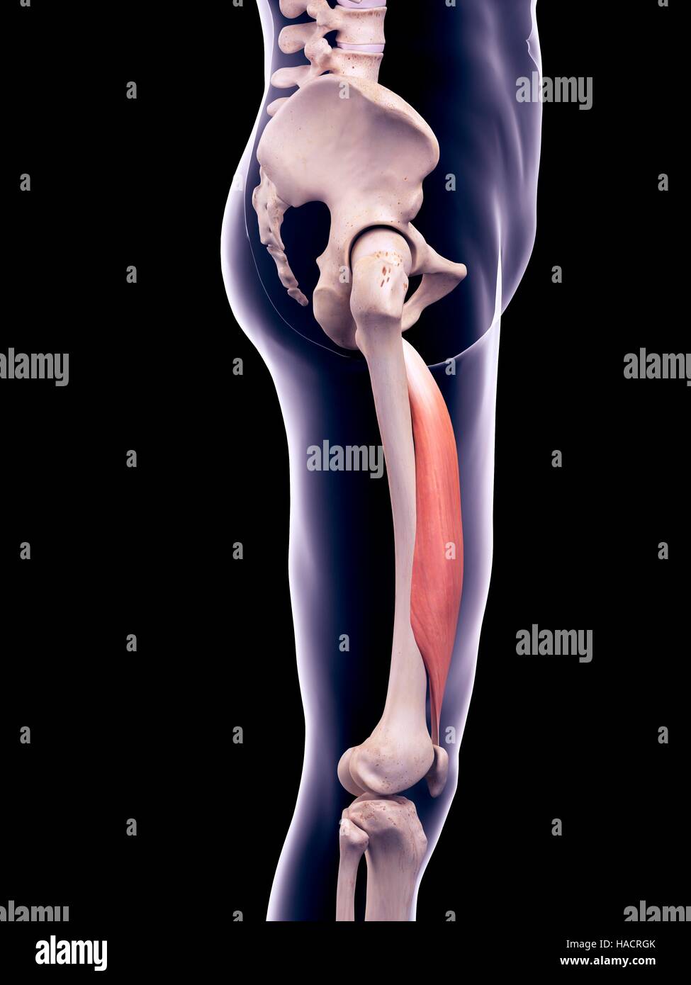 Illustration of the vastus intermedius muscle. Stock Photohttps://www.alamy.com/image-license-details/?v=1https://www.alamy.com/stock-photo-illustration-of-the-vastus-intermedius-muscle-126901059.html
RFHACRGK–Illustration of the vastus intermedius muscle.
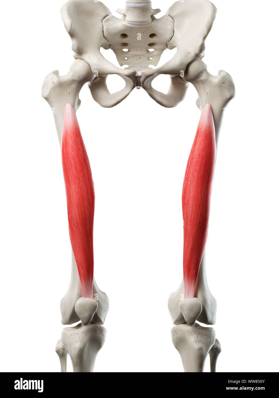 Vastus intermedius muscle, illustration Stock Photohttps://www.alamy.com/image-license-details/?v=1https://www.alamy.com/vastus-intermedius-muscle-illustration-image273701483.html
RFWW850Y–Vastus intermedius muscle, illustration
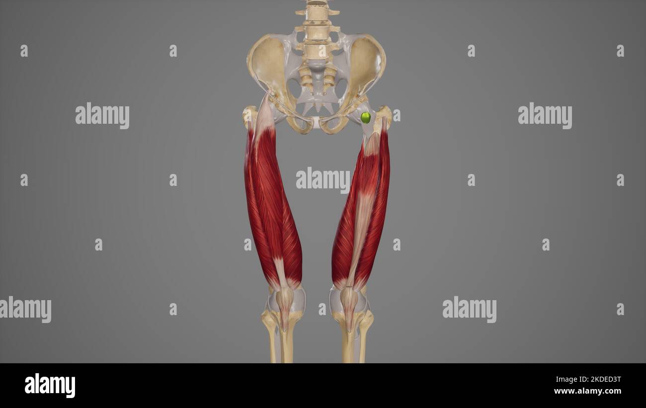 Anterior Quadriceps Femoris Muscles Stock Photohttps://www.alamy.com/image-license-details/?v=1https://www.alamy.com/anterior-quadriceps-femoris-muscles-image490198460.html
RF2KDED3T–Anterior Quadriceps Femoris Muscles
 medical accurate illustration of the vastus intermedius Stock Photohttps://www.alamy.com/image-license-details/?v=1https://www.alamy.com/stock-photo-medical-accurate-illustration-of-the-vastus-intermedius-89743100.html
RFF6046M–medical accurate illustration of the vastus intermedius
 medically accurate illustration of the intermedius Stock Photohttps://www.alamy.com/image-license-details/?v=1https://www.alamy.com/stock-photo-medically-accurate-illustration-of-the-intermedius-89761053.html
RFF60Y3W–medically accurate illustration of the intermedius
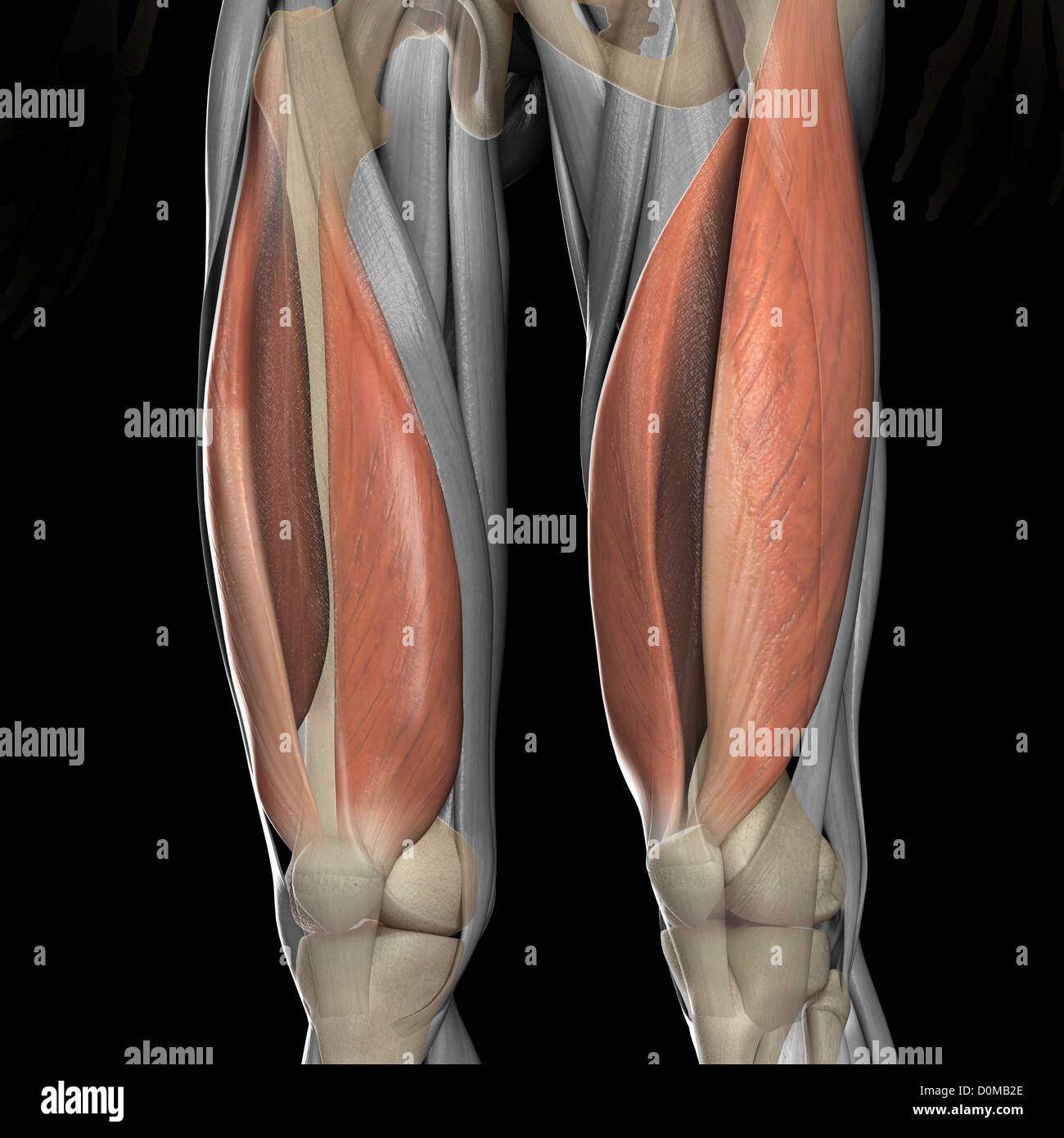 A human model showing the vastus medialis and lateralis muscles. Stock Photohttps://www.alamy.com/image-license-details/?v=1https://www.alamy.com/stock-photo-a-human-model-showing-the-vastus-medialis-and-lateralis-muscles-52078838.html
RMD0MB2E–A human model showing the vastus medialis and lateralis muscles.
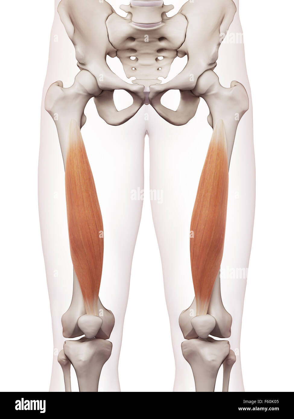 medically accurate muscle illustration of the vastus intermedius Stock Photohttps://www.alamy.com/image-license-details/?v=1https://www.alamy.com/stock-photo-medically-accurate-muscle-illustration-of-the-vastus-intermedius-89754677.html
RFF60K05–medically accurate muscle illustration of the vastus intermedius
 Vastus Lateralis Muscle anatomy for medical concept 3D illustration Stock Photohttps://www.alamy.com/image-license-details/?v=1https://www.alamy.com/vastus-lateralis-muscle-anatomy-for-medical-concept-3d-illustration-image502043263.html
RF2M4P18F–Vastus Lateralis Muscle anatomy for medical concept 3D illustration
 Young athletic woman doing exercise with fitness ball in the gym, one person, african american, close up, black model, portrait of sporty black woman, Stock Photohttps://www.alamy.com/image-license-details/?v=1https://www.alamy.com/young-athletic-woman-doing-exercise-with-fitness-ball-in-the-gym-one-person-african-american-close-up-black-model-portrait-of-sporty-black-woman-image429096726.html
RF2FX3172–Young athletic woman doing exercise with fitness ball in the gym, one person, african american, close up, black model, portrait of sporty black woman,
 Applied anatomy and kinesiology, the mechanism of muscular movement . he exter-nus. The tendon of origin is a flatsheet arising from the linea asperaand the tendon of insertion is thesame sheet to which the othersjoin. Action.—The line of pull is justlike that of the externus except thatit is directed diagonally inward in-stead of outward. Isolated actioncauses inward displacement of thepatella and paralysis makes thesubject liable to outward displace-ment. VASTUS INTERMEDIUS. A companion of the two preced-ing, lying between them and beneaththe rectus femoris. Origin.—The surface of the uppert Stock Photohttps://www.alamy.com/image-license-details/?v=1https://www.alamy.com/applied-anatomy-and-kinesiology-the-mechanism-of-muscular-movement-he-exter-nus-the-tendon-of-origin-is-a-flatsheet-arising-from-the-linea-asperaand-the-tendon-of-insertion-is-thesame-sheet-to-which-the-othersjoin-actionthe-line-of-pull-is-justlike-that-of-the-externus-except-thatit-is-directed-diagonally-inward-in-stead-of-outward-isolated-actioncauses-inward-displacement-of-thepatella-and-paralysis-makes-thesubject-liable-to-outward-displace-ment-vastus-intermedius-a-companion-of-the-two-preced-ing-lying-between-them-and-beneaththe-rectus-femoris-originthe-surface-of-the-uppert-image342684367.html
RM2AWEHBB–Applied anatomy and kinesiology, the mechanism of muscular movement . he exter-nus. The tendon of origin is a flatsheet arising from the linea asperaand the tendon of insertion is thesame sheet to which the othersjoin. Action.—The line of pull is justlike that of the externus except thatit is directed diagonally inward in-stead of outward. Isolated actioncauses inward displacement of thepatella and paralysis makes thesubject liable to outward displace-ment. VASTUS INTERMEDIUS. A companion of the two preced-ing, lying between them and beneaththe rectus femoris. Origin.—The surface of the uppert
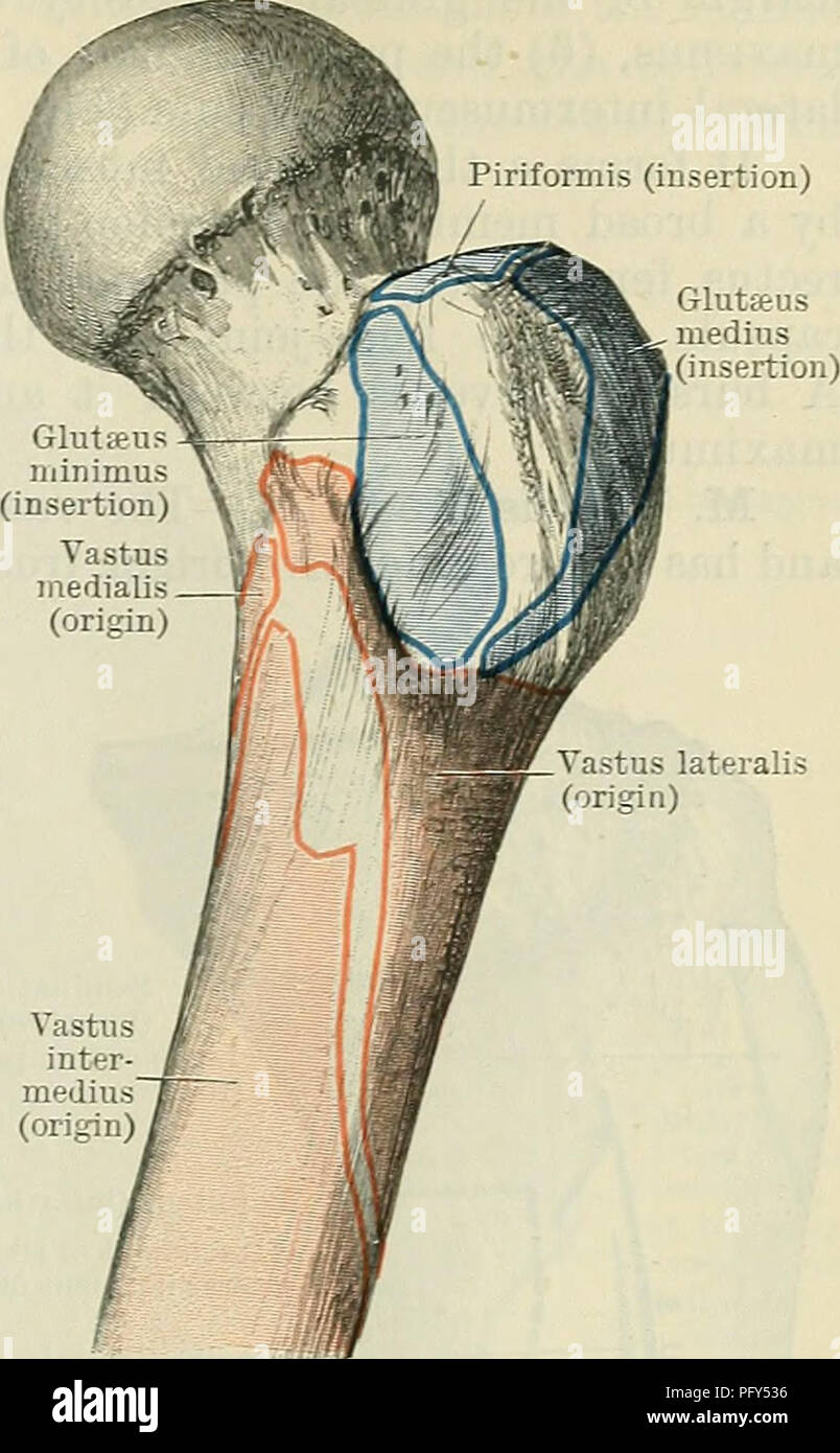 . Cunningham's Text-book of anatomy. Anatomy. formis (insertion) MUSCLES ON THE ANTEEIOK ASPECT OF THE THIGH. 407 muscle. The vastus intermedius envelops the femur, and is concealed by the other muscles. M. Rectus Femoris. — The rectus femoris has a double tendinous origin. (1) The straight head arises from the inferior anterior spine of the ilium (Fig. 366, p. 412): (2) the reflected head springs from a rough groove on the dorsum ilii just above the highest part of the acetabulum (Eig. 366, p. 412). A bursa lies beneath this head of origin. The two heads, bound together and connected to the c Stock Photohttps://www.alamy.com/image-license-details/?v=1https://www.alamy.com/cunninghams-text-book-of-anatomy-anatomy-formis-insertion-muscles-on-the-anteeiok-aspect-of-the-thigh-407-muscle-the-vastus-intermedius-envelops-the-femur-and-is-concealed-by-the-other-muscles-m-rectus-femoris-the-rectus-femoris-has-a-double-tendinous-origin-1-the-straight-head-arises-from-the-inferior-anterior-spine-of-the-ilium-fig-366-p-412-2-the-reflected-head-springs-from-a-rough-groove-on-the-dorsum-ilii-just-above-the-highest-part-of-the-acetabulum-eig-366-p-412-a-bursa-lies-beneath-this-head-of-origin-the-two-heads-bound-together-and-connected-to-the-c-image216340970.html
RMPFY536–. Cunningham's Text-book of anatomy. Anatomy. formis (insertion) MUSCLES ON THE ANTEEIOK ASPECT OF THE THIGH. 407 muscle. The vastus intermedius envelops the femur, and is concealed by the other muscles. M. Rectus Femoris. — The rectus femoris has a double tendinous origin. (1) The straight head arises from the inferior anterior spine of the ilium (Fig. 366, p. 412): (2) the reflected head springs from a rough groove on the dorsum ilii just above the highest part of the acetabulum (Eig. 366, p. 412). A bursa lies beneath this head of origin. The two heads, bound together and connected to the c
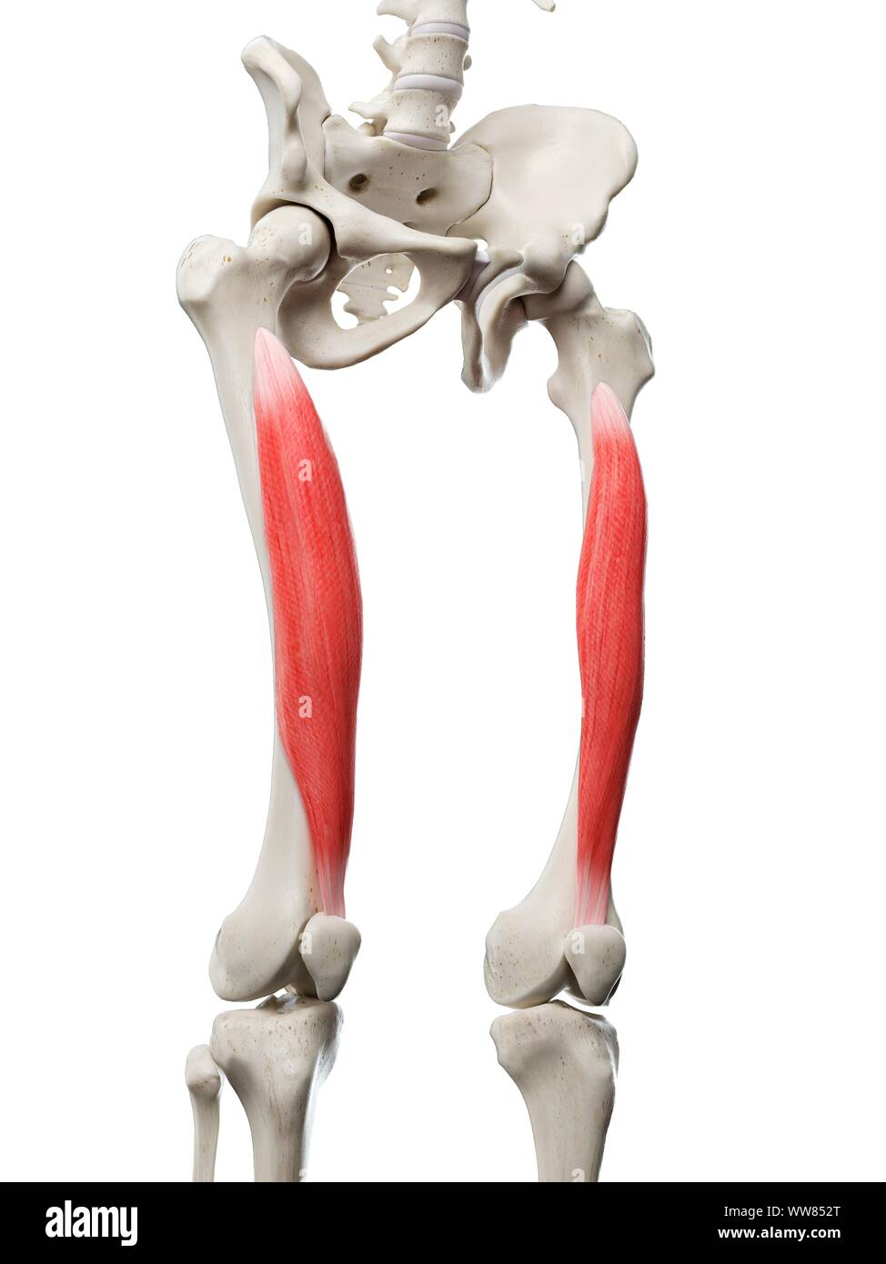 Vastus intermedius muscle, illustration Stock Photohttps://www.alamy.com/image-license-details/?v=1https://www.alamy.com/vastus-intermedius-muscle-illustration-image273701536.html
RFWW852T–Vastus intermedius muscle, illustration
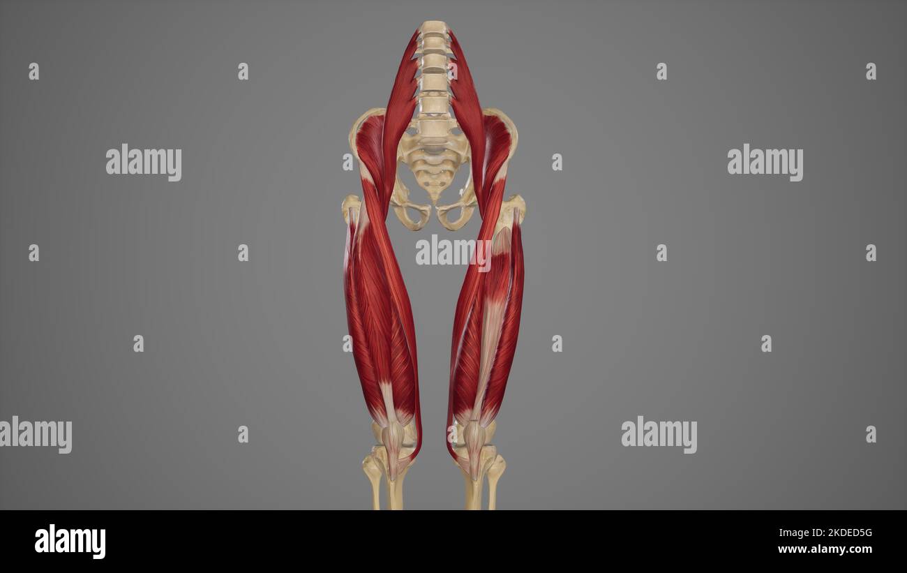 Anterior View of Anterior Thigh Muscles Stock Photohttps://www.alamy.com/image-license-details/?v=1https://www.alamy.com/anterior-view-of-anterior-thigh-muscles-image490198508.html
RF2KDED5G–Anterior View of Anterior Thigh Muscles
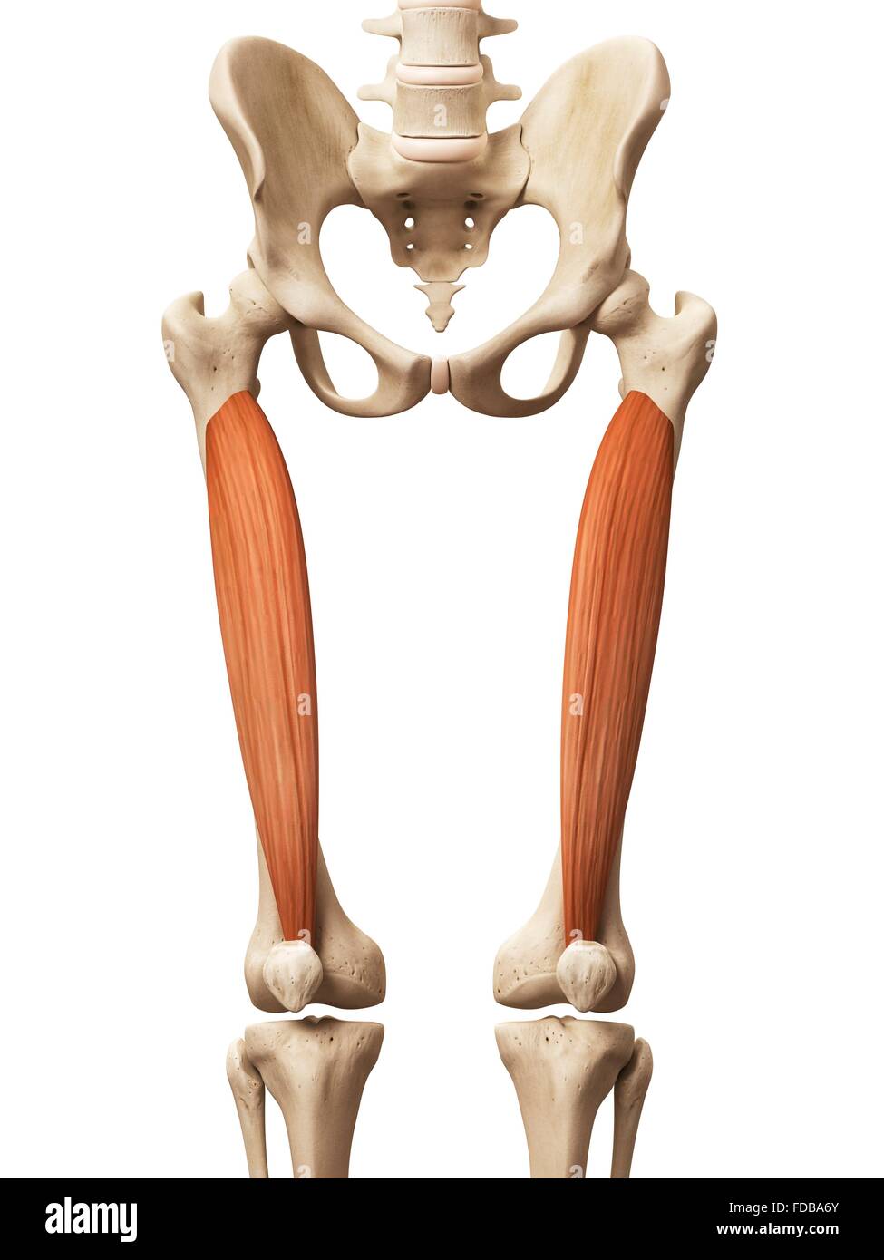 Human leg muscles (vastus intermedius), illustration. Stock Photohttps://www.alamy.com/image-license-details/?v=1https://www.alamy.com/stock-photo-human-leg-muscles-vastus-intermedius-illustration-94291875.html
RFFDBA6Y–Human leg muscles (vastus intermedius), illustration.
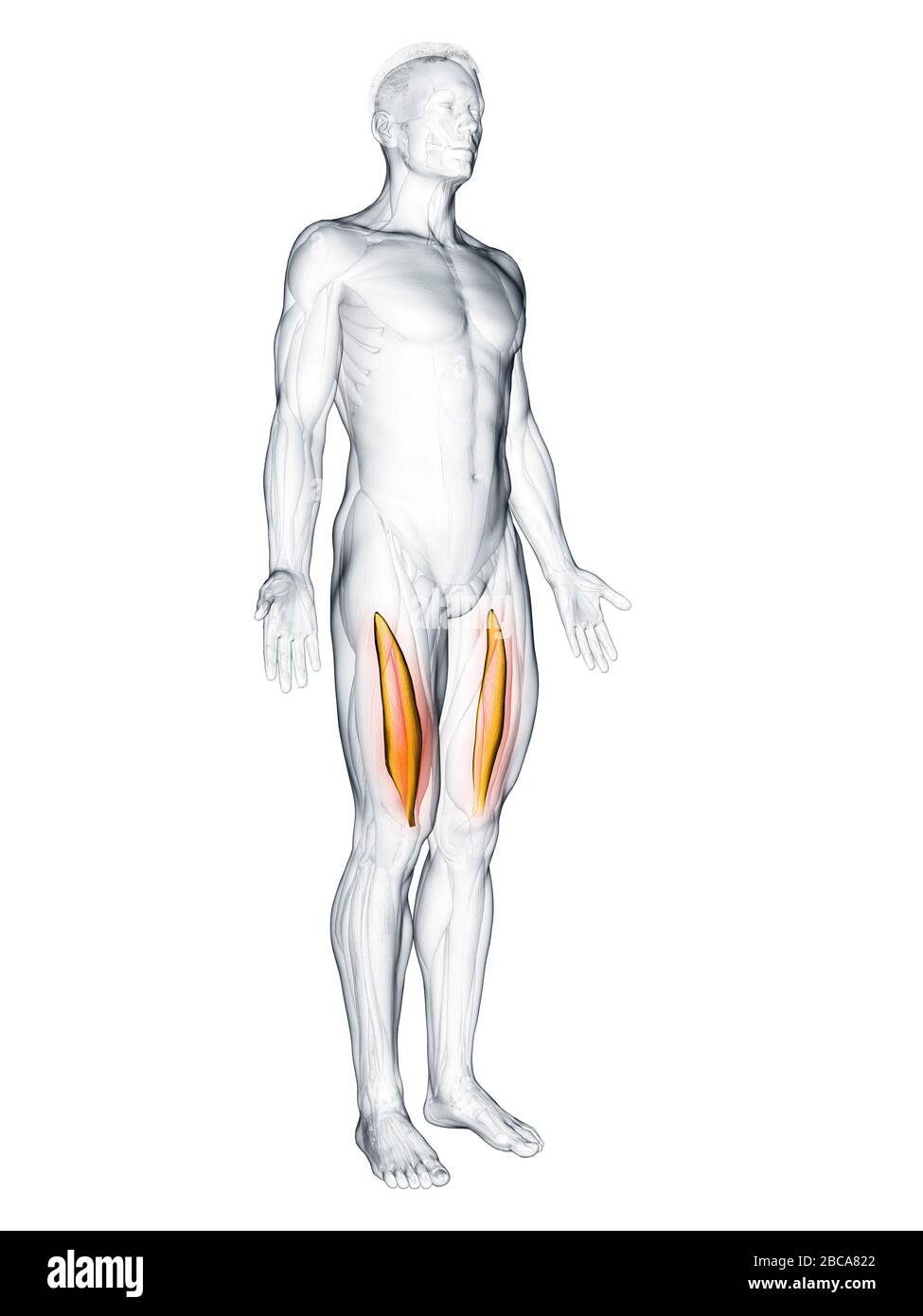 Vastus intermedius muscle, illustration. Stock Photohttps://www.alamy.com/image-license-details/?v=1https://www.alamy.com/vastus-intermedius-muscle-illustration-image351809082.html
RF2BCA822–Vastus intermedius muscle, illustration.
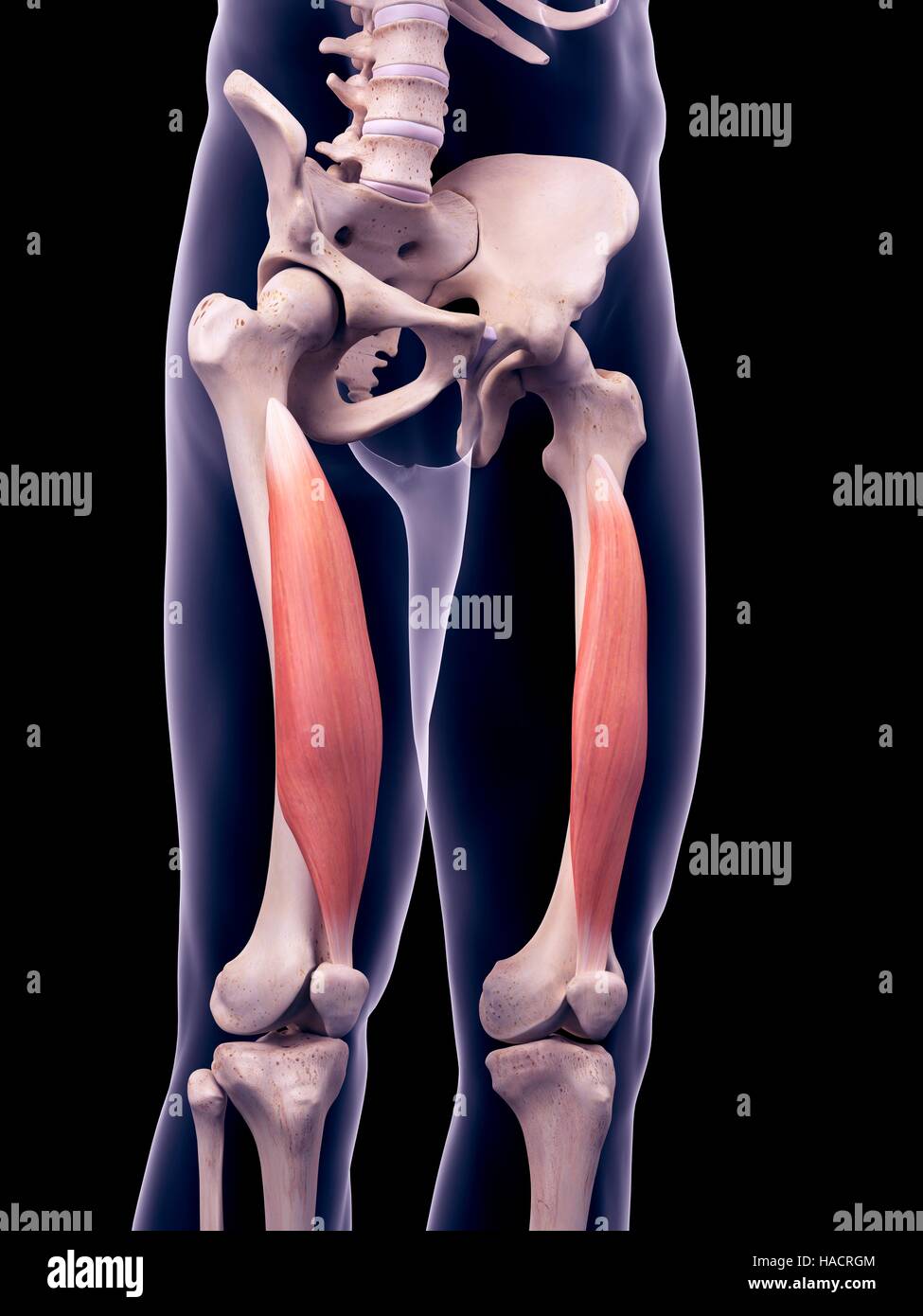 Illustration of the vastus intermedius muscles. Stock Photohttps://www.alamy.com/image-license-details/?v=1https://www.alamy.com/stock-photo-illustration-of-the-vastus-intermedius-muscles-126901060.html
RFHACRGM–Illustration of the vastus intermedius muscles.
 medically accurate muscle illustration of the vastus intermedius Stock Photohttps://www.alamy.com/image-license-details/?v=1https://www.alamy.com/stock-photo-medically-accurate-muscle-illustration-of-the-vastus-intermedius-89754679.html
RFF60K07–medically accurate muscle illustration of the vastus intermedius
 A human model showing the vastus medialis and lateralis muscles. Stock Photohttps://www.alamy.com/image-license-details/?v=1https://www.alamy.com/stock-photo-a-human-model-showing-the-vastus-medialis-and-lateralis-muscles-52078753.html
RMD0MAYD–A human model showing the vastus medialis and lateralis muscles.
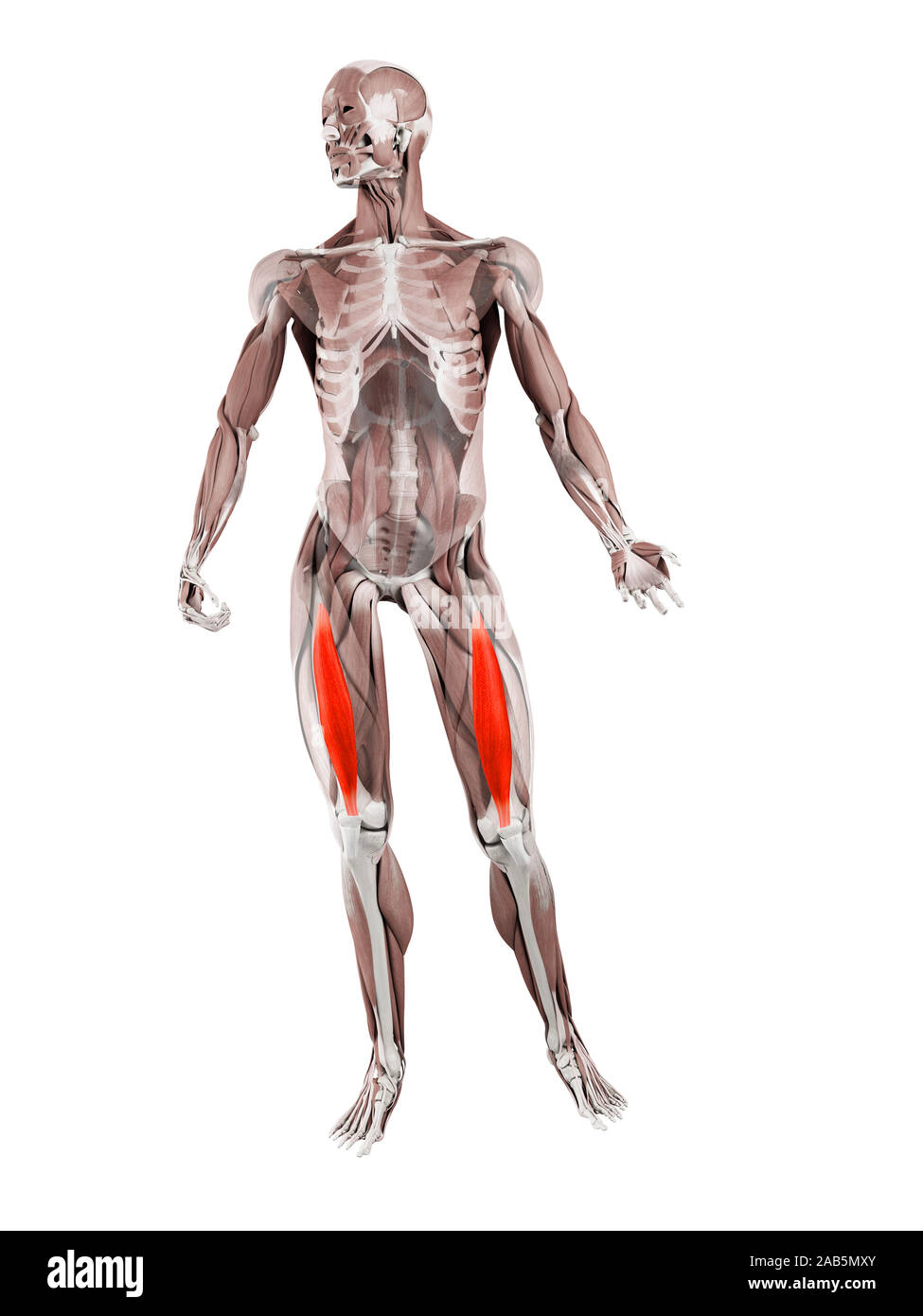 3d rendered muscle illustration of the vastus intermedius Stock Photohttps://www.alamy.com/image-license-details/?v=1https://www.alamy.com/3d-rendered-muscle-illustration-of-the-vastus-intermedius-image333884403.html
RF2AB5MXY–3d rendered muscle illustration of the vastus intermedius
 Slim sporty girl exercising with fitness ball in fitness gym. one person, close up portrait, african american, natural hair model, black model, liftin Stock Photohttps://www.alamy.com/image-license-details/?v=1https://www.alamy.com/slim-sporty-girl-exercising-with-fitness-ball-in-fitness-gym-one-person-close-up-portrait-african-american-natural-hair-model-black-model-liftin-image429096589.html
RF2FX3125–Slim sporty girl exercising with fitness ball in fitness gym. one person, close up portrait, african american, natural hair model, black model, liftin
 3d rendered muscle illustration of the vastus intermedius Stock Photohttps://www.alamy.com/image-license-details/?v=1https://www.alamy.com/3d-rendered-muscle-illustration-of-the-vastus-intermedius-image333875584.html
RF2AB59M0–3d rendered muscle illustration of the vastus intermedius
 Vastus Medialis Muscle anatomy for medical concept 3D illustration Stock Photohttps://www.alamy.com/image-license-details/?v=1https://www.alamy.com/vastus-medialis-muscle-anatomy-for-medical-concept-3d-illustration-image502043272.html
RF2M4P18T–Vastus Medialis Muscle anatomy for medical concept 3D illustration
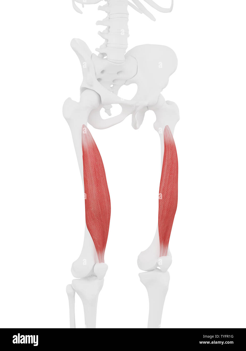 3d rendered medically accurate illustration of the Vastus Intermedius Stock Photohttps://www.alamy.com/image-license-details/?v=1https://www.alamy.com/3d-rendered-medically-accurate-illustration-of-the-vastus-intermedius-image257888220.html
RFTYFR1G–3d rendered medically accurate illustration of the Vastus Intermedius
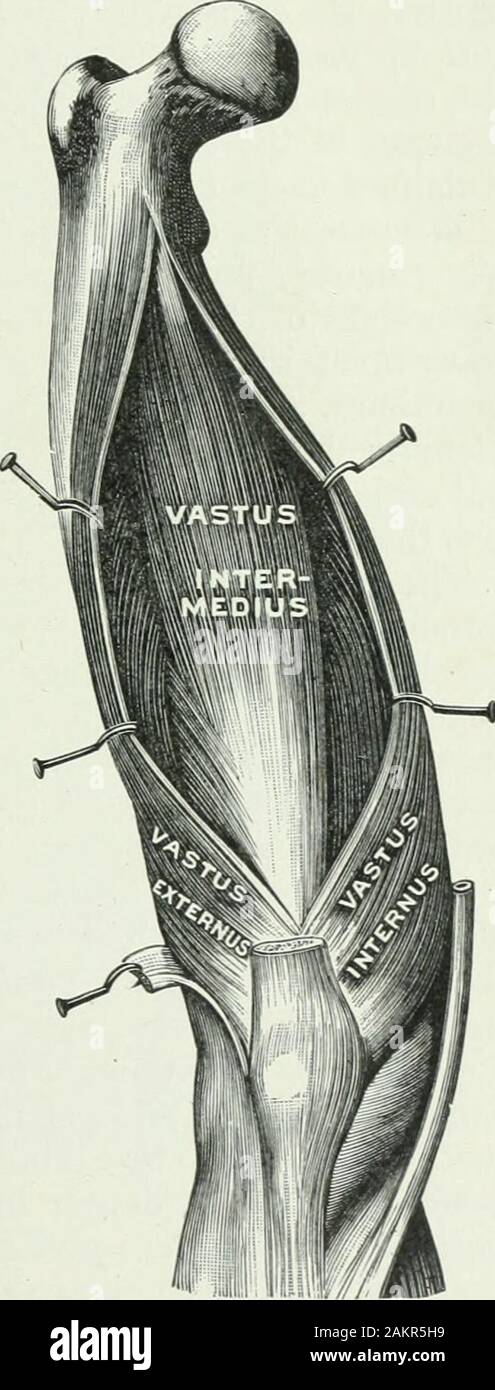 Applied anatomy and kinesiology; the mechanism of muscular movement . der of the patella. Structure.—Similar to the exter-nus. The tendon of origin is a flatsheet arising from the linea asperaand the tendon of insertion is thesame sheet to which the othersjoin. Action.—^The line of pull is justlike that of the externus except thatit is directed diagonally inward in-stead of outward. Isolated actioncauses inward displacement of thepatella and paralysis makes thesubject liable to outward displace-ment. VASTUS INTERMEDIUS. A companion of the two preced-ing, lying between them and beneaththe rectu Stock Photohttps://www.alamy.com/image-license-details/?v=1https://www.alamy.com/applied-anatomy-and-kinesiology-the-mechanism-of-muscular-movement-der-of-the-patella-structuresimilar-to-the-exter-nus-the-tendon-of-origin-is-a-flatsheet-arising-from-the-linea-asperaand-the-tendon-of-insertion-is-thesame-sheet-to-which-the-othersjoin-actionthe-line-of-pull-is-justlike-that-of-the-externus-except-thatit-is-directed-diagonally-inward-in-stead-of-outward-isolated-actioncauses-inward-displacement-of-thepatella-and-paralysis-makes-thesubject-liable-to-outward-displace-ment-vastus-intermedius-a-companion-of-the-two-preced-ing-lying-between-them-and-beneaththe-rectu-image339184757.html
RM2AKR5H9–Applied anatomy and kinesiology; the mechanism of muscular movement . der of the patella. Structure.—Similar to the exter-nus. The tendon of origin is a flatsheet arising from the linea asperaand the tendon of insertion is thesame sheet to which the othersjoin. Action.—^The line of pull is justlike that of the externus except thatit is directed diagonally inward in-stead of outward. Isolated actioncauses inward displacement of thepatella and paralysis makes thesubject liable to outward displace-ment. VASTUS INTERMEDIUS. A companion of the two preced-ing, lying between them and beneaththe rectu
 3D Illustration, Muscle is a soft tissue, Muscle cells contain proteins , producing a contraction that changes both the length and the shape of the ce Stock Photohttps://www.alamy.com/image-license-details/?v=1https://www.alamy.com/3d-illustration-muscle-is-a-soft-tissue-muscle-cells-contain-proteins-producing-a-contraction-that-changes-both-the-length-and-the-shape-of-the-ce-image395465915.html
RF2DYB0PK–3D Illustration, Muscle is a soft tissue, Muscle cells contain proteins , producing a contraction that changes both the length and the shape of the ce
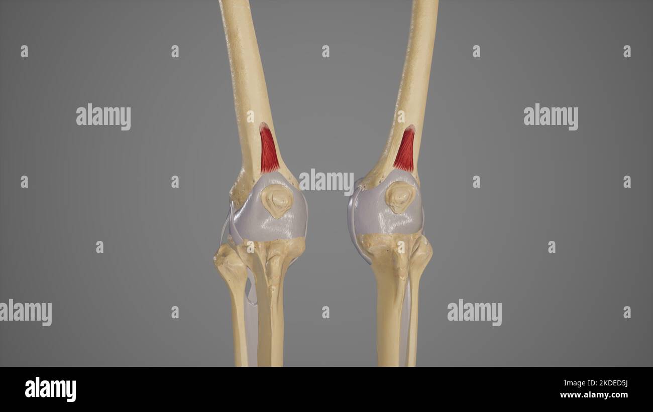 Medical Illustration of Articularis Genus Muscle Stock Photohttps://www.alamy.com/image-license-details/?v=1https://www.alamy.com/medical-illustration-of-articularis-genus-muscle-image490198510.html
RF2KDED5J–Medical Illustration of Articularis Genus Muscle
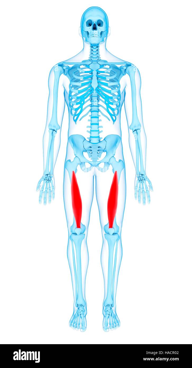 Illustration of the vastus intermedius muscles. Stock Photohttps://www.alamy.com/image-license-details/?v=1https://www.alamy.com/stock-photo-illustration-of-the-vastus-intermedius-muscles-126900594.html
RFHACR02–Illustration of the vastus intermedius muscles.
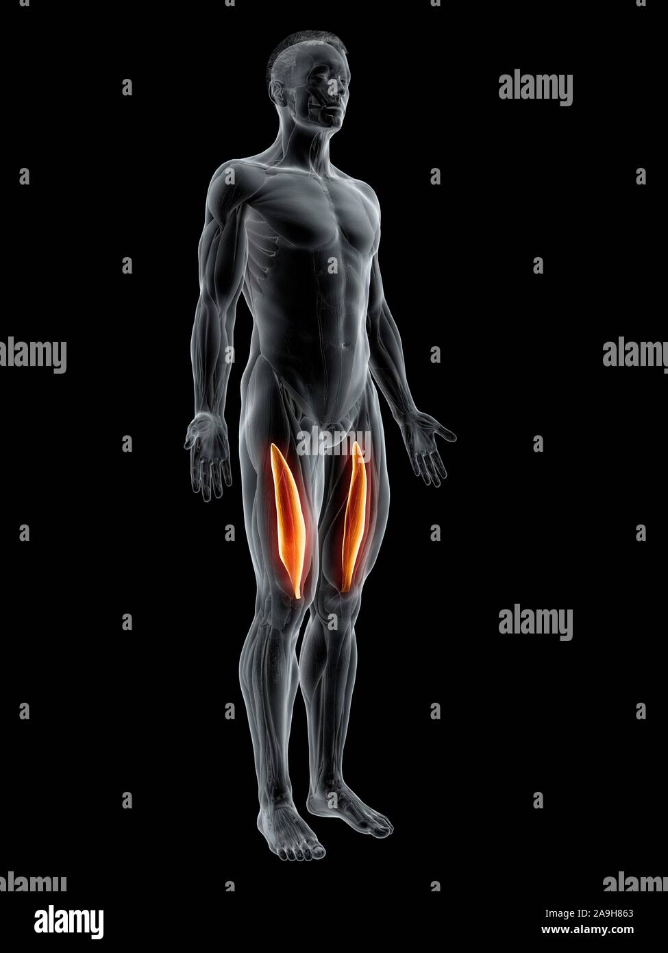 Vastus intermedius muscle, illustration Stock Photohttps://www.alamy.com/image-license-details/?v=1https://www.alamy.com/vastus-intermedius-muscle-illustration-image332908523.html
RF2A9H863–Vastus intermedius muscle, illustration
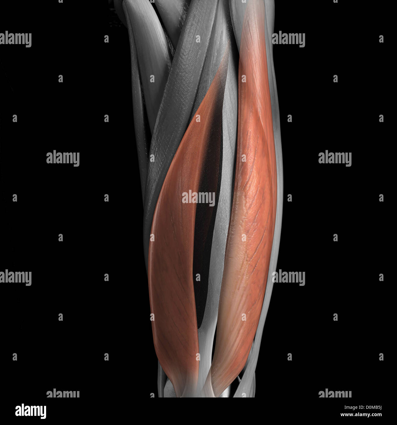 A human model showing the vastus medialis and lateralis muscles. Stock Photohttps://www.alamy.com/image-license-details/?v=1https://www.alamy.com/stock-photo-a-human-model-showing-the-vastus-medialis-and-lateralis-muscles-52078926.html
RMD0MB5J–A human model showing the vastus medialis and lateralis muscles.
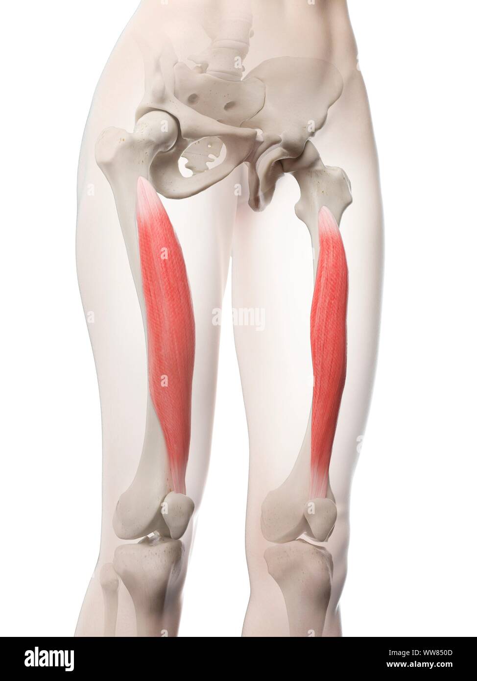 Vastus intermedius muscle, illustration Stock Photohttps://www.alamy.com/image-license-details/?v=1https://www.alamy.com/vastus-intermedius-muscle-illustration-image273701469.html
RFWW850D–Vastus intermedius muscle, illustration
 Fit sporty girl exercising with fitness ball, lifting, one person, close up, black woman, african american, background, fitness center Stock Photohttps://www.alamy.com/image-license-details/?v=1https://www.alamy.com/fit-sporty-girl-exercising-with-fitness-ball-lifting-one-person-close-up-black-woman-african-american-background-fitness-center-image429096605.html
RF2FX312N–Fit sporty girl exercising with fitness ball, lifting, one person, close up, black woman, african american, background, fitness center
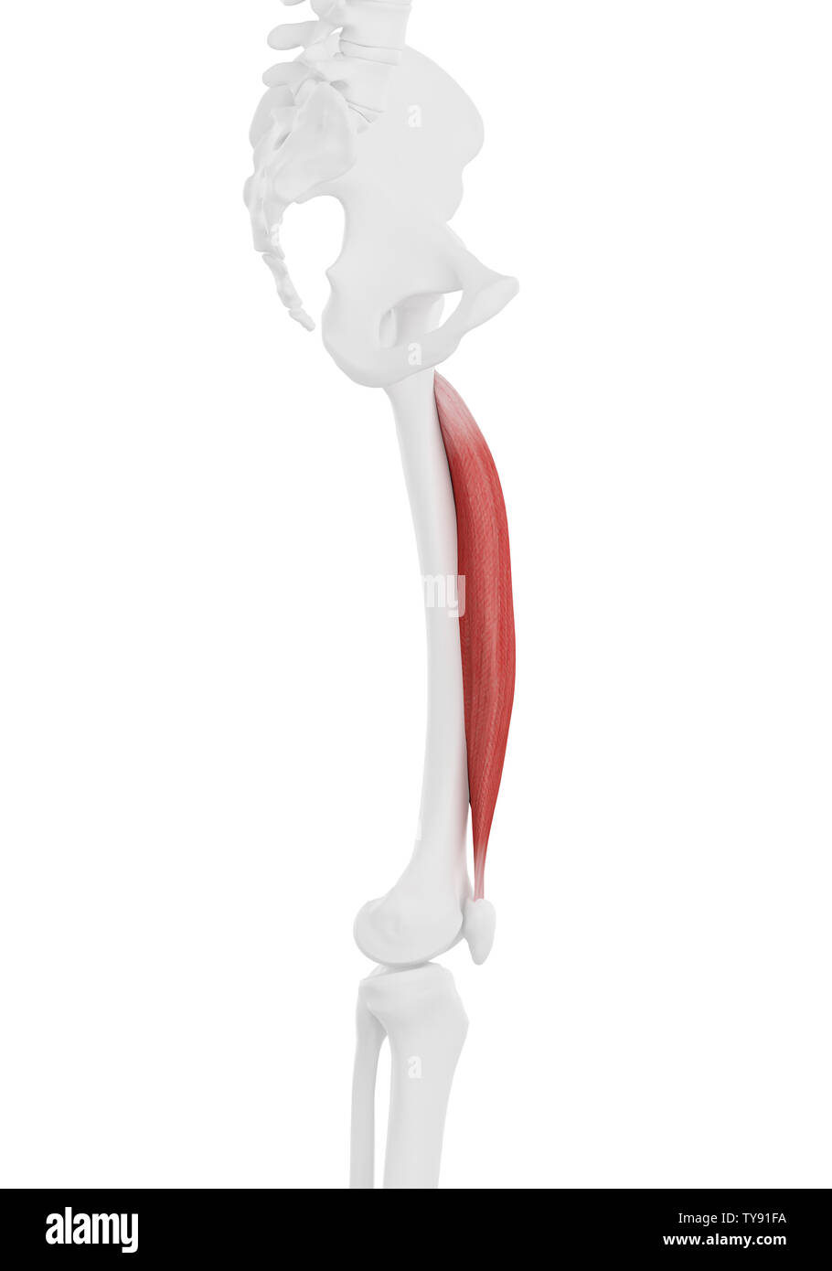 3d rendered medically accurate illustration of the Vastus Intermedius Stock Photohttps://www.alamy.com/image-license-details/?v=1https://www.alamy.com/3d-rendered-medically-accurate-illustration-of-the-vastus-intermedius-image257739646.html
RFTY91FA–3d rendered medically accurate illustration of the Vastus Intermedius
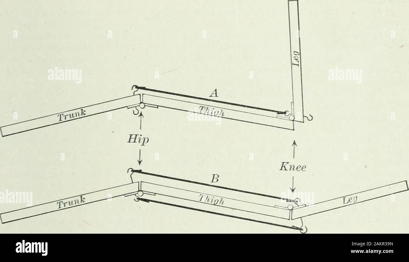 Applied anatomy and kinesiology; the mechanism of muscular movement . and leave the other cord in place, itwill extend both joints as before. We are thus confronted by theproblem, How can the hamstring-muscles, which are flexors of theknee, cause extension of the knee? How can a cord tied across two VASTUS INTERMEDIUS 185 joints give to a muscle that is primarily a flexor of a joint theability to extend it? Dr. Lombard has explained this apparent contradiction by show-ing that the two-joint muscles of the thigh have better leverage asextensors than as flexors. The hamstring muscles have better Stock Photohttps://www.alamy.com/image-license-details/?v=1https://www.alamy.com/applied-anatomy-and-kinesiology-the-mechanism-of-muscular-movement-and-leave-the-other-cord-in-place-itwill-extend-both-joints-as-before-we-are-thus-confronted-by-theproblem-how-can-the-hamstring-muscles-which-are-flexors-of-theknee-cause-extension-of-the-knee-how-can-a-cord-tied-across-two-vastus-intermedius-185-joints-give-to-a-muscle-that-is-primarily-a-flexor-of-a-joint-theability-to-extend-it-dr-lombard-has-explained-this-apparent-contradiction-by-show-ing-that-the-two-joint-muscles-of-the-thigh-have-better-leverage-asextensors-than-as-flexors-the-hamstring-muscles-have-better-image339182977.html
RM2AKR39N–Applied anatomy and kinesiology; the mechanism of muscular movement . and leave the other cord in place, itwill extend both joints as before. We are thus confronted by theproblem, How can the hamstring-muscles, which are flexors of theknee, cause extension of the knee? How can a cord tied across two VASTUS INTERMEDIUS 185 joints give to a muscle that is primarily a flexor of a joint theability to extend it? Dr. Lombard has explained this apparent contradiction by show-ing that the two-joint muscles of the thigh have better leverage asextensors than as flexors. The hamstring muscles have better
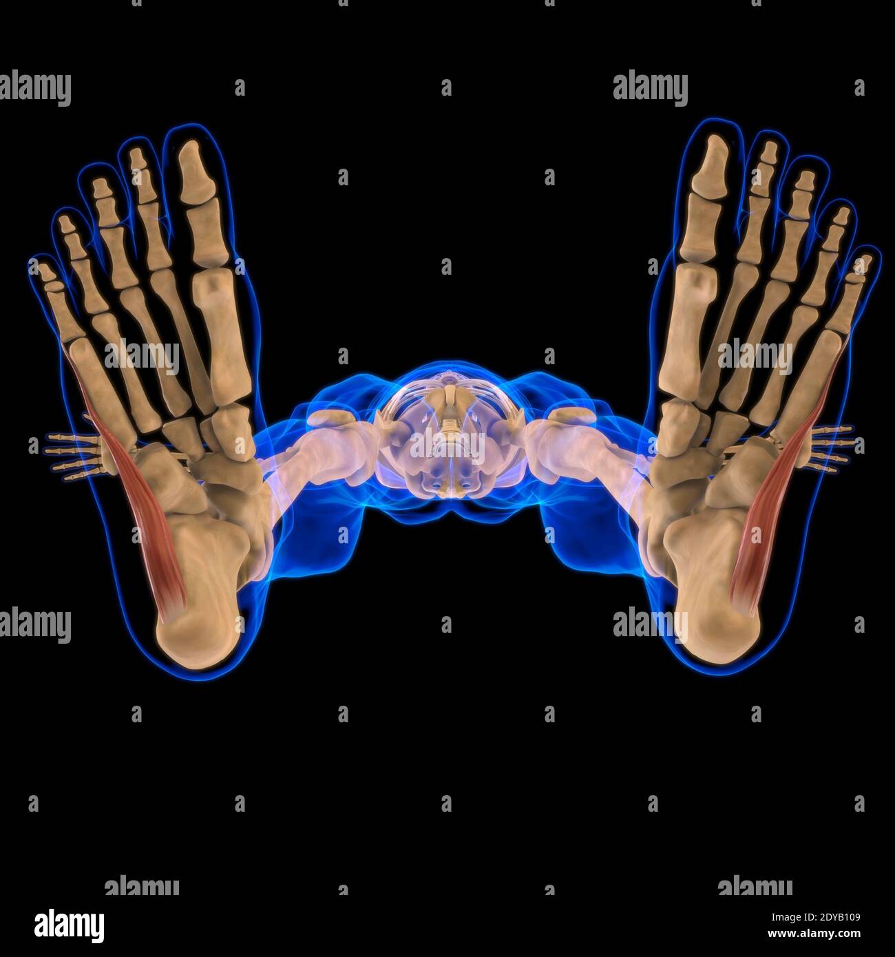 3D Illustration, Muscle is a soft tissue, Muscle cells contain proteins , producing a contraction that changes both the length and the shape of the ce Stock Photohttps://www.alamy.com/image-license-details/?v=1https://www.alamy.com/3d-illustration-muscle-is-a-soft-tissue-muscle-cells-contain-proteins-producing-a-contraction-that-changes-both-the-length-and-the-shape-of-the-ce-image395466073.html
RF2DYB109–3D Illustration, Muscle is a soft tissue, Muscle cells contain proteins , producing a contraction that changes both the length and the shape of the ce
 Pectineus Muscle anatomy for medical concept 3D illustration Stock Photohttps://www.alamy.com/image-license-details/?v=1https://www.alamy.com/pectineus-muscle-anatomy-for-medical-concept-3d-illustration-image502043386.html
RF2M4P1CX–Pectineus Muscle anatomy for medical concept 3D illustration
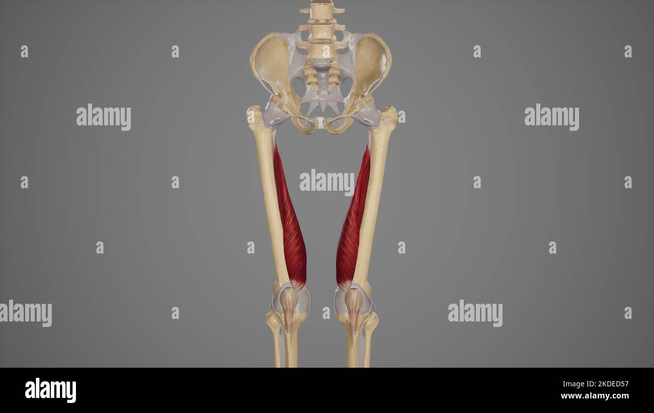 Medical Illustration of Vastus Medialis Muscle Stock Photohttps://www.alamy.com/image-license-details/?v=1https://www.alamy.com/medical-illustration-of-vastus-medialis-muscle-image490198499.html
RF2KDED57–Medical Illustration of Vastus Medialis Muscle
 Vastus intermedius muscle, illustration Stock Photohttps://www.alamy.com/image-license-details/?v=1https://www.alamy.com/vastus-intermedius-muscle-illustration-image273701477.html
RFWW850N–Vastus intermedius muscle, illustration
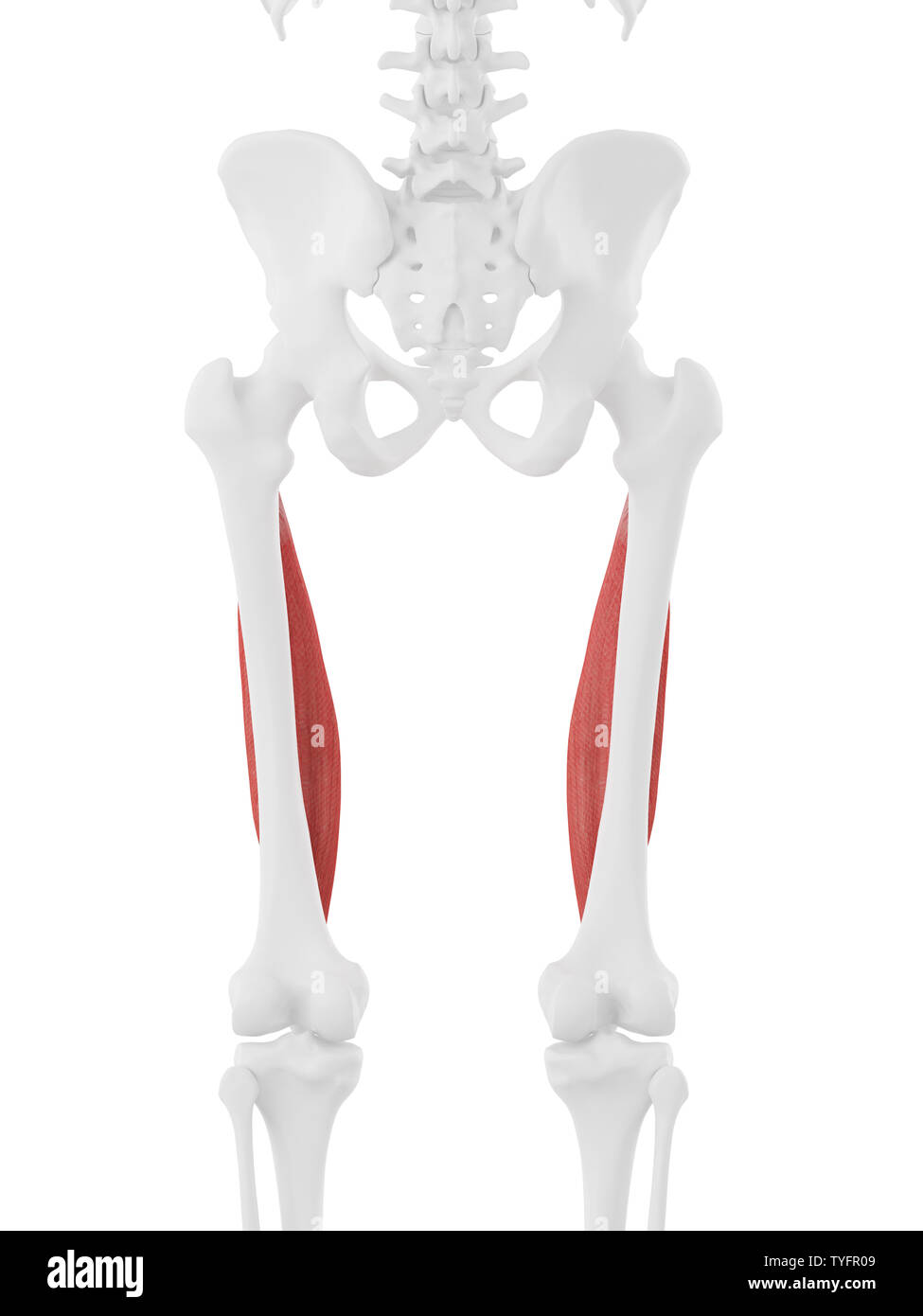 3d rendered medically accurate illustration of the Vastus Intermedius Stock Photohttps://www.alamy.com/image-license-details/?v=1https://www.alamy.com/3d-rendered-medically-accurate-illustration-of-the-vastus-intermedius-image257888185.html
RFTYFR09–3d rendered medically accurate illustration of the Vastus Intermedius
 Applied anatomy and kinesiology, the mechanism of muscular movement . and leave the other cord in place, itwill extend both joints as before. We are thus confronted by theproblem, How can the hamstring-muscles, which are flexors of theknee, cause extension of the knee? How can a cord tied across two VASTUS INTERMEDIUS 185 joints give to a muscle that is primarily a flexor of a joint theability to extend it? Dr. Lombard has explained this apparent contradiction by show-ing that the two-joint muscles of the thigh have better leverage asextensors than as flexors. The hamstring muscles have better Stock Photohttps://www.alamy.com/image-license-details/?v=1https://www.alamy.com/applied-anatomy-and-kinesiology-the-mechanism-of-muscular-movement-and-leave-the-other-cord-in-place-itwill-extend-both-joints-as-before-we-are-thus-confronted-by-theproblem-how-can-the-hamstring-muscles-which-are-flexors-of-theknee-cause-extension-of-the-knee-how-can-a-cord-tied-across-two-vastus-intermedius-185-joints-give-to-a-muscle-that-is-primarily-a-flexor-of-a-joint-theability-to-extend-it-dr-lombard-has-explained-this-apparent-contradiction-by-show-ing-that-the-two-joint-muscles-of-the-thigh-have-better-leverage-asextensors-than-as-flexors-the-hamstring-muscles-have-better-image342682195.html
RM2AWEEHR–Applied anatomy and kinesiology, the mechanism of muscular movement . and leave the other cord in place, itwill extend both joints as before. We are thus confronted by theproblem, How can the hamstring-muscles, which are flexors of theknee, cause extension of the knee? How can a cord tied across two VASTUS INTERMEDIUS 185 joints give to a muscle that is primarily a flexor of a joint theability to extend it? Dr. Lombard has explained this apparent contradiction by show-ing that the two-joint muscles of the thigh have better leverage asextensors than as flexors. The hamstring muscles have better
 Gastrocnemius Muscle anatomy for medical concept 3D illustration Stock Photohttps://www.alamy.com/image-license-details/?v=1https://www.alamy.com/gastrocnemius-muscle-anatomy-for-medical-concept-3d-illustration-image502043384.html
RF2M4P1CT–Gastrocnemius Muscle anatomy for medical concept 3D illustration
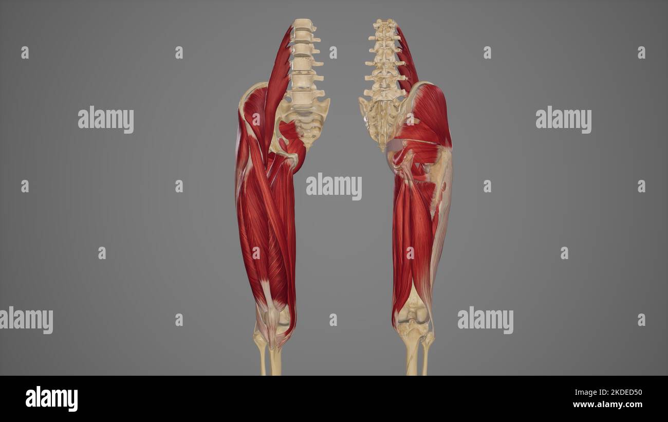 Anterior and Posterior View of Thigh Muscles Stock Photohttps://www.alamy.com/image-license-details/?v=1https://www.alamy.com/anterior-and-posterior-view-of-thigh-muscles-image490198492.html
RF2KDED50–Anterior and Posterior View of Thigh Muscles
 Vastus intermedius muscle, illustration Stock Photohttps://www.alamy.com/image-license-details/?v=1https://www.alamy.com/vastus-intermedius-muscle-illustration-image273701523.html
RFWW852B–Vastus intermedius muscle, illustration
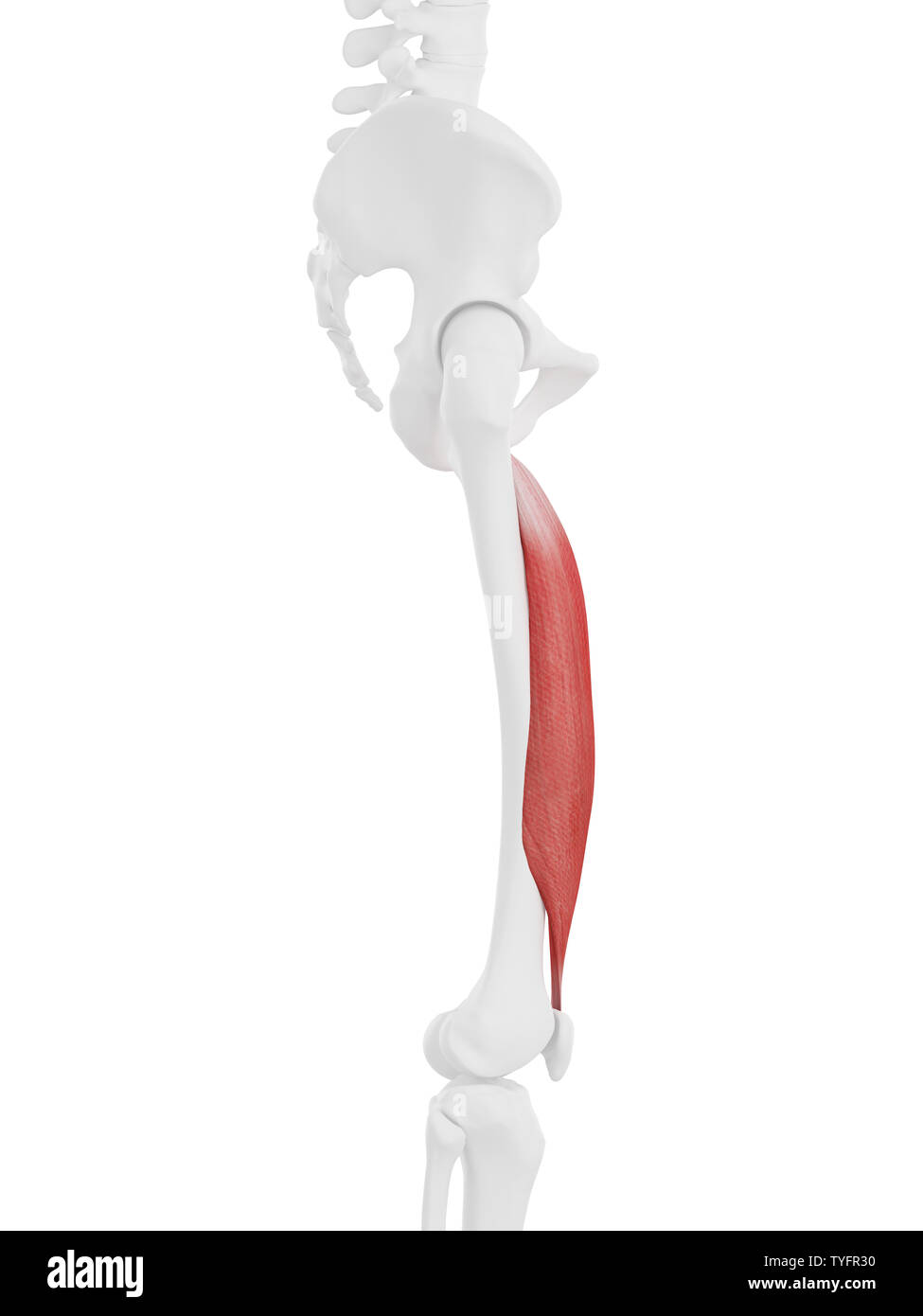 3d rendered medically accurate illustration of the Vastus Intermedius Stock Photohttps://www.alamy.com/image-license-details/?v=1https://www.alamy.com/3d-rendered-medically-accurate-illustration-of-the-vastus-intermedius-image257888260.html
RFTYFR30–3d rendered medically accurate illustration of the Vastus Intermedius
 . Applied anatomy and kinesiology. the patella. Structure.—Similar to the exter-nus. The tendon of origin is a flatsheet arising from the linea asperaand the tendon of insertion is thesame sheet to which the othersjoin. Action.—^The line of pull is justlike that of the externus except thatit is directed diagonally inward in-stead of outward. Isolated actioncauses inward displacement of thepatella and paralysis makes thesubject liable to outward displace-ment. VASTUS INTERMEDIUS. A companion of the two preced-ing, lying between them and beneaththe rectus femoris. Origin.—^The surface of the upp Stock Photohttps://www.alamy.com/image-license-details/?v=1https://www.alamy.com/applied-anatomy-and-kinesiology-the-patella-structuresimilar-to-the-exter-nus-the-tendon-of-origin-is-a-flatsheet-arising-from-the-linea-asperaand-the-tendon-of-insertion-is-thesame-sheet-to-which-the-othersjoin-actionthe-line-of-pull-is-justlike-that-of-the-externus-except-thatit-is-directed-diagonally-inward-in-stead-of-outward-isolated-actioncauses-inward-displacement-of-thepatella-and-paralysis-makes-thesubject-liable-to-outward-displace-ment-vastus-intermedius-a-companion-of-the-two-preced-ing-lying-between-them-and-beneaththe-rectus-femoris-originthe-surface-of-the-upp-image370744088.html
RM2CF4RT8–. Applied anatomy and kinesiology. the patella. Structure.—Similar to the exter-nus. The tendon of origin is a flatsheet arising from the linea asperaand the tendon of insertion is thesame sheet to which the othersjoin. Action.—^The line of pull is justlike that of the externus except thatit is directed diagonally inward in-stead of outward. Isolated actioncauses inward displacement of thepatella and paralysis makes thesubject liable to outward displace-ment. VASTUS INTERMEDIUS. A companion of the two preced-ing, lying between them and beneaththe rectus femoris. Origin.—^The surface of the upp
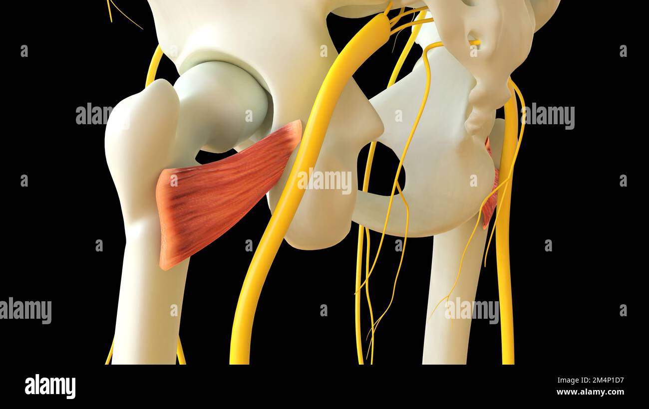 Quadriceps Femoris Muscle anatomy for medical concept 3D illustration Stock Photohttps://www.alamy.com/image-license-details/?v=1https://www.alamy.com/quadriceps-femoris-muscle-anatomy-for-medical-concept-3d-illustration-image502043395.html
RF2M4P1D7–Quadriceps Femoris Muscle anatomy for medical concept 3D illustration
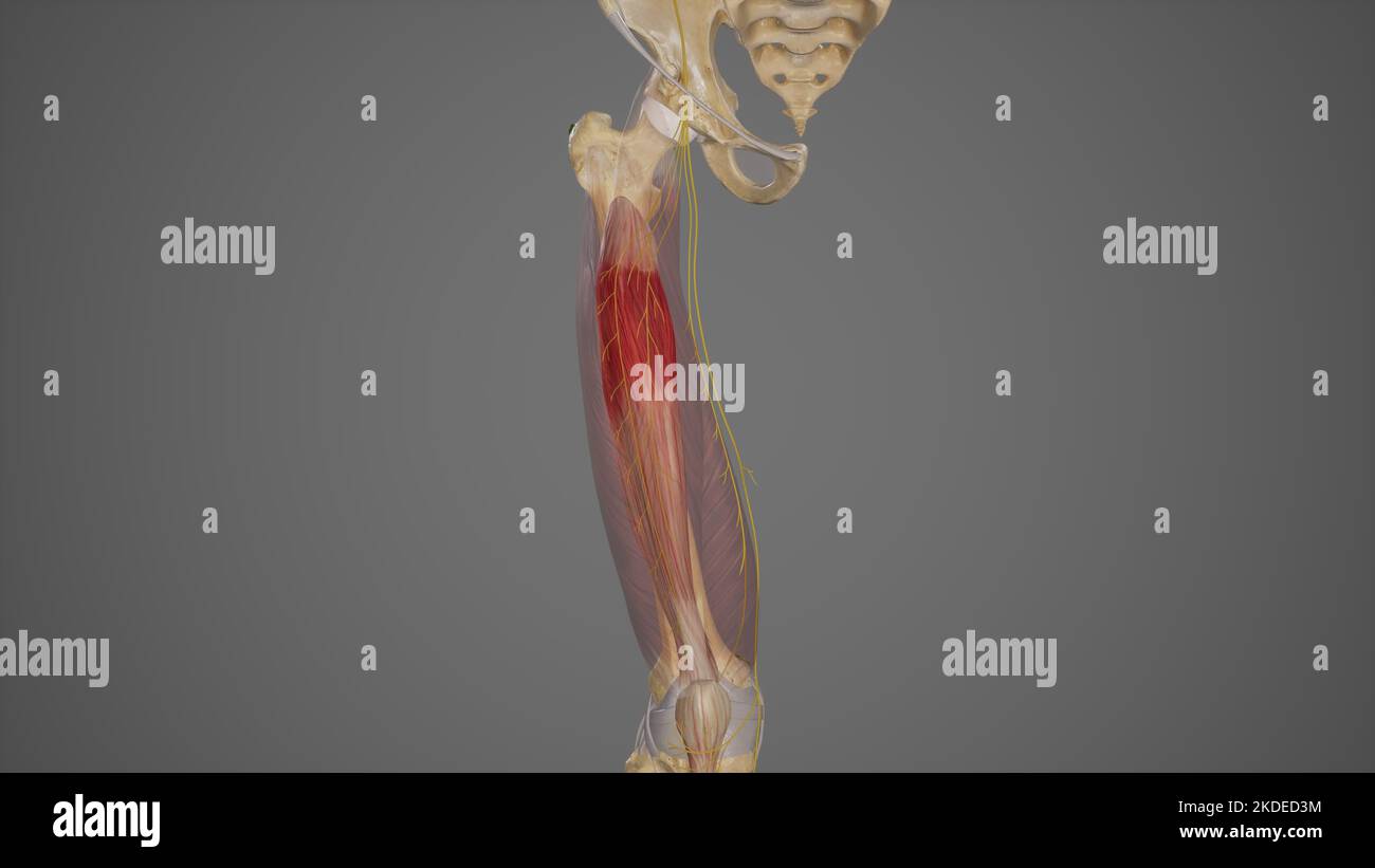 Branches of Posterior Division of Femoral Nerve Stock Photohttps://www.alamy.com/image-license-details/?v=1https://www.alamy.com/branches-of-posterior-division-of-femoral-nerve-image490198456.html
RF2KDED3M–Branches of Posterior Division of Femoral Nerve
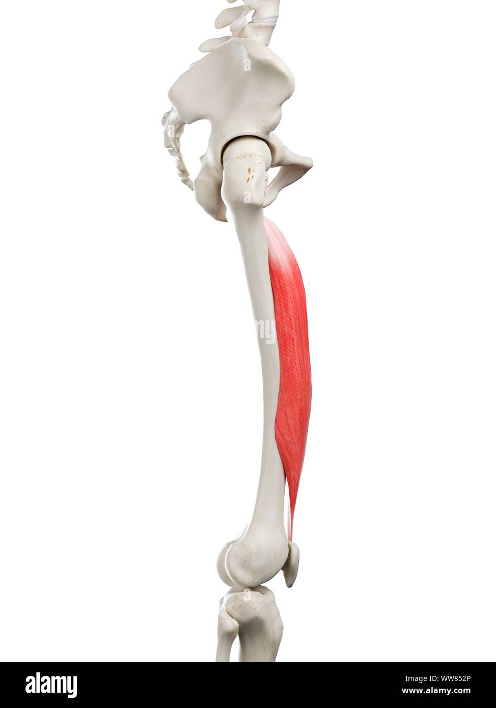 Vastus intermedius muscle, illustration Stock Photohttps://www.alamy.com/image-license-details/?v=1https://www.alamy.com/vastus-intermedius-muscle-illustration-image273701534.html
RFWW852P–Vastus intermedius muscle, illustration
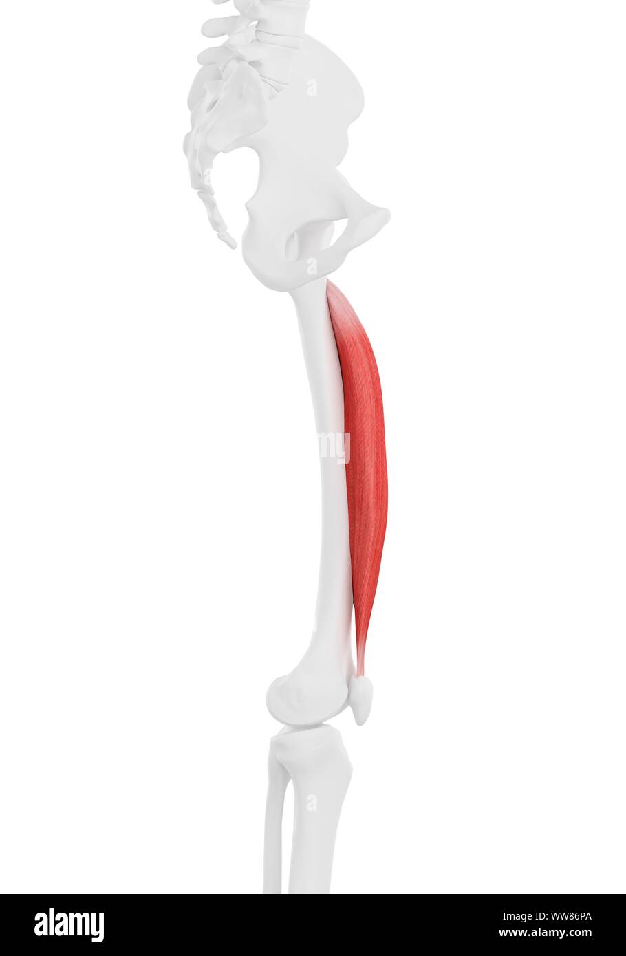 Vastus intermedius muscle, illustration Stock Photohttps://www.alamy.com/image-license-details/?v=1https://www.alamy.com/vastus-intermedius-muscle-illustration-image273702866.html
RFWW86PA–Vastus intermedius muscle, illustration
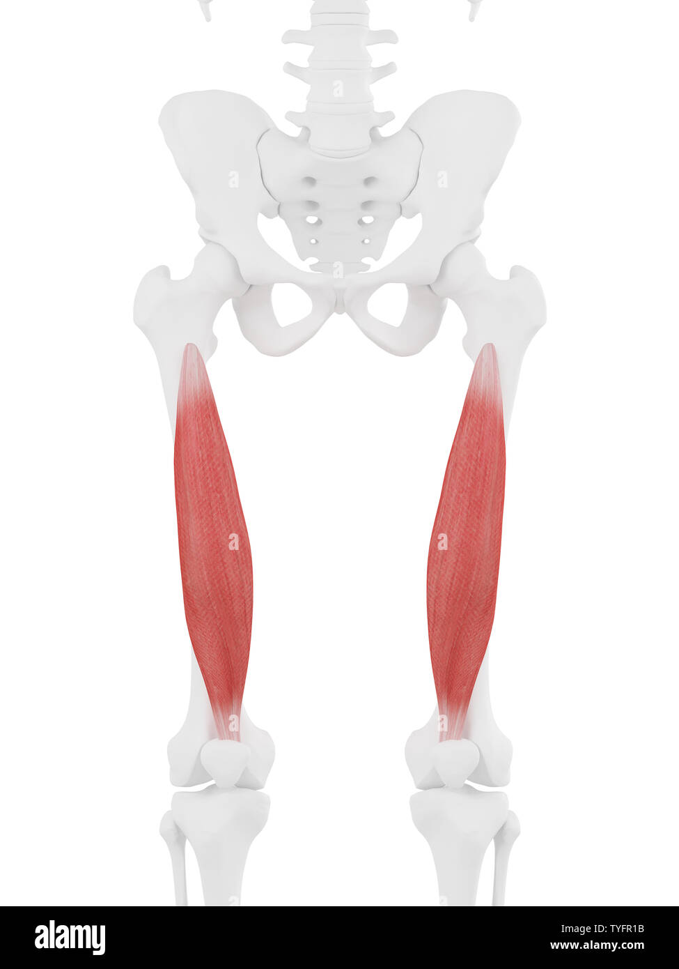 3d rendered medically accurate illustration of the Vastus Intermedius Stock Photohttps://www.alamy.com/image-license-details/?v=1https://www.alamy.com/3d-rendered-medically-accurate-illustration-of-the-vastus-intermedius-image257888215.html
RFTYFR1B–3d rendered medically accurate illustration of the Vastus Intermedius
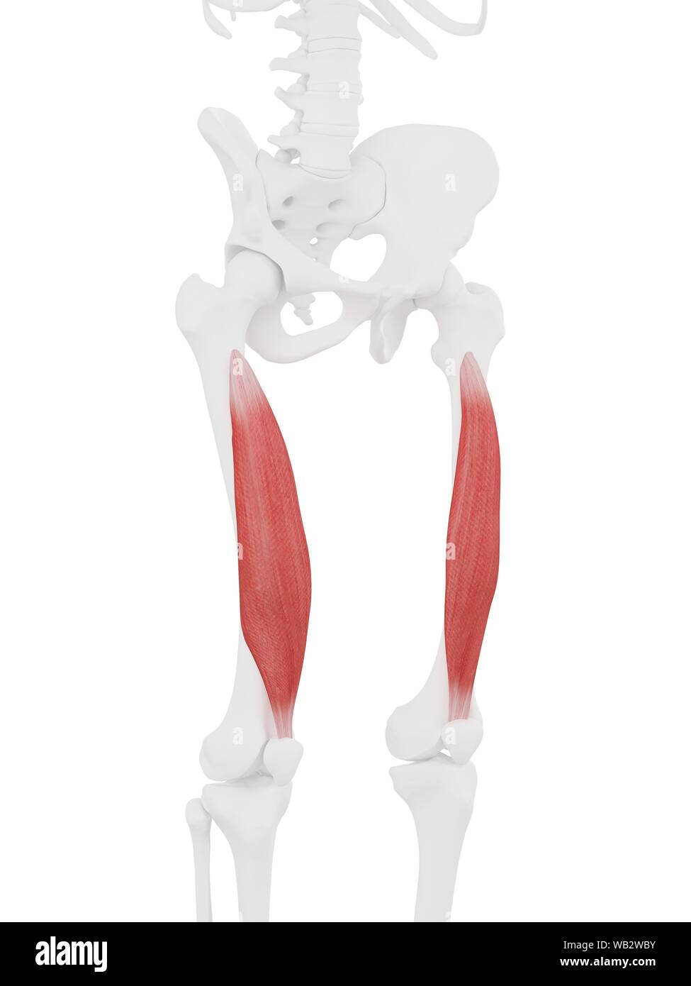 Vastus intermedius muscle, computer illustration. Stock Photohttps://www.alamy.com/image-license-details/?v=1https://www.alamy.com/vastus-intermedius-muscle-computer-illustration-image264980575.html
RFWB2WBY–Vastus intermedius muscle, computer illustration.
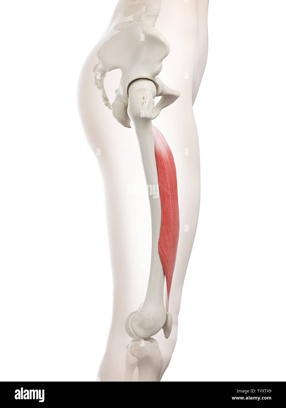 3d rendered medically accurate illustration of a womans Vastus Intermedius Stock Photohttps://www.alamy.com/image-license-details/?v=1https://www.alamy.com/3d-rendered-medically-accurate-illustration-of-a-womans-vastus-intermedius-image258131169.html
RFTYXTX9–3d rendered medically accurate illustration of a womans Vastus Intermedius
 . Applied anatomy and kinesiology. and leave the other cord in place, itwill extend both joints as before. We are thus confronted by theproblem, How can the hamstring muscles, which are flexors of theknee, cause extension of the knee? How can a cord tied across two VASTUS INTERMEDIUS 167 joints give to a muscle that is primarily a flexor of a joint theability to extend it? Dr. Lombard has explained this apparent contradiction by show-ing that the two-joint muscles of the thigh have better leverage asextensors than as flexors. The hamstring muscles have betterleverage at the hip and the rectus Stock Photohttps://www.alamy.com/image-license-details/?v=1https://www.alamy.com/applied-anatomy-and-kinesiology-and-leave-the-other-cord-in-place-itwill-extend-both-joints-as-before-we-are-thus-confronted-by-theproblem-how-can-the-hamstring-muscles-which-are-flexors-of-theknee-cause-extension-of-the-knee-how-can-a-cord-tied-across-two-vastus-intermedius-167-joints-give-to-a-muscle-that-is-primarily-a-flexor-of-a-joint-theability-to-extend-it-dr-lombard-has-explained-this-apparent-contradiction-by-show-ing-that-the-two-joint-muscles-of-the-thigh-have-better-leverage-asextensors-than-as-flexors-the-hamstring-muscles-have-betterleverage-at-the-hip-and-the-rectus-image370743002.html
RM2CF4PDE–. Applied anatomy and kinesiology. and leave the other cord in place, itwill extend both joints as before. We are thus confronted by theproblem, How can the hamstring muscles, which are flexors of theknee, cause extension of the knee? How can a cord tied across two VASTUS INTERMEDIUS 167 joints give to a muscle that is primarily a flexor of a joint theability to extend it? Dr. Lombard has explained this apparent contradiction by show-ing that the two-joint muscles of the thigh have better leverage asextensors than as flexors. The hamstring muscles have betterleverage at the hip and the rectus
 Gluteus Medius Muscle anatomy for medical concept 3D illustration Stock Photohttps://www.alamy.com/image-license-details/?v=1https://www.alamy.com/gluteus-medius-muscle-anatomy-for-medical-concept-3d-illustration-image502043372.html
RF2M4P1CC–Gluteus Medius Muscle anatomy for medical concept 3D illustration
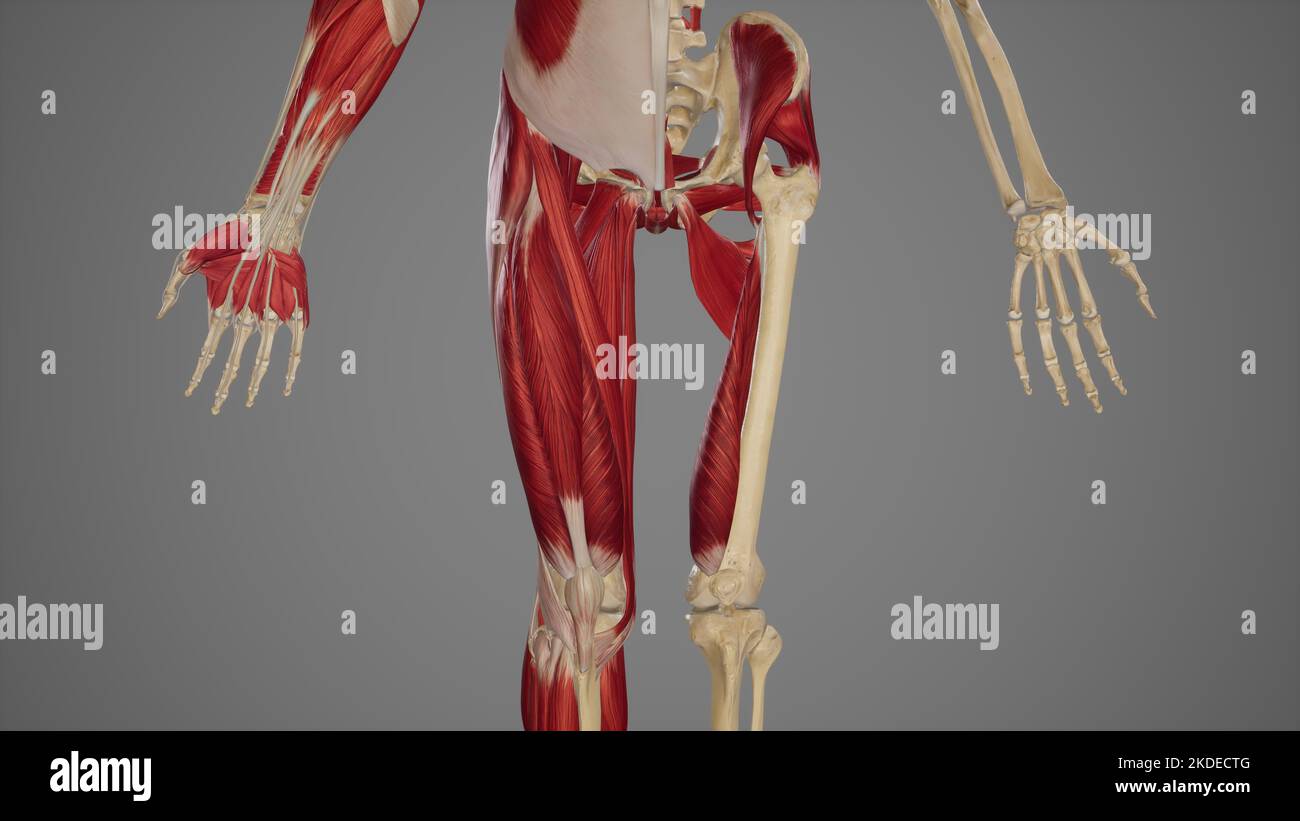 Thigh Muscles Stock Photohttps://www.alamy.com/image-license-details/?v=1https://www.alamy.com/thigh-muscles-image490198256.html
RF2KDECTG–Thigh Muscles
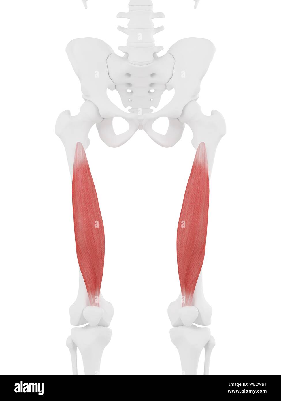 Vastus intermedius muscle, computer illustration. Stock Photohttps://www.alamy.com/image-license-details/?v=1https://www.alamy.com/vastus-intermedius-muscle-computer-illustration-image264980572.html
RFWB2WBT–Vastus intermedius muscle, computer illustration.
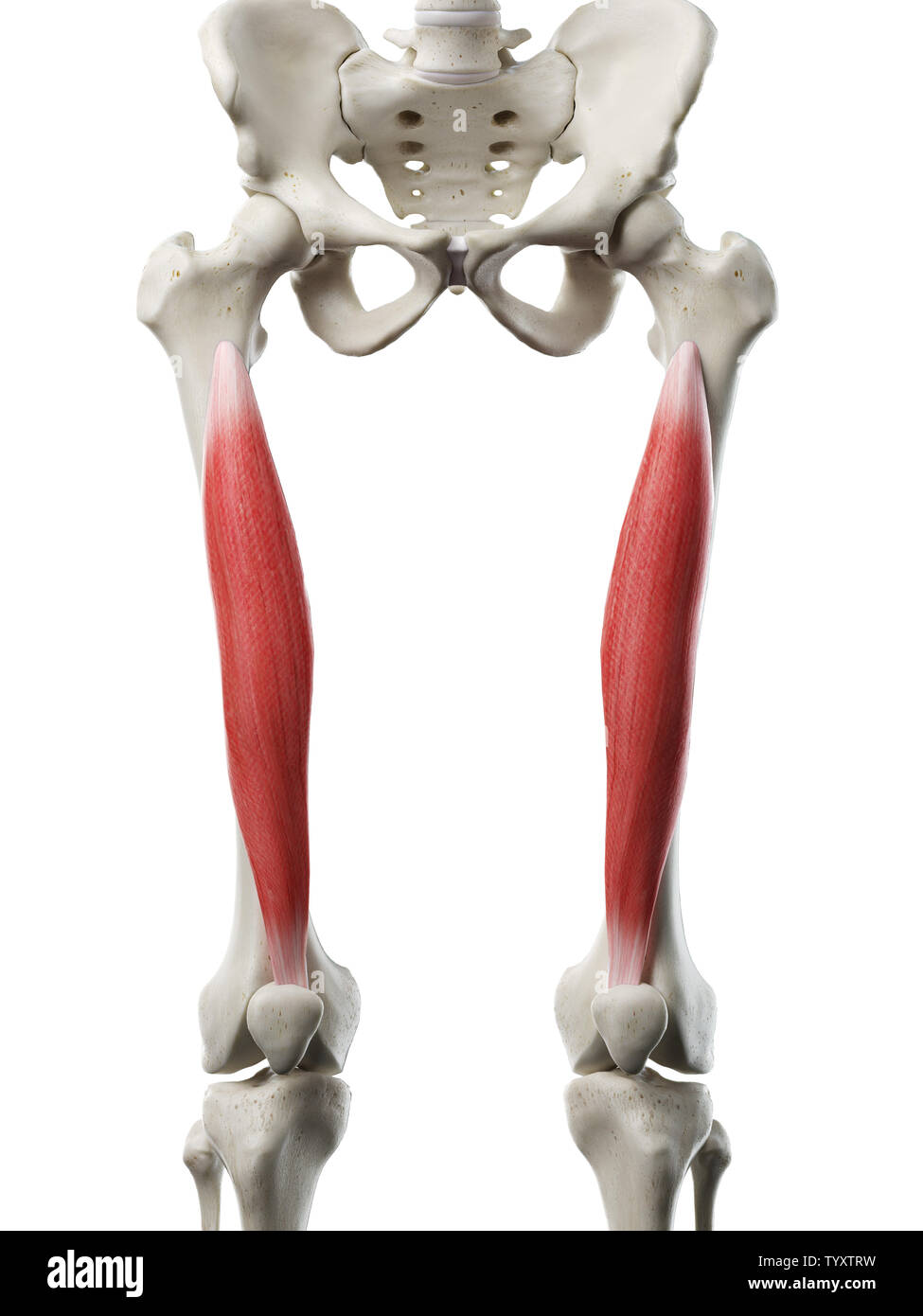 3d rendered medically accurate illustration of a womans Vastus Intermedius Stock Photohttps://www.alamy.com/image-license-details/?v=1https://www.alamy.com/3d-rendered-medically-accurate-illustration-of-a-womans-vastus-intermedius-image258131101.html
RFTYXTRW–3d rendered medically accurate illustration of a womans Vastus Intermedius
 . The anatomy of the domestic animals. Veterinary anatomy. 330 FASCI.E AND MUSCLES OF THE HORSE nishes insertion to fibers of the vasti. The tendon of insertion is formed bj' the union of these tendinous layers on the lower part of the muscle. The lower portion of the muscle is pennate, the fibers on either side converging on the tendon at an acute angle. Relations.—Medially, the iliacus, sartorius, and vastus medialis; laterally, the tensor fascise latae, glutei, and vastus lateralis; posteriorly, the hip joint and the vastus intermedius; anteriorly, the fascia lata and the skin. The anterior Stock Photohttps://www.alamy.com/image-license-details/?v=1https://www.alamy.com/the-anatomy-of-the-domestic-animals-veterinary-anatomy-330-fascie-and-muscles-of-the-horse-nishes-insertion-to-fibers-of-the-vasti-the-tendon-of-insertion-is-formed-bj-the-union-of-these-tendinous-layers-on-the-lower-part-of-the-muscle-the-lower-portion-of-the-muscle-is-pennate-the-fibers-on-either-side-converging-on-the-tendon-at-an-acute-angle-relationsmedially-the-iliacus-sartorius-and-vastus-medialis-laterally-the-tensor-fascise-latae-glutei-and-vastus-lateralis-posteriorly-the-hip-joint-and-the-vastus-intermedius-anteriorly-the-fascia-lata-and-the-skin-the-anterior-image236800991.html
RMRN7627–. The anatomy of the domestic animals. Veterinary anatomy. 330 FASCI.E AND MUSCLES OF THE HORSE nishes insertion to fibers of the vasti. The tendon of insertion is formed bj' the union of these tendinous layers on the lower part of the muscle. The lower portion of the muscle is pennate, the fibers on either side converging on the tendon at an acute angle. Relations.—Medially, the iliacus, sartorius, and vastus medialis; laterally, the tensor fascise latae, glutei, and vastus lateralis; posteriorly, the hip joint and the vastus intermedius; anteriorly, the fascia lata and the skin. The anterior
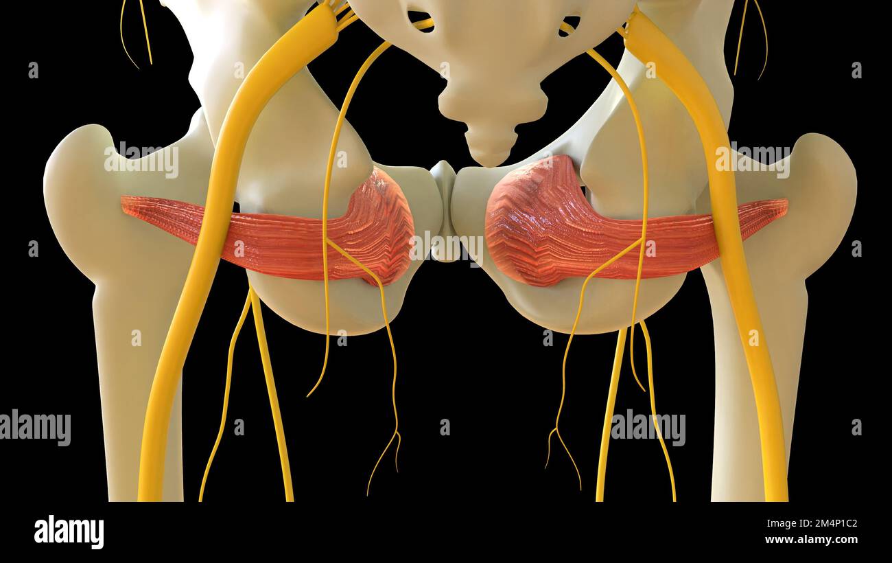 Obturator Internus Muscle anatomy for medical concept 3D illustration Stock Photohttps://www.alamy.com/image-license-details/?v=1https://www.alamy.com/obturator-internus-muscle-anatomy-for-medical-concept-3d-illustration-image502043362.html
RF2M4P1C2–Obturator Internus Muscle anatomy for medical concept 3D illustration
 Vastus intermedius muscle, computer illustration. Stock Photohttps://www.alamy.com/image-license-details/?v=1https://www.alamy.com/vastus-intermedius-muscle-computer-illustration-image264980569.html
RFWB2WBN–Vastus intermedius muscle, computer illustration.
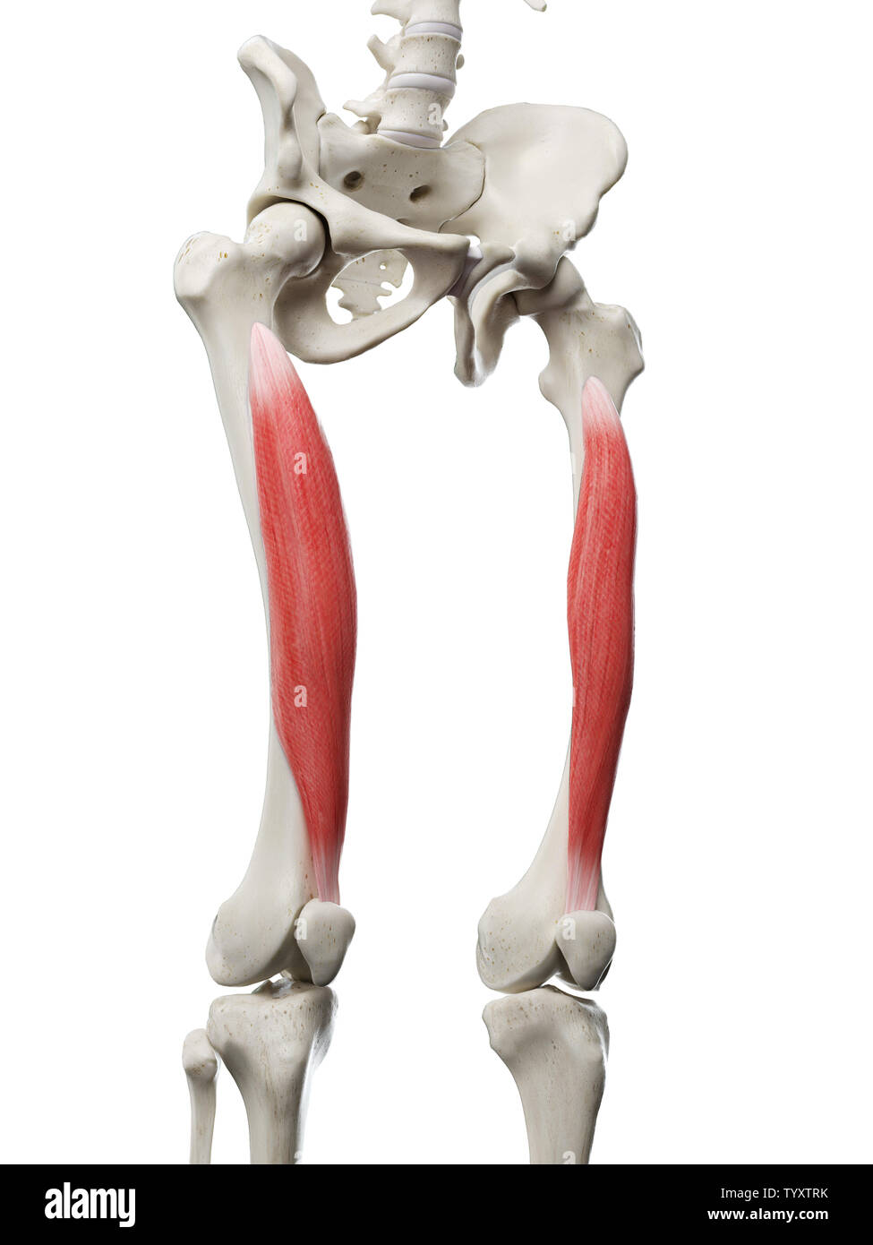 3d rendered medically accurate illustration of a womans Vastus Intermedius Stock Photohttps://www.alamy.com/image-license-details/?v=1https://www.alamy.com/3d-rendered-medically-accurate-illustration-of-a-womans-vastus-intermedius-image258131095.html
RFTYXTRK–3d rendered medically accurate illustration of a womans Vastus Intermedius
 . The anatomy of the domestic animals . Veterinary anatomy. 332 fascijE and muscles of the horse proximal part of the medial patellar ligament. (2) The tendon of the rectus femoris. Action.—To extend the stifle joint. Structure.—This is very similar to that of the vastus lateralis. It is, how- ever, more difficult to separate from the intermedins, because many fibers of the latter arise on the tendinous sheet which covers the contact surface, of the medial Shaft of femur Gluteus profundus Trochanter major {anterior part) Prepubic teyidon Obturator externus Vastus lateralis Vastus intermedius R Stock Photohttps://www.alamy.com/image-license-details/?v=1https://www.alamy.com/the-anatomy-of-the-domestic-animals-veterinary-anatomy-332-fascije-and-muscles-of-the-horse-proximal-part-of-the-medial-patellar-ligament-2-the-tendon-of-the-rectus-femoris-actionto-extend-the-stifle-joint-structurethis-is-very-similar-to-that-of-the-vastus-lateralis-it-is-how-ever-more-difficult-to-separate-from-the-intermedins-because-many-fibers-of-the-latter-arise-on-the-tendinous-sheet-which-covers-the-contact-surface-of-the-medial-shaft-of-femur-gluteus-profundus-trochanter-major-anterior-part-prepubic-teyidon-obturator-externus-vastus-lateralis-vastus-intermedius-r-image232325919.html
RMRDYA27–. The anatomy of the domestic animals . Veterinary anatomy. 332 fascijE and muscles of the horse proximal part of the medial patellar ligament. (2) The tendon of the rectus femoris. Action.—To extend the stifle joint. Structure.—This is very similar to that of the vastus lateralis. It is, how- ever, more difficult to separate from the intermedins, because many fibers of the latter arise on the tendinous sheet which covers the contact surface, of the medial Shaft of femur Gluteus profundus Trochanter major {anterior part) Prepubic teyidon Obturator externus Vastus lateralis Vastus intermedius R
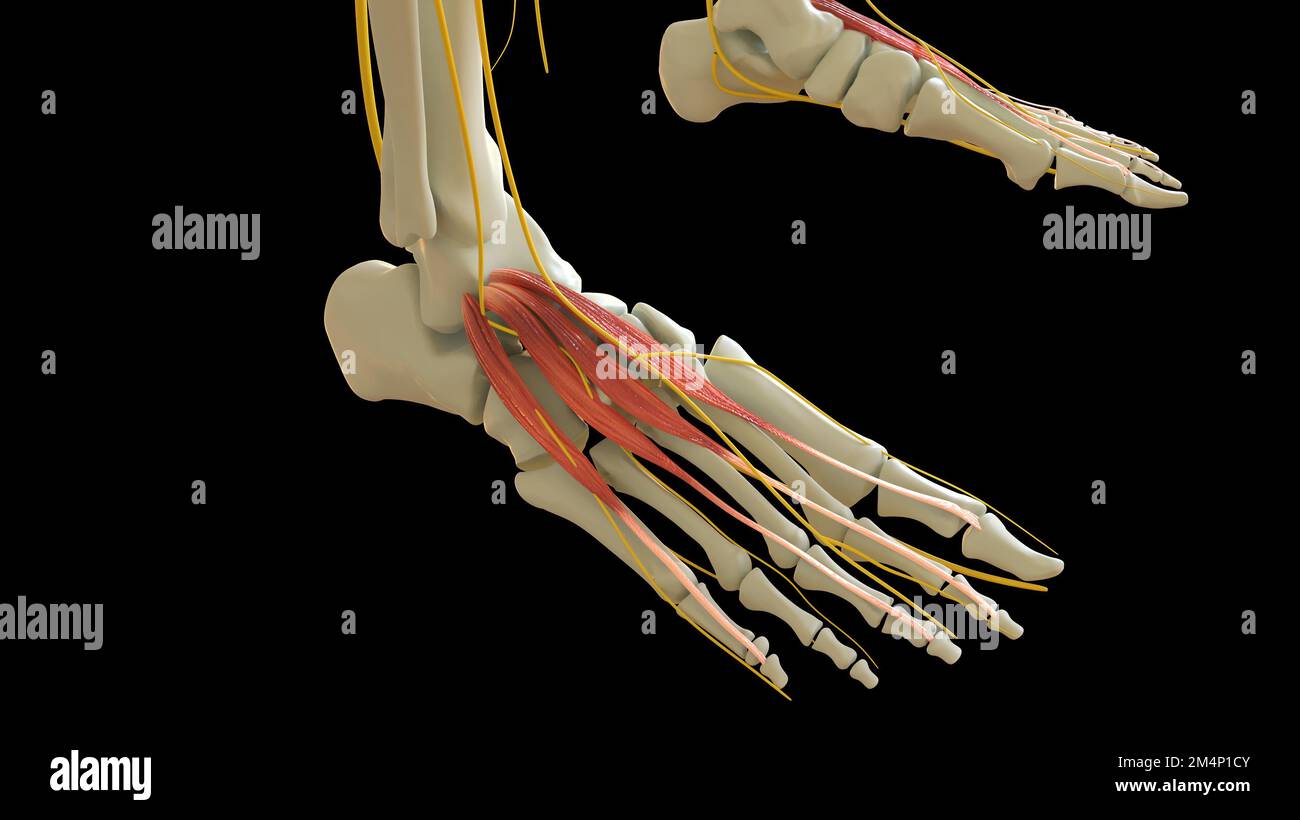 Extensor Hallucis Brevis Muscle anatomy for medical concept 3D illustration Stock Photohttps://www.alamy.com/image-license-details/?v=1https://www.alamy.com/extensor-hallucis-brevis-muscle-anatomy-for-medical-concept-3d-illustration-image502043387.html
RF2M4P1CY–Extensor Hallucis Brevis Muscle anatomy for medical concept 3D illustration
 Vastus intermedius muscle, computer illustration. Stock Photohttps://www.alamy.com/image-license-details/?v=1https://www.alamy.com/vastus-intermedius-muscle-computer-illustration-image264980573.html
RFWB2WBW–Vastus intermedius muscle, computer illustration.
 3d rendered medically accurate illustration of a womans Vastus Intermedius Stock Photohttps://www.alamy.com/image-license-details/?v=1https://www.alamy.com/3d-rendered-medically-accurate-illustration-of-a-womans-vastus-intermedius-image258131131.html
RFTYXTTY–3d rendered medically accurate illustration of a womans Vastus Intermedius
 Human leg muscles (vastus intermedius), illustration. Stock Photohttps://www.alamy.com/image-license-details/?v=1https://www.alamy.com/stock-photo-human-leg-muscles-vastus-intermedius-illustration-94291880.html
RFFDBA74–Human leg muscles (vastus intermedius), illustration.
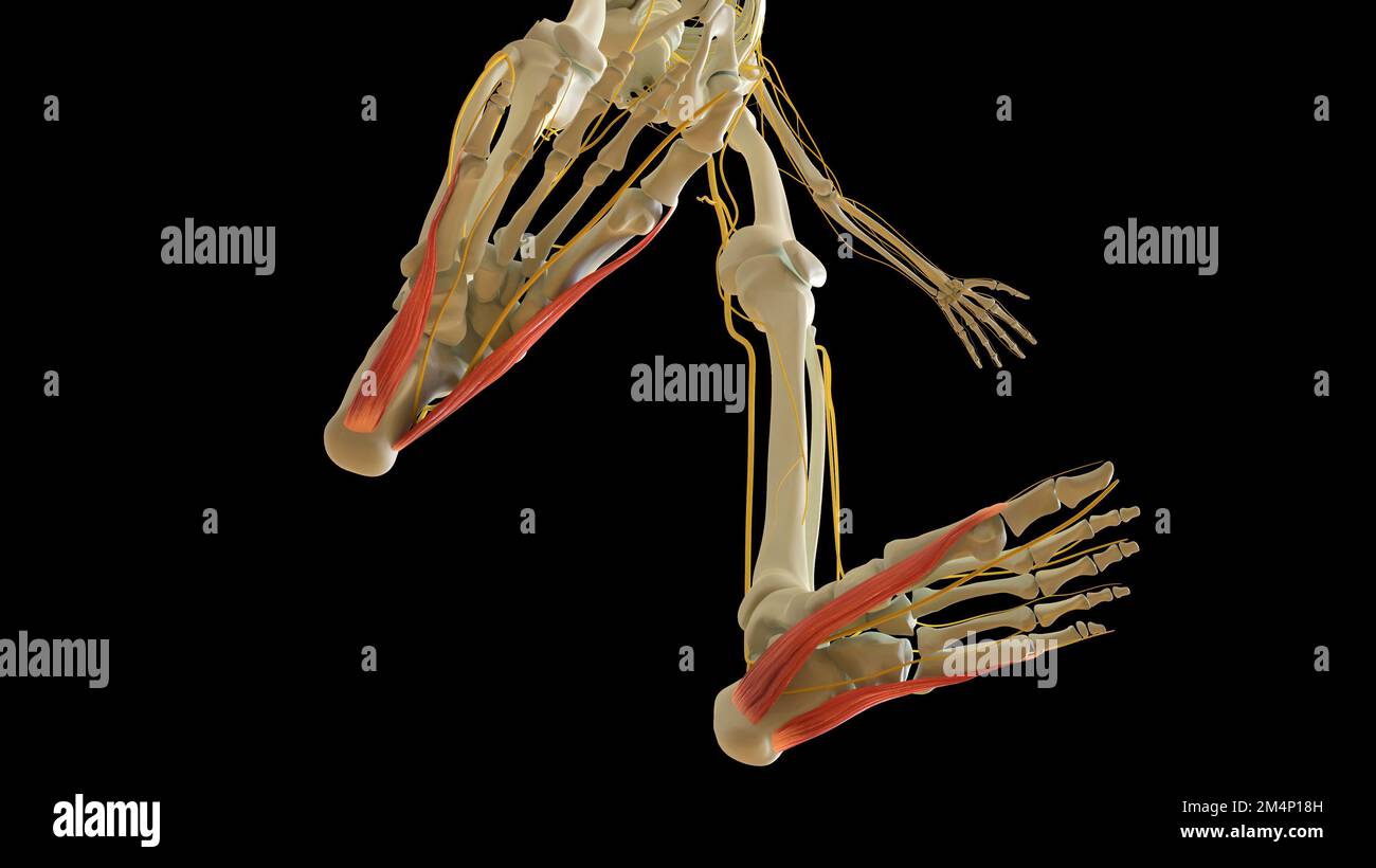 Abductor Hallucis and Digiti Minimi Leg Muscles anatomy for medical concept 3D illustration Stock Photohttps://www.alamy.com/image-license-details/?v=1https://www.alamy.com/abductor-hallucis-and-digiti-minimi-leg-muscles-anatomy-for-medical-concept-3d-illustration-image502043265.html
RF2M4P18H–Abductor Hallucis and Digiti Minimi Leg Muscles anatomy for medical concept 3D illustration
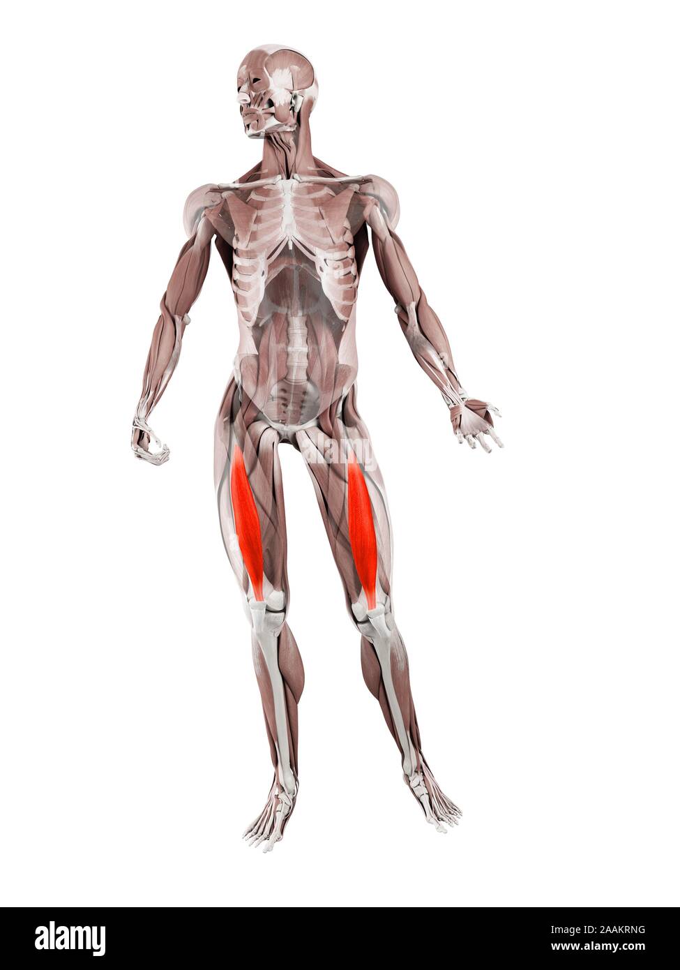 Vastus intermedius muscle, computer illustration. Stock Photohttps://www.alamy.com/image-license-details/?v=1https://www.alamy.com/vastus-intermedius-muscle-computer-illustration-image333579276.html
RF2AAKRNG–Vastus intermedius muscle, computer illustration.
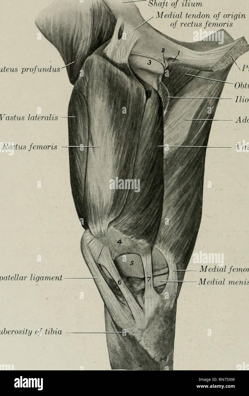 . The anatomy of the domestic animals. Veterinary anatomy. 330 FASCIA AND MUSCLES OF THE HORSE nishes insertion to fibers of the vasti. The tendon of insertion is formed by the union of these tendinous layers on the lower part of the muscle. The lower portion of the muscle is pennate, the fibers on either side converging on the tendon at an acute angle. Relations.—Medially, the iliacus, sartorius, and vastus medialis; laterally, the tensor fasciae latae, glutei, and vastus lateralis; posteriorly, the hip joint and the vastus intermedius; anteriorly, the fascia lata and the skin. The anterior f Stock Photohttps://www.alamy.com/image-license-details/?v=1https://www.alamy.com/the-anatomy-of-the-domestic-animals-veterinary-anatomy-330-fascia-and-muscles-of-the-horse-nishes-insertion-to-fibers-of-the-vasti-the-tendon-of-insertion-is-formed-by-the-union-of-these-tendinous-layers-on-the-lower-part-of-the-muscle-the-lower-portion-of-the-muscle-is-pennate-the-fibers-on-either-side-converging-on-the-tendon-at-an-acute-angle-relationsmedially-the-iliacus-sartorius-and-vastus-medialis-laterally-the-tensor-fasciae-latae-glutei-and-vastus-lateralis-posteriorly-the-hip-joint-and-the-vastus-intermedius-anteriorly-the-fascia-lata-and-the-skin-the-anterior-f-image236800897.html
RMRN75XW–. The anatomy of the domestic animals. Veterinary anatomy. 330 FASCIA AND MUSCLES OF THE HORSE nishes insertion to fibers of the vasti. The tendon of insertion is formed by the union of these tendinous layers on the lower part of the muscle. The lower portion of the muscle is pennate, the fibers on either side converging on the tendon at an acute angle. Relations.—Medially, the iliacus, sartorius, and vastus medialis; laterally, the tensor fasciae latae, glutei, and vastus lateralis; posteriorly, the hip joint and the vastus intermedius; anteriorly, the fascia lata and the skin. The anterior f
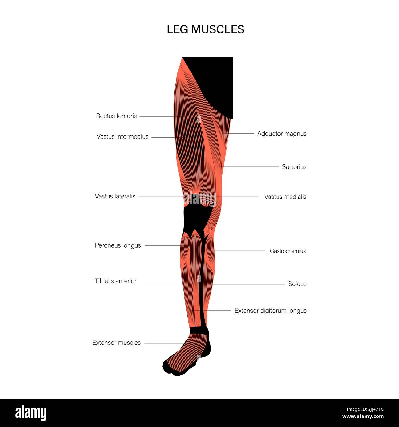 Leg anatomy, illustration. Stock Photohttps://www.alamy.com/image-license-details/?v=1https://www.alamy.com/leg-anatomy-illustration-image475837728.html
RF2JJ47TG–Leg anatomy, illustration.
 3d rendered medically accurate illustration of a womans Vastus Intermedius Stock Photohttps://www.alamy.com/image-license-details/?v=1https://www.alamy.com/3d-rendered-medically-accurate-illustration-of-a-womans-vastus-intermedius-image258131115.html
RFTYXTTB–3d rendered medically accurate illustration of a womans Vastus Intermedius
 Semimembranosus Muscle anatomy for medical concept 3D illustration Stock Photohttps://www.alamy.com/image-license-details/?v=1https://www.alamy.com/semimembranosus-muscle-anatomy-for-medical-concept-3d-illustration-image502043260.html
RF2M4P18C–Semimembranosus Muscle anatomy for medical concept 3D illustration
 . Cunningham's Text-book of anatomy. Anatomy. terior surface of the proximal part^of the Left Femur. Vastus mediai Saphenous nerve^ Femoral vessel: Sartorius Adductor lokgus Adductor magnds Gracilis. Rectus femoris Vastus lateralis Vastus intermedius Femur Femoris (short head) SEMIMEMBRANOSUS' Biceps Femoris (long head) Semitendinostj Sciatic nerve Fig. 362.—Transverse Section of the Thigh (Hunter's Adductor Canal). M. Vastus Lateralis.—The vastus lateralis has an origin, partly fleshy, partly membranous, from (1) the capsule of the hip-joint) (2) the tubercle of the femur, (3) a concave surfa Stock Photohttps://www.alamy.com/image-license-details/?v=1https://www.alamy.com/cunninghams-text-book-of-anatomy-anatomy-terior-surface-of-the-proximal-partof-the-left-femur-vastus-mediai-saphenous-nerve-femoral-vessel-sartorius-adductor-lokgus-adductor-magnds-gracilis-rectus-femoris-vastus-lateralis-vastus-intermedius-femur-femoris-short-head-semimembranosus-biceps-femoris-long-head-semitendinostj-sciatic-nerve-fig-362transverse-section-of-the-thigh-hunters-adductor-canal-m-vastus-lateralisthe-vastus-lateralis-has-an-origin-partly-fleshy-partly-membranous-from-1-the-capsule-of-the-hip-joint-2-the-tubercle-of-the-femur-3-a-concave-surfa-image231880935.html
RMRD72DY–. Cunningham's Text-book of anatomy. Anatomy. terior surface of the proximal part^of the Left Femur. Vastus mediai Saphenous nerve^ Femoral vessel: Sartorius Adductor lokgus Adductor magnds Gracilis. Rectus femoris Vastus lateralis Vastus intermedius Femur Femoris (short head) SEMIMEMBRANOSUS' Biceps Femoris (long head) Semitendinostj Sciatic nerve Fig. 362.—Transverse Section of the Thigh (Hunter's Adductor Canal). M. Vastus Lateralis.—The vastus lateralis has an origin, partly fleshy, partly membranous, from (1) the capsule of the hip-joint) (2) the tubercle of the femur, (3) a concave surfa
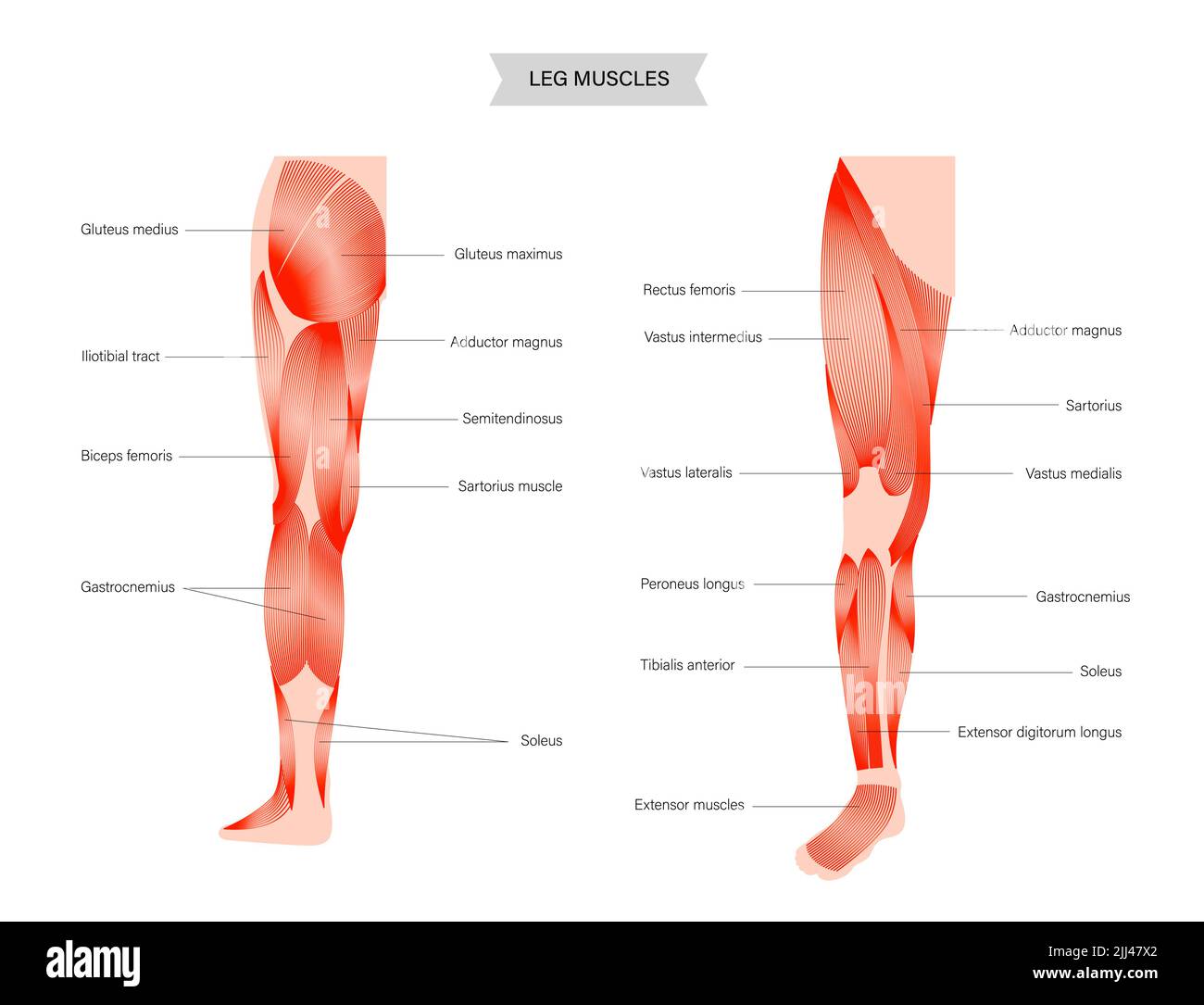 Leg anatomy, illustration. Stock Photohttps://www.alamy.com/image-license-details/?v=1https://www.alamy.com/leg-anatomy-illustration-image475837770.html
RF2JJ47X2–Leg anatomy, illustration.
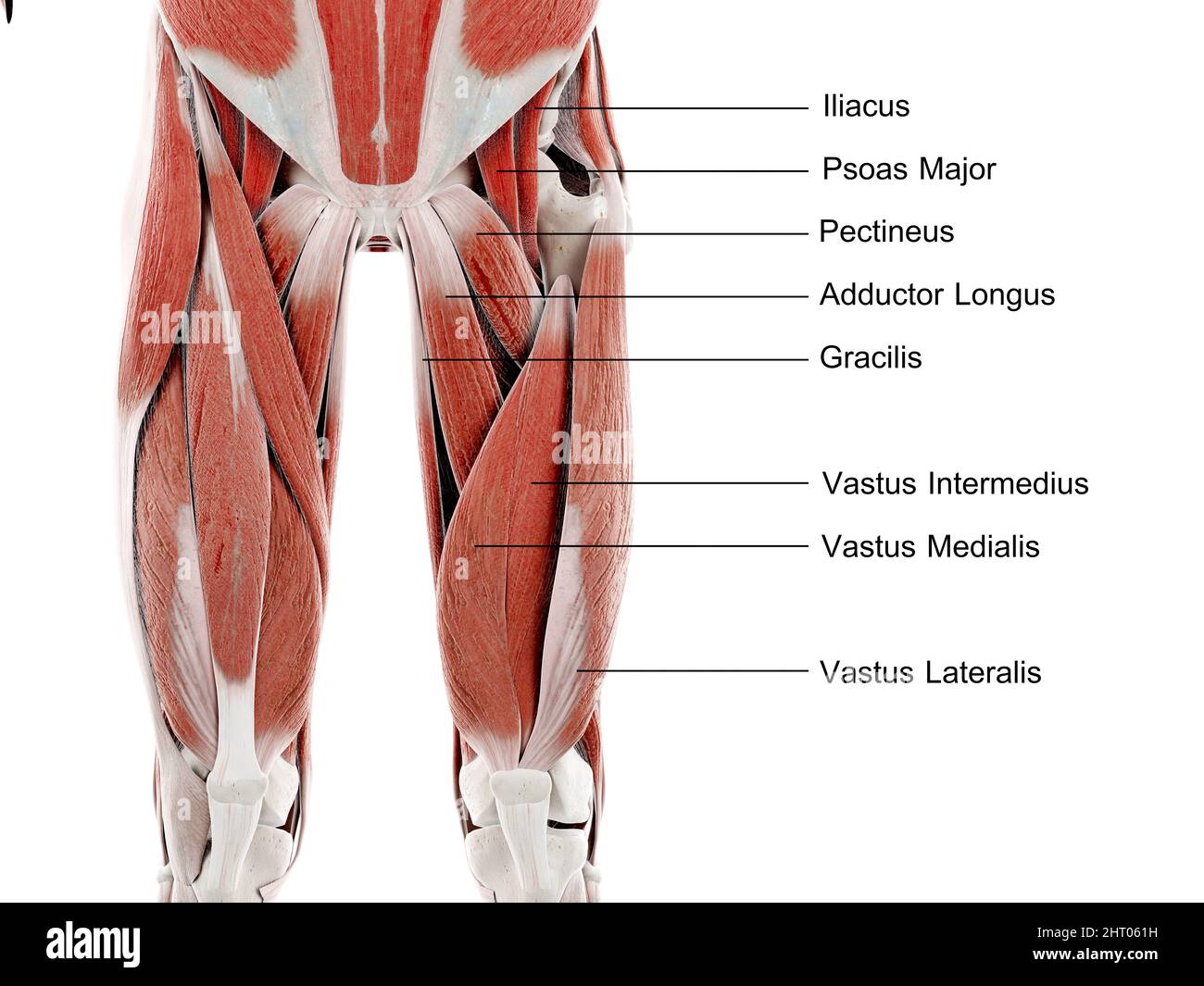 Muscles of the upper leg, illustration Stock Photohttps://www.alamy.com/image-license-details/?v=1https://www.alamy.com/muscles-of-the-upper-leg-illustration-image462226061.html
RF2HT061H–Muscles of the upper leg, illustration
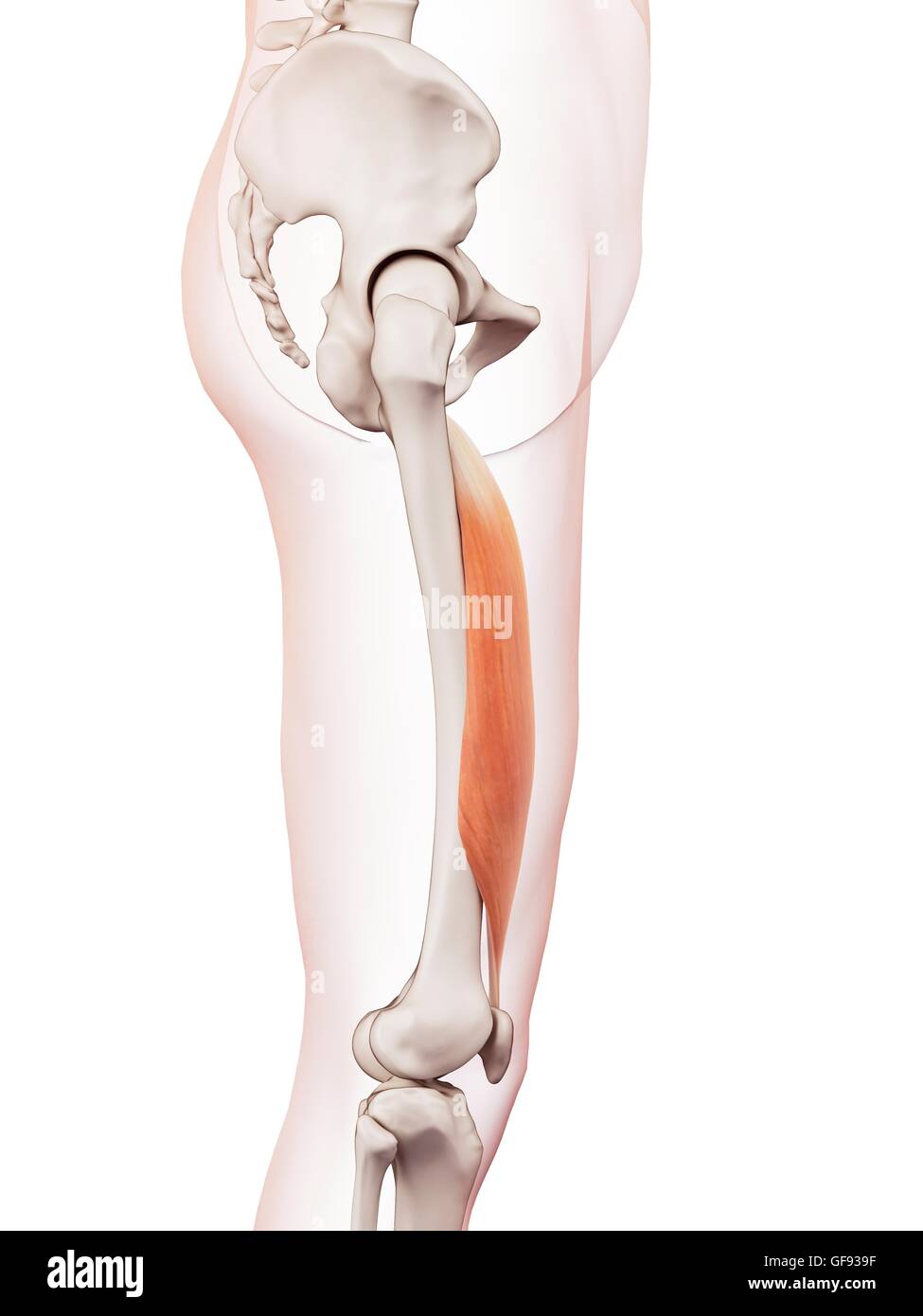 Human leg muscles, illustration. Stock Photohttps://www.alamy.com/image-license-details/?v=1https://www.alamy.com/stock-photo-human-leg-muscles-illustration-112682235.html
RFGF939F–Human leg muscles, illustration.
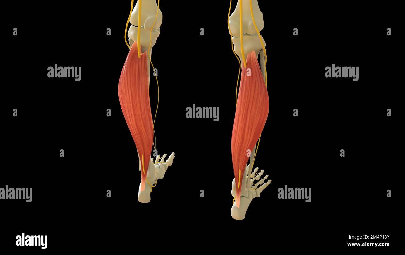 Soleus Muscle anatomy for medical concept 3D illustration Stock Photohttps://www.alamy.com/image-license-details/?v=1https://www.alamy.com/soleus-muscle-anatomy-for-medical-concept-3d-illustration-image502043275.html
RF2M4P18Y–Soleus Muscle anatomy for medical concept 3D illustration
 . An atlas of human anatomy for students and physicians. Anatomy. 352 THE MUSCLES OF THE LOWER EXTREMITY Surface of origin of the gluteus medius muscle Surface of origin of the gluteus minimus muscle Rectus femoris muscle (proximal extremity) Tendon of the gluteus medius muscle Tendon of the gluteus minimus muscle Vastus intemus muscle ivi. vastus raedialis Crureus muscle M. vastus intermedius vastus extemus muscle- M. vastus lateralis Quadriceps extensor cruris muscle Rectus femoris muscle - (distal extremity) Biceps flexor cruris muscle M. biceps femoris. Crest of the ilium Crista iliaca A Stock Photohttps://www.alamy.com/image-license-details/?v=1https://www.alamy.com/an-atlas-of-human-anatomy-for-students-and-physicians-anatomy-352-the-muscles-of-the-lower-extremity-surface-of-origin-of-the-gluteus-medius-muscle-surface-of-origin-of-the-gluteus-minimus-muscle-rectus-femoris-muscle-proximal-extremity-tendon-of-the-gluteus-medius-muscle-tendon-of-the-gluteus-minimus-muscle-vastus-intemus-muscle-ivi-vastus-raedialis-crureus-muscle-m-vastus-intermedius-vastus-extemus-muscle-m-vastus-lateralis-quadriceps-extensor-cruris-muscle-rectus-femoris-muscle-distal-extremity-biceps-flexor-cruris-muscle-m-biceps-femoris-crest-of-the-ilium-crista-iliaca-a-image235399815.html
RMRJYAT7–. An atlas of human anatomy for students and physicians. Anatomy. 352 THE MUSCLES OF THE LOWER EXTREMITY Surface of origin of the gluteus medius muscle Surface of origin of the gluteus minimus muscle Rectus femoris muscle (proximal extremity) Tendon of the gluteus medius muscle Tendon of the gluteus minimus muscle Vastus intemus muscle ivi. vastus raedialis Crureus muscle M. vastus intermedius vastus extemus muscle- M. vastus lateralis Quadriceps extensor cruris muscle Rectus femoris muscle - (distal extremity) Biceps flexor cruris muscle M. biceps femoris. Crest of the ilium Crista iliaca A
 Human leg muscles, illustration. Stock Photohttps://www.alamy.com/image-license-details/?v=1https://www.alamy.com/stock-photo-human-leg-muscles-illustration-112682234.html
RFGF939E–Human leg muscles, illustration.
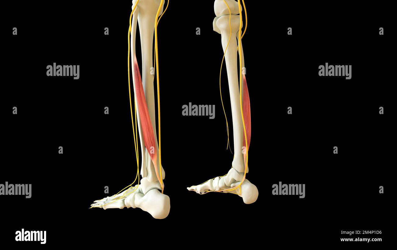 Flexor Hallucis Longus Muscle anatomy for medical concept 3D illustration Stock Photohttps://www.alamy.com/image-license-details/?v=1https://www.alamy.com/flexor-hallucis-longus-muscle-anatomy-for-medical-concept-3d-illustration-image502043394.html
RF2M4P1D6–Flexor Hallucis Longus Muscle anatomy for medical concept 3D illustration
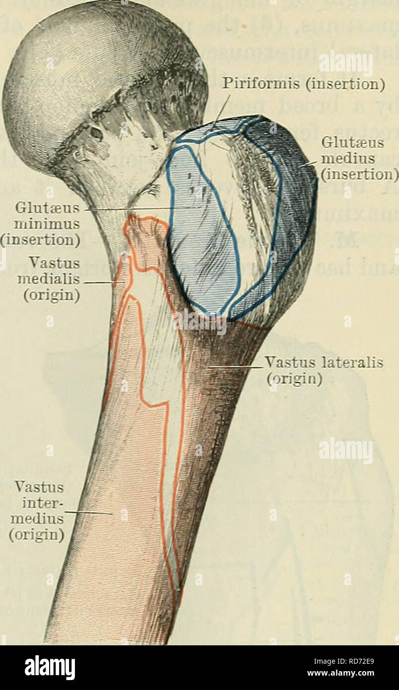 . Cunningham's Text-book of anatomy. Anatomy. formis (insertion) MUSCLES ON THE ANTEEIOK ASPECT OF THE THIGH. 407 muscle. The vastus intermedius envelops the femur, and is concealed by the other muscles. M. Rectus Femoris. — The rectus femoris has a double tendinous origin. (1) The straight head arises from the inferior anterior spine of the ilium (Fig. 366, p. 412): (2) the reflected head springs from a rough groove on the dorsum ilii just above the highest part of the acetabulum (Eig. 366, p. 412). A bursa lies beneath this head of origin. The two heads, bound together and connected to the c Stock Photohttps://www.alamy.com/image-license-details/?v=1https://www.alamy.com/cunninghams-text-book-of-anatomy-anatomy-formis-insertion-muscles-on-the-anteeiok-aspect-of-the-thigh-407-muscle-the-vastus-intermedius-envelops-the-femur-and-is-concealed-by-the-other-muscles-m-rectus-femoris-the-rectus-femoris-has-a-double-tendinous-origin-1-the-straight-head-arises-from-the-inferior-anterior-spine-of-the-ilium-fig-366-p-412-2-the-reflected-head-springs-from-a-rough-groove-on-the-dorsum-ilii-just-above-the-highest-part-of-the-acetabulum-eig-366-p-412-a-bursa-lies-beneath-this-head-of-origin-the-two-heads-bound-together-and-connected-to-the-c-image231880945.html
RMRD72E9–. Cunningham's Text-book of anatomy. Anatomy. formis (insertion) MUSCLES ON THE ANTEEIOK ASPECT OF THE THIGH. 407 muscle. The vastus intermedius envelops the femur, and is concealed by the other muscles. M. Rectus Femoris. — The rectus femoris has a double tendinous origin. (1) The straight head arises from the inferior anterior spine of the ilium (Fig. 366, p. 412): (2) the reflected head springs from a rough groove on the dorsum ilii just above the highest part of the acetabulum (Eig. 366, p. 412). A bursa lies beneath this head of origin. The two heads, bound together and connected to the c
