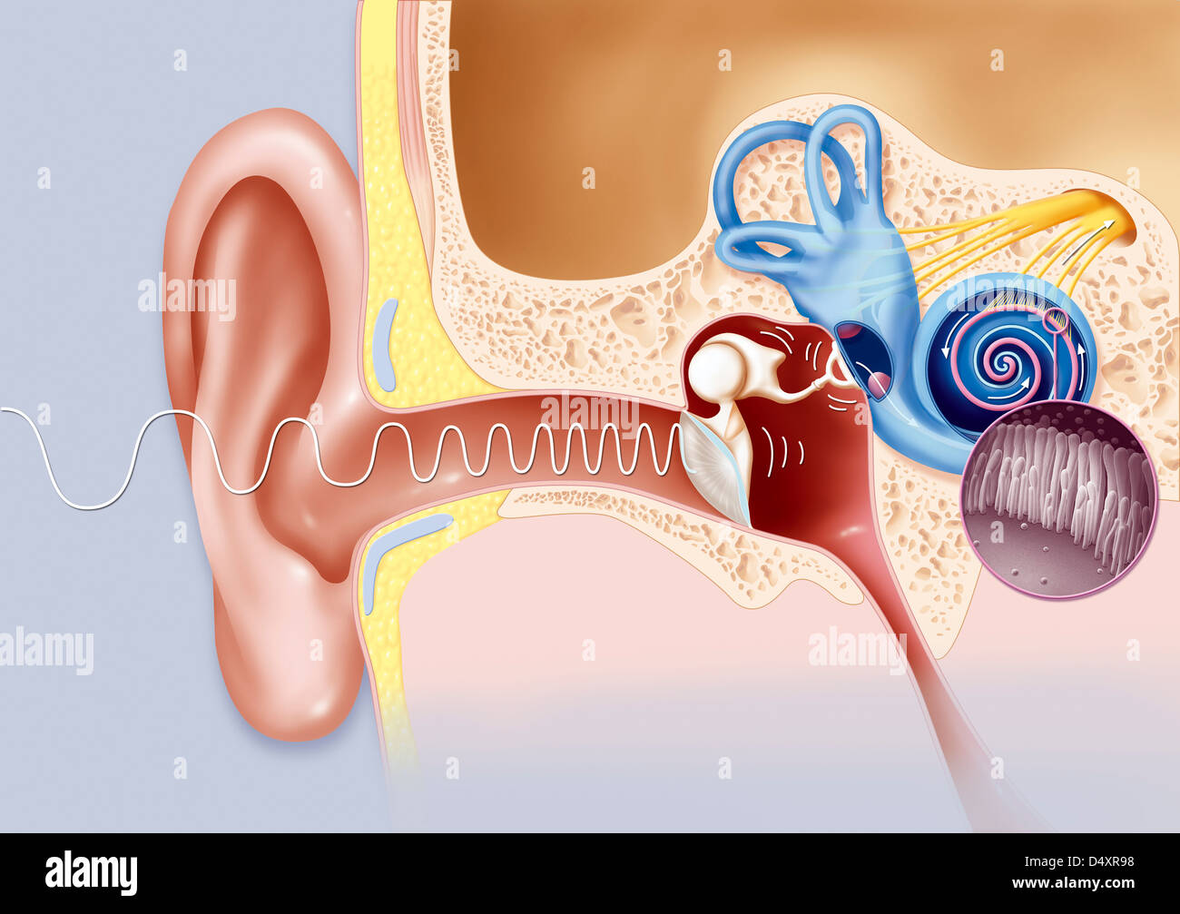
Quick filters:
Vestibulocochlear Stock Photos and Images
 Illustration of the cranial nerves anatomy. The cranial nerves are a set of 12 paired nerves that arise directly from the brain. The first two nerves (olfactory and optic) arise from the cerebrum, whereas the remaining ten emerge from the brainstem. The names of the cranial nerves relate to their function and they are numerically identified in roman numerals. Stock Photohttps://www.alamy.com/image-license-details/?v=1https://www.alamy.com/illustration-of-the-cranial-nerves-anatomy-the-cranial-nerves-are-a-set-of-12-paired-nerves-that-arise-directly-from-the-brain-the-first-two-nerves-olfactory-and-optic-arise-from-the-cerebrum-whereas-the-remaining-ten-emerge-from-the-brainstem-the-names-of-the-cranial-nerves-relate-to-their-function-and-they-are-numerically-identified-in-roman-numerals-image628777660.html
RF2YEY890–Illustration of the cranial nerves anatomy. The cranial nerves are a set of 12 paired nerves that arise directly from the brain. The first two nerves (olfactory and optic) arise from the cerebrum, whereas the remaining ten emerge from the brainstem. The names of the cranial nerves relate to their function and they are numerically identified in roman numerals.
 3d rendered medically accurate illustration of the interior brain anatomy Stock Photohttps://www.alamy.com/image-license-details/?v=1https://www.alamy.com/3d-rendered-medically-accurate-illustration-of-the-interior-brain-anatomy-image365977630.html
RF2C7BM5J–3d rendered medically accurate illustration of the interior brain anatomy
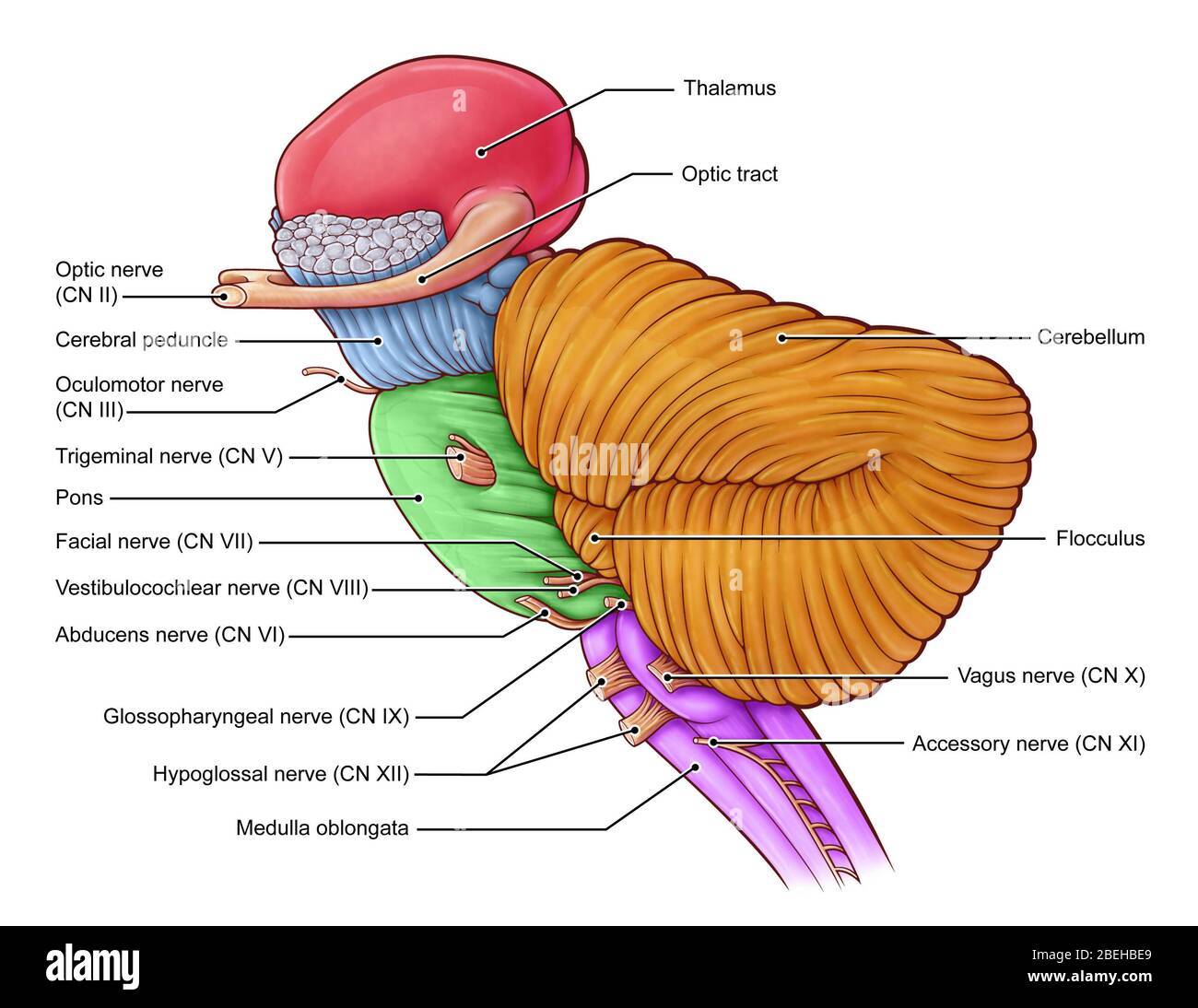 Diencephalon and Brainstem, illustration Stock Photohttps://www.alamy.com/image-license-details/?v=1https://www.alamy.com/diencephalon-and-brainstem-illustration-image353194753.html
RM2BEHBE9–Diencephalon and Brainstem, illustration
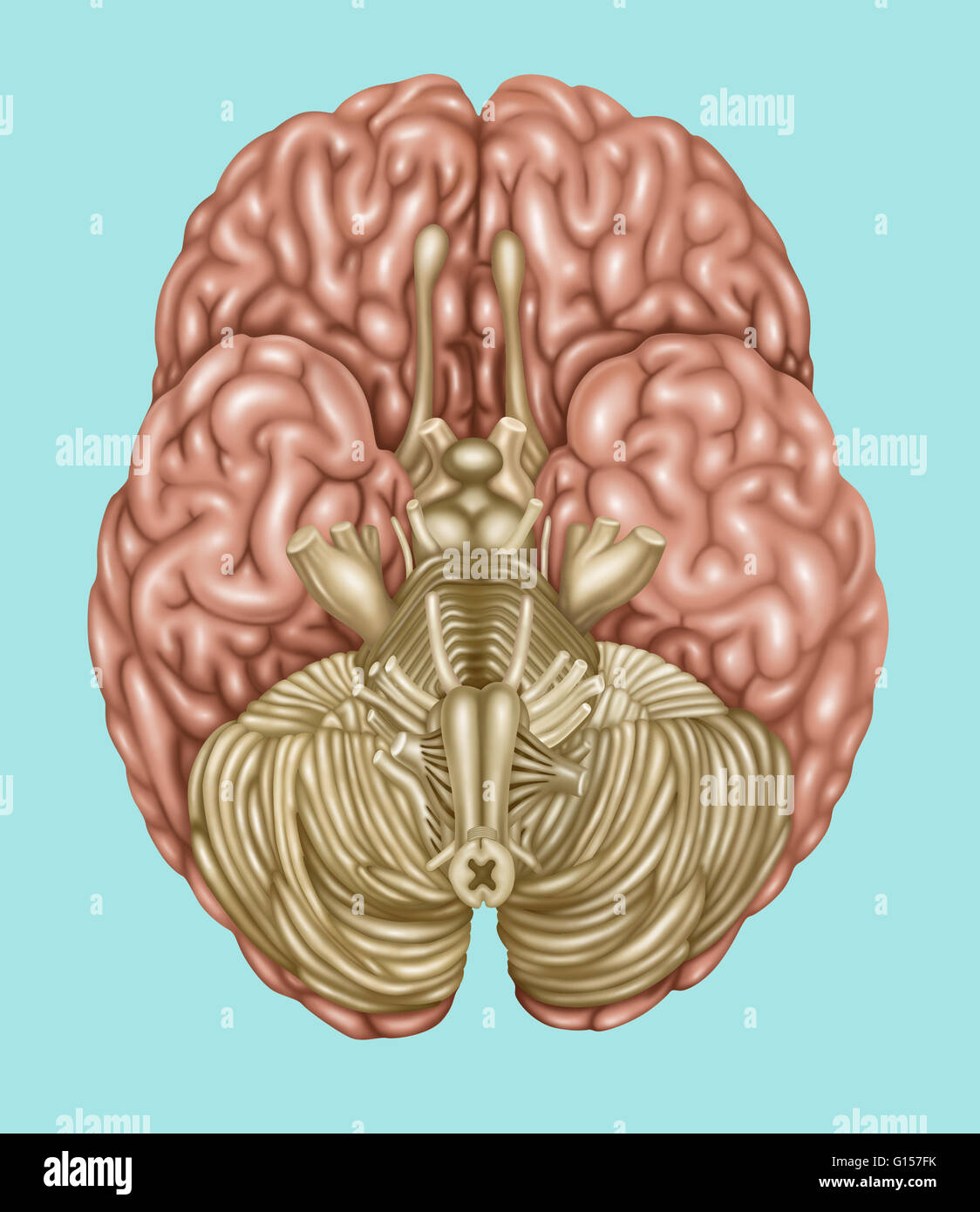 Illustration showing anatomy of the brain, inferior (underside) view. The following areas can be found in this view: olfactory, optic, oculomotor, trochlear, trigeminal, abducens vestibulocochlear, hypoglossal, accessory, facial, glossopharyngeal, and vag Stock Photohttps://www.alamy.com/image-license-details/?v=1https://www.alamy.com/stock-photo-illustration-showing-anatomy-of-the-brain-inferior-underside-view-103992551.html
RMG157FK–Illustration showing anatomy of the brain, inferior (underside) view. The following areas can be found in this view: olfactory, optic, oculomotor, trochlear, trigeminal, abducens vestibulocochlear, hypoglossal, accessory, facial, glossopharyngeal, and vag
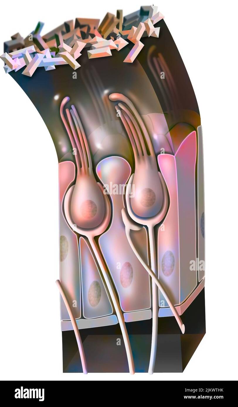 Functioning of the macule: organ of static balance (position of the head). Stock Photohttps://www.alamy.com/image-license-details/?v=1https://www.alamy.com/functioning-of-the-macule-organ-of-static-balance-position-of-the-head-image476926511.html
RF2JKWTHK–Functioning of the macule: organ of static balance (position of the head).
 Anatomy of the human ear. Stock Photohttps://www.alamy.com/image-license-details/?v=1https://www.alamy.com/anatomy-of-the-human-ear-image240162409.html
RFRXM9GW–Anatomy of the human ear.
 Illustration of the cranial nerves anatomy. The cranial nerves are a set of 12 paired nerves that arise directly from the brain. The first two nerves (olfactory and optic) arise from the cerebrum, whereas the remaining ten emerge from the brainstem. The names of the cranial nerves relate to their function and they are numerically identified in roman numerals. Stock Photohttps://www.alamy.com/image-license-details/?v=1https://www.alamy.com/illustration-of-the-cranial-nerves-anatomy-the-cranial-nerves-are-a-set-of-12-paired-nerves-that-arise-directly-from-the-brain-the-first-two-nerves-olfactory-and-optic-arise-from-the-cerebrum-whereas-the-remaining-ten-emerge-from-the-brainstem-the-names-of-the-cranial-nerves-relate-to-their-function-and-they-are-numerically-identified-in-roman-numerals-image628777672.html
RF2YEY89C–Illustration of the cranial nerves anatomy. The cranial nerves are a set of 12 paired nerves that arise directly from the brain. The first two nerves (olfactory and optic) arise from the cerebrum, whereas the remaining ten emerge from the brainstem. The names of the cranial nerves relate to their function and they are numerically identified in roman numerals.
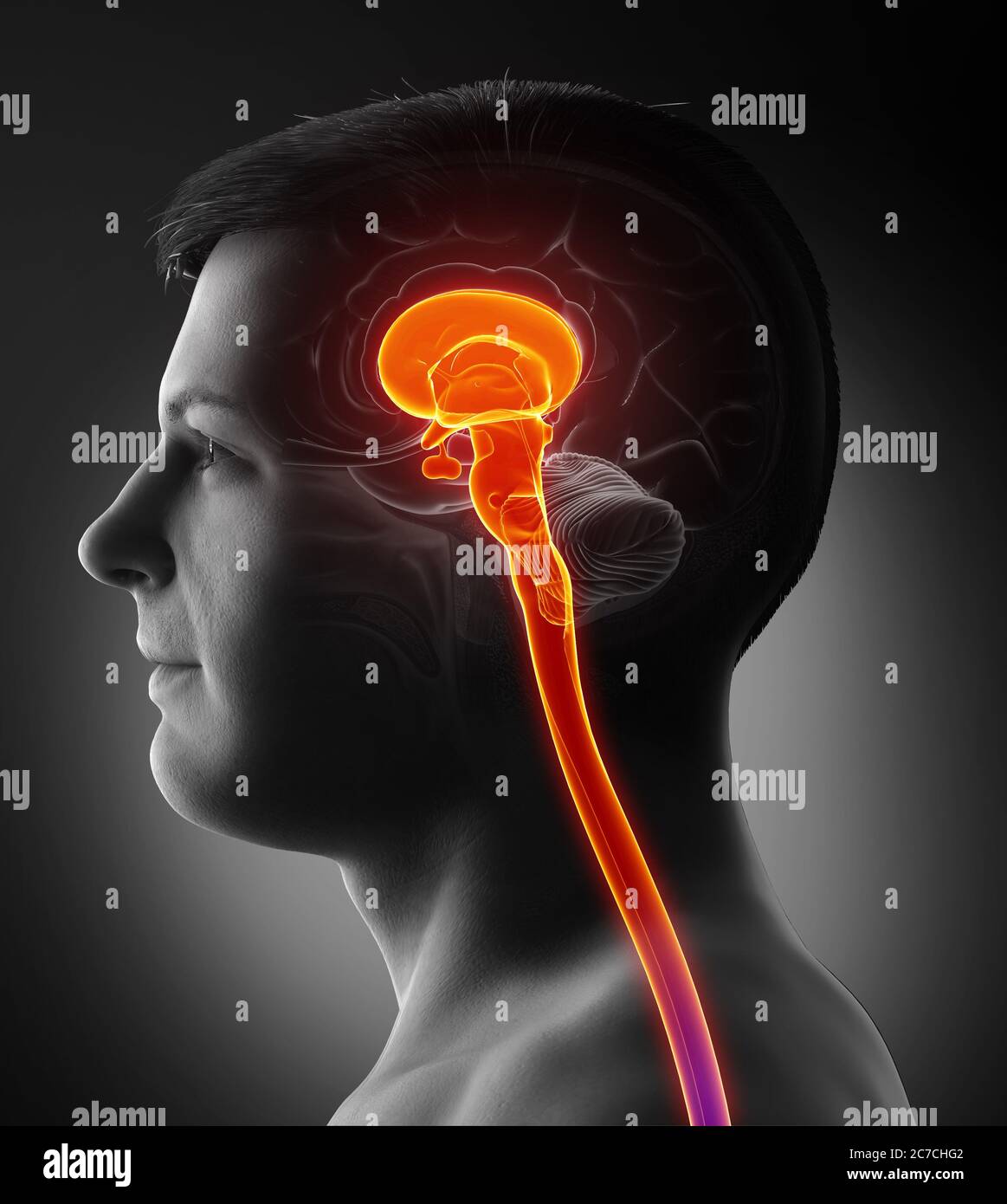 3d rendering medical illustration of brainstem Stock Photohttps://www.alamy.com/image-license-details/?v=1https://www.alamy.com/3d-rendering-medical-illustration-of-brainstem-image365997522.html
RF2C7CHG2–3d rendering medical illustration of brainstem
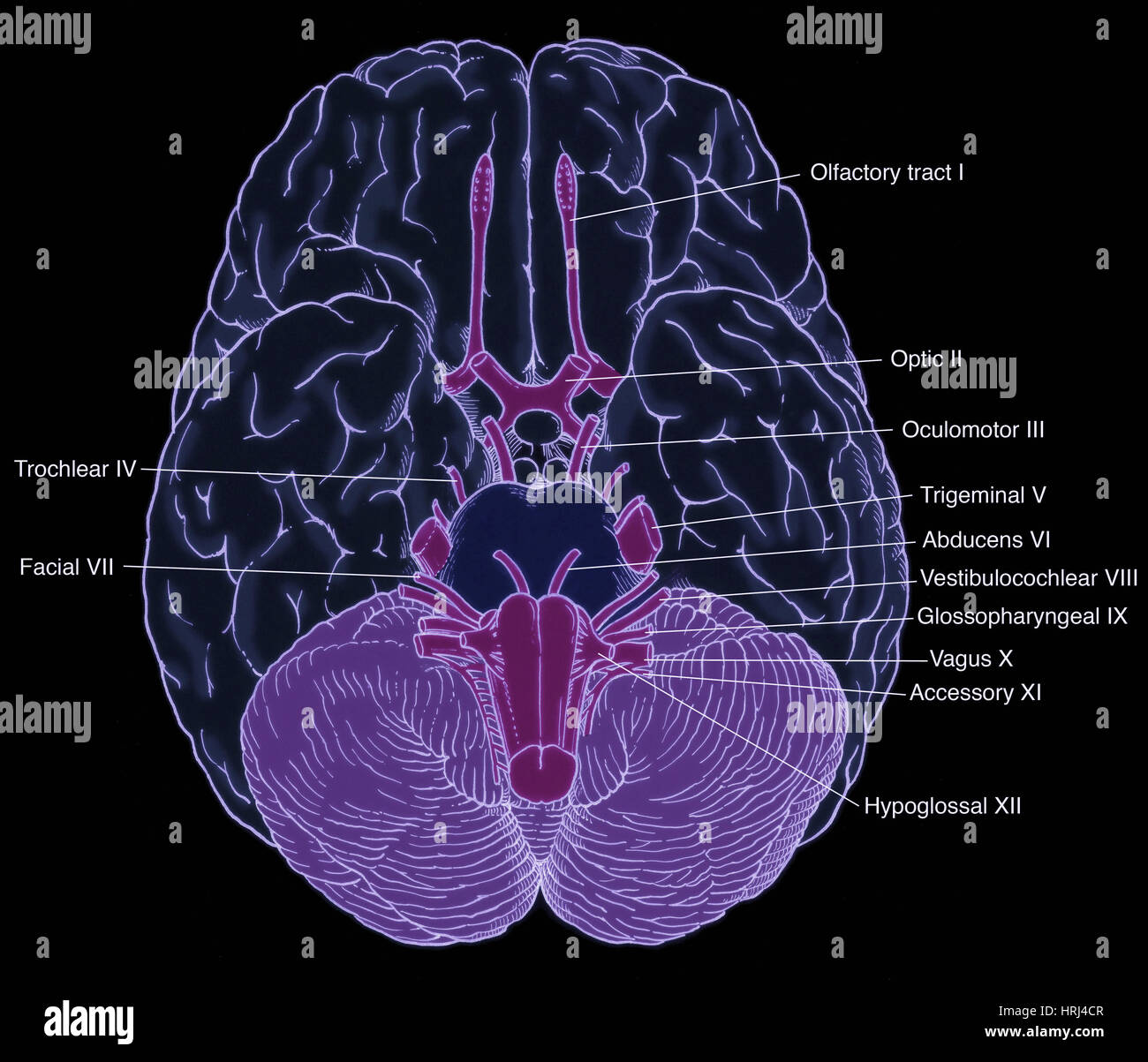 Illustration of Cranial Nerves Stock Photohttps://www.alamy.com/image-license-details/?v=1https://www.alamy.com/stock-photo-illustration-of-cranial-nerves-135008295.html
RMHRJ4CR–Illustration of Cranial Nerves
 Functioning of the macule: organ of static balance (position of the head). Stock Photohttps://www.alamy.com/image-license-details/?v=1https://www.alamy.com/functioning-of-the-macule-organ-of-static-balance-position-of-the-head-image476926488.html
RF2JKWTGT–Functioning of the macule: organ of static balance (position of the head).
 Anatomy of the human ear. Stock Photohttps://www.alamy.com/image-license-details/?v=1https://www.alamy.com/anatomy-of-the-human-ear-image240162233.html
RFRXM9AH–Anatomy of the human ear.
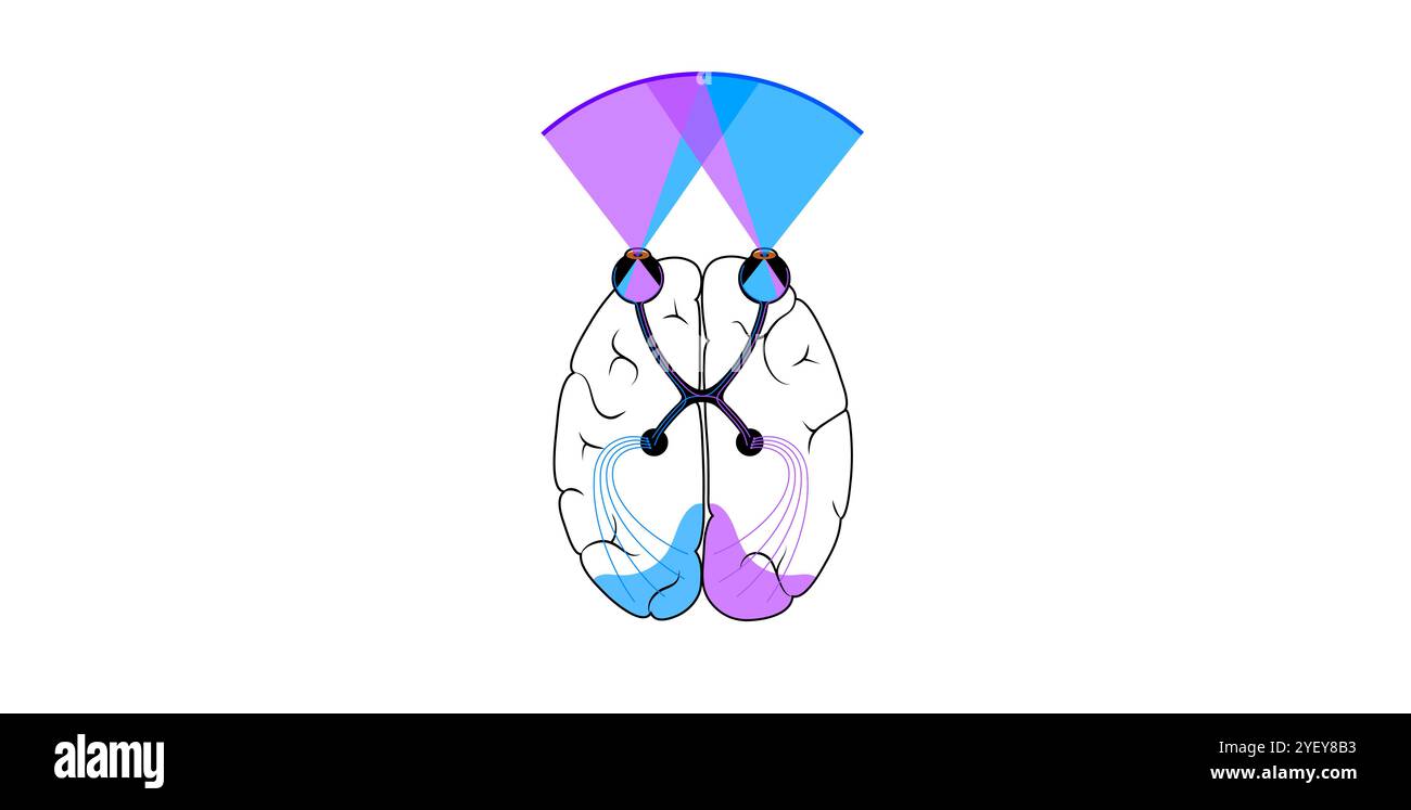 Illustration of the optic nerve anatomy. The optic nerves send visual messages from eye to brain. Stock Photohttps://www.alamy.com/image-license-details/?v=1https://www.alamy.com/illustration-of-the-optic-nerve-anatomy-the-optic-nerves-send-visual-messages-from-eye-to-brain-image628777719.html
RF2YEY8B3–Illustration of the optic nerve anatomy. The optic nerves send visual messages from eye to brain.
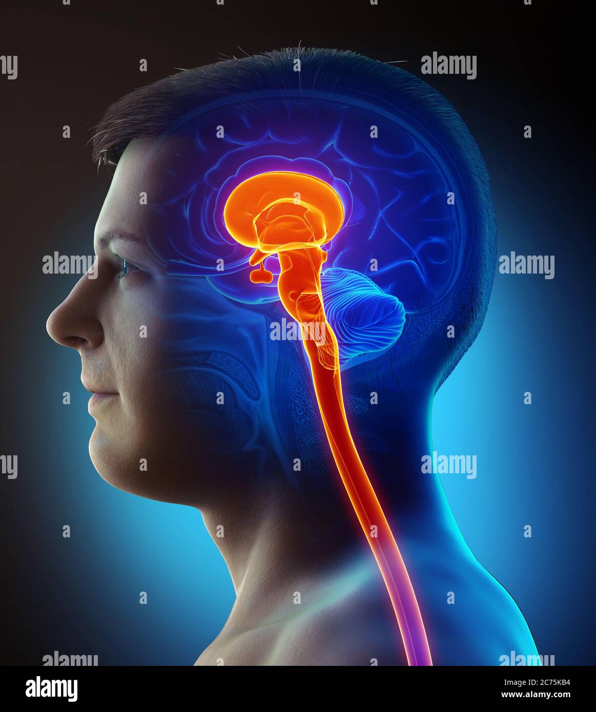 3d rendering medical illustration of brainstem Stock Photohttps://www.alamy.com/image-license-details/?v=1https://www.alamy.com/3d-rendering-medical-illustration-of-brainstem-image365845288.html
RF2C75KB4–3d rendering medical illustration of brainstem
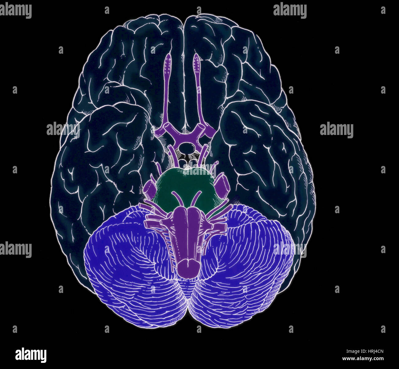 Illustration of Cranial Nerves Stock Photohttps://www.alamy.com/image-license-details/?v=1https://www.alamy.com/stock-photo-illustration-of-cranial-nerves-135008293.html
RMHRJ4CN–Illustration of Cranial Nerves
 Anatomy of the macule showing the cells (ciliates, supports). Stock Photohttps://www.alamy.com/image-license-details/?v=1https://www.alamy.com/anatomy-of-the-macule-showing-the-cells-ciliates-supports-image476926482.html
RF2JKWTGJ–Anatomy of the macule showing the cells (ciliates, supports).
 Anatomy of the human ear. Stock Photohttps://www.alamy.com/image-license-details/?v=1https://www.alamy.com/anatomy-of-the-human-ear-image240162351.html
RFRXM9ER–Anatomy of the human ear.
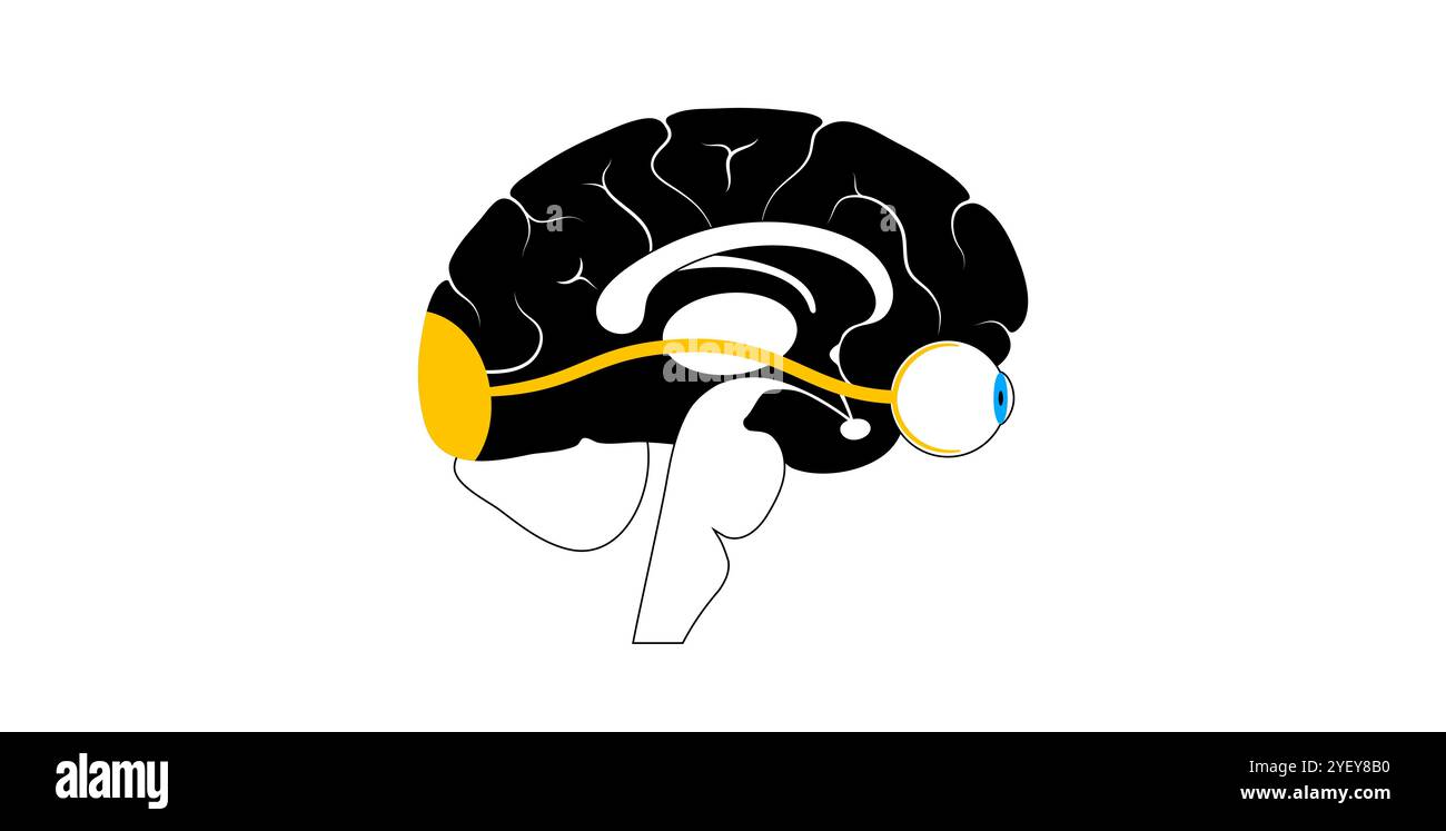 Illustration of the optic nerve anatomy. The optic nerves send visual messages from eye to brain. Stock Photohttps://www.alamy.com/image-license-details/?v=1https://www.alamy.com/illustration-of-the-optic-nerve-anatomy-the-optic-nerves-send-visual-messages-from-eye-to-brain-image628777716.html
RF2YEY8B0–Illustration of the optic nerve anatomy. The optic nerves send visual messages from eye to brain.
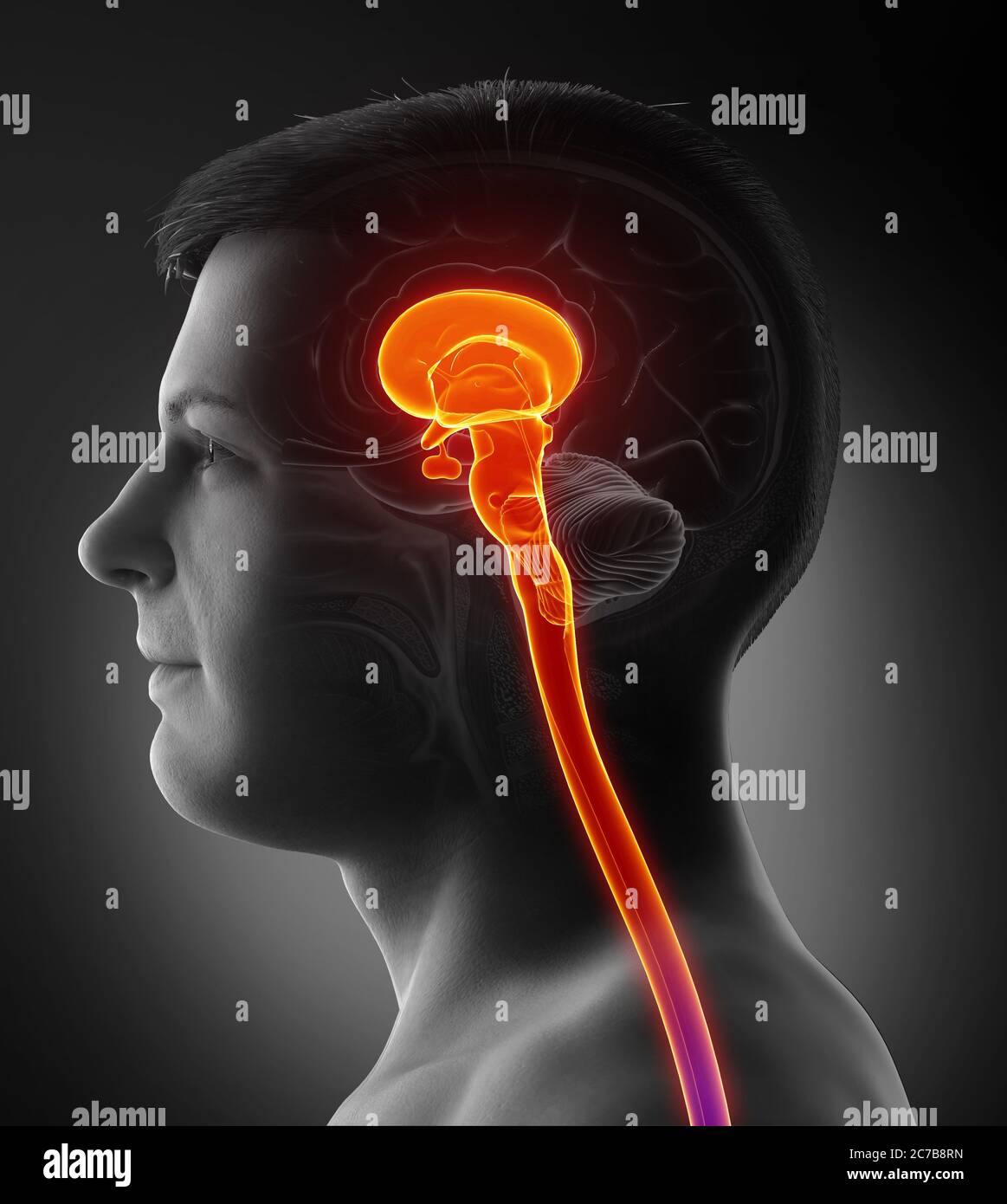 3d rendering medical illustration of brainstem Stock Photohttps://www.alamy.com/image-license-details/?v=1https://www.alamy.com/3d-rendering-medical-illustration-of-brainstem-image365968729.html
RF2C7B8RN–3d rendering medical illustration of brainstem
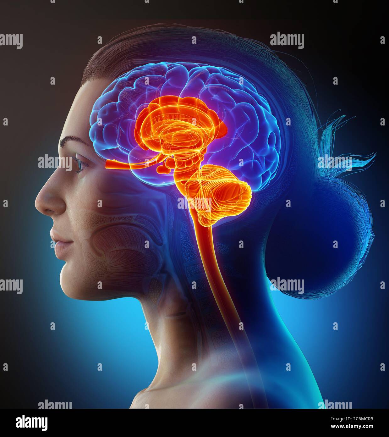 3d rendered medically accurate illustration of the interior brain anatomy Stock Photohttps://www.alamy.com/image-license-details/?v=1https://www.alamy.com/3d-rendered-medically-accurate-illustration-of-the-interior-brain-anatomy-image365554761.html
RF2C6MCR5–3d rendered medically accurate illustration of the interior brain anatomy
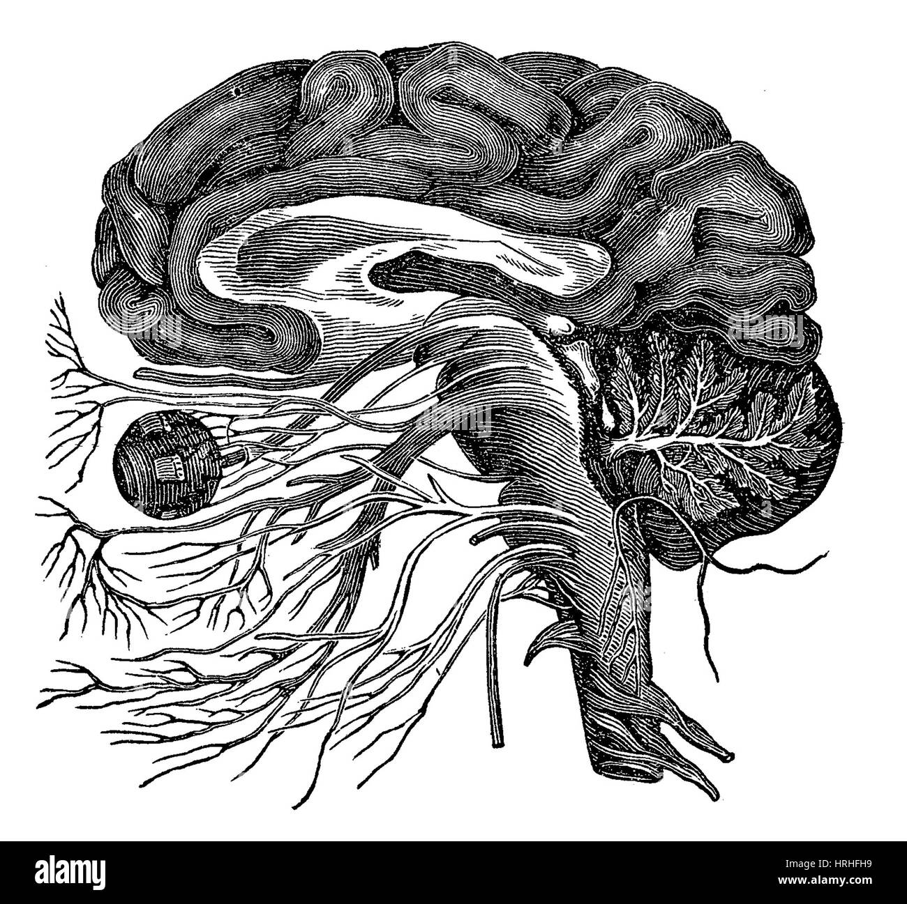 Brain and Cranial Nerves Stock Photohttps://www.alamy.com/image-license-details/?v=1https://www.alamy.com/stock-photo-brain-and-cranial-nerves-134995093.html
RMHRHFH9–Brain and Cranial Nerves
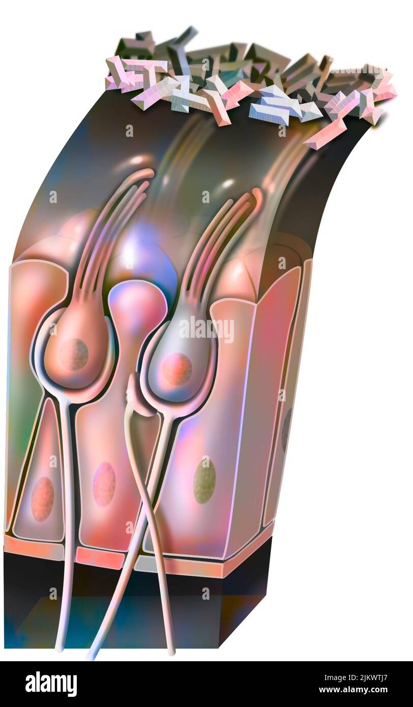 Functioning of the macule: organ of static balance (position of the head). Stock Photohttps://www.alamy.com/image-license-details/?v=1https://www.alamy.com/functioning-of-the-macule-organ-of-static-balance-position-of-the-head-image476926527.html
RF2JKWTJ7–Functioning of the macule: organ of static balance (position of the head).
 Anatomy of the human ear. Stock Photohttps://www.alamy.com/image-license-details/?v=1https://www.alamy.com/anatomy-of-the-human-ear-image240162419.html
RFRXM9H7–Anatomy of the human ear.
 Illustration of the cranial nerves anatomy. The cranial nerves are a set of 12 paired nerves that arise directly from the brain. The first two nerves (olfactory and optic) arise from the cerebrum, whereas the remaining ten emerge from the brainstem. The names of the cranial nerves relate to their function and they are numerically identified in roman numerals. Stock Photohttps://www.alamy.com/image-license-details/?v=1https://www.alamy.com/illustration-of-the-cranial-nerves-anatomy-the-cranial-nerves-are-a-set-of-12-paired-nerves-that-arise-directly-from-the-brain-the-first-two-nerves-olfactory-and-optic-arise-from-the-cerebrum-whereas-the-remaining-ten-emerge-from-the-brainstem-the-names-of-the-cranial-nerves-relate-to-their-function-and-they-are-numerically-identified-in-roman-numerals-image628777664.html
RF2YEY894–Illustration of the cranial nerves anatomy. The cranial nerves are a set of 12 paired nerves that arise directly from the brain. The first two nerves (olfactory and optic) arise from the cerebrum, whereas the remaining ten emerge from the brainstem. The names of the cranial nerves relate to their function and they are numerically identified in roman numerals.
 3d rendered medically accurate illustration of the interior brain anatomy Stock Photohttps://www.alamy.com/image-license-details/?v=1https://www.alamy.com/3d-rendered-medically-accurate-illustration-of-the-interior-brain-anatomy-image365889522.html
RF2C77KPX–3d rendered medically accurate illustration of the interior brain anatomy
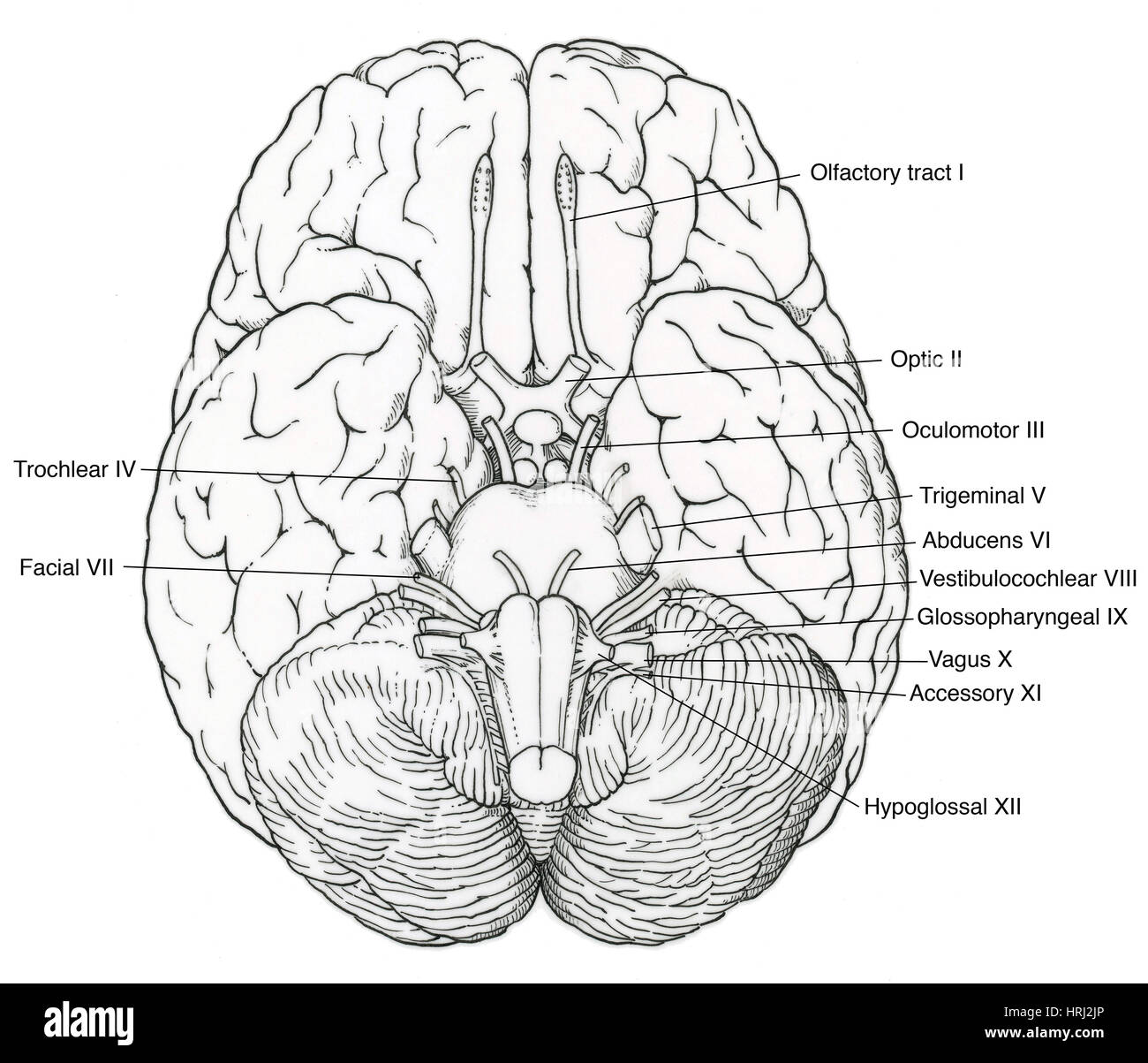 Illustration of Cranial Nerves Stock Photohttps://www.alamy.com/image-license-details/?v=1https://www.alamy.com/stock-photo-illustration-of-cranial-nerves-135006894.html
RMHRJ2JP–Illustration of Cranial Nerves
 Ear cup including: ear canal, eardrum and ossicles, cochlea. Stock Photohttps://www.alamy.com/image-license-details/?v=1https://www.alamy.com/ear-cup-including-ear-canal-eardrum-and-ossicles-cochlea-image476924829.html
RF2JKWPDH–Ear cup including: ear canal, eardrum and ossicles, cochlea.
 Anatomy of the human ear. Stock Photohttps://www.alamy.com/image-license-details/?v=1https://www.alamy.com/anatomy-of-the-human-ear-image240162440.html
RFRXM9J0–Anatomy of the human ear.
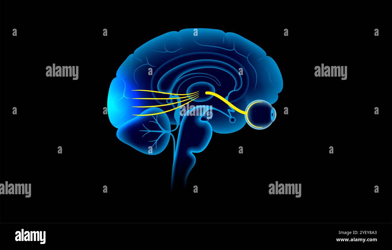 Illustration of the optic nerve anatomy. The optic nerves send visual messages from eye to brain. Stock Photohttps://www.alamy.com/image-license-details/?v=1https://www.alamy.com/illustration-of-the-optic-nerve-anatomy-the-optic-nerves-send-visual-messages-from-eye-to-brain-image628777691.html
RF2YEY8A3–Illustration of the optic nerve anatomy. The optic nerves send visual messages from eye to brain.
 3d rendered medically accurate illustration of the interior brain anatomy Stock Photohttps://www.alamy.com/image-license-details/?v=1https://www.alamy.com/3d-rendered-medically-accurate-illustration-of-the-interior-brain-anatomy-image366057488.html
RF2C7FA1M–3d rendered medically accurate illustration of the interior brain anatomy
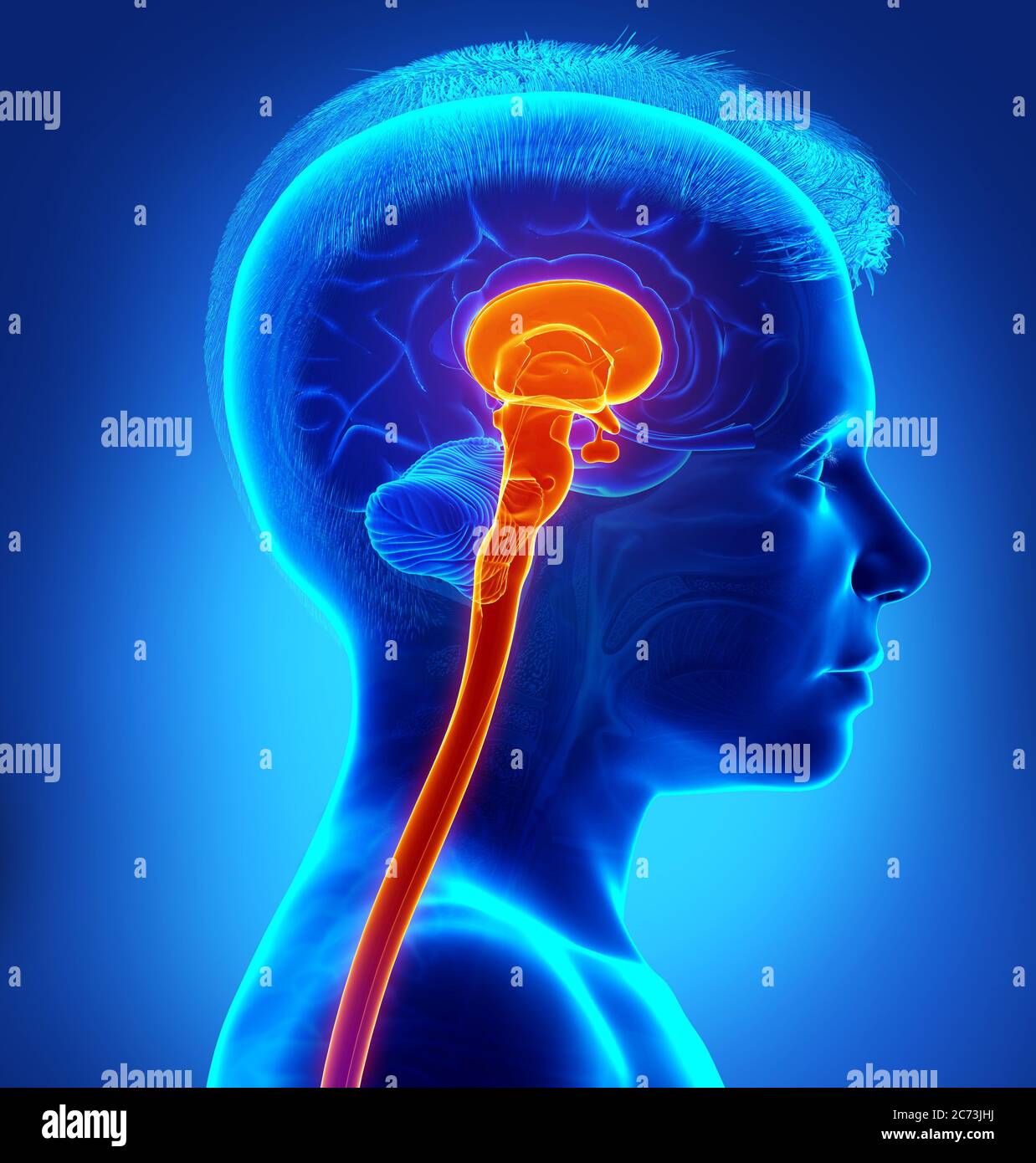 3d rendering medical illustration of brainstem Stock Photohttps://www.alamy.com/image-license-details/?v=1https://www.alamy.com/3d-rendering-medical-illustration-of-brainstem-image365800782.html
RF2C73JHJ–3d rendering medical illustration of brainstem
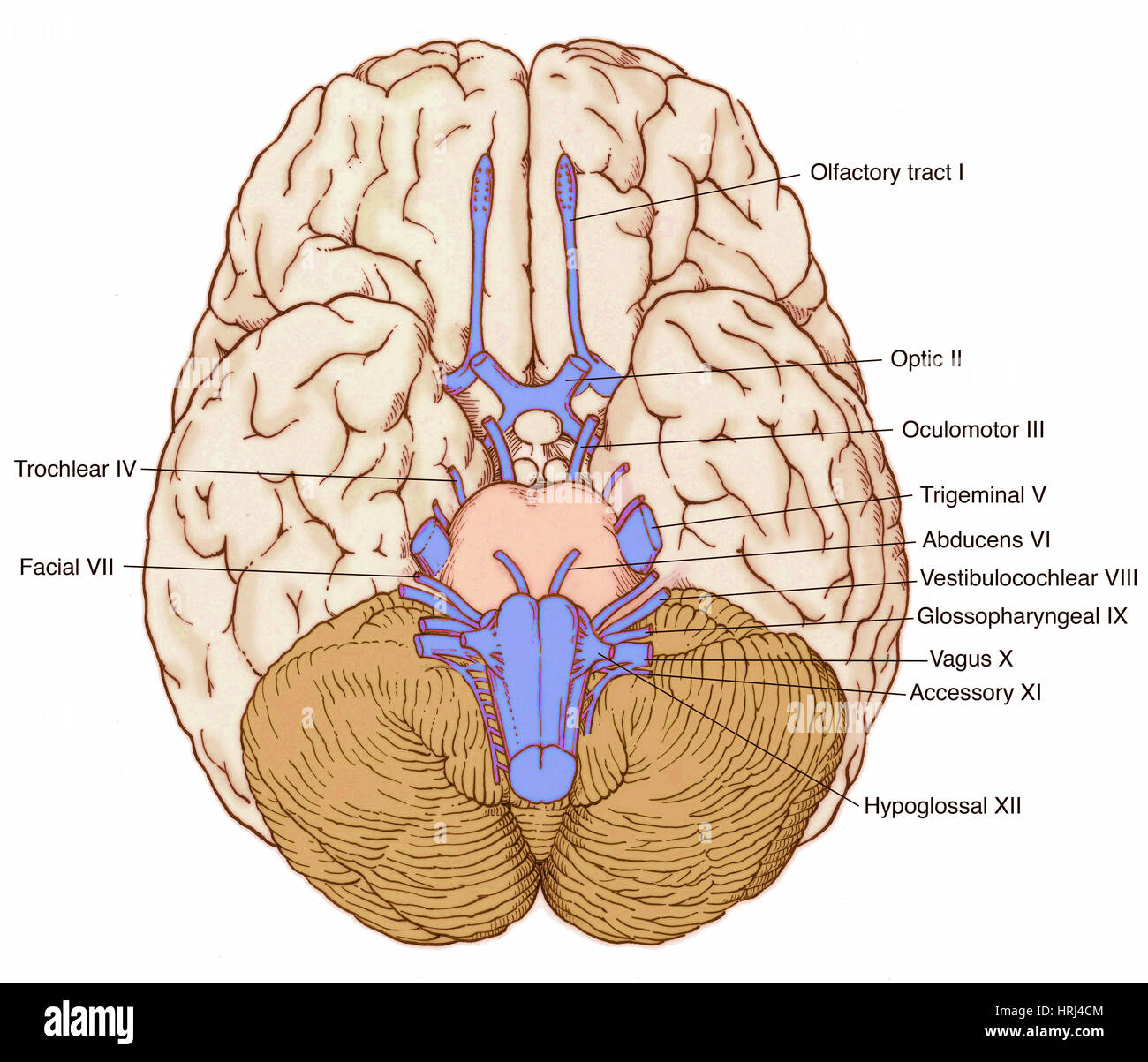 Illustration of Cranial Nerves Stock Photohttps://www.alamy.com/image-license-details/?v=1https://www.alamy.com/stock-photo-illustration-of-cranial-nerves-135008292.html
RMHRJ4CM–Illustration of Cranial Nerves
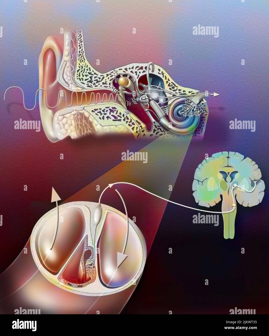 Anatomy of the ear with zoom of the organ of hearing. Stock Photohttps://www.alamy.com/image-license-details/?v=1https://www.alamy.com/anatomy-of-the-ear-with-zoom-of-the-organ-of-hearing-image476926105.html
RF2JKWT35–Anatomy of the ear with zoom of the organ of hearing.
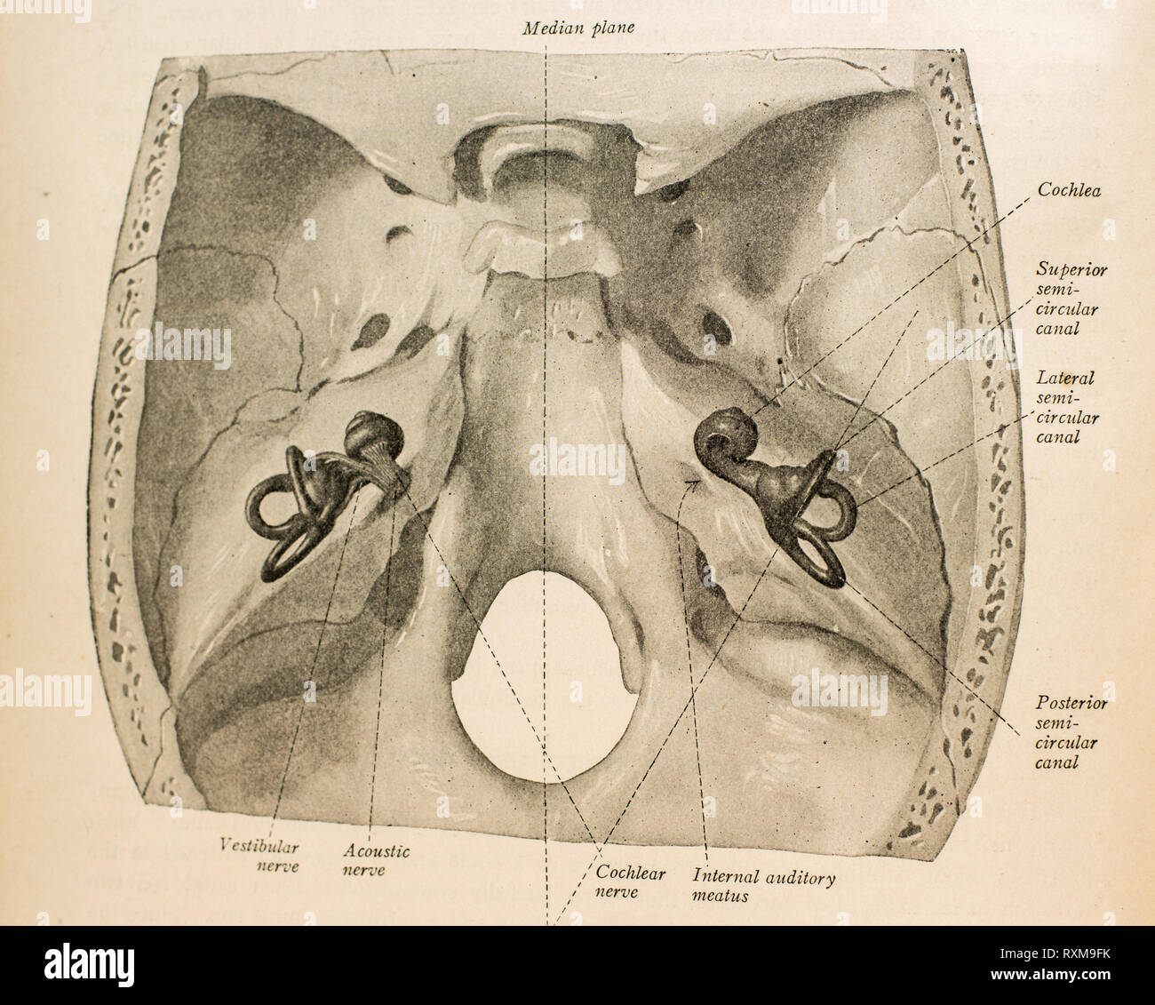 Anatomy of the human ear. Stock Photohttps://www.alamy.com/image-license-details/?v=1https://www.alamy.com/anatomy-of-the-human-ear-image240162375.html
RFRXM9FK–Anatomy of the human ear.
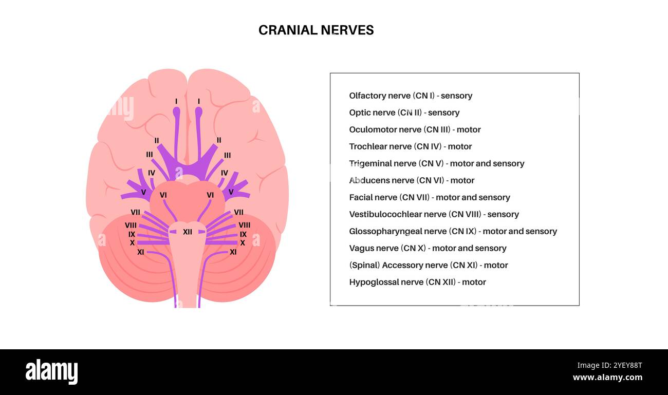 Illustration of the cranial nerves anatomy. The cranial nerves are a set of 12 paired nerves that arise directly from the brain. The first two nerves (olfactory and optic) arise from the cerebrum, whereas the remaining ten emerge from the brainstem. The names of the cranial nerves relate to their function and they are numerically identified in roman numerals. Stock Photohttps://www.alamy.com/image-license-details/?v=1https://www.alamy.com/illustration-of-the-cranial-nerves-anatomy-the-cranial-nerves-are-a-set-of-12-paired-nerves-that-arise-directly-from-the-brain-the-first-two-nerves-olfactory-and-optic-arise-from-the-cerebrum-whereas-the-remaining-ten-emerge-from-the-brainstem-the-names-of-the-cranial-nerves-relate-to-their-function-and-they-are-numerically-identified-in-roman-numerals-image628777656.html
RF2YEY88T–Illustration of the cranial nerves anatomy. The cranial nerves are a set of 12 paired nerves that arise directly from the brain. The first two nerves (olfactory and optic) arise from the cerebrum, whereas the remaining ten emerge from the brainstem. The names of the cranial nerves relate to their function and they are numerically identified in roman numerals.
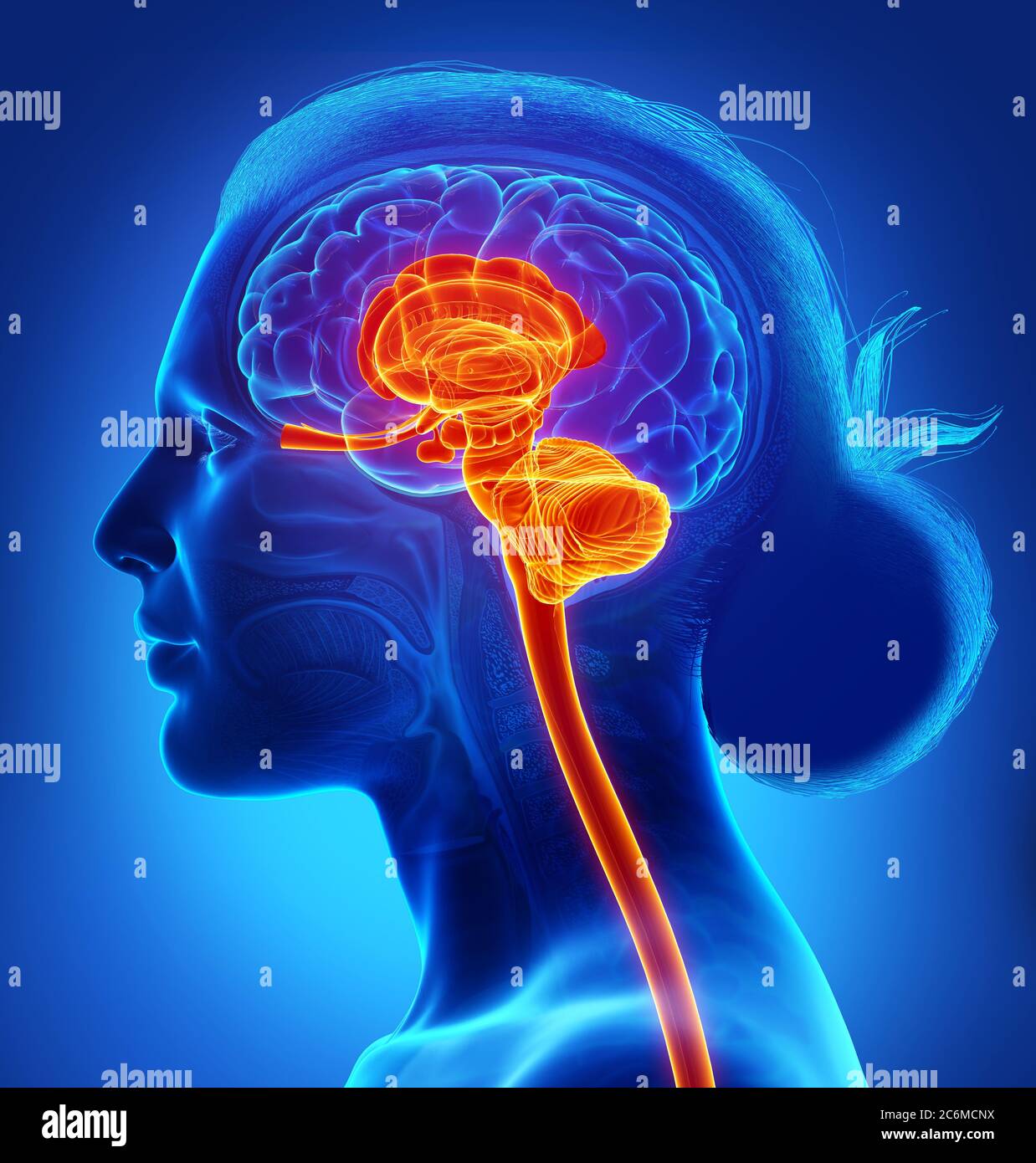 3d rendered medically accurate illustration of the interior brain anatomy Stock Photohttps://www.alamy.com/image-license-details/?v=1https://www.alamy.com/3d-rendered-medically-accurate-illustration-of-the-interior-brain-anatomy-image365554726.html
RF2C6MCNX–3d rendered medically accurate illustration of the interior brain anatomy
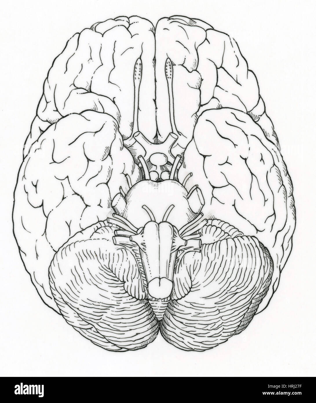 Illustration of Cranial Nerves Stock Photohttps://www.alamy.com/image-license-details/?v=1https://www.alamy.com/stock-photo-illustration-of-cranial-nerves-135006579.html
RMHRJ27F–Illustration of Cranial Nerves
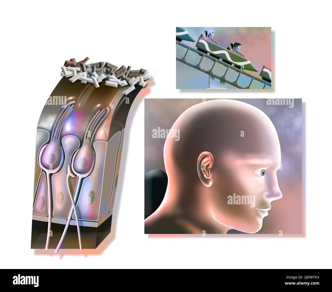 Functioning of the macule: organ of static balance (position of the head). Stock Photohttps://www.alamy.com/image-license-details/?v=1https://www.alamy.com/functioning-of-the-macule-organ-of-static-balance-position-of-the-head-image476926574.html
RF2JKWTKX–Functioning of the macule: organ of static balance (position of the head).
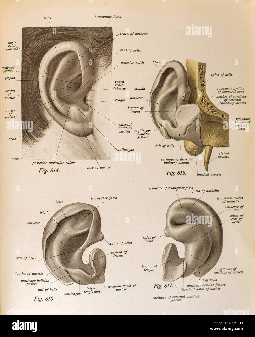 Anatomy of the human ear. Stock Photohttps://www.alamy.com/image-license-details/?v=1https://www.alamy.com/anatomy-of-the-human-ear-image240162309.html
RFRXM9D9–Anatomy of the human ear.
 Illustration of the cranial nerves anatomy. The cranial nerves are a set of 12 paired nerves that arise directly from the brain. The first two nerves (olfactory and optic) arise from the cerebrum, whereas the remaining ten emerge from the brainstem. The names of the cranial nerves relate to their function and they are numerically identified in roman numerals. Stock Photohttps://www.alamy.com/image-license-details/?v=1https://www.alamy.com/illustration-of-the-cranial-nerves-anatomy-the-cranial-nerves-are-a-set-of-12-paired-nerves-that-arise-directly-from-the-brain-the-first-two-nerves-olfactory-and-optic-arise-from-the-cerebrum-whereas-the-remaining-ten-emerge-from-the-brainstem-the-names-of-the-cranial-nerves-relate-to-their-function-and-they-are-numerically-identified-in-roman-numerals-image628777668.html
RF2YEY898–Illustration of the cranial nerves anatomy. The cranial nerves are a set of 12 paired nerves that arise directly from the brain. The first two nerves (olfactory and optic) arise from the cerebrum, whereas the remaining ten emerge from the brainstem. The names of the cranial nerves relate to their function and they are numerically identified in roman numerals.
 3d rendering medical illustration of brainstem Stock Photohttps://www.alamy.com/image-license-details/?v=1https://www.alamy.com/3d-rendering-medical-illustration-of-brainstem-image365964760.html
RF2C7B3P0–3d rendering medical illustration of brainstem
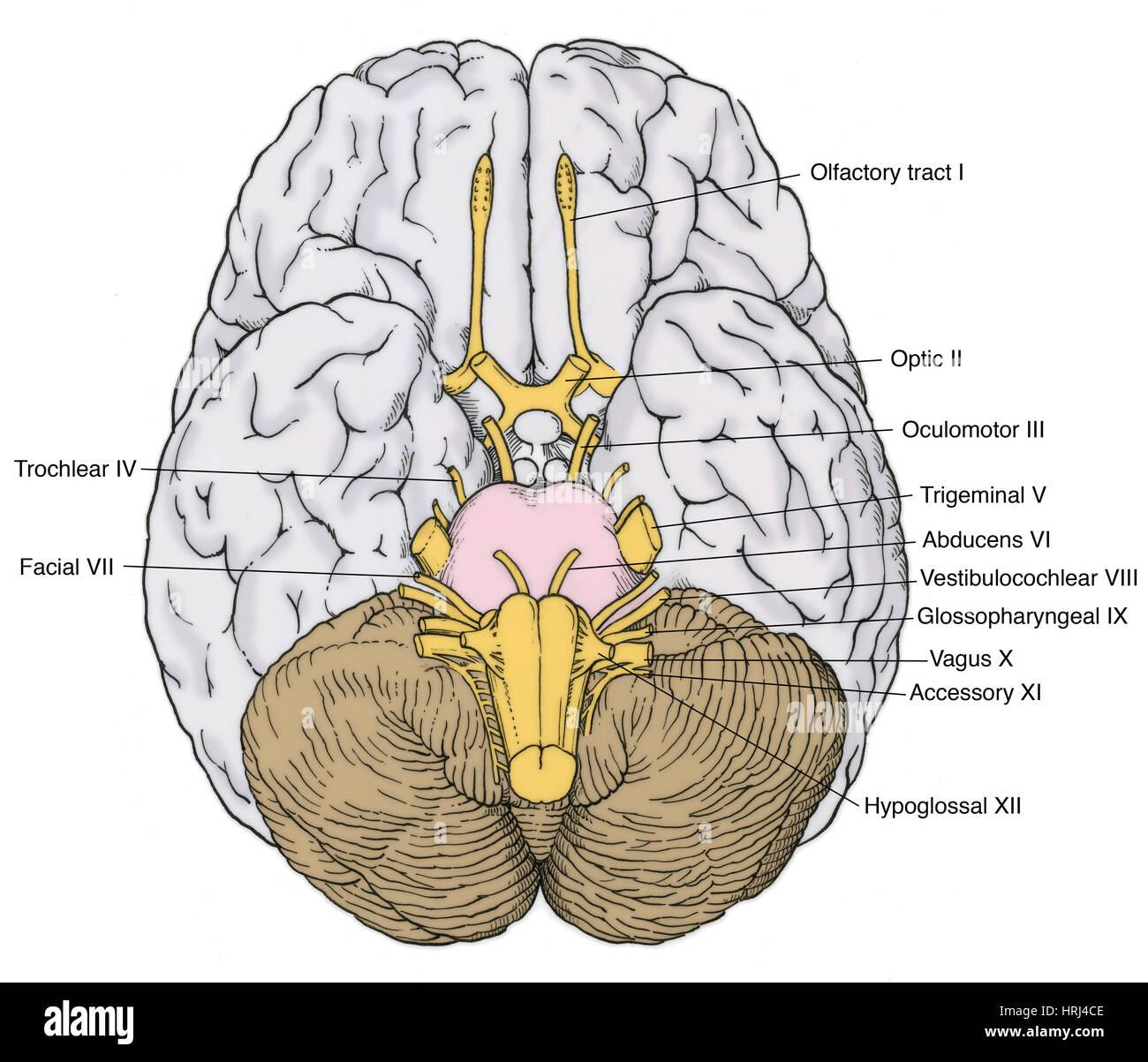 Illustration of Cranial Nerves Stock Photohttps://www.alamy.com/image-license-details/?v=1https://www.alamy.com/stock-photo-illustration-of-cranial-nerves-135008286.html
RMHRJ4CE–Illustration of Cranial Nerves
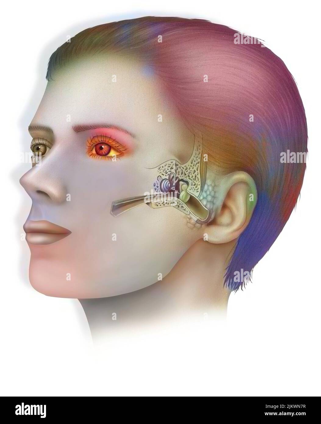 Anatomy of the inner ear showing the eardrum, the cochlea. Stock Photohttps://www.alamy.com/image-license-details/?v=1https://www.alamy.com/anatomy-of-the-inner-ear-showing-the-eardrum-the-cochlea-image476923883.html
RF2JKWN7R–Anatomy of the inner ear showing the eardrum, the cochlea.
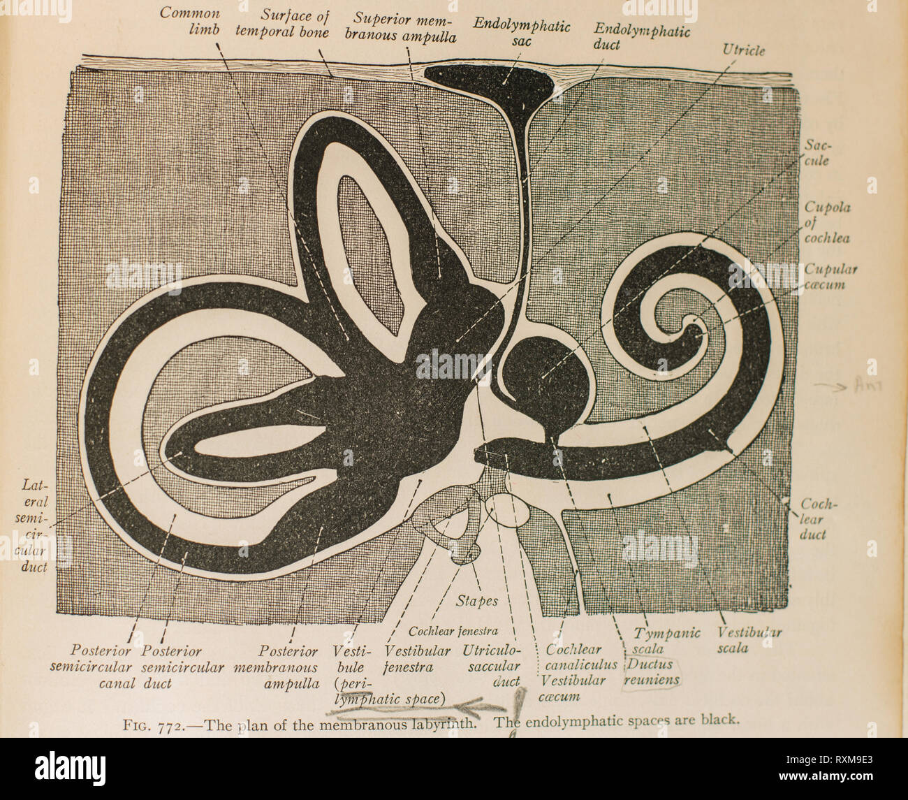 Anatomy of the human ear. Stock Photohttps://www.alamy.com/image-license-details/?v=1https://www.alamy.com/anatomy-of-the-human-ear-image240162331.html
RFRXM9E3–Anatomy of the human ear.
 Illustration of the cranial nerves anatomy. The cranial nerves are a set of 12 paired nerves that arise directly from the brain. The first two nerves (olfactory and optic) arise from the cerebrum, whereas the remaining ten emerge from the brainstem. The names of the cranial nerves relate to their function and they are numerically identified in roman numerals. Stock Photohttps://www.alamy.com/image-license-details/?v=1https://www.alamy.com/illustration-of-the-cranial-nerves-anatomy-the-cranial-nerves-are-a-set-of-12-paired-nerves-that-arise-directly-from-the-brain-the-first-two-nerves-olfactory-and-optic-arise-from-the-cerebrum-whereas-the-remaining-ten-emerge-from-the-brainstem-the-names-of-the-cranial-nerves-relate-to-their-function-and-they-are-numerically-identified-in-roman-numerals-image628777667.html
RF2YEY897–Illustration of the cranial nerves anatomy. The cranial nerves are a set of 12 paired nerves that arise directly from the brain. The first two nerves (olfactory and optic) arise from the cerebrum, whereas the remaining ten emerge from the brainstem. The names of the cranial nerves relate to their function and they are numerically identified in roman numerals.
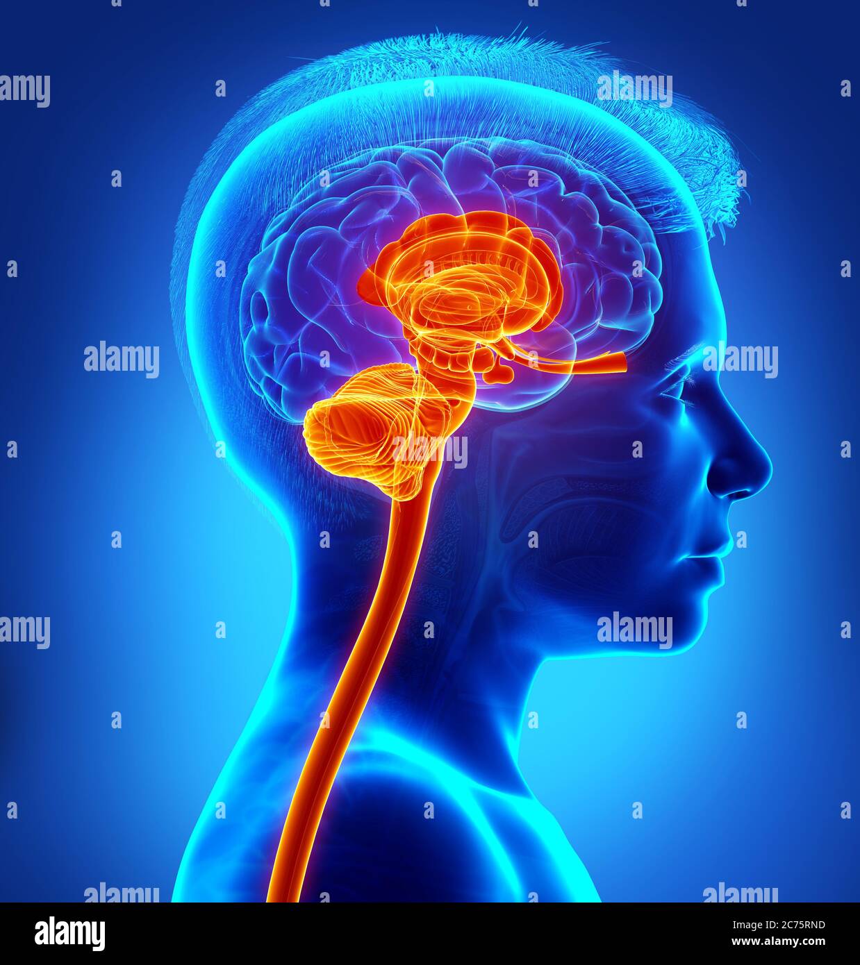 3d rendered medically accurate illustration of the interior brain anatomy Stock Photohttps://www.alamy.com/image-license-details/?v=1https://www.alamy.com/3d-rendered-medically-accurate-illustration-of-the-interior-brain-anatomy-image365848713.html
RF2C75RND–3d rendered medically accurate illustration of the interior brain anatomy
 Undersurface of the Brain Stock Photohttps://www.alamy.com/image-license-details/?v=1https://www.alamy.com/stock-photo-undersurface-of-the-brain-134995082.html
RMHRHFGX–Undersurface of the Brain
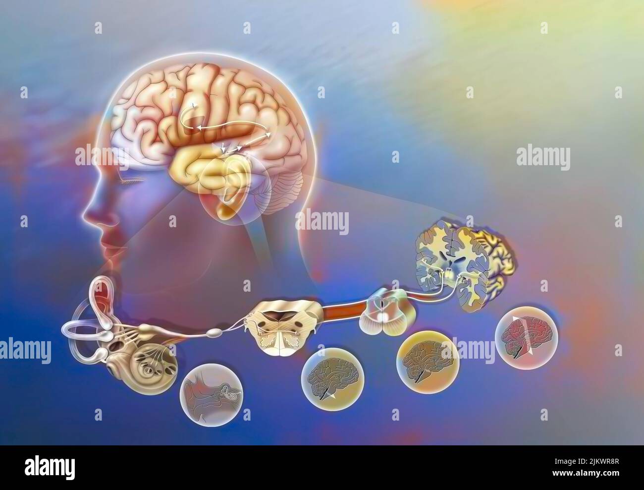 How a sound when it has reached the cochlea spreads through the brain. Stock Photohttps://www.alamy.com/image-license-details/?v=1https://www.alamy.com/how-a-sound-when-it-has-reached-the-cochlea-spreads-through-the-brain-image476925479.html
RF2JKWR8R–How a sound when it has reached the cochlea spreads through the brain.
 Anatomy of the human ear. Stock Photohttps://www.alamy.com/image-license-details/?v=1https://www.alamy.com/anatomy-of-the-human-ear-image240162432.html
RFRXM9HM–Anatomy of the human ear.
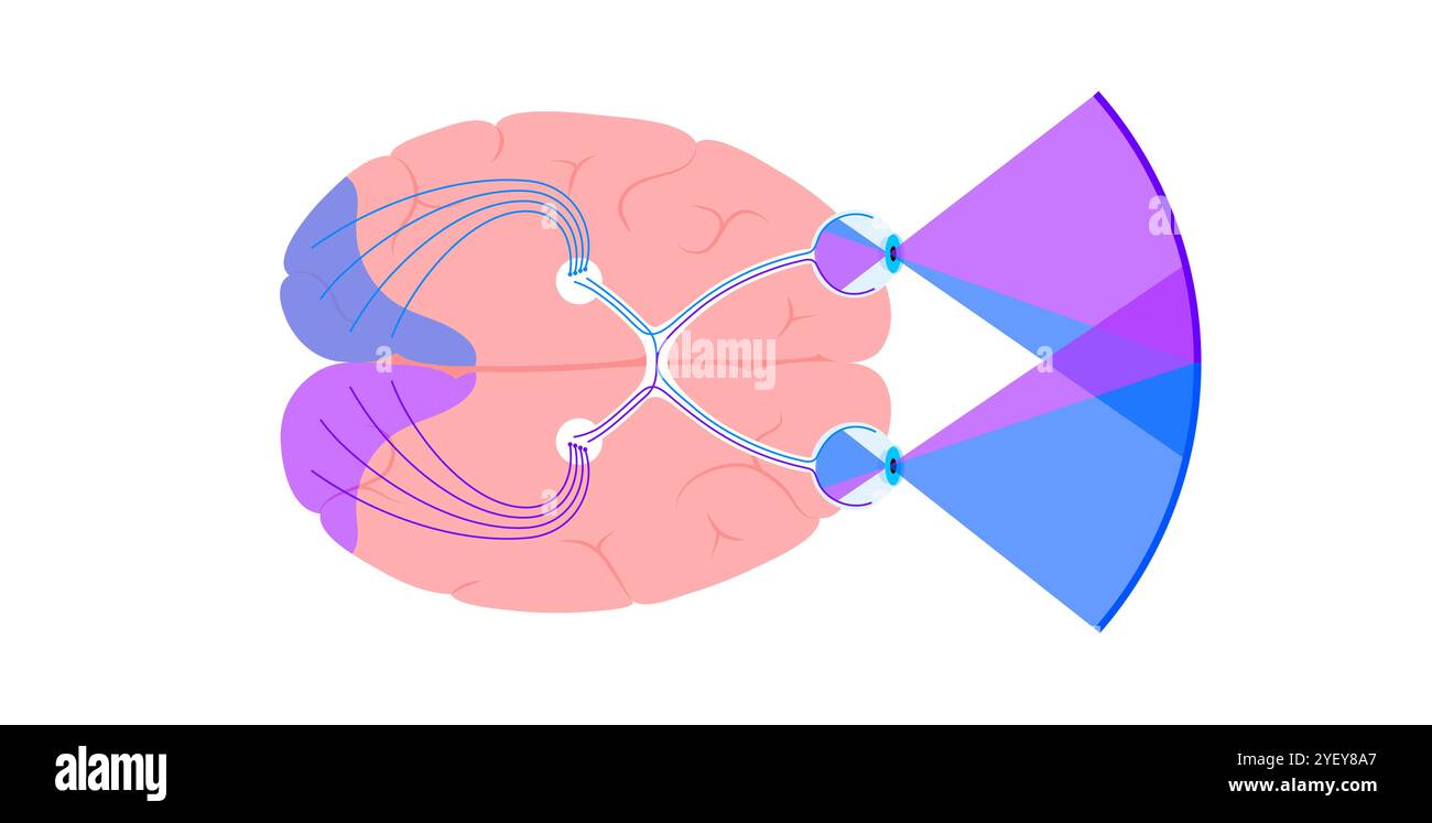 Illustration of the optic nerve anatomy. The optic nerves send visual messages from eye to brain. Stock Photohttps://www.alamy.com/image-license-details/?v=1https://www.alamy.com/illustration-of-the-optic-nerve-anatomy-the-optic-nerves-send-visual-messages-from-eye-to-brain-image628777695.html
RF2YEY8A7–Illustration of the optic nerve anatomy. The optic nerves send visual messages from eye to brain.
 3d rendering medical illustration of brainstem Stock Photohttps://www.alamy.com/image-license-details/?v=1https://www.alamy.com/3d-rendering-medical-illustration-of-brainstem-image365926522.html
RF2C79B0A–3d rendering medical illustration of brainstem
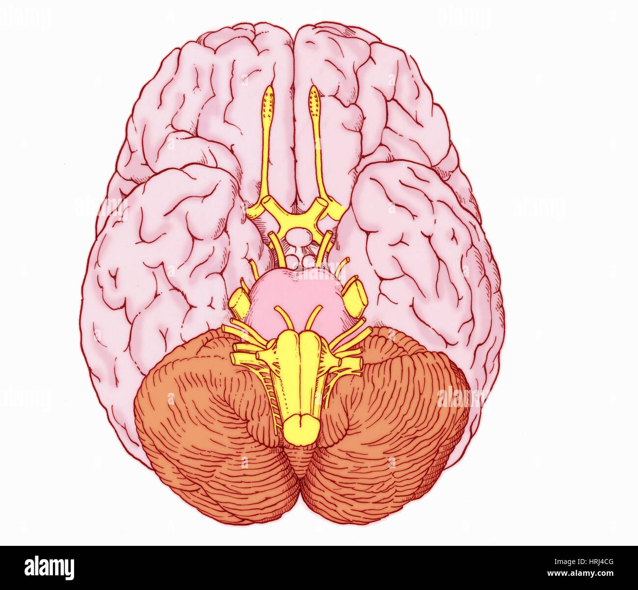 Illustration of Cranial Nerves Stock Photohttps://www.alamy.com/image-license-details/?v=1https://www.alamy.com/stock-photo-illustration-of-cranial-nerves-135008288.html
RMHRJ4CG–Illustration of Cranial Nerves
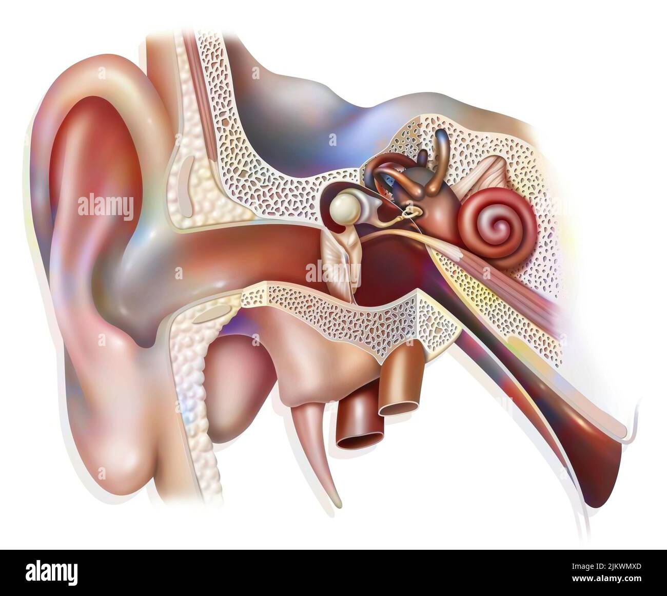 Anatomy of the inner ear showing the eardrum, the cochlea. Stock Photohttps://www.alamy.com/image-license-details/?v=1https://www.alamy.com/anatomy-of-the-inner-ear-showing-the-eardrum-the-cochlea-image476923621.html
RF2JKWMXD–Anatomy of the inner ear showing the eardrum, the cochlea.
 Anatomy of the human ear. Stock Photohttps://www.alamy.com/image-license-details/?v=1https://www.alamy.com/anatomy-of-the-human-ear-image240162276.html
RFRXM9C4–Anatomy of the human ear.
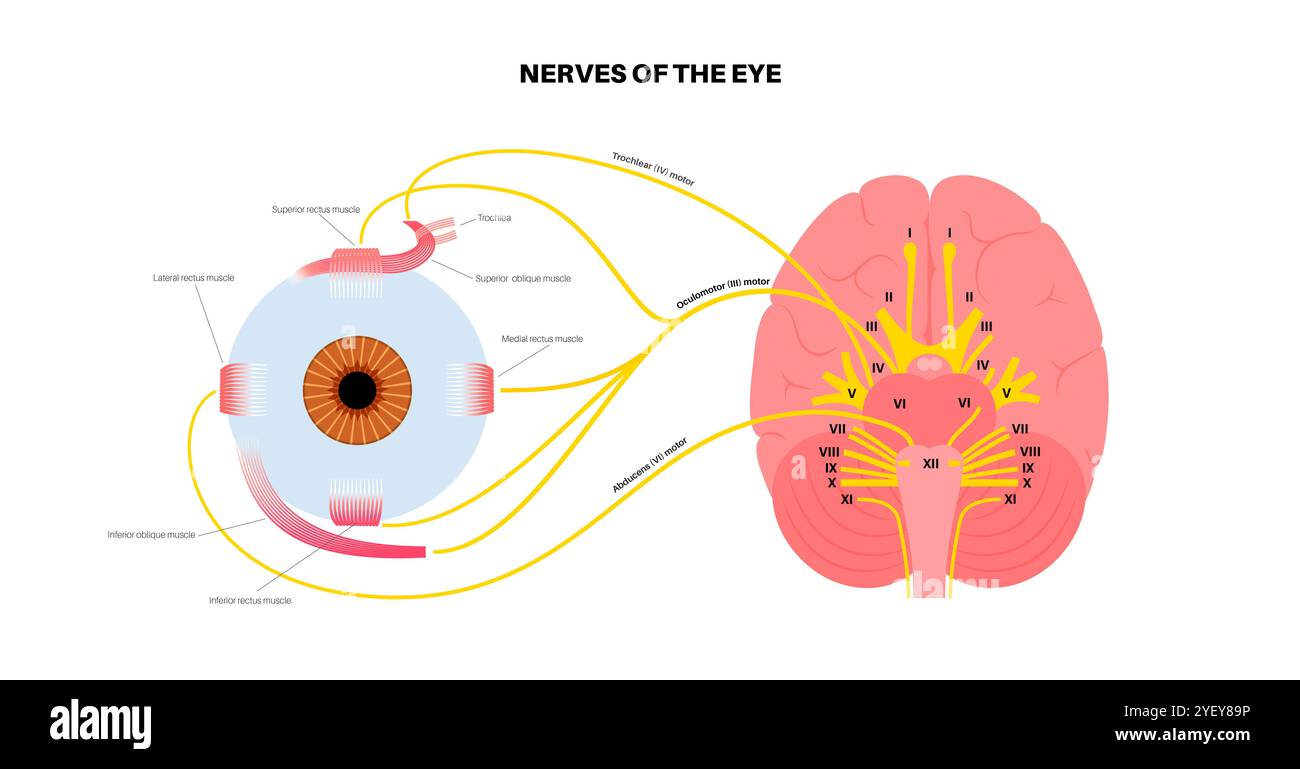 Illustration of the motor nerves of the eye, including the abducens, trochlear and oculomotor nerves in the human brain. These nerves innervate motor, sensory, and autonomic structures in the eyes. Stock Photohttps://www.alamy.com/image-license-details/?v=1https://www.alamy.com/illustration-of-the-motor-nerves-of-the-eye-including-the-abducens-trochlear-and-oculomotor-nerves-in-the-human-brain-these-nerves-innervate-motor-sensory-and-autonomic-structures-in-the-eyes-image628777682.html
RF2YEY89P–Illustration of the motor nerves of the eye, including the abducens, trochlear and oculomotor nerves in the human brain. These nerves innervate motor, sensory, and autonomic structures in the eyes.
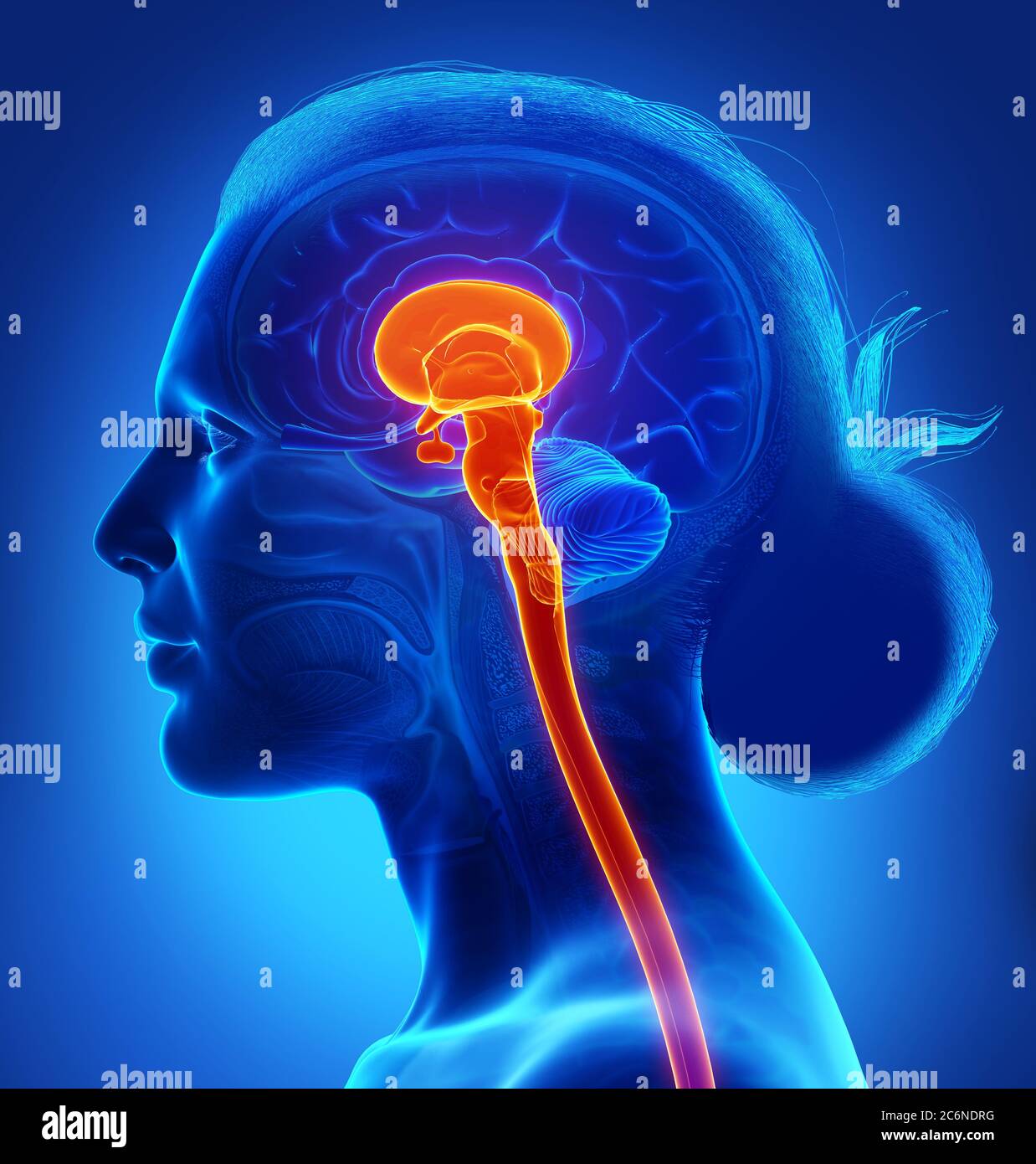 3d rendering medical illustration of brainstem Stock Photohttps://www.alamy.com/image-license-details/?v=1https://www.alamy.com/3d-rendering-medical-illustration-of-brainstem-image365577508.html
RF2C6NDRG–3d rendering medical illustration of brainstem
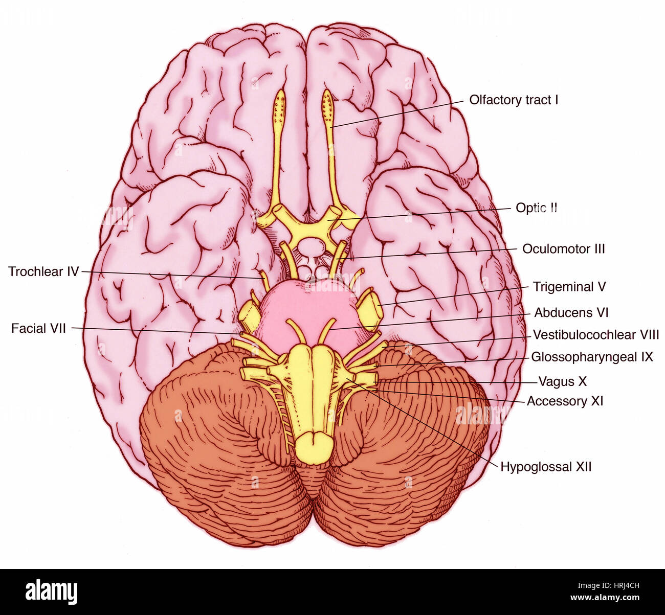 Illustration of Cranial Nerves Stock Photohttps://www.alamy.com/image-license-details/?v=1https://www.alamy.com/stock-photo-illustration-of-cranial-nerves-135008289.html
RMHRJ4CH–Illustration of Cranial Nerves
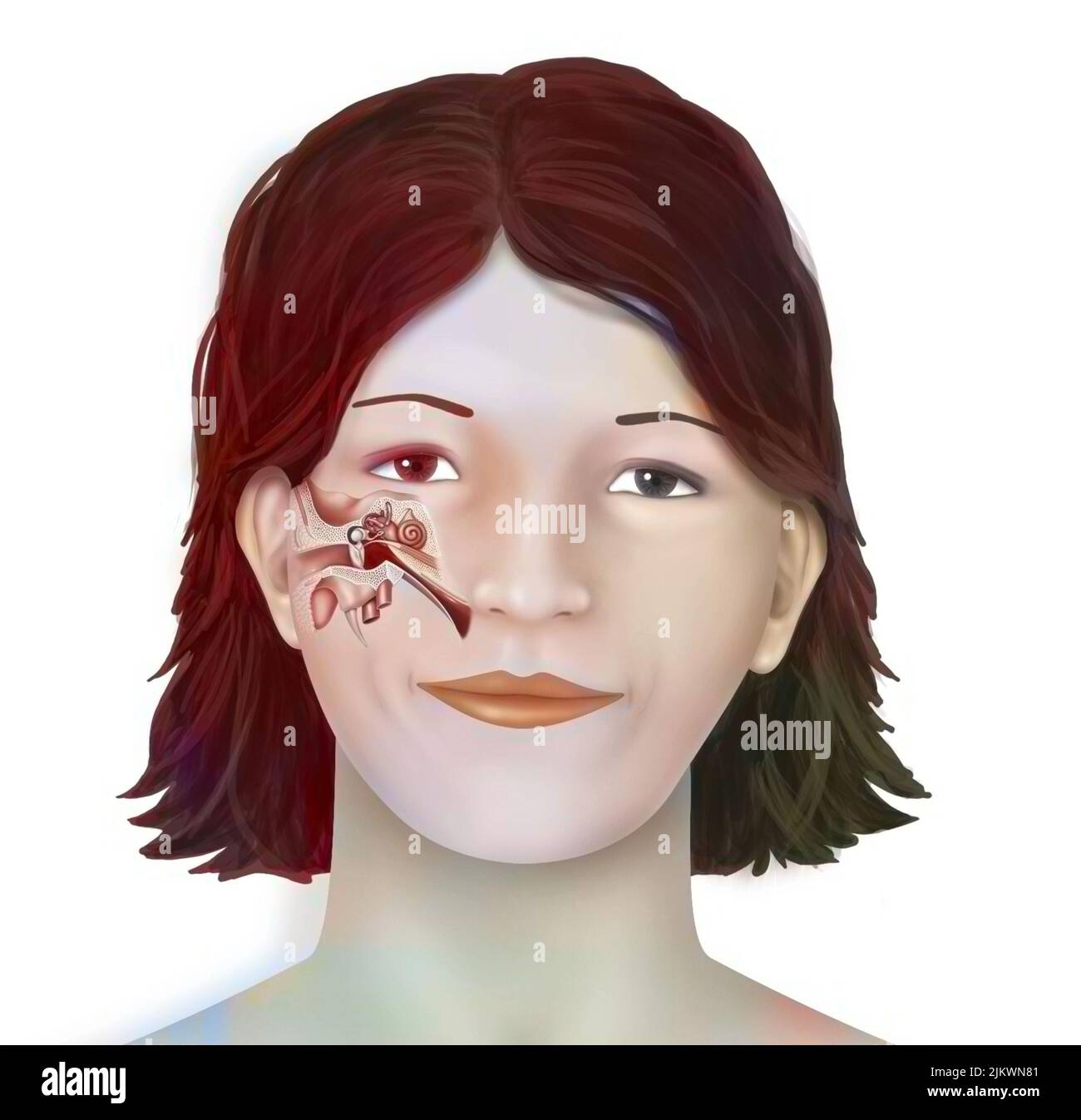 Anatomy of the inner ear showing the eardrum, the cochlea. Stock Photohttps://www.alamy.com/image-license-details/?v=1https://www.alamy.com/anatomy-of-the-inner-ear-showing-the-eardrum-the-cochlea-image476923889.html
RF2JKWN81–Anatomy of the inner ear showing the eardrum, the cochlea.
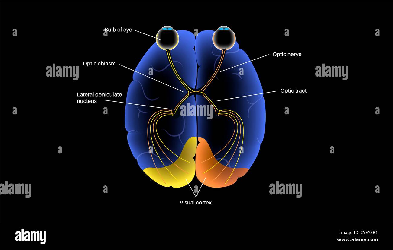 Illustration of the optic nerve anatomy. The optic nerves send visual messages from eye to brain. Stock Photohttps://www.alamy.com/image-license-details/?v=1https://www.alamy.com/illustration-of-the-optic-nerve-anatomy-the-optic-nerves-send-visual-messages-from-eye-to-brain-image628777717.html
RF2YEY8B1–Illustration of the optic nerve anatomy. The optic nerves send visual messages from eye to brain.
 3d rendered medically accurate illustration of the interior brain anatomy Stock Photohttps://www.alamy.com/image-license-details/?v=1https://www.alamy.com/3d-rendered-medically-accurate-illustration-of-the-interior-brain-anatomy-image365991995.html
RF2C7CAEK–3d rendered medically accurate illustration of the interior brain anatomy
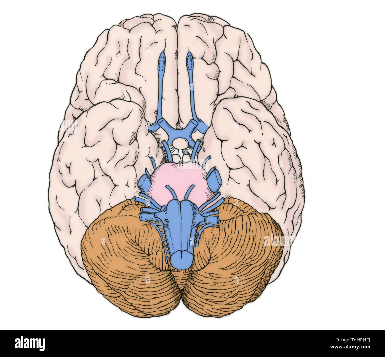 Illustration of Cranial Nerves Stock Photohttps://www.alamy.com/image-license-details/?v=1https://www.alamy.com/stock-photo-illustration-of-cranial-nerves-135008290.html
RMHRJ4CJ–Illustration of Cranial Nerves
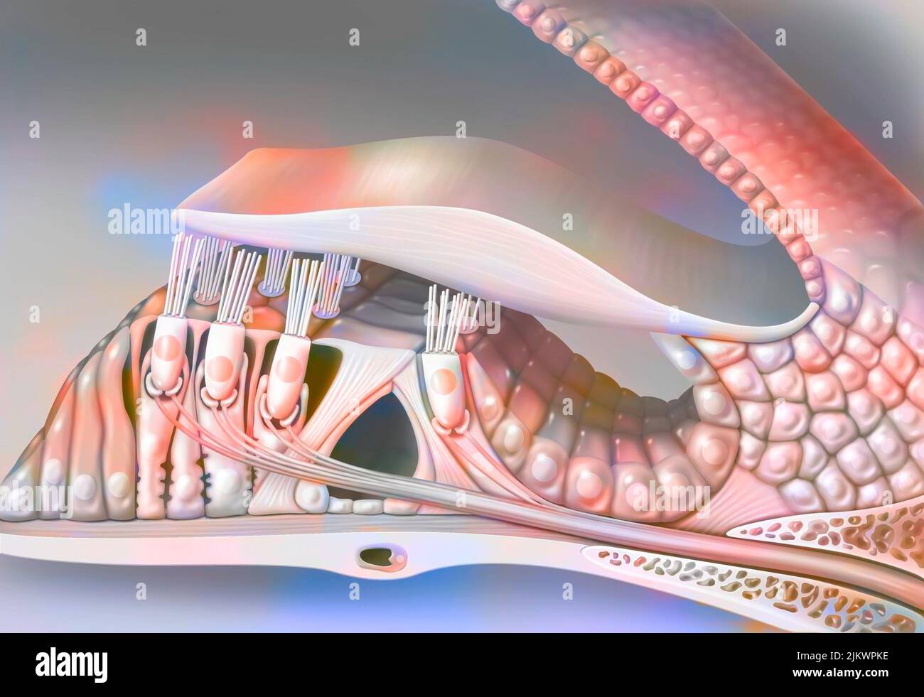 The organ of Corti (cut through a coil of the cochlea). Stock Photohttps://www.alamy.com/image-license-details/?v=1https://www.alamy.com/the-organ-of-corti-cut-through-a-coil-of-the-cochlea-image476924994.html
RF2JKWPKE–The organ of Corti (cut through a coil of the cochlea).
 Illustration of the oculomotor nerve anatomy in the human brain. The oculomotor nerve divides into superior and inferior branches in the anterior part of the cavernous sinus. Stock Photohttps://www.alamy.com/image-license-details/?v=1https://www.alamy.com/illustration-of-the-oculomotor-nerve-anatomy-in-the-human-brain-the-oculomotor-nerve-divides-into-superior-and-inferior-branches-in-the-anterior-part-of-the-cavernous-sinus-image628777681.html
RF2YEY89N–Illustration of the oculomotor nerve anatomy in the human brain. The oculomotor nerve divides into superior and inferior branches in the anterior part of the cavernous sinus.
 3d rendered medically accurate illustration of the interior brain anatomy Stock Photohttps://www.alamy.com/image-license-details/?v=1https://www.alamy.com/3d-rendered-medically-accurate-illustration-of-the-interior-brain-anatomy-image365571939.html
RF2C6N6MK–3d rendered medically accurate illustration of the interior brain anatomy
 Illustration of Cranial Nerves Stock Photohttps://www.alamy.com/image-license-details/?v=1https://www.alamy.com/stock-photo-illustration-of-cranial-nerves-135008285.html
RMHRJ4CD–Illustration of Cranial Nerves
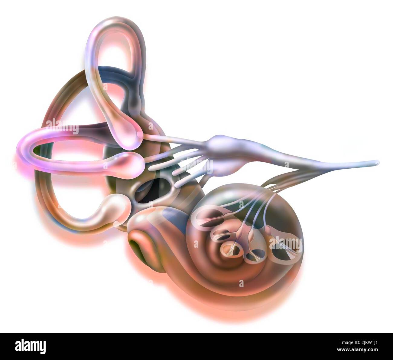 Inner ear and vestibular apparatus with semicircular canals, macule. Stock Photohttps://www.alamy.com/image-license-details/?v=1https://www.alamy.com/inner-ear-and-vestibular-apparatus-with-semicircular-canals-macule-image476926521.html
RF2JKWTJ1–Inner ear and vestibular apparatus with semicircular canals, macule.
 Illustration of the motor nerves of the eye, including the abducens, trochlear and oculomotor nerves in the human brain. These nerves innervate motor, sensory, and autonomic structures in the eyes. Stock Photohttps://www.alamy.com/image-license-details/?v=1https://www.alamy.com/illustration-of-the-motor-nerves-of-the-eye-including-the-abducens-trochlear-and-oculomotor-nerves-in-the-human-brain-these-nerves-innervate-motor-sensory-and-autonomic-structures-in-the-eyes-image628777708.html
RF2YEY8AM–Illustration of the motor nerves of the eye, including the abducens, trochlear and oculomotor nerves in the human brain. These nerves innervate motor, sensory, and autonomic structures in the eyes.
 3d rendering medical illustration of brainstem Stock Photohttps://www.alamy.com/image-license-details/?v=1https://www.alamy.com/3d-rendering-medical-illustration-of-brainstem-image365975798.html
RF2C7BHT6–3d rendering medical illustration of brainstem
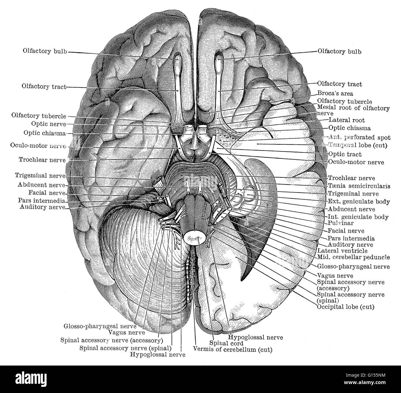 Illustration of the undersurface of the brain showing the nerves. This is an historical illustration from the 1890's. Stock Photohttps://www.alamy.com/image-license-details/?v=1https://www.alamy.com/stock-photo-illustration-of-the-undersurface-of-the-brain-showing-the-nerves-this-103991152.html
RMG155NM–Illustration of the undersurface of the brain showing the nerves. This is an historical illustration from the 1890's.
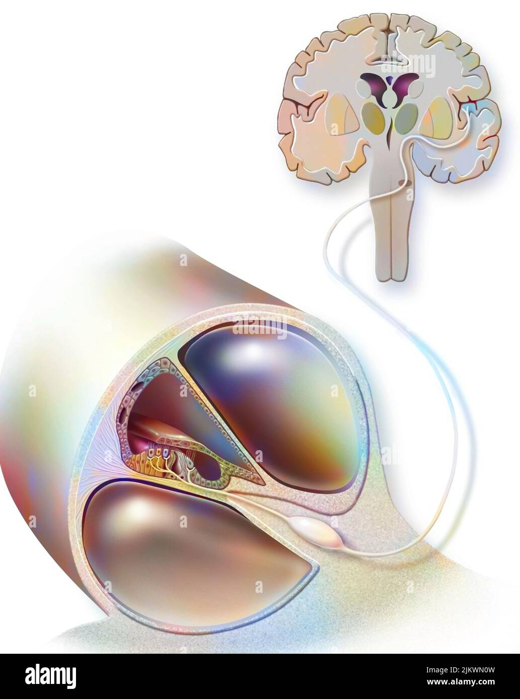 Path of sound from the organ of Corti to the auditory area of the brain. Stock Photohttps://www.alamy.com/image-license-details/?v=1https://www.alamy.com/path-of-sound-from-the-organ-of-corti-to-the-auditory-area-of-the-brain-image476923689.html
RF2JKWN0W–Path of sound from the organ of Corti to the auditory area of the brain.
 Illustration of the oculomotor nerve anatomy in the human brain. The oculomotor nerve divides into superior and inferior branches in the anterior part of the cavernous sinus. Stock Photohttps://www.alamy.com/image-license-details/?v=1https://www.alamy.com/illustration-of-the-oculomotor-nerve-anatomy-in-the-human-brain-the-oculomotor-nerve-divides-into-superior-and-inferior-branches-in-the-anterior-part-of-the-cavernous-sinus-image628777712.html
RF2YEY8AT–Illustration of the oculomotor nerve anatomy in the human brain. The oculomotor nerve divides into superior and inferior branches in the anterior part of the cavernous sinus.
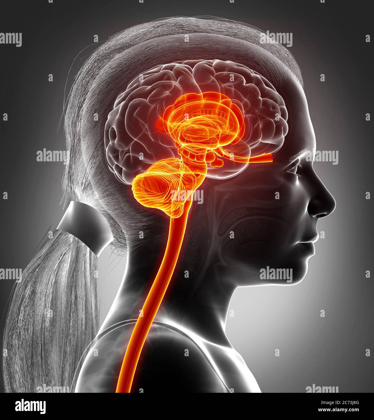 3d rendered medically accurate illustration of the interior brain anatomy Stock Photohttps://www.alamy.com/image-license-details/?v=1https://www.alamy.com/3d-rendered-medically-accurate-illustration-of-the-interior-brain-anatomy-image365800528.html
RF2C73J8G–3d rendered medically accurate illustration of the interior brain anatomy
 Color enhanced illustration of the undersurface of the brain showing the nerves. This is an historical illustration from the 1890's. Stock Photohttps://www.alamy.com/image-license-details/?v=1https://www.alamy.com/stock-photo-color-enhanced-illustration-of-the-undersurface-of-the-brain-showing-103991153.html
RMG155NN–Color enhanced illustration of the undersurface of the brain showing the nerves. This is an historical illustration from the 1890's.
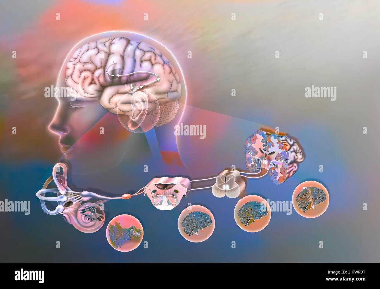 How a sound when it has reached the cochlea spreads through the brain. Stock Photohttps://www.alamy.com/image-license-details/?v=1https://www.alamy.com/how-a-sound-when-it-has-reached-the-cochlea-spreads-through-the-brain-image476925508.html
RF2JKWR9T–How a sound when it has reached the cochlea spreads through the brain.
 Illustration of the oculomotor nerve anatomy in the human brain. The oculomotor nerve divides into superior and inferior branches in the anterior part of the cavernous sinus. Stock Photohttps://www.alamy.com/image-license-details/?v=1https://www.alamy.com/illustration-of-the-oculomotor-nerve-anatomy-in-the-human-brain-the-oculomotor-nerve-divides-into-superior-and-inferior-branches-in-the-anterior-part-of-the-cavernous-sinus-image628777679.html
RF2YEY89K–Illustration of the oculomotor nerve anatomy in the human brain. The oculomotor nerve divides into superior and inferior branches in the anterior part of the cavernous sinus.
 3d rendered medically accurate illustration of the interior brain anatomy Stock Photohttps://www.alamy.com/image-license-details/?v=1https://www.alamy.com/3d-rendered-medically-accurate-illustration-of-the-interior-brain-anatomy-image365801061.html
RF2C73JYH–3d rendered medically accurate illustration of the interior brain anatomy
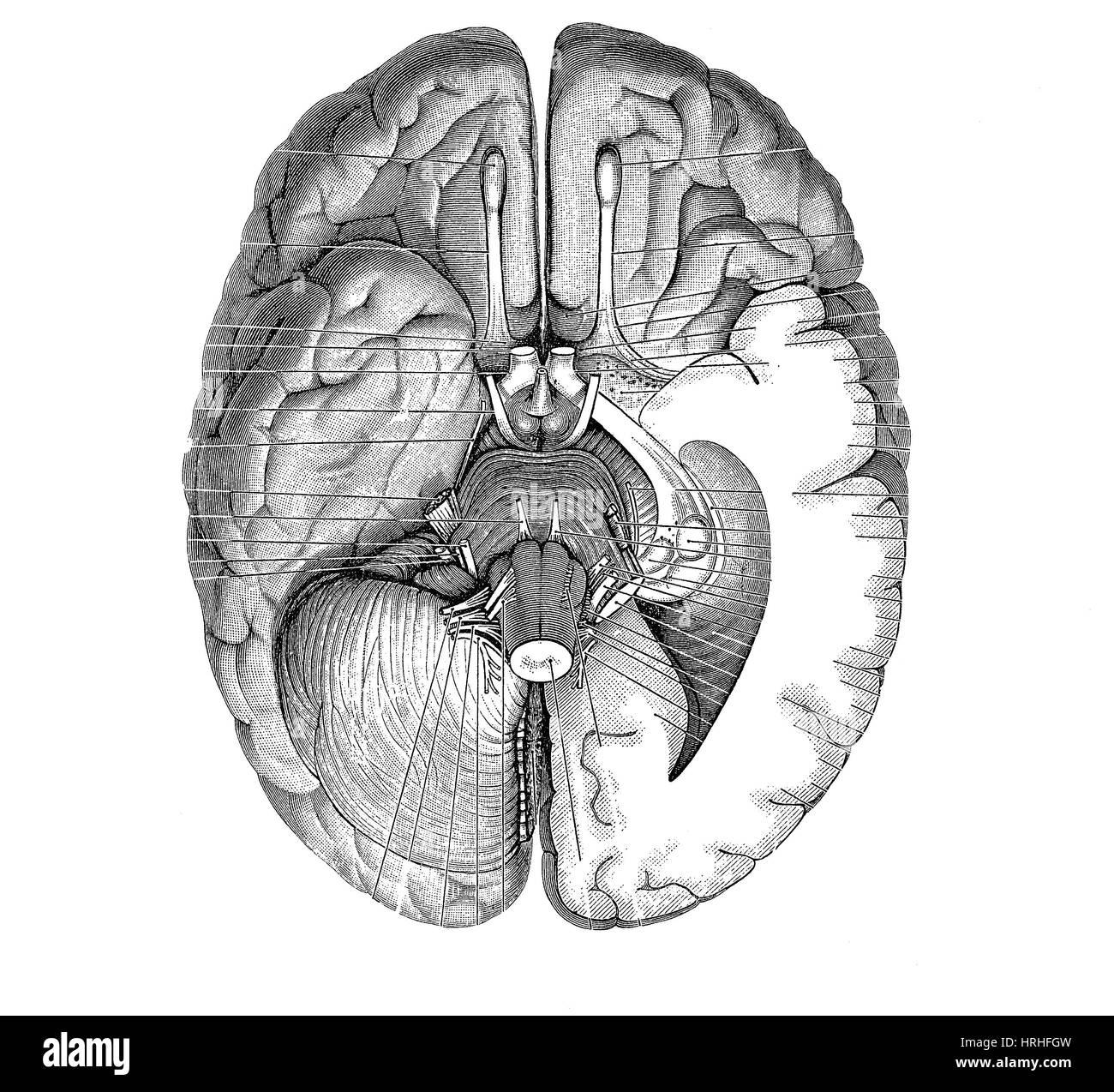 Undersurface of the Brain Stock Photohttps://www.alamy.com/image-license-details/?v=1https://www.alamy.com/stock-photo-undersurface-of-the-brain-134995081.html
RMHRHFGW–Undersurface of the Brain
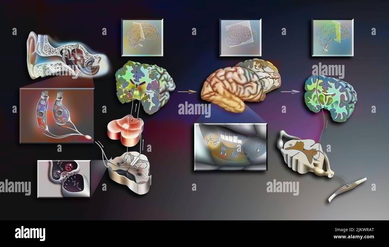 Perception and interpretation of sound in the brain. Stock Photohttps://www.alamy.com/image-license-details/?v=1https://www.alamy.com/perception-and-interpretation-of-sound-in-the-brain-image476925536.html
RF2JKWRAT–Perception and interpretation of sound in the brain.
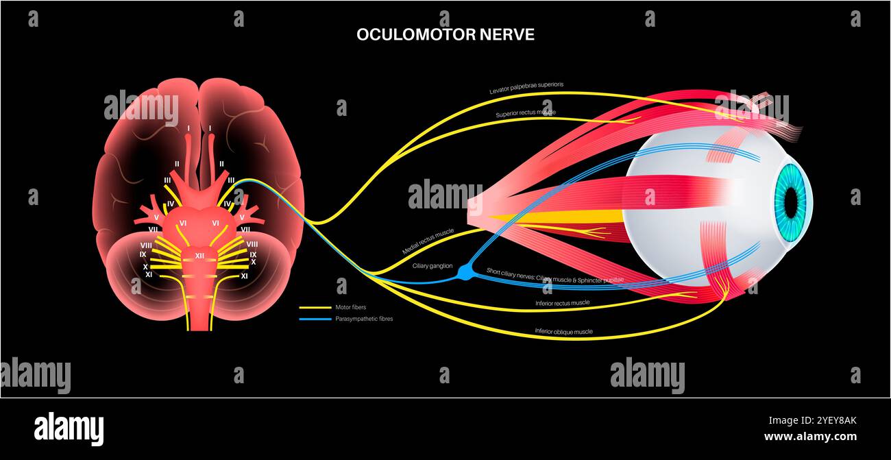 Illustration of the oculomotor nerve anatomy in the human brain. The oculomotor nerve divides into superior and inferior branches in the anterior part of the cavernous sinus. Stock Photohttps://www.alamy.com/image-license-details/?v=1https://www.alamy.com/illustration-of-the-oculomotor-nerve-anatomy-in-the-human-brain-the-oculomotor-nerve-divides-into-superior-and-inferior-branches-in-the-anterior-part-of-the-cavernous-sinus-image628777707.html
RF2YEY8AK–Illustration of the oculomotor nerve anatomy in the human brain. The oculomotor nerve divides into superior and inferior branches in the anterior part of the cavernous sinus.
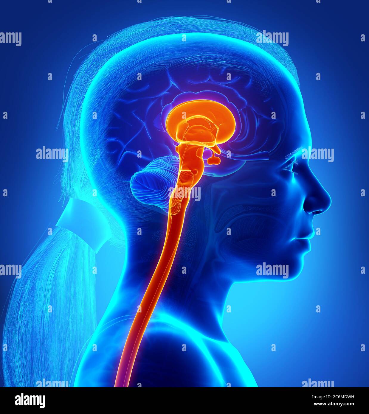 3d rendering medical illustration of brainstem Stock Photohttps://www.alamy.com/image-license-details/?v=1https://www.alamy.com/3d-rendering-medical-illustration-of-brainstem-image365555613.html
RF2C6MDWH–3d rendering medical illustration of brainstem
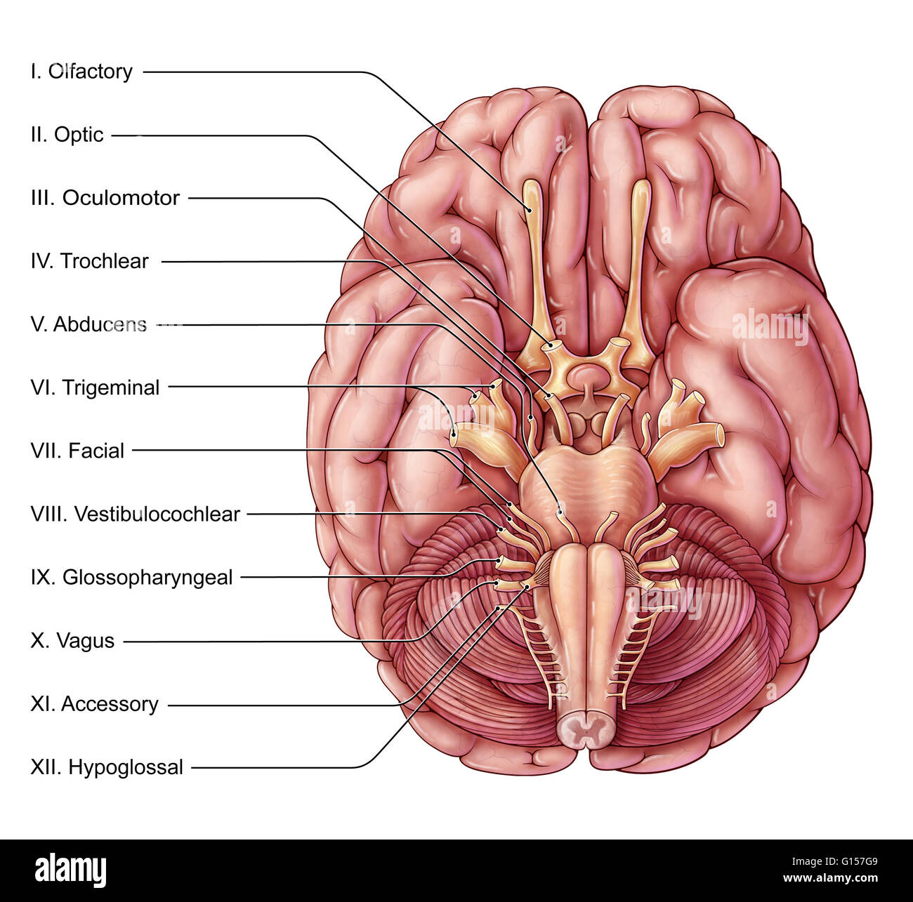 An illustration of the brain from an inferior (basal) view showing the twelve pairs of cranial nerves. Stock Photohttps://www.alamy.com/image-license-details/?v=1https://www.alamy.com/stock-photo-an-illustration-of-the-brain-from-an-inferior-basal-view-showing-the-103992569.html
RMG157G9–An illustration of the brain from an inferior (basal) view showing the twelve pairs of cranial nerves.
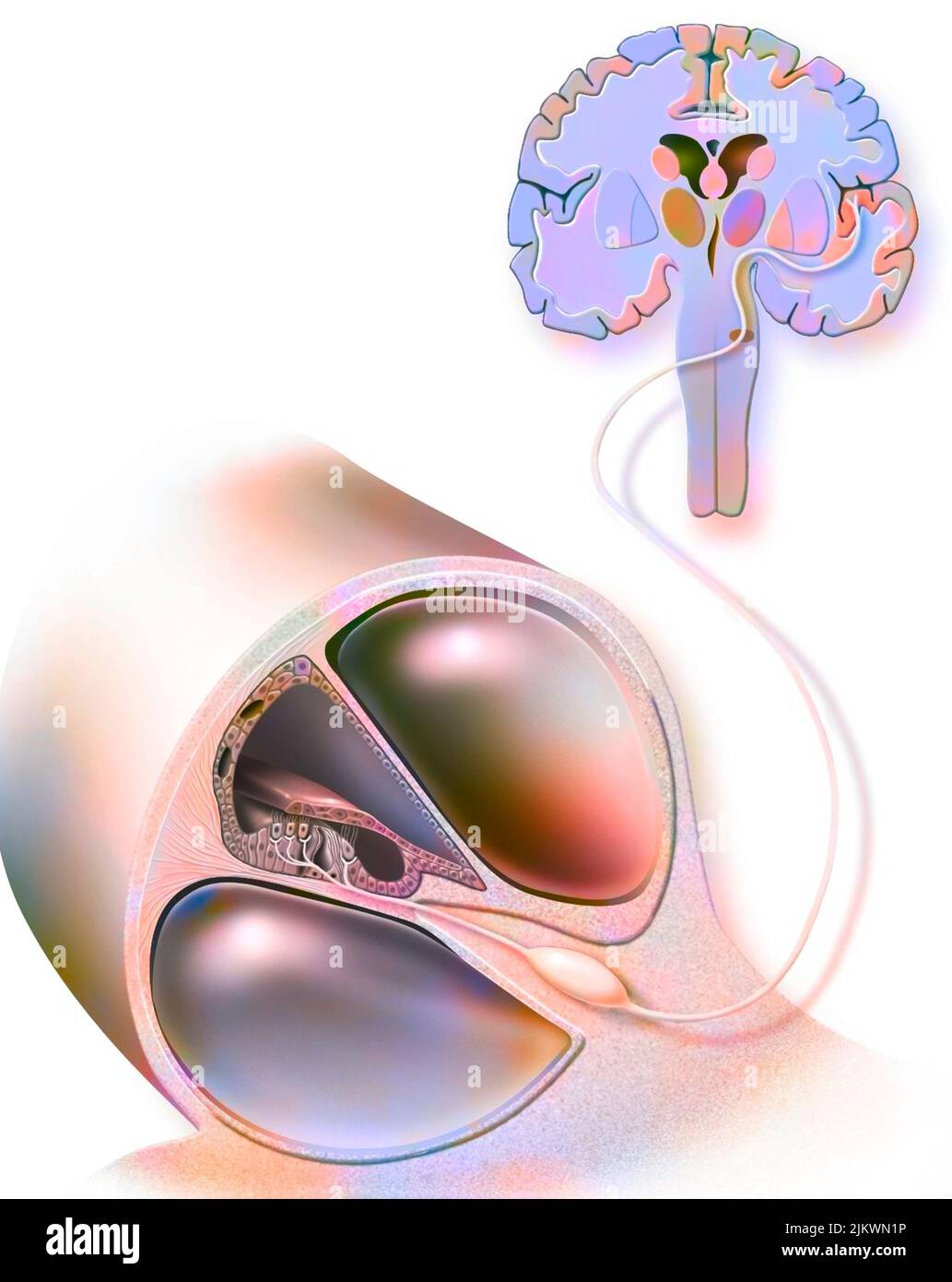 Path of sound from the organ of Corti to the auditory area of the brain. Stock Photohttps://www.alamy.com/image-license-details/?v=1https://www.alamy.com/path-of-sound-from-the-organ-of-corti-to-the-auditory-area-of-the-brain-image476923714.html
RF2JKWN1P–Path of sound from the organ of Corti to the auditory area of the brain.
 Illustration of the motor nerves of the eye, including the abducens, trochlear and oculomotor nerves in the human brain. These nerves innervate motor, sensory, and autonomic structures in the eyes. Stock Photohttps://www.alamy.com/image-license-details/?v=1https://www.alamy.com/illustration-of-the-motor-nerves-of-the-eye-including-the-abducens-trochlear-and-oculomotor-nerves-in-the-human-brain-these-nerves-innervate-motor-sensory-and-autonomic-structures-in-the-eyes-image628777701.html
RF2YEY8AD–Illustration of the motor nerves of the eye, including the abducens, trochlear and oculomotor nerves in the human brain. These nerves innervate motor, sensory, and autonomic structures in the eyes.
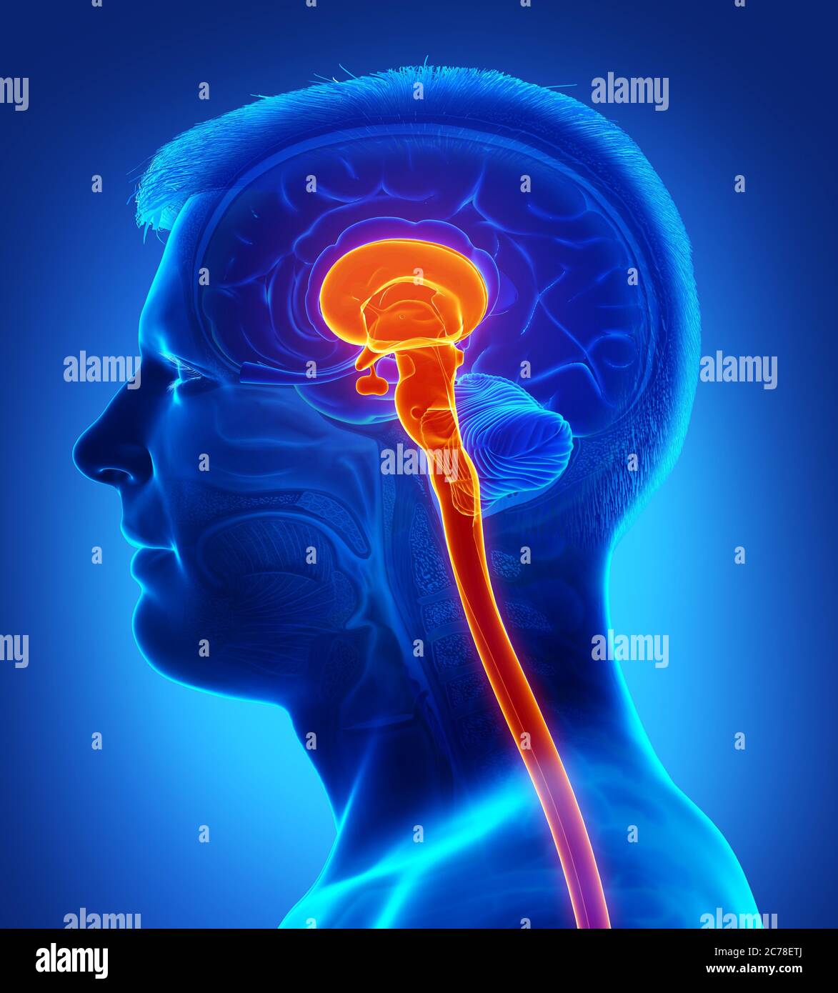 3d rendering medical illustration of brainstem Stock Photohttps://www.alamy.com/image-license-details/?v=1https://www.alamy.com/3d-rendering-medical-illustration-of-brainstem-image365907602.html
RF2C78ETJ–3d rendering medical illustration of brainstem
 3d rendered medically accurate illustration of the interior brain anatomy Stock Photohttps://www.alamy.com/image-license-details/?v=1https://www.alamy.com/3d-rendered-medically-accurate-illustration-of-the-interior-brain-anatomy-image365565097.html
RF2C6MX09–3d rendered medically accurate illustration of the interior brain anatomy
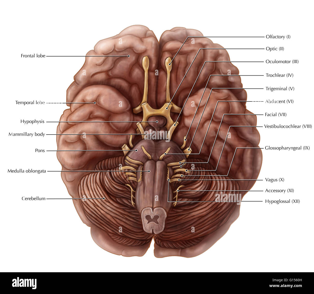 An illustrated view of the inferior surface of the brain depicting the 12 cranial nerves, cerebrum, pons, cerebellum, medulla oblongata and pituitary gland. Stock Photohttps://www.alamy.com/image-license-details/?v=1https://www.alamy.com/stock-photo-an-illustrated-view-of-the-inferior-surface-of-the-brain-depicting-103991345.html
RMG1560H–An illustrated view of the inferior surface of the brain depicting the 12 cranial nerves, cerebrum, pons, cerebellum, medulla oblongata and pituitary gland.
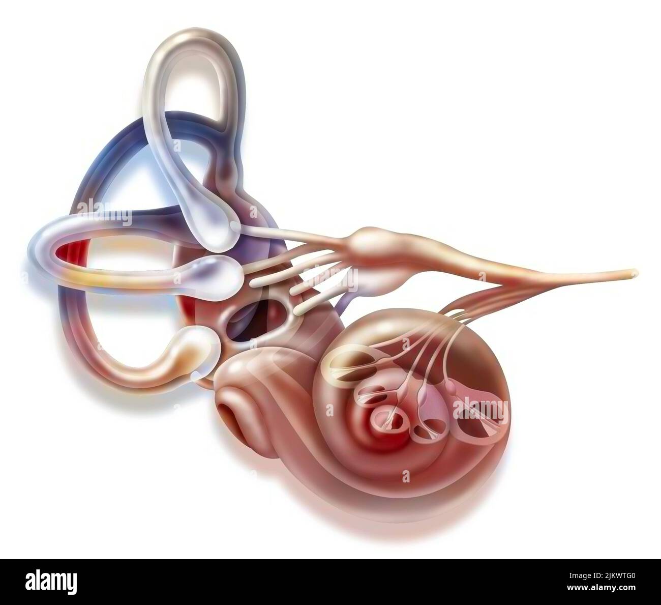 Inner ear and vestibular apparatus with semicircular canals, macule. Stock Photohttps://www.alamy.com/image-license-details/?v=1https://www.alamy.com/inner-ear-and-vestibular-apparatus-with-semicircular-canals-macule-image476926464.html
RF2JKWTG0–Inner ear and vestibular apparatus with semicircular canals, macule.
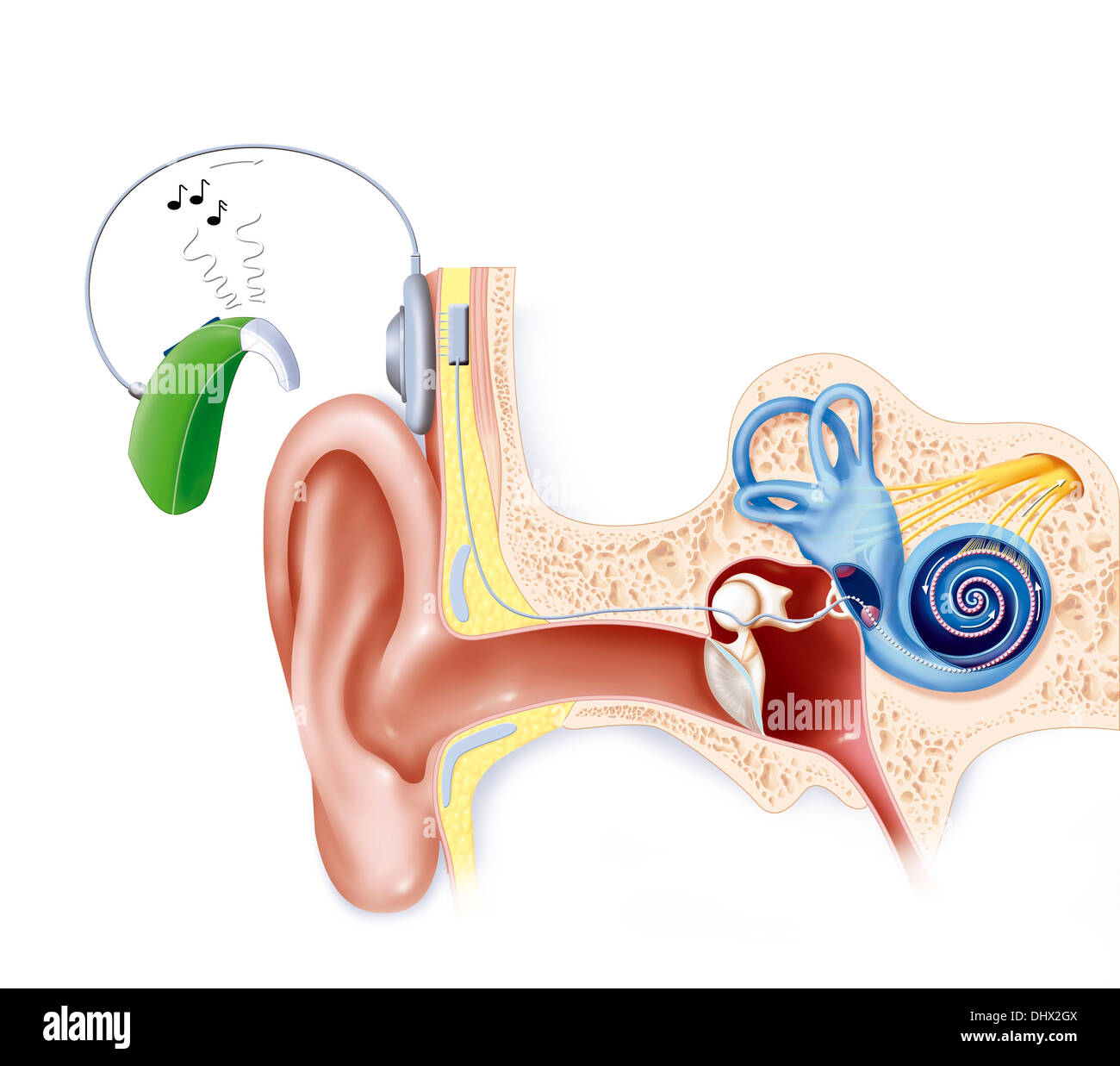 COCHLEAR IMPLANT, ILLUSTRATION Stock Photohttps://www.alamy.com/image-license-details/?v=1https://www.alamy.com/cochlear-implant-illustration-image62653050.html
RMDHX2GX–COCHLEAR IMPLANT, ILLUSTRATION
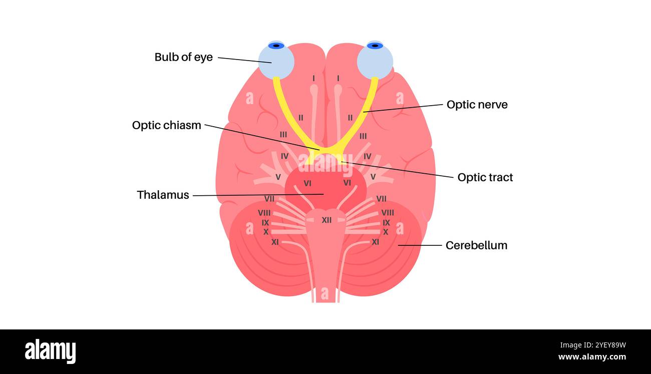 Illustration of the optic nerve anatomy. The optic nerves send visual messages from eye to brain. Stock Photohttps://www.alamy.com/image-license-details/?v=1https://www.alamy.com/illustration-of-the-optic-nerve-anatomy-the-optic-nerves-send-visual-messages-from-eye-to-brain-image628777685.html
RF2YEY89W–Illustration of the optic nerve anatomy. The optic nerves send visual messages from eye to brain.
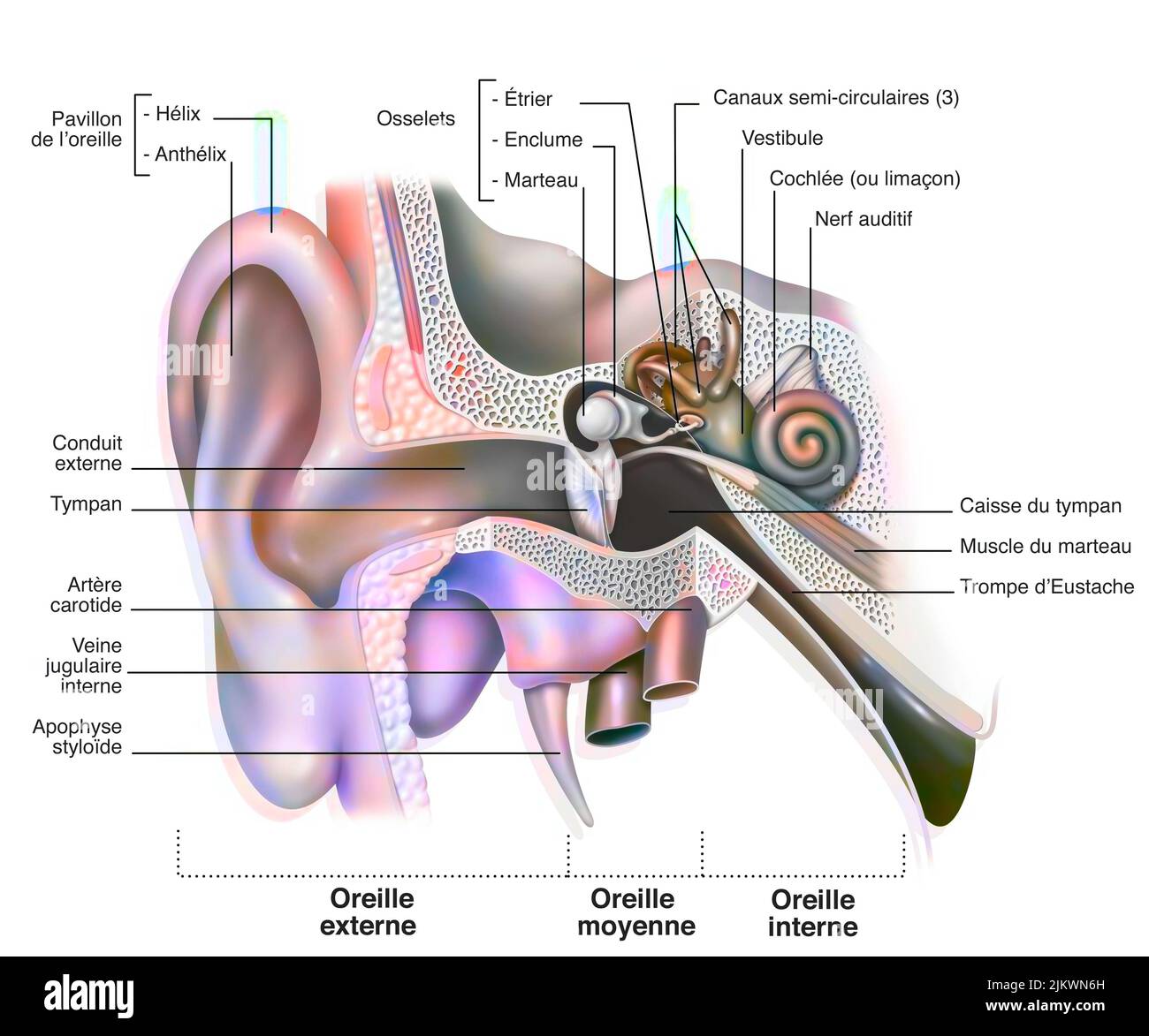 Anatomy of the inner ear showing the eardrum, the cochlea. Stock Photohttps://www.alamy.com/image-license-details/?v=1https://www.alamy.com/anatomy-of-the-inner-ear-showing-the-eardrum-the-cochlea-image476923849.html
RF2JKWN6H–Anatomy of the inner ear showing the eardrum, the cochlea.
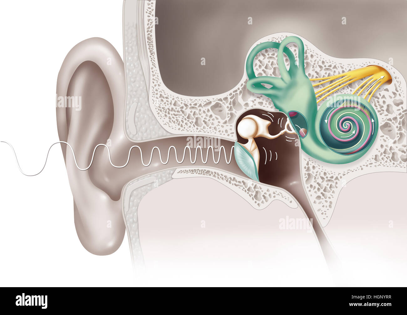 HEARING, DRAWING Stock Photohttps://www.alamy.com/image-license-details/?v=1https://www.alamy.com/stock-photo-hearing-drawing-130789899.html
RMHGNYRR–HEARING, DRAWING
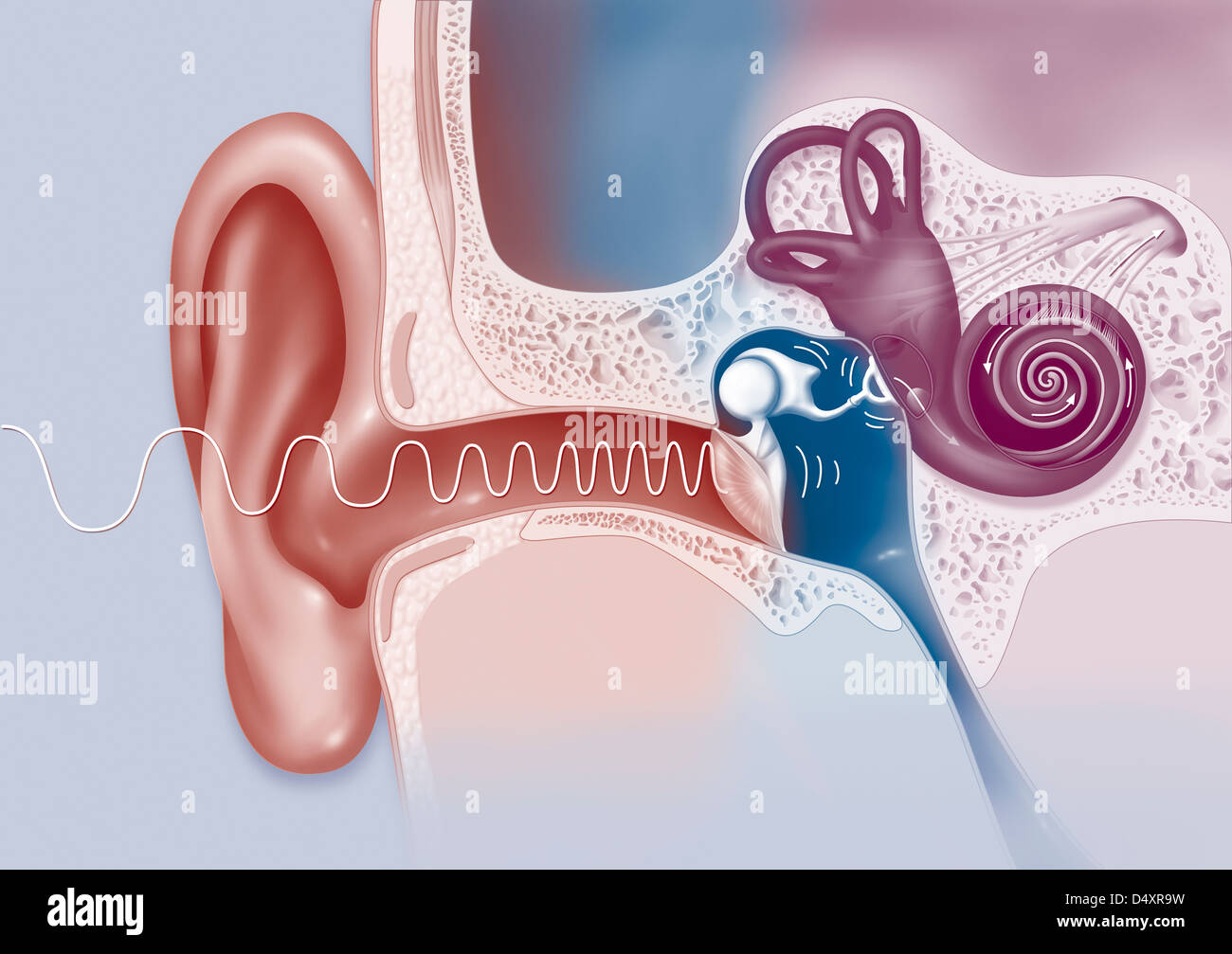 EAR, DRAWING Stock Photohttps://www.alamy.com/image-license-details/?v=1https://www.alamy.com/stock-photo-ear-drawing-54678789.html
RMD4XR9W–EAR, DRAWING
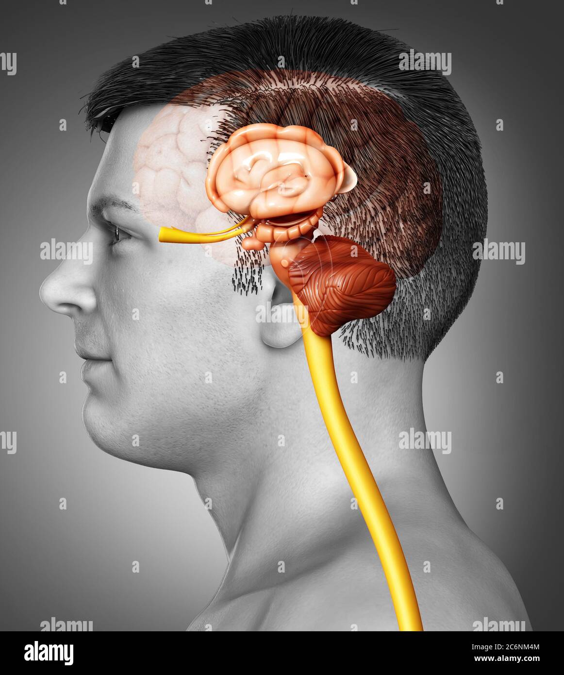 3d rendered medically accurate illustration of the interior brain anatomy Stock Photohttps://www.alamy.com/image-license-details/?v=1https://www.alamy.com/3d-rendered-medically-accurate-illustration-of-the-interior-brain-anatomy-image365582468.html
RF2C6NM4M–3d rendered medically accurate illustration of the interior brain anatomy
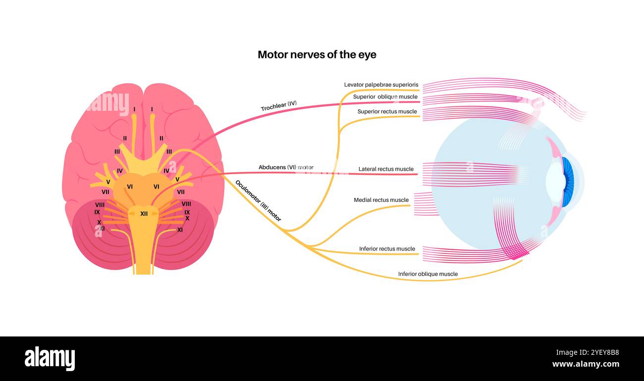 Illustration of the motor nerves of the eye, including the abducens, trochlear and oculomotor nerves in the human brain. These nerves innervate motor, sensory, and autonomic structures in the eyes. Stock Photohttps://www.alamy.com/image-license-details/?v=1https://www.alamy.com/illustration-of-the-motor-nerves-of-the-eye-including-the-abducens-trochlear-and-oculomotor-nerves-in-the-human-brain-these-nerves-innervate-motor-sensory-and-autonomic-structures-in-the-eyes-image628777724.html
RF2YEY8B8–Illustration of the motor nerves of the eye, including the abducens, trochlear and oculomotor nerves in the human brain. These nerves innervate motor, sensory, and autonomic structures in the eyes.
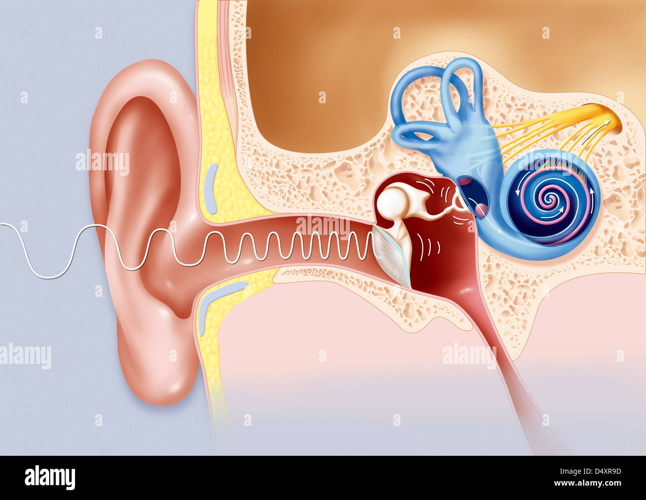 EAR, DRAWING Stock Photohttps://www.alamy.com/image-license-details/?v=1https://www.alamy.com/stock-photo-ear-drawing-54678777.html
RMD4XR9D–EAR, DRAWING
 3d rendered medically accurate illustration of the interior brain anatomy Stock Photohttps://www.alamy.com/image-license-details/?v=1https://www.alamy.com/3d-rendered-medically-accurate-illustration-of-the-interior-brain-anatomy-image365799644.html
RF2C73H50–3d rendered medically accurate illustration of the interior brain anatomy
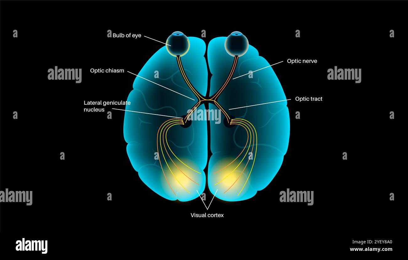 Illustration of the optic nerve anatomy. The optic nerves send visual messages from eye to brain. Stock Photohttps://www.alamy.com/image-license-details/?v=1https://www.alamy.com/illustration-of-the-optic-nerve-anatomy-the-optic-nerves-send-visual-messages-from-eye-to-brain-image628777688.html
RF2YEY8A0–Illustration of the optic nerve anatomy. The optic nerves send visual messages from eye to brain.
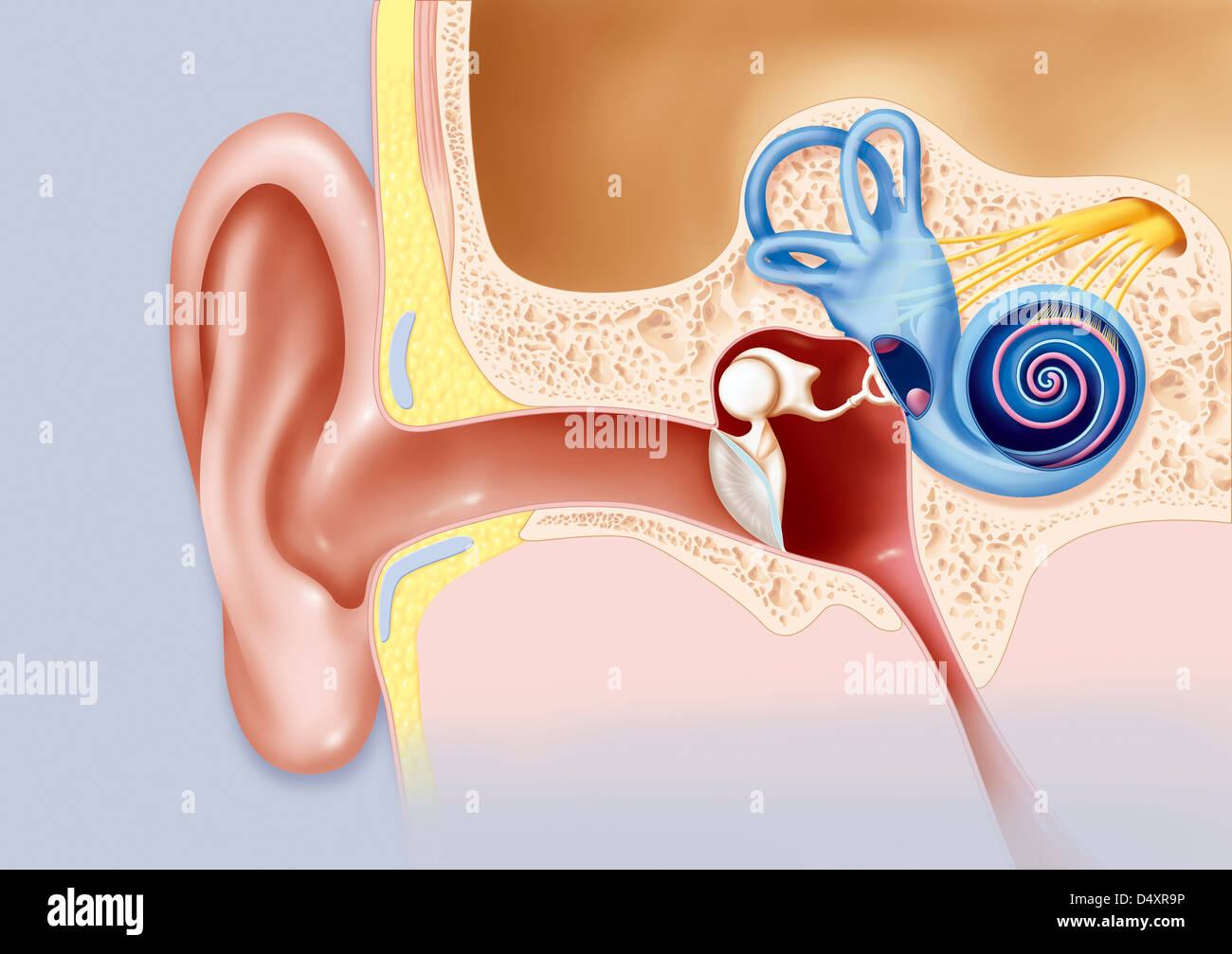 EAR, DRAWING Stock Photohttps://www.alamy.com/image-license-details/?v=1https://www.alamy.com/stock-photo-ear-drawing-54678786.html
RMD4XR9P–EAR, DRAWING
 3d rendered medically accurate illustration of the interior brain anatomy Stock Photohttps://www.alamy.com/image-license-details/?v=1https://www.alamy.com/3d-rendered-medically-accurate-illustration-of-the-interior-brain-anatomy-image365578038.html
RF2C6NEEE–3d rendered medically accurate illustration of the interior brain anatomy
 Illustration of the motor nerves of the eye, including the abducens, trochlear and oculomotor nerves in the human brain. These nerves innervate motor, sensory, and autonomic structures in the eyes. Stock Photohttps://www.alamy.com/image-license-details/?v=1https://www.alamy.com/illustration-of-the-motor-nerves-of-the-eye-including-the-abducens-trochlear-and-oculomotor-nerves-in-the-human-brain-these-nerves-innervate-motor-sensory-and-autonomic-structures-in-the-eyes-image628777683.html
RF2YEY89R–Illustration of the motor nerves of the eye, including the abducens, trochlear and oculomotor nerves in the human brain. These nerves innervate motor, sensory, and autonomic structures in the eyes.
