Quick filters:
Vocal tract Stock Photos and Images
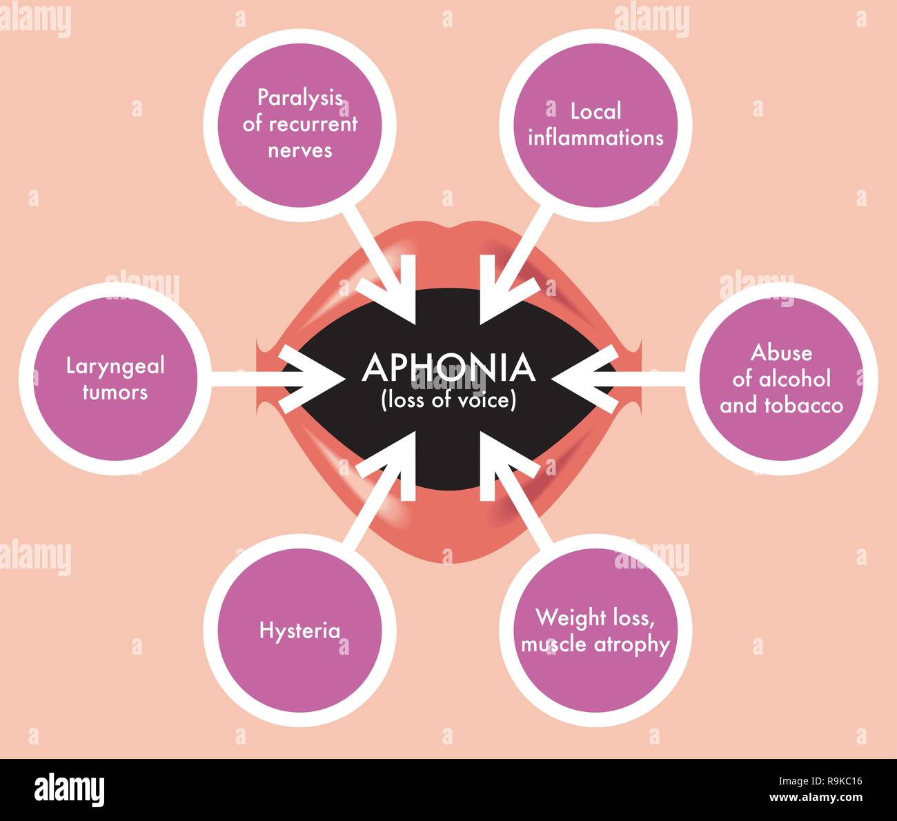 A medical diagram illustrating Aphonia, the loss of voice. Stock Vectorhttps://www.alamy.com/image-license-details/?v=1https://www.alamy.com/a-medical-diagram-illustrating-aphonia-the-loss-of-voice-image229693218.html
RFR9KC16–A medical diagram illustrating Aphonia, the loss of voice.
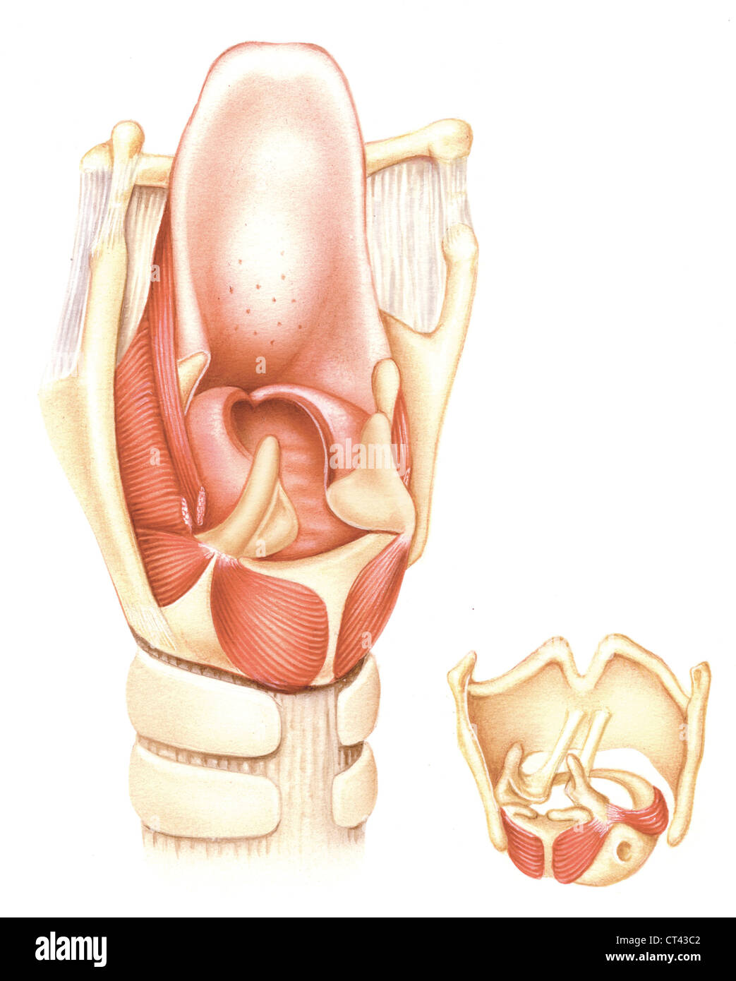 Larynx, drawing Stock Photohttps://www.alamy.com/image-license-details/?v=1https://www.alamy.com/stock-photo-larynx-drawing-49262978.html
RMCT43C2–Larynx, drawing
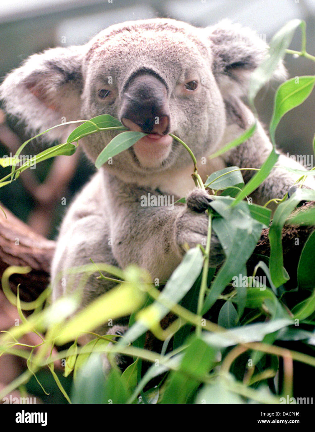 (FILE) An undated archive photo shows a male koala eating eucalyptus at the zoo in Duisburg, Germany. During mating season, the best part of the koala is the voicebox: With the help of their vocal tract, the somewhat sluggish marsupial can fake its body size, according to researchers from Austria and Australia in the 'Journal of Experimental Biology'. Scientists discovered that the Stock Photohttps://www.alamy.com/image-license-details/?v=1https://www.alamy.com/stock-photo-filean-undated-archive-photo-shows-a-male-koala-eating-eucalyptus-58058818.html
RMDACPH6–(FILE) An undated archive photo shows a male koala eating eucalyptus at the zoo in Duisburg, Germany. During mating season, the best part of the koala is the voicebox: With the help of their vocal tract, the somewhat sluggish marsupial can fake its body size, according to researchers from Austria and Australia in the 'Journal of Experimental Biology'. Scientists discovered that the
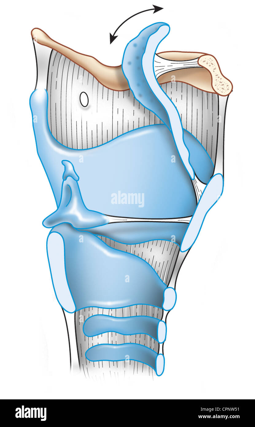 LARYNX, DRAWING Stock Photohttps://www.alamy.com/image-license-details/?v=1https://www.alamy.com/stock-photo-larynx-drawing-48423901.html
RMCPNW51–LARYNX, DRAWING
 sore throat vocal cords tense woman can't speak she sticks out tongue looks up wrinkled hand holds throat can't say Stock Photohttps://www.alamy.com/image-license-details/?v=1https://www.alamy.com/sore-throat-vocal-cords-tense-woman-cant-speak-she-sticks-out-tongue-looks-up-wrinkled-hand-holds-throat-cant-say-image539928348.html
RF2PABT38–sore throat vocal cords tense woman can't speak she sticks out tongue looks up wrinkled hand holds throat can't say
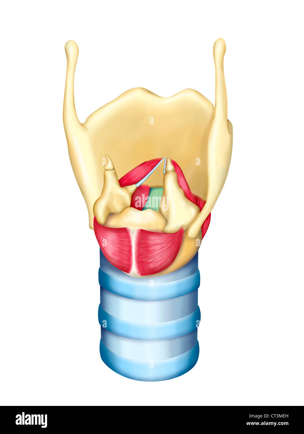 LARYNX, DRAWING Stock Photohttps://www.alamy.com/image-license-details/?v=1https://www.alamy.com/stock-photo-larynx-drawing-49254425.html
RMCT3MEH–LARYNX, DRAWING
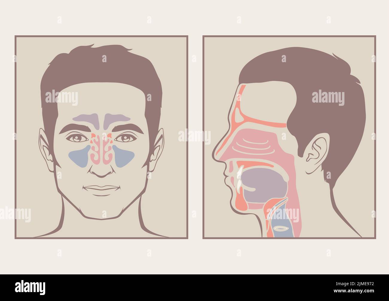 Nose, throat anatomy Stock Photohttps://www.alamy.com/image-license-details/?v=1https://www.alamy.com/nose-throat-anatomy-image477287638.html
RF2JME972–Nose, throat anatomy
 The larynx is an organ in the top of the neck of tetrapods involved in breathing, producing sound, and protecting the trachea against food aspiration Stock Photohttps://www.alamy.com/image-license-details/?v=1https://www.alamy.com/the-larynx-is-an-organ-in-the-top-of-the-neck-of-tetrapods-involved-in-breathing-producing-sound-and-protecting-the-trachea-against-food-aspiration-image433750489.html
RF2G5K14W–The larynx is an organ in the top of the neck of tetrapods involved in breathing, producing sound, and protecting the trachea against food aspiration
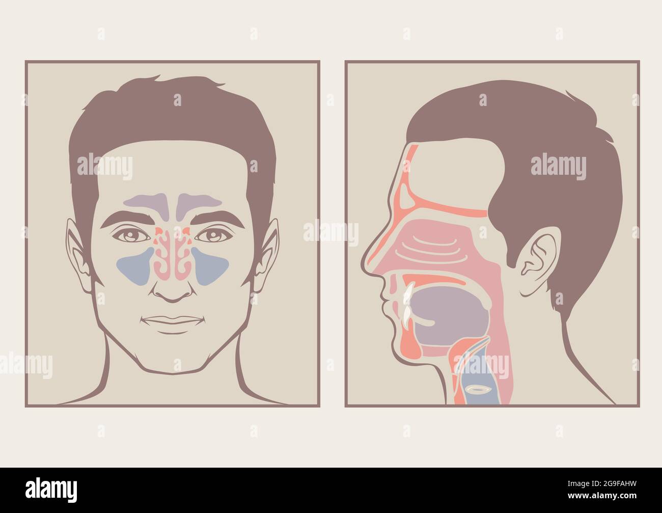 nose, throat anatomy, human mouth, respiratory system Stock Vectorhttps://www.alamy.com/image-license-details/?v=1https://www.alamy.com/nose-throat-anatomy-human-mouth-respiratory-system-image436128725.html
RF2G9FAHW–nose, throat anatomy, human mouth, respiratory system
 Detail of nose pencil drawing on white paper Stock Photohttps://www.alamy.com/image-license-details/?v=1https://www.alamy.com/detail-of-nose-pencil-drawing-on-white-paper-image62367858.html
RFDHD2RE–Detail of nose pencil drawing on white paper
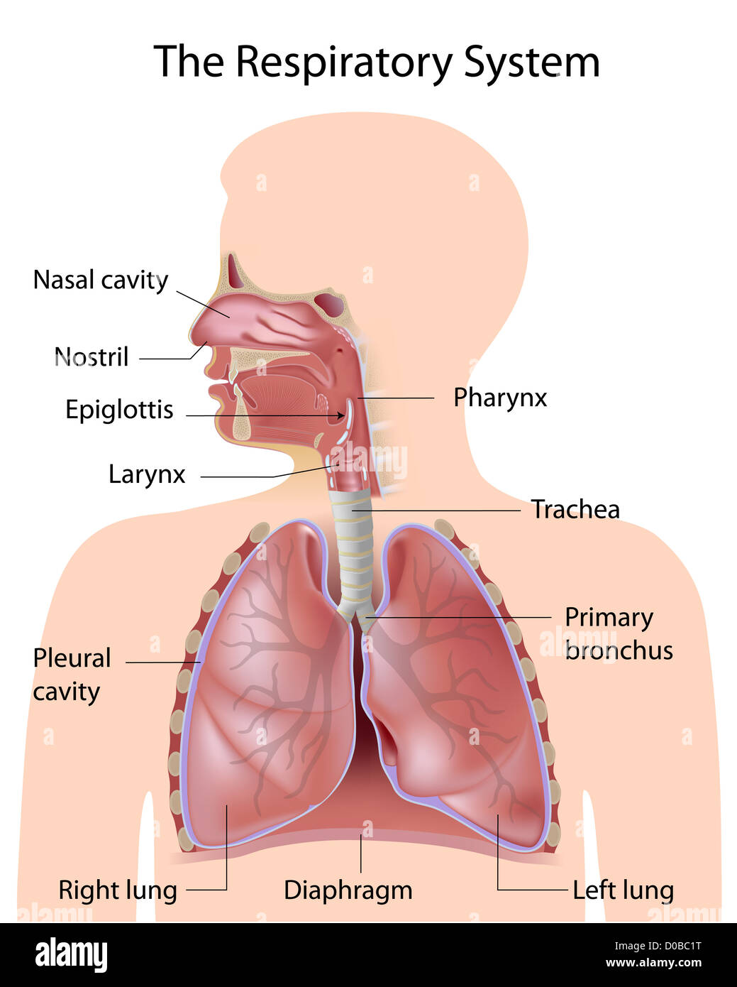 The respiratory system, labeled Stock Photohttps://www.alamy.com/image-license-details/?v=1https://www.alamy.com/stock-photo-the-respiratory-system-labeled-51882036.html
RFD0BC1T–The respiratory system, labeled
RF2B28B05–Rhinopharynx structure line icon, concept sign, outline vector illustration, linear symbol.
 Detail of nose pencil drawing on white paper Stock Photohttps://www.alamy.com/image-license-details/?v=1https://www.alamy.com/stock-photo-detail-of-nose-pencil-drawing-on-white-paper-144267021.html
RFJAKX1H–Detail of nose pencil drawing on white paper
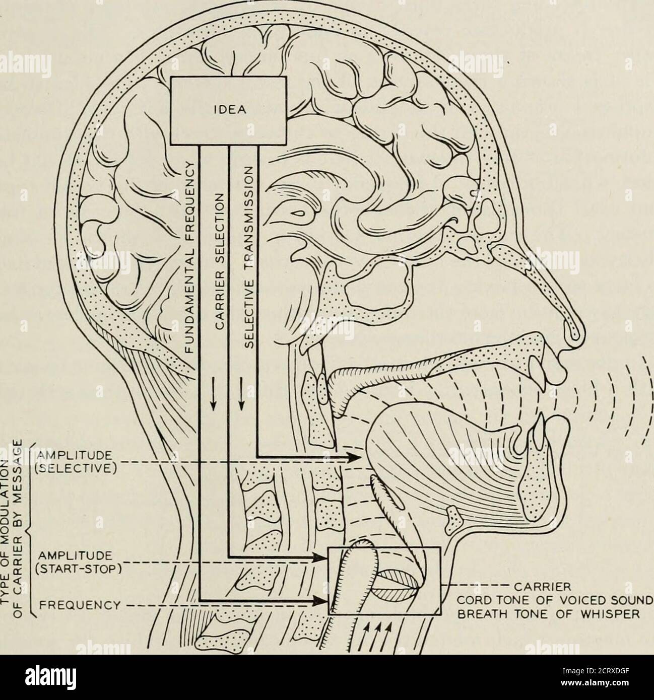 . The Bell System technical journal . ular actions in the vocal tract. Never-theless these motions contain the dynamic speech information as isproved by their interpretation by lip readers to the extent visibilitypermits. Another method of demonstrating the information contentof certain of these motions is the artificial injection of a sound streaminto the back of the mouth for a carrier whereby intelligible speech 2 The information referred to is that in the communication of intelligence. Thereis, however, static information in the carrier itself. This serves for station identi-fication in ra Stock Photohttps://www.alamy.com/image-license-details/?v=1https://www.alamy.com/the-bell-system-technical-journal-ular-actions-in-the-vocal-tract-never-theless-these-motions-contain-the-dynamic-speech-information-as-isproved-by-their-interpretation-by-lip-readers-to-the-extent-visibilitypermits-another-method-of-demonstrating-the-information-contentof-certain-of-these-motions-is-the-artificial-injection-of-a-sound-streaminto-the-back-of-the-mouth-for-a-carrier-whereby-intelligible-speech-2-the-information-referred-to-is-that-in-the-communication-of-intelligence-thereis-however-static-information-in-the-carrier-itself-this-serves-for-station-identi-fication-in-ra-image376136223.html
RM2CRXDGF–. The Bell System technical journal . ular actions in the vocal tract. Never-theless these motions contain the dynamic speech information as isproved by their interpretation by lip readers to the extent visibilitypermits. Another method of demonstrating the information contentof certain of these motions is the artificial injection of a sound streaminto the back of the mouth for a carrier whereby intelligible speech 2 The information referred to is that in the communication of intelligence. Thereis, however, static information in the carrier itself. This serves for station identi-fication in ra
 X-ray view of a man resting with red highlighted nasal sinus and throat Stock Photohttps://www.alamy.com/image-license-details/?v=1https://www.alamy.com/stock-photo-x-ray-view-of-a-man-resting-with-red-highlighted-nasal-sinus-and-throat-30481547.html
RFBNGFEK–X-ray view of a man resting with red highlighted nasal sinus and throat
![Infography on the human larynx. [Adobe InDesign (.indd); 4795x3543]. Stock Photo Infography on the human larynx. [Adobe InDesign (.indd); 4795x3543]. Stock Photo](https://c8.alamy.com/comp/2NEC3Y9/infography-on-the-human-larynx-adobe-indesign-indd-4795x3543-2NEC3Y9.jpg) Infography on the human larynx. [Adobe InDesign (.indd); 4795x3543]. Stock Photohttps://www.alamy.com/image-license-details/?v=1https://www.alamy.com/infography-on-the-human-larynx-adobe-indesign-indd-4795x3543-image525182765.html
Infography on the human larynx. [Adobe InDesign (.indd); 4795x3543]. Stock Photohttps://www.alamy.com/image-license-details/?v=1https://www.alamy.com/infography-on-the-human-larynx-adobe-indesign-indd-4795x3543-image525182765.htmlRM2NEC3Y9–Infography on the human larynx. [Adobe InDesign (.indd); 4795x3543].
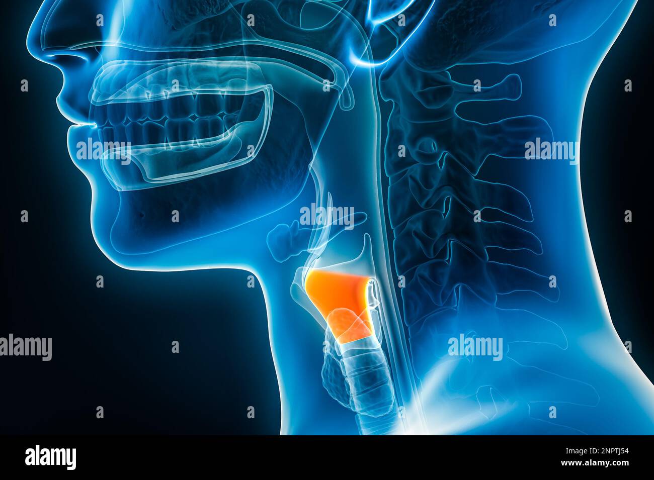 Xray lateral or profile view of the larynx or voice box 3D rendering illustration with male body contours. Human organ anatomy, laryngitis, medical, b Stock Photohttps://www.alamy.com/image-license-details/?v=1https://www.alamy.com/xray-lateral-or-profile-view-of-the-larynx-or-voice-box-3d-rendering-illustration-with-male-body-contours-human-organ-anatomy-laryngitis-medical-b-image530374576.html
RF2NPTJ54–Xray lateral or profile view of the larynx or voice box 3D rendering illustration with male body contours. Human organ anatomy, laryngitis, medical, b
 Rose-ringed or Ring-necked Parakeets (Psittacula krameri). Twenty one days old chicks. Aviary birds. Stock Photohttps://www.alamy.com/image-license-details/?v=1https://www.alamy.com/rose-ringed-or-ring-necked-parakeets-psittacula-krameri-twenty-one-image69067169.html
RME0A7TH–Rose-ringed or Ring-necked Parakeets (Psittacula krameri). Twenty one days old chicks. Aviary birds.
RF2HCF82N–Nasopharynx icon. Cartoon vector flat illustration, isolated on white background. Nose anatomy
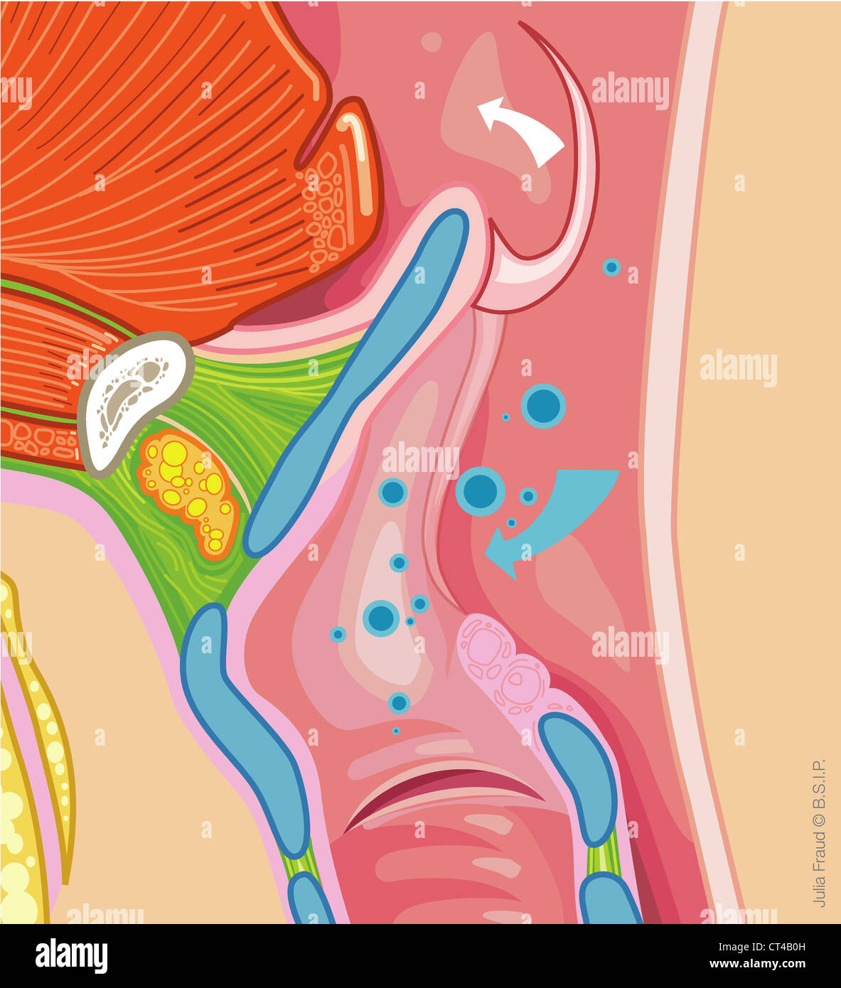 PHARYNX, DRAWING Stock Photohttps://www.alamy.com/image-license-details/?v=1https://www.alamy.com/stock-photo-pharynx-drawing-49268929.html
RMCT4B0H–PHARYNX, DRAWING
 DIGESTIVE SYSTEM Stock Photohttps://www.alamy.com/image-license-details/?v=1https://www.alamy.com/stock-photo-digestive-system-53860946.html
RMD3HG56–DIGESTIVE SYSTEM
 sore throat vocal cords tense woman can't speak she sticks out tongue looks up wrinkled hand holds throat can't say word real person treatment medicine scratchy throat swallowed choked 40-50 years old Stock Photohttps://www.alamy.com/image-license-details/?v=1https://www.alamy.com/sore-throat-vocal-cords-tense-woman-cant-speak-she-sticks-out-tongue-looks-up-wrinkled-hand-holds-throat-cant-say-word-real-person-treatment-medicine-scratchy-throat-swallowed-choked-40-50-years-old-image504346693.html
RF2M8EY9W–sore throat vocal cords tense woman can't speak she sticks out tongue looks up wrinkled hand holds throat can't say word real person treatment medicine scratchy throat swallowed choked 40-50 years old
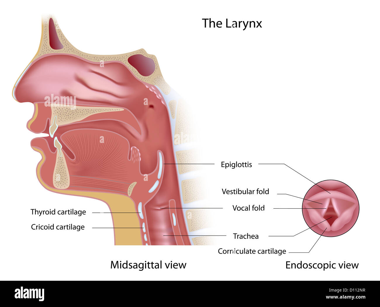 Anatomy of voice box midsagittal and endoscopic view Stock Photohttps://www.alamy.com/image-license-details/?v=1https://www.alamy.com/stock-photo-anatomy-of-voice-box-midsagittal-and-endoscopic-view-52269891.html
RFD112NR–Anatomy of voice box midsagittal and endoscopic view
 . Kirkes' handbook of physiology . Fig. Fig. 32. Fig. 31.—A Small Piece of the Liver of the Horse. (Cadiat.) Fig. 32.—Glandular Epithelium. Small lobule of a mucous gland of the tongue, showingnucleated glandular cells. X 200. (V. D. Harris.) and also the sinuses and ducts in connection with it and the lachrymal sac,the upper surface of the soft palate and the naso-pharynx, the Eustachian tubeand tympanum, the larynx, except over the vocal cords, to the finest sub-divisions of the bronchi. In part of this tract, however, the epithelium is inseveral layers, of which only the most superficial is Stock Photohttps://www.alamy.com/image-license-details/?v=1https://www.alamy.com/kirkes-handbook-of-physiology-fig-fig-32-fig-31a-small-piece-of-the-liver-of-the-horse-cadiat-fig-32glandular-epithelium-small-lobule-of-a-mucous-gland-of-the-tongue-showingnucleated-glandular-cells-x-200-v-d-harris-and-also-the-sinuses-and-ducts-in-connection-with-it-and-the-lachrymal-sacthe-upper-surface-of-the-soft-palate-and-the-naso-pharynx-the-eustachian-tubeand-tympanum-the-larynx-except-over-the-vocal-cords-to-the-finest-sub-divisions-of-the-bronchi-in-part-of-this-tract-however-the-epithelium-is-inseveral-layers-of-which-only-the-most-superficial-is-image370001631.html
RM2CDY0RY–. Kirkes' handbook of physiology . Fig. Fig. 32. Fig. 31.—A Small Piece of the Liver of the Horse. (Cadiat.) Fig. 32.—Glandular Epithelium. Small lobule of a mucous gland of the tongue, showingnucleated glandular cells. X 200. (V. D. Harris.) and also the sinuses and ducts in connection with it and the lachrymal sac,the upper surface of the soft palate and the naso-pharynx, the Eustachian tubeand tympanum, the larynx, except over the vocal cords, to the finest sub-divisions of the bronchi. In part of this tract, however, the epithelium is inseveral layers, of which only the most superficial is
 X-ray view of a man resting with view of lungs Stock Photohttps://www.alamy.com/image-license-details/?v=1https://www.alamy.com/stock-photo-x-ray-view-of-a-man-resting-with-view-of-lungs-30481537.html
RFBNGFE9–X-ray view of a man resting with view of lungs
 ANATOMY, HEAD Stock Photohttps://www.alamy.com/image-license-details/?v=1https://www.alamy.com/stock-photo-anatomy-head-53861065.html
RMD3HG9D–ANATOMY, HEAD
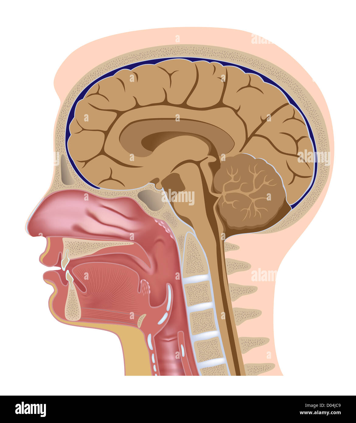 Median section of human head Stock Photohttps://www.alamy.com/image-license-details/?v=1https://www.alamy.com/stock-photo-median-section-of-human-head-51733369.html
RFD04JC9–Median section of human head
 . Kirkes' handbook of physiology . Fig. Fig. 32. Fig. 31.—A Small Piece of the Liver of the Horse. (Cadiat.) Fig. 32.—Glandular Epithelium. Small lobule of a mucous gland of the tongue, showingnucleated glandular cells. X 200. (V. D. Harris.) and also the sinuses and ducts in connection with it and the lachrymal sac,the upper surface of the soft palate and the naso-pharynx, the Eustachian tubeand tympanum, the larynx, except over the vocal cords, to the finest sub-divisions of the bronchi. In part of this tract, however, the epithelium is inseveral layers, of which only the most superficial is Stock Photohttps://www.alamy.com/image-license-details/?v=1https://www.alamy.com/kirkes-handbook-of-physiology-fig-fig-32-fig-31a-small-piece-of-the-liver-of-the-horse-cadiat-fig-32glandular-epithelium-small-lobule-of-a-mucous-gland-of-the-tongue-showingnucleated-glandular-cells-x-200-v-d-harris-and-also-the-sinuses-and-ducts-in-connection-with-it-and-the-lachrymal-sacthe-upper-surface-of-the-soft-palate-and-the-naso-pharynx-the-eustachian-tubeand-tympanum-the-larynx-except-over-the-vocal-cords-to-the-finest-sub-divisions-of-the-bronchi-in-part-of-this-tract-however-the-epithelium-is-inseveral-layers-of-which-only-the-most-superficial-is-image370001492.html
RM2CDY0K0–. Kirkes' handbook of physiology . Fig. Fig. 32. Fig. 31.—A Small Piece of the Liver of the Horse. (Cadiat.) Fig. 32.—Glandular Epithelium. Small lobule of a mucous gland of the tongue, showingnucleated glandular cells. X 200. (V. D. Harris.) and also the sinuses and ducts in connection with it and the lachrymal sac,the upper surface of the soft palate and the naso-pharynx, the Eustachian tubeand tympanum, the larynx, except over the vocal cords, to the finest sub-divisions of the bronchi. In part of this tract, however, the epithelium is inseveral layers, of which only the most superficial is
 ANATOMY, HEAD Stock Photohttps://www.alamy.com/image-license-details/?v=1https://www.alamy.com/stock-photo-anatomy-head-53861063.html
RMD3HG9B–ANATOMY, HEAD
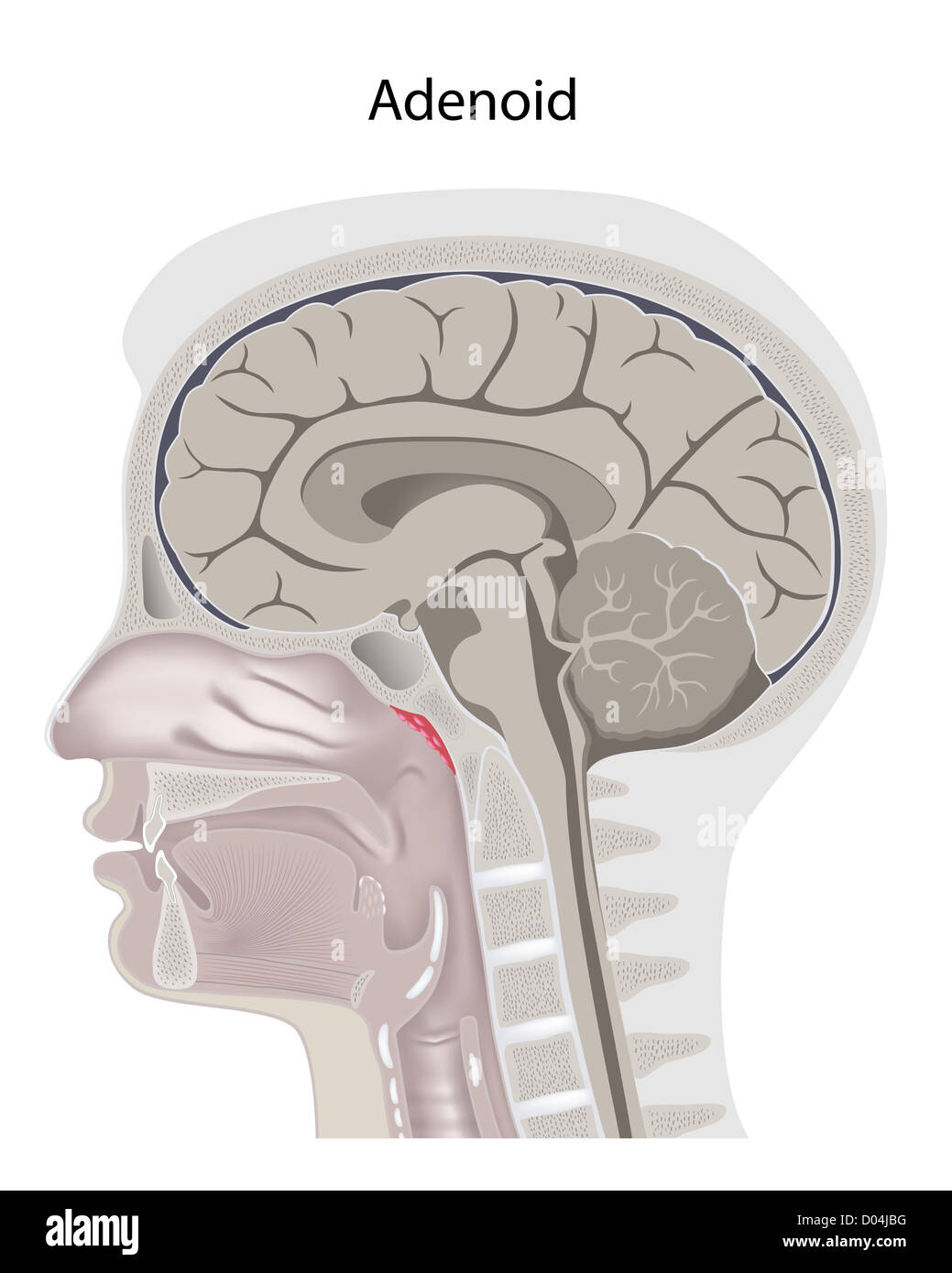 Adenoid location in the head Stock Photohttps://www.alamy.com/image-license-details/?v=1https://www.alamy.com/stock-photo-adenoid-location-in-the-head-51733348.html
RFD04JBG–Adenoid location in the head
 . Chordate morphology. Morphology (Animals); Chordata. placenta which is attached by folds to the uterine wall and which absorbs materials from the maternal blood (Figure 7-16). Many teleosts are viviparous (live-bearing) and show the same range of adaptations as the sharks. .Some other terms can be introduced here which apply to the conditions described. Oviparous describes those animals which lay eggs. Ovoviviparous describes the retention of eggs within the reproductive tract, or some specialized area (pits in skin, vocal sacs, or brood pouches), until the young hatch or complete their yolk Stock Photohttps://www.alamy.com/image-license-details/?v=1https://www.alamy.com/chordate-morphology-morphology-animals-chordata-placenta-which-is-attached-by-folds-to-the-uterine-wall-and-which-absorbs-materials-from-the-maternal-blood-figure-7-16-many-teleosts-are-viviparous-live-bearing-and-show-the-same-range-of-adaptations-as-the-sharks-some-other-terms-can-be-introduced-here-which-apply-to-the-conditions-described-oviparous-describes-those-animals-which-lay-eggs-ovoviviparous-describes-the-retention-of-eggs-within-the-reproductive-tract-or-some-specialized-area-pits-in-skin-vocal-sacs-or-brood-pouches-until-the-young-hatch-or-complete-their-yolk-image234952322.html
RMRJ702A–. Chordate morphology. Morphology (Animals); Chordata. placenta which is attached by folds to the uterine wall and which absorbs materials from the maternal blood (Figure 7-16). Many teleosts are viviparous (live-bearing) and show the same range of adaptations as the sharks. .Some other terms can be introduced here which apply to the conditions described. Oviparous describes those animals which lay eggs. Ovoviviparous describes the retention of eggs within the reproductive tract, or some specialized area (pits in skin, vocal sacs, or brood pouches), until the young hatch or complete their yolk
 The respiratory system, non-labeled Stock Photohttps://www.alamy.com/image-license-details/?v=1https://www.alamy.com/stock-photo-the-respiratory-system-non-labeled-51882030.html
RFD0BC1J–The respiratory system, non-labeled
 . The anatomy of the frog. Frogs -- Anatomy; Amphibians -- Anatomy. 282 THE ALIMENTARY TRACT, ETC. Fig. 183. G. Muscles of the tongue, from the ventral sui'face. G M. genio-^lossus. Gj} Straight fibres of the AI. genio-glossus. G//1 Curved fibres of the M. genio-glossus. Hy and ////i M. hyoglossus, Z Borders of the tongue. The M. hyoglossus is the retractor of the toiigaie, the M. fjenio-ytossiis the protractor. (For mucous membrane of the tong'ue, see organ of taste. The vocal sacs are described with the organs of voice and respiration.) B. The Oesophagus and Stomach (Figs. 184, 185, 189, 194 Stock Photohttps://www.alamy.com/image-license-details/?v=1https://www.alamy.com/the-anatomy-of-the-frog-frogs-anatomy-amphibians-anatomy-282-the-alimentary-tract-etc-fig-183-g-muscles-of-the-tongue-from-the-ventral-suiface-g-m-genio-lossus-gj-straight-fibres-of-the-ai-genio-glossus-g1-curved-fibres-of-the-m-genio-glossus-hy-and-i-m-hyoglossus-z-borders-of-the-tongue-the-m-hyoglossus-is-the-retractor-of-the-toiigaie-the-m-fjenio-ytossiis-the-protractor-for-mucous-membrane-of-the-tongue-see-organ-of-taste-the-vocal-sacs-are-described-with-the-organs-of-voice-and-respiration-b-the-oesophagus-and-stomach-figs-184-185-189-194-image236803759.html
RMRN79H3–. The anatomy of the frog. Frogs -- Anatomy; Amphibians -- Anatomy. 282 THE ALIMENTARY TRACT, ETC. Fig. 183. G. Muscles of the tongue, from the ventral sui'face. G M. genio-^lossus. Gj} Straight fibres of the AI. genio-glossus. G//1 Curved fibres of the M. genio-glossus. Hy and ////i M. hyoglossus, Z Borders of the tongue. The M. hyoglossus is the retractor of the toiigaie, the M. fjenio-ytossiis the protractor. (For mucous membrane of the tong'ue, see organ of taste. The vocal sacs are described with the organs of voice and respiration.) B. The Oesophagus and Stomach (Figs. 184, 185, 189, 194
