Quick filters:
X ray view Stock Photos and Images
 Unprecedented X-ray View of Supernova Remains Stock Photohttps://www.alamy.com/image-license-details/?v=1https://www.alamy.com/stock-photo-unprecedented-x-ray-view-of-supernova-remains-77419331.html
RMEDXN43–Unprecedented X-ray View of Supernova Remains
 Doctor or scientist with x-ray machine scans her own body to see her skeleton and heart. Vector cartoon illustration. Stock Vectorhttps://www.alamy.com/image-license-details/?v=1https://www.alamy.com/stock-photo-doctor-or-scientist-with-x-ray-machine-scans-her-own-body-to-see-her-118064969.html
RFGT2921–Doctor or scientist with x-ray machine scans her own body to see her skeleton and heart. Vector cartoon illustration.
 X-ray view of human hand. Stock Photohttps://www.alamy.com/image-license-details/?v=1https://www.alamy.com/stock-photo-x-ray-view-of-human-hand-57642485.html
RFD9NRG5–X-ray view of human hand.
 x-ray view of luggage with inside arms. 3d render. Stock Photohttps://www.alamy.com/image-license-details/?v=1https://www.alamy.com/x-ray-view-of-luggage-with-inside-arms-3d-render-image429108325.html
RF2FX3G19–x-ray view of luggage with inside arms. 3d render.
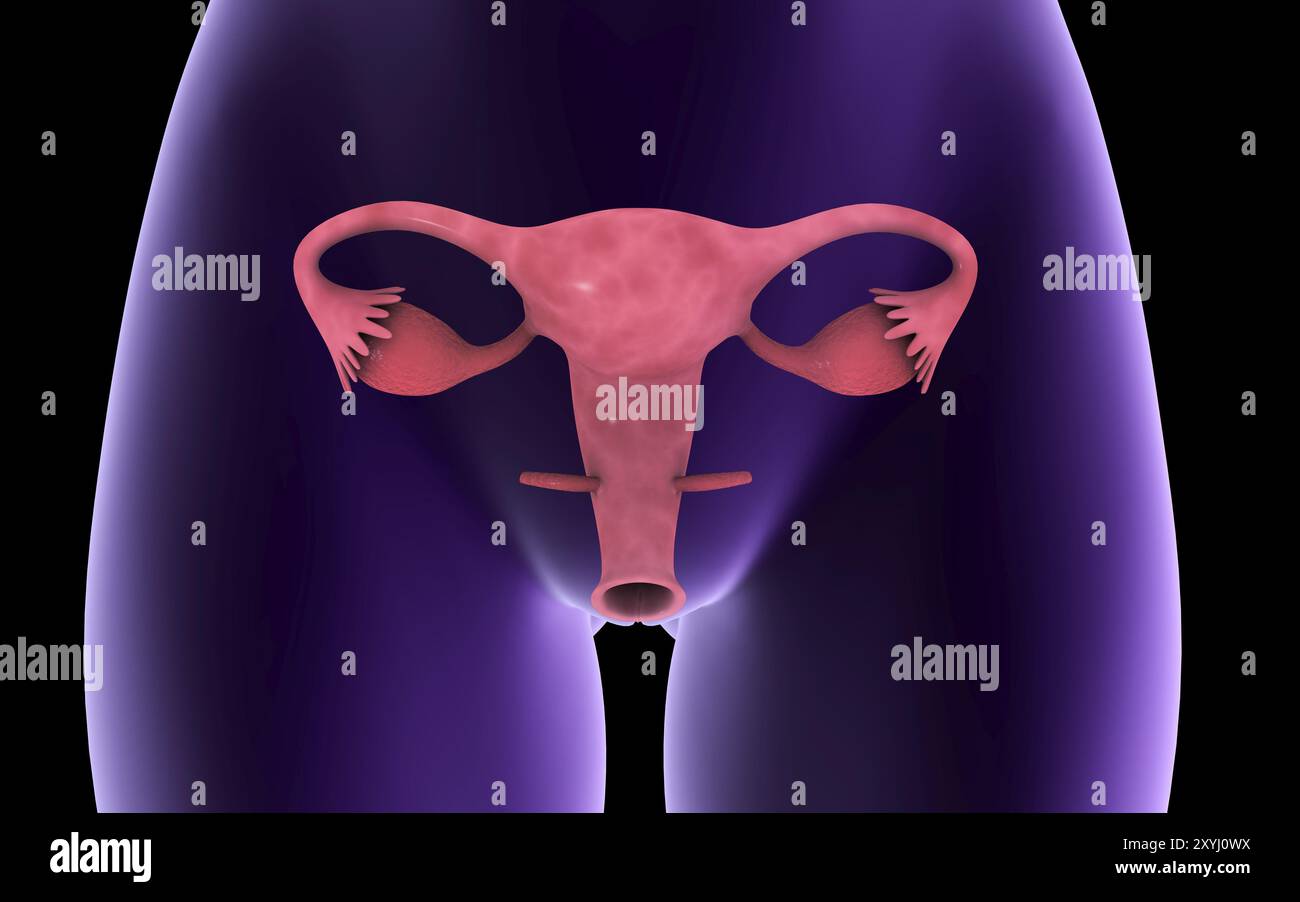 Female reproductive organ, x-ray view Stock Photohttps://www.alamy.com/image-license-details/?v=1https://www.alamy.com/female-reproductive-organ-x-ray-view-image619354454.html
RM2XYJ0WX–Female reproductive organ, x-ray view
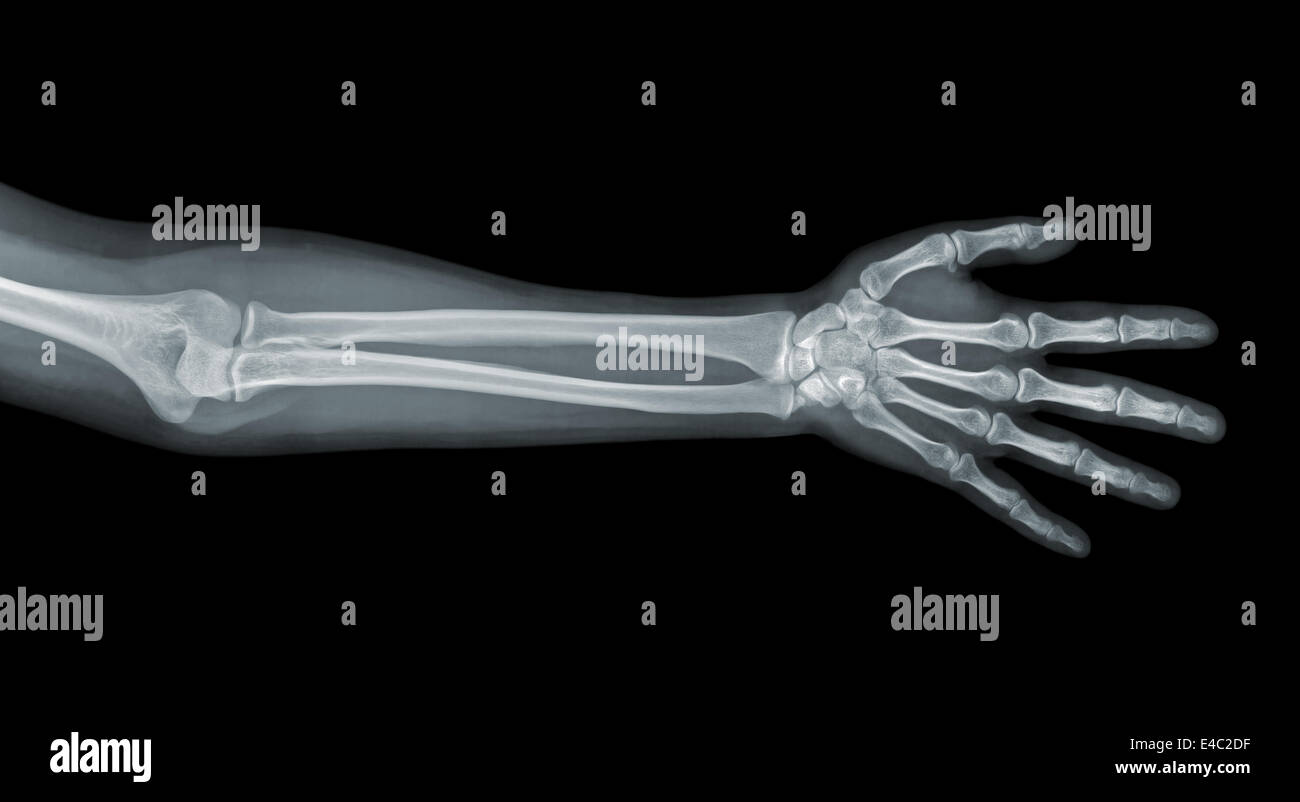 Hand x-ray view Stock Photohttps://www.alamy.com/image-license-details/?v=1https://www.alamy.com/stock-photo-hand-x-ray-view-71565467.html
RFE4C2DF–Hand x-ray view
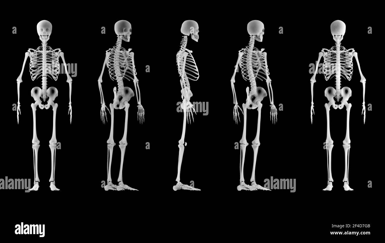 X-ray view of a human skeleton. Medical examination and body scan. Human anatomy and body bones. 360 degree view of a skeleton. 3d render Stock Photohttps://www.alamy.com/image-license-details/?v=1https://www.alamy.com/x-ray-view-of-a-human-skeleton-medical-examination-and-body-scan-human-anatomy-and-body-bones-360-degree-view-of-a-skeleton-3d-render-image415798779.html
RF2F4D7GB–X-ray view of a human skeleton. Medical examination and body scan. Human anatomy and body bones. 360 degree view of a skeleton. 3d render
 Vertebral joint, medically 3D artwork, x-ray view Stock Photohttps://www.alamy.com/image-license-details/?v=1https://www.alamy.com/vertebral-joint-medically-3d-artwork-x-ray-view-image230076580.html
RFRA8W0M–Vertebral joint, medically 3D artwork, x-ray view
 Hip replacement implant installed in the pelvis bone. X-ray view. Medically accurate 3D illustration Stock Photohttps://www.alamy.com/image-license-details/?v=1https://www.alamy.com/stock-image-hip-replacement-implant-installed-in-the-pelvis-bone-x-ray-view-medically-164433553.html
RFKFEGJW–Hip replacement implant installed in the pelvis bone. X-ray view. Medically accurate 3D illustration
 Pituitary gland or neurohypophysis x-ray view 3D rendering illustration. Human brain, nervous and endocrine system anatomy, medical, healthcare, scien Stock Photohttps://www.alamy.com/image-license-details/?v=1https://www.alamy.com/pituitary-gland-or-neurohypophysis-x-ray-view-3d-rendering-illustration-human-brain-nervous-and-endocrine-system-anatomy-medical-healthcare-scien-image571867882.html
RF2T6AR8X–Pituitary gland or neurohypophysis x-ray view 3D rendering illustration. Human brain, nervous and endocrine system anatomy, medical, healthcare, scien
 Hand x-ray view on black. Stock Photohttps://www.alamy.com/image-license-details/?v=1https://www.alamy.com/hand-x-ray-view-on-black-image158642657.html
RFK62P95–Hand x-ray view on black.
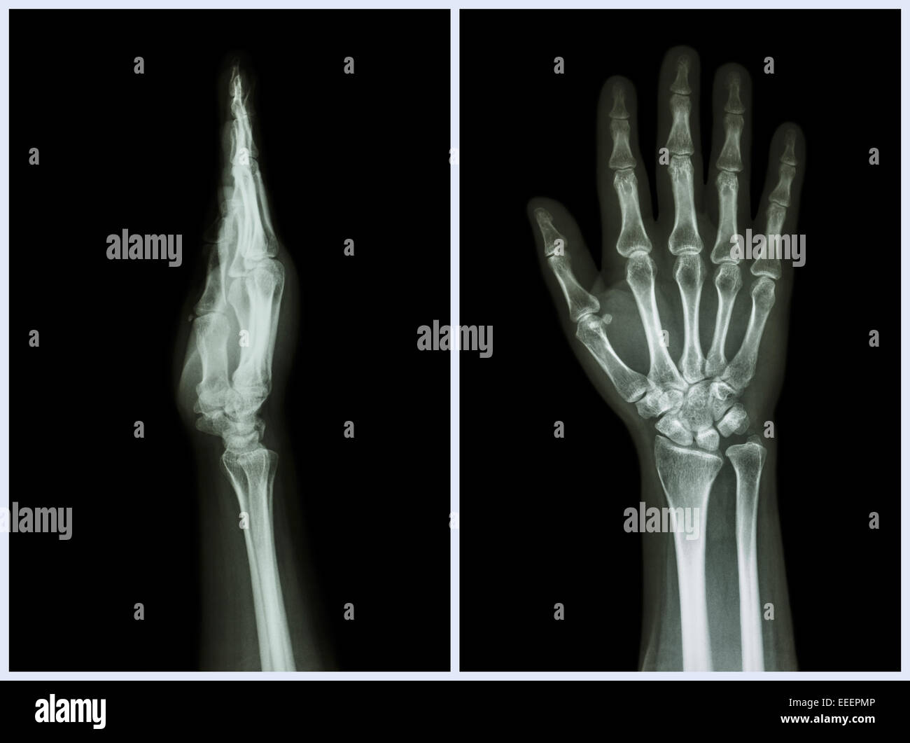 X-Ray Hands ( front & side view ) : Normal human hands Stock Photohttps://www.alamy.com/image-license-details/?v=1https://www.alamy.com/stock-photo-x-ray-hands-front-side-view-normal-human-hands-77771814.html
RFEEEPMP–X-Ray Hands ( front & side view ) : Normal human hands
 Results instrumental studies of patient in the hospital. Stock Photohttps://www.alamy.com/image-license-details/?v=1https://www.alamy.com/results-instrumental-studies-of-patient-in-the-hospital-image457656877.html
RF2HGG20D–Results instrumental studies of patient in the hospital.
 Wire-frame and X-Ray view of Tuk Tuk one of Asia famous transportation vehicle especially in Thailand and Cambodia 3D rendered. Stock Photohttps://www.alamy.com/image-license-details/?v=1https://www.alamy.com/wire-frame-and-x-ray-view-of-tuk-tuk-one-of-asia-famous-transportation-vehicle-especially-in-thailand-and-cambodia-3d-rendered-image232651512.html
RFREE5AG–Wire-frame and X-Ray view of Tuk Tuk one of Asia famous transportation vehicle especially in Thailand and Cambodia 3D rendered.
 X-Ray view of a city Stock Photohttps://www.alamy.com/image-license-details/?v=1https://www.alamy.com/x-ray-view-of-a-city-image616548422.html
RF2XR25PE–X-Ray view of a city
 Fish-Shaped Applique, 1400-1532. Central Andes, Central Coast, Ychsma (Pachacamac) people. Cotton and camelid fiber, tapestry weave with areas of eccentric weft floats; overall: 26 x 56.5 cm (10 1/4 x 22 1/4 in.); mounted: 32.4 x 62.9 cm (12 3/4 x 24 3/4 in.). This fish-shaped textile, a complete weaving with 33 finished edges, was stitched with others like it to a mantle, a shawl-like garment that was a staple of ancient Andean wardrobe. The partial “x-ray view,” which emphasizes the bony white teeth and spine, is unique to the style of the Ychsma (yeach-mah), who lived on Peru’s central coa Stock Photohttps://www.alamy.com/image-license-details/?v=1https://www.alamy.com/fish-shaped-applique-1400-1532-central-andes-central-coast-ychsma-pachacamac-people-cotton-and-camelid-fiber-tapestry-weave-with-areas-of-eccentric-weft-floats-overall-26-x-565-cm-10-14-x-22-14-in-mounted-324-x-629-cm-12-34-x-24-34-in-this-fish-shaped-textile-a-complete-weaving-with-33-finished-edges-was-stitched-with-others-like-it-to-a-mantle-a-shawl-like-garment-that-was-a-staple-of-ancient-andean-wardrobe-the-partial-x-ray-view-which-emphasizes-the-bony-white-teeth-and-spine-is-unique-to-the-style-of-the-ychsma-yeach-mah-who-lived-on-perus-central-coa-image448081150.html
RM2H0YT26–Fish-Shaped Applique, 1400-1532. Central Andes, Central Coast, Ychsma (Pachacamac) people. Cotton and camelid fiber, tapestry weave with areas of eccentric weft floats; overall: 26 x 56.5 cm (10 1/4 x 22 1/4 in.); mounted: 32.4 x 62.9 cm (12 3/4 x 24 3/4 in.). This fish-shaped textile, a complete weaving with 33 finished edges, was stitched with others like it to a mantle, a shawl-like garment that was a staple of ancient Andean wardrobe. The partial “x-ray view,” which emphasizes the bony white teeth and spine, is unique to the style of the Ychsma (yeach-mah), who lived on Peru’s central coa
 Young female doctor checks an X-ray. View from the back. Stock Photohttps://www.alamy.com/image-license-details/?v=1https://www.alamy.com/young-female-doctor-checks-an-x-ray-view-from-the-back-image424888246.html
RF2FK7986–Young female doctor checks an X-ray. View from the back.
 Inspired by View of Japan, Albumen silver photograph mounted on, Japan, late 19th-early 20th century, with mounting: 4 1/4 x 6 7/16 in., 10.8 x 16.3 cm, Reimagined by Artotop. Classic art reinvented with a modern twist. Design of warm cheerful glowing of brightness and light ray radiance. Photography inspired by surrealism and futurism, embracing dynamic energy of modern technology, movement, speed and revolutionize culture Stock Photohttps://www.alamy.com/image-license-details/?v=1https://www.alamy.com/inspired-by-view-of-japan-albumen-silver-photograph-mounted-on-japan-late-19th-early-20th-century-with-mounting-4-14-x-6-716-in-108-x-163-cm-reimagined-by-artotop-classic-art-reinvented-with-a-modern-twist-design-of-warm-cheerful-glowing-of-brightness-and-light-ray-radiance-photography-inspired-by-surrealism-and-futurism-embracing-dynamic-energy-of-modern-technology-movement-speed-and-revolutionize-culture-image459266827.html
RF2HK5BEK–Inspired by View of Japan, Albumen silver photograph mounted on, Japan, late 19th-early 20th century, with mounting: 4 1/4 x 6 7/16 in., 10.8 x 16.3 cm, Reimagined by Artotop. Classic art reinvented with a modern twist. Design of warm cheerful glowing of brightness and light ray radiance. Photography inspired by surrealism and futurism, embracing dynamic energy of modern technology, movement, speed and revolutionize culture
 Overhead view of light bulb, X-ray. Stock Photohttps://www.alamy.com/image-license-details/?v=1https://www.alamy.com/overhead-view-of-light-bulb-x-ray-image356003971.html
RF2BK5AKF–Overhead view of light bulb, X-ray.
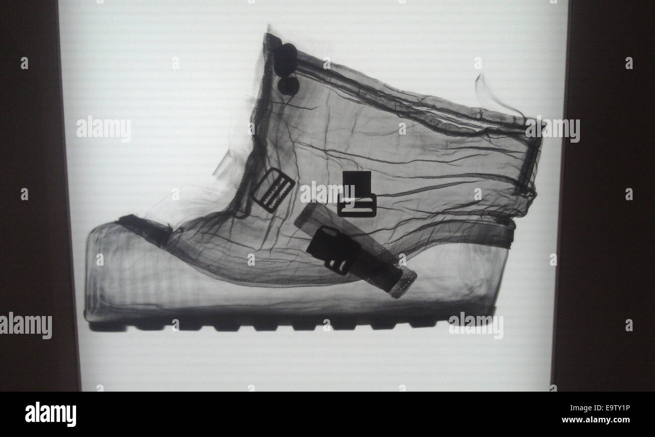 An x-ray view of a spacesuit boot. This photo is from the 'Suited for Space' social event at the National Air and Space Museum on July 25, 2013. The exhibit, which covered the spacesuits used by astronauts during their journeys into space, was open throug Stock Photohttps://www.alamy.com/image-license-details/?v=1https://www.alamy.com/stock-photo-an-x-ray-view-of-a-spacesuit-boot-this-photo-is-from-the-suited-for-74921442.html
RME9TY1P–An x-ray view of a spacesuit boot. This photo is from the 'Suited for Space' social event at the National Air and Space Museum on July 25, 2013. The exhibit, which covered the spacesuits used by astronauts during their journeys into space, was open throug
 x-ray view of luggage with inside arms. 3d render. Stock Photohttps://www.alamy.com/image-license-details/?v=1https://www.alamy.com/x-ray-view-of-luggage-with-inside-arms-3d-render-image448636060.html
RM2H1W3TC–x-ray view of luggage with inside arms. 3d render.
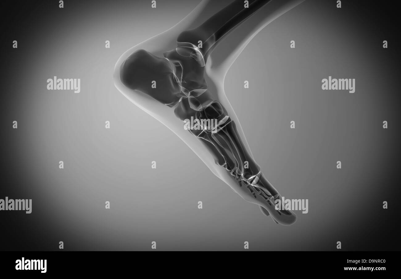 X-ray view of human foot. Stock Photohttps://www.alamy.com/image-license-details/?v=1https://www.alamy.com/stock-photo-x-ray-view-of-human-foot-57642368.html
RFD9NRC0–X-ray view of human foot.
 3D-Illustration of a glowing human male hand in an x-ray view Stock Photohttps://www.alamy.com/image-license-details/?v=1https://www.alamy.com/3d-illustration-of-a-glowing-human-male-hand-in-an-x-ray-view-image548237732.html
RF2PRXAR0–3D-Illustration of a glowing human male hand in an x-ray view
 Female reproductive organ, x-ray view Stock Photohttps://www.alamy.com/image-license-details/?v=1https://www.alamy.com/female-reproductive-organ-x-ray-view-image619578616.html
RM2Y006RM–Female reproductive organ, x-ray view
 anatomy of human spine in x-ray view Stock Photohttps://www.alamy.com/image-license-details/?v=1https://www.alamy.com/stock-photo-anatomy-of-human-spine-in-x-ray-view-103457525.html
RMG08W3H–anatomy of human spine in x-ray view
 X-ray view of an umbilical cord placed on a black background Stock Photohttps://www.alamy.com/image-license-details/?v=1https://www.alamy.com/stock-photo-x-ray-view-of-an-umbilical-cord-placed-on-a-black-background-74464022.html
RFE943HA–X-ray view of an umbilical cord placed on a black background
 knee joint, medically artwork, x-ray view Stock Photohttps://www.alamy.com/image-license-details/?v=1https://www.alamy.com/knee-joint-medically-artwork-x-ray-view-image230076646.html
RFRA8W32–knee joint, medically artwork, x-ray view
 Hip replacement implant installed in the pelvis bone. X-ray view. Medically accurate 3D illustration Stock Photohttps://www.alamy.com/image-license-details/?v=1https://www.alamy.com/hip-replacement-implant-installed-in-the-pelvis-bone-x-ray-view-medically-accurate-3d-illustration-image228492105.html
RFR7MM09–Hip replacement implant installed in the pelvis bone. X-ray view. Medically accurate 3D illustration
 Forebrain or prosencephalon x-ray view 3D rendering illustration. Human brain and nervous system anatomy, medical, healthcare, biology, science, neuro Stock Photohttps://www.alamy.com/image-license-details/?v=1https://www.alamy.com/forebrain-or-prosencephalon-x-ray-view-3d-rendering-illustration-human-brain-and-nervous-system-anatomy-medical-healthcare-biology-science-neuro-image571501198.html
RF2T5P3H2–Forebrain or prosencephalon x-ray view 3D rendering illustration. Human brain and nervous system anatomy, medical, healthcare, biology, science, neuro
 yellow human lungs on x-ray view, cg medicine 3d illustration Stock Photohttps://www.alamy.com/image-license-details/?v=1https://www.alamy.com/yellow-human-lungs-on-x-ray-view-cg-medicine-3d-illustration-image417924056.html
RF2F7X2B4–yellow human lungs on x-ray view, cg medicine 3d illustration
 technological transparent set of hand or palm anatomy beautiful aesthetic pose - 3d illustration of hand set in x ray view from different angles and p Stock Photohttps://www.alamy.com/image-license-details/?v=1https://www.alamy.com/technological-transparent-set-of-hand-or-palm-anatomy-beautiful-aesthetic-pose-3d-illustration-of-hand-set-in-x-ray-view-from-different-angles-and-p-image480003422.html
RF2JTX17A–technological transparent set of hand or palm anatomy beautiful aesthetic pose - 3d illustration of hand set in x ray view from different angles and p
 x-ray view engine in the white car Stock Photohttps://www.alamy.com/image-license-details/?v=1https://www.alamy.com/x-ray-view-engine-in-the-white-car-image365342854.html
RF2C6APF2–x-ray view engine in the white car
 Wire-frame and X-Ray view of Tuk Tuk one of Asia famous transportation vehicle especially in Thailand and Cambodia 3D rendered. Stock Photohttps://www.alamy.com/image-license-details/?v=1https://www.alamy.com/wire-frame-and-x-ray-view-of-tuk-tuk-one-of-asia-famous-transportation-vehicle-especially-in-thailand-and-cambodia-3d-rendered-image232651648.html
RFREE5FC–Wire-frame and X-Ray view of Tuk Tuk one of Asia famous transportation vehicle especially in Thailand and Cambodia 3D rendered.
 Side view of smiling doctor with an x-ray Stock Photohttps://www.alamy.com/image-license-details/?v=1https://www.alamy.com/stock-photo-side-view-of-smiling-doctor-with-an-x-ray-43990775.html
RFCFFXK3–Side view of smiling doctor with an x-ray
 X-ray photograph of a foot taken by a photographer during World War One. The image is numbered as 6074 in the collection. The shot shows a detailed x-ray view of the foot. The description mentions that the photo was taken for official purposes and is labeled as 'R 11 AU SOTES.' Stock Photohttps://www.alamy.com/image-license-details/?v=1https://www.alamy.com/x-ray-photograph-of-a-foot-taken-by-a-photographer-during-world-war-one-the-image-is-numbered-as-6074-in-the-collection-the-shot-shows-a-detailed-x-ray-view-of-the-foot-the-description-mentions-that-the-photo-was-taken-for-official-purposes-and-is-labeled-as-r-11-au-sotes-image558128533.html
RM2RC0XHW–X-ray photograph of a foot taken by a photographer during World War One. The image is numbered as 6074 in the collection. The shot shows a detailed x-ray view of the foot. The description mentions that the photo was taken for official purposes and is labeled as 'R 11 AU SOTES.'
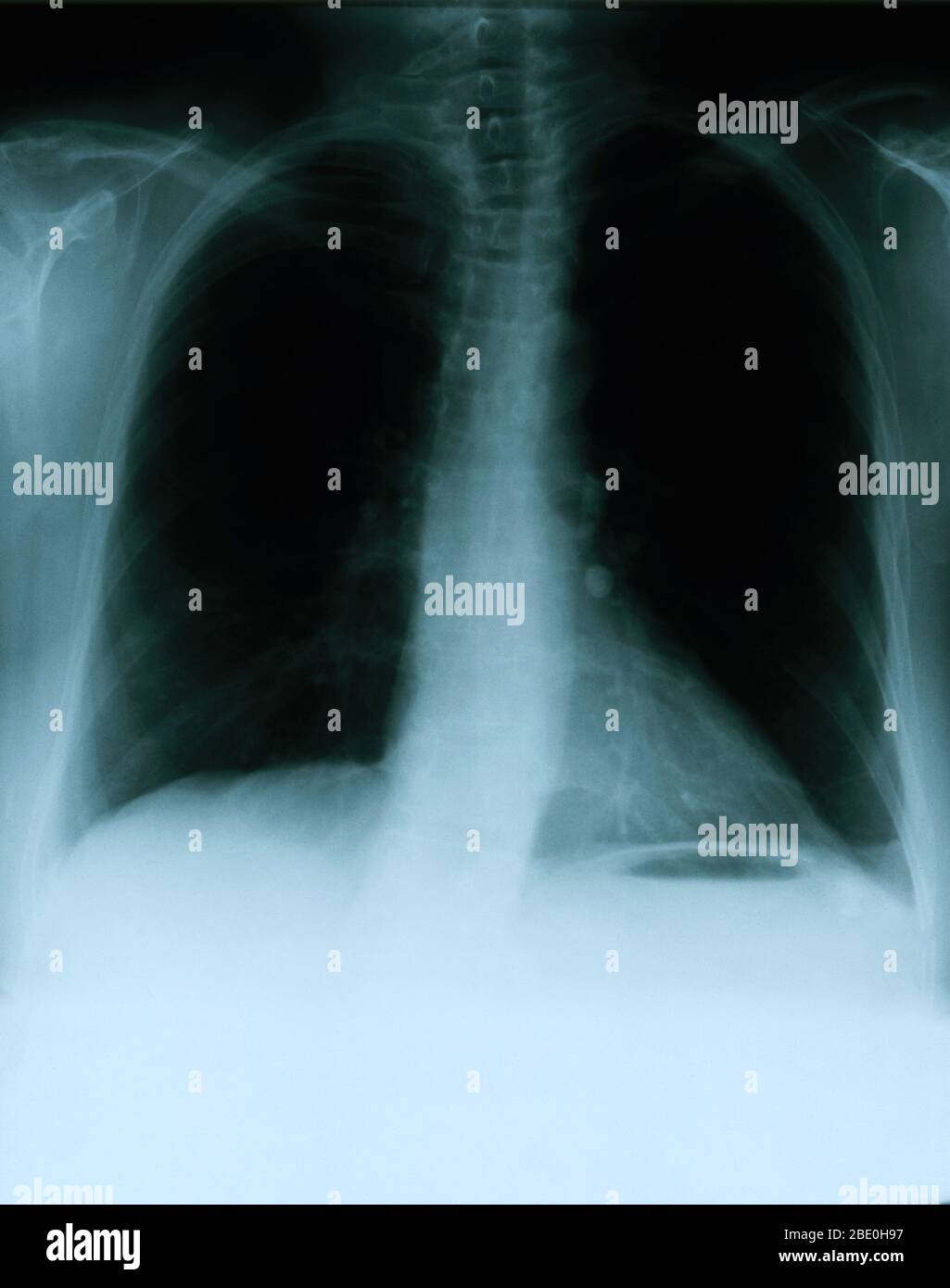 X-ray showing a frontal view of the chest of a 54 year old female. The x-ray shows a calcified left hilar lymph node which most likely resulted from prior granulomatous disease. Also noticeable is a vague area of increased density within the lateral aspect of the right apex, and a mild scoliotic deformity of the dorsal spine. Stock Photohttps://www.alamy.com/image-license-details/?v=1https://www.alamy.com/x-ray-showing-a-frontal-view-of-the-chest-of-a-54-year-old-female-the-x-ray-shows-a-calcified-left-hilar-lymph-node-which-most-likely-resulted-from-prior-granulomatous-disease-also-noticeable-is-a-vague-area-of-increased-density-within-the-lateral-aspect-of-the-right-apex-and-a-mild-scoliotic-deformity-of-the-dorsal-spine-image352826131.html
RM2BE0H97–X-ray showing a frontal view of the chest of a 54 year old female. The x-ray shows a calcified left hilar lymph node which most likely resulted from prior granulomatous disease. Also noticeable is a vague area of increased density within the lateral aspect of the right apex, and a mild scoliotic deformity of the dorsal spine.
 Stroke . film x-ray of human skull lateral view with stroke . blank area at right side . Stock Photohttps://www.alamy.com/image-license-details/?v=1https://www.alamy.com/stock-photo-stroke-film-x-ray-of-human-skull-lateral-view-with-stroke-blank-area-134137903.html
RFHP6E7B–Stroke . film x-ray of human skull lateral view with stroke . blank area at right side .
 Taxi cab angled plan view, X-ray. Stock Photohttps://www.alamy.com/image-license-details/?v=1https://www.alamy.com/taxi-cab-angled-plan-view-x-ray-image356003918.html
RF2BK5AHJ–Taxi cab angled plan view, X-ray.
 Spacesuit boot x-ray view Stock Photohttps://www.alamy.com/image-license-details/?v=1https://www.alamy.com/spacesuit-boot-x-ray-view-image68968552.html
RME05P2G–Spacesuit boot x-ray view
 Doctor, X-ray picture, knee, diagnosis Stock Photohttps://www.alamy.com/image-license-details/?v=1https://www.alamy.com/stock-photo-doctor-x-ray-picture-knee-diagnosis-123171149.html
RMH4AX1H–Doctor, X-ray picture, knee, diagnosis
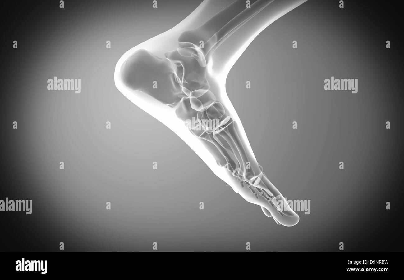 X-ray view of human foot. Stock Photohttps://www.alamy.com/image-license-details/?v=1https://www.alamy.com/stock-photo-x-ray-view-of-human-foot-57642365.html
RFD9NRBW–X-ray view of human foot.
 3D-Illustration of a glowing human male hand in an x-ray view Stock Photohttps://www.alamy.com/image-license-details/?v=1https://www.alamy.com/3d-illustration-of-a-glowing-human-male-hand-in-an-x-ray-view-image548657896.html
RF2PTHEMT–3D-Illustration of a glowing human male hand in an x-ray view
 X-ray view of human skeleton showing teeth and gums Stock Photohttps://www.alamy.com/image-license-details/?v=1https://www.alamy.com/x-ray-view-of-human-skeleton-showing-teeth-and-gums-image619199607.html
RM2XYAYBK–X-ray view of human skeleton showing teeth and gums
 anatomy of human lungs in x-ray view Stock Photohttps://www.alamy.com/image-license-details/?v=1https://www.alamy.com/stock-photo-anatomy-of-human-lungs-in-x-ray-view-103457228.html
RMG08TN0–anatomy of human lungs in x-ray view
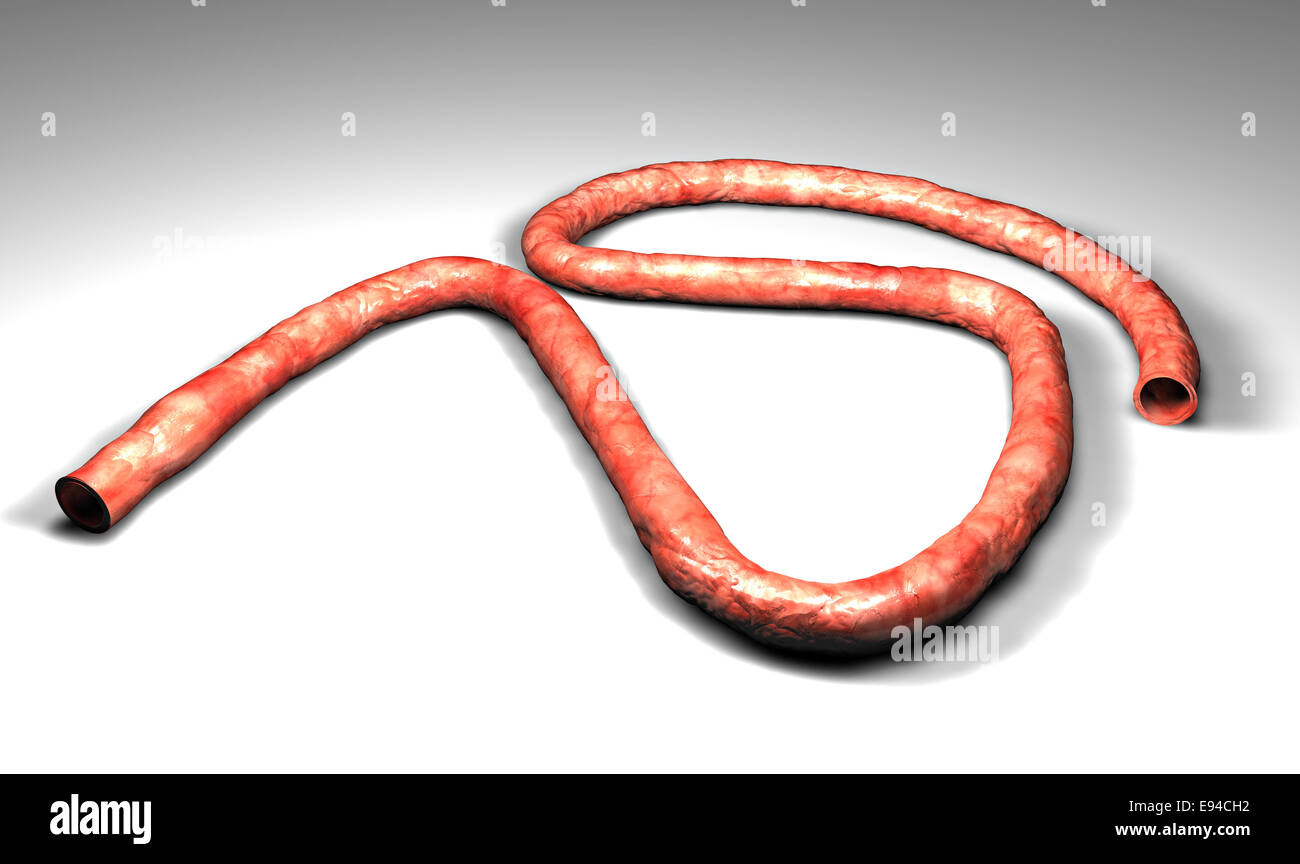 X-ray view of an umbilical cord placed on a white background Stock Photohttps://www.alamy.com/image-license-details/?v=1https://www.alamy.com/stock-photo-x-ray-view-of-an-umbilical-cord-placed-on-a-white-background-74471070.html
RFE94CH2–X-ray view of an umbilical cord placed on a white background
 wrist, medically 3D artwork, x-ray view, radiography Stock Photohttps://www.alamy.com/image-license-details/?v=1https://www.alamy.com/wrist-medically-3d-artwork-x-ray-view-radiography-image230076593.html
RFRA8W15–wrist, medically 3D artwork, x-ray view, radiography
 Hip replacement implant installed in the pelvis bone. X-ray view. Medically accurate 3D illustration Stock Photohttps://www.alamy.com/image-license-details/?v=1https://www.alamy.com/hip-replacement-implant-installed-in-the-pelvis-bone-x-ray-view-medically-accurate-3d-illustration-image228491989.html
RFR7MKT5–Hip replacement implant installed in the pelvis bone. X-ray view. Medically accurate 3D illustration
 Midbrain or mesencephalon lateral x-ray view 3D rendering illustration. Human brain and nervous system anatomy, medical, healthcare, biology, science, Stock Photohttps://www.alamy.com/image-license-details/?v=1https://www.alamy.com/midbrain-or-mesencephalon-lateral-x-ray-view-3d-rendering-illustration-human-brain-and-nervous-system-anatomy-medical-healthcare-biology-science-image571007495.html
RF2T4YHTR–Midbrain or mesencephalon lateral x-ray view 3D rendering illustration. Human brain and nervous system anatomy, medical, healthcare, biology, science,
 X-ray view of the respiratory tract in a human. The upper airways are inflamed and therefore red. Pencil drawn illustration. Stock Photohttps://www.alamy.com/image-license-details/?v=1https://www.alamy.com/x-ray-view-of-the-respiratory-tract-in-a-human-the-upper-airways-are-inflamed-and-therefore-red-pencil-drawn-illustration-image246663082.html
RFT98D7P–X-ray view of the respiratory tract in a human. The upper airways are inflamed and therefore red. Pencil drawn illustration.
 technological transparent set of hand or palm anatomy beautiful aesthetic pose - 3d illustration of hand set in x ray view from different angles and p Stock Photohttps://www.alamy.com/image-license-details/?v=1https://www.alamy.com/technological-transparent-set-of-hand-or-palm-anatomy-beautiful-aesthetic-pose-3d-illustration-of-hand-set-in-x-ray-view-from-different-angles-and-p-image480003420.html
RF2JTX178–technological transparent set of hand or palm anatomy beautiful aesthetic pose - 3d illustration of hand set in x ray view from different angles and p
 Portrait of female doctor with x-ray image behind windowpane Stock Photohttps://www.alamy.com/image-license-details/?v=1https://www.alamy.com/portrait-of-female-doctor-with-x-ray-image-behind-windowpane-image327753131.html
RF2A16CCY–Portrait of female doctor with x-ray image behind windowpane
 Wire-frame and X-Ray view of Tuk Tuk one of Asia famous transportation vehicle especially in Thailand and Cambodia 3D rendered. Stock Photohttps://www.alamy.com/image-license-details/?v=1https://www.alamy.com/wire-frame-and-x-ray-view-of-tuk-tuk-one-of-asia-famous-transportation-vehicle-especially-in-thailand-and-cambodia-3d-rendered-image232540412.html
RFRE93JM–Wire-frame and X-Ray view of Tuk Tuk one of Asia famous transportation vehicle especially in Thailand and Cambodia 3D rendered.
 the god stands in three-quarter front view with the head to the right. In his hair a ray crown, in his left hand a bow, in his right hand an arrow which he takes out of the tube and under his right arm an unidentified object, ground line, gem, intaglio, ring stone, stone, carnelian, Color: orange, transparent, Shape: oval, standing, Machined: Maaskant-Kleibrink, M., Catalog of the engraved gems in the Royal Coin Cabinet, 1978, p.60, fig.2, type F1, 11 x 9 mm. Thickness 2 mm., Germany Stock Photohttps://www.alamy.com/image-license-details/?v=1https://www.alamy.com/the-god-stands-in-three-quarter-front-view-with-the-head-to-the-right-in-his-hair-a-ray-crown-in-his-left-hand-a-bow-in-his-right-hand-an-arrow-which-he-takes-out-of-the-tube-and-under-his-right-arm-an-unidentified-object-ground-line-gem-intaglio-ring-stone-stone-carnelian-color-orange-transparent-shape-oval-standing-machined-maaskant-kleibrink-m-catalog-of-the-engraved-gems-in-the-royal-coin-cabinet-1978-p60-fig2-type-f1-11-x-9-mm-thickness-2-mm-germany-image344541922.html
RM2B0F6MJ–the god stands in three-quarter front view with the head to the right. In his hair a ray crown, in his left hand a bow, in his right hand an arrow which he takes out of the tube and under his right arm an unidentified object, ground line, gem, intaglio, ring stone, stone, carnelian, Color: orange, transparent, Shape: oval, standing, Machined: Maaskant-Kleibrink, M., Catalog of the engraved gems in the Royal Coin Cabinet, 1978, p.60, fig.2, type F1, 11 x 9 mm. Thickness 2 mm., Germany
 Side view of smiling female doctor with x-ray Stock Photohttps://www.alamy.com/image-license-details/?v=1https://www.alamy.com/stock-photo-side-view-of-smiling-female-doctor-with-x-ray-51148537.html
RFCY60DD–Side view of smiling female doctor with x-ray
 X-ray showing a frontal view of the chest of a 54 year old female. The x-ray shows a calcified left hilar lymph node which most likely resulted from prior granulomatous disease. Also noticeable is a vague area of increased density within the lateral aspect of the right apex, and a mild scoliotic deformity of the dorsal spine. Stock Photohttps://www.alamy.com/image-license-details/?v=1https://www.alamy.com/x-ray-showing-a-frontal-view-of-the-chest-of-a-54-year-old-female-the-x-ray-shows-a-calcified-left-hilar-lymph-node-which-most-likely-resulted-from-prior-granulomatous-disease-also-noticeable-is-a-vague-area-of-increased-density-within-the-lateral-aspect-of-the-right-apex-and-a-mild-scoliotic-deformity-of-the-dorsal-spine-image352834615.html
RM2BE1047–X-ray showing a frontal view of the chest of a 54 year old female. The x-ray shows a calcified left hilar lymph node which most likely resulted from prior granulomatous disease. Also noticeable is a vague area of increased density within the lateral aspect of the right apex, and a mild scoliotic deformity of the dorsal spine.
 Film X-ray of normal human skull . lateral view . blank area at right side . Stock Photohttps://www.alamy.com/image-license-details/?v=1https://www.alamy.com/stock-photo-film-x-ray-of-normal-human-skull-lateral-view-blank-area-at-right-134137900.html
RFHP6E78–Film X-ray of normal human skull . lateral view . blank area at right side .
 Medically Accurate Healthy Tooth X-Ray View on a black background. 3d Rendering Stock Photohttps://www.alamy.com/image-license-details/?v=1https://www.alamy.com/medically-accurate-healthy-tooth-x-ray-view-on-a-black-background-3d-rendering-image548723120.html
RF2PTMDX8–Medically Accurate Healthy Tooth X-Ray View on a black background. 3d Rendering
 Internal view of office block levels, X-ray. Stock Photohttps://www.alamy.com/image-license-details/?v=1https://www.alamy.com/internal-view-of-office-block-levels-x-ray-image364972343.html
RF2C5NWXF–Internal view of office block levels, X-ray.
 Doctor, X-ray picture, looking Stock Photohttps://www.alamy.com/image-license-details/?v=1https://www.alamy.com/stock-photo-doctor-x-ray-picture-looking-123172863.html
RMH4B06R–Doctor, X-ray picture, looking
 X-ray view of human foot. Stock Photohttps://www.alamy.com/image-license-details/?v=1https://www.alamy.com/stock-photo-x-ray-view-of-human-foot-57642343.html
RFD9NRB3–X-ray view of human foot.
 3D-Illustration of a glowing human male hand in an x-ray view Stock Photohttps://www.alamy.com/image-license-details/?v=1https://www.alamy.com/3d-illustration-of-a-glowing-human-male-hand-in-an-x-ray-view-image548237742.html
RF2PRXARA–3D-Illustration of a glowing human male hand in an x-ray view
 X-ray view of human foot Stock Photohttps://www.alamy.com/image-license-details/?v=1https://www.alamy.com/x-ray-view-of-human-foot-image619197108.html
RM2XYAT6C–X-ray view of human foot
 human liver in x-ray view Stock Photohttps://www.alamy.com/image-license-details/?v=1https://www.alamy.com/stock-photo-human-liver-in-x-ray-view-103457238.html
RMG08TNA–human liver in x-ray view
 Human body seen from behind with a x-ray view of the spine Stock Photohttps://www.alamy.com/image-license-details/?v=1https://www.alamy.com/stock-photo-human-body-seen-from-behind-with-a-x-ray-view-of-the-spine-85493804.html
RFEY2G64–Human body seen from behind with a x-ray view of the spine
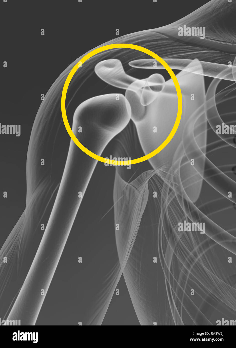 Shoulder joint, medically 3D artwork, x-ray view, radiograph Stock Photohttps://www.alamy.com/image-license-details/?v=1https://www.alamy.com/shoulder-joint-medically-3d-artwork-x-ray-view-radiograph-image230076634.html
RFRA8W2J–Shoulder joint, medically 3D artwork, x-ray view, radiograph
 Dental titanium implant, x-ray view Stock Photohttps://www.alamy.com/image-license-details/?v=1https://www.alamy.com/stock-image-dental-titanium-implant-x-ray-view-164433523.html
RFKFEGHR–Dental titanium implant, x-ray view
 Ventricles and cerebral aqueduct lateral x-ray view 3D rendering illustration. Human brain and ventricular system anatomy, medical, healthcare, scienc Stock Photohttps://www.alamy.com/image-license-details/?v=1https://www.alamy.com/ventricles-and-cerebral-aqueduct-lateral-x-ray-view-3d-rendering-illustration-human-brain-and-ventricular-system-anatomy-medical-healthcare-scienc-image572718989.html
RF2T7NGWH–Ventricles and cerebral aqueduct lateral x-ray view 3D rendering illustration. Human brain and ventricular system anatomy, medical, healthcare, scienc
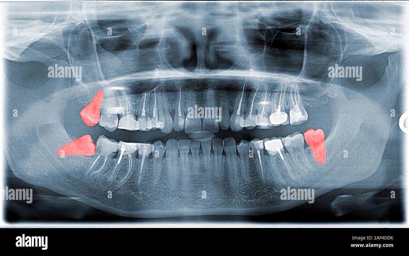 Full Frame View Of Dental Jaw X-ray With Wishdom Teeth Stock Photohttps://www.alamy.com/image-license-details/?v=1https://www.alamy.com/full-frame-view-of-dental-jaw-x-ray-with-wishdom-teeth-image336315215.html
RF2AF4DDK–Full Frame View Of Dental Jaw X-ray With Wishdom Teeth
 technological transparent set of hand or palm anatomy beautiful aesthetic pose - 3d illustration of hand set in x ray view from different angles and p Stock Photohttps://www.alamy.com/image-license-details/?v=1https://www.alamy.com/technological-transparent-set-of-hand-or-palm-anatomy-beautiful-aesthetic-pose-3d-illustration-of-hand-set-in-x-ray-view-from-different-angles-and-p-image480003432.html
RF2JTX17M–technological transparent set of hand or palm anatomy beautiful aesthetic pose - 3d illustration of hand set in x ray view from different angles and p
 Portrait of happy female doctor with x-ray image behind windowpane Stock Photohttps://www.alamy.com/image-license-details/?v=1https://www.alamy.com/portrait-of-happy-female-doctor-with-x-ray-image-behind-windowpane-image327751725.html
RF2A16AJN–Portrait of happy female doctor with x-ray image behind windowpane
 Wire-frame and X-Ray view of Tuk Tuk one of Asia famous transportation vehicle especially in Thailand and Cambodia 3D rendered. Stock Photohttps://www.alamy.com/image-license-details/?v=1https://www.alamy.com/wire-frame-and-x-ray-view-of-tuk-tuk-one-of-asia-famous-transportation-vehicle-especially-in-thailand-and-cambodia-3d-rendered-image232540346.html
RFRE93GA–Wire-frame and X-Ray view of Tuk Tuk one of Asia famous transportation vehicle especially in Thailand and Cambodia 3D rendered.
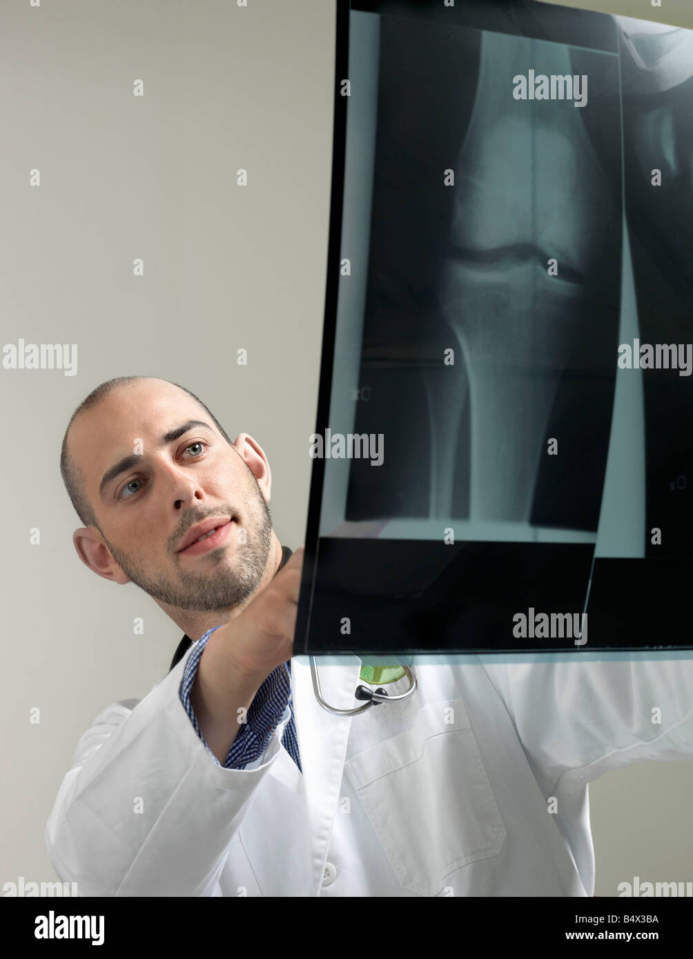 Doctor with x-ray Stock Photohttps://www.alamy.com/image-license-details/?v=1https://www.alamy.com/stock-photo-doctor-with-x-ray-20242414.html
RFB4X3BA–Doctor with x-ray
 X-ray view in the upper airways Stock Photohttps://www.alamy.com/image-license-details/?v=1https://www.alamy.com/stock-photo-x-ray-view-in-the-upper-airways-144360548.html
RFJAT59T–X-ray view in the upper airways
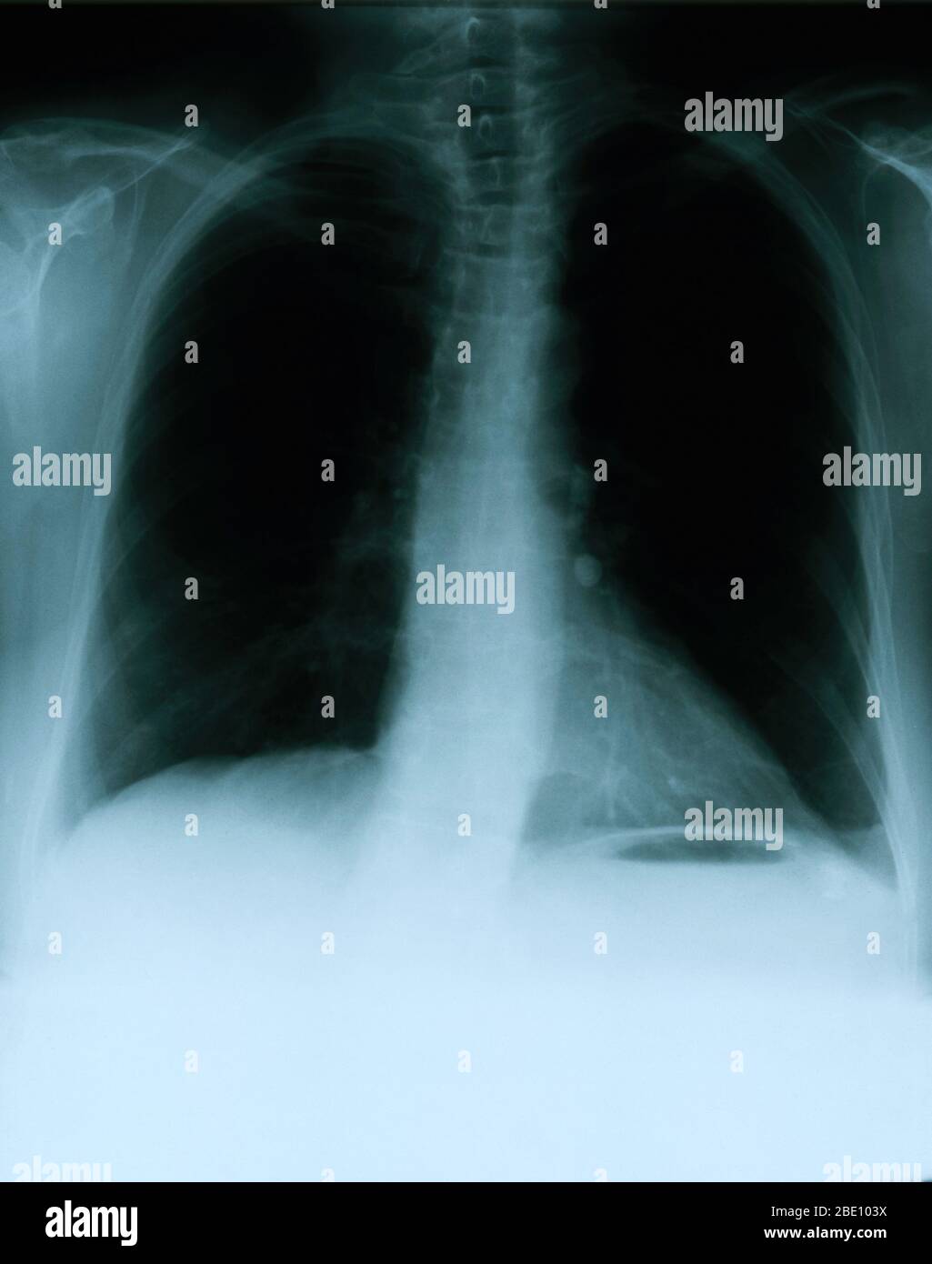 X-ray showing a frontal view of the chest of a 54 year old female. The x-ray shows a calcified left hilar lymph node which most likely resulted from prior granulomatous disease. Also noticeable is a vague area of increased density within the lateral aspect of the right apex, and a mild scoliotic deformity of the dorsal spine. Stock Photohttps://www.alamy.com/image-license-details/?v=1https://www.alamy.com/x-ray-showing-a-frontal-view-of-the-chest-of-a-54-year-old-female-the-x-ray-shows-a-calcified-left-hilar-lymph-node-which-most-likely-resulted-from-prior-granulomatous-disease-also-noticeable-is-a-vague-area-of-increased-density-within-the-lateral-aspect-of-the-right-apex-and-a-mild-scoliotic-deformity-of-the-dorsal-spine-image352834606.html
RM2BE103X–X-ray showing a frontal view of the chest of a 54 year old female. The x-ray shows a calcified left hilar lymph node which most likely resulted from prior granulomatous disease. Also noticeable is a vague area of increased density within the lateral aspect of the right apex, and a mild scoliotic deformity of the dorsal spine.
 Stroke . film x-ray of human skull lateral view with stroke . blank area at right side . Stock Photohttps://www.alamy.com/image-license-details/?v=1https://www.alamy.com/stock-photo-stroke-film-x-ray-of-human-skull-lateral-view-with-stroke-blank-area-134137901.html
RFHP6E79–Stroke . film x-ray of human skull lateral view with stroke . blank area at right side .
 Medically Accurate Healthy Tooth X-Ray View on a black background. 3d Rendering Stock Photohttps://www.alamy.com/image-license-details/?v=1https://www.alamy.com/medically-accurate-healthy-tooth-x-ray-view-on-a-black-background-3d-rendering-image548724301.html
RF2PTMFCD–Medically Accurate Healthy Tooth X-Ray View on a black background. 3d Rendering
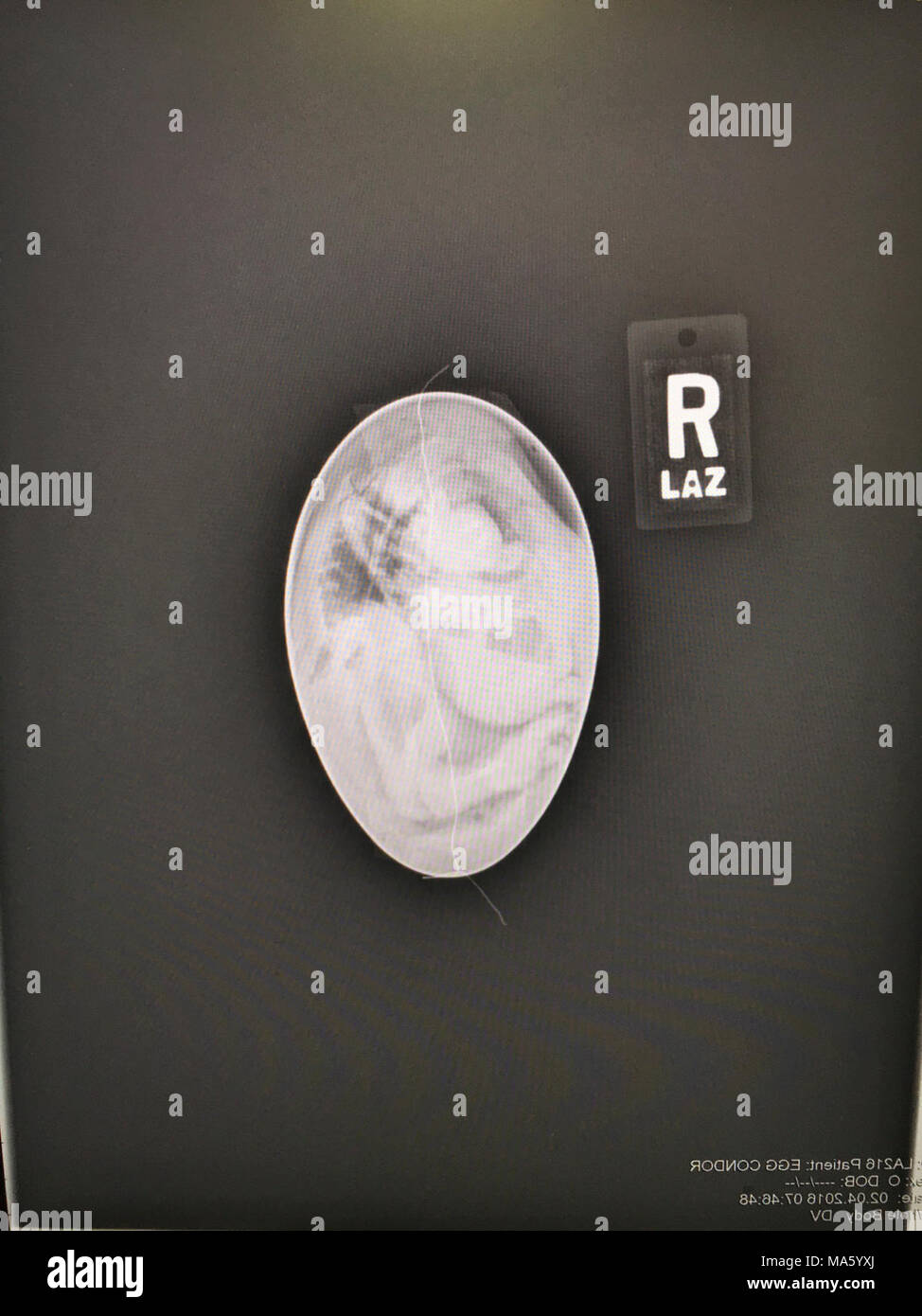 X-ray view of inside California condor egg 216. Stock Photohttps://www.alamy.com/image-license-details/?v=1https://www.alamy.com/x-ray-view-of-inside-california-condor-egg-216-image178381914.html
RMMA5YXJ–X-ray view of inside California condor egg 216.
 Doctor, X-ray picture, patient Stock Photohttps://www.alamy.com/image-license-details/?v=1https://www.alamy.com/stock-photo-doctor-x-ray-picture-patient-123172861.html
RMH4B06N–Doctor, X-ray picture, patient
 X-ray view of human foot. Stock Photohttps://www.alamy.com/image-license-details/?v=1https://www.alamy.com/stock-photo-x-ray-view-of-human-foot-57642347.html
RFD9NRB7–X-ray view of human foot.
 3D-Illustration of a glowing human male hand in an x-ray view Stock Photohttps://www.alamy.com/image-license-details/?v=1https://www.alamy.com/3d-illustration-of-a-glowing-human-male-hand-in-an-x-ray-view-image548657894.html
RF2PTHEMP–3D-Illustration of a glowing human male hand in an x-ray view
 X-ray view of human foot Stock Photohttps://www.alamy.com/image-license-details/?v=1https://www.alamy.com/x-ray-view-of-human-foot-image619792128.html
RM2Y09Y54–X-ray view of human foot
 Inspired by View near Fishkill, from 'The Hudson River Portfolio', William Guy Wall, Irish, 1792–after 1864, John Hill, American (born England), 1770–1850, 1823–24, Hand-colored aquatint, etching, and engraving, Fishkill, New York, United States, North and Central America, Prints, image: 16 1/4 x 21, Reimagined by Artotop. Classic art reinvented with a modern twist. Design of warm cheerful glowing of brightness and light ray radiance. Photography inspired by surrealism and futurism, embracing dynamic energy of modern technology, movement, speed and revolutionize culture Stock Photohttps://www.alamy.com/image-license-details/?v=1https://www.alamy.com/inspired-by-view-near-fishkill-from-the-hudson-river-portfolio-william-guy-wall-irish-1792after-1864-john-hill-american-born-england-17701850-182324-hand-colored-aquatint-etching-and-engraving-fishkill-new-york-united-states-north-and-central-america-prints-image-16-14-x-21-reimagined-by-artotop-classic-art-reinvented-with-a-modern-twist-design-of-warm-cheerful-glowing-of-brightness-and-light-ray-radiance-photography-inspired-by-surrealism-and-futurism-embracing-dynamic-energy-of-modern-technology-movement-speed-and-revolutionize-culture-image459283688.html
RF2HK650T–Inspired by View near Fishkill, from 'The Hudson River Portfolio', William Guy Wall, Irish, 1792–after 1864, John Hill, American (born England), 1770–1850, 1823–24, Hand-colored aquatint, etching, and engraving, Fishkill, New York, United States, North and Central America, Prints, image: 16 1/4 x 21, Reimagined by Artotop. Classic art reinvented with a modern twist. Design of warm cheerful glowing of brightness and light ray radiance. Photography inspired by surrealism and futurism, embracing dynamic energy of modern technology, movement, speed and revolutionize culture
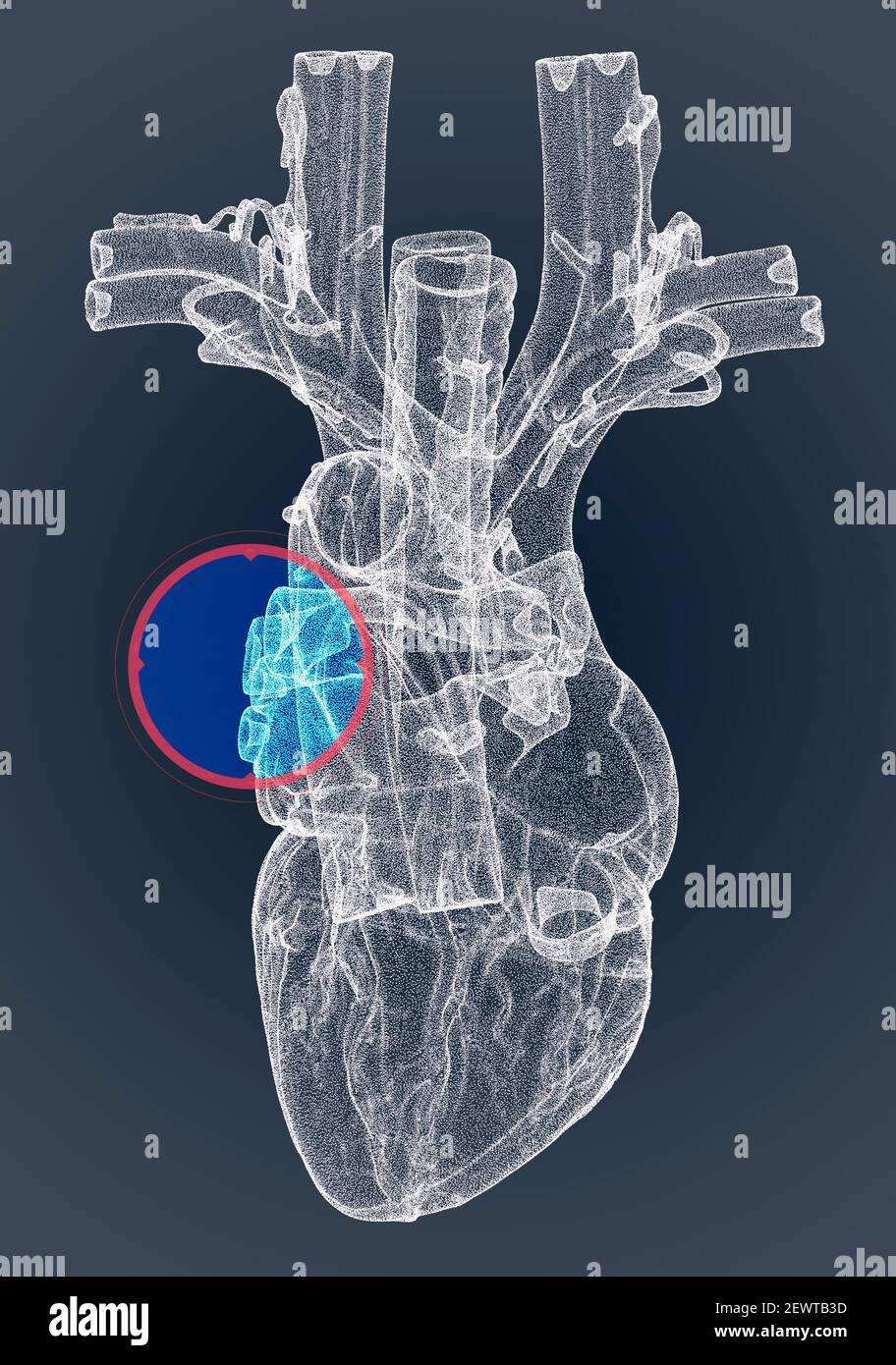 Heart, ventricles, human anatomy, cardiac ventricles. Human body, section. X-ray view. HUD. Advanced Scientific Devices. Hologram. Scanner. 3d render Stock Photohttps://www.alamy.com/image-license-details/?v=1https://www.alamy.com/heart-ventricles-human-anatomy-cardiac-ventricles-human-body-section-x-ray-view-hud-advanced-scientific-devices-hologram-scanner-3d-render-image411740433.html
RF2EWTB3D–Heart, ventricles, human anatomy, cardiac ventricles. Human body, section. X-ray view. HUD. Advanced Scientific Devices. Hologram. Scanner. 3d render
 Medically 3D illustration showing x-ray view of a kidneyon blue background Stock Photohttps://www.alamy.com/image-license-details/?v=1https://www.alamy.com/medically-3d-illustration-showing-x-ray-view-of-a-kidneyon-blue-background-image349810741.html
RF2B9374N–Medically 3D illustration showing x-ray view of a kidneyon blue background
 Dental titanium implant, x-ray view Stock Photohttps://www.alamy.com/image-license-details/?v=1https://www.alamy.com/stock-image-dental-titanium-implant-x-ray-view-162659833.html
RFKCHP7N–Dental titanium implant, x-ray view
 Dentate gyrus x-ray view 3D rendering illustration. Human brain and nervous system anatomy, medical, healthcare, biology, science, neuroscience, neuro Stock Photohttps://www.alamy.com/image-license-details/?v=1https://www.alamy.com/dentate-gyrus-x-ray-view-3d-rendering-illustration-human-brain-and-nervous-system-anatomy-medical-healthcare-biology-science-neuroscience-neuro-image571715135.html
RF2T63TDK–Dentate gyrus x-ray view 3D rendering illustration. Human brain and nervous system anatomy, medical, healthcare, biology, science, neuroscience, neuro
 X-ray of knee joint on a negatoscope, X-ray as a type of diagnosis for the treatment of diseases and injuries of the joints of the legs Stock Photohttps://www.alamy.com/image-license-details/?v=1https://www.alamy.com/x-ray-of-knee-joint-on-a-negatoscope-x-ray-as-a-type-of-diagnosis-for-the-treatment-of-diseases-and-injuries-of-the-joints-of-the-legs-image382506345.html
RF2D68JMW–X-ray of knee joint on a negatoscope, X-ray as a type of diagnosis for the treatment of diseases and injuries of the joints of the legs
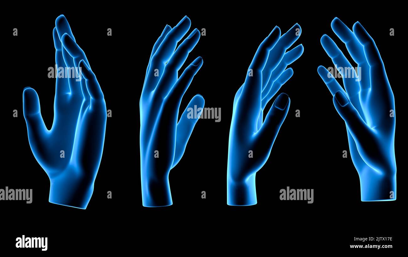 technological transparent set of hand or palm anatomy beautiful aesthetic pose - 3d illustration of hand set in x ray view from different angles and p Stock Photohttps://www.alamy.com/image-license-details/?v=1https://www.alamy.com/technological-transparent-set-of-hand-or-palm-anatomy-beautiful-aesthetic-pose-3d-illustration-of-hand-set-in-x-ray-view-from-different-angles-and-p-image480003426.html
RF2JTX17E–technological transparent set of hand or palm anatomy beautiful aesthetic pose - 3d illustration of hand set in x ray view from different angles and p
 A view of a stethoscope through an x-ray Stock Photohttps://www.alamy.com/image-license-details/?v=1https://www.alamy.com/stock-photo-a-view-of-a-stethoscope-through-an-x-ray-104047252.html
RFG17N98–A view of a stethoscope through an x-ray
 Wire-frame and X-Ray view of Tuk Tuk one of Asia famous transportation vehicle especially in Thailand and Cambodia 3D rendered. Stock Photohttps://www.alamy.com/image-license-details/?v=1https://www.alamy.com/wire-frame-and-x-ray-view-of-tuk-tuk-one-of-asia-famous-transportation-vehicle-especially-in-thailand-and-cambodia-3d-rendered-image232540663.html
RFRE93YK–Wire-frame and X-Ray view of Tuk Tuk one of Asia famous transportation vehicle especially in Thailand and Cambodia 3D rendered.
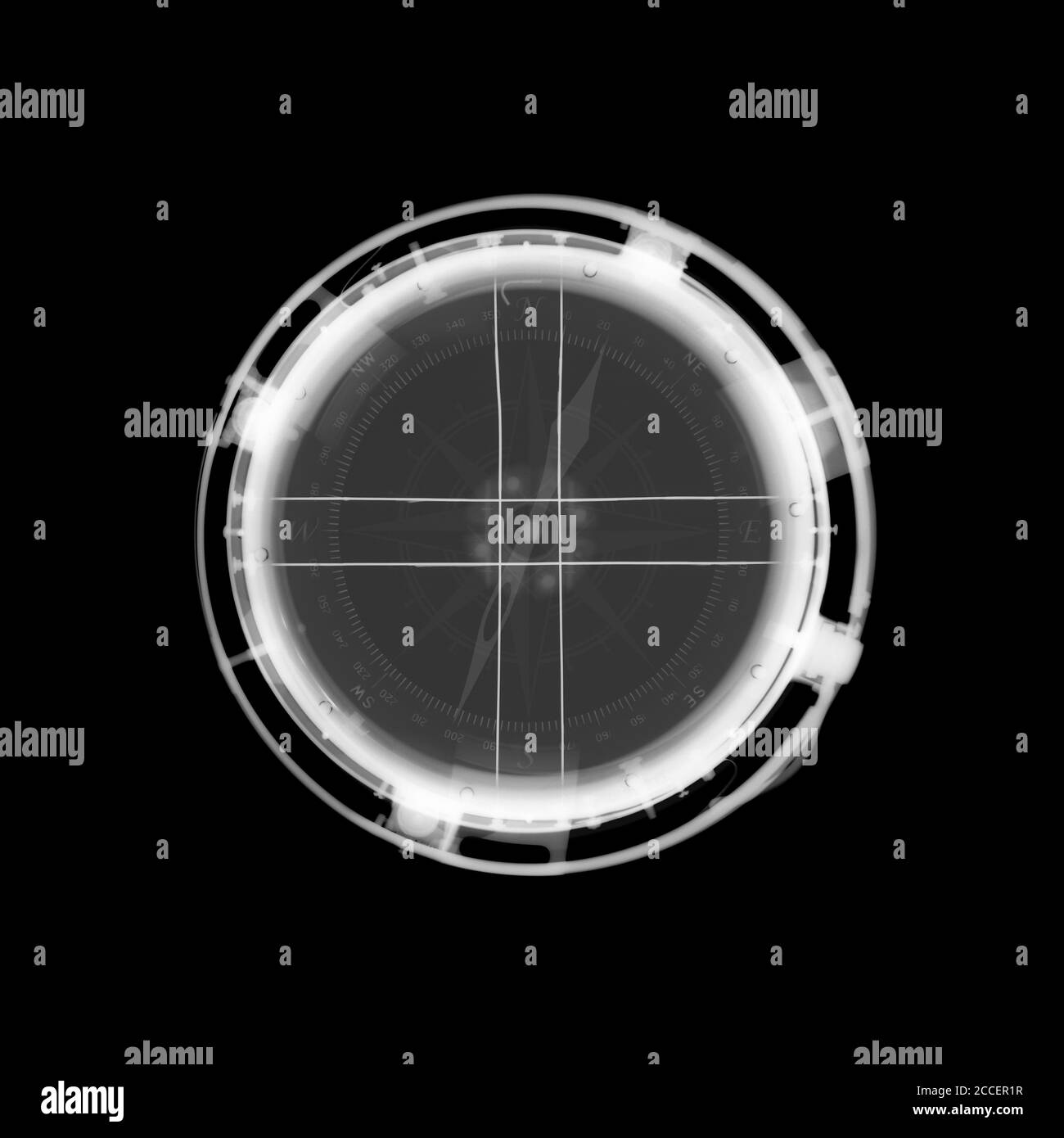 Compass, X-ray Stock Photohttps://www.alamy.com/image-license-details/?v=1https://www.alamy.com/compass-x-ray-image369119011.html
RF2CCER1R–Compass, X-ray
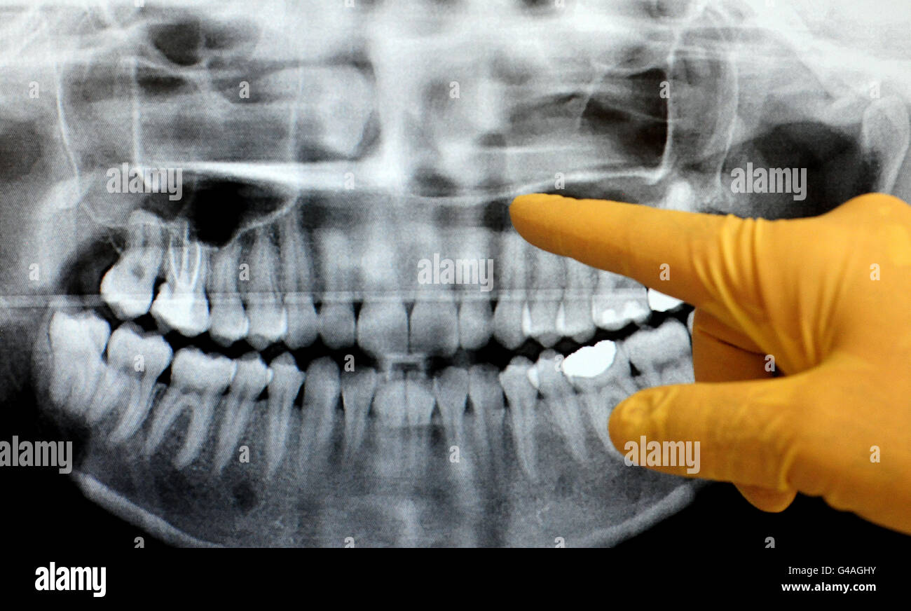 General view of dentist checking an x-ray.. General view of dentist checking an x-ray. Stock Photohttps://www.alamy.com/image-license-details/?v=1https://www.alamy.com/stock-photo-general-view-of-dentist-checking-an-x-ray-general-view-of-dentist-105953399.html
RMG4AGHY–General view of dentist checking an x-ray.. General view of dentist checking an x-ray.
 X-ray showing a frontal view of the chest of a 54 year old female. The x-ray shows a calcified left hilar lymph node which most likely resulted from prior granulomatous disease. Also noticeable is a vague area of increased density within the lateral aspect of the right apex, and a mild scoliotic deformity of the dorsal spine. Stock Photohttps://www.alamy.com/image-license-details/?v=1https://www.alamy.com/x-ray-showing-a-frontal-view-of-the-chest-of-a-54-year-old-female-the-x-ray-shows-a-calcified-left-hilar-lymph-node-which-most-likely-resulted-from-prior-granulomatous-disease-also-noticeable-is-a-vague-area-of-increased-density-within-the-lateral-aspect-of-the-right-apex-and-a-mild-scoliotic-deformity-of-the-dorsal-spine-image352826142.html
RM2BE0H9J–X-ray showing a frontal view of the chest of a 54 year old female. The x-ray shows a calcified left hilar lymph node which most likely resulted from prior granulomatous disease. Also noticeable is a vague area of increased density within the lateral aspect of the right apex, and a mild scoliotic deformity of the dorsal spine.
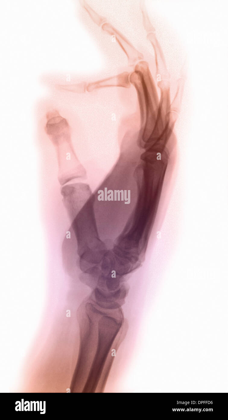 normal x-ray of the hand, lateral view Stock Photohttps://www.alamy.com/image-license-details/?v=1https://www.alamy.com/normal-x-ray-of-the-hand-lateral-view-image65494946.html
RFDPFFD6–normal x-ray of the hand, lateral view
 Medically Accurate Aching Tooth X-Ray View with Red Zone of Pain on a black background. 3d Rendering Stock Photohttps://www.alamy.com/image-license-details/?v=1https://www.alamy.com/medically-accurate-aching-tooth-x-ray-view-with-red-zone-of-pain-on-a-black-background-3d-rendering-image548722218.html
RF2PTMCP2–Medically Accurate Aching Tooth X-Ray View with Red Zone of Pain on a black background. 3d Rendering
 A view from below of a shasta daisy (Chrysanthemum x superbum) in summer Stock Photohttps://www.alamy.com/image-license-details/?v=1https://www.alamy.com/stock-photo-a-view-from-below-of-a-shasta-daisy-chrysanthemum-x-superbum-in-summer-22717997.html
RMB8XW11–A view from below of a shasta daisy (Chrysanthemum x superbum) in summer
 Doctor, X-ray picture, patient Stock Photohttps://www.alamy.com/image-license-details/?v=1https://www.alamy.com/stock-photo-doctor-x-ray-picture-patient-123172859.html
RMH4B06K–Doctor, X-ray picture, patient
 X-ray view of human foot. Stock Photohttps://www.alamy.com/image-license-details/?v=1https://www.alamy.com/stock-photo-x-ray-view-of-human-foot-57642356.html
RFD9NRBG–X-ray view of human foot.

