Quick filters:
Xiphisternum Stock Photos and Images
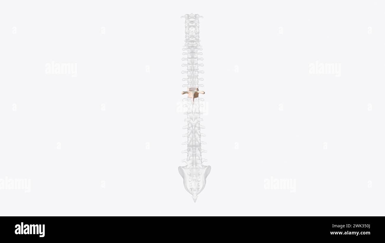 The eighth thoracic vertebra is, together with the ninth thoracic vertebra, at the same level as the xiphisternum 3d illustration Stock Photohttps://www.alamy.com/image-license-details/?v=1https://www.alamy.com/the-eighth-thoracic-vertebra-is-together-with-the-ninth-thoracic-vertebra-at-the-same-level-as-the-xiphisternum-3d-illustration-image596900770.html
RF2WK350J–The eighth thoracic vertebra is, together with the ninth thoracic vertebra, at the same level as the xiphisternum 3d illustration
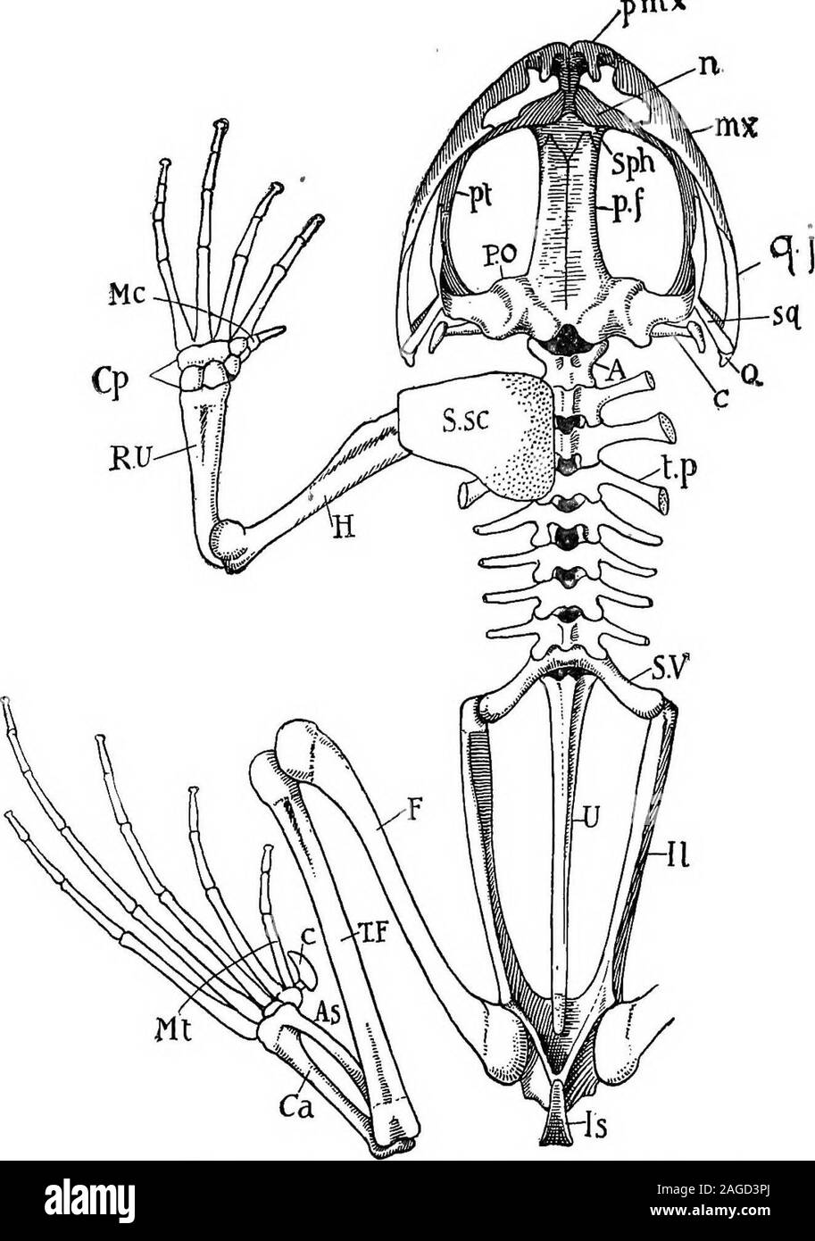 . Outlines of zoology. is formed by thejunction of scapula and coracoid. Between the median ends of the coracoids lie two fusedcartilaginous epicoracoids, behind which is a bony part ofthe sternum, prolonged posteriorly into a notched cartila-ginous xiphisternum. Anteriorly lies a bony portion calledthe omosternum, which is prolonged forwards into an epi-sternum cartilage. This sternum does not arise like that ofhigher Vertebrates, from a fusion of the ventral ends of ribs.Indeed, there are no ribs in the frog, unless they be minuterudiments at the ends of the transverse processes. The true fr Stock Photohttps://www.alamy.com/image-license-details/?v=1https://www.alamy.com/outlines-of-zoology-is-formed-by-thejunction-of-scapula-and-coracoid-between-the-median-ends-of-the-coracoids-lie-two-fusedcartilaginous-epicoracoids-behind-which-is-a-bony-part-ofthe-sternum-prolonged-posteriorly-into-a-notched-cartila-ginous-xiphisternum-anteriorly-lies-a-bony-portion-calledthe-omosternum-which-is-prolonged-forwards-into-an-epi-sternum-cartilage-this-sternum-does-not-arise-like-that-ofhigher-vertebrates-from-a-fusion-of-the-ventral-ends-of-ribsindeed-there-are-no-ribs-in-the-frog-unless-they-be-minuterudiments-at-the-ends-of-the-transverse-processes-the-true-fr-image337119850.html
RM2AGD3PJ–. Outlines of zoology. is formed by thejunction of scapula and coracoid. Between the median ends of the coracoids lie two fusedcartilaginous epicoracoids, behind which is a bony part ofthe sternum, prolonged posteriorly into a notched cartila-ginous xiphisternum. Anteriorly lies a bony portion calledthe omosternum, which is prolonged forwards into an epi-sternum cartilage. This sternum does not arise like that ofhigher Vertebrates, from a fusion of the ventral ends of ribs.Indeed, there are no ribs in the frog, unless they be minuterudiments at the ends of the transverse processes. The true fr
 . A manual of zoology. XII PHYLUM CHORDATA 415 Passing forwards from the anterior ends of the united epicoracoids is a rod of bone, the episternum {Ep), tipped by a rounded plate of cartilage, the omosternum ; and passing backwards from their posterior ends is a similar but larger bony rod, the sternum {St), also tipped by a cartilaginous plate, to which the name xiphisternum (K?i) is applied.. Fig. 249. — Rana esculenta. The shoulder girdle from the ventral aspect. Co, coracoid: Co', epicoraeoid; CI, clavicle; G, glenoid cavity; £/>, episternum; Fe, fenestra between procoracoid and coracoi Stock Photohttps://www.alamy.com/image-license-details/?v=1https://www.alamy.com/a-manual-of-zoology-xii-phylum-chordata-415-passing-forwards-from-the-anterior-ends-of-the-united-epicoracoids-is-a-rod-of-bone-the-episternum-ep-tipped-by-a-rounded-plate-of-cartilage-the-omosternum-and-passing-backwards-from-their-posterior-ends-is-a-similar-but-larger-bony-rod-the-sternum-st-also-tipped-by-a-cartilaginous-plate-to-which-the-name-xiphisternum-ki-is-applied-fig-249-rana-esculenta-the-shoulder-girdle-from-the-ventral-aspect-co-coracoid-co-epicoraeoid-ci-clavicle-g-glenoid-cavity-gt-episternum-fe-fenestra-between-procoracoid-and-coracoi-image216446434.html
RMPG3YHP–. A manual of zoology. XII PHYLUM CHORDATA 415 Passing forwards from the anterior ends of the united epicoracoids is a rod of bone, the episternum {Ep), tipped by a rounded plate of cartilage, the omosternum ; and passing backwards from their posterior ends is a similar but larger bony rod, the sternum {St), also tipped by a cartilaginous plate, to which the name xiphisternum (K?i) is applied.. Fig. 249. — Rana esculenta. The shoulder girdle from the ventral aspect. Co, coracoid: Co', epicoraeoid; CI, clavicle; G, glenoid cavity; £/>, episternum; Fe, fenestra between procoracoid and coracoi
![. Coracoid Epicoracoid Sternum Xiphisternum Fig. 4.âPectoral Girdle of Xenopus and Rana. [In the Frog the sternum is composed of the following parts :ââ (i) The omosternum consisting of tivo parts, an anterior cartilaginous part (sometimes called the omosternum) and a posterior bony part (sometimes called the episternum). (ii) The two epicoracoids consisting of cartilage, (Hi) The metasternum consisting of two parts, an anterior bony part (sometimes called the sternum) and a posterior cartilaginous part (sometimes called the xiphisternum).'] Make a sketch of the pectoral girdle. 10 Stock Photo . Coracoid Epicoracoid Sternum Xiphisternum Fig. 4.âPectoral Girdle of Xenopus and Rana. [In the Frog the sternum is composed of the following parts :ââ (i) The omosternum consisting of tivo parts, an anterior cartilaginous part (sometimes called the omosternum) and a posterior bony part (sometimes called the episternum). (ii) The two epicoracoids consisting of cartilage, (Hi) The metasternum consisting of two parts, an anterior bony part (sometimes called the sternum) and a posterior cartilaginous part (sometimes called the xiphisternum).'] Make a sketch of the pectoral girdle. 10 Stock Photo](https://c8.alamy.com/comp/MCKTTB/coracoid-epicoracoid-sternum-xiphisternum-fig-4pectoral-girdle-of-xenopus-and-rana-in-the-frog-the-sternum-is-composed-of-the-following-parts-i-the-omosternum-consisting-of-tivo-parts-an-anterior-cartilaginous-part-sometimes-called-the-omosternum-and-a-posterior-bony-part-sometimes-called-the-episternum-ii-the-two-epicoracoids-consisting-of-cartilage-hi-the-metasternum-consisting-of-two-parts-an-anterior-bony-part-sometimes-called-the-sternum-and-a-posterior-cartilaginous-part-sometimes-called-the-xiphisternum-make-a-sketch-of-the-pectoral-girdle-10-MCKTTB.jpg) . Coracoid Epicoracoid Sternum Xiphisternum Fig. 4.âPectoral Girdle of Xenopus and Rana. [In the Frog the sternum is composed of the following parts :ââ (i) The omosternum consisting of tivo parts, an anterior cartilaginous part (sometimes called the omosternum) and a posterior bony part (sometimes called the episternum). (ii) The two epicoracoids consisting of cartilage, (Hi) The metasternum consisting of two parts, an anterior bony part (sometimes called the sternum) and a posterior cartilaginous part (sometimes called the xiphisternum).'] Make a sketch of the pectoral girdle. 10 Stock Photohttps://www.alamy.com/image-license-details/?v=1https://www.alamy.com/coracoid-epicoracoid-sternum-xiphisternum-fig-4pectoral-girdle-of-xenopus-and-rana-in-the-frog-the-sternum-is-composed-of-the-following-parts-i-the-omosternum-consisting-of-tivo-parts-an-anterior-cartilaginous-part-sometimes-called-the-omosternum-and-a-posterior-bony-part-sometimes-called-the-episternum-ii-the-two-epicoracoids-consisting-of-cartilage-hi-the-metasternum-consisting-of-two-parts-an-anterior-bony-part-sometimes-called-the-sternum-and-a-posterior-cartilaginous-part-sometimes-called-the-xiphisternum-make-a-sketch-of-the-pectoral-girdle-10-image179916139.html
. Coracoid Epicoracoid Sternum Xiphisternum Fig. 4.âPectoral Girdle of Xenopus and Rana. [In the Frog the sternum is composed of the following parts :ââ (i) The omosternum consisting of tivo parts, an anterior cartilaginous part (sometimes called the omosternum) and a posterior bony part (sometimes called the episternum). (ii) The two epicoracoids consisting of cartilage, (Hi) The metasternum consisting of two parts, an anterior bony part (sometimes called the sternum) and a posterior cartilaginous part (sometimes called the xiphisternum).'] Make a sketch of the pectoral girdle. 10 Stock Photohttps://www.alamy.com/image-license-details/?v=1https://www.alamy.com/coracoid-epicoracoid-sternum-xiphisternum-fig-4pectoral-girdle-of-xenopus-and-rana-in-the-frog-the-sternum-is-composed-of-the-following-parts-i-the-omosternum-consisting-of-tivo-parts-an-anterior-cartilaginous-part-sometimes-called-the-omosternum-and-a-posterior-bony-part-sometimes-called-the-episternum-ii-the-two-epicoracoids-consisting-of-cartilage-hi-the-metasternum-consisting-of-two-parts-an-anterior-bony-part-sometimes-called-the-sternum-and-a-posterior-cartilaginous-part-sometimes-called-the-xiphisternum-make-a-sketch-of-the-pectoral-girdle-10-image179916139.htmlRMMCKTTB–. Coracoid Epicoracoid Sternum Xiphisternum Fig. 4.âPectoral Girdle of Xenopus and Rana. [In the Frog the sternum is composed of the following parts :ââ (i) The omosternum consisting of tivo parts, an anterior cartilaginous part (sometimes called the omosternum) and a posterior bony part (sometimes called the episternum). (ii) The two epicoracoids consisting of cartilage, (Hi) The metasternum consisting of two parts, an anterior bony part (sometimes called the sternum) and a posterior cartilaginous part (sometimes called the xiphisternum).'] Make a sketch of the pectoral girdle. 10
 The eighth thoracic vertebra is, together with the ninth thoracic vertebra, at the same level as the xiphisternum 3d illustration Stock Photohttps://www.alamy.com/image-license-details/?v=1https://www.alamy.com/the-eighth-thoracic-vertebra-is-together-with-the-ninth-thoracic-vertebra-at-the-same-level-as-the-xiphisternum-3d-illustration-image596900772.html
RF2WK350M–The eighth thoracic vertebra is, together with the ninth thoracic vertebra, at the same level as the xiphisternum 3d illustration
 . Outlines of zoology. Fig. 316.—Pectoral girdle oi Rana esniknta.—After Ecker. The cartilaginous parts are dotted. Ep.^ Episternum ; oni., omo-sternuni; £p,c., epicoracoids ; St., sternum ; jr., xiphisternum ;cl., clavicle with underlying precoracoid cartilage; co., cora-coid; Sc, scapula; S.sc, supra-scapula; Gl., glenoid cavityfor humerus. proximal and three distal elements, and a central piecewedged in between them, five metacarpal bones, of whichthe first—corresponding to the absent thumb—is very. Fig. 317.—Side view of frogs pelvis.—After Eckfr.//., Ilium; Is., ischium ; Pb., pubis ; Ac, Stock Photohttps://www.alamy.com/image-license-details/?v=1https://www.alamy.com/outlines-of-zoology-fig-316pectoral-girdle-oi-rana-esnikntaafter-ecker-the-cartilaginous-parts-are-dotted-ep-episternum-oni-omo-sternuni-pc-epicoracoids-st-sternum-jr-xiphisternum-cl-clavicle-with-underlying-precoracoid-cartilage-co-cora-coid-sc-scapula-ssc-supra-scapula-gl-glenoid-cavityfor-humerus-proximal-and-three-distal-elements-and-a-central-piecewedged-in-between-them-five-metacarpal-bones-of-whichthe-firstcorresponding-to-the-absent-thumbis-very-fig-317side-view-of-frogs-pelvisafter-eckfr-ilium-is-ischium-pb-pubis-ac-image337119030.html
RM2AGD2NA–. Outlines of zoology. Fig. 316.—Pectoral girdle oi Rana esniknta.—After Ecker. The cartilaginous parts are dotted. Ep.^ Episternum ; oni., omo-sternuni; £p,c., epicoracoids ; St., sternum ; jr., xiphisternum ;cl., clavicle with underlying precoracoid cartilage; co., cora-coid; Sc, scapula; S.sc, supra-scapula; Gl., glenoid cavityfor humerus. proximal and three distal elements, and a central piecewedged in between them, five metacarpal bones, of whichthe first—corresponding to the absent thumb—is very. Fig. 317.—Side view of frogs pelvis.—After Eckfr.//., Ilium; Is., ischium ; Pb., pubis ; Ac,
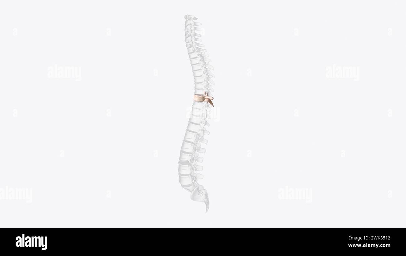 The eighth thoracic vertebra is, together with the ninth thoracic vertebra, at the same level as the xiphisternum 3d illustration Stock Photohttps://www.alamy.com/image-license-details/?v=1https://www.alamy.com/the-eighth-thoracic-vertebra-is-together-with-the-ninth-thoracic-vertebra-at-the-same-level-as-the-xiphisternum-3d-illustration-image596900782.html
RF2WK3512–The eighth thoracic vertebra is, together with the ninth thoracic vertebra, at the same level as the xiphisternum 3d illustration
 . An introduction to the osteology of the mammalia . and terminating anteriorly in aconical rounded projection. The segments of the mesosternum are elongated, andmore or less four-sided, contracted at the middle, andwidening at each extremity. They ossify, according toParker, ectosteally, or from without inwards, the bony deposit commencing in the inner layerof the perichondrium, as in the shaftsof long bones ; and they remain per-manently distinct from each other. The xiphisternum is long, narrow,and flat, and generally ends in an ex-panded flattened cartilage. In the Pinnipedia, the prestern Stock Photohttps://www.alamy.com/image-license-details/?v=1https://www.alamy.com/an-introduction-to-the-osteology-of-the-mammalia-and-terminating-anteriorly-in-aconical-rounded-projection-the-segments-of-the-mesosternum-are-elongated-andmore-or-less-four-sided-contracted-at-the-middle-andwidening-at-each-extremity-they-ossify-according-toparker-ectosteally-or-from-without-inwards-the-bony-deposit-commencing-in-the-inner-layerof-the-perichondrium-as-in-the-shaftsof-long-bones-and-they-remain-per-manently-distinct-from-each-other-the-xiphisternum-is-long-narrowand-flat-and-generally-ends-in-an-ex-panded-flattened-cartilage-in-the-pinnipedia-the-prestern-image369744577.html
RM2CDF8YD–. An introduction to the osteology of the mammalia . and terminating anteriorly in aconical rounded projection. The segments of the mesosternum are elongated, andmore or less four-sided, contracted at the middle, andwidening at each extremity. They ossify, according toParker, ectosteally, or from without inwards, the bony deposit commencing in the inner layerof the perichondrium, as in the shaftsof long bones ; and they remain per-manently distinct from each other. The xiphisternum is long, narrow,and flat, and generally ends in an ex-panded flattened cartilage. In the Pinnipedia, the prestern
 The eighth thoracic vertebra is, together with the ninth thoracic vertebra, at the same level as the xiphisternum 3d illustration Stock Photohttps://www.alamy.com/image-license-details/?v=1https://www.alamy.com/the-eighth-thoracic-vertebra-is-together-with-the-ninth-thoracic-vertebra-at-the-same-level-as-the-xiphisternum-3d-illustration-image596900777.html
RF2WK350W–The eighth thoracic vertebra is, together with the ninth thoracic vertebra, at the same level as the xiphisternum 3d illustration
![. An introduction to the osteology of the mammalia . body of the sternum or gladiolus. 3. Xiphisternum, xiphoid or ensiform process of the ster-num. The mesosternum is usually composed of several distinctsegments, which may become ankylosed together, but moreoften permanently retain their individuality, being con-nected either by fibrous tissue or by synovial joints. 1 For much valuable information upon the structure and developmentof the sternum, see W. K. Parkers Monograph on the Shoulder-girdleand Sternum of the Vertebrata, published by the Ray Society, 1868. CHAP. VII.] GENERAL CHARACTERS. Stock Photo . An introduction to the osteology of the mammalia . body of the sternum or gladiolus. 3. Xiphisternum, xiphoid or ensiform process of the ster-num. The mesosternum is usually composed of several distinctsegments, which may become ankylosed together, but moreoften permanently retain their individuality, being con-nected either by fibrous tissue or by synovial joints. 1 For much valuable information upon the structure and developmentof the sternum, see W. K. Parkers Monograph on the Shoulder-girdleand Sternum of the Vertebrata, published by the Ray Society, 1868. CHAP. VII.] GENERAL CHARACTERS. Stock Photo](https://c8.alamy.com/comp/2CDF917/an-introduction-to-the-osteology-of-the-mammalia-body-of-the-sternum-or-gladiolus-3-xiphisternum-xiphoid-or-ensiform-process-of-the-ster-num-the-mesosternum-is-usually-composed-of-several-distinctsegments-which-may-become-ankylosed-together-but-moreoften-permanently-retain-their-individuality-being-con-nected-either-by-fibrous-tissue-or-by-synovial-joints-1-for-much-valuable-information-upon-the-structure-and-developmentof-the-sternum-see-w-k-parkers-monograph-on-the-shoulder-girdleand-sternum-of-the-vertebrata-published-by-the-ray-society-1868-chap-vii-general-characters-2CDF917.jpg) . An introduction to the osteology of the mammalia . body of the sternum or gladiolus. 3. Xiphisternum, xiphoid or ensiform process of the ster-num. The mesosternum is usually composed of several distinctsegments, which may become ankylosed together, but moreoften permanently retain their individuality, being con-nected either by fibrous tissue or by synovial joints. 1 For much valuable information upon the structure and developmentof the sternum, see W. K. Parkers Monograph on the Shoulder-girdleand Sternum of the Vertebrata, published by the Ray Society, 1868. CHAP. VII.] GENERAL CHARACTERS. Stock Photohttps://www.alamy.com/image-license-details/?v=1https://www.alamy.com/an-introduction-to-the-osteology-of-the-mammalia-body-of-the-sternum-or-gladiolus-3-xiphisternum-xiphoid-or-ensiform-process-of-the-ster-num-the-mesosternum-is-usually-composed-of-several-distinctsegments-which-may-become-ankylosed-together-but-moreoften-permanently-retain-their-individuality-being-con-nected-either-by-fibrous-tissue-or-by-synovial-joints-1-for-much-valuable-information-upon-the-structure-and-developmentof-the-sternum-see-w-k-parkers-monograph-on-the-shoulder-girdleand-sternum-of-the-vertebrata-published-by-the-ray-society-1868-chap-vii-general-characters-image369744627.html
. An introduction to the osteology of the mammalia . body of the sternum or gladiolus. 3. Xiphisternum, xiphoid or ensiform process of the ster-num. The mesosternum is usually composed of several distinctsegments, which may become ankylosed together, but moreoften permanently retain their individuality, being con-nected either by fibrous tissue or by synovial joints. 1 For much valuable information upon the structure and developmentof the sternum, see W. K. Parkers Monograph on the Shoulder-girdleand Sternum of the Vertebrata, published by the Ray Society, 1868. CHAP. VII.] GENERAL CHARACTERS. Stock Photohttps://www.alamy.com/image-license-details/?v=1https://www.alamy.com/an-introduction-to-the-osteology-of-the-mammalia-body-of-the-sternum-or-gladiolus-3-xiphisternum-xiphoid-or-ensiform-process-of-the-ster-num-the-mesosternum-is-usually-composed-of-several-distinctsegments-which-may-become-ankylosed-together-but-moreoften-permanently-retain-their-individuality-being-con-nected-either-by-fibrous-tissue-or-by-synovial-joints-1-for-much-valuable-information-upon-the-structure-and-developmentof-the-sternum-see-w-k-parkers-monograph-on-the-shoulder-girdleand-sternum-of-the-vertebrata-published-by-the-ray-society-1868-chap-vii-general-characters-image369744627.htmlRM2CDF917–. An introduction to the osteology of the mammalia . body of the sternum or gladiolus. 3. Xiphisternum, xiphoid or ensiform process of the ster-num. The mesosternum is usually composed of several distinctsegments, which may become ankylosed together, but moreoften permanently retain their individuality, being con-nected either by fibrous tissue or by synovial joints. 1 For much valuable information upon the structure and developmentof the sternum, see W. K. Parkers Monograph on the Shoulder-girdleand Sternum of the Vertebrata, published by the Ray Society, 1868. CHAP. VII.] GENERAL CHARACTERS.
 The eighth thoracic vertebra is, together with the ninth thoracic vertebra, at the same level as the xiphisternum 3d illustration Stock Photohttps://www.alamy.com/image-license-details/?v=1https://www.alamy.com/the-eighth-thoracic-vertebra-is-together-with-the-ninth-thoracic-vertebra-at-the-same-level-as-the-xiphisternum-3d-illustration-image596900774.html
RF2WK350P–The eighth thoracic vertebra is, together with the ninth thoracic vertebra, at the same level as the xiphisternum 3d illustration
 . An introduction to the osteology of the mammalia . sterm7m.sternum (Sorex). In RJiyncJwcyon it is broadin front, narrow posteriorly, strongly keeled below, and with two horn-like processes projectingoutwards and forwards between the attachment of theclavicles and the first pair of ribs. The mesosternum is usually narrow, as in the Carnivora,but in the Hedgehog, where it consists of three segments,it is broad and flat posteriorly, and to the last segment three VII. INSECT1VORA. 95 ribs are attached. In this genus the xiphisternum is rudi-mentary, whereas in the Shrews it is long and ends in a Stock Photohttps://www.alamy.com/image-license-details/?v=1https://www.alamy.com/an-introduction-to-the-osteology-of-the-mammalia-sterm7msternum-sorex-in-rjiyncjwcyon-it-is-broadin-front-narrow-posteriorly-strongly-keeled-below-and-with-two-horn-like-processes-projectingoutwards-and-forwards-between-the-attachment-of-theclavicles-and-the-first-pair-of-ribs-the-mesosternum-is-usually-narrow-as-in-the-carnivorabut-in-the-hedgehog-where-it-consists-of-three-segmentsit-is-broad-and-flat-posteriorly-and-to-the-last-segment-three-vii-insect1vora-95-ribs-are-attached-in-this-genus-the-xiphisternum-is-rudi-mentary-whereas-in-the-shrews-it-is-long-and-ends-in-a-image369744469.html
RM2CDF8RH–. An introduction to the osteology of the mammalia . sterm7m.sternum (Sorex). In RJiyncJwcyon it is broadin front, narrow posteriorly, strongly keeled below, and with two horn-like processes projectingoutwards and forwards between the attachment of theclavicles and the first pair of ribs. The mesosternum is usually narrow, as in the Carnivora,but in the Hedgehog, where it consists of three segments,it is broad and flat posteriorly, and to the last segment three VII. INSECT1VORA. 95 ribs are attached. In this genus the xiphisternum is rudi-mentary, whereas in the Shrews it is long and ends in a
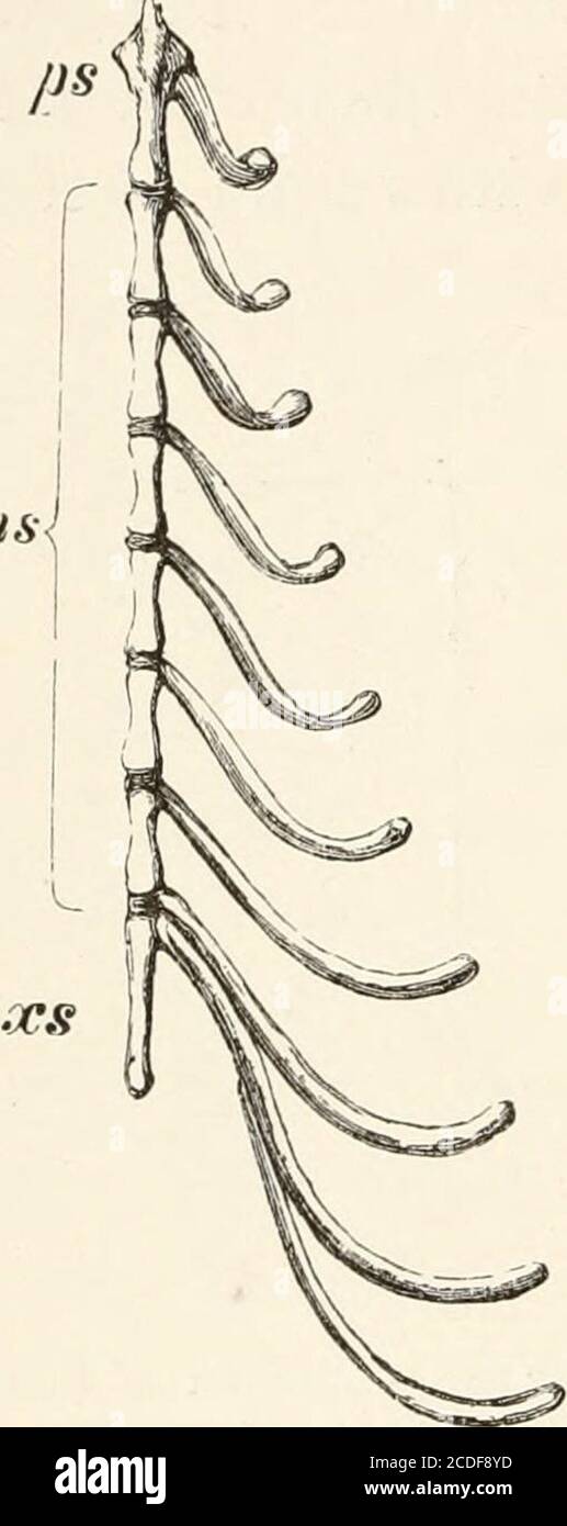
![. An introduction to the osteology of the mammalia . FIG. 45.—Sternum and ribs of the Great Armadillo (Priodou gigas), .ps presternum ; xs xiphisternum. In the MONOTREMATA the intermediate ribs are wellmarked (see Fig. 44, p. 105), and only partly ossified byendostosis, while the sternal ribs (except the first) are,according to Parker, strongly ossified ectosteally, as in Birds. VIII.] MONO TREMA TA. The hinder sternal ribs are very broad and flat. TheEchidna has 16, and the Ornithorhynchus 17 pairs ofvertebral ribs; they do not divide above into head andtubercle, but are attached only to the Stock Photo . An introduction to the osteology of the mammalia . FIG. 45.—Sternum and ribs of the Great Armadillo (Priodou gigas), .ps presternum ; xs xiphisternum. In the MONOTREMATA the intermediate ribs are wellmarked (see Fig. 44, p. 105), and only partly ossified byendostosis, while the sternal ribs (except the first) are,according to Parker, strongly ossified ectosteally, as in Birds. VIII.] MONO TREMA TA. The hinder sternal ribs are very broad and flat. TheEchidna has 16, and the Ornithorhynchus 17 pairs ofvertebral ribs; they do not divide above into head andtubercle, but are attached only to the Stock Photo](https://c8.alamy.com/comp/2CDF70R/an-introduction-to-the-osteology-of-the-mammalia-fig-45sternum-and-ribs-of-the-great-armadillo-priodou-gigas-ps-presternum-xs-xiphisternum-in-the-monotremata-the-intermediate-ribs-are-wellmarked-see-fig-44-p-105-and-only-partly-ossified-byendostosis-while-the-sternal-ribs-except-the-first-areaccording-to-parker-strongly-ossified-ectosteally-as-in-birds-viii-mono-trema-ta-the-hinder-sternal-ribs-are-very-broad-and-flat-theechidna-has-16-and-the-ornithorhynchus-17-pairs-ofvertebral-ribs-they-do-not-divide-above-into-head-andtubercle-but-are-attached-only-to-the-2CDF70R.jpg) . An introduction to the osteology of the mammalia . FIG. 45.—Sternum and ribs of the Great Armadillo (Priodou gigas), .ps presternum ; xs xiphisternum. In the MONOTREMATA the intermediate ribs are wellmarked (see Fig. 44, p. 105), and only partly ossified byendostosis, while the sternal ribs (except the first) are,according to Parker, strongly ossified ectosteally, as in Birds. VIII.] MONO TREMA TA. The hinder sternal ribs are very broad and flat. TheEchidna has 16, and the Ornithorhynchus 17 pairs ofvertebral ribs; they do not divide above into head andtubercle, but are attached only to the Stock Photohttps://www.alamy.com/image-license-details/?v=1https://www.alamy.com/an-introduction-to-the-osteology-of-the-mammalia-fig-45sternum-and-ribs-of-the-great-armadillo-priodou-gigas-ps-presternum-xs-xiphisternum-in-the-monotremata-the-intermediate-ribs-are-wellmarked-see-fig-44-p-105-and-only-partly-ossified-byendostosis-while-the-sternal-ribs-except-the-first-areaccording-to-parker-strongly-ossified-ectosteally-as-in-birds-viii-mono-trema-ta-the-hinder-sternal-ribs-are-very-broad-and-flat-theechidna-has-16-and-the-ornithorhynchus-17-pairs-ofvertebral-ribs-they-do-not-divide-above-into-head-andtubercle-but-are-attached-only-to-the-image369743047.html
. An introduction to the osteology of the mammalia . FIG. 45.—Sternum and ribs of the Great Armadillo (Priodou gigas), .ps presternum ; xs xiphisternum. In the MONOTREMATA the intermediate ribs are wellmarked (see Fig. 44, p. 105), and only partly ossified byendostosis, while the sternal ribs (except the first) are,according to Parker, strongly ossified ectosteally, as in Birds. VIII.] MONO TREMA TA. The hinder sternal ribs are very broad and flat. TheEchidna has 16, and the Ornithorhynchus 17 pairs ofvertebral ribs; they do not divide above into head andtubercle, but are attached only to the Stock Photohttps://www.alamy.com/image-license-details/?v=1https://www.alamy.com/an-introduction-to-the-osteology-of-the-mammalia-fig-45sternum-and-ribs-of-the-great-armadillo-priodou-gigas-ps-presternum-xs-xiphisternum-in-the-monotremata-the-intermediate-ribs-are-wellmarked-see-fig-44-p-105-and-only-partly-ossified-byendostosis-while-the-sternal-ribs-except-the-first-areaccording-to-parker-strongly-ossified-ectosteally-as-in-birds-viii-mono-trema-ta-the-hinder-sternal-ribs-are-very-broad-and-flat-theechidna-has-16-and-the-ornithorhynchus-17-pairs-ofvertebral-ribs-they-do-not-divide-above-into-head-andtubercle-but-are-attached-only-to-the-image369743047.htmlRM2CDF70R–. An introduction to the osteology of the mammalia . FIG. 45.—Sternum and ribs of the Great Armadillo (Priodou gigas), .ps presternum ; xs xiphisternum. In the MONOTREMATA the intermediate ribs are wellmarked (see Fig. 44, p. 105), and only partly ossified byendostosis, while the sternal ribs (except the first) are,according to Parker, strongly ossified ectosteally, as in Birds. VIII.] MONO TREMA TA. The hinder sternal ribs are very broad and flat. TheEchidna has 16, and the Ornithorhynchus 17 pairs ofvertebral ribs; they do not divide above into head andtubercle, but are attached only to the
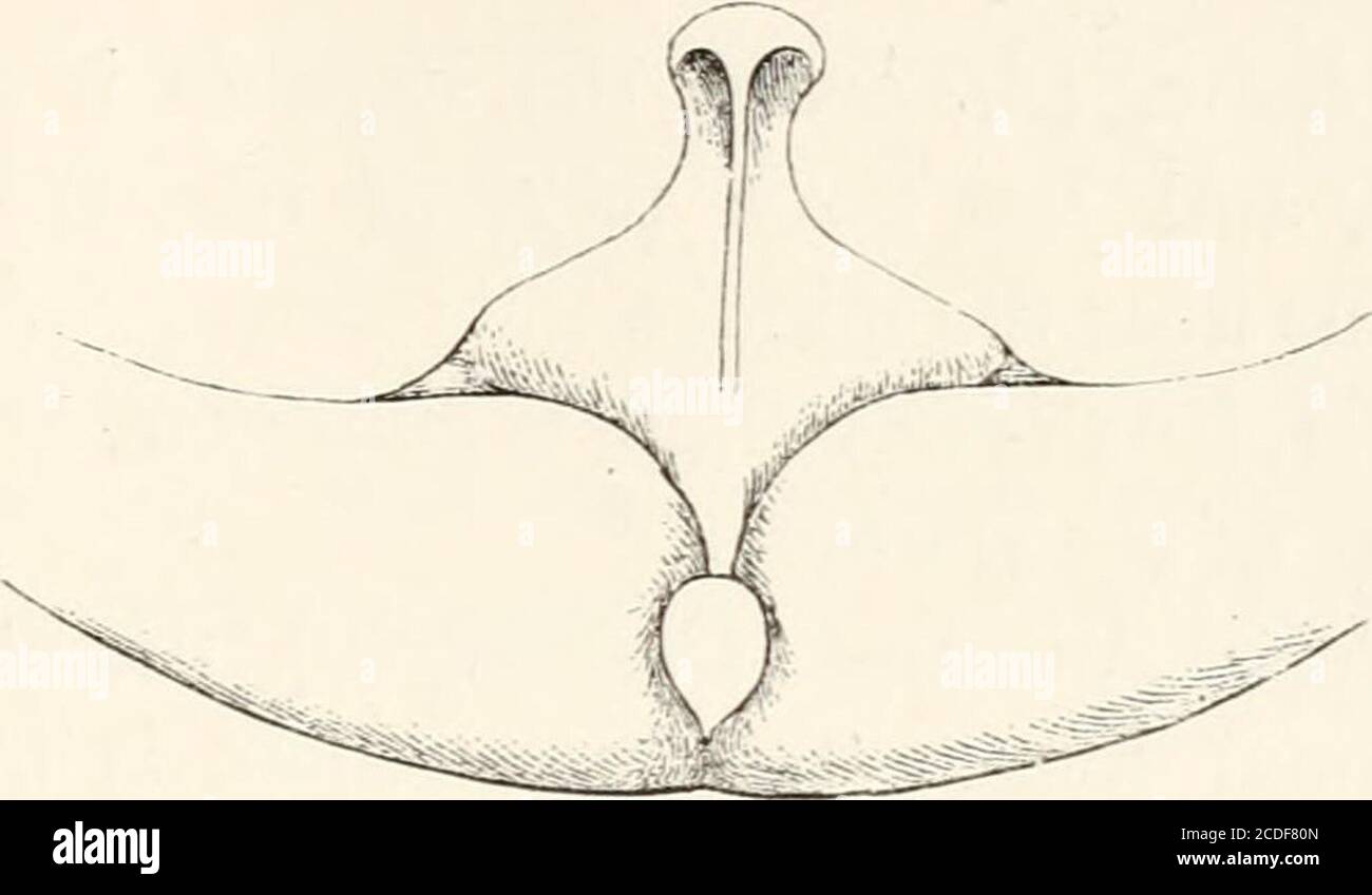 . An introduction to the osteology of the mammalia . FIG. 39.—Sternum of Common Rorqual or FinWhale {Balcenoptera musculus), jV FIG. 40.—Sternum of Pike Whale(Balcenoptera rostra to), TV the ossified portion at one period appears deeply notched infront ; as the bone meets across the middle line anteriorly,this notch usually becomes converted into a hole (seeFig. 39), which finally closes with complete maturity. 1 In the cartilaginous sternum of a young BaLznoptera sibbaldiiProfessor Turner found the xiphisternum to be quite distinct from thepresternum, and connected with it by fibrous tissue. Stock Photohttps://www.alamy.com/image-license-details/?v=1https://www.alamy.com/an-introduction-to-the-osteology-of-the-mammalia-fig-39sternum-of-common-rorqual-or-finwhale-balcenoptera-musculus-jv-fig-40sternum-of-pike-whalebalcenoptera-rostra-to-tv-the-ossified-portion-at-one-period-appears-deeply-notched-infront-as-the-bone-meets-across-the-middle-line-anteriorlythis-notch-usually-becomes-converted-into-a-hole-seefig-39-which-finally-closes-with-complete-maturity-1-in-the-cartilaginous-sternum-of-a-young-balznoptera-sibbaldiiprofessor-turner-found-the-xiphisternum-to-be-quite-distinct-from-thepresternum-and-connected-with-it-by-fibrous-tissue-image369743829.html
RM2CDF80N–. An introduction to the osteology of the mammalia . FIG. 39.—Sternum of Common Rorqual or FinWhale {Balcenoptera musculus), jV FIG. 40.—Sternum of Pike Whale(Balcenoptera rostra to), TV the ossified portion at one period appears deeply notched infront ; as the bone meets across the middle line anteriorly,this notch usually becomes converted into a hole (seeFig. 39), which finally closes with complete maturity. 1 In the cartilaginous sternum of a young BaLznoptera sibbaldiiProfessor Turner found the xiphisternum to be quite distinct from thepresternum, and connected with it by fibrous tissue.
 . Regional anesthesia : its technic and clinical application . Fig. 273.—Costo-iliac block for appendectomy (McBurney-Weirs incision). Thetwo dots mark the points of entrance of the needle, and the interrupted line the direc-tion of the anesthetic wall created across the nerve supply of the region. 3. Field-block C (Battle-Jalaguier-Kammerer Incision).—^With thepatient lying in the same position as before, i. e., on his back, whealsare raised along the right costal margin, from the xiphisternum to aboutthe level of the tip of the eleventh rib, and from that point downward 38o REGIONAL ANESTHES Stock Photohttps://www.alamy.com/image-license-details/?v=1https://www.alamy.com/regional-anesthesia-its-technic-and-clinical-application-fig-273costo-iliac-block-for-appendectomy-mcburney-weirs-incision-thetwo-dots-mark-the-points-of-entrance-of-the-needle-and-the-interrupted-line-the-direc-tion-of-the-anesthetic-wall-created-across-the-nerve-supply-of-the-region-3-field-block-c-battle-jalaguier-kammerer-incisionwith-thepatient-lying-in-the-same-position-as-before-i-e-on-his-back-whealsare-raised-along-the-right-costal-margin-from-the-xiphisternum-to-aboutthe-level-of-the-tip-of-the-eleventh-rib-and-from-that-point-downward-38o-regional-anesthes-image370058681.html
RM2CE1HHD–. Regional anesthesia : its technic and clinical application . Fig. 273.—Costo-iliac block for appendectomy (McBurney-Weirs incision). Thetwo dots mark the points of entrance of the needle, and the interrupted line the direc-tion of the anesthetic wall created across the nerve supply of the region. 3. Field-block C (Battle-Jalaguier-Kammerer Incision).—^With thepatient lying in the same position as before, i. e., on his back, whealsare raised along the right costal margin, from the xiphisternum to aboutthe level of the tip of the eleventh rib, and from that point downward 38o REGIONAL ANESTHES
 . An atlas of human anatomy for students and physicians. Anatomy. THE AXIAL SKELETON 41 Clavicular notch Incisura clavicularis Interclavicular notch Incisura jugularis Gladiolo-enaiform articulation. The manubrium (presternum) Sternal synchondrosis (manu- " brio-gladiolal articulation) Synchondrosis sternalis Articular facet for a rib Incisura costalis ,, The body of the sternum or gladiolus (mesosternum) Articular facet for a rib Incisura costalis Ensiform or xiphoid process (metasternum, xiphisternum) Processus xiphoideus -•Clavicular notch Incisura clavicularis Manubrium Manubrium ster Stock Photohttps://www.alamy.com/image-license-details/?v=1https://www.alamy.com/an-atlas-of-human-anatomy-for-students-and-physicians-anatomy-the-axial-skeleton-41-clavicular-notch-incisura-clavicularis-interclavicular-notch-incisura-jugularis-gladiolo-enaiform-articulation-the-manubrium-presternum-sternal-synchondrosis-manu-quot-brio-gladiolal-articulation-synchondrosis-sternalis-articular-facet-for-a-rib-incisura-costalis-the-body-of-the-sternum-or-gladiolus-mesosternum-articular-facet-for-a-rib-incisura-costalis-ensiform-or-xiphoid-process-metasternum-xiphisternum-processus-xiphoideus-clavicular-notch-incisura-clavicularis-manubrium-manubrium-ster-image235400468.html
RMRJYBKG–. An atlas of human anatomy for students and physicians. Anatomy. THE AXIAL SKELETON 41 Clavicular notch Incisura clavicularis Interclavicular notch Incisura jugularis Gladiolo-enaiform articulation. The manubrium (presternum) Sternal synchondrosis (manu- " brio-gladiolal articulation) Synchondrosis sternalis Articular facet for a rib Incisura costalis ,, The body of the sternum or gladiolus (mesosternum) Articular facet for a rib Incisura costalis Ensiform or xiphoid process (metasternum, xiphisternum) Processus xiphoideus -•Clavicular notch Incisura clavicularis Manubrium Manubrium ster
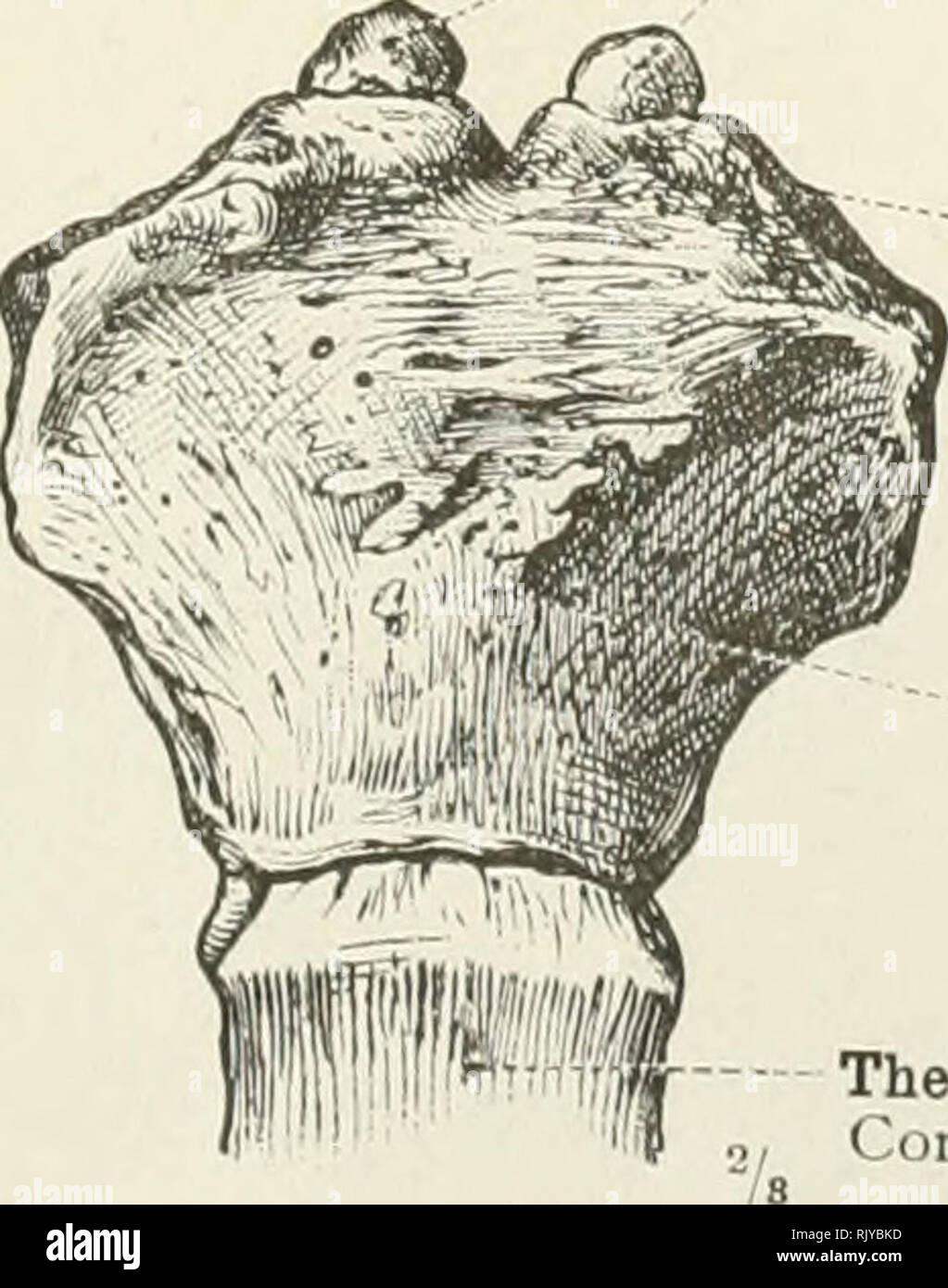 . An atlas of human anatomy for students and physicians. Anatomy. The manubrium (presternum) Sternal synchondrosis (manu- " brio-gladiolal articulation) Synchondrosis sternalis Articular facet for a rib Incisura costalis ,, The body of the sternum or gladiolus (mesosternum) Articular facet for a rib Incisura costalis Ensiform or xiphoid process (metasternum, xiphisternum) Processus xiphoideus -â¢Clavicular notch Incisura clavicularis Manubrium Manubrium sterni -Angle of the sternum1 Angulus sterni Body of the sternum or gladiolus Corpus sterni Articular facets for the ribs Incisure costal Stock Photohttps://www.alamy.com/image-license-details/?v=1https://www.alamy.com/an-atlas-of-human-anatomy-for-students-and-physicians-anatomy-the-manubrium-presternum-sternal-synchondrosis-manu-quot-brio-gladiolal-articulation-synchondrosis-sternalis-articular-facet-for-a-rib-incisura-costalis-the-body-of-the-sternum-or-gladiolus-mesosternum-articular-facet-for-a-rib-incisura-costalis-ensiform-or-xiphoid-process-metasternum-xiphisternum-processus-xiphoideus-clavicular-notch-incisura-clavicularis-manubrium-manubrium-sterni-angle-of-the-sternum1-angulus-sterni-body-of-the-sternum-or-gladiolus-corpus-sterni-articular-facets-for-the-ribs-incisure-costal-image235400465.html
RMRJYBKD–. An atlas of human anatomy for students and physicians. Anatomy. The manubrium (presternum) Sternal synchondrosis (manu- " brio-gladiolal articulation) Synchondrosis sternalis Articular facet for a rib Incisura costalis ,, The body of the sternum or gladiolus (mesosternum) Articular facet for a rib Incisura costalis Ensiform or xiphoid process (metasternum, xiphisternum) Processus xiphoideus -â¢Clavicular notch Incisura clavicularis Manubrium Manubrium sterni -Angle of the sternum1 Angulus sterni Body of the sternum or gladiolus Corpus sterni Articular facets for the ribs Incisure costal
 . Outlines of zoology. Zoology. THE LIMBS AND GIRDLES. 535 sented. The glenoid cavity with which the humerus articulates is formed as usual by the junction of scapula and coracoid. Between the median ends of the coracoids lie two cartilaginous epicoracoids, behind which is a bony part of the sternum, prolonged posteriorly into a notched cartila- ginous xiphisternum. Anteriorly lies a bony portion called the omosternum, which is prolonged forwards into an epi- sternum cartilage. This sternum does not arise like that of higher Vertebrates from a fusion of the ventral ends of ribs. Indeed, there Stock Photohttps://www.alamy.com/image-license-details/?v=1https://www.alamy.com/outlines-of-zoology-zoology-the-limbs-and-girdles-535-sented-the-glenoid-cavity-with-which-the-humerus-articulates-is-formed-as-usual-by-the-junction-of-scapula-and-coracoid-between-the-median-ends-of-the-coracoids-lie-two-cartilaginous-epicoracoids-behind-which-is-a-bony-part-of-the-sternum-prolonged-posteriorly-into-a-notched-cartila-ginous-xiphisternum-anteriorly-lies-a-bony-portion-called-the-omosternum-which-is-prolonged-forwards-into-an-epi-sternum-cartilage-this-sternum-does-not-arise-like-that-of-higher-vertebrates-from-a-fusion-of-the-ventral-ends-of-ribs-indeed-there-image232345623.html
RMRE075Y–. Outlines of zoology. Zoology. THE LIMBS AND GIRDLES. 535 sented. The glenoid cavity with which the humerus articulates is formed as usual by the junction of scapula and coracoid. Between the median ends of the coracoids lie two cartilaginous epicoracoids, behind which is a bony part of the sternum, prolonged posteriorly into a notched cartila- ginous xiphisternum. Anteriorly lies a bony portion called the omosternum, which is prolonged forwards into an epi- sternum cartilage. This sternum does not arise like that of higher Vertebrates from a fusion of the ventral ends of ribs. Indeed, there
 . Elementary text-book of zoology. Scapula. Coracoid. Xiphisternum. View with dorsal parts bent downwards. Bone is black and cartilage dotted. The presence of this urostyle, the single sacral vertebra and the small number of vertebrae are the important peculiarities of the vertebral column. The vestigial ribs are also to be noted. Fig. 249.—Fore-limb of Rana. Phalanges of Digits. Humerus.. IVIetacarpal.s. CarpaLs. Note fusion of radius and ulna and absence of poUex, a metacarpal only remaining. The peripheral (or appendicular) skeleton consists of the two limb-girdles and limbs. These are cons Stock Photohttps://www.alamy.com/image-license-details/?v=1https://www.alamy.com/elementary-text-book-of-zoology-scapula-coracoid-xiphisternum-view-with-dorsal-parts-bent-downwards-bone-is-black-and-cartilage-dotted-the-presence-of-this-urostyle-the-single-sacral-vertebra-and-the-small-number-of-vertebrae-are-the-important-peculiarities-of-the-vertebral-column-the-vestigial-ribs-are-also-to-be-noted-fig-249fore-limb-of-rana-phalanges-of-digits-humerus-ivietacarpals-carpals-note-fusion-of-radius-and-ulna-and-absence-of-pouex-a-metacarpal-only-remaining-the-peripheral-or-appendicular-skeleton-consists-of-the-two-limb-girdles-and-limbs-these-are-cons-image232088751.html
RMRDGFFY–. Elementary text-book of zoology. Scapula. Coracoid. Xiphisternum. View with dorsal parts bent downwards. Bone is black and cartilage dotted. The presence of this urostyle, the single sacral vertebra and the small number of vertebrae are the important peculiarities of the vertebral column. The vestigial ribs are also to be noted. Fig. 249.—Fore-limb of Rana. Phalanges of Digits. Humerus.. IVIetacarpal.s. CarpaLs. Note fusion of radius and ulna and absence of poUex, a metacarpal only remaining. The peripheral (or appendicular) skeleton consists of the two limb-girdles and limbs. These are cons
 . The biology of the frog. Frogs. THE SKELETON 239 portion, the suprascapida, which is composed of cartilage which is more or less calcified at the base. The supra-. FlG. 67. — Middle part of the shoulder girdle of the frog from below. Co, coracoid; Co', epicoracoid; CI, clavicle; Ep, episternum ; G, glenoid cavity; Fe, fenestra; KC, cartilage between scapula and clavicle; Kn, xiphisternum; m, junction of epicoracoids; S, scapula; 6'/, sternum. (After Wiedersheim.) scapula articulates below with the long scapula, which is oblong and constricted in the middle ; the posterior side of the lower e Stock Photohttps://www.alamy.com/image-license-details/?v=1https://www.alamy.com/the-biology-of-the-frog-frogs-the-skeleton-239-portion-the-suprascapida-which-is-composed-of-cartilage-which-is-more-or-less-calcified-at-the-base-the-supra-flg-67-middle-part-of-the-shoulder-girdle-of-the-frog-from-below-co-coracoid-co-epicoracoid-ci-clavicle-ep-episternum-g-glenoid-cavity-fe-fenestra-kc-cartilage-between-scapula-and-clavicle-kn-xiphisternum-m-junction-of-epicoracoids-s-scapula-6-sternum-after-wiedersheim-scapula-articulates-below-with-the-long-scapula-which-is-oblong-and-constricted-in-the-middle-the-posterior-side-of-the-lower-e-image234606693.html
RMRHK76D–. The biology of the frog. Frogs. THE SKELETON 239 portion, the suprascapida, which is composed of cartilage which is more or less calcified at the base. The supra-. FlG. 67. — Middle part of the shoulder girdle of the frog from below. Co, coracoid; Co', epicoracoid; CI, clavicle; Ep, episternum ; G, glenoid cavity; Fe, fenestra; KC, cartilage between scapula and clavicle; Kn, xiphisternum; m, junction of epicoracoids; S, scapula; 6'/, sternum. (After Wiedersheim.) scapula articulates below with the long scapula, which is oblong and constricted in the middle ; the posterior side of the lower e
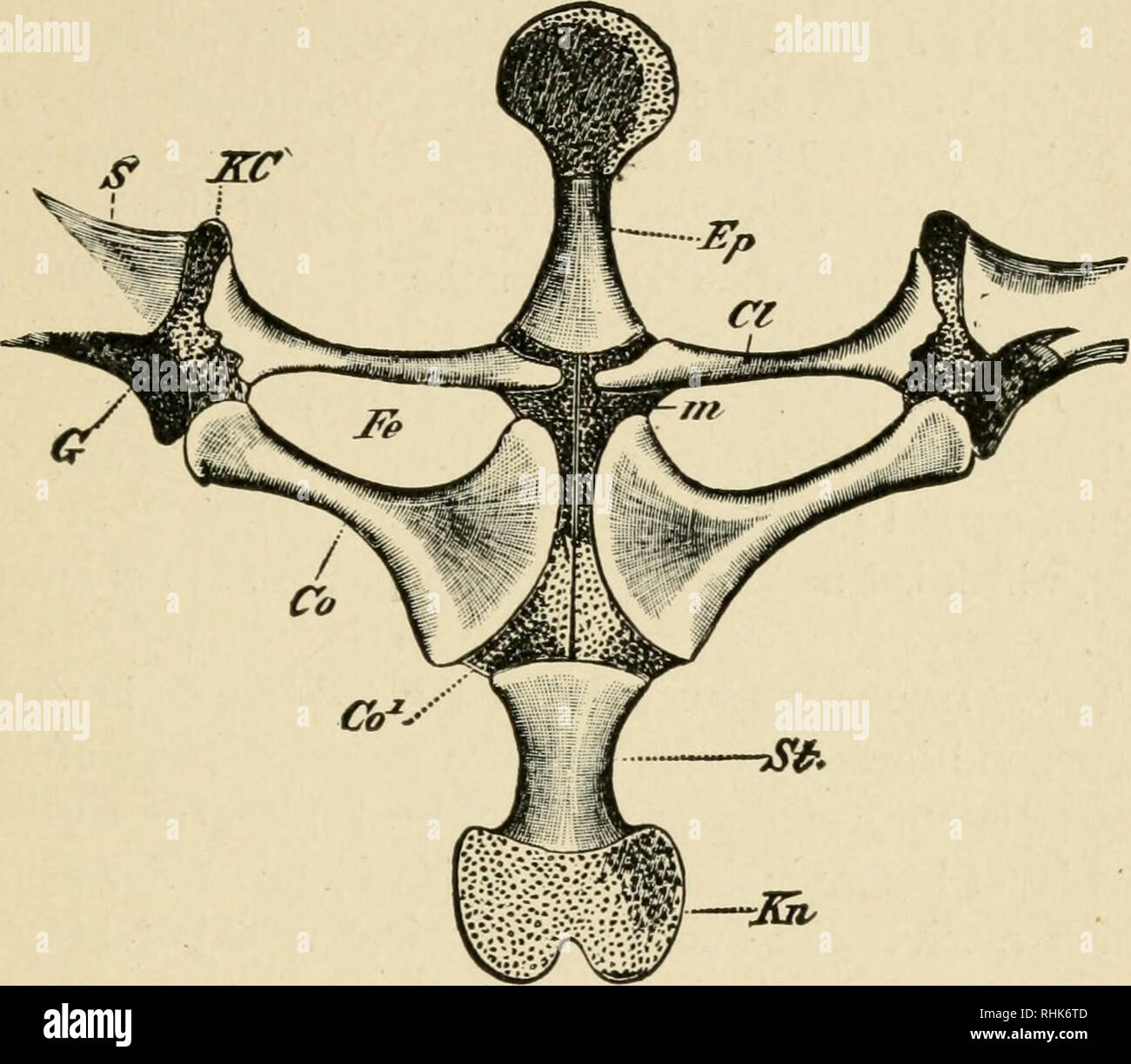 . The biology of the frog. Frogs. XIII THE SKELETON 239 portion, the suprascapula, which is composed of cartilage which is more or less calcified at the base. The supra-. FlG. 67. —Middle part of the shoulder girdle of the frog from below. Co, coracoid; Co epicoracoid; CI, clavicle; Ep, episternum; G, glenoid cavity; Fe, fenestra; KC, cartilage between scapula and clavicle; Kn, xiphisternum; w, junction of epicoracoids; S, scapula; 5/, sternum. (After Wiedersheim.) scapula articulates below with the long scapula, which is oblong and constricted in the middle; the posterior side of the lower e Stock Photohttps://www.alamy.com/image-license-details/?v=1https://www.alamy.com/the-biology-of-the-frog-frogs-xiii-the-skeleton-239-portion-the-suprascapula-which-is-composed-of-cartilage-which-is-more-or-less-calcified-at-the-base-the-supra-flg-67-middle-part-of-the-shoulder-girdle-of-the-frog-from-below-co-coracoid-co-epicoracoid-ci-clavicle-ep-episternum-g-glenoid-cavity-fe-fenestra-kc-cartilage-between-scapula-and-clavicle-kn-xiphisternum-w-junction-of-epicoracoids-s-scapula-5-sternum-after-wiedersheim-scapula-articulates-below-with-the-long-scapula-which-is-oblong-and-constricted-in-the-middle-the-posterior-side-of-the-lower-e-image234606413.html
RMRHK6TD–. The biology of the frog. Frogs. XIII THE SKELETON 239 portion, the suprascapula, which is composed of cartilage which is more or less calcified at the base. The supra-. FlG. 67. —Middle part of the shoulder girdle of the frog from below. Co, coracoid; Co epicoracoid; CI, clavicle; Ep, episternum; G, glenoid cavity; Fe, fenestra; KC, cartilage between scapula and clavicle; Kn, xiphisternum; w, junction of epicoracoids; S, scapula; 5/, sternum. (After Wiedersheim.) scapula articulates below with the long scapula, which is oblong and constricted in the middle; the posterior side of the lower e
 . The biology of the frog. Frogs. XIII THE SKELETON 239 portion, the suprascapula, which is composed of cartilage which is more or less calcified at the base. The supra-. FIG. 67.— Middle part of the shoulder girdle of the frog from below. Co, coracoid; Co', epicoracoid; Cl, clavicle; Ep, episternum; G, glenoid cavity; Fe, fenestra; A'C, cartilage between scapula and clavicle; Kn, xiphisternum; m, junction of epicoracoids; 6", scapula; 5/, sternum. (After Wiedersheim.) scapula articulates below with the long scapiila, which is oblong and constricted in the middle ; the posterior side of t Stock Photohttps://www.alamy.com/image-license-details/?v=1https://www.alamy.com/the-biology-of-the-frog-frogs-xiii-the-skeleton-239-portion-the-suprascapula-which-is-composed-of-cartilage-which-is-more-or-less-calcified-at-the-base-the-supra-fig-67-middle-part-of-the-shoulder-girdle-of-the-frog-from-below-co-coracoid-co-epicoracoid-cl-clavicle-ep-episternum-g-glenoid-cavity-fe-fenestra-ac-cartilage-between-scapula-and-clavicle-kn-xiphisternum-m-junction-of-epicoracoids-6quot-scapula-5-sternum-after-wiedersheim-scapula-articulates-below-with-the-long-scapiila-which-is-oblong-and-constricted-in-the-middle-the-posterior-side-of-t-image234606589.html
RMRHK72N–. The biology of the frog. Frogs. XIII THE SKELETON 239 portion, the suprascapula, which is composed of cartilage which is more or less calcified at the base. The supra-. FIG. 67.— Middle part of the shoulder girdle of the frog from below. Co, coracoid; Co', epicoracoid; Cl, clavicle; Ep, episternum; G, glenoid cavity; Fe, fenestra; A'C, cartilage between scapula and clavicle; Kn, xiphisternum; m, junction of epicoracoids; 6", scapula; 5/, sternum. (After Wiedersheim.) scapula articulates below with the long scapiila, which is oblong and constricted in the middle ; the posterior side of t
![. Biology of the vertebrates : a comparative study of man and his animal allies. Vertebrates; Vertebrates -- Anatomy; Anatomy, Comparative. 55° Biology of the Vertebrates Scapula Ompsternum ]ff â,''''/SuprascapuJa JUfksto^S Clavicle. Glenoid Fossa Xiphisternum^ Fig. 462. Sternum and pectoral girdle of a frog, Rana, ventral view. Cartilage stippled. (From Sayles, Manual for Comparative Anatomy, copyright 1938, by permission of The Macmillan Company, publishers.) Suprascapula Clavi Suprascapula- Fenestrae-^ Epicoracoid Cartilage-r-r-c^T '--? .fill/ Supracoracoid Foramen'"^' Glenoid Cavity Stock Photo . Biology of the vertebrates : a comparative study of man and his animal allies. Vertebrates; Vertebrates -- Anatomy; Anatomy, Comparative. 55° Biology of the Vertebrates Scapula Ompsternum ]ff â,''''/SuprascapuJa JUfksto^S Clavicle. Glenoid Fossa Xiphisternum^ Fig. 462. Sternum and pectoral girdle of a frog, Rana, ventral view. Cartilage stippled. (From Sayles, Manual for Comparative Anatomy, copyright 1938, by permission of The Macmillan Company, publishers.) Suprascapula Clavi Suprascapula- Fenestrae-^ Epicoracoid Cartilage-r-r-c^T '--? .fill/ Supracoracoid Foramen'"^' Glenoid Cavity Stock Photo](https://c8.alamy.com/comp/RHJNDJ/biology-of-the-vertebrates-a-comparative-study-of-man-and-his-animal-allies-vertebrates-vertebrates-anatomy-anatomy-comparative-55-biology-of-the-vertebrates-scapula-ompsternum-ff-suprascapuja-jufkstos-clavicle-glenoid-fossa-xiphisternum-fig-462-sternum-and-pectoral-girdle-of-a-frog-rana-ventral-view-cartilage-stippled-from-sayles-manual-for-comparative-anatomy-copyright-1938-by-permission-of-the-macmillan-company-publishers-suprascapula-clavi-suprascapula-fenestrae-epicoracoid-cartilage-r-r-ct-fill-supracoracoid-foramenquot-glenoid-cavity-RHJNDJ.jpg) . Biology of the vertebrates : a comparative study of man and his animal allies. Vertebrates; Vertebrates -- Anatomy; Anatomy, Comparative. 55° Biology of the Vertebrates Scapula Ompsternum ]ff â,''''/SuprascapuJa JUfksto^S Clavicle. Glenoid Fossa Xiphisternum^ Fig. 462. Sternum and pectoral girdle of a frog, Rana, ventral view. Cartilage stippled. (From Sayles, Manual for Comparative Anatomy, copyright 1938, by permission of The Macmillan Company, publishers.) Suprascapula Clavi Suprascapula- Fenestrae-^ Epicoracoid Cartilage-r-r-c^T '--? .fill/ Supracoracoid Foramen'"^' Glenoid Cavity Stock Photohttps://www.alamy.com/image-license-details/?v=1https://www.alamy.com/biology-of-the-vertebrates-a-comparative-study-of-man-and-his-animal-allies-vertebrates-vertebrates-anatomy-anatomy-comparative-55-biology-of-the-vertebrates-scapula-ompsternum-ff-suprascapuja-jufkstos-clavicle-glenoid-fossa-xiphisternum-fig-462-sternum-and-pectoral-girdle-of-a-frog-rana-ventral-view-cartilage-stippled-from-sayles-manual-for-comparative-anatomy-copyright-1938-by-permission-of-the-macmillan-company-publishers-suprascapula-clavi-suprascapula-fenestrae-epicoracoid-cartilage-r-r-ct-fill-supracoracoid-foramenquot-glenoid-cavity-image234595918.html
. Biology of the vertebrates : a comparative study of man and his animal allies. Vertebrates; Vertebrates -- Anatomy; Anatomy, Comparative. 55° Biology of the Vertebrates Scapula Ompsternum ]ff â,''''/SuprascapuJa JUfksto^S Clavicle. Glenoid Fossa Xiphisternum^ Fig. 462. Sternum and pectoral girdle of a frog, Rana, ventral view. Cartilage stippled. (From Sayles, Manual for Comparative Anatomy, copyright 1938, by permission of The Macmillan Company, publishers.) Suprascapula Clavi Suprascapula- Fenestrae-^ Epicoracoid Cartilage-r-r-c^T '--? .fill/ Supracoracoid Foramen'"^' Glenoid Cavity Stock Photohttps://www.alamy.com/image-license-details/?v=1https://www.alamy.com/biology-of-the-vertebrates-a-comparative-study-of-man-and-his-animal-allies-vertebrates-vertebrates-anatomy-anatomy-comparative-55-biology-of-the-vertebrates-scapula-ompsternum-ff-suprascapuja-jufkstos-clavicle-glenoid-fossa-xiphisternum-fig-462-sternum-and-pectoral-girdle-of-a-frog-rana-ventral-view-cartilage-stippled-from-sayles-manual-for-comparative-anatomy-copyright-1938-by-permission-of-the-macmillan-company-publishers-suprascapula-clavi-suprascapula-fenestrae-epicoracoid-cartilage-r-r-ct-fill-supracoracoid-foramenquot-glenoid-cavity-image234595918.htmlRMRHJNDJ–. Biology of the vertebrates : a comparative study of man and his animal allies. Vertebrates; Vertebrates -- Anatomy; Anatomy, Comparative. 55° Biology of the Vertebrates Scapula Ompsternum ]ff â,''''/SuprascapuJa JUfksto^S Clavicle. Glenoid Fossa Xiphisternum^ Fig. 462. Sternum and pectoral girdle of a frog, Rana, ventral view. Cartilage stippled. (From Sayles, Manual for Comparative Anatomy, copyright 1938, by permission of The Macmillan Company, publishers.) Suprascapula Clavi Suprascapula- Fenestrae-^ Epicoracoid Cartilage-r-r-c^T '--? .fill/ Supracoracoid Foramen'"^' Glenoid Cavity
 . Text book of vertebrate zoology. Vertebrates; Anatomy, Comparative. SKELETON. 149 ployed as a means of dividing birds into Ratitae and Carinatae. It is interesting to find a keel existing in the bats and in the fossil pterodactyls. In the mammals the sternum is more elongate, and more ribs contribute to its formation than in the sauropsida. It may consist of as many separate sternebrae as there are ribs connected with it, or these may so unite that but three separate bones can be recognized, a manubrium in front, a body in the middle, and an ensiform process (xiphisternum) behind, the latter Stock Photohttps://www.alamy.com/image-license-details/?v=1https://www.alamy.com/text-book-of-vertebrate-zoology-vertebrates-anatomy-comparative-skeleton-149-ployed-as-a-means-of-dividing-birds-into-ratitae-and-carinatae-it-is-interesting-to-find-a-keel-existing-in-the-bats-and-in-the-fossil-pterodactyls-in-the-mammals-the-sternum-is-more-elongate-and-more-ribs-contribute-to-its-formation-than-in-the-sauropsida-it-may-consist-of-as-many-separate-sternebrae-as-there-are-ribs-connected-with-it-or-these-may-so-unite-that-but-three-separate-bones-can-be-recognized-a-manubrium-in-front-a-body-in-the-middle-and-an-ensiform-process-xiphisternum-behind-the-latter-image232252722.html
RMRDT0M2–. Text book of vertebrate zoology. Vertebrates; Anatomy, Comparative. SKELETON. 149 ployed as a means of dividing birds into Ratitae and Carinatae. It is interesting to find a keel existing in the bats and in the fossil pterodactyls. In the mammals the sternum is more elongate, and more ribs contribute to its formation than in the sauropsida. It may consist of as many separate sternebrae as there are ribs connected with it, or these may so unite that but three separate bones can be recognized, a manubrium in front, a body in the middle, and an ensiform process (xiphisternum) behind, the latter
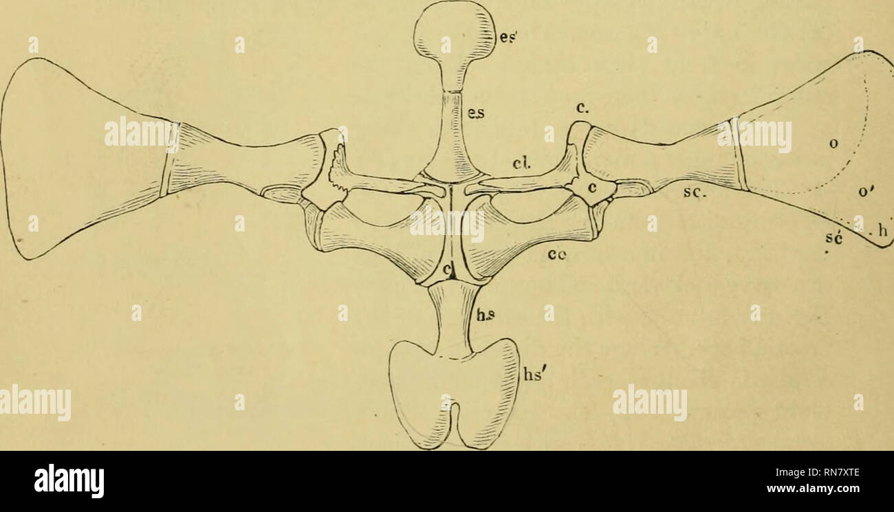 . The anatomy of the frog. Frogs -- Anatomy; Amphibians -- Anatomy. Shoulder-girdle and sternum of Rana esciilenta, tw ice nat. size. The scapula and suprascapula are turned outwards. Connecting cartilage. h s. Sternum proper- Clavicles and precoracnids. W Xiphisternum^l^ Coracoid. o Bone. ^ Omostei-num. o' Calcified cartilage. Episternum. sc. Scapula. Hyaline cartilage. sc' Snprascapula.. Please note that these images are extracted from scanned page images that may have been digitally enhanced for readability - coloration and appearance of these illustrations may not perfectly resemble the or Stock Photohttps://www.alamy.com/image-license-details/?v=1https://www.alamy.com/the-anatomy-of-the-frog-frogs-anatomy-amphibians-anatomy-shoulder-girdle-and-sternum-of-rana-esciilenta-tw-ice-nat-size-the-scapula-and-suprascapula-are-turned-outwards-connecting-cartilage-h-s-sternum-proper-clavicles-and-precoracnids-w-xiphisternuml-coracoid-o-bone-omostei-num-o-calcified-cartilage-episternum-sc-scapula-hyaline-cartilage-sc-snprascapula-please-note-that-these-images-are-extracted-from-scanned-page-images-that-may-have-been-digitally-enhanced-for-readability-coloration-and-appearance-of-these-illustrations-may-not-perfectly-resemble-the-or-image236817294.html
RMRN7XTE–. The anatomy of the frog. Frogs -- Anatomy; Amphibians -- Anatomy. Shoulder-girdle and sternum of Rana esciilenta, tw ice nat. size. The scapula and suprascapula are turned outwards. Connecting cartilage. h s. Sternum proper- Clavicles and precoracnids. W Xiphisternum^l^ Coracoid. o Bone. ^ Omostei-num. o' Calcified cartilage. Episternum. sc. Scapula. Hyaline cartilage. sc' Snprascapula.. Please note that these images are extracted from scanned page images that may have been digitally enhanced for readability - coloration and appearance of these illustrations may not perfectly resemble the or
![. Elementary text-book of zoology [electronic resource]. Zoology. Xiphisternum. View with dorsal parts bent downwards. Bone is black and cartilage dotted. fore limb has two peculiarities. The radius and ulna are united into one bone, and there is no pre-axial digit or thumb. Fig, 248.—Fore-limb of Ran a.. Note fusion of radius and ulna and absence of pollex, a metacarpal onlj- remaining. The pelvic o^irdle has a very long forwardly-diiected ilium articulating with the sacral vertebra. The puhes and ischia Fig. 249.—Pelvic Girdle of Rana. Ilium.. Please note that these images are extracted from Stock Photo . Elementary text-book of zoology [electronic resource]. Zoology. Xiphisternum. View with dorsal parts bent downwards. Bone is black and cartilage dotted. fore limb has two peculiarities. The radius and ulna are united into one bone, and there is no pre-axial digit or thumb. Fig, 248.—Fore-limb of Ran a.. Note fusion of radius and ulna and absence of pollex, a metacarpal onlj- remaining. The pelvic o^irdle has a very long forwardly-diiected ilium articulating with the sacral vertebra. The puhes and ischia Fig. 249.—Pelvic Girdle of Rana. Ilium.. Please note that these images are extracted from Stock Photo](https://c8.alamy.com/comp/RJN9D1/elementary-text-book-of-zoology-electronic-resource-zoology-xiphisternum-view-with-dorsal-parts-bent-downwards-bone-is-black-and-cartilage-dotted-fore-limb-has-two-peculiarities-the-radius-and-ulna-are-united-into-one-bone-and-there-is-no-pre-axial-digit-or-thumb-fig-248fore-limb-of-ran-a-note-fusion-of-radius-and-ulna-and-absence-of-pollex-a-metacarpal-onlj-remaining-the-pelvic-oirdle-has-a-very-long-forwardly-diiected-ilium-articulating-with-the-sacral-vertebra-the-puhes-and-ischia-fig-249pelvic-girdle-of-rana-ilium-please-note-that-these-images-are-extracted-from-RJN9D1.jpg) . Elementary text-book of zoology [electronic resource]. Zoology. Xiphisternum. View with dorsal parts bent downwards. Bone is black and cartilage dotted. fore limb has two peculiarities. The radius and ulna are united into one bone, and there is no pre-axial digit or thumb. Fig, 248.—Fore-limb of Ran a.. Note fusion of radius and ulna and absence of pollex, a metacarpal onlj- remaining. The pelvic o^irdle has a very long forwardly-diiected ilium articulating with the sacral vertebra. The puhes and ischia Fig. 249.—Pelvic Girdle of Rana. Ilium.. Please note that these images are extracted from Stock Photohttps://www.alamy.com/image-license-details/?v=1https://www.alamy.com/elementary-text-book-of-zoology-electronic-resource-zoology-xiphisternum-view-with-dorsal-parts-bent-downwards-bone-is-black-and-cartilage-dotted-fore-limb-has-two-peculiarities-the-radius-and-ulna-are-united-into-one-bone-and-there-is-no-pre-axial-digit-or-thumb-fig-248fore-limb-of-ran-a-note-fusion-of-radius-and-ulna-and-absence-of-pollex-a-metacarpal-onlj-remaining-the-pelvic-oirdle-has-a-very-long-forwardly-diiected-ilium-articulating-with-the-sacral-vertebra-the-puhes-and-ischia-fig-249pelvic-girdle-of-rana-ilium-please-note-that-these-images-are-extracted-from-image235267005.html
. Elementary text-book of zoology [electronic resource]. Zoology. Xiphisternum. View with dorsal parts bent downwards. Bone is black and cartilage dotted. fore limb has two peculiarities. The radius and ulna are united into one bone, and there is no pre-axial digit or thumb. Fig, 248.—Fore-limb of Ran a.. Note fusion of radius and ulna and absence of pollex, a metacarpal onlj- remaining. The pelvic o^irdle has a very long forwardly-diiected ilium articulating with the sacral vertebra. The puhes and ischia Fig. 249.—Pelvic Girdle of Rana. Ilium.. Please note that these images are extracted from Stock Photohttps://www.alamy.com/image-license-details/?v=1https://www.alamy.com/elementary-text-book-of-zoology-electronic-resource-zoology-xiphisternum-view-with-dorsal-parts-bent-downwards-bone-is-black-and-cartilage-dotted-fore-limb-has-two-peculiarities-the-radius-and-ulna-are-united-into-one-bone-and-there-is-no-pre-axial-digit-or-thumb-fig-248fore-limb-of-ran-a-note-fusion-of-radius-and-ulna-and-absence-of-pollex-a-metacarpal-onlj-remaining-the-pelvic-oirdle-has-a-very-long-forwardly-diiected-ilium-articulating-with-the-sacral-vertebra-the-puhes-and-ischia-fig-249pelvic-girdle-of-rana-ilium-please-note-that-these-images-are-extracted-from-image235267005.htmlRMRJN9D1–. Elementary text-book of zoology [electronic resource]. Zoology. Xiphisternum. View with dorsal parts bent downwards. Bone is black and cartilage dotted. fore limb has two peculiarities. The radius and ulna are united into one bone, and there is no pre-axial digit or thumb. Fig, 248.—Fore-limb of Ran a.. Note fusion of radius and ulna and absence of pollex, a metacarpal onlj- remaining. The pelvic o^irdle has a very long forwardly-diiected ilium articulating with the sacral vertebra. The puhes and ischia Fig. 249.—Pelvic Girdle of Rana. Ilium.. Please note that these images are extracted from
 . A manual of zoology. XII PHYLUM CHORDATA 415 Passing forwards from the anterior ends of the united epicoracoids is a rod of bone, the episternum {Ep), tipped by a rounded plate of cartilage, the omosternum ; and passing backwards from their posterior ends is a similar but larger bony rod, the sternum {St), also tipped by a cartilaginous plate, to which the name xiphisternum (K?i) is applied.. Fig. 249. — Rana esculenta. The shoulder girdle from the ventral aspect. Co, coracoid: Co', epicoraeoid; CI, clavicle; G, glenoid cavity; £/>, episternum; Fe, fenestra between procoracoid and coracoi Stock Photohttps://www.alamy.com/image-license-details/?v=1https://www.alamy.com/a-manual-of-zoology-xii-phylum-chordata-415-passing-forwards-from-the-anterior-ends-of-the-united-epicoracoids-is-a-rod-of-bone-the-episternum-ep-tipped-by-a-rounded-plate-of-cartilage-the-omosternum-and-passing-backwards-from-their-posterior-ends-is-a-similar-but-larger-bony-rod-the-sternum-st-also-tipped-by-a-cartilaginous-plate-to-which-the-name-xiphisternum-ki-is-applied-fig-249-rana-esculenta-the-shoulder-girdle-from-the-ventral-aspect-co-coracoid-co-epicoraeoid-ci-clavicle-g-glenoid-cavity-gt-episternum-fe-fenestra-between-procoracoid-and-coracoi-image232132382.html
RMRDJF66–. A manual of zoology. XII PHYLUM CHORDATA 415 Passing forwards from the anterior ends of the united epicoracoids is a rod of bone, the episternum {Ep), tipped by a rounded plate of cartilage, the omosternum ; and passing backwards from their posterior ends is a similar but larger bony rod, the sternum {St), also tipped by a cartilaginous plate, to which the name xiphisternum (K?i) is applied.. Fig. 249. — Rana esculenta. The shoulder girdle from the ventral aspect. Co, coracoid: Co', epicoraeoid; CI, clavicle; G, glenoid cavity; £/>, episternum; Fe, fenestra between procoracoid and coracoi
 . Biology of the vertebrates : a comparative study of man and his animal allies. Vertebrates; Vertebrates -- Anatomy; Anatomy, Comparative. Glenoid Fossa Xiphisternum^ Fig. 462. Sternum and pectoral girdle of a frog, Rana, ventral view. Cartilage stippled. (From Sayles, Manual for Comparative Anatomy, copyright 1938, by permission of The Macmillan Company, publishers.) Suprascapula Clavi Suprascapula- Fenestrae-^ Epicoracoid Cartilage-r-r-c^T '--? .fill/ Supracoracoid Foramen'"^' Glenoid Cavity Interclavicle §: « A Sternum -,y- y Fenestrae // . // Scapula I / / / Glenoid Cavity > ' Stock Photohttps://www.alamy.com/image-license-details/?v=1https://www.alamy.com/biology-of-the-vertebrates-a-comparative-study-of-man-and-his-animal-allies-vertebrates-vertebrates-anatomy-anatomy-comparative-glenoid-fossa-xiphisternum-fig-462-sternum-and-pectoral-girdle-of-a-frog-rana-ventral-view-cartilage-stippled-from-sayles-manual-for-comparative-anatomy-copyright-1938-by-permission-of-the-macmillan-company-publishers-suprascapula-clavi-suprascapula-fenestrae-epicoracoid-cartilage-r-r-ct-fill-supracoracoid-foramenquot-glenoid-cavity-interclavicle-a-sternum-y-y-fenestrae-scapula-i-glenoid-cavity-gt-image234595904.html
RMRHJND4–. Biology of the vertebrates : a comparative study of man and his animal allies. Vertebrates; Vertebrates -- Anatomy; Anatomy, Comparative. Glenoid Fossa Xiphisternum^ Fig. 462. Sternum and pectoral girdle of a frog, Rana, ventral view. Cartilage stippled. (From Sayles, Manual for Comparative Anatomy, copyright 1938, by permission of The Macmillan Company, publishers.) Suprascapula Clavi Suprascapula- Fenestrae-^ Epicoracoid Cartilage-r-r-c^T '--? .fill/ Supracoracoid Foramen'"^' Glenoid Cavity Interclavicle §: « A Sternum -,y- y Fenestrae // . // Scapula I / / / Glenoid Cavity > '
 . A manual of elementary zoology . Zoology. APPENDIX 557 submaxillaris muscles; xiphisternum; anterior abdominal and cutaneous veins ; hypoglossal nerves (Fig. 21). Cut through the muscles of the ventral body-wall, and through the pectoral girdle, on one side of the middle line. Ligature doubly the anterior abdominal vein near the liver ; cut between the ligatures ; carefully cut through the muscles which still hold down the pectoral girdle; turn back the body-wall, and pin it. Arrange the viscera for drawing to show as much as possible, noting: heart; lungs, liver, gall bladder, stomach, smal Stock Photohttps://www.alamy.com/image-license-details/?v=1https://www.alamy.com/a-manual-of-elementary-zoology-zoology-appendix-557-submaxillaris-muscles-xiphisternum-anterior-abdominal-and-cutaneous-veins-hypoglossal-nerves-fig-21-cut-through-the-muscles-of-the-ventral-body-wall-and-through-the-pectoral-girdle-on-one-side-of-the-middle-line-ligature-doubly-the-anterior-abdominal-vein-near-the-liver-cut-between-the-ligatures-carefully-cut-through-the-muscles-which-still-hold-down-the-pectoral-girdle-turn-back-the-body-wall-and-pin-it-arrange-the-viscera-for-drawing-to-show-as-much-as-possible-noting-heart-lungs-liver-gall-bladder-stomach-smal-image232116181.html
RMRDHPFH–. A manual of elementary zoology . Zoology. APPENDIX 557 submaxillaris muscles; xiphisternum; anterior abdominal and cutaneous veins ; hypoglossal nerves (Fig. 21). Cut through the muscles of the ventral body-wall, and through the pectoral girdle, on one side of the middle line. Ligature doubly the anterior abdominal vein near the liver ; cut between the ligatures ; carefully cut through the muscles which still hold down the pectoral girdle; turn back the body-wall, and pin it. Arrange the viscera for drawing to show as much as possible, noting: heart; lungs, liver, gall bladder, stomach, smal
 . Outlines of zoology. Zoology. THE LIMBS AND GIRDLES. 537 fibulare—and three imperfectly ossified distal elements, five metatarsal bones, and five toes. The first toe or hallux. FlG. 228.—Pectoral girdle of Rana esculenta. —After Ecker. The cartilaginous parts are dotted, it/*., Episternum; om., omo- sternum ; Ep.c, epicoracoids ; St., sternum ; x., xiphisternum ; cl., clavicle with underlying precoracoid cartilage; co., cora- coid ; Sc, scapula; S.sc., supra-sc'apula; 67., glenoid cavity for humerus. has two phalanges, the second also two, the third three, the fourth four, the fifth three, a Stock Photohttps://www.alamy.com/image-license-details/?v=1https://www.alamy.com/outlines-of-zoology-zoology-the-limbs-and-girdles-537-fibulareand-three-imperfectly-ossified-distal-elements-five-metatarsal-bones-and-five-toes-the-first-toe-or-hallux-flg-228pectoral-girdle-of-rana-esculenta-after-ecker-the-cartilaginous-parts-are-dotted-it-episternum-om-omo-sternum-epc-epicoracoids-st-sternum-x-xiphisternum-cl-clavicle-with-underlying-precoracoid-cartilage-co-cora-coid-sc-scapula-ssc-supra-scapula-67-glenoid-cavity-for-humerus-has-two-phalanges-the-second-also-two-the-third-three-the-fourth-four-the-fifth-three-a-image232213411.html
RMRDP6G3–. Outlines of zoology. Zoology. THE LIMBS AND GIRDLES. 537 fibulare—and three imperfectly ossified distal elements, five metatarsal bones, and five toes. The first toe or hallux. FlG. 228.—Pectoral girdle of Rana esculenta. —After Ecker. The cartilaginous parts are dotted, it/*., Episternum; om., omo- sternum ; Ep.c, epicoracoids ; St., sternum ; x., xiphisternum ; cl., clavicle with underlying precoracoid cartilage; co., cora- coid ; Sc, scapula; S.sc., supra-sc'apula; 67., glenoid cavity for humerus. has two phalanges, the second also two, the third three, the fourth four, the fifth three, a
![. Elementary text-book of zoology [electronic resource]. Zoology. Xiphisternum. View with dorsal parts bent downwards. Bone is black and cartilage dotted. fore limb has two peculiarities. The radius and ulna are united into one bone, and there is no pre-axial digit or thumb. Fig, 248.—Fore-limb of Ran a.. Please note that these images are extracted from scanned page images that may have been digitally enhanced for readability - coloration and appearance of these illustrations may not perfectly resemble the original work.. Masterman, Arthur Thomas; Parsons, John Herbert, Sir, 1868-1957, donor; Stock Photo . Elementary text-book of zoology [electronic resource]. Zoology. Xiphisternum. View with dorsal parts bent downwards. Bone is black and cartilage dotted. fore limb has two peculiarities. The radius and ulna are united into one bone, and there is no pre-axial digit or thumb. Fig, 248.—Fore-limb of Ran a.. Please note that these images are extracted from scanned page images that may have been digitally enhanced for readability - coloration and appearance of these illustrations may not perfectly resemble the original work.. Masterman, Arthur Thomas; Parsons, John Herbert, Sir, 1868-1957, donor; Stock Photo](https://c8.alamy.com/comp/RJN9DN/elementary-text-book-of-zoology-electronic-resource-zoology-xiphisternum-view-with-dorsal-parts-bent-downwards-bone-is-black-and-cartilage-dotted-fore-limb-has-two-peculiarities-the-radius-and-ulna-are-united-into-one-bone-and-there-is-no-pre-axial-digit-or-thumb-fig-248fore-limb-of-ran-a-please-note-that-these-images-are-extracted-from-scanned-page-images-that-may-have-been-digitally-enhanced-for-readability-coloration-and-appearance-of-these-illustrations-may-not-perfectly-resemble-the-original-work-masterman-arthur-thomas-parsons-john-herbert-sir-1868-1957-donor-RJN9DN.jpg) . Elementary text-book of zoology [electronic resource]. Zoology. Xiphisternum. View with dorsal parts bent downwards. Bone is black and cartilage dotted. fore limb has two peculiarities. The radius and ulna are united into one bone, and there is no pre-axial digit or thumb. Fig, 248.—Fore-limb of Ran a.. Please note that these images are extracted from scanned page images that may have been digitally enhanced for readability - coloration and appearance of these illustrations may not perfectly resemble the original work.. Masterman, Arthur Thomas; Parsons, John Herbert, Sir, 1868-1957, donor; Stock Photohttps://www.alamy.com/image-license-details/?v=1https://www.alamy.com/elementary-text-book-of-zoology-electronic-resource-zoology-xiphisternum-view-with-dorsal-parts-bent-downwards-bone-is-black-and-cartilage-dotted-fore-limb-has-two-peculiarities-the-radius-and-ulna-are-united-into-one-bone-and-there-is-no-pre-axial-digit-or-thumb-fig-248fore-limb-of-ran-a-please-note-that-these-images-are-extracted-from-scanned-page-images-that-may-have-been-digitally-enhanced-for-readability-coloration-and-appearance-of-these-illustrations-may-not-perfectly-resemble-the-original-work-masterman-arthur-thomas-parsons-john-herbert-sir-1868-1957-donor-image235267025.html
. Elementary text-book of zoology [electronic resource]. Zoology. Xiphisternum. View with dorsal parts bent downwards. Bone is black and cartilage dotted. fore limb has two peculiarities. The radius and ulna are united into one bone, and there is no pre-axial digit or thumb. Fig, 248.—Fore-limb of Ran a.. Please note that these images are extracted from scanned page images that may have been digitally enhanced for readability - coloration and appearance of these illustrations may not perfectly resemble the original work.. Masterman, Arthur Thomas; Parsons, John Herbert, Sir, 1868-1957, donor; Stock Photohttps://www.alamy.com/image-license-details/?v=1https://www.alamy.com/elementary-text-book-of-zoology-electronic-resource-zoology-xiphisternum-view-with-dorsal-parts-bent-downwards-bone-is-black-and-cartilage-dotted-fore-limb-has-two-peculiarities-the-radius-and-ulna-are-united-into-one-bone-and-there-is-no-pre-axial-digit-or-thumb-fig-248fore-limb-of-ran-a-please-note-that-these-images-are-extracted-from-scanned-page-images-that-may-have-been-digitally-enhanced-for-readability-coloration-and-appearance-of-these-illustrations-may-not-perfectly-resemble-the-original-work-masterman-arthur-thomas-parsons-john-herbert-sir-1868-1957-donor-image235267025.htmlRMRJN9DN–. Elementary text-book of zoology [electronic resource]. Zoology. Xiphisternum. View with dorsal parts bent downwards. Bone is black and cartilage dotted. fore limb has two peculiarities. The radius and ulna are united into one bone, and there is no pre-axial digit or thumb. Fig, 248.—Fore-limb of Ran a.. Please note that these images are extracted from scanned page images that may have been digitally enhanced for readability - coloration and appearance of these illustrations may not perfectly resemble the original work.. Masterman, Arthur Thomas; Parsons, John Herbert, Sir, 1868-1957, donor;
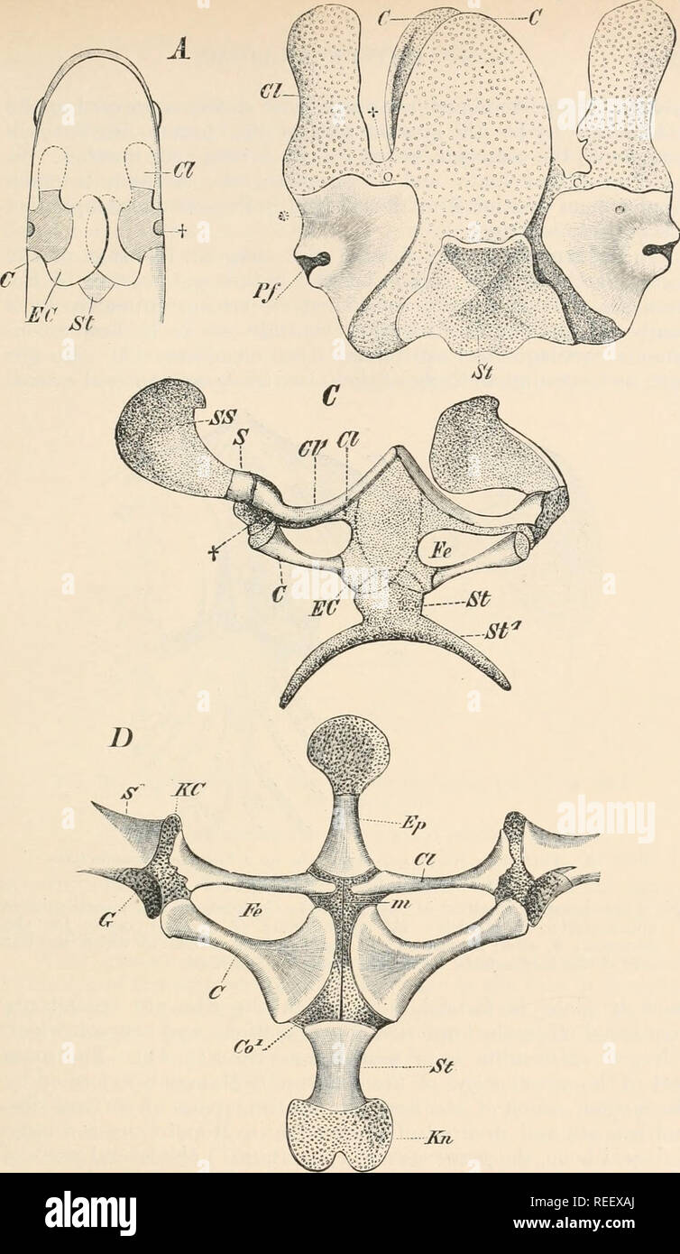 . Comparative anatomy of vertebrates. Anatomy, Comparative; Vertebrates. Km. 55.—PECTORAL ARCH OF VARIOUS AMPHIBIANS. From the ventral side. A—Urodele (diagrammatic) ; B—Axolotl (Amblystoma) ; C—Bomlnnnin,- iynews ; U, J'nnn ewii/cnta. C, coracoirl; Cl, procoracoid; C71 (Cl in U), clavicle ; EC, Co1, epicoracoid ; Ep, omostermun ; Fe, fenestra between procoracoid and coracoid bars ; /n, cartilaginous xiphisternum ; t, Pf, O, glenoid cavity for the humerus ; S, scapula ; SS, supraacapula; St, Sfl, sternum. *, j (in B) indicate nerve- apertures.. Please note that these images are extracted from Stock Photohttps://www.alamy.com/image-license-details/?v=1https://www.alamy.com/comparative-anatomy-of-vertebrates-anatomy-comparative-vertebrates-km-55pectoral-arch-of-various-amphibians-from-the-ventral-side-aurodele-diagrammatic-baxolotl-amblystoma-cbomlnnnin-iynews-u-jnnn-ewiicnta-c-coracoirl-cl-procoracoid-c71-cl-in-u-clavicle-ec-co1-epicoracoid-ep-omostermun-fe-fenestra-between-procoracoid-and-coracoid-bars-n-cartilaginous-xiphisternum-t-pf-o-glenoid-cavity-for-the-humerus-s-scapula-ss-supraacapula-st-sfl-sternum-j-in-b-indicate-nerve-apertures-please-note-that-these-images-are-extracted-from-image232667978.html
RMREEXAJ–. Comparative anatomy of vertebrates. Anatomy, Comparative; Vertebrates. Km. 55.—PECTORAL ARCH OF VARIOUS AMPHIBIANS. From the ventral side. A—Urodele (diagrammatic) ; B—Axolotl (Amblystoma) ; C—Bomlnnnin,- iynews ; U, J'nnn ewii/cnta. C, coracoirl; Cl, procoracoid; C71 (Cl in U), clavicle ; EC, Co1, epicoracoid ; Ep, omostermun ; Fe, fenestra between procoracoid and coracoid bars ; /n, cartilaginous xiphisternum ; t, Pf, O, glenoid cavity for the humerus ; S, scapula ; SS, supraacapula; St, Sfl, sternum. *, j (in B) indicate nerve- apertures.. Please note that these images are extracted from
 . Outlines of zoology. Zoology. FlG. 228.—Pectoral girdle of Rana esculenta. —After Ecker. The cartilaginous parts are dotted, it/*., Episternum; om., omo- sternum ; Ep.c, epicoracoids ; St., sternum ; x., xiphisternum ; cl., clavicle with underlying precoracoid cartilage; co., cora- coid ; Sc, scapula; S.sc., supra-sc'apula; 67., glenoid cavity for humerus. has two phalanges, the second also two, the third three, the fourth four, the fifth three, and, finally, outside the hallux there is a " calcar," which looks like an extra toe, and con-. FlG. 229.—Side view of frog's pelvis.—Afte Stock Photohttps://www.alamy.com/image-license-details/?v=1https://www.alamy.com/outlines-of-zoology-zoology-flg-228pectoral-girdle-of-rana-esculenta-after-ecker-the-cartilaginous-parts-are-dotted-it-episternum-om-omo-sternum-epc-epicoracoids-st-sternum-x-xiphisternum-cl-clavicle-with-underlying-precoracoid-cartilage-co-cora-coid-sc-scapula-ssc-supra-scapula-67-glenoid-cavity-for-humerus-has-two-phalanges-the-second-also-two-the-third-three-the-fourth-four-the-fifth-three-and-finally-outside-the-hallux-there-is-a-quot-calcarquot-which-looks-like-an-extra-toe-and-con-flg-229side-view-of-frogs-pelvisafte-image232213406.html
RMRDP6FX–. Outlines of zoology. Zoology. FlG. 228.—Pectoral girdle of Rana esculenta. —After Ecker. The cartilaginous parts are dotted, it/*., Episternum; om., omo- sternum ; Ep.c, epicoracoids ; St., sternum ; x., xiphisternum ; cl., clavicle with underlying precoracoid cartilage; co., cora- coid ; Sc, scapula; S.sc., supra-sc'apula; 67., glenoid cavity for humerus. has two phalanges, the second also two, the third three, the fourth four, the fifth three, and, finally, outside the hallux there is a " calcar," which looks like an extra toe, and con-. FlG. 229.—Side view of frog's pelvis.—Afte
 . Comparative anatomy of vertebrates. Anatomy, Comparative; Vertebrates -- Anatomy. FIG. 56.—Sternum, etc., of Iguana tuberculata, after Blan- chard. c, coracoid; c/, clavicle; e, episternum; h, humerus; pc, procoracoid; x, xiphisternum. FIG. 57.—Sternum of guinea pig. sr, sternal rib; st, sterne- brae; vr, vertebral rib, x, xiphi- sternum. In the mammals the number of ribs connected with the sternum is greater than in the lower classes. The sternebrae may remain dis- tinct throughout life (fig. 57) or, as in man, they may fuse into fewer elements, the xiphoid process being unconnected with th Stock Photohttps://www.alamy.com/image-license-details/?v=1https://www.alamy.com/comparative-anatomy-of-vertebrates-anatomy-comparative-vertebrates-anatomy-fig-56sternum-etc-of-iguana-tuberculata-after-blan-chard-c-coracoid-c-clavicle-e-episternum-h-humerus-pc-procoracoid-x-xiphisternum-fig-57sternum-of-guinea-pig-sr-sternal-rib-st-sterne-brae-vr-vertebral-rib-x-xiphi-sternum-in-the-mammals-the-number-of-ribs-connected-with-the-sternum-is-greater-than-in-the-lower-classes-the-sternebrae-may-remain-dis-tinct-throughout-life-fig-57-or-as-in-man-they-may-fuse-into-fewer-elements-the-xiphoid-process-being-unconnected-with-th-image232680478.html
RMREFE92–. Comparative anatomy of vertebrates. Anatomy, Comparative; Vertebrates -- Anatomy. FIG. 56.—Sternum, etc., of Iguana tuberculata, after Blan- chard. c, coracoid; c/, clavicle; e, episternum; h, humerus; pc, procoracoid; x, xiphisternum. FIG. 57.—Sternum of guinea pig. sr, sternal rib; st, sterne- brae; vr, vertebral rib, x, xiphi- sternum. In the mammals the number of ribs connected with the sternum is greater than in the lower classes. The sternebrae may remain dis- tinct throughout life (fig. 57) or, as in man, they may fuse into fewer elements, the xiphoid process being unconnected with th
 . The anatomy of the frog. Frogs -- Anatomy; Amphibians -- Anatomy. MUSCLES OF THE TEUNK. 69 The whole muscle is attached by its most anterior fibres to the cartilage of the xiphisternum, the rest passing- into an aponeurosis Fig. 63.. Muscles of trunk of Rana cscitlcnta, from the right side. cd M. ciitaiieus fenioris. d M. deltoideus. d.m. M. depressor maxillae. i M. inf ra.spinatiis. Id M. latiss. dorsi. oe M. obliquus abdom. externus. oe' Scapular origin of same. ss M. siibscaimlaris. t M. triceps brachii. which, inseparably connected with the Imcrlpiiones iendineae, traverses the lower sur Stock Photohttps://www.alamy.com/image-license-details/?v=1https://www.alamy.com/the-anatomy-of-the-frog-frogs-anatomy-amphibians-anatomy-muscles-of-the-teunk-69-the-whole-muscle-is-attached-by-its-most-anterior-fibres-to-the-cartilage-of-the-xiphisternum-the-rest-passing-into-an-aponeurosis-fig-63-muscles-of-trunk-of-rana-cscitlcnta-from-the-right-side-cd-m-ciitaiieus-fenioris-d-m-deltoideus-dm-m-depressor-maxillae-i-m-inf-raspinatiis-id-m-latiss-dorsi-oe-m-obliquus-abdom-externus-oe-scapular-origin-of-same-ss-m-siibscaimlaris-t-m-triceps-brachii-which-inseparably-connected-with-the-imcrlpiiones-iendineae-traverses-the-lower-sur-image236816401.html
RMRN7WMH–. The anatomy of the frog. Frogs -- Anatomy; Amphibians -- Anatomy. MUSCLES OF THE TEUNK. 69 The whole muscle is attached by its most anterior fibres to the cartilage of the xiphisternum, the rest passing- into an aponeurosis Fig. 63.. Muscles of trunk of Rana cscitlcnta, from the right side. cd M. ciitaiieus fenioris. d M. deltoideus. d.m. M. depressor maxillae. i M. inf ra.spinatiis. Id M. latiss. dorsi. oe M. obliquus abdom. externus. oe' Scapular origin of same. ss M. siibscaimlaris. t M. triceps brachii. which, inseparably connected with the Imcrlpiiones iendineae, traverses the lower sur
 . Text book of vertebrate zoology. Vertebrates; Anatomy, Comparative. Fig. 286. ventral portion of girdle of Rana, heim, illustrating Sternum and the shoulder after Wieders- t h e firmister- nous type of sternum, cl, clavicle; co, cora- coid ; ec, epicoracoid; g, glenoid fossa; os, omosternum; s, ventral part of scapula; st, sternum; x, xiphisternum. Fig. 285. Shoulder girdle of Bombinator igneus, showing the ar- ciferous type, after Wiedersheim. c, clavicle ; co, coracoid; ec, epi- coracoid ; g, glenoid fossa; pc, pro- coracoid; s, scapula; j-.r, supra- scapula; st, sternum. or toothless frog Stock Photohttps://www.alamy.com/image-license-details/?v=1https://www.alamy.com/text-book-of-vertebrate-zoology-vertebrates-anatomy-comparative-fig-286-ventral-portion-of-girdle-of-rana-heim-illustrating-sternum-and-the-shoulder-after-wieders-t-h-e-firmister-nous-type-of-sternum-cl-clavicle-co-cora-coid-ec-epicoracoid-g-glenoid-fossa-os-omosternum-s-ventral-part-of-scapula-st-sternum-x-xiphisternum-fig-285-shoulder-girdle-of-bombinator-igneus-showing-the-ar-ciferous-type-after-wiedersheim-c-clavicle-co-coracoid-ec-epi-coracoid-g-glenoid-fossa-pc-pro-coracoid-s-scapula-j-r-supra-scapula-st-sternum-or-toothless-frog-image232234851.html
RMRDR5WR–. Text book of vertebrate zoology. Vertebrates; Anatomy, Comparative. Fig. 286. ventral portion of girdle of Rana, heim, illustrating Sternum and the shoulder after Wieders- t h e firmister- nous type of sternum, cl, clavicle; co, cora- coid ; ec, epicoracoid; g, glenoid fossa; os, omosternum; s, ventral part of scapula; st, sternum; x, xiphisternum. Fig. 285. Shoulder girdle of Bombinator igneus, showing the ar- ciferous type, after Wiedersheim. c, clavicle ; co, coracoid; ec, epi- coracoid ; g, glenoid fossa; pc, pro- coracoid; s, scapula; j-.r, supra- scapula; st, sternum. or toothless frog
 . Bulletin of the Museum of Comparative Zoology at Harvard College. Zoology. C3 'CANISVA04 -*RJ< aw yovt. 7 A-7. 0 10 20 mmiuum —, m m 'A9 A8&, AMPHICYON Figure 5 Kibs and Xiphisternum Amphicyon. A-l, anterior view, third right rib head; A-2, articular view of third right rib head; A-3, posterior view of third right rib head; A-4, posterior view of eleventh left rib head; A-5, articular view of eleventh left rib head; A-6, anterior view of eleventh left rib head. Comparable views. Please note that these images are extracted from scanned page images that may have been digitally enhanced Stock Photohttps://www.alamy.com/image-license-details/?v=1https://www.alamy.com/bulletin-of-the-museum-of-comparative-zoology-at-harvard-college-zoology-c3-canisva04-rjlt-aw-yovt-7-a-7-0-10-20-mmiuum-m-m-a9-a8amp-amphicyon-figure-5-kibs-and-xiphisternum-amphicyon-a-l-anterior-view-third-right-rib-head-a-2-articular-view-of-third-right-rib-head-a-3-posterior-view-of-third-right-rib-head-a-4-posterior-view-of-eleventh-left-rib-head-a-5-articular-view-of-eleventh-left-rib-head-a-6-anterior-view-of-eleventh-left-rib-head-comparable-views-please-note-that-these-images-are-extracted-from-scanned-page-images-that-may-have-been-digitally-enhanced-image233913132.html
RMRGFJGC–. Bulletin of the Museum of Comparative Zoology at Harvard College. Zoology. C3 'CANISVA04 -*RJ< aw yovt. 7 A-7. 0 10 20 mmiuum —, m m 'A9 A8&, AMPHICYON Figure 5 Kibs and Xiphisternum Amphicyon. A-l, anterior view, third right rib head; A-2, articular view of third right rib head; A-3, posterior view of third right rib head; A-4, posterior view of eleventh left rib head; A-5, articular view of eleventh left rib head; A-6, anterior view of eleventh left rib head. Comparable views. Please note that these images are extracted from scanned page images that may have been digitally enhanced
 . Dissection of the platana and the frog. Xenopus laevis; Rana fuscigula. osternum trnum Epicoracoid. Coracoid Epicoracoid Sternum Xiphisternum Fig. 4.âPectoral Girdle of Xenopus and Rana. [In the Frog the sternum is composed of the following parts :ââ (i) The omosternum consisting of tivo parts, an anterior cartilaginous part (sometimes called the omosternum) and a posterior bony part (sometimes called the episternum). (ii) The two epicoracoids consisting of cartilage, (Hi) The metasternum consisting of two parts, an anterior bony part (sometimes called the sternum) and a posterior cartilagin Stock Photohttps://www.alamy.com/image-license-details/?v=1https://www.alamy.com/dissection-of-the-platana-and-the-frog-xenopus-laevis-rana-fuscigula-osternum-trnum-epicoracoid-coracoid-epicoracoid-sternum-xiphisternum-fig-4pectoral-girdle-of-xenopus-and-rana-in-the-frog-the-sternum-is-composed-of-the-following-parts-i-the-omosternum-consisting-of-tivo-parts-an-anterior-cartilaginous-part-sometimes-called-the-omosternum-and-a-posterior-bony-part-sometimes-called-the-episternum-ii-the-two-epicoracoids-consisting-of-cartilage-hi-the-metasternum-consisting-of-two-parts-an-anterior-bony-part-sometimes-called-the-sternum-and-a-posterior-cartilagin-image231400400.html
RMRCD5G0–. Dissection of the platana and the frog. Xenopus laevis; Rana fuscigula. osternum trnum Epicoracoid. Coracoid Epicoracoid Sternum Xiphisternum Fig. 4.âPectoral Girdle of Xenopus and Rana. [In the Frog the sternum is composed of the following parts :ââ (i) The omosternum consisting of tivo parts, an anterior cartilaginous part (sometimes called the omosternum) and a posterior bony part (sometimes called the episternum). (ii) The two epicoracoids consisting of cartilage, (Hi) The metasternum consisting of two parts, an anterior bony part (sometimes called the sternum) and a posterior cartilagin