Quick filters:
Xylem tissue Stock Photos and Images
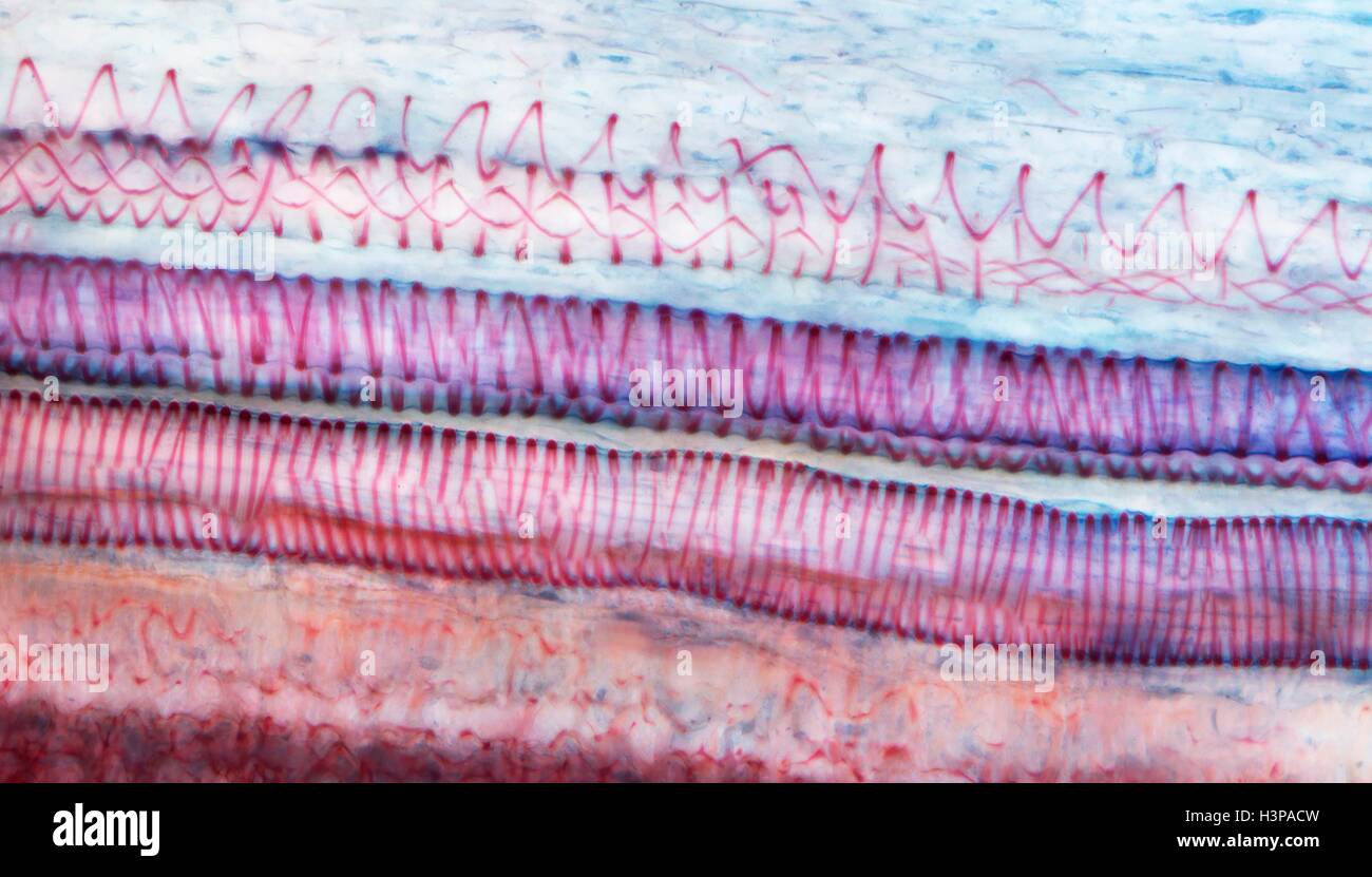 Xylem tissue. Light micrograph (LM) of a section through sunflower(helianthus annuus) tissue showing spiral tracheids, a type of xylem. Tracheids are long tubular cells with lignin, a material that provides support, in the cell walls. Spiral thickening of the cells can be seen. Tracheids conduct water from the roots of a plant along the stems to the leaves. Magnification: x210 when printed 10cm wide. Stock Photohttps://www.alamy.com/image-license-details/?v=1https://www.alamy.com/stock-photo-xylem-tissue-light-micrograph-lm-of-a-section-through-sunflowerhelianthus-122807689.html
RFH3PACW–Xylem tissue. Light micrograph (LM) of a section through sunflower(helianthus annuus) tissue showing spiral tracheids, a type of xylem. Tracheids are long tubular cells with lignin, a material that provides support, in the cell walls. Spiral thickening of the cells can be seen. Tracheids conduct water from the roots of a plant along the stems to the leaves. Magnification: x210 when printed 10cm wide.
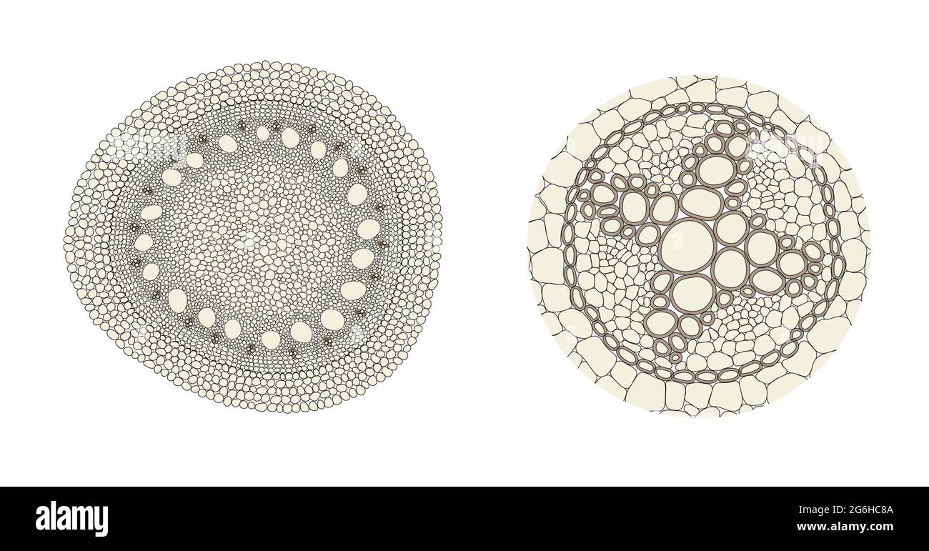 Plant root comparison on monocot (left) and dicot (right) Stock Photohttps://www.alamy.com/image-license-details/?v=1https://www.alamy.com/plant-root-comparison-on-monocot-left-and-dicot-right-image434329962.html
RF2G6HC8A–Plant root comparison on monocot (left) and dicot (right)
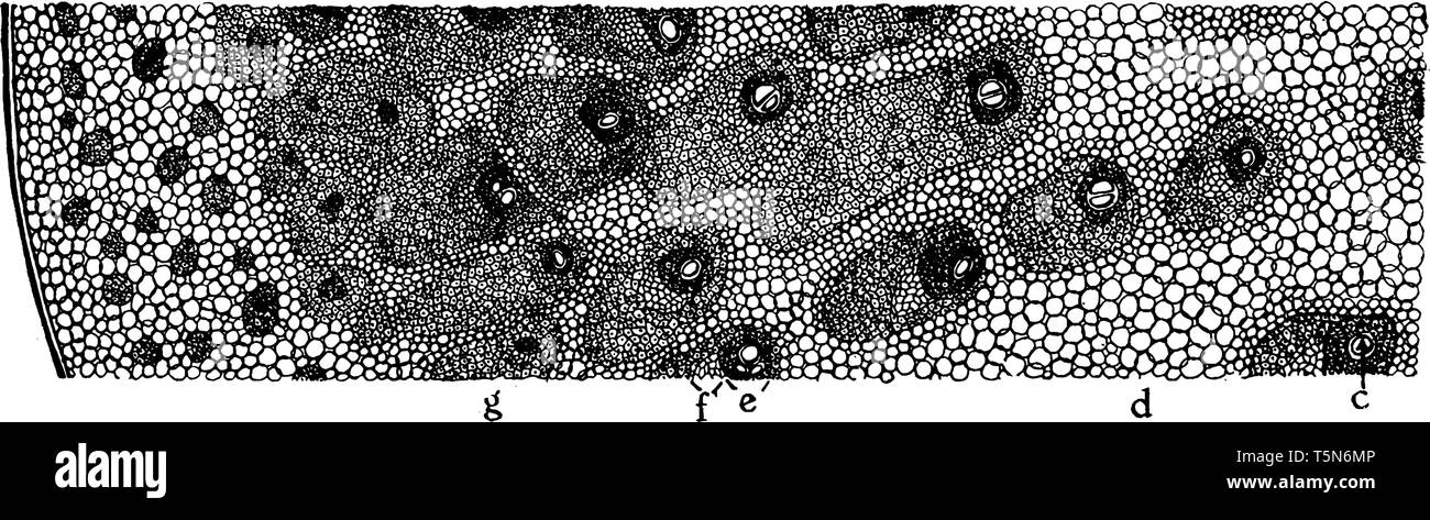 A diagram of Cross section of a portion of palm stem. The xylem tissue is produced on the inside of the cambium layer, vintage line drawing or engravi Stock Vectorhttps://www.alamy.com/image-license-details/?v=1https://www.alamy.com/a-diagram-of-cross-section-of-a-portion-of-palm-stem-the-xylem-tissue-is-produced-on-the-inside-of-the-cambium-layer-vintage-line-drawing-or-engravi-image244484710.html
RFT5N6MP–A diagram of Cross section of a portion of palm stem. The xylem tissue is produced on the inside of the cambium layer, vintage line drawing or engravi
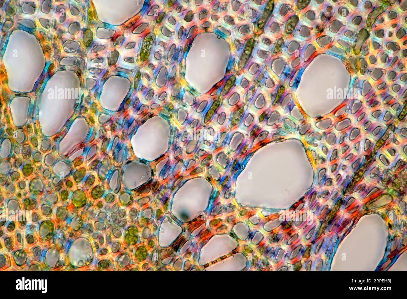 The image presents oak xylem tissue in the transversal cross-section of the stalk, photographed through the microscope in polarized light at a magnifi Stock Photohttps://www.alamy.com/image-license-details/?v=1https://www.alamy.com/the-image-presents-oak-xylem-tissue-in-the-transversal-cross-section-of-the-stalk-photographed-through-the-microscope-in-polarized-light-at-a-magnifi-image564575190.html
RM2RPEHBJ–The image presents oak xylem tissue in the transversal cross-section of the stalk, photographed through the microscope in polarized light at a magnifi
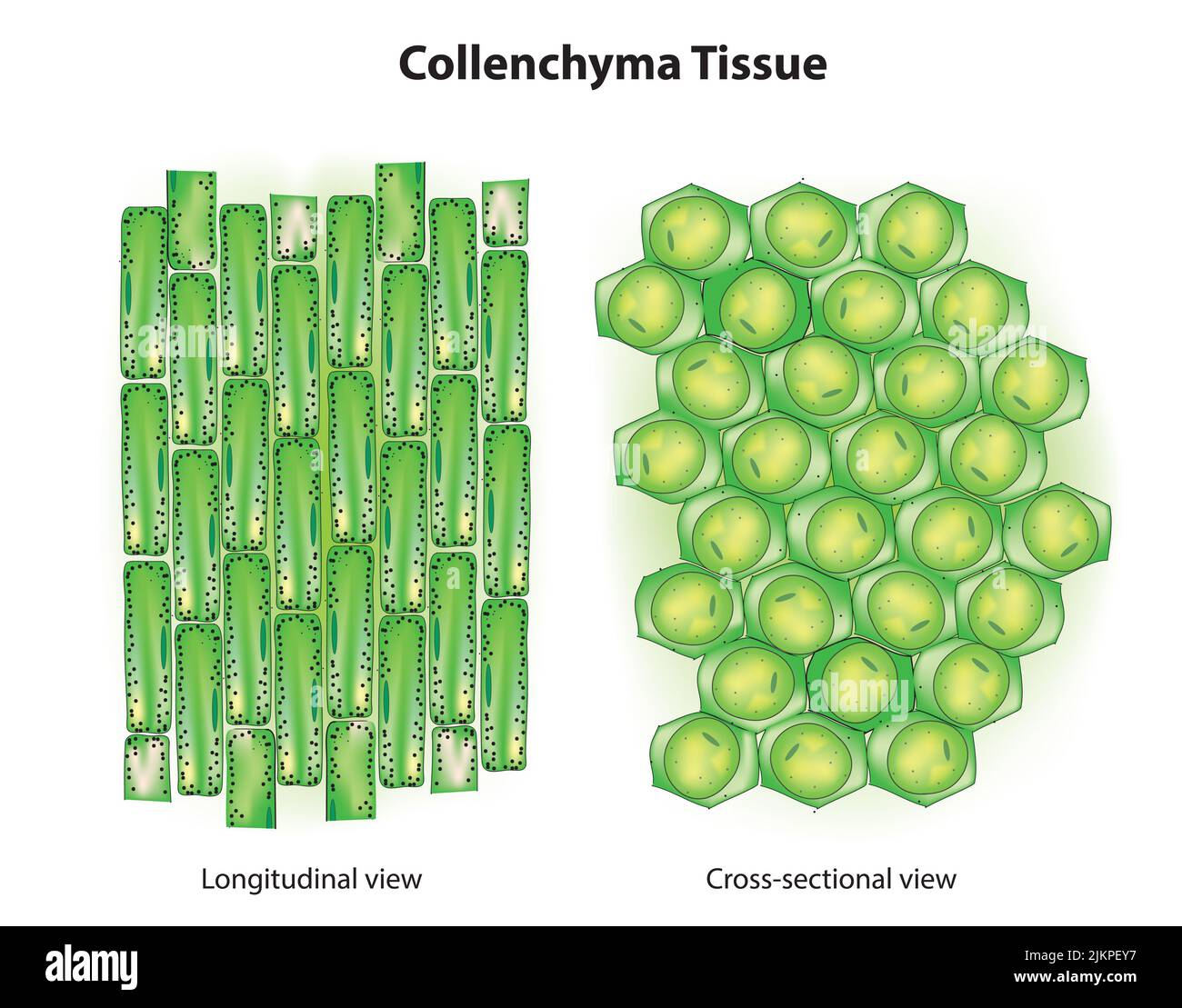 Collenchyma tissue anatomy Stock Photohttps://www.alamy.com/image-license-details/?v=1https://www.alamy.com/collenchyma-tissue-anatomy-image476853083.html
RF2JKPEY7–Collenchyma tissue anatomy
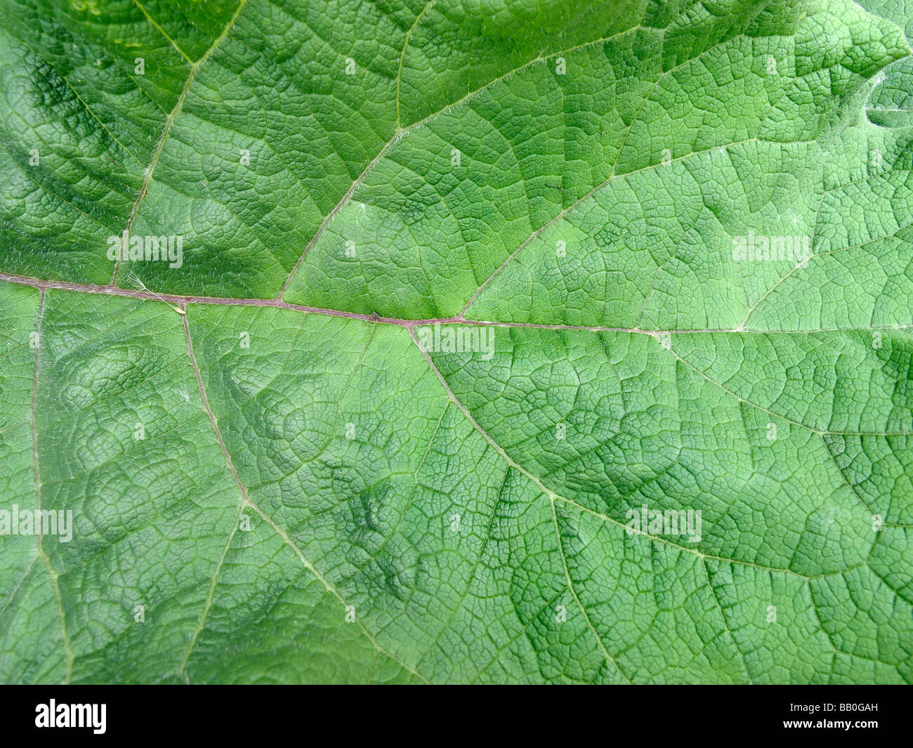 Close-up of leaf in Abbey Fish Ponds Nature Reserve Oxfordshire Stock Photohttps://www.alamy.com/image-license-details/?v=1https://www.alamy.com/stock-photo-close-up-of-leaf-in-abbey-fish-ponds-nature-reserve-oxfordshire-23984425.html
RMBB0GAH–Close-up of leaf in Abbey Fish Ponds Nature Reserve Oxfordshire
 Xylem and phloem water and minerals transportation system outline diagram, Scientific Designing Of Xylem And Phloem Scheme, Nutrient And Mineral Stock Photohttps://www.alamy.com/image-license-details/?v=1https://www.alamy.com/xylem-and-phloem-water-and-minerals-transportation-system-outline-diagram-scientific-designing-of-xylem-and-phloem-scheme-nutrient-and-mineral-image623348742.html
RF2Y63YK2–Xylem and phloem water and minerals transportation system outline diagram, Scientific Designing Of Xylem And Phloem Scheme, Nutrient And Mineral
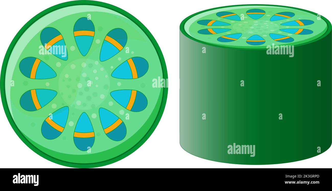 vascular tissue system of plant. xylem and phloem. Cross section of plant stem. Top view and side view. Vector illustration. Stock Vectorhttps://www.alamy.com/image-license-details/?v=1https://www.alamy.com/vascular-tissue-system-of-plant-xylem-and-phloem-cross-section-of-plant-stem-top-view-and-side-view-vector-illustration-image484104165.html
RF2K3GRPD–vascular tissue system of plant. xylem and phloem. Cross section of plant stem. Top view and side view. Vector illustration.
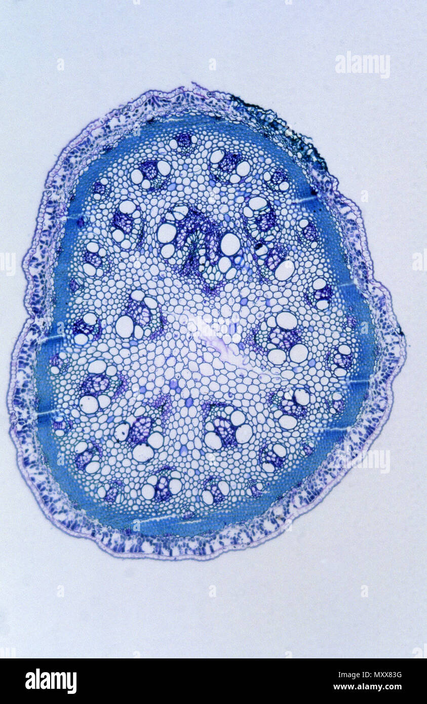 Stoma. Section cross. Asparagus stem. 18x Stock Photohttps://www.alamy.com/image-license-details/?v=1https://www.alamy.com/stoma-section-cross-asparagus-stem-18x-image188661860.html
RFMXX83G–Stoma. Section cross. Asparagus stem. 18x
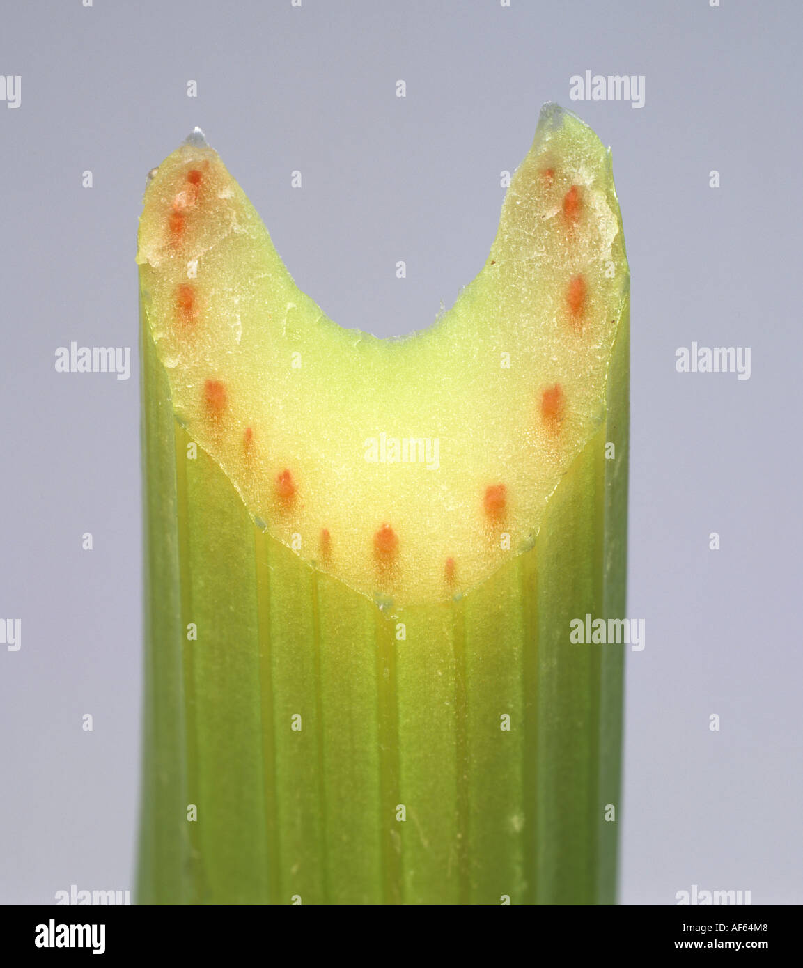 Section through a celery stem to show red dye taken up through the vascular bundles Stock Photohttps://www.alamy.com/image-license-details/?v=1https://www.alamy.com/section-through-a-celery-stem-to-show-red-dye-taken-up-through-the-image7910727.html
RMAF64M8–Section through a celery stem to show red dye taken up through the vascular bundles
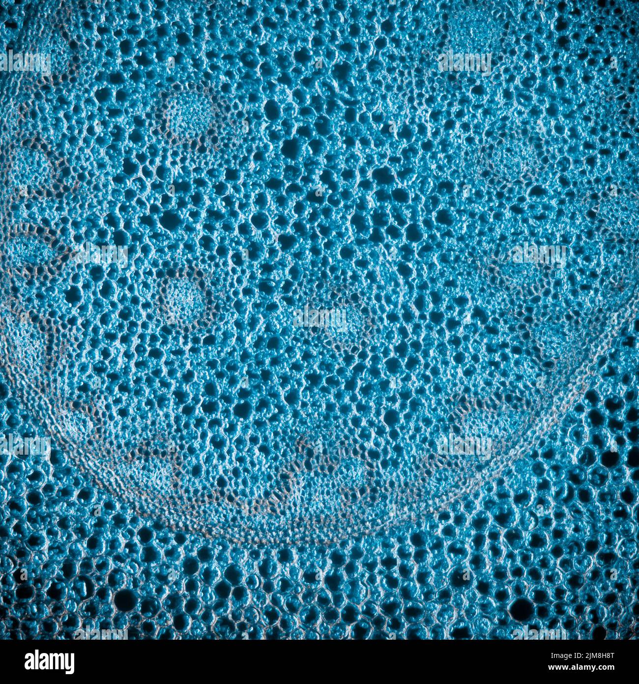 Science micrograph plant root tissue Stock Photohttps://www.alamy.com/image-license-details/?v=1https://www.alamy.com/science-micrograph-plant-root-tissue-image477162248.html
RF2JM8H8T–Science micrograph plant root tissue
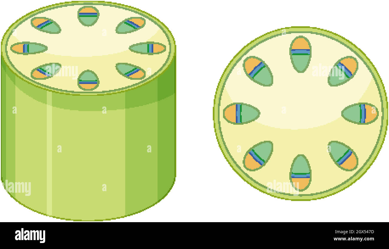 Elements of vascular tissue system in plants Stock Vectorhttps://www.alamy.com/image-license-details/?v=1https://www.alamy.com/elements-of-vascular-tissue-system-in-plants-image446353361.html
RF2GX547D–Elements of vascular tissue system in plants
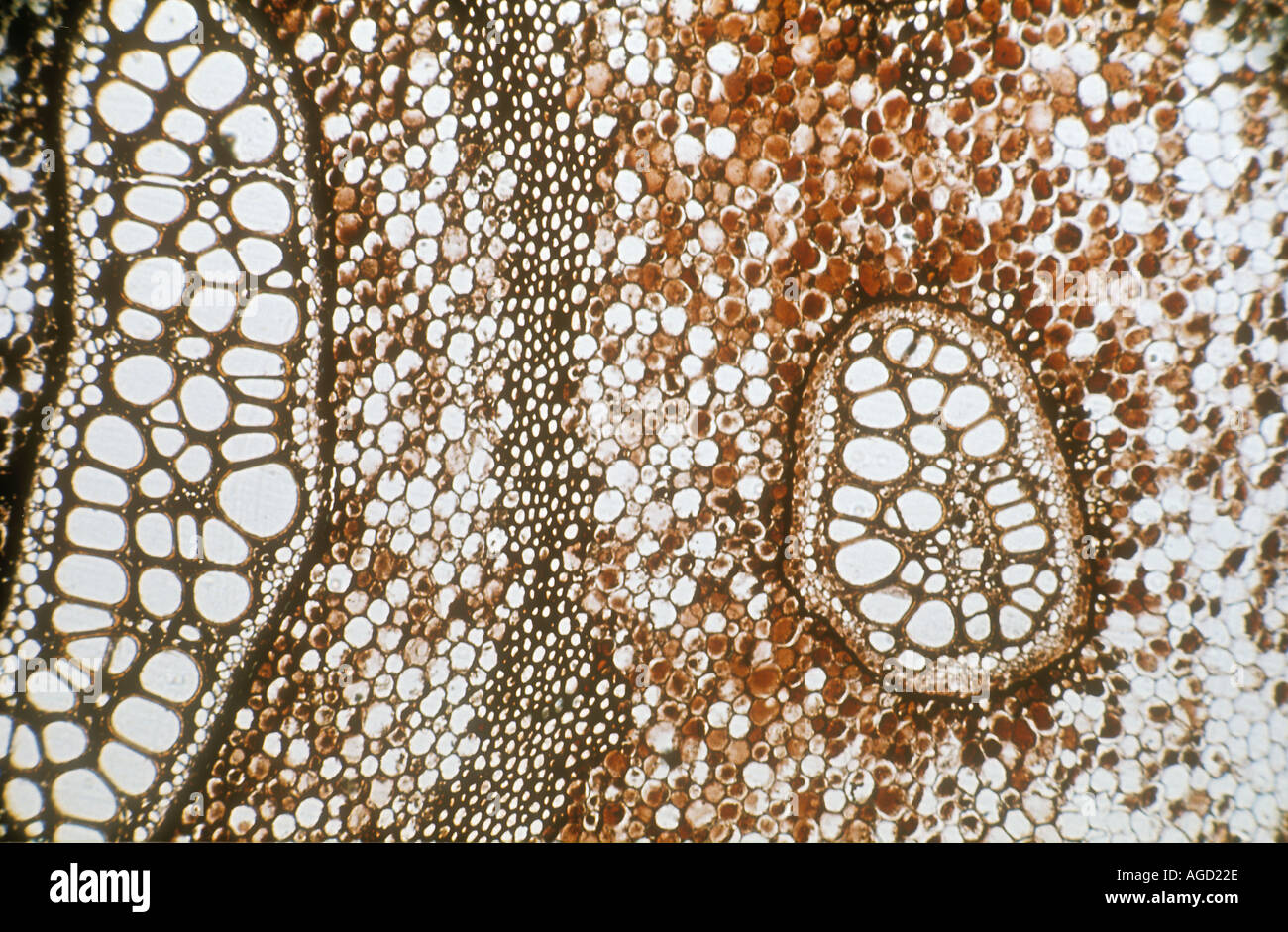 Microscope transverse section through Pteridium rhizome showing xylem cells Stock Photohttps://www.alamy.com/image-license-details/?v=1https://www.alamy.com/microscope-transverse-section-through-pteridium-rhizome-showing-xylem-image1102381.html
RMAGD22E–Microscope transverse section through Pteridium rhizome showing xylem cells
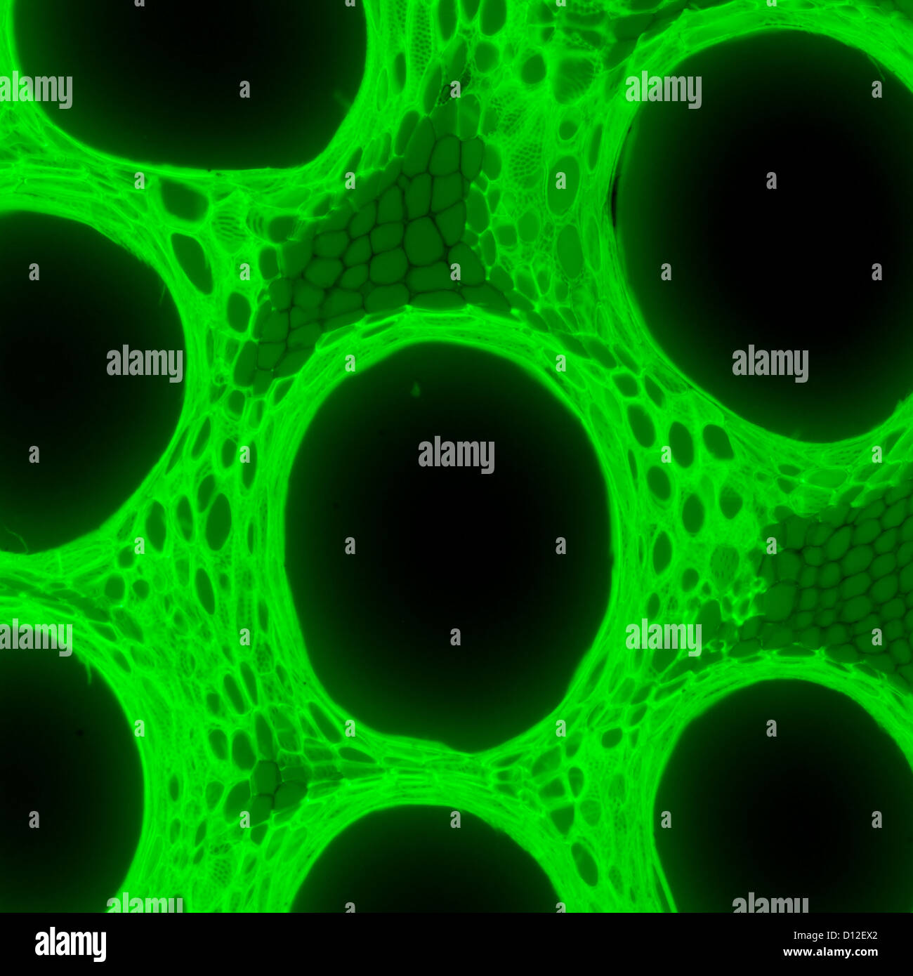 micrograph plant tissue, stem of pumpkin,with green fluorescence Stock Photohttps://www.alamy.com/image-license-details/?v=1https://www.alamy.com/stock-photo-micrograph-plant-tissue-stem-of-pumpkinwith-green-fluorescence-52301370.html
RFD12EX2–micrograph plant tissue, stem of pumpkin,with green fluorescence
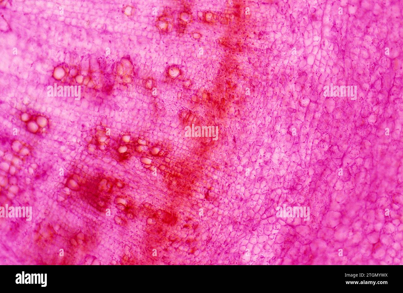 Cambium is a growth tissue of the plants with secondary growth. Is found bettween xylem (left) and phloem (right). Photomicrograph. Stock Photohttps://www.alamy.com/image-license-details/?v=1https://www.alamy.com/cambium-is-a-growth-tissue-of-the-plants-with-secondary-growth-is-found-bettween-xylem-left-and-phloem-right-photomicrograph-image578237574.html
RF2TGMYWX–Cambium is a growth tissue of the plants with secondary growth. Is found bettween xylem (left) and phloem (right). Photomicrograph.
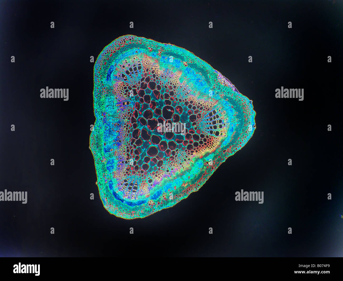 soybean stem cs Stock Photohttps://www.alamy.com/image-license-details/?v=1https://www.alamy.com/stock-photo-soybean-stem-cs-17367597.html
RFB074F9–soybean stem cs
![A leaf is the main appendage of the stem of a vascular plant [1], usually laterally above the ground, and specialized for photosynthesis. Stock Photo A leaf is the main appendage of the stem of a vascular plant [1], usually laterally above the ground, and specialized for photosynthesis. Stock Photo](https://c8.alamy.com/comp/2PHC002/a-leaf-is-the-main-appendage-of-the-stem-of-a-vascular-plant-1-usually-laterally-above-the-ground-and-specialized-for-photosynthesis-2PHC002.jpg) A leaf is the main appendage of the stem of a vascular plant [1], usually laterally above the ground, and specialized for photosynthesis. Stock Photohttps://www.alamy.com/image-license-details/?v=1https://www.alamy.com/a-leaf-is-the-main-appendage-of-the-stem-of-a-vascular-plant-1-usually-laterally-above-the-ground-and-specialized-for-photosynthesis-image544233986.html
A leaf is the main appendage of the stem of a vascular plant [1], usually laterally above the ground, and specialized for photosynthesis. Stock Photohttps://www.alamy.com/image-license-details/?v=1https://www.alamy.com/a-leaf-is-the-main-appendage-of-the-stem-of-a-vascular-plant-1-usually-laterally-above-the-ground-and-specialized-for-photosynthesis-image544233986.htmlRF2PHC002–A leaf is the main appendage of the stem of a vascular plant [1], usually laterally above the ground, and specialized for photosynthesis.
 A close up of the whorls and natural patterning on an oak tree in Calfiornia, USA. Stock Photohttps://www.alamy.com/image-license-details/?v=1https://www.alamy.com/a-close-up-of-the-whorls-and-natural-patterning-on-an-oak-tree-in-calfiornia-usa-image633730915.html
RF2YR0X6Y–A close up of the whorls and natural patterning on an oak tree in Calfiornia, USA.
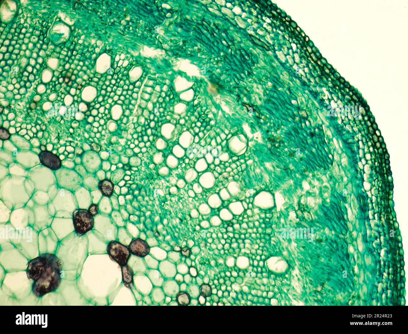 Stem of cotton x.s. details under biological optical misroscope Stock Photohttps://www.alamy.com/image-license-details/?v=1https://www.alamy.com/stem-of-cotton-xs-details-under-biological-optical-misroscope-image552066987.html
RF2R24R23–Stem of cotton x.s. details under biological optical misroscope
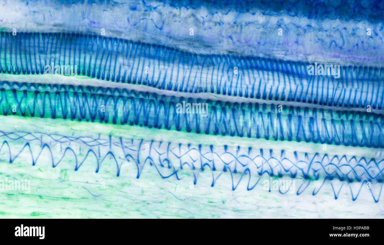 Xylem tissue. Light micrograph (LM) of a section through sunflower(helianthus annuus) tissue showing spiral tracheids, a type of xylem. Tracheids are long tubular cells with lignin, a material that provides support, in the cell walls. Spiral thickening of the cells can be seen. Tracheids conduct water from the roots of a plant along the stems to the leaves. Magnification: x210 when printed 10cm wide. Stock Photohttps://www.alamy.com/image-license-details/?v=1https://www.alamy.com/stock-photo-xylem-tissue-light-micrograph-lm-of-a-section-through-sunflowerhelianthus-122807647.html
RFH3PABB–Xylem tissue. Light micrograph (LM) of a section through sunflower(helianthus annuus) tissue showing spiral tracheids, a type of xylem. Tracheids are long tubular cells with lignin, a material that provides support, in the cell walls. Spiral thickening of the cells can be seen. Tracheids conduct water from the roots of a plant along the stems to the leaves. Magnification: x210 when printed 10cm wide.
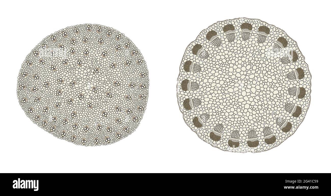 Plant Stem comparison of monocot (left) and dicot (right) Stock Photohttps://www.alamy.com/image-license-details/?v=1https://www.alamy.com/plant-stem-comparison-of-monocot-left-and-dicot-right-image432749333.html
RF2G41C59–Plant Stem comparison of monocot (left) and dicot (right)
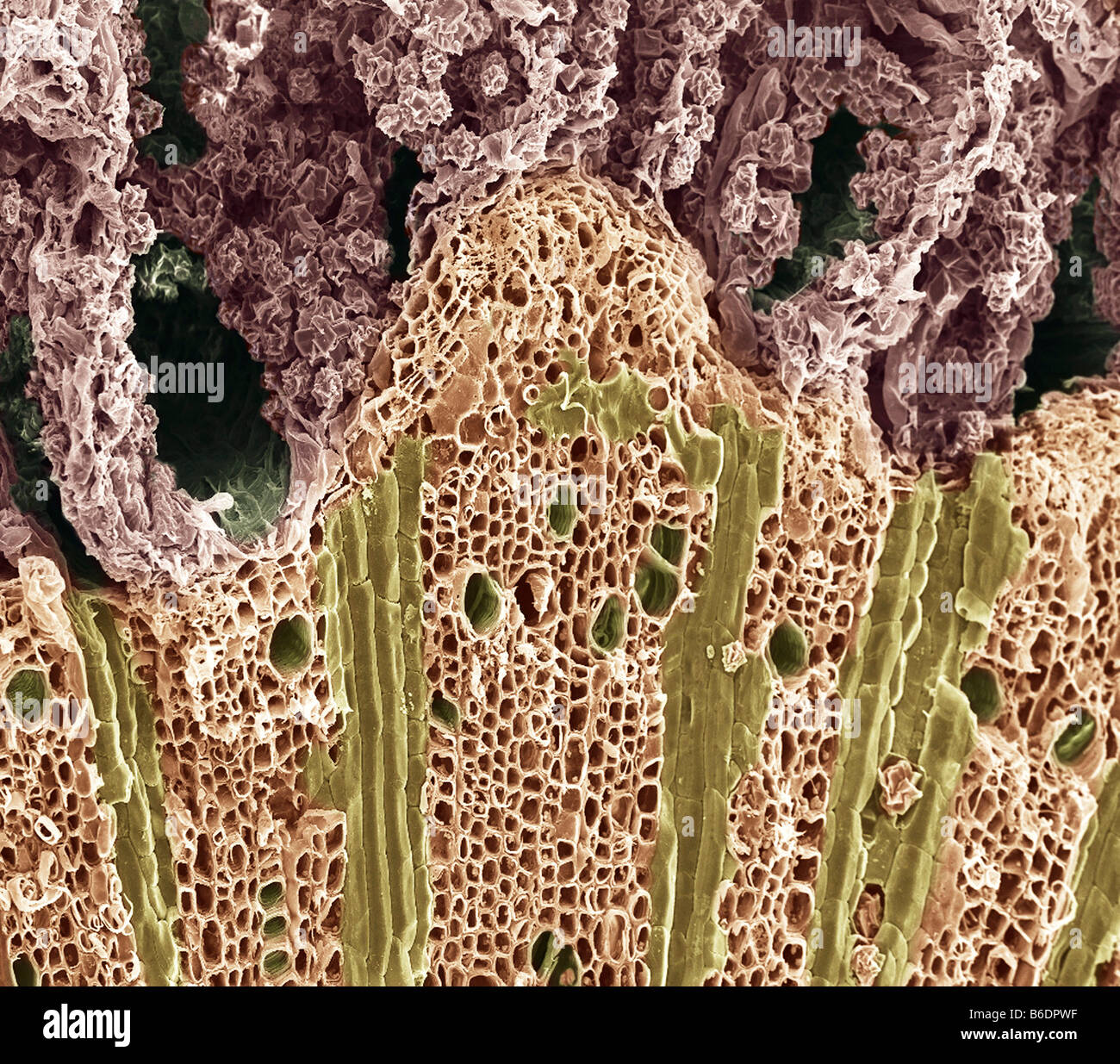 Wood. Coloured scanning electron micrograph (SEM) of wood showing phloem vessels (dark green holes) and xylem tissue (below). Stock Photohttps://www.alamy.com/image-license-details/?v=1https://www.alamy.com/stock-photo-wood-coloured-scanning-electron-micrograph-sem-of-wood-showing-phloem-21201643.html
RFB6DPWF–Wood. Coloured scanning electron micrograph (SEM) of wood showing phloem vessels (dark green holes) and xylem tissue (below).
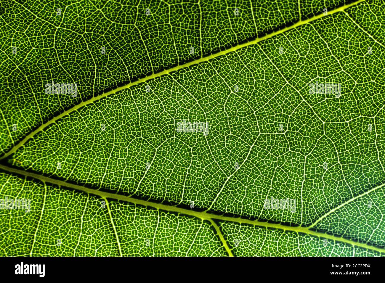 Vascular Tissue in a Leaf Stock Photohttps://www.alamy.com/image-license-details/?v=1https://www.alamy.com/vascular-tissue-in-a-leaf-image368855142.html
RF2CC2PDX–Vascular Tissue in a Leaf
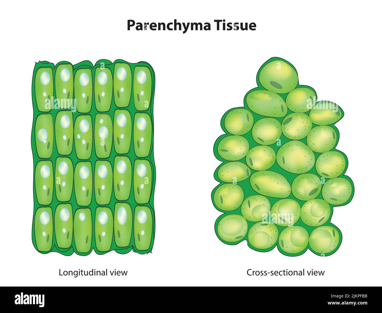 Parenchyma Tissue Stock Photohttps://www.alamy.com/image-license-details/?v=1https://www.alamy.com/parenchyma-tissue-image476853423.html
RF2JKPFBB–Parenchyma Tissue
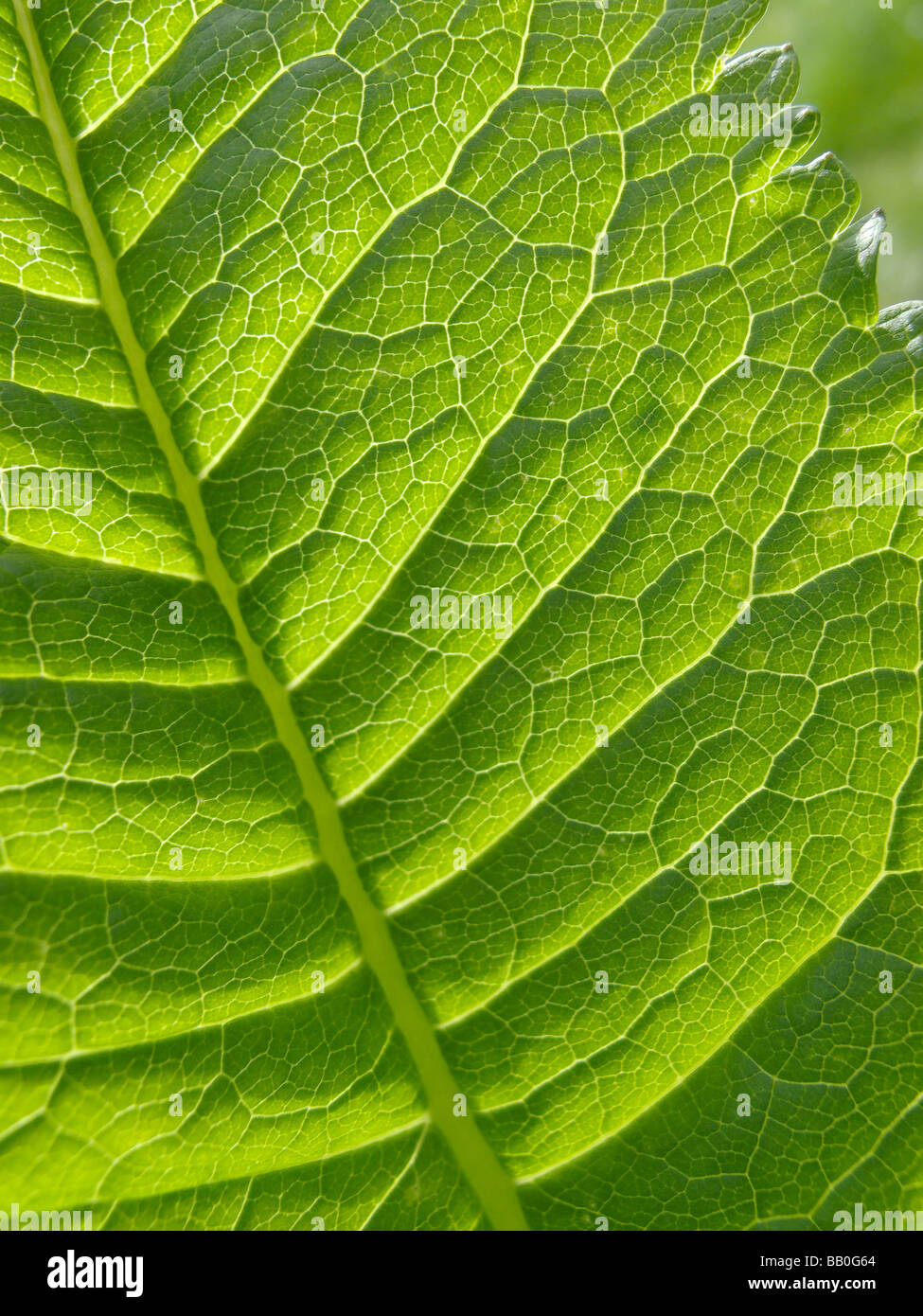 Close-up of leaf 2 in Abbey Fish Ponds Nature Reserve Oxfordshire Stock Photohttps://www.alamy.com/image-license-details/?v=1https://www.alamy.com/stock-photo-close-up-of-leaf-2-in-abbey-fish-ponds-nature-reserve-oxfordshire-23984300.html
RMBB0G64–Close-up of leaf 2 in Abbey Fish Ponds Nature Reserve Oxfordshire
 A Diagram to show the relationship of the food-conducting tissues of the leaf with those of the stem; and in the stem the relationship of these tissue Stock Vectorhttps://www.alamy.com/image-license-details/?v=1https://www.alamy.com/a-diagram-to-show-the-relationship-of-the-food-conducting-tissues-of-the-leaf-with-those-of-the-stem-and-in-the-stem-the-relationship-of-these-tissue-image244626787.html
RFT5YKXY–A Diagram to show the relationship of the food-conducting tissues of the leaf with those of the stem; and in the stem the relationship of these tissue
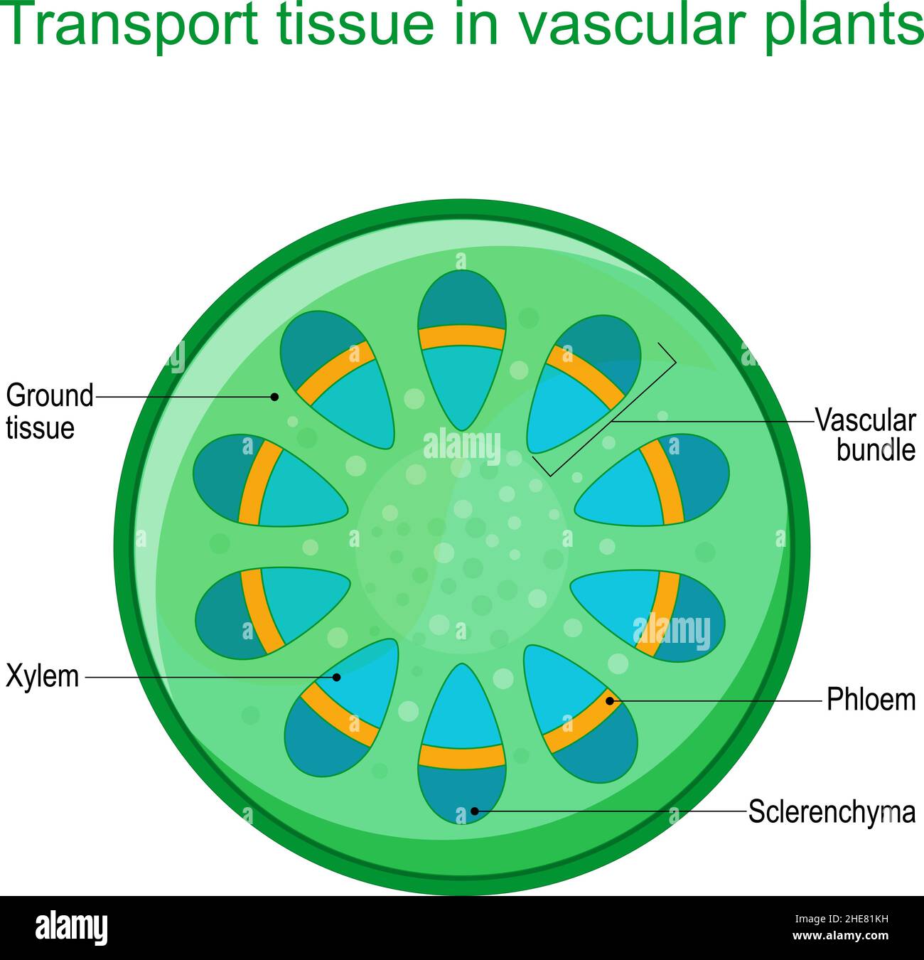 Cross section of vascular tissue system of a plant. Dicot vascular bundles of xylem and phloem are arranged in a ring. Vector diagram for education Stock Vectorhttps://www.alamy.com/image-license-details/?v=1https://www.alamy.com/cross-section-of-vascular-tissue-system-of-a-plant-dicot-vascular-bundles-of-xylem-and-phloem-are-arranged-in-a-ring-vector-diagram-for-education-image456251701.html
RF2HE81KH–Cross section of vascular tissue system of a plant. Dicot vascular bundles of xylem and phloem are arranged in a ring. Vector diagram for education
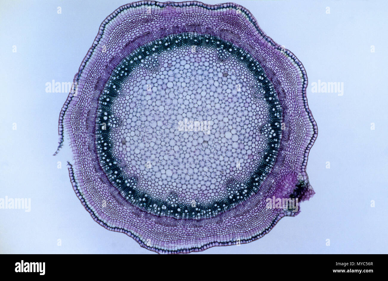 Dicotyledon stem Stock Photohttps://www.alamy.com/image-license-details/?v=1https://www.alamy.com/dicotyledon-stem-image188966927.html
RFMYC56R–Dicotyledon stem
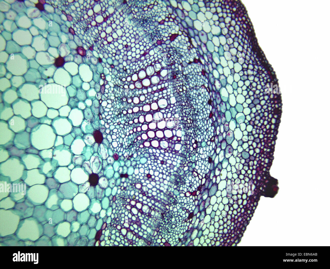 cotton (Gossypium spec.), cross section of a stem of a cotton plant, 100 x, detail of xylem, phloem and cambium Stock Photohttps://www.alamy.com/image-license-details/?v=1https://www.alamy.com/stock-photo-cotton-gossypium-spec-cross-section-of-a-stem-of-a-cotton-plant-100-76068675.html
RMEBN6AB–cotton (Gossypium spec.), cross section of a stem of a cotton plant, 100 x, detail of xylem, phloem and cambium
 science micrograph plant root tissue Stock Photohttps://www.alamy.com/image-license-details/?v=1https://www.alamy.com/stock-photo-science-micrograph-plant-root-tissue-50693185.html
RFCXD7JW–science micrograph plant root tissue
 Elements of vascular tissue system in plants Stock Vectorhttps://www.alamy.com/image-license-details/?v=1https://www.alamy.com/elements-of-vascular-tissue-system-in-plants-image446055332.html
RF2GWKG3G–Elements of vascular tissue system in plants
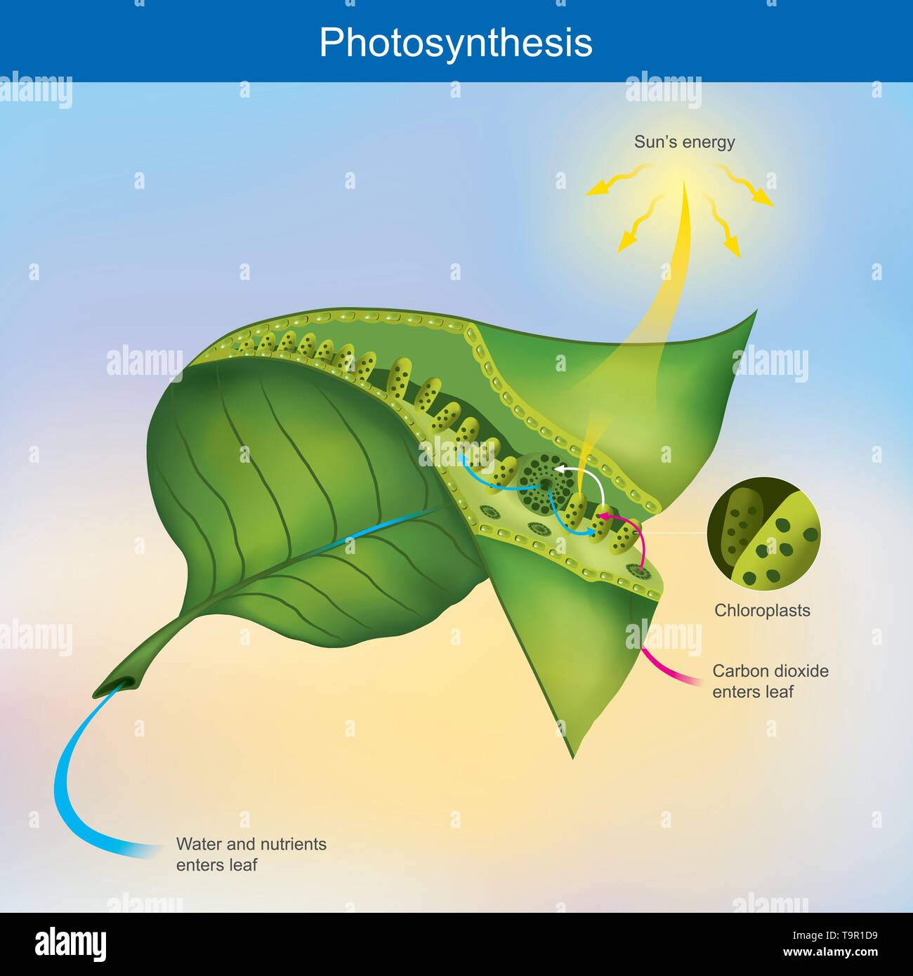 Photosynthesis is a process by plants and other organisms use to convert light energy into chemical energy. Stock Vectorhttps://www.alamy.com/image-license-details/?v=1https://www.alamy.com/photosynthesis-is-a-process-by-plants-and-other-organisms-use-to-convert-light-energy-into-chemical-energy-image246983109.html
RFT9R1D9–Photosynthesis is a process by plants and other organisms use to convert light energy into chemical energy.
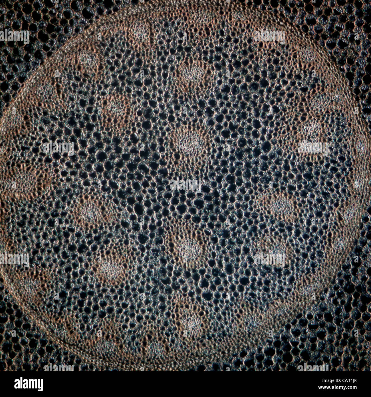 science micrograph plant root tissue background Stock Photohttps://www.alamy.com/image-license-details/?v=1https://www.alamy.com/stock-photo-science-micrograph-plant-root-tissue-background-50315295.html
RFCWT1JR–science micrograph plant root tissue background
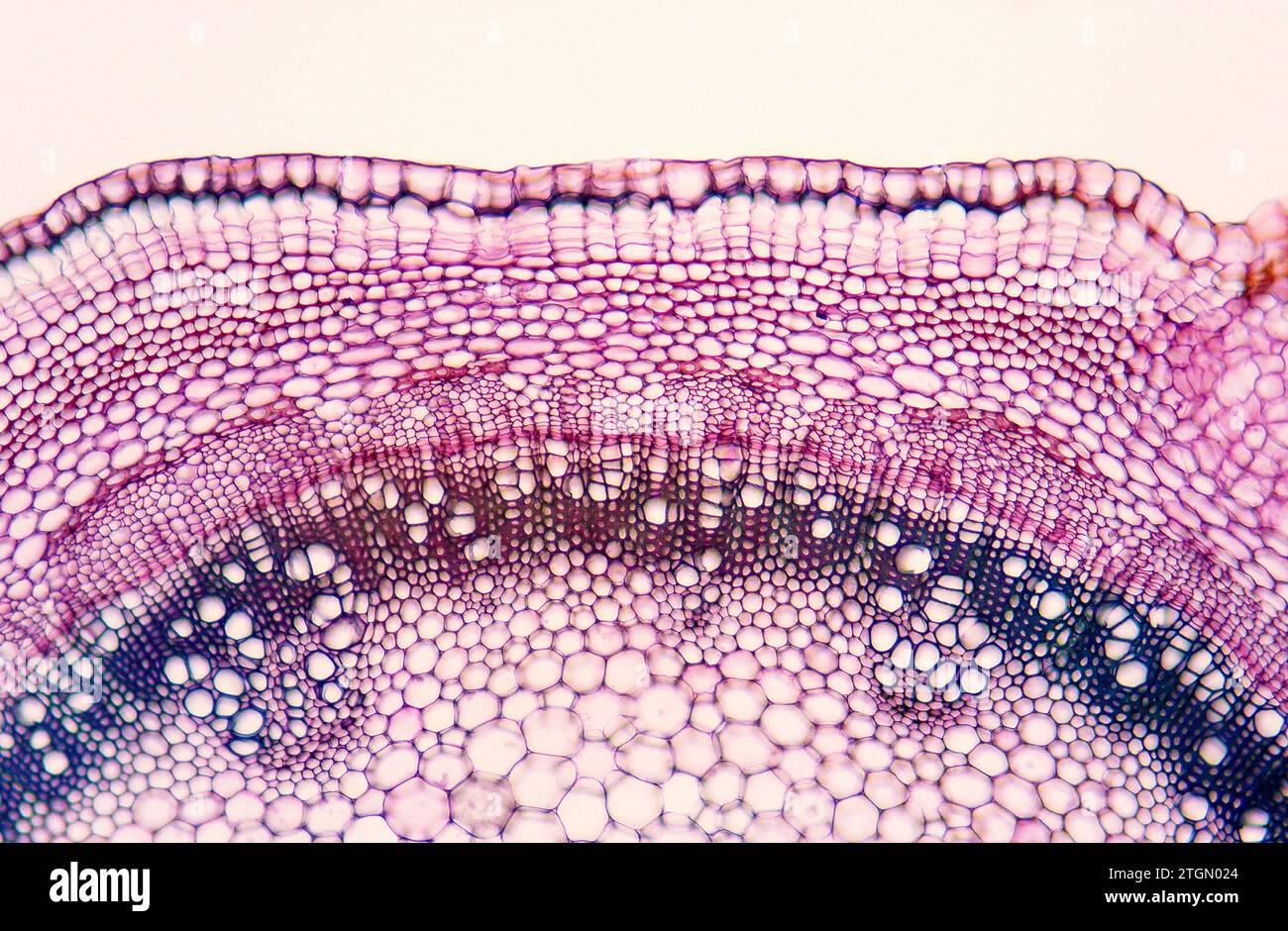 Cambium is a growth tissue of the plants with secondary growth. Is found bettween xylem (above) and phloem (below). Photomicrograph. Stock Photohttps://www.alamy.com/image-license-details/?v=1https://www.alamy.com/cambium-is-a-growth-tissue-of-the-plants-with-secondary-growth-is-found-bettween-xylem-above-and-phloem-below-photomicrograph-image578237692.html
RF2TGN024–Cambium is a growth tissue of the plants with secondary growth. Is found bettween xylem (above) and phloem (below). Photomicrograph.
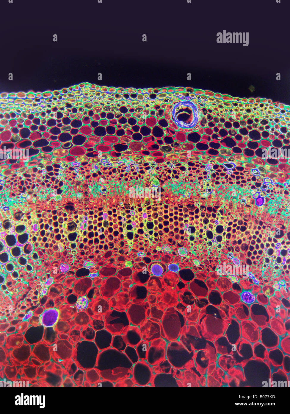 Microscopic HP cotton stem Gossypium sp Stock Photohttps://www.alamy.com/image-license-details/?v=1https://www.alamy.com/stock-photo-microscopic-hp-cotton-stem-gossypium-sp-17366929.html
RFB073KD–Microscopic HP cotton stem Gossypium sp
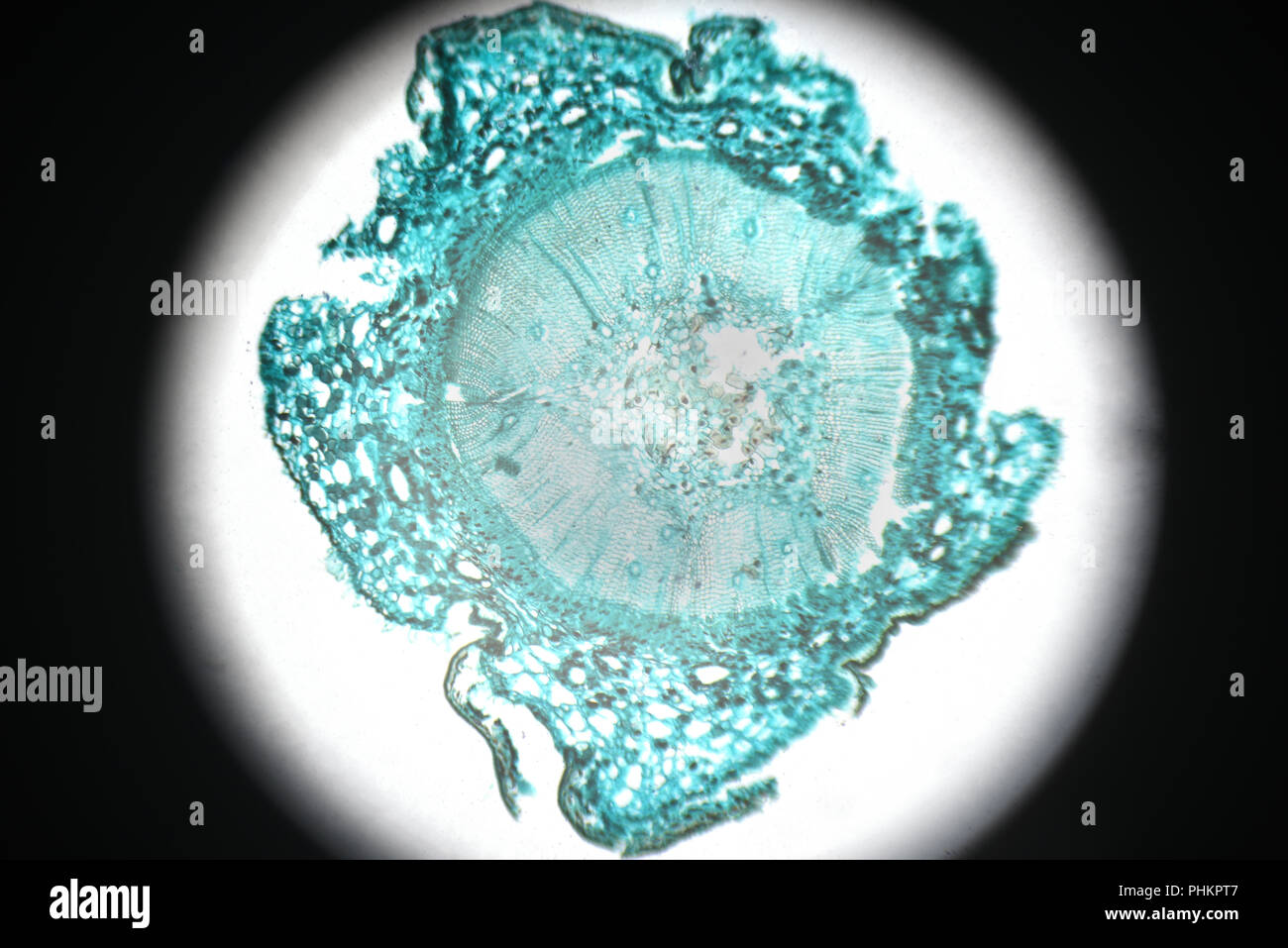 microscopy Pine Stem cross section Stock Photohttps://www.alamy.com/image-license-details/?v=1https://www.alamy.com/microscopy-pine-stem-cross-section-image217408583.html
RFPHKPT7–microscopy Pine Stem cross section
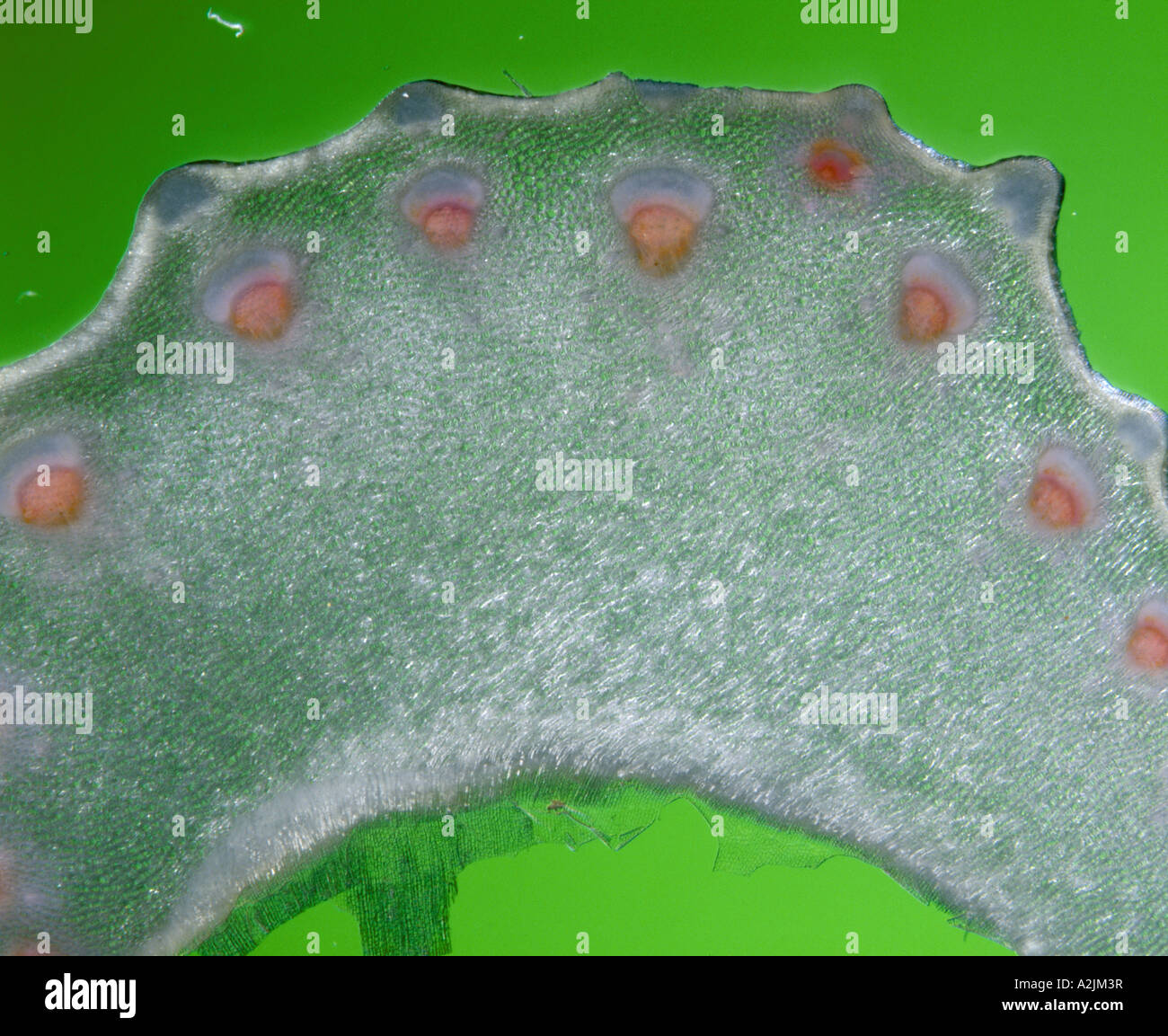 FIBROVASCULAR BUNDLES. CONDUCTIVE TISSUE -- XYLEM AND PHLOEM. CELERY PLACED IN BLUE FOOD COLORING DYE IN WATER Stock Photohttps://www.alamy.com/image-license-details/?v=1https://www.alamy.com/stock-photo-fibrovascular-bundles-conductive-tissue-xylem-and-phloem-celery-placed-10537450.html
RMA2JM3R–FIBROVASCULAR BUNDLES. CONDUCTIVE TISSUE -- XYLEM AND PHLOEM. CELERY PLACED IN BLUE FOOD COLORING DYE IN WATER
 Still life with golden leaf, acorn cup, and detail of fallen tree showing intricate tissue below its bark. Stock Photohttps://www.alamy.com/image-license-details/?v=1https://www.alamy.com/still-life-with-golden-leaf-acorn-cup-and-detail-of-fallen-tree-showing-intricate-tissue-below-its-bark-image243501074.html
RMT44C2X–Still life with golden leaf, acorn cup, and detail of fallen tree showing intricate tissue below its bark.
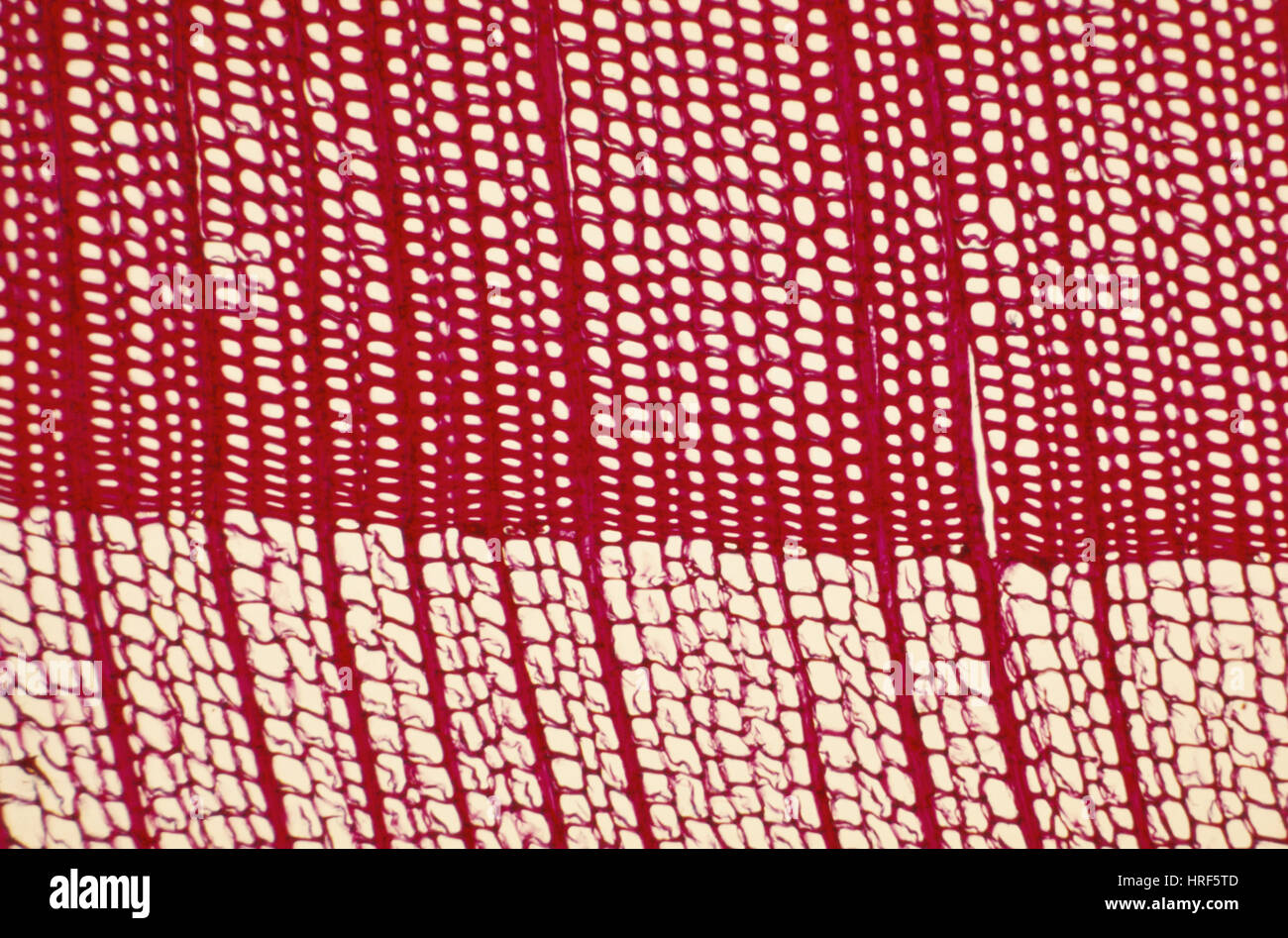 Growth Rings Stock Photohttps://www.alamy.com/image-license-details/?v=1https://www.alamy.com/stock-photo-growth-rings-134943549.html
RMHRF5TD–Growth Rings
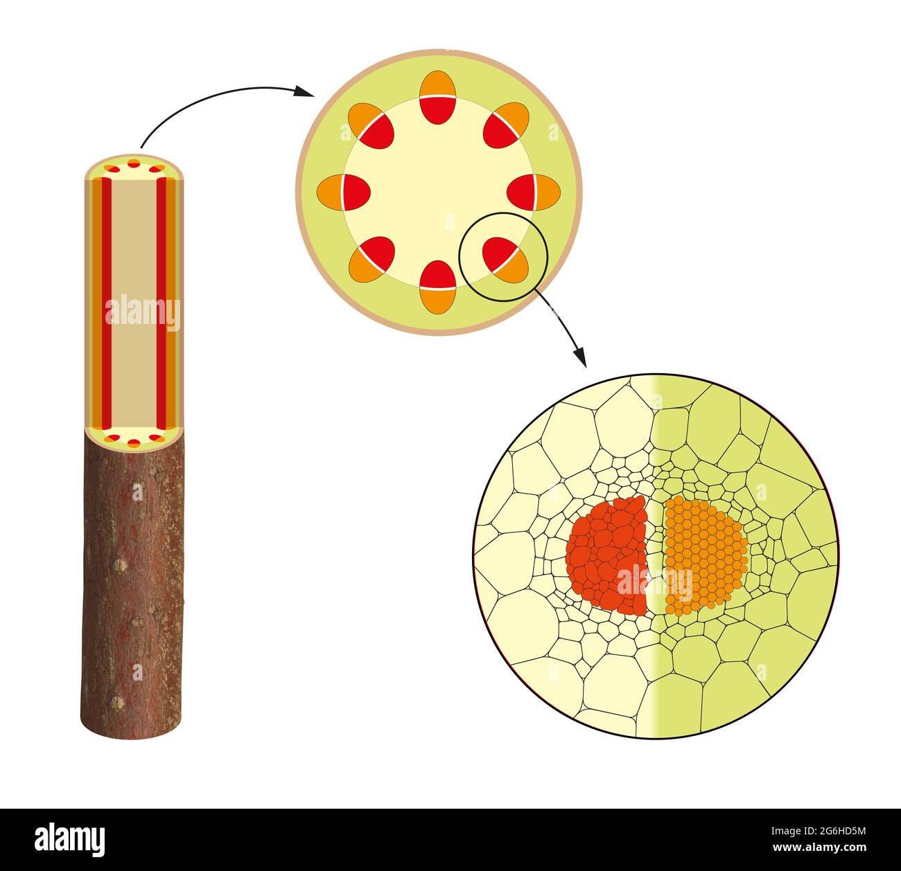 Cross section cut of plant stem. Xylem, phloem Stock Photohttps://www.alamy.com/image-license-details/?v=1https://www.alamy.com/cross-section-cut-of-plant-stem-xylem-phloem-image434330672.html
RF2G6HD5M–Cross section cut of plant stem. Xylem, phloem
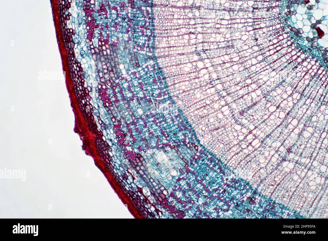 Plant stem, light micrograph Stock Photohttps://www.alamy.com/image-license-details/?v=1https://www.alamy.com/plant-stem-light-micrograph-image456891326.html
RF2HF95FA–Plant stem, light micrograph
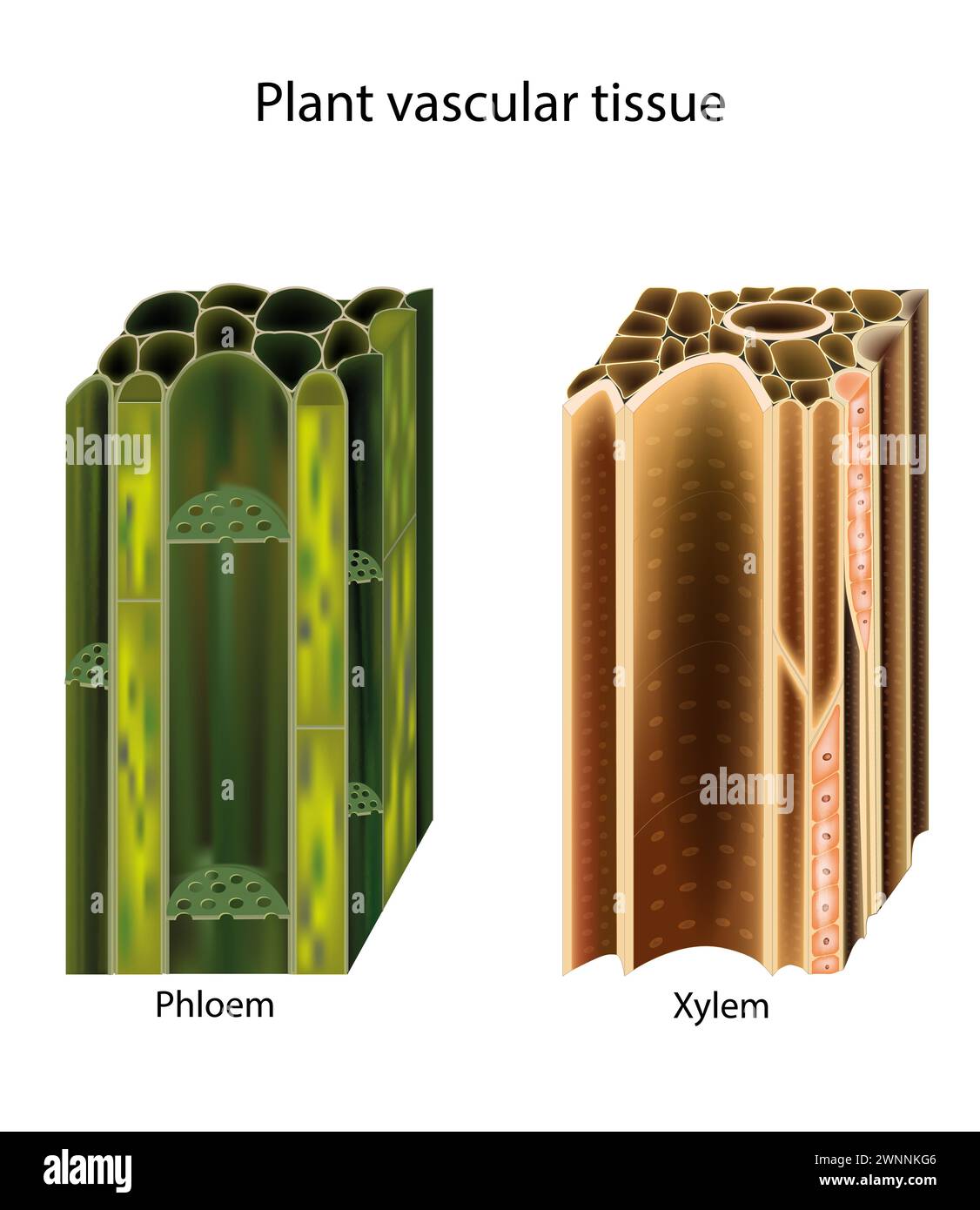 Plant vascular tissue. Xylem and phloem. Cross section showing vascular bundles. Translocation Stock Vectorhttps://www.alamy.com/image-license-details/?v=1https://www.alamy.com/plant-vascular-tissue-xylem-and-phloem-cross-section-showing-vascular-bundles-translocation-image598536630.html
RF2WNNKG6–Plant vascular tissue. Xylem and phloem. Cross section showing vascular bundles. Translocation
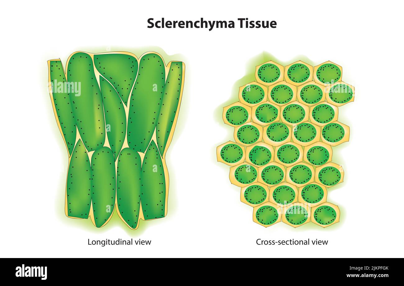 Sclerenchyma tissue Stock Photohttps://www.alamy.com/image-license-details/?v=1https://www.alamy.com/sclerenchyma-tissue-image476853571.html
RF2JKPFGK–Sclerenchyma tissue
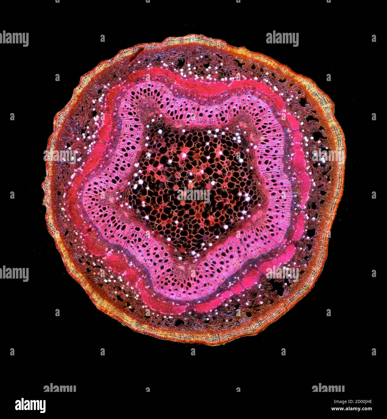 Poplar tree twigs. Populus sp. stem sections TS. Stained sections Stock Photohttps://www.alamy.com/image-license-details/?v=1https://www.alamy.com/poplar-tree-twigs-populus-sp-stem-sections-ts-stained-sections-image378642698.html
RM2D00JHE–Poplar tree twigs. Populus sp. stem sections TS. Stained sections
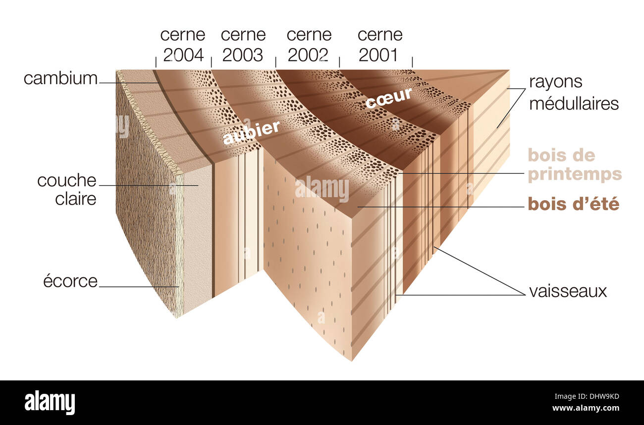 TREE TRUNK, ILLUSTRATION Stock Photohttps://www.alamy.com/image-license-details/?v=1https://www.alamy.com/tree-trunk-illustration-image62636657.html
RMDHW9KD–TREE TRUNK, ILLUSTRATION
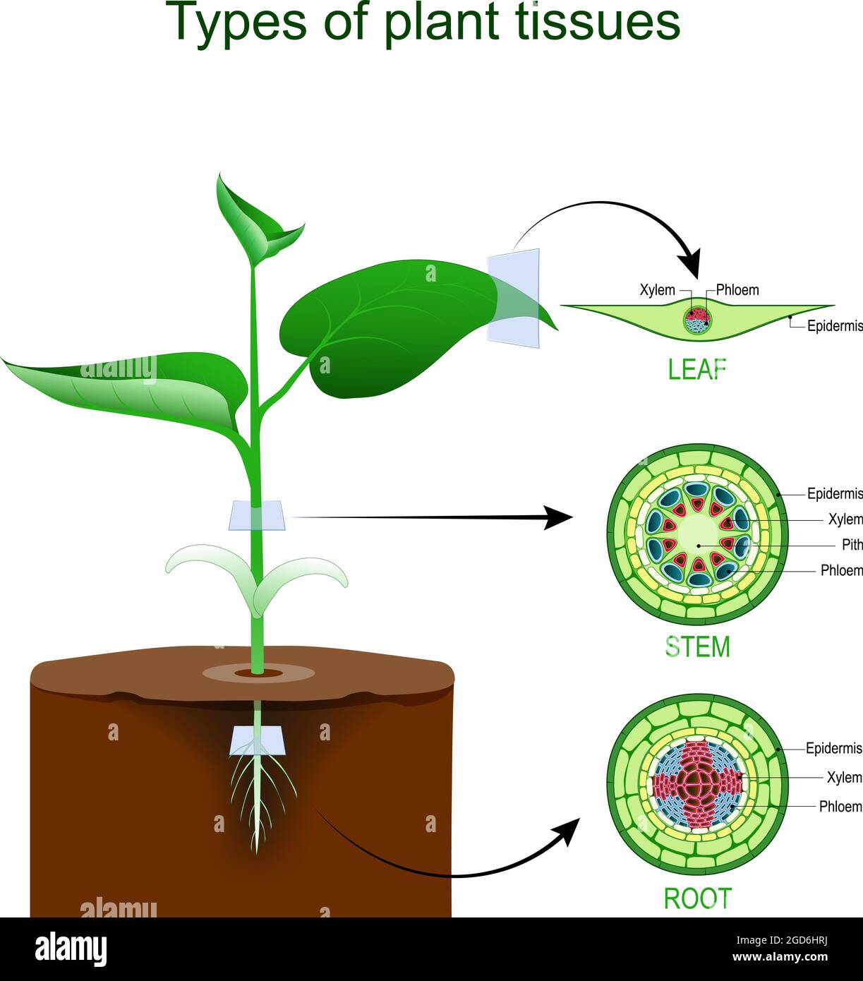 Types of plant tissues. Anatomy of a Plant Body. Cross section of Root, stem and leaf of a green plant. Vector illustration. Poster for education Stock Vectorhttps://www.alamy.com/image-license-details/?v=1https://www.alamy.com/types-of-plant-tissues-anatomy-of-a-plant-body-cross-section-of-root-stem-and-leaf-of-a-green-plant-vector-illustration-poster-for-education-image438395430.html
RF2GD6HRJ–Types of plant tissues. Anatomy of a Plant Body. Cross section of Root, stem and leaf of a green plant. Vector illustration. Poster for education
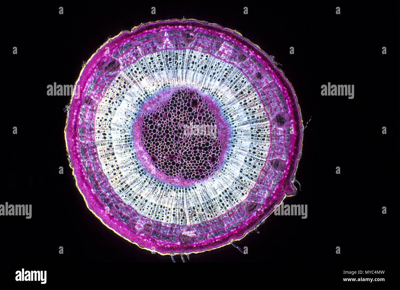 Xylem Stock Photohttps://www.alamy.com/image-license-details/?v=1https://www.alamy.com/xylem-image188966537.html
RFMYC4MW–Xylem
 Types of plant tissue vector illustration. Labeled educational structure scheme. Biological closeup with cross section and longitudinal views. Parenchyma, collenchyma and sclerenchyma description info Stock Vectorhttps://www.alamy.com/image-license-details/?v=1https://www.alamy.com/types-of-plant-tissue-vector-illustration-labeled-educational-structure-scheme-biological-closeup-with-cross-section-and-longitudinal-views-parenchyma-collenchyma-and-sclerenchyma-description-info-image608640739.html
RF2XA5YD7–Types of plant tissue vector illustration. Labeled educational structure scheme. Biological closeup with cross section and longitudinal views. Parenchyma, collenchyma and sclerenchyma description info
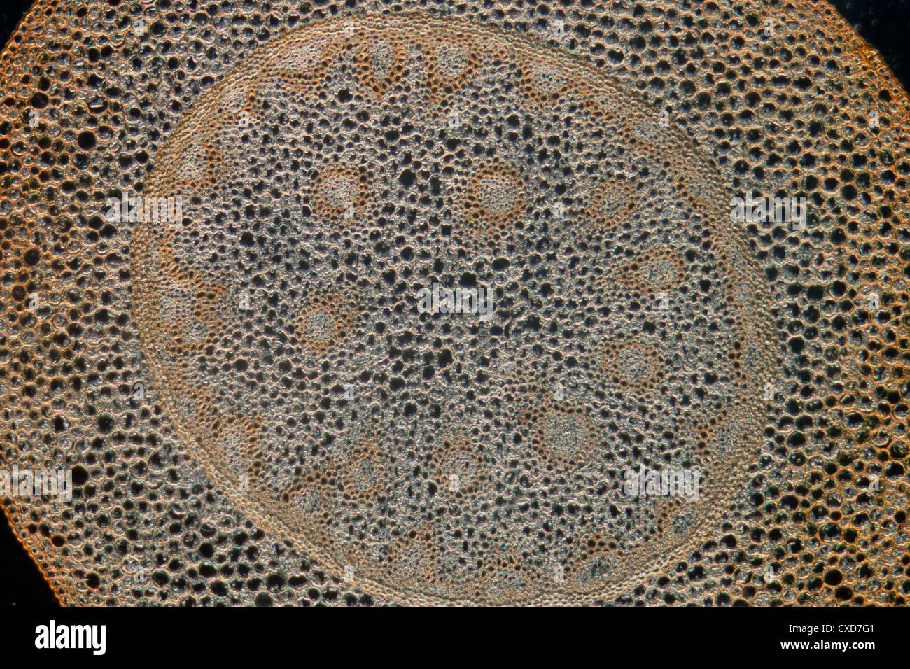 science micrograph plant root tissue Stock Photohttps://www.alamy.com/image-license-details/?v=1https://www.alamy.com/stock-photo-science-micrograph-plant-root-tissue-50693105.html
RFCXD7G1–science micrograph plant root tissue
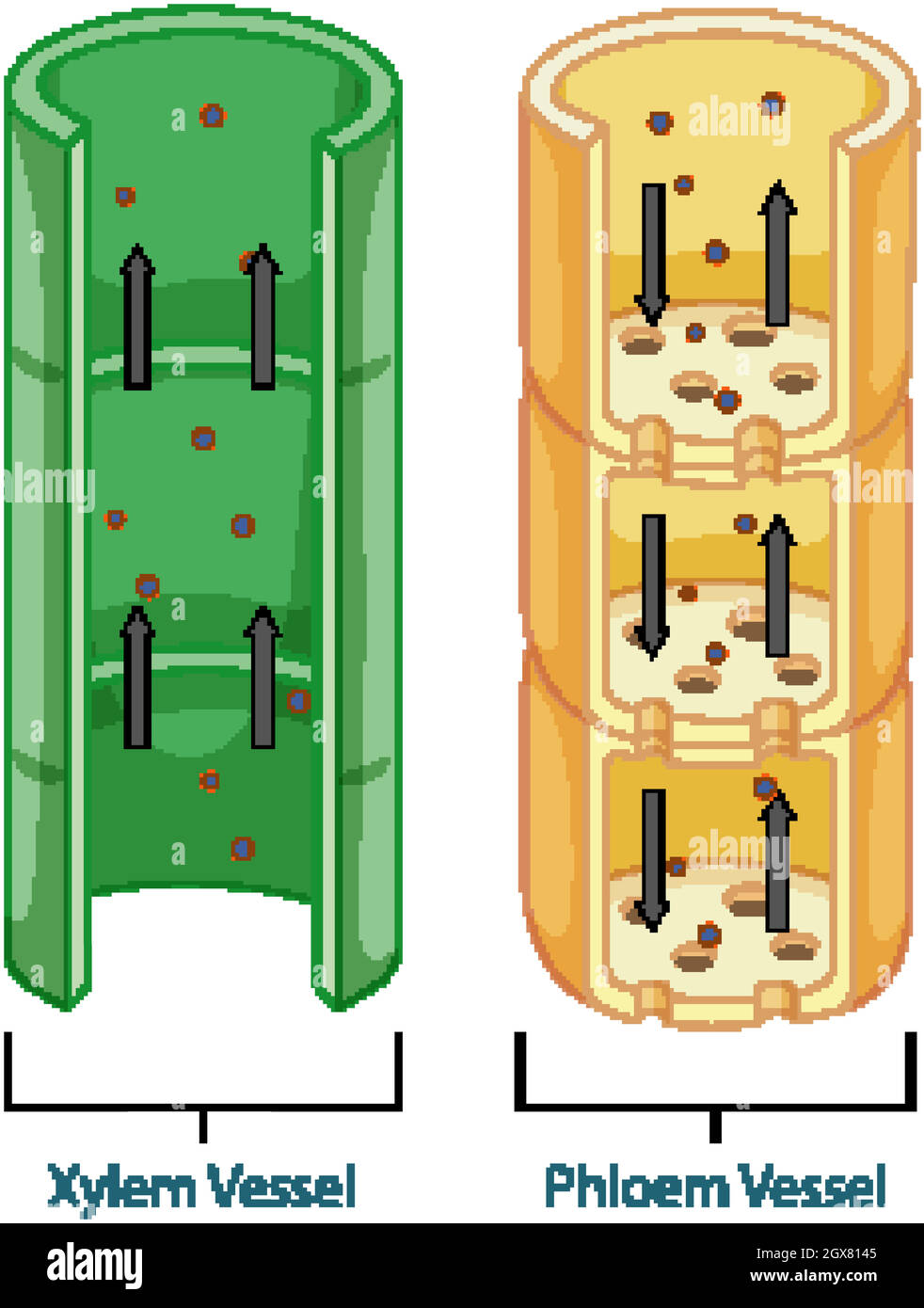 Diagram showing vascular tissue system in plants Stock Vectorhttps://www.alamy.com/image-license-details/?v=1https://www.alamy.com/diagram-showing-vascular-tissue-system-in-plants-image446416773.html
RF2GX8145–Diagram showing vascular tissue system in plants
 Close up view of a cell wall - biology background Stock Photohttps://www.alamy.com/image-license-details/?v=1https://www.alamy.com/stock-photo-close-up-view-of-a-cell-wall-biology-background-74511123.html
RFE967KF–Close up view of a cell wall - biology background
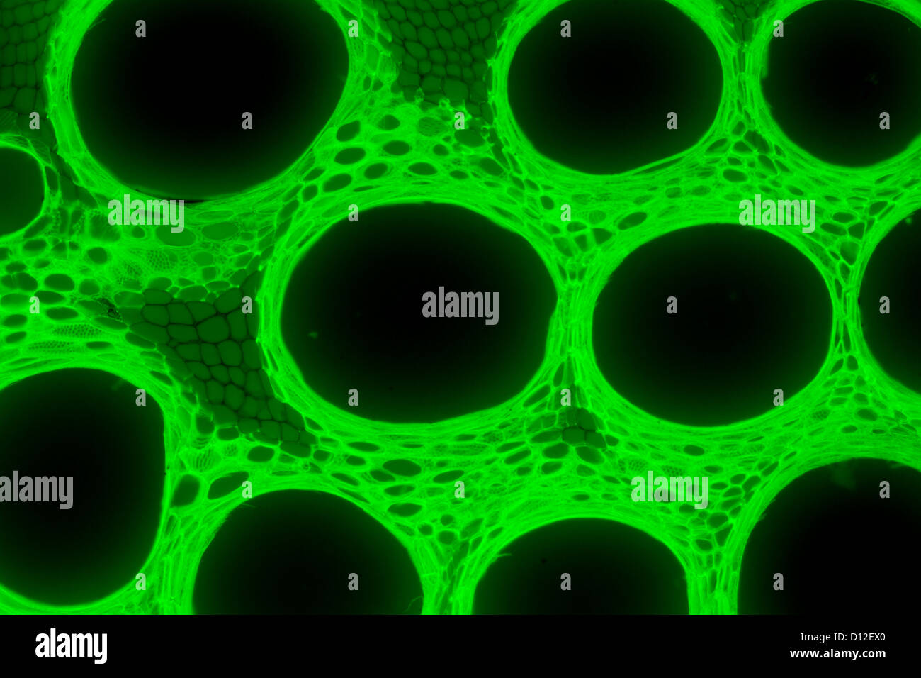 micrograph plant tissue, stem of pumpkin,with green fluorescence Stock Photohttps://www.alamy.com/image-license-details/?v=1https://www.alamy.com/stock-photo-micrograph-plant-tissue-stem-of-pumpkinwith-green-fluorescence-52301368.html
RFD12EX0–micrograph plant tissue, stem of pumpkin,with green fluorescence
 White mulberry stem with spiral xylem (red up), phloem (red down) and abscission tissue (red right). Photomicrograph X 50 at 10 cm wide. Stock Photohttps://www.alamy.com/image-license-details/?v=1https://www.alamy.com/white-mulberry-stem-with-spiral-xylem-red-up-phloem-red-down-and-abscission-tissue-red-right-photomicrograph-x-50-at-10-cm-wide-image588791267.html
RF2W5WN7F–White mulberry stem with spiral xylem (red up), phloem (red down) and abscission tissue (red right). Photomicrograph X 50 at 10 cm wide.
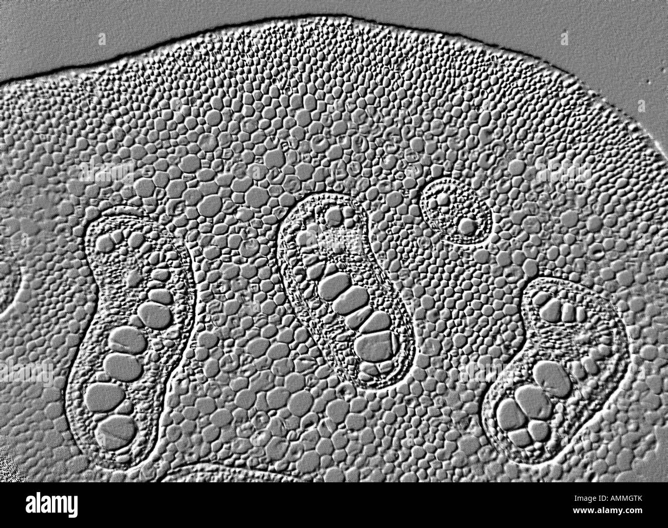 fern stem Stock Photohttps://www.alamy.com/image-license-details/?v=1https://www.alamy.com/stock-photo-fern-stem-15296802.html
RFAMMGTK–fern stem
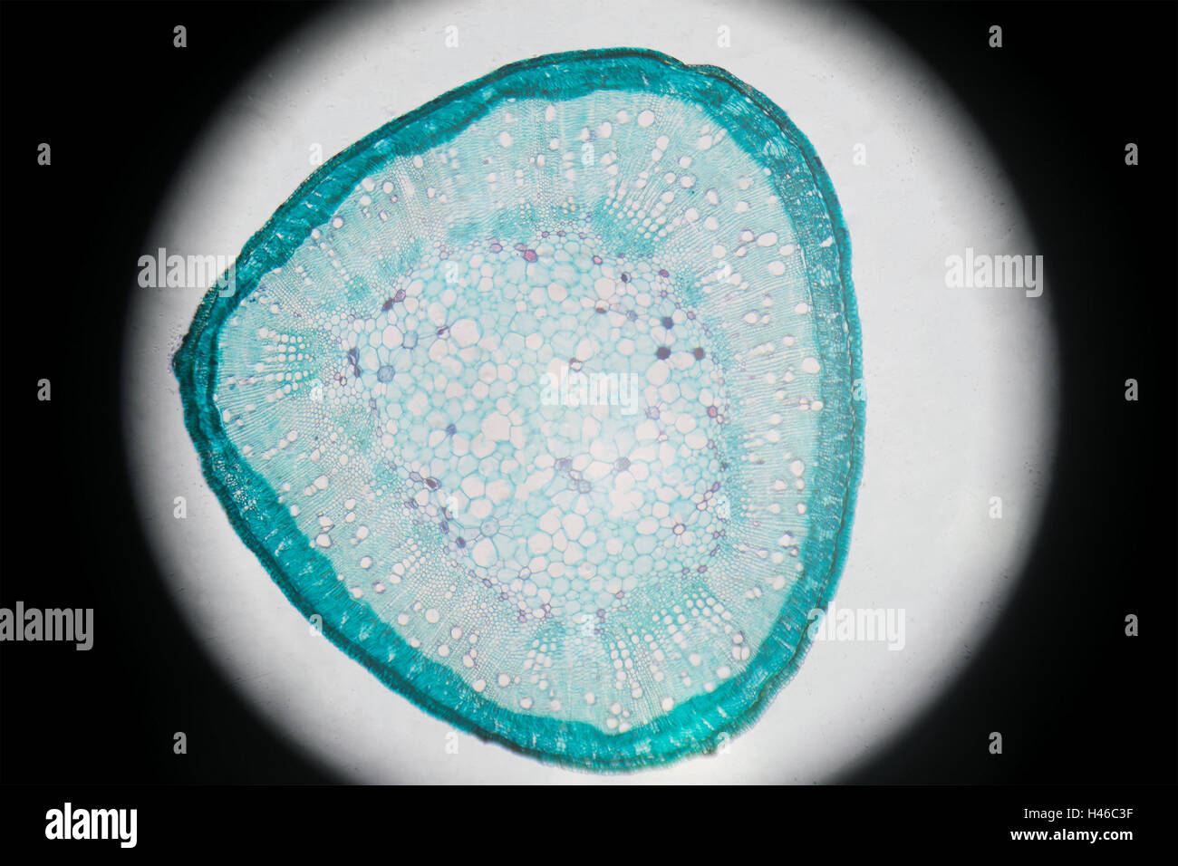 Microscopic photography of Steam of Cotton, cross section. Stock Photohttps://www.alamy.com/image-license-details/?v=1https://www.alamy.com/stock-photo-microscopic-photography-of-steam-of-cotton-cross-section-123072419.html
RFH46C3F–Microscopic photography of Steam of Cotton, cross section.
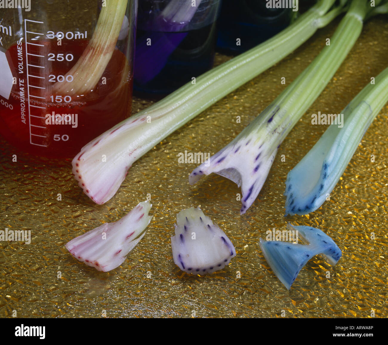 CELERY PLACED IN COLORED WATER WHICH IS CONDUCTED UPWARD TO LEAVES THROUGH XYLEM (FIBROVASCULAR BUNDLES) Stock Photohttps://www.alamy.com/image-license-details/?v=1https://www.alamy.com/stock-photo-celery-placed-in-colored-water-which-is-conducted-upward-to-leaves-16131909.html
RMARWA8P–CELERY PLACED IN COLORED WATER WHICH IS CONDUCTED UPWARD TO LEAVES THROUGH XYLEM (FIBROVASCULAR BUNDLES)
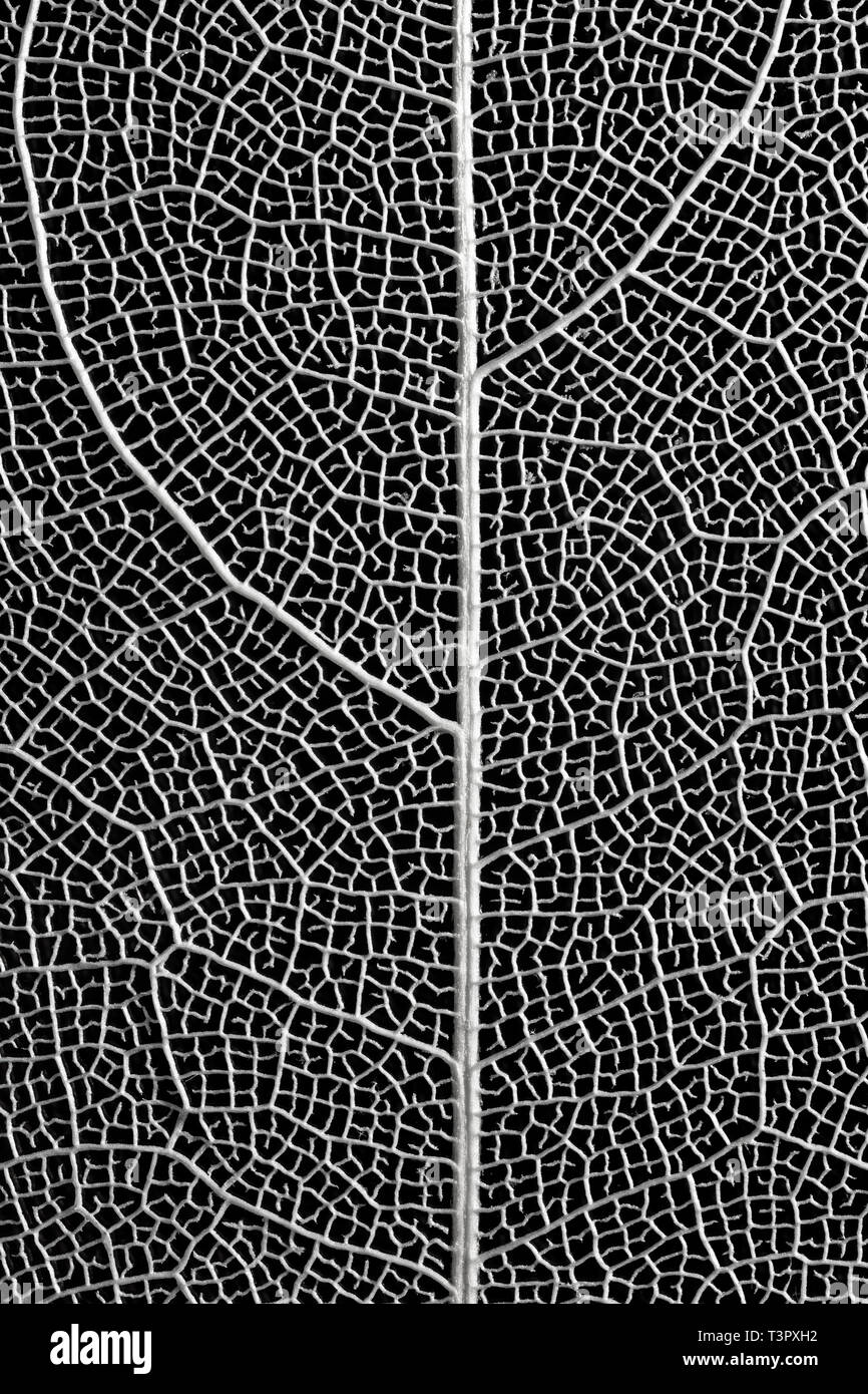 Skeletonised tree leaf showing leaf veiin structure. Stock Photohttps://www.alamy.com/image-license-details/?v=1https://www.alamy.com/skeletonised-tree-leaf-showing-leaf-veiin-structure-image243292926.html
RFT3PXH2–Skeletonised tree leaf showing leaf veiin structure.
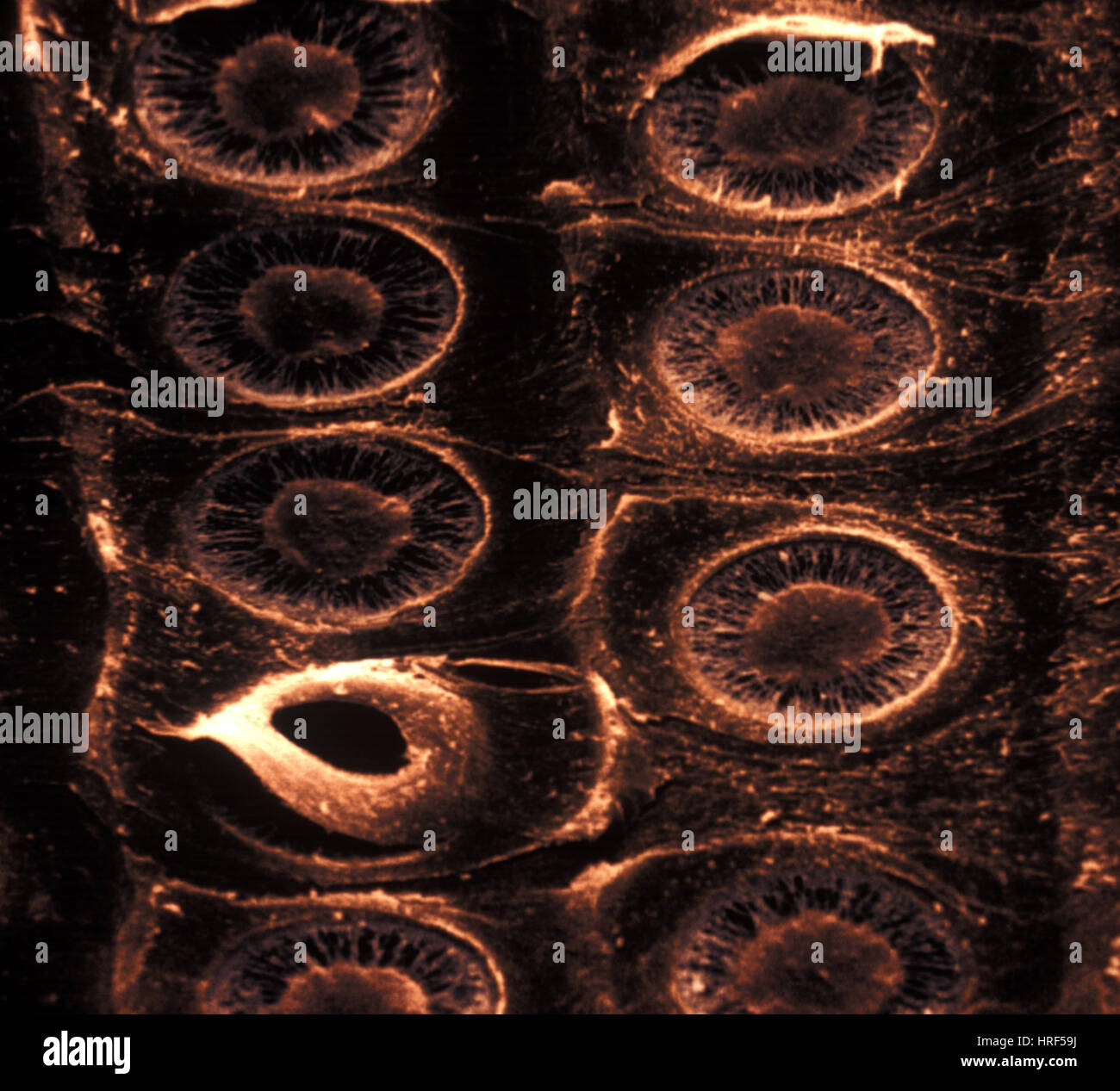 Xylem Stock Photohttps://www.alamy.com/image-license-details/?v=1https://www.alamy.com/stock-photo-xylem-134943134.html
RMHRF59J–Xylem
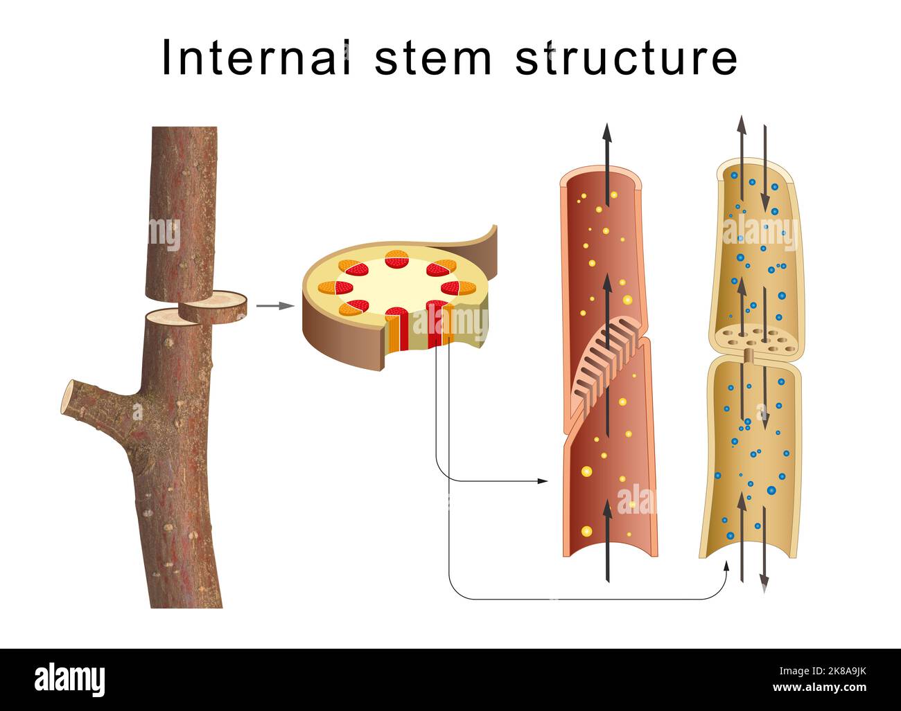 Internal anatomy of the tree stem Stock Photohttps://www.alamy.com/image-license-details/?v=1https://www.alamy.com/internal-anatomy-of-the-tree-stem-image487034651.html
RF2K8A9JK–Internal anatomy of the tree stem
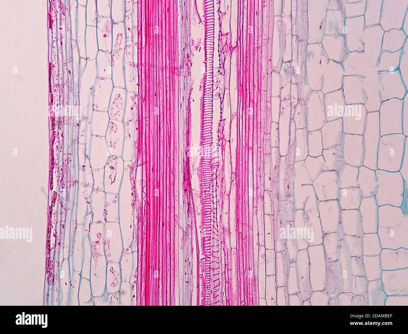 Plant vascular tissue, light micrograph. Stock Photohttps://www.alamy.com/image-license-details/?v=1https://www.alamy.com/plant-vascular-tissue-light-micrograph-image385222727.html
RF2DAMBEF–Plant vascular tissue, light micrograph.
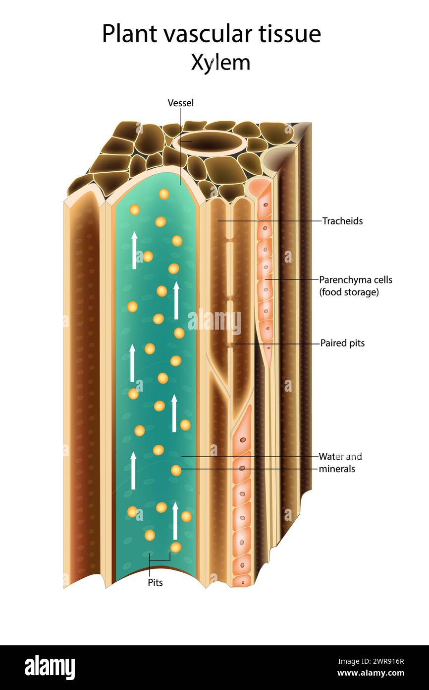 Plant vascular tissue Xylem. Cross section showing vascular bundles. Translocation vascular plants Stock Photohttps://www.alamy.com/image-license-details/?v=1https://www.alamy.com/plant-vascular-tissue-xylem-cross-section-showing-vascular-bundles-translocation-vascular-plants-image599488143.html
RF2WR916R–Plant vascular tissue Xylem. Cross section showing vascular bundles. Translocation vascular plants
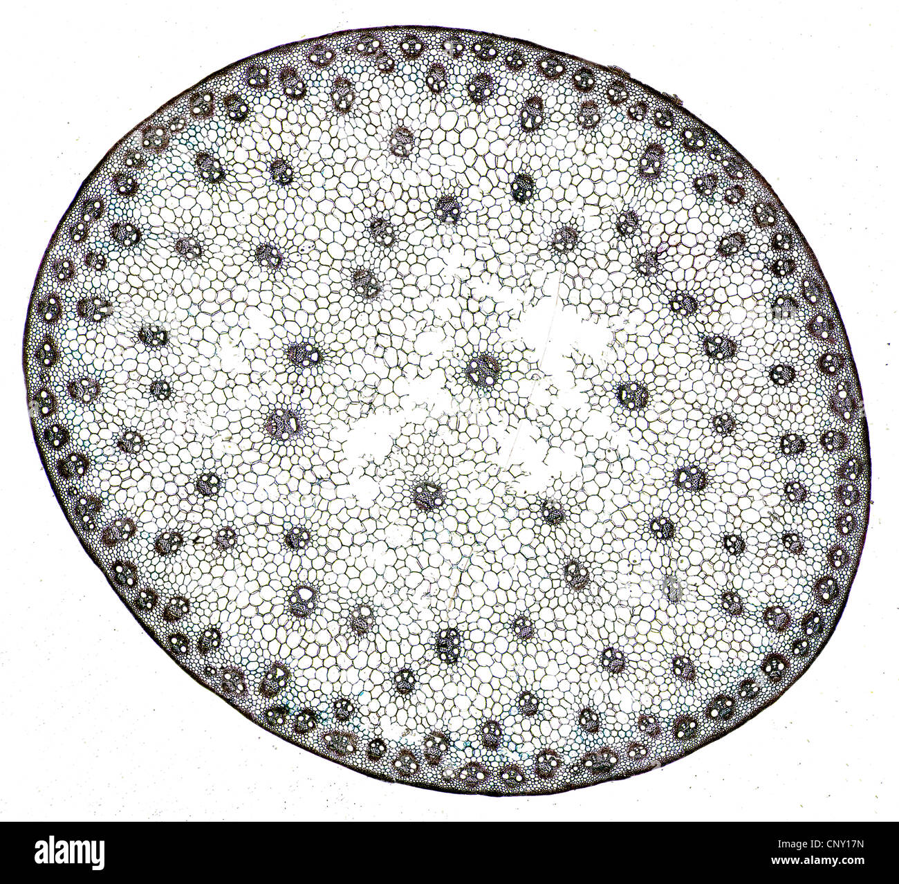 Cross section of a monocot stem with cells and vessel elements clearly visible Stock Photohttps://www.alamy.com/image-license-details/?v=1https://www.alamy.com/stock-photo-cross-section-of-a-monocot-stem-with-cells-and-vessel-elements-clearly-47922217.html
RMCNY17N–Cross section of a monocot stem with cells and vessel elements clearly visible
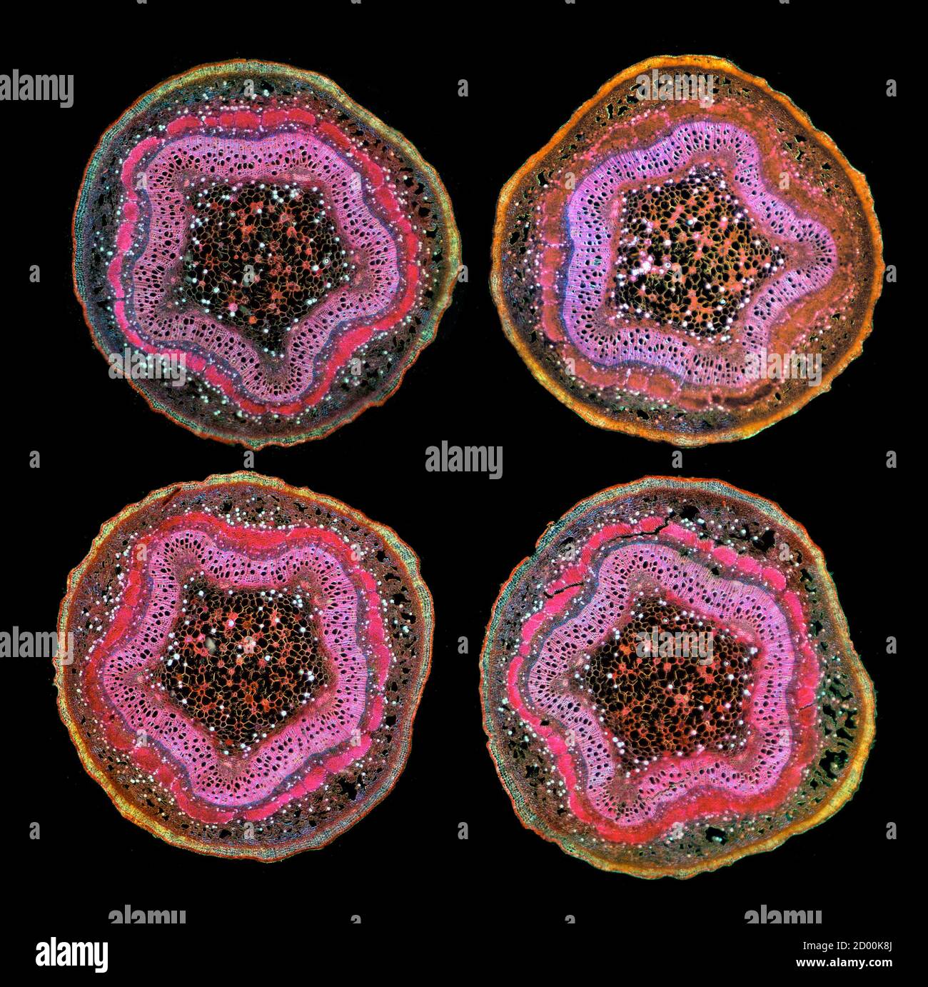 Poplar tree twigs. Populus sp. stem sections TS. Stained sections Stock Photohttps://www.alamy.com/image-license-details/?v=1https://www.alamy.com/poplar-tree-twigs-populus-sp-stem-sections-ts-stained-sections-image378643234.html
RM2D00K8J–Poplar tree twigs. Populus sp. stem sections TS. Stained sections
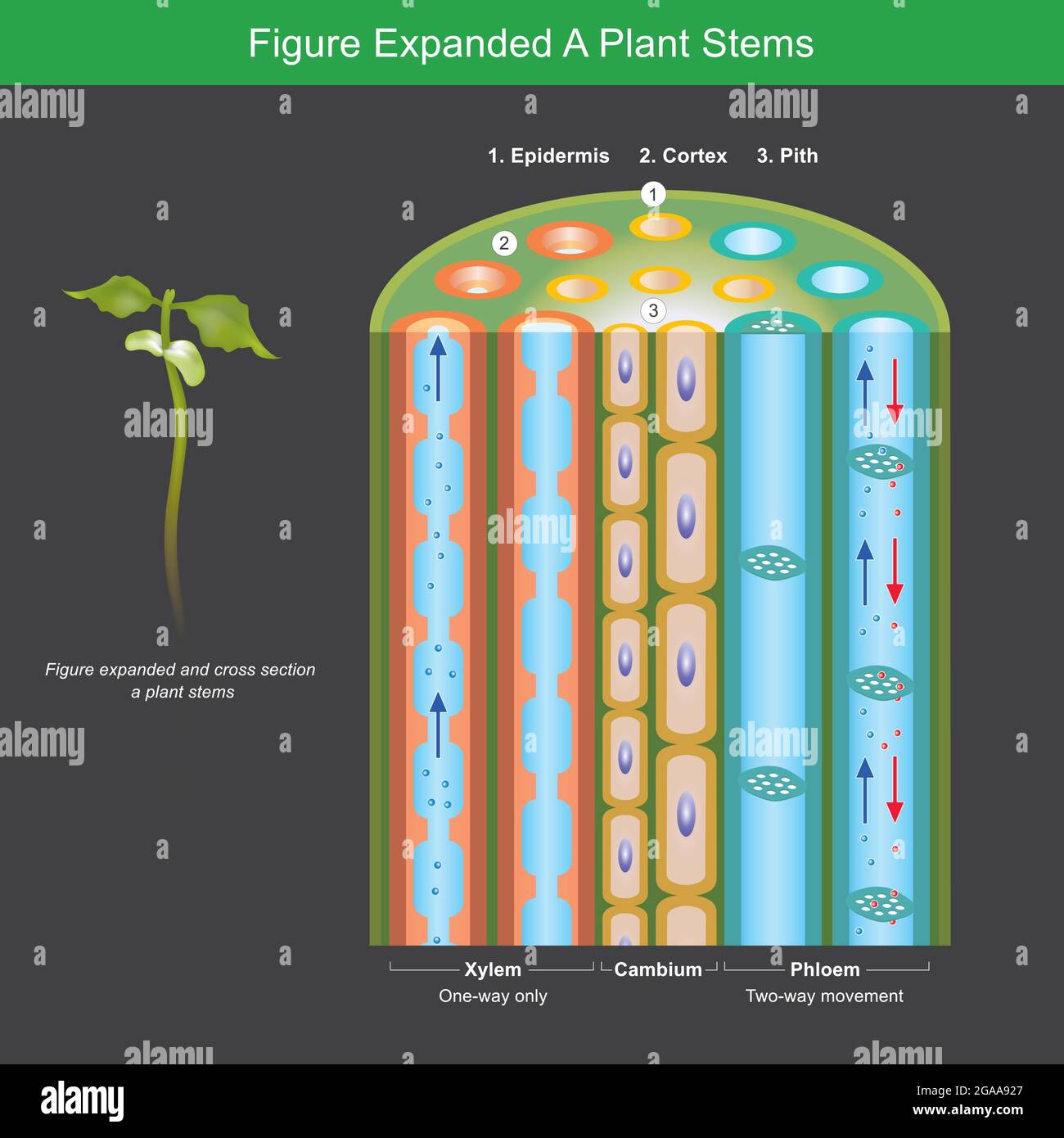 Figure Expanded A Plant Stems. Figure expanded for explain a plants transport nutrient and water in stems. Illustration. Stock Vectorhttps://www.alamy.com/image-license-details/?v=1https://www.alamy.com/figure-expanded-a-plant-stems-figure-expanded-for-explain-a-plants-transport-nutrient-and-water-in-stems-illustration-image436632399.html
RF2GAA927–Figure Expanded A Plant Stems. Figure expanded for explain a plants transport nutrient and water in stems. Illustration.
 Wood Stock Photohttps://www.alamy.com/image-license-details/?v=1https://www.alamy.com/stock-photo-wood-72322804.html
RME5JGD8–Wood
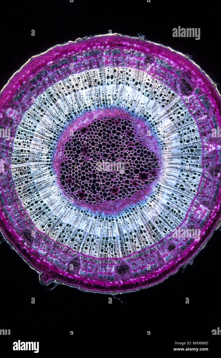 Xylem.9x Stock Photohttps://www.alamy.com/image-license-details/?v=1https://www.alamy.com/xylem9x-image188662333.html
RFMXX8MD–Xylem.9x
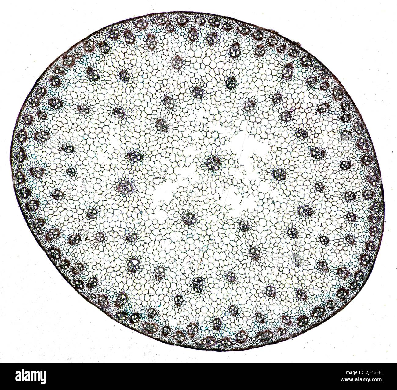 Cross section of a monocot stem with cells and vessel elements clearly visible. Stock Photohttps://www.alamy.com/image-license-details/?v=1https://www.alamy.com/cross-section-of-a-monocot-stem-with-cells-and-vessel-elements-clearly-visible-image473924517.html
RM2JF13FH–Cross section of a monocot stem with cells and vessel elements clearly visible.
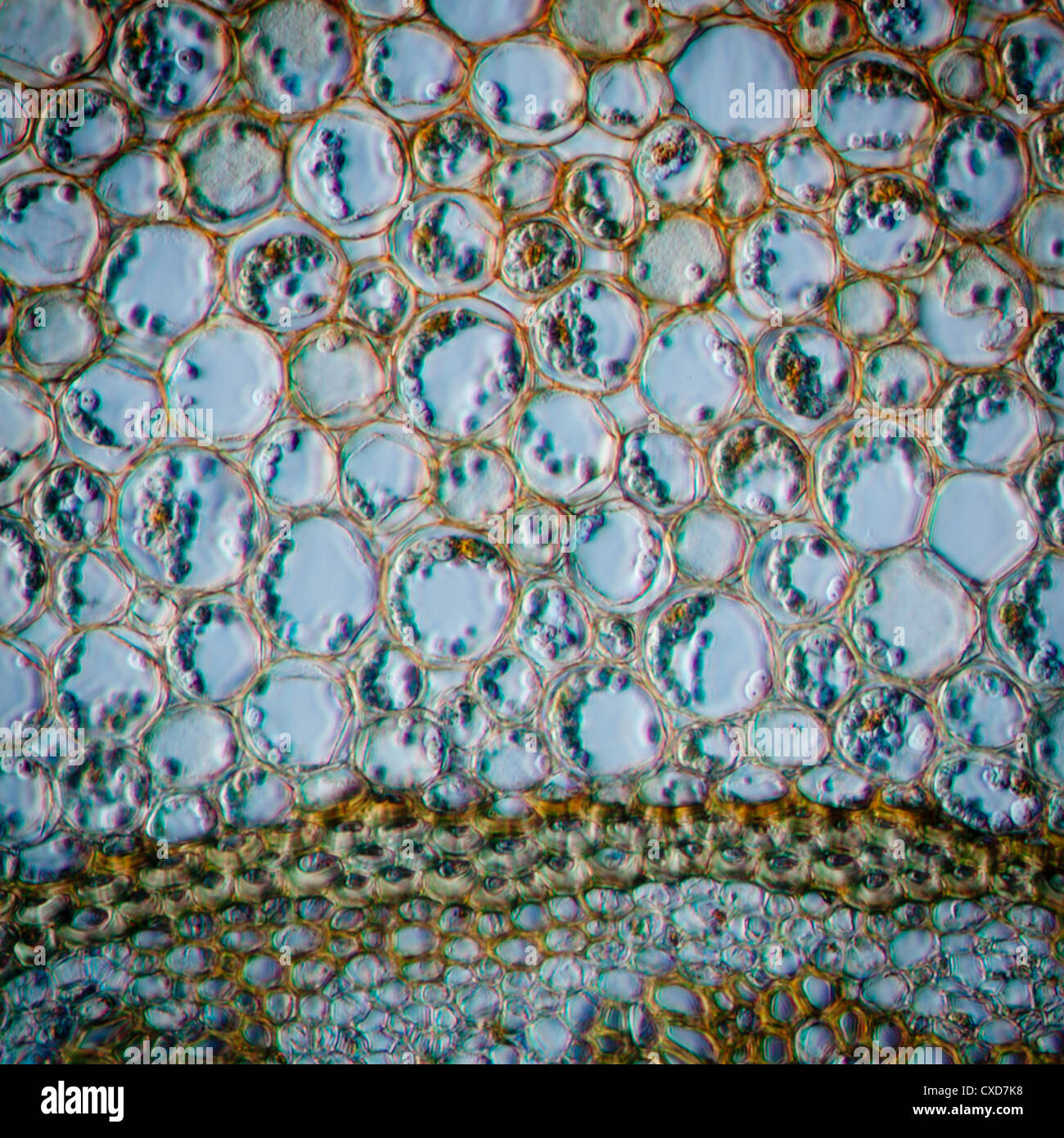 science micrograph plant root tissue Stock Photohttps://www.alamy.com/image-license-details/?v=1https://www.alamy.com/stock-photo-science-micrograph-plant-root-tissue-50693196.html
RFCXD7K8–science micrograph plant root tissue
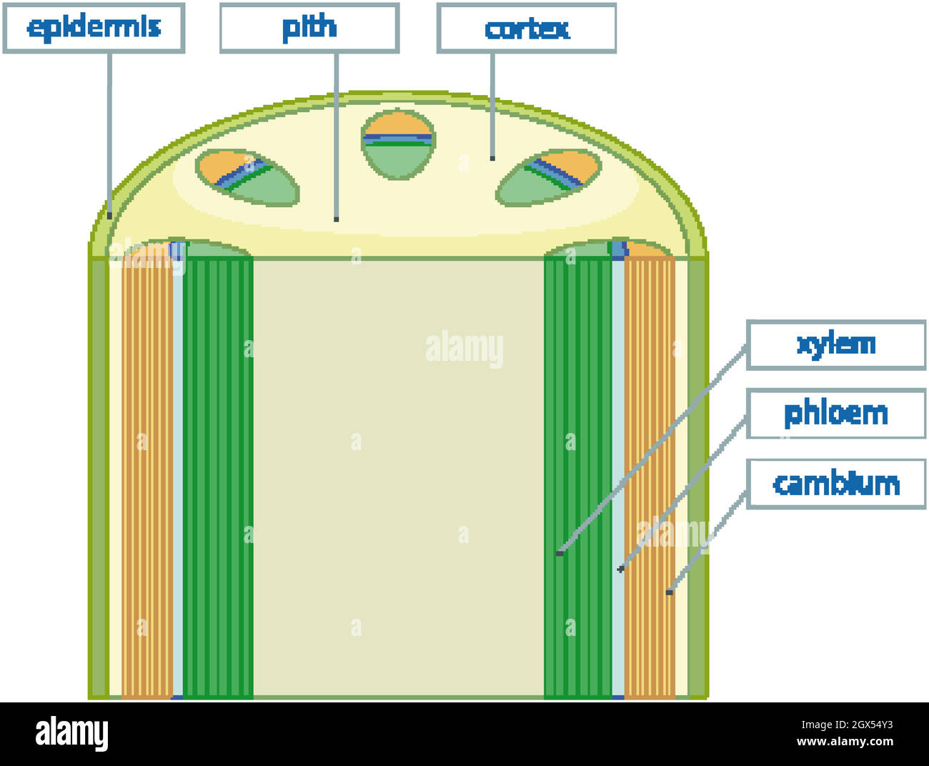 Diagram showing vascular tissue system in plants Stock Vectorhttps://www.alamy.com/image-license-details/?v=1https://www.alamy.com/diagram-showing-vascular-tissue-system-in-plants-image446353911.html
RF2GX54Y3–Diagram showing vascular tissue system in plants
 Close up view of a cell wall - biology background Stock Photohttps://www.alamy.com/image-license-details/?v=1https://www.alamy.com/stock-photo-close-up-view-of-a-cell-wall-biology-background-74511122.html
RFE967KE–Close up view of a cell wall - biology background
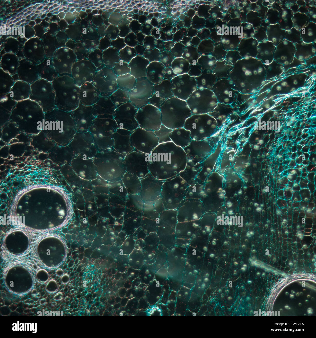 microscopy micrograph plant tissue, stem of pumpkin, magnification 100X Stock Photohttps://www.alamy.com/image-license-details/?v=1https://www.alamy.com/stock-photo-microscopy-micrograph-plant-tissue-stem-of-pumpkin-magnification-100x-50315590.html
RFCWT21A–microscopy micrograph plant tissue, stem of pumpkin, magnification 100X
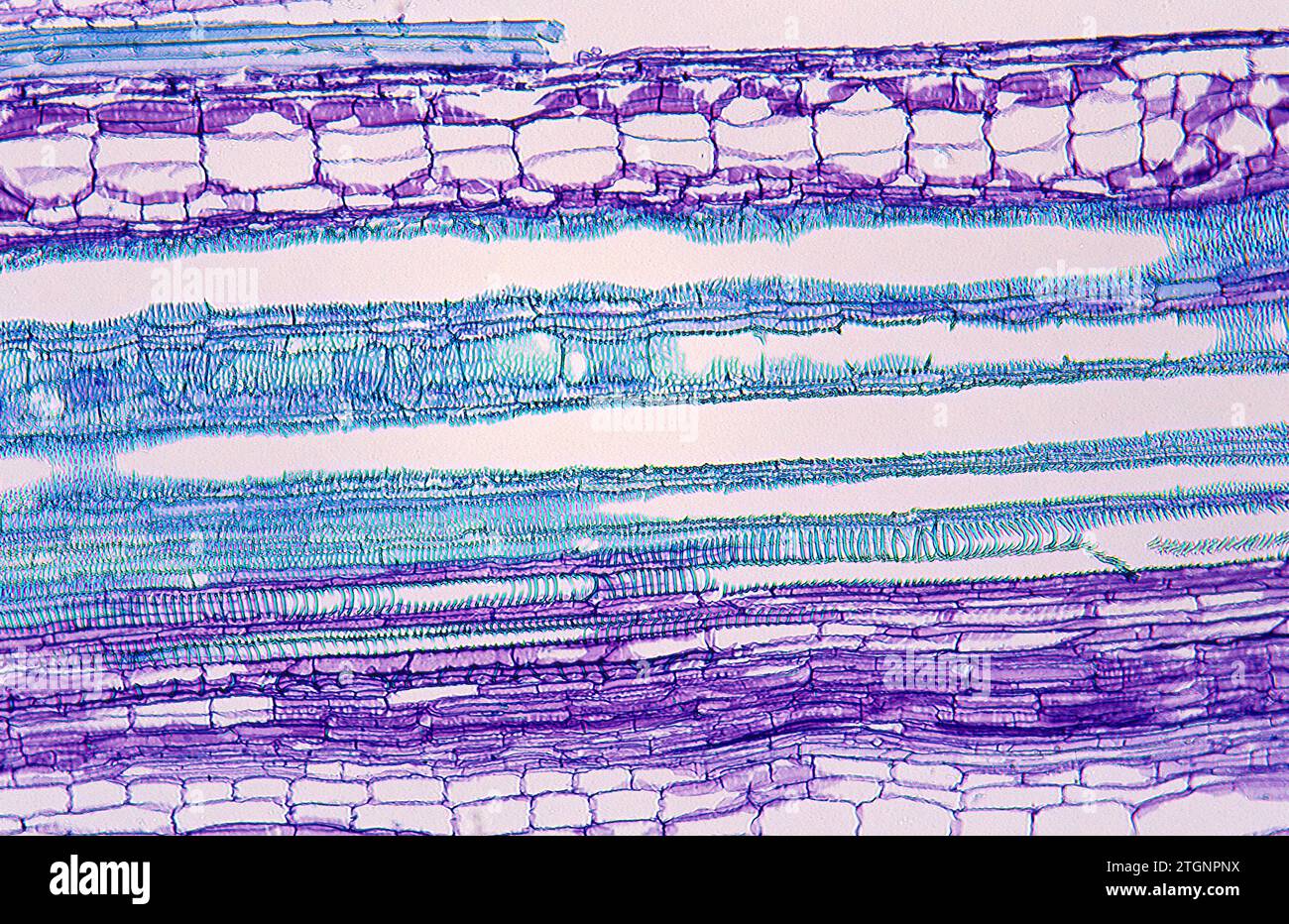 Phloem (purple) and xylem (blue) on a stem longitudinal section. Photomicrograph of pumpkin tissue. Stock Photohttps://www.alamy.com/image-license-details/?v=1https://www.alamy.com/phloem-purple-and-xylem-blue-on-a-stem-longitudinal-section-photomicrograph-of-pumpkin-tissue-image578255494.html
RF2TGNPNX–Phloem (purple) and xylem (blue) on a stem longitudinal section. Photomicrograph of pumpkin tissue.
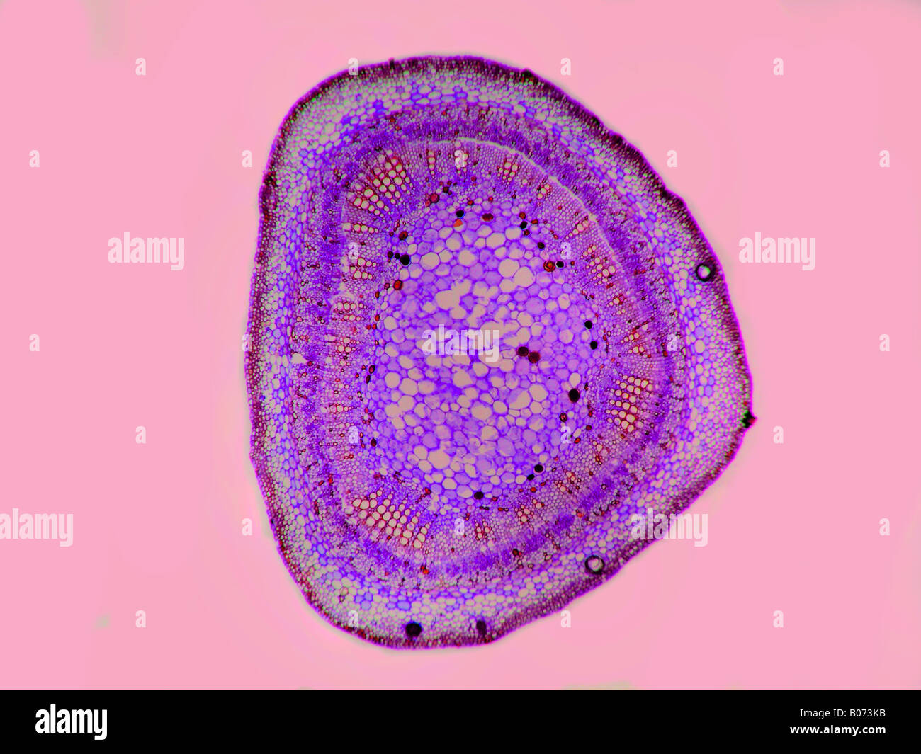 A cotton leafstem Gossypium sp Stock Photohttps://www.alamy.com/image-license-details/?v=1https://www.alamy.com/stock-photo-a-cotton-leafstem-gossypium-sp-17366927.html
RFB073KB–A cotton leafstem Gossypium sp
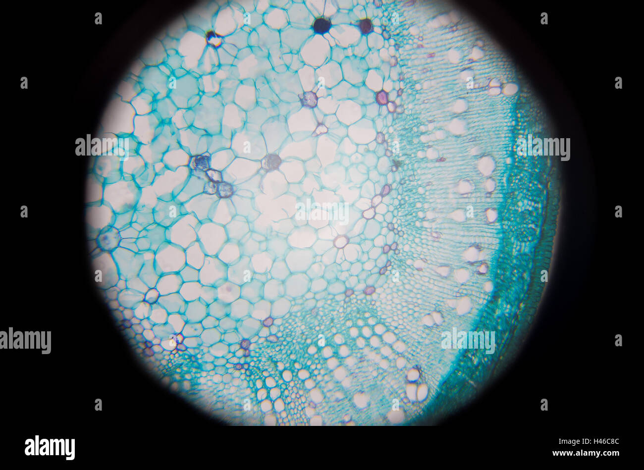 Microscopic photography of Steam of wood dicotyledon, cross section. Stock Photohttps://www.alamy.com/image-license-details/?v=1https://www.alamy.com/stock-photo-microscopic-photography-of-steam-of-wood-dicotyledon-cross-section-123072556.html
RFH46C8C–Microscopic photography of Steam of wood dicotyledon, cross section.
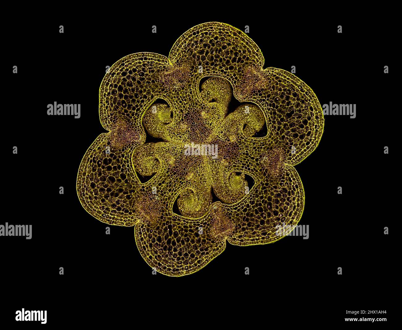 cross section cut slice of plant stem under the microscope – microscopic view of plant cells for botanic education – high quality Stock Photohttps://www.alamy.com/image-license-details/?v=1https://www.alamy.com/cross-section-cut-slice-of-plant-stem-under-the-microscope-microscopic-view-of-plant-cells-for-botanic-education-high-quality-image463480896.html
RF2HX1AH4–cross section cut slice of plant stem under the microscope – microscopic view of plant cells for botanic education – high quality
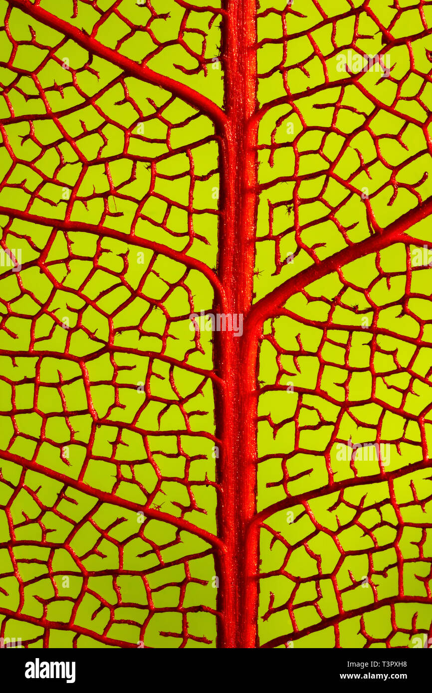 Skeletonised tree leaf showing leaf vein structure Stock Photohttps://www.alamy.com/image-license-details/?v=1https://www.alamy.com/skeletonised-tree-leaf-showing-leaf-vein-structure-image243292932.html
RFT3PXH8–Skeletonised tree leaf showing leaf vein structure
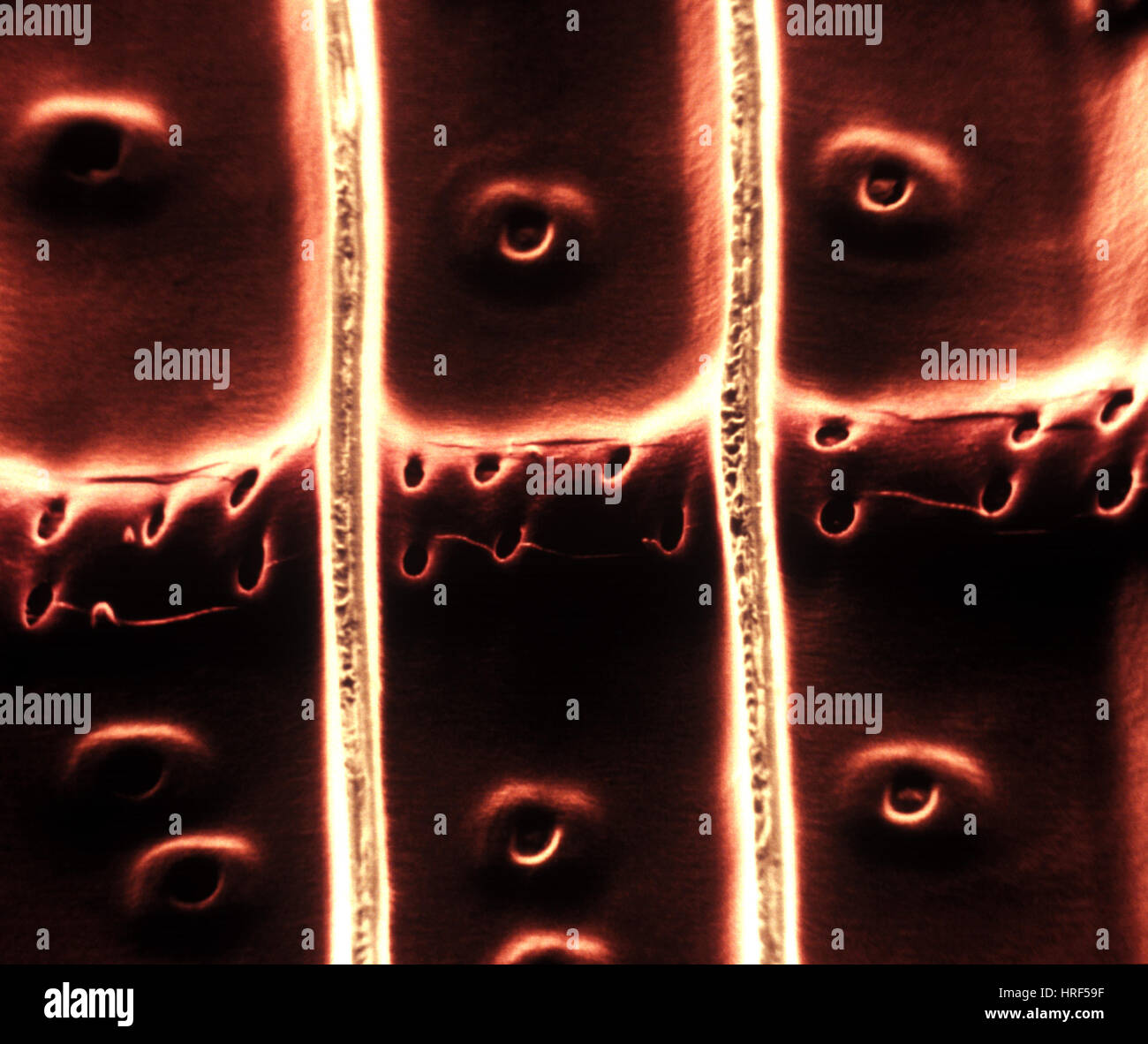 Xylem Stock Photohttps://www.alamy.com/image-license-details/?v=1https://www.alamy.com/stock-photo-xylem-134943131.html
RMHRF59F–Xylem
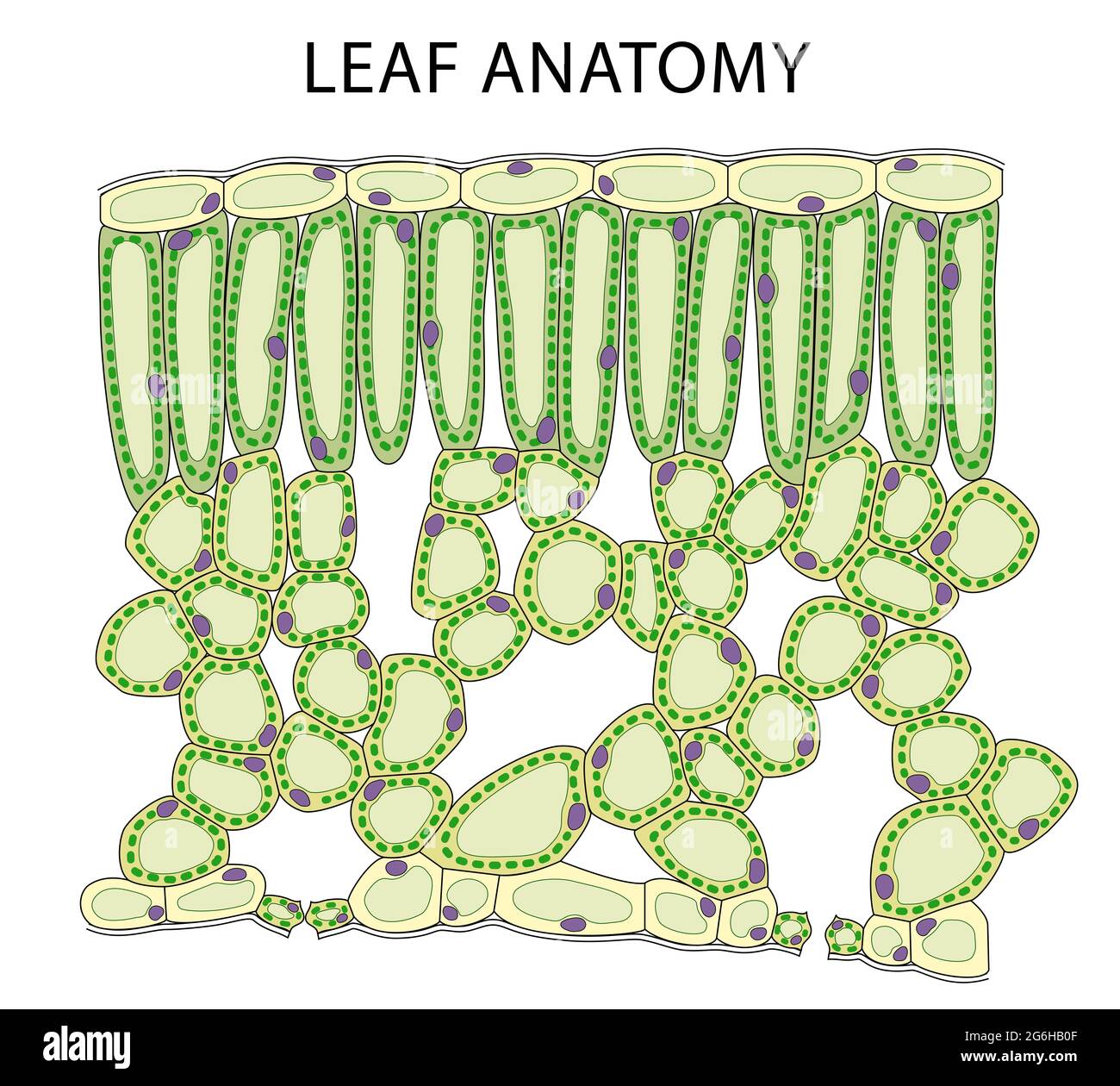 Education Chart of Biology for Cross Section of Leaf Diagram Stock Photohttps://www.alamy.com/image-license-details/?v=1https://www.alamy.com/education-chart-of-biology-for-cross-section-of-leaf-diagram-image434328959.html
RF2G6HB0F–Education Chart of Biology for Cross Section of Leaf Diagram
 Plant vascular tissue, light micrograph. Stock Photohttps://www.alamy.com/image-license-details/?v=1https://www.alamy.com/plant-vascular-tissue-light-micrograph-image385222722.html
RF2DAMBEA–Plant vascular tissue, light micrograph.
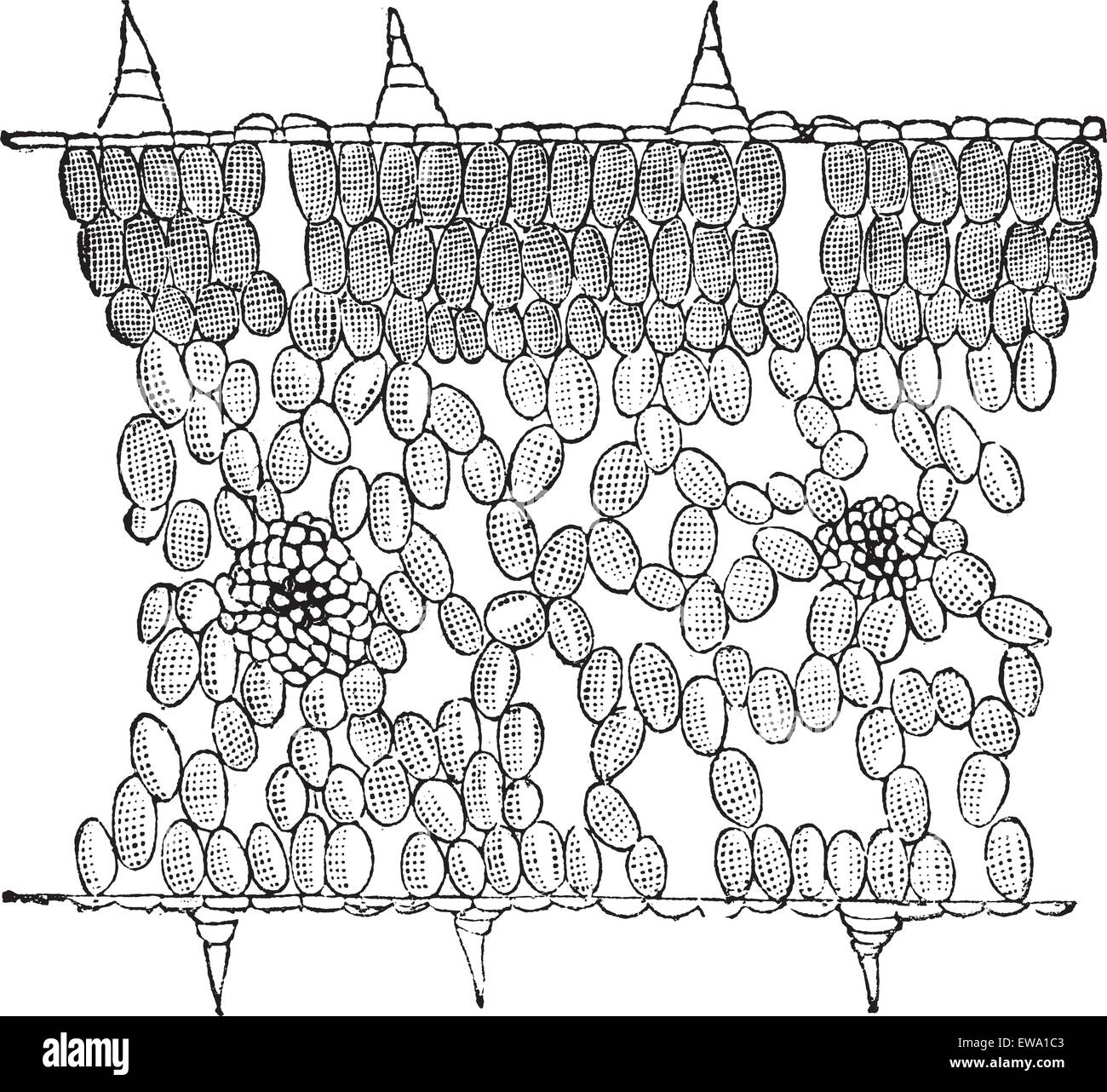 Vertical Cross-section of a Leaf, magnified, vintage engraved illustration. Trousset encyclopedia (1886 - 1891). Stock Vectorhttps://www.alamy.com/image-license-details/?v=1https://www.alamy.com/stock-photo-vertical-cross-section-of-a-leaf-magnified-vintage-engraved-illustration-84428515.html
RFEWA1C3–Vertical Cross-section of a Leaf, magnified, vintage engraved illustration. Trousset encyclopedia (1886 - 1891).
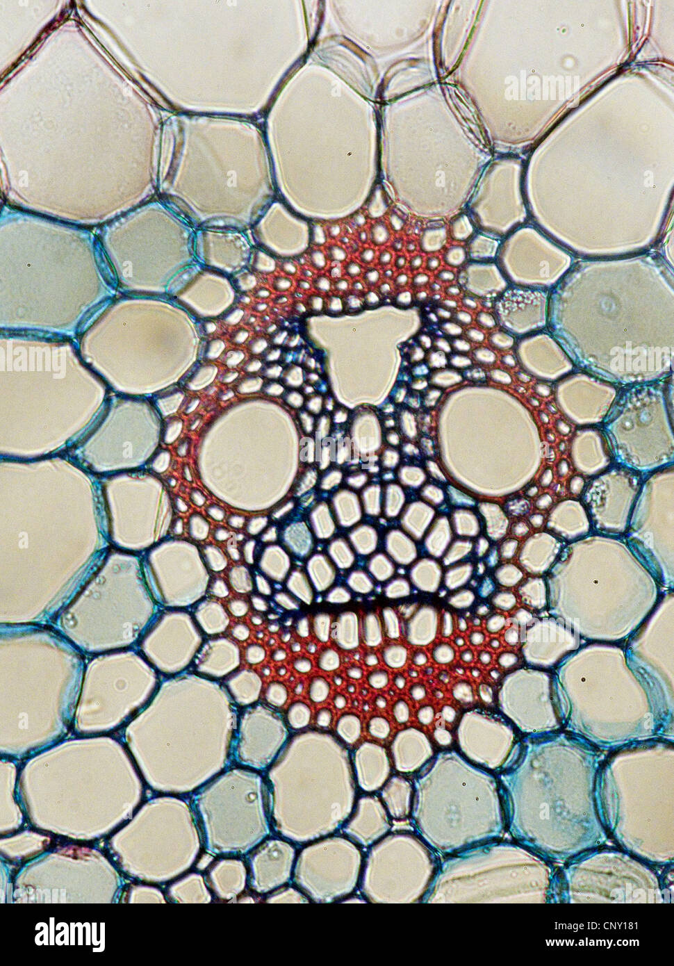 Indian corn, maize (Zea mays), Vascular bundle from maize Stock Photohttps://www.alamy.com/image-license-details/?v=1https://www.alamy.com/stock-photo-indian-corn-maize-zea-mays-vascular-bundle-from-maize-47922225.html
RMCNY181–Indian corn, maize (Zea mays), Vascular bundle from maize
 Carline thistle, Carlina acaulis, stem section TS. Stock Photohttps://www.alamy.com/image-license-details/?v=1https://www.alamy.com/carline-thistle-carlina-acaulis-stem-section-ts-image378635745.html
RM2D009N5–Carline thistle, Carlina acaulis, stem section TS.
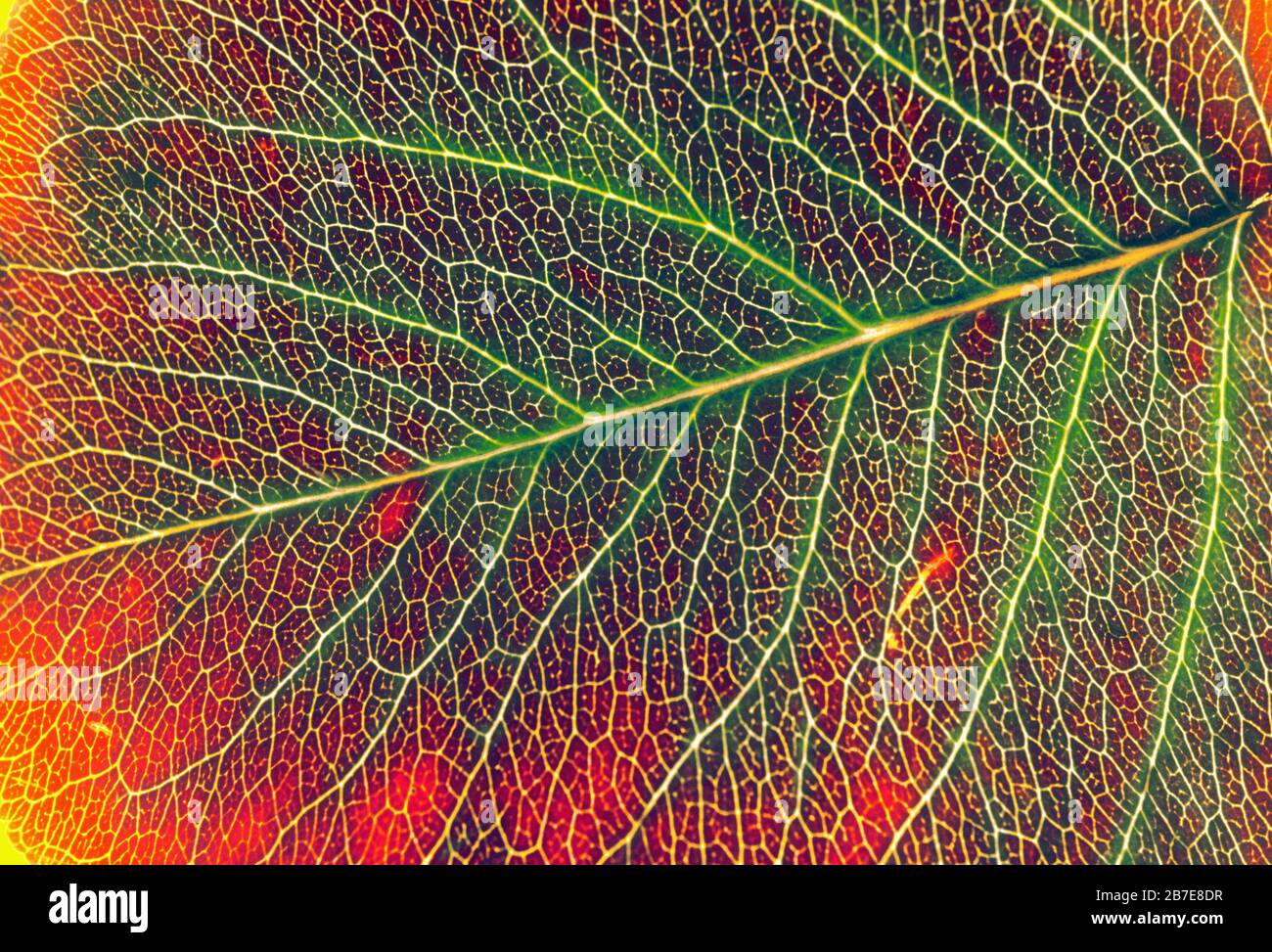 Vein pattern in leaf Stock Photohttps://www.alamy.com/image-license-details/?v=1https://www.alamy.com/vein-pattern-in-leaf-image348823939.html
RF2B7E8DR–Vein pattern in leaf
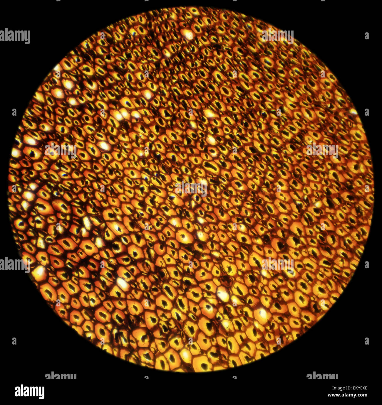 Plasmodesmata slice under the microscope, (Plasmodesma Sec.) 100x Stock Photohttps://www.alamy.com/image-license-details/?v=1https://www.alamy.com/stock-photo-plasmodesmata-slice-under-the-microscope-plasmodesma-sec-100x-81124358.html
RFEKYEXE–Plasmodesmata slice under the microscope, (Plasmodesma Sec.) 100x
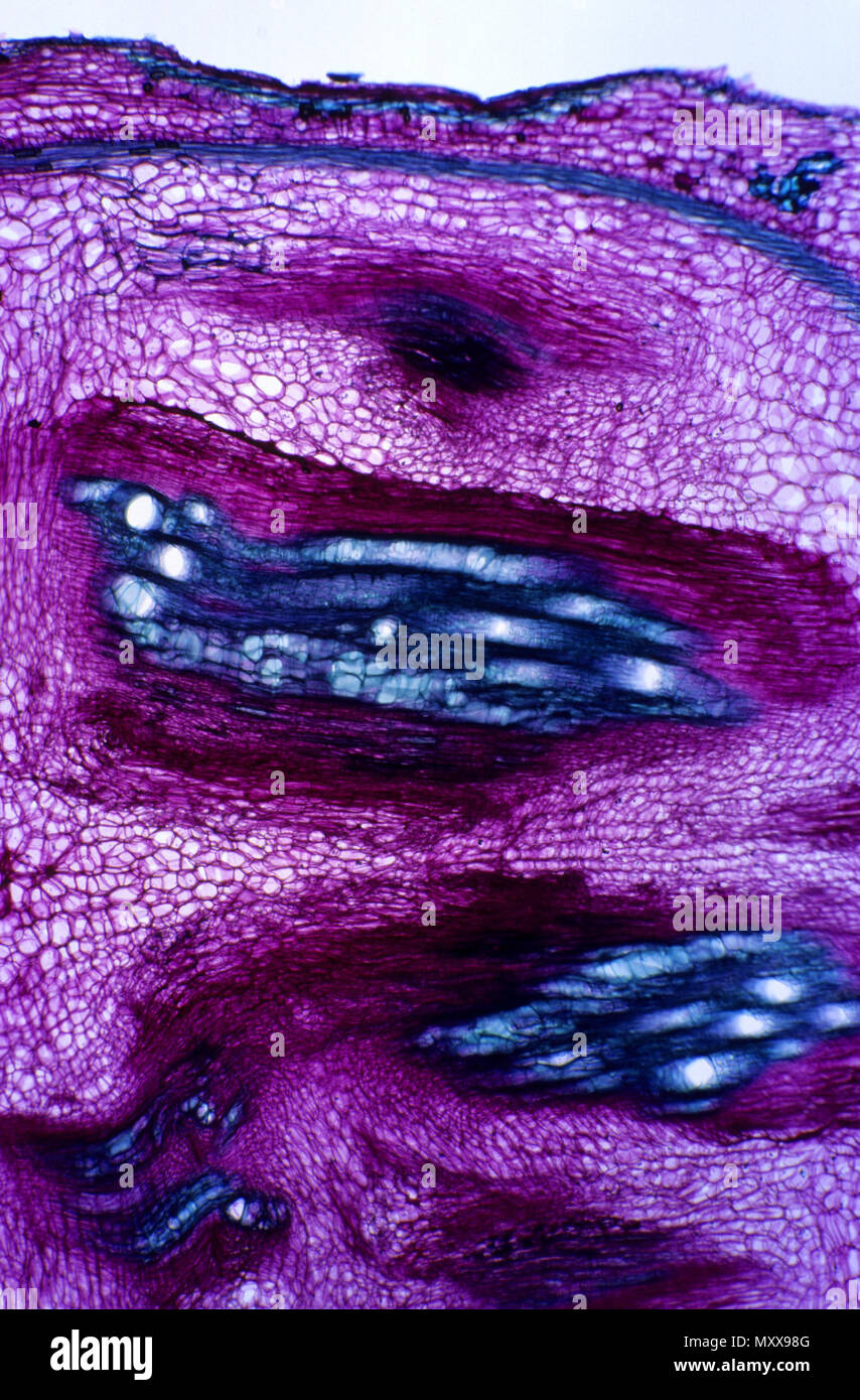 Xylem Stock Photohttps://www.alamy.com/image-license-details/?v=1https://www.alamy.com/xylem-image188662784.html
RFMXX98G–Xylem
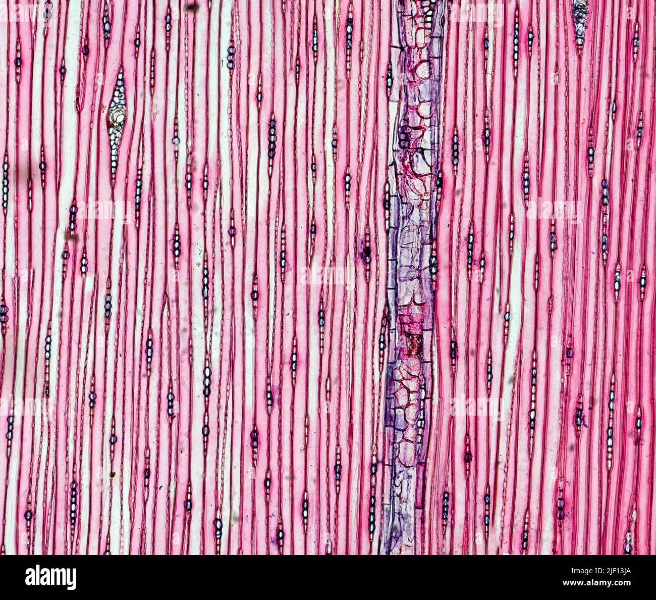 Longitudinal section of stem from Pine (Pinus sylvestris) with one vascular tube clearly visible. Stock Photohttps://www.alamy.com/image-license-details/?v=1https://www.alamy.com/longitudinal-section-of-stem-from-pine-pinus-sylvestris-with-one-vascular-tube-clearly-visible-image473924594.html
RM2JF13JA–Longitudinal section of stem from Pine (Pinus sylvestris) with one vascular tube clearly visible.
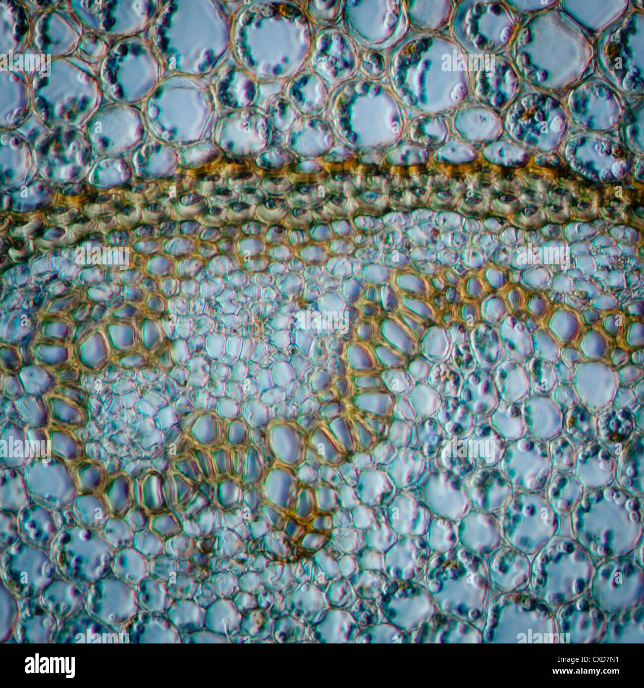 science micrograph plant root tissue Stock Photohttps://www.alamy.com/image-license-details/?v=1https://www.alamy.com/stock-photo-science-micrograph-plant-root-tissue-50693245.html
RFCXD7N1–science micrograph plant root tissue
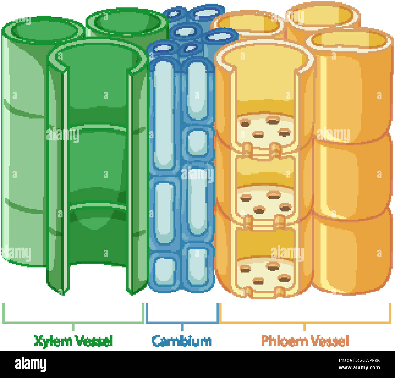 Diagram showing vascular tissue system in plants Stock Vectorhttps://www.alamy.com/image-license-details/?v=1https://www.alamy.com/diagram-showing-vascular-tissue-system-in-plants-image446126819.html
RF2GWPR8K–Diagram showing vascular tissue system in plants
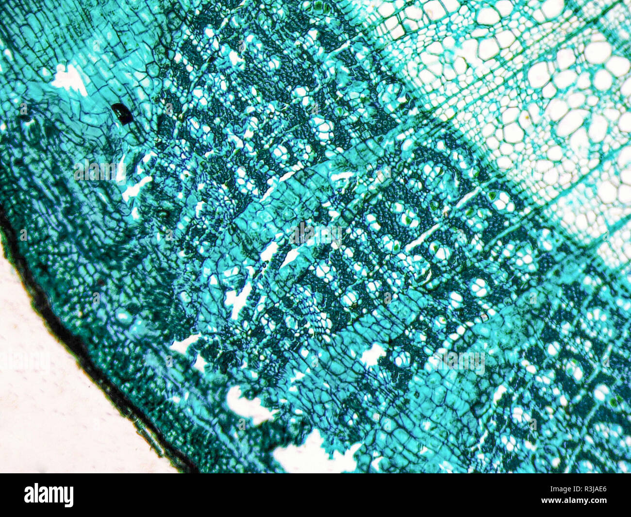 Stock Photohttps://www.alamy.com/image-license-details/?v=1https://www.alamy.com/-image225982126.html
RFR3JAE6–
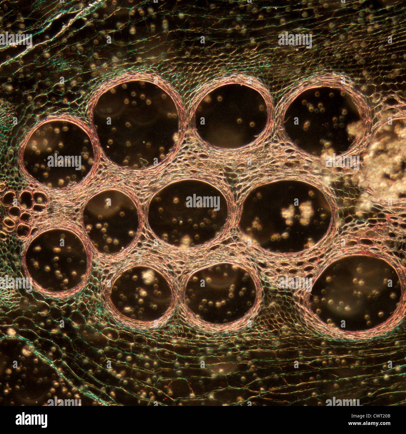 microscopy micrograph plant tissue, stem of pumpkin, magnification 100X Stock Photohttps://www.alamy.com/image-license-details/?v=1https://www.alamy.com/stock-photo-microscopy-micrograph-plant-tissue-stem-of-pumpkin-magnification-100x-50315563.html
RFCWT20B–microscopy micrograph plant tissue, stem of pumpkin, magnification 100X
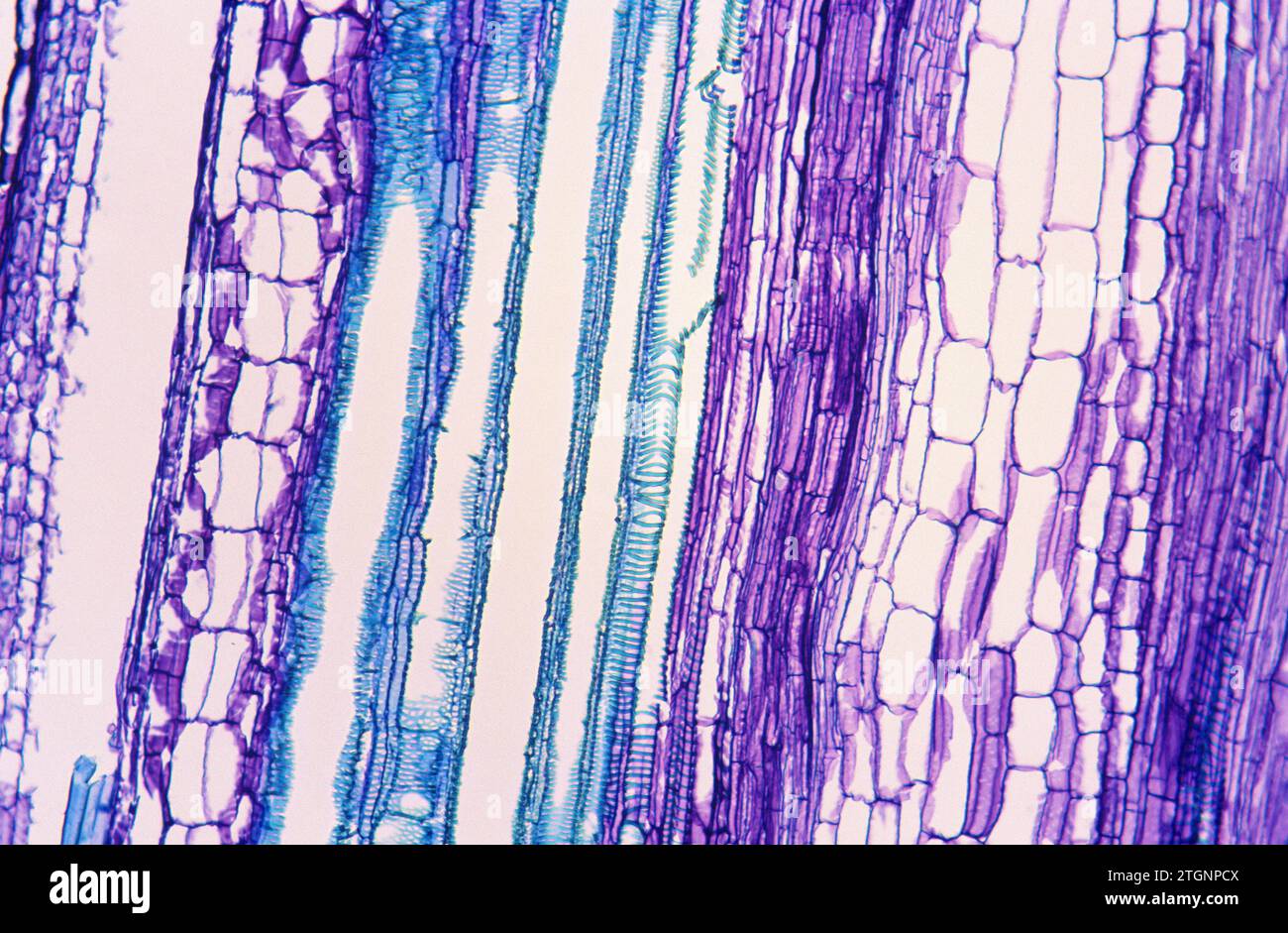 Phloem (purple) and xylem (blue) on a stem longitudinal section. Photomicrograph of pumpkin tissue. Stock Photohttps://www.alamy.com/image-license-details/?v=1https://www.alamy.com/phloem-purple-and-xylem-blue-on-a-stem-longitudinal-section-photomicrograph-of-pumpkin-tissue-image578255242.html
RF2TGNPCX–Phloem (purple) and xylem (blue) on a stem longitudinal section. Photomicrograph of pumpkin tissue.
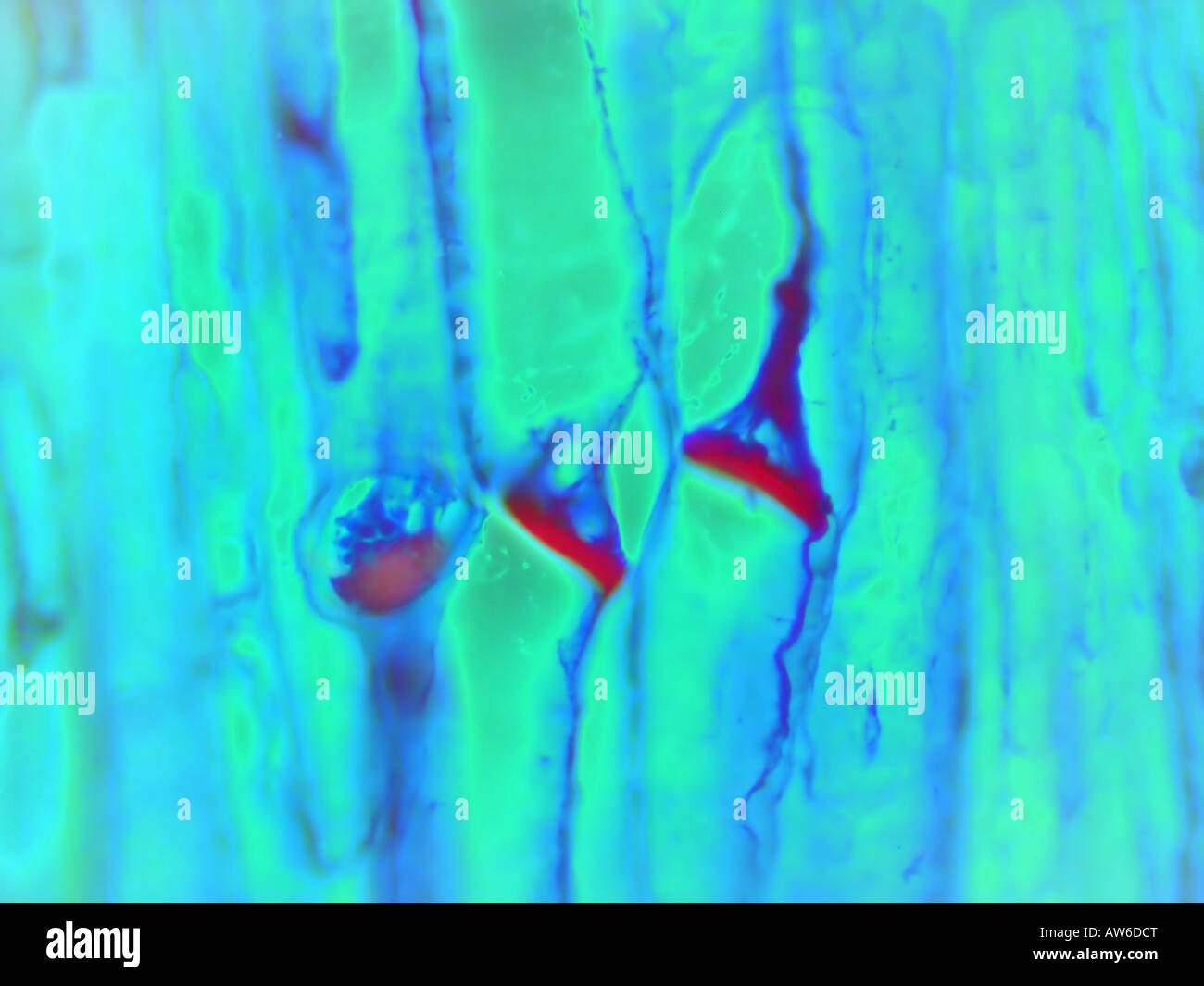 Sieve plates Stock Photohttps://www.alamy.com/image-license-details/?v=1https://www.alamy.com/stock-photo-sieve-plates-16481063.html
RFAW6DCT–Sieve plates
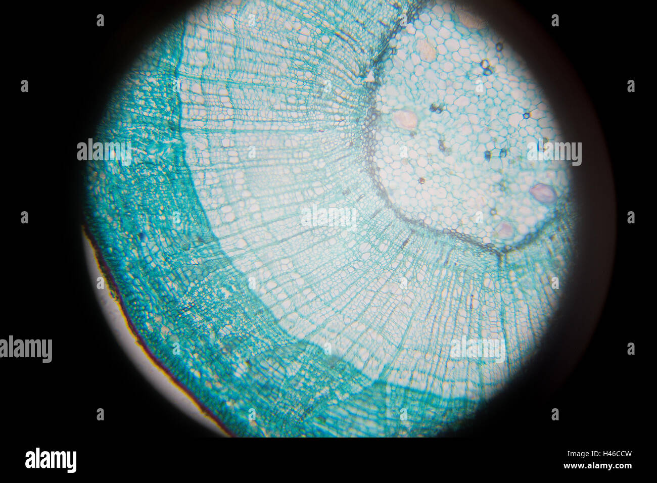 Microscopic photography of Steam of wood dicotyledon, cross section. Stock Photohttps://www.alamy.com/image-license-details/?v=1https://www.alamy.com/stock-photo-microscopic-photography-of-steam-of-wood-dicotyledon-cross-section-123072681.html
RFH46CCW–Microscopic photography of Steam of wood dicotyledon, cross section.
 cross section cut slice of plant stem under the microscope – microscopic view of plant cells for botanic education – high quality Stock Photohttps://www.alamy.com/image-license-details/?v=1https://www.alamy.com/cross-section-cut-slice-of-plant-stem-under-the-microscope-microscopic-view-of-plant-cells-for-botanic-education-high-quality-image463482667.html
RF2HX1CTB–cross section cut slice of plant stem under the microscope – microscopic view of plant cells for botanic education – high quality
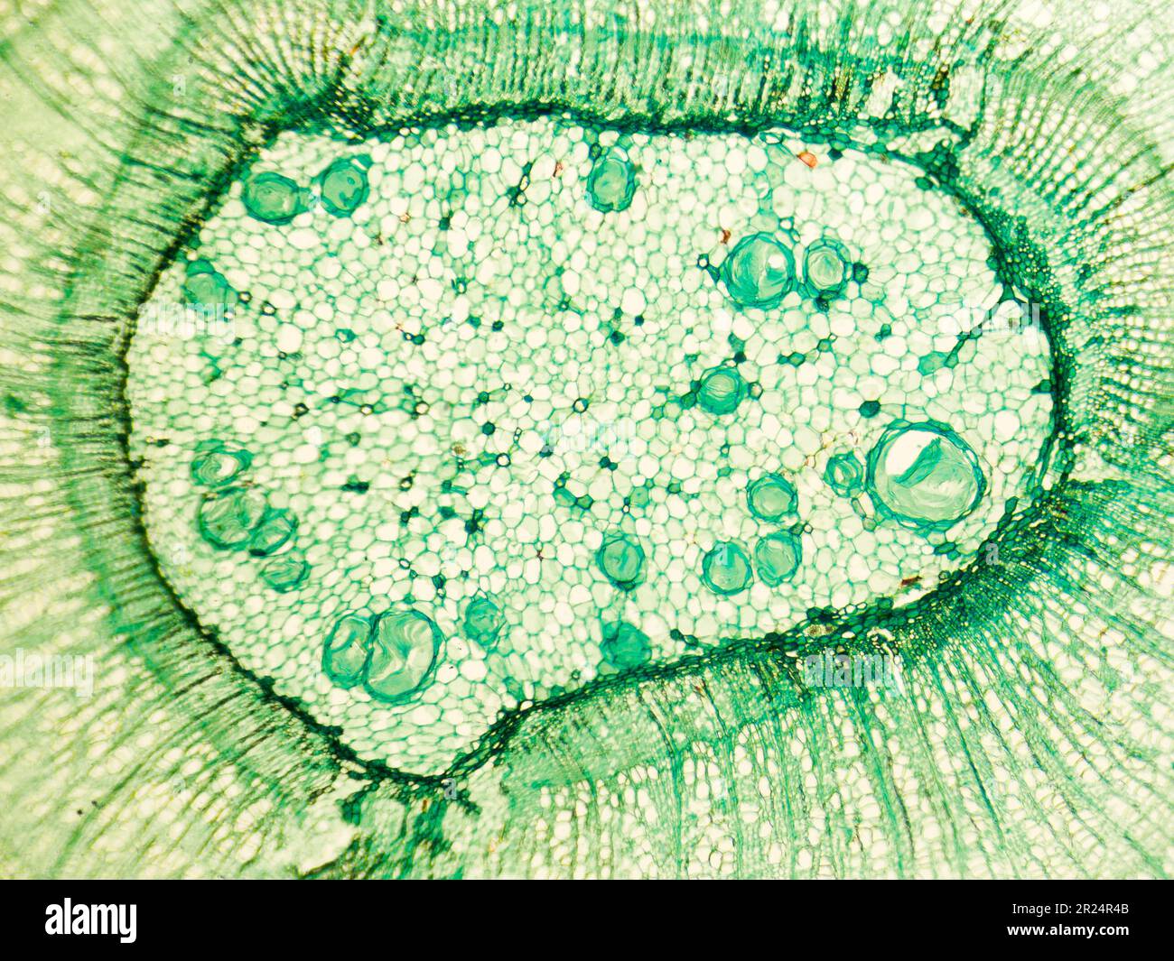 stem of wood discotyledon x.s. details under biological optical misroscope Stock Photohttps://www.alamy.com/image-license-details/?v=1https://www.alamy.com/stem-of-wood-discotyledon-xs-details-under-biological-optical-misroscope-image552067051.html
RF2R24R4B–stem of wood discotyledon x.s. details under biological optical misroscope
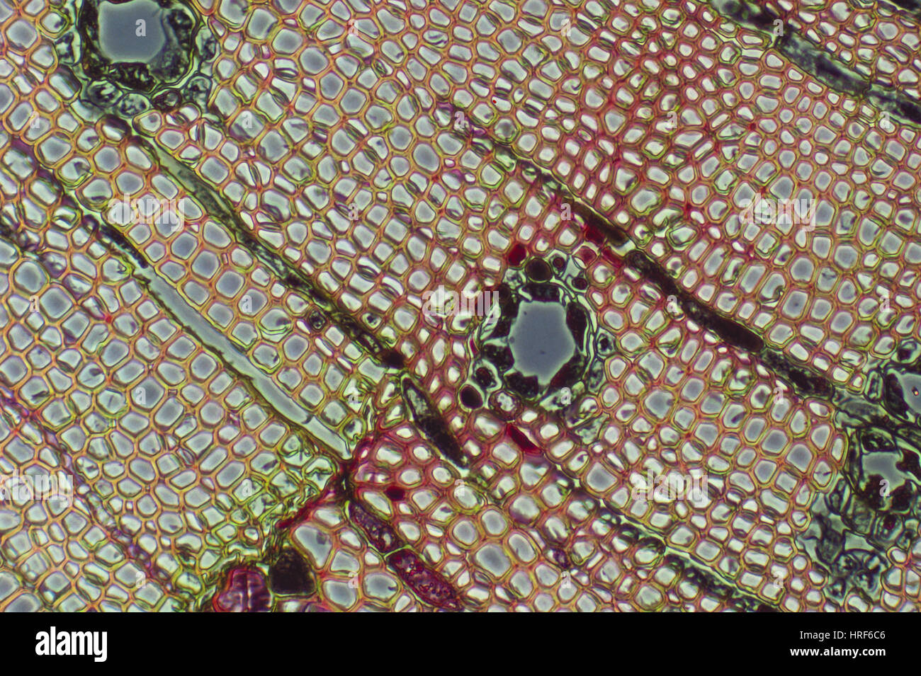 Pine Xylem Stock Photohttps://www.alamy.com/image-license-details/?v=1https://www.alamy.com/stock-photo-pine-xylem-134943990.html
RMHRF6C6–Pine Xylem
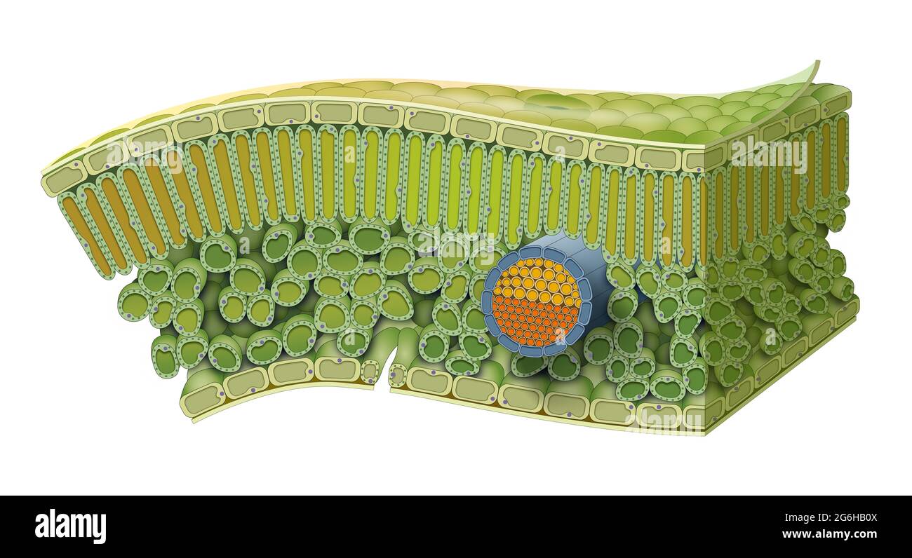 Cellular Structure of Leaf. Internal Leaf Structure a leaf is made of many layers that are sandwiched between two layers of tough skin cells Stock Photohttps://www.alamy.com/image-license-details/?v=1https://www.alamy.com/cellular-structure-of-leaf-internal-leaf-structure-a-leaf-is-made-of-many-layers-that-are-sandwiched-between-two-layers-of-tough-skin-cells-image434328970.html
RF2G6HB0X–Cellular Structure of Leaf. Internal Leaf Structure a leaf is made of many layers that are sandwiched between two layers of tough skin cells
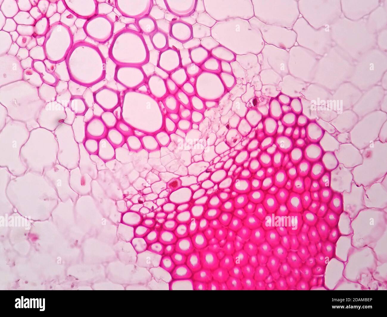 Plant vascular tissue, light micrograph. Stock Photohttps://www.alamy.com/image-license-details/?v=1https://www.alamy.com/plant-vascular-tissue-light-micrograph-image385222734.html
RF2DAMBEP–Plant vascular tissue, light micrograph.
 grafting technique apply on a grapes fruit tree, wrap with plastic, to sustain the moisture Stock Photohttps://www.alamy.com/image-license-details/?v=1https://www.alamy.com/grafting-technique-apply-on-a-grapes-fruit-tree-wrap-with-plastic-to-sustain-the-moisture-image609154823.html
RF2XB1B5B–grafting technique apply on a grapes fruit tree, wrap with plastic, to sustain the moisture
 Indian corn, maize (Zea mays), Vascular bundles in a sprout of maize Stock Photohttps://www.alamy.com/image-license-details/?v=1https://www.alamy.com/stock-photo-indian-corn-maize-zea-mays-vascular-bundles-in-a-sprout-of-maize-47950390.html
RMCP095X–Indian corn, maize (Zea mays), Vascular bundles in a sprout of maize