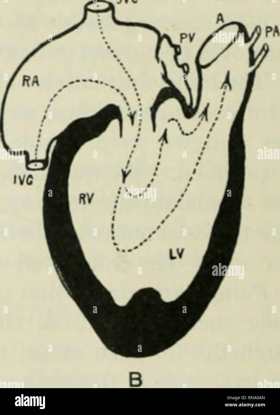
. The anatomical record. Anatomy; Anatomy. Fig. A Diagrammatic sketch showing probable circulation in case 1. Fig. B Diagrammatic sketch showing probable circulation in case 2. IVC, inferior vena cava; RA. right atrium; LA, left atrium, SVC, superior vena cava; A, aorta; DA, ductus arteriosus; PA, pulmonary arteries; PV, pul- monary veins; LV, left ventricle; RV, right ventricle. We believe, however, that the amount of blood reaching the lungs by any one of the above two routes, though probably was greater in amount than in the first case, j-et it was without doubt insufficient to support life
Image details
Contributor:
Library Book Collection / Alamy Stock PhotoImage ID:
RNA0ANFile size:
7.1 MB (148.8 KB Compressed download)Releases:
Model - no | Property - noDo I need a release?Dimensions:
1344 x 1859 px | 22.8 x 31.5 cm | 9 x 12.4 inches | 150dpiMore information:
This image is a public domain image, which means either that copyright has expired in the image or the copyright holder has waived their copyright. Alamy charges you a fee for access to the high resolution copy of the image.
This image could have imperfections as it’s either historical or reportage.
. The anatomical record. Anatomy; Anatomy. Fig. A Diagrammatic sketch showing probable circulation in case 1. Fig. B Diagrammatic sketch showing probable circulation in case 2. IVC, inferior vena cava; RA. right atrium; LA, left atrium, SVC, superior vena cava; A, aorta; DA, ductus arteriosus; PA, pulmonary arteries; PV, pul- monary veins; LV, left ventricle; RV, right ventricle. We believe, however, that the amount of blood reaching the lungs by any one of the above two routes, though probably was greater in amount than in the first case, j-et it was without doubt insufficient to support life for any long period. We base this opinion on the great reduction of size of the pulmonary arteries and veins (less than one-third the normal dimensions) and the diminutive collapsed condition of the left atrium, both. Please note that these images are extracted from scanned page images that may have been digitally enhanced for readability - coloration and appearance of these illustrations may not perfectly resemble the original work.. Bardeen, Charles Russell, 1871-1935, ed; Boyden, Edward A. (Edward Allen), 1886-1976; Bremer, John Lewis, 1874- ed; Hardesty, Irving, b. 1866, ed; American Association of Anatomists; American Society of Zoologists; Wistar Institute of Anatomy and Biology. [New York, etc. ] A. R. Liss [etc. ]