. The anatomy of the domestic animals . Veterinary anatomy. 190 SKELETON OF THE DOG extensively with the corresponding process of the malar. The articular surface for the condyle of the mandible consists of a transverse groove which is continued upon the front of the large postglenoid process. Behind the latter is the lower opening of the temporal canal. There is no condyle. The mastoid part is small, but bears a distinct mastoid process. The external acoustic meatus is wide and very short, so that one can see into the tympanum in the dry skull. The bulla ossea is very large and is rounded and
Image details
Contributor:
The Book Worm / Alamy Stock PhotoImage ID:
RDYAN3File size:
7.2 MB (297.7 KB Compressed download)Releases:
Model - no | Property - noDo I need a release?Dimensions:
1202 x 2080 px | 20.4 x 35.2 cm | 8 x 13.9 inches | 150dpiMore information:
This image is a public domain image, which means either that copyright has expired in the image or the copyright holder has waived their copyright. Alamy charges you a fee for access to the high resolution copy of the image.
This image could have imperfections as it’s either historical or reportage.
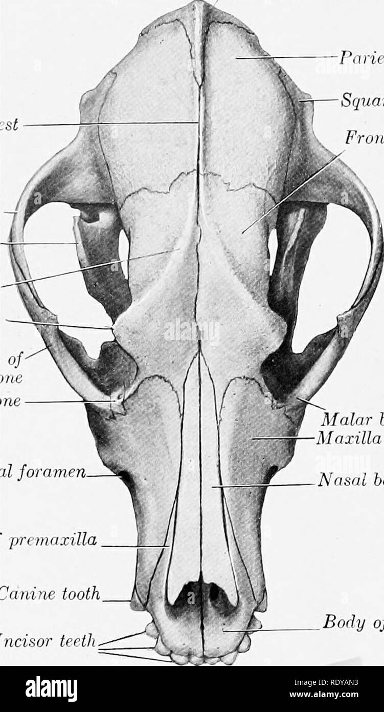
. The anatomy of the domestic animals . Veterinary anatomy. 190 SKELETON OF THE DOG extensively with the corresponding process of the malar. The articular surface for the condyle of the mandible consists of a transverse groove which is continued upon the front of the large postglenoid process. Behind the latter is the lower opening of the temporal canal. There is no condyle. The mastoid part is small, but bears a distinct mastoid process. The external acoustic meatus is wide and very short, so that one can see into the tympanum in the dry skull. The bulla ossea is very large and is rounded and smooth; its medial side is united to the basilar part of the occipital bone. Above this junction and roofed in by the union of the petrous part and the basioccipital is the petro-basilar canal (Canalis petrobasilaris) ; this transmits a vein from the floor of the cranium to the foramen lacerum posterius. The latter opens into a narrow depression behind the IjuUa ossea. It transmits the Interparietal bone Parietal bone Squamous te?nporal bone Parietal crest ^^J |i sKk „ trontal bone Zygomatic process of temporal hone Coronoid process Frontal crest Supraorbital process Zygomatic process malar bo Lacrimal bo. Infraorbital foramen. Nasal process of premaxilla Canine tooth Incisor teeth. Malar bone Maxilla Nasal bone Body of premaxilla Fig. 209.—Skull of Dog; Dohsal View. ninth, tenth, and eleventh cranial nerves. The carotid canal branches off from the petro-basilar, passes forward lateral to it through the medial part of the bulla ossea, and opens in front at the carotid foramen; it transmits the internal carotid artery. The Eustachian opening is immediately lateral to the carotid foramen. The muscular and hyoid processes are extremely rudimentary. The petrous part projects into the cranial cavity and forms a sharp prominent petrosal crest. The medial surface presents a deep floccular fossa above the internal acoustic meatus. The anterior surface is also free. The anterior an