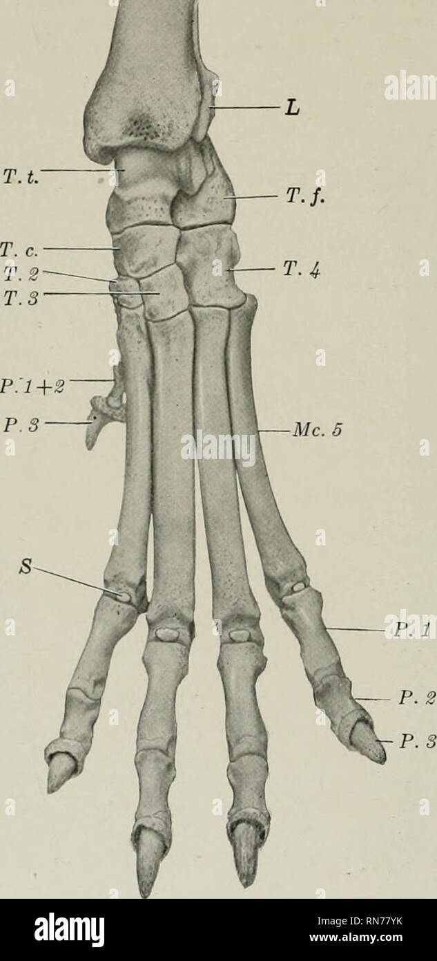
. The anatomy of the domestic animals. Veterinary anatomy. 206 THE ARTICULATIONS OR JOINTS first tarsal and furnishes insertion to the tibialis anterior muscle. In some cases it fuses with the first tarsal; when the first digit is well developed, its metatarsal may resemble the others (except in size) or be reduced in its proximal part to a fibrous band. The other metatarsals are a little longer than the corresponding metacarpals. Their proximal ends are elongated from before backward and have plantar projections, which in the case of the third and fourth usually have facets. Fig. 228.—Skeleto

Image details
Contributor:
Library Book Collection / Alamy Stock PhotoImage ID:
RN77YKFile size:
7.1 MB (180.9 KB Compressed download)Releases:
Model - no | Property - noDo I need a release?Dimensions:
1103 x 2265 px | 18.7 x 38.4 cm | 7.4 x 15.1 inches | 150dpiMore information:
This image is a public domain image, which means either that copyright has expired in the image or the copyright holder has waived their copyright. Alamy charges you a fee for access to the high resolution copy of the image.
This image could have imperfections as it’s either historical or reportage.
. The anatomy of the domestic animals. Veterinary anatomy. 206 THE ARTICULATIONS OR JOINTS first tarsal and furnishes insertion to the tibialis anterior muscle. In some cases it fuses with the first tarsal; when the first digit is well developed, its metatarsal may resemble the others (except in size) or be reduced in its proximal part to a fibrous band. The other metatarsals are a little longer than the corresponding metacarpals. Their proximal ends are elongated from before backward and have plantar projections, which in the case of the third and fourth usually have facets. Fig. 228.—Skeleton op Distal Part of Left Pelvic Limb of Dog; Dorsal View. L, Lateral malleolus (distal end of fibula); T. t., tibial tarsal bone; T. /., fibular tarsal bone; T. c, central tarsal bone; T. 3, T. 3, T. 4, second, third, and fourth tarsal bones: P. 1 + S, fused first and second phalanges, and P. S, third phalanx, of first digit; Mc. 5, fifth metacarpal bone; P. 1, P. 2, P. 3, phalanges of fifth digit; S, dorsal sesamoid. for articulation with two small rounded sesamoid bones. In other respects they resemble the metacarpals. The first digit is often absent. When present, its development varies and it contains one or two phalanges. In other cases—especially in very large dogs— a sixth digit is present; it does not articulate with the metatarsus, but is attached by fibrous tissue. The phalanges of the other digits resemble those of the tho- racic limb. Ossification of the metatarsal bones and phalanges is comjjlete at five or six months.. Please note that these images are extracted from scanned page images that may have been digitally enhanced for readability - coloration and appearance of these illustrations may not perfectly resemble the original work.. Sisson, Septimus, 1865-1924. Philadelphia, London, W. B. Saunders Company