. The anatomy of the domestic animals . Veterinary anatomy. BONES OF THE SKULL—CRANIUM 167 downward, and is continuous with the temporal crest. Two divergent ridges pass upward from the foramen magnum, and the surface between them is concave and smooth. The greater part of the cerebral surface of the squamous part is united with the parietal bones, but a ventral concave area faces into the cranial cavity. The foramen magnum is almost triangular, and is narrow above, where it is flanked by two small tuberosities. The paramastoid processes are extremely long and pro- ject almost straight ventral
Image details
Contributor:
The Book Worm / Alamy Stock PhotoImage ID:
RDYB9BFile size:
7.1 MB (288.6 KB Compressed download)Releases:
Model - no | Property - noDo I need a release?Dimensions:
1752 x 1426 px | 29.7 x 24.1 cm | 11.7 x 9.5 inches | 150dpiMore information:
This image is a public domain image, which means either that copyright has expired in the image or the copyright holder has waived their copyright. Alamy charges you a fee for access to the high resolution copy of the image.
This image could have imperfections as it’s either historical or reportage.
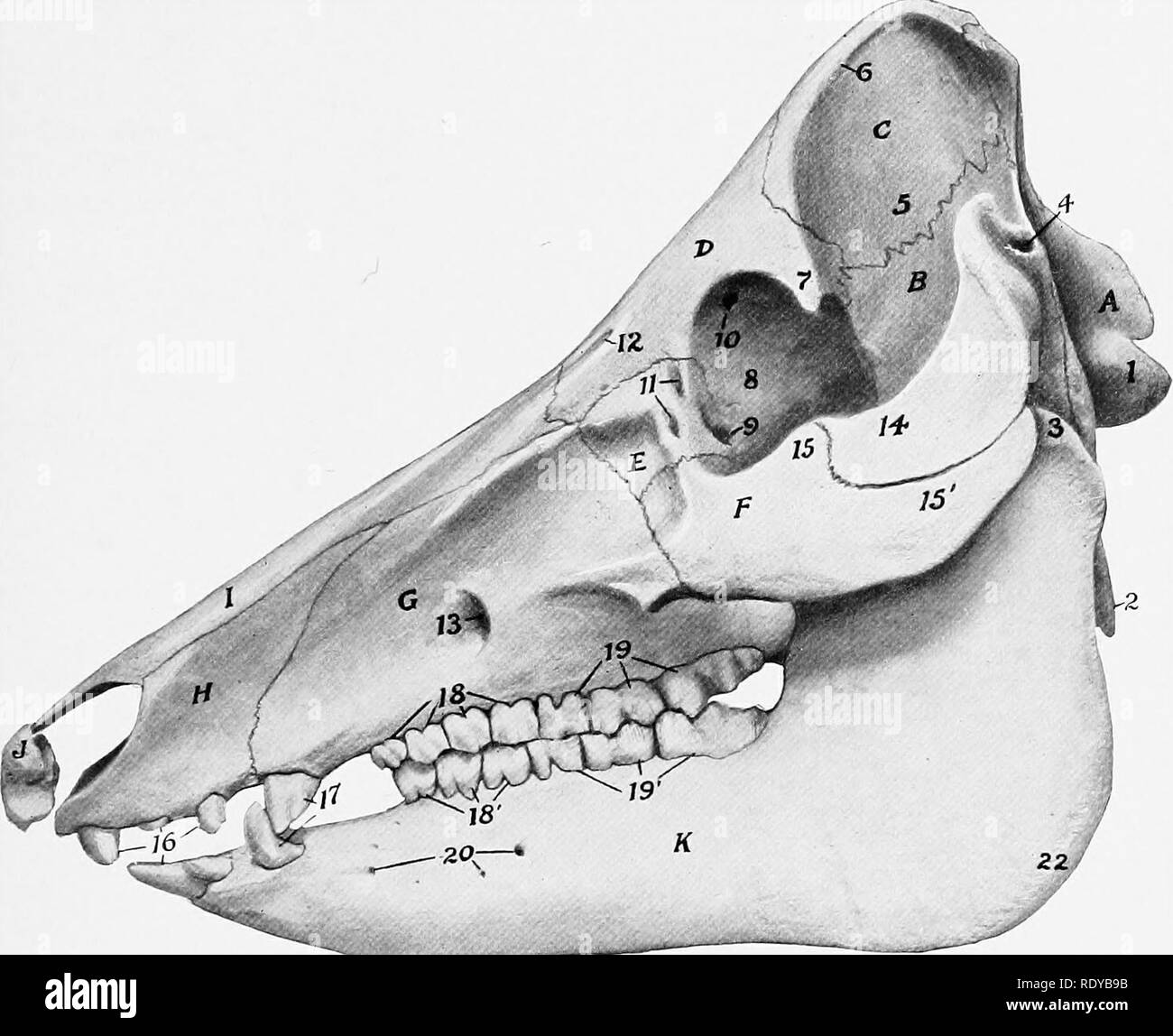
. The anatomy of the domestic animals . Veterinary anatomy. BONES OF THE SKULL—CRANIUM 167 downward, and is continuous with the temporal crest. Two divergent ridges pass upward from the foramen magnum, and the surface between them is concave and smooth. The greater part of the cerebral surface of the squamous part is united with the parietal bones, but a ventral concave area faces into the cranial cavity. The foramen magnum is almost triangular, and is narrow above, where it is flanked by two small tuberosities. The paramastoid processes are extremely long and pro- ject almost straight ventrally. The hypoglossal foramen is at the medial side of the root of the process. The basilar part is short and wide; its ventral surface. sj - Fig. 177.—Skull of Pig; Lateral View. A, Occipital bone; B, squamous temporal bone; C, parietal bone; D, frontal bone; E^ lacrimal bone; F, malar bone; G', maxilla; H, premaxilla; /, nasal bone; J, os rostri; K, mandible; 1, occipital condyle; 2, paramastoid proc- ess; 3, condyle of mandible; 4, meatus acusticus externus; 5, temporal fossa; 6, parietal crest; 7, supraorbital process; 8, orbital part of frontal bone; 9, fossa for origin of ventral oblique muscle of eyeball; 10, orbital opening of supraorbital canal; 11, lacrimal foramina; 12, supraorbital foramen and groove; 13, infraorbital foramen; 14, zygomatic process of temporal bone; 15, temporal, and 15', zygomatic, process of malar bone; 16, incisor teeth; 17, canine teetli; 18, IS', premolars; 19, 19', molars; 20, mental foramina; 21, mental prominence; 22, angle of mandible. bears a thin median ridge and two lateral imprints or tubercles which converge at the junction with the sphenoid bone. The interparietal bone fuses before birth with the occipital. The internal occipital protuberance is absent. The parietal bone is overlapped by the occipital bone behind and concurs in the formation of the nuchal crest. Its external surface is divided by the parietal crest into two parts. T