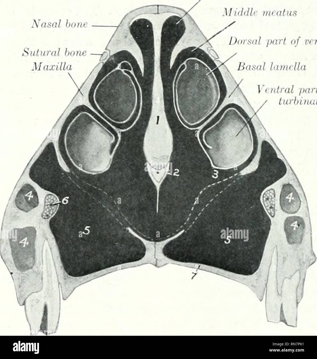
. The anatomy of the domestic animals. Veterinary anatomy. Fig. 138.—Cranium of Jebset Cow, Nuchal View. The Fig. l.TO—Cranium of Polled ..-gus Cow. Ndchal Sawn Off. 1, Foramen magnum; 2, occipital condyle; 3, paramastoid process: 4, bulla ossea; .'». meatus acusticua extornus; 6, mastoid foramen; 7, external occipital protuberance; 8, median occipital crest; 9, liuea nuchse superior; 10, frontal Nasal bone Dorsal miatiia Mid.Ui mmtiis Fig. 140.—CRoaa-i ? Nasal Reg Dnisiil part iif i'( Hlrdl lurhinalc Basal lanalln 'i idral part of I'cidral tiirhinaU-'. ?KULL OF Ox. Section Pa Third Cheek
Image details
Contributor:
Library Book Collection / Alamy Stock PhotoImage ID:
RN7P61File size:
7.1 MB (243.5 KB Compressed download)Releases:
Model - no | Property - noDo I need a release?Dimensions:
1536 x 1627 px | 26 x 27.6 cm | 10.2 x 10.8 inches | 150dpiMore information:
This image is a public domain image, which means either that copyright has expired in the image or the copyright holder has waived their copyright. Alamy charges you a fee for access to the high resolution copy of the image.
This image could have imperfections as it’s either historical or reportage.
. The anatomy of the domestic animals. Veterinary anatomy. Fig. 138.—Cranium of Jebset Cow, Nuchal View. The Fig. l.TO—Cranium of Polled ..-gus Cow. Ndchal Sawn Off. 1, Foramen magnum; 2, occipital condyle; 3, paramastoid process: 4, bulla ossea; .'». meatus acusticua extornus; 6, mastoid foramen; 7, external occipital protuberance; 8, median occipital crest; 9, liuea nuchse superior; 10, frontal Nasal bone Dorsal miatiia Mid.Ui mmtiis Fig. 140.—CRoaa-i ? Nasal Reg Dnisiil part iif i'( Hlrdl lurhinalc Basal lanalln 'i idral part of I'cidral tiirhinaU-'. ?KULL OF Ox. Section Pa Third Cheek Tooth. 1, Cartilage of .sGi)tum nasi; 2. vomer; 3, ventral meatus; 4. anterior extremity of maxillary sinus; 5, palatine sinus; 6, infraorbital canal and nerve; 7, palatine process of maxilla. Dotted lines indicate mucous membrane which closes gap in bony floor of nasal canty. Tlu' cranial cavity is shorter and its long axis is more ohliciuc than in tiie horse, but it is relatively high and wide. The anterior fossa lies at a much higher level. Please note that these images are extracted from scanned page images that may have been digitally enhanced for readability - coloration and appearance of these illustrations may not perfectly resemble the original work.. Sisson, Septimus, 1865-1924. Philadelphia Saunders