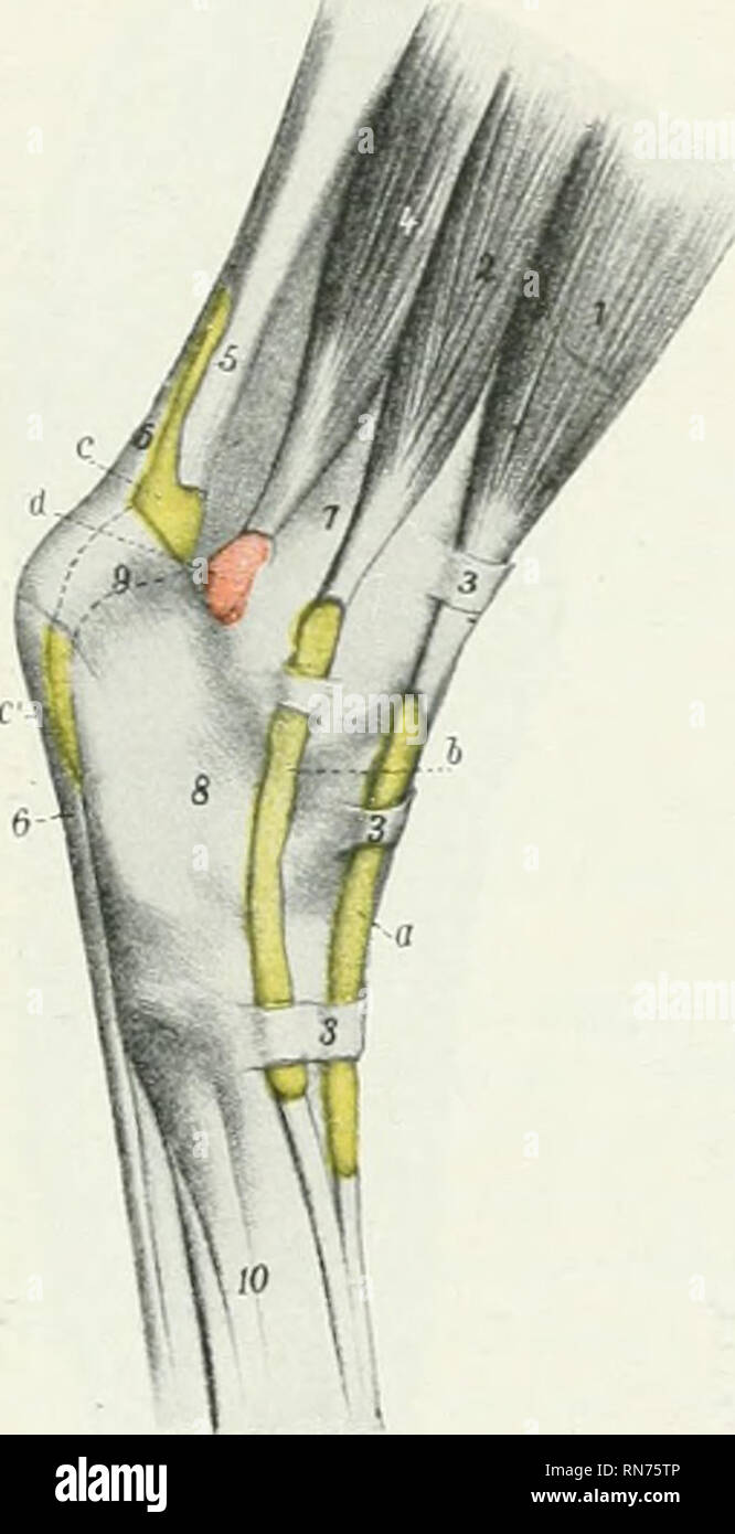
. The anatomy of the domestic animals. Veterinary anatomy. FiG. 295.—Injected Synovial Sheaths a OF Tarsal Region of Horse; Latera J BCRS-? View. a, Syno^aa! sheath of long digital extensor; b, synovial sheath of lateral digital extensor; c, c', bursa under superfioiiil Mexur tendon; d, capsule of hock joint; 1. long extensor; ^^ lateral extensor; 3, 3, 3, annular hgaments; ^, deep dis^ital flexor; -5, tendon of gastroc- nemius; 6, superScial flexor tendon; 7, tibia; S, tarsus; 9, tuber calcis; 10, metatarsus. (After EUenberger, in I^isering's Atlas.) Fig. 294.—Injected Synovial Sheaths and Bt
Image details
Contributor:
Library Book Collection / Alamy Stock PhotoImage ID:
RN75TPFile size:
7.1 MB (187.6 KB Compressed download)Releases:
Model - no | Property - noDo I need a release?Dimensions:
1132 x 2207 px | 19.2 x 37.4 cm | 7.5 x 14.7 inches | 150dpiMore information:
This image is a public domain image, which means either that copyright has expired in the image or the copyright holder has waived their copyright. Alamy charges you a fee for access to the high resolution copy of the image.
This image could have imperfections as it’s either historical or reportage.
. The anatomy of the domestic animals. Veterinary anatomy. FiG. 295.—Injected Synovial Sheaths a OF Tarsal Region of Horse; Latera J BCRS-? View. a, Syno^aa! sheath of long digital extensor; b, synovial sheath of lateral digital extensor; c, c', bursa under superfioiiil Mexur tendon; d, capsule of hock joint; 1. long extensor; ^^ lateral extensor; 3, 3, 3, annular hgaments; ^, deep dis^ital flexor; -5, tendon of gastroc- nemius; 6, superScial flexor tendon; 7, tibia; S, tarsus; 9, tuber calcis; 10, metatarsus. (After EUenberger, in I^isering's Atlas.) Fig. 294.—Injected Synovial Sheaths and BtjRs^ OF Tarsal Region of Horse; Medll View. o. Synovial sheath of peroneus tertius and tibialis anterior; h, bursa under medial (cunean) tendon of tibialis anterior; c, synovial sheath of flexor longus; d, tarsal sheath of deep flexor; e, e', bursa under superficial flexor tendon; /, /', tibio-tarsal joint capsule; 1, long extensor; 2, tibialis anterior; 3', medial (cun- ean) tendon of 2; 3, flexor longus; 4< deep digital flexor; 5, superficial flexor tendon; G, gastrocnemius tendon; 7, tibia; 5, tarsus; 0, tuber calcis; 10, large metatarsal bone; 11, medial small metatarsal bone; i 3, 13', fascial bands. (After EUenberger, in Leisering's Atlas.) tion. The latter emerges between the branches of the peroneus tertius and bi- furcates, the anterior branch being inserted into the large metatarsal bone, the medial one into the first tarsal bone. The tendon is provided with a synovial sheath at its emergence, and a bursa is interposed between the medial l^ranch and the medial ligament of the hock.^ Relatiotis.—Superficially', the long and lateral extensors, the peroneus tertius, and the deep peroneal nerve; deeply, the tibia, the deep flexor, and the anterior tibial vessels, * In surgical works the medial branch is termed the cunean tendon; it is sometimes resected for the relief of bone spaan.. Please note that these images are extracted from scanned page images that may