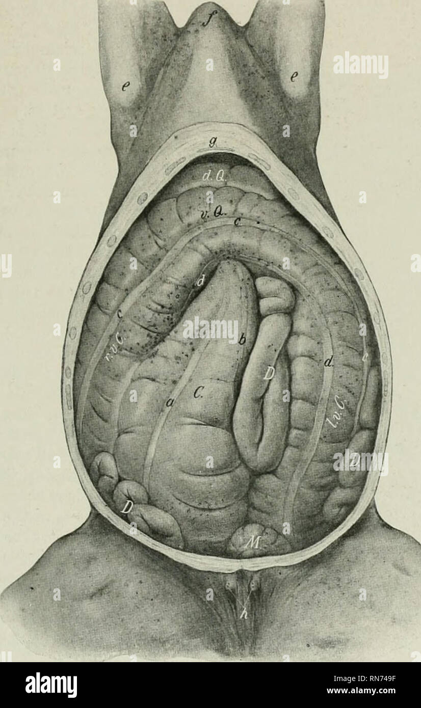
. The anatomy of the domestic animals. Veterinary anatomy. THE GREAT COLON 427 the medial surface of the base of the caecum it turns dorsally and to the left behind the left sac of the stomach; here it becomes constricted, and joins the small colon lielow the left kidney. The sternal flexure extends forward to a point opposite to the ventral part of the seventh rib, and the diaphragmatic flexure as far as the sixth intercostal space on the right of the median plane. The caliber of the great colon varies greatly at different jjoints. At its origin. Fig. 367.—.AlBdominal Viscera of Horse; ^'E^•T

Image details
Contributor:
Library Book Collection / Alamy Stock PhotoImage ID:
RN749FFile size:
7.1 MB (294.3 KB Compressed download)Releases:
Model - no | Property - noDo I need a release?Dimensions:
1260 x 1982 px | 21.3 x 33.6 cm | 8.4 x 13.2 inches | 150dpiMore information:
This image is a public domain image, which means either that copyright has expired in the image or the copyright holder has waived their copyright. Alamy charges you a fee for access to the high resolution copy of the image.
This image could have imperfections as it’s either historical or reportage.
. The anatomy of the domestic animals. Veterinary anatomy. THE GREAT COLON 427 the medial surface of the base of the caecum it turns dorsally and to the left behind the left sac of the stomach; here it becomes constricted, and joins the small colon lielow the left kidney. The sternal flexure extends forward to a point opposite to the ventral part of the seventh rib, and the diaphragmatic flexure as far as the sixth intercostal space on the right of the median plane. The caliber of the great colon varies greatly at different jjoints. At its origin. Fig. 367.—.AlBdominal Viscera of Horse; ^'E^•TRAL View. The ventral wall and part of the lateral walls of the abdomen are removed. C, Caecum; r.v.C, right ventral part of colon; v.Q., sternal flexure of colon; [Lv. C, left ventral part of colon; d.Q., diaphragmatic flexure of colon; D., small intestine: M, , small colon; a. ventral free band of caecum: b, medial band of CEBCum; c, lateral band of ventral part of colon d, ventral band of ventral part of colon; e, point of elbow; /, anterior end of sternal region; g, xiphoid cartilage; h, teats. (After Ellenberger-Baum, Top. Anat. d. Pferdes.) it is only about two to three inches (ca. 5 to 7.5 cm.) in diameter.' This soon increases to about eight to ten inches (ca. 20 to 25 cm.) for the ventral parts. Beyond the pelvic flexure the diameter is reduced to about three or four inches (ca. 8 to 9 cm.). Near the diaphragmatic flexure the caliber rapidly increases, and reaches its maximum in the last part, where it forms a large sacculation, which may 1 Usually there is a sacculation of considerable size wliich succeeds the constricted origin.. Please note that these images are extracted from scanned page images that may have been digitally enhanced for readability - coloration and appearance of these illustrations may not perfectly resemble the original work.. Sisson, Septimus, 1865-1924. Philadelphia, London, W. B. Saunders Company