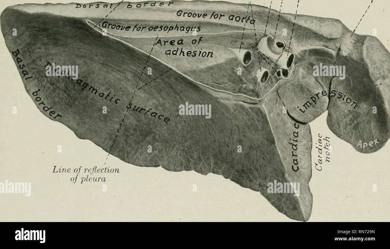
. The anatomy of the domestic animals. Veterinary anatomy. THE LUNGS 533 The ventral border (Margo ventralis) is thin and short; it occupies the angular space between the mediastinum and the ventral parts of the sternal ribs (Recessus costo-mediastinalis). It presents, opposite to the heart, the cardiac notch (Incisura cardiaca). On the left lung this notch is opposite to the ribs from the third to tlie sixth, so that a considerable area of the pericardium here lies in direct contact with the chest-wall. On the right lung the notch is much smaller, and extends from the third rib to the fourth
Image details
Contributor:
Library Book Collection / Alamy Stock PhotoImage ID:
RN729NFile size:
7.2 MB (263.6 KB Compressed download)Releases:
Model - no | Property - noDo I need a release?Dimensions:
2094 x 1194 px | 35.5 x 20.2 cm | 14 x 8 inches | 150dpiMore information:
This image is a public domain image, which means either that copyright has expired in the image or the copyright holder has waived their copyright. Alamy charges you a fee for access to the high resolution copy of the image.
This image could have imperfections as it’s either historical or reportage.
. The anatomy of the domestic animals. Veterinary anatomy. THE LUNGS 533 The ventral border (Margo ventralis) is thin and short; it occupies the angular space between the mediastinum and the ventral parts of the sternal ribs (Recessus costo-mediastinalis). It presents, opposite to the heart, the cardiac notch (Incisura cardiaca). On the left lung this notch is opposite to the ribs from the third to tlie sixth, so that a considerable area of the pericardium here lies in direct contact with the chest-wall. On the right lung the notch is much smaller, and extends from the third rib to the fourth intercostal space. The left cardiac notch is usually quadrilateral; its highest part is about four to five inches (ca. 10-12 cm.) above the sternal ends of the fourth and fifth ribs. The right notch is usually- triangular; its apex is about three inches (ca. 7-8 cm.) above the level of the sternal end of the ribs at the third intercostal space. In some cases a fissure partially marks off the apex from the body of the lung. The base of the lung (Basis pulmonis) is oval in outline; its surface (Facies Ligament of lung {cut) Bronchial artery I Bronchus Pulmonary I i Pulmonary Vascular veins j j artery impression ^''^<'<'^/or aori/x ' I '', , .'? oesophagus < . , ' ' / -. lAne of reJlectio7i of pleura Fin. 475.—Left LrxG of Horse; Mediastinal and Diaphhagmatic Surfaces. Organ hardened in situ. Vascular impression for common dorso-cervico-vertebral ^ diaphragmatica) is deeply concave in adaptation to the thoracic surface of the diaphragm. Laterally and dorsally it is limited by a thin convex basal border (Margo basalis) which fits into the narrow recess (Sinus phrenico-costalis) between the diaphragm and the lateral chest-wall. The position of this border, of course, varies during respiration. In the deepest inspiration it may reach the bottom of this recess. In dissecting-room sulDJects the distance between the border and the diaphrag- matic line of reflection of the pleura