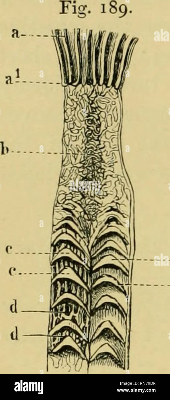
. The anatomy of the frog. Frogs -- Anatomy; Amphibians -- Anatomy. 288 THE ALIMENTARY TRACT, ETC.. these folds and between them are smaller, secondary folds, partly irregularly arranged, partly longitvidinal [d d). Towards the middle of the small intestine this valvular arrangement is lost, to be replaced by an irregular net-like folding ; beyond this longitudinal folds arise, which proceed in a sinuous course towards the large intestine. The whole mucous membrane, both on the folds and otherwise, is covered with a simple layer of columnar epithelium, which is con- tinued into numerous simple

Image details
Contributor:
Library Book Collection / Alamy Stock PhotoImage ID:
RN79DRFile size:
7.2 MB (251.3 KB Compressed download)Releases:
Model - no | Property - noDo I need a release?Dimensions:
1068 x 2340 px | 18.1 x 39.6 cm | 7.1 x 15.6 inches | 150dpiMore information:
This image is a public domain image, which means either that copyright has expired in the image or the copyright holder has waived their copyright. Alamy charges you a fee for access to the high resolution copy of the image.
This image could have imperfections as it’s either historical or reportage.
. The anatomy of the frog. Frogs -- Anatomy; Amphibians -- Anatomy. 288 THE ALIMENTARY TRACT, ETC.. these folds and between them are smaller, secondary folds, partly irregularly arranged, partly longitvidinal [d d). Towards the middle of the small intestine this valvular arrangement is lost, to be replaced by an irregular net-like folding ; beyond this longitudinal folds arise, which proceed in a sinuous course towards the large intestine. The whole mucous membrane, both on the folds and otherwise, is covered with a simple layer of columnar epithelium, which is con- tinued into numerous simple follicles (glands of Lieberkiihn) found throughout the mucous membrane of the small intestine. The cells are placed on a basement membrane, which rests on a thin layer of loose connective-tissue, intervening betAveen the epithelial coat and the muse/dan's mucosae. The ejiithelial cells are intermixed with a large number of goblet- cells, and have between them fine processes from the connective-tissue corpuscles of the subjacent layer; many of these processes extend to or even l)eyond the free margin of the epithelial cells. The individual cells are columnar, possessed of a well-marked cell-wall, and have distinct, large, oval nuclei, containing one or more nucleoli. The protoplasmic contents are granular, and with proper treatment show a very distinct intra- cellular network. The free margins of the cells are sharply marked off from the cell-contents, and are more firmly attached to the cor- responding portions of adjacent cells than the rest of the cell-wall. This margin has a longitudinal striation, which owdng to the im- portant function performed by this part of the intestine, namely, absorption of the fat, has been the subject of many important investigations. [In the following brief summary of the earlier researches on the minute structure of the intestinal epithelium, in which the intestine of the frog was chiefly used, the memoirs in which these investigations are reco