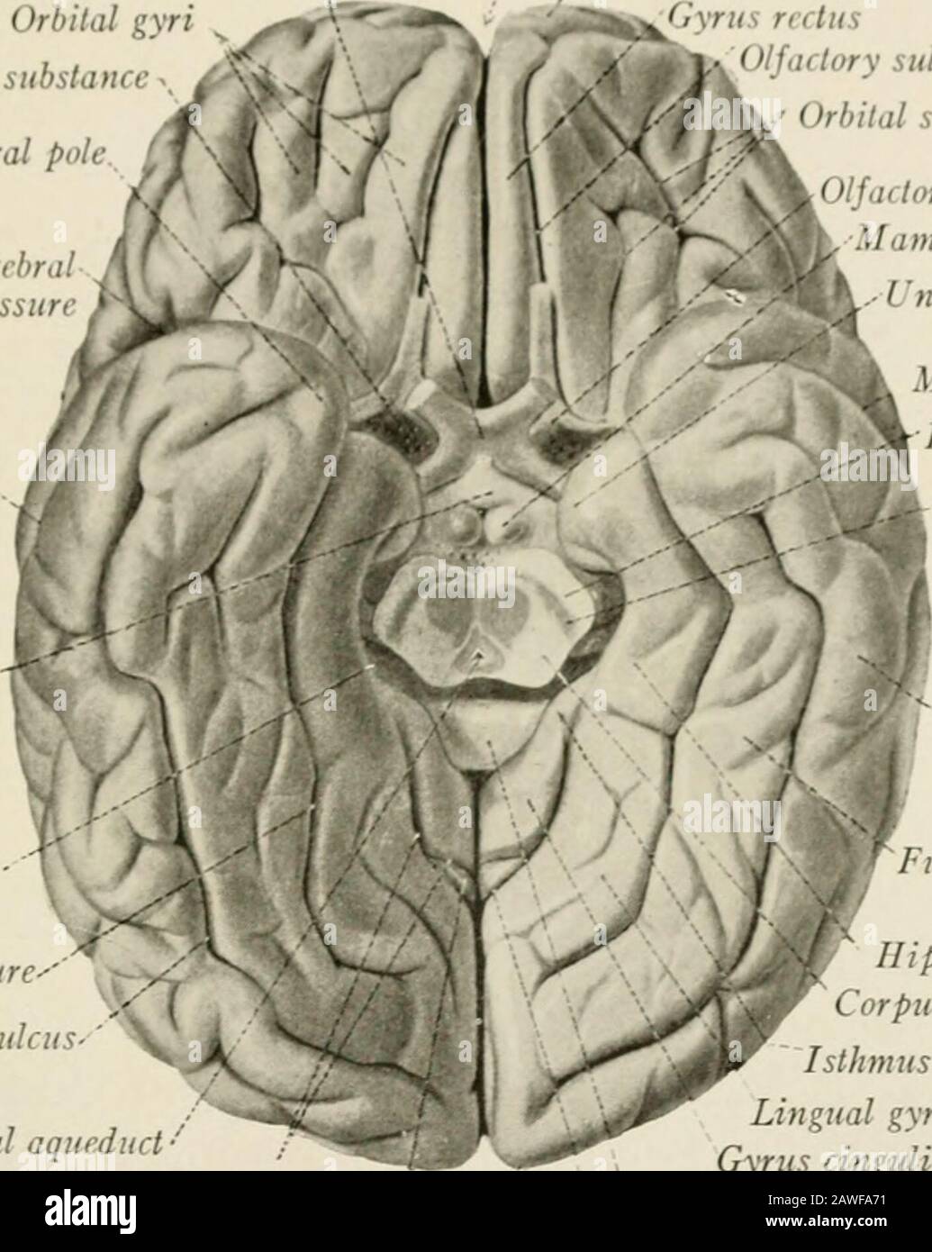
The anatomy of the nervous system, from the standpoint of development and function . urfacesof the cerebral hemisphere to the frontal, parietal, occipital, and temporallobes, as indicated in Fig. 171. According to this scheme the gyrus fornicatus 1111 EXTERNAL CONFIGURATION OP I III CEREBRAL II I. MI-Ill I 241 stand- by it-di ami i- sometimes designated a- tin- limbic lobe. This plan ofsubdivision, which was based n the erroneous belief that all portions of thegyrus fornicatus belonged t the rhinencephalon, should In- abandoned. Asimpler ami more logical arrangement assigns the hippocampal gyr

Image details
Contributor:
The Reading Room / Alamy Stock PhotoImage ID:
2AWFA71File size:
7.1 MB (275.4 KB Compressed download)Releases:
Model - no | Property - noDo I need a release?Dimensions:
1411 x 1771 px | 23.9 x 30 cm | 9.4 x 11.8 inches | 150dpiMore information:
This image is a public domain image, which means either that copyright has expired in the image or the copyright holder has waived their copyright. Alamy charges you a fee for access to the high resolution copy of the image.
This image could have imperfections as it’s either historical or reportage.
The anatomy of the nervous system, from the standpoint of development and function . urfacesof the cerebral hemisphere to the frontal, parietal, occipital, and temporallobes, as indicated in Fig. 171. According to this scheme the gyrus fornicatus 1111 EXTERNAL CONFIGURATION OP I III CEREBRAL II I. MI-Ill I 241 stand- by it-di ami i- sometimes designated a- tin- limbic lobe. This plan ofsubdivision, which was based n the erroneous belief that all portions of thegyrus fornicatus belonged t the rhinencephalon, should In- abandoned. Asimpler ami more logical arrangement assigns the hippocampal gyrus and uncusto the temporal lobe and divides the gyrus cinguli between the frontal andparietal lobes. Optic chiasma Orbital gyriAnterior perforated substance .. /. mporal pole Lateral cerebral(Sylvian) fissure Longitudinal fi urt of cerebrum frontal pole Gyrus rectus ^^0//./( lory sulcus M Vj. Orbital sulci &/ V^k Olfn lory trigone Mammillary body.-1 ncus Middle temporal sulcus- Tuber cinereum Hippocampal fissure- T Collateral fissure-Inferior temporal sulcus. Cerebral aqueduct • Collateral fissure Cuneus Middle temporal sulcusBase of cerebral peduncleSubstantia nigra Inferior temporal gyrus Fusiform gyrus Hippocampal gyrusCorpus quadrigeminumIsthmus of gyrus fornicatusLingual gyrusGyrus cinguliSplenium of corpus callosumParieto-occipital fissureOccipital pole (Sobotta-McMurrich.) Fig. 172.—Basal aspect of the human cerebral hemisphere The basal surface of the hemisphere (Fig. 172) consists of two parts: (1)the ventral surface of the temporal lobe, whose sulci and gyri have been de-scribed in a preceding paragraph, and which rests upon the tentorium cerebelliand the floor of the middle cranial fossa; and (2) the orbital surface of the frontallobe resting upon the floor of the anterior cranial fossa. The latter surfacepresents near its medial border the olfactory sulcus, a straight, deep furrow, directed rostrally and somewhat medially, that lodges the olfactory tract andbul