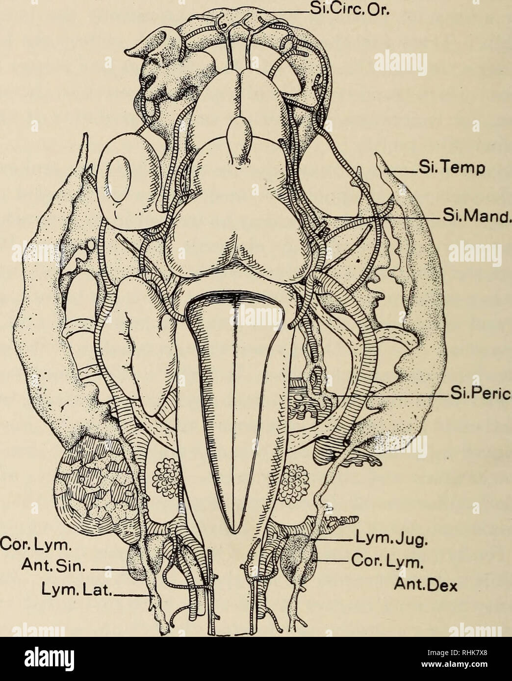
. The biology of the amphibia. Amphibians. 196 THE BIOLOGY OF THE AMPHIBIA of the tadpole, emptying into the vertebral vein, and a caudal pair, one on either side of the coccyx, pumping the lymph into a branch of the ischiadic vein. These hearts may be readily observed by removing the skin from the end of the coccyx.. Fig. 75.—Reconstruction of the lymphatic vessels of the head of a toad tadpole showing their relation to the larger blood vessels. Cor.Lym.Ant.Dex., right anterior lymph heart; Cor.Lym.Ant.Sin., left anterior lymph heart; Lym. Jug., lymphatica jugularis; Lym.Lat., lymphatica late
Image details
Contributor:
Library Book Collection / Alamy Stock PhotoImage ID:
RHK7X8File size:
7.2 MB (428.3 KB Compressed download)Releases:
Model - no | Property - noDo I need a release?Dimensions:
1417 x 1764 px | 24 x 29.9 cm | 9.4 x 11.8 inches | 150dpiMore information:
This image is a public domain image, which means either that copyright has expired in the image or the copyright holder has waived their copyright. Alamy charges you a fee for access to the high resolution copy of the image.
This image could have imperfections as it’s either historical or reportage.
. The biology of the amphibia. Amphibians. 196 THE BIOLOGY OF THE AMPHIBIA of the tadpole, emptying into the vertebral vein, and a caudal pair, one on either side of the coccyx, pumping the lymph into a branch of the ischiadic vein. These hearts may be readily observed by removing the skin from the end of the coccyx.. Fig. 75.—Reconstruction of the lymphatic vessels of the head of a toad tadpole showing their relation to the larger blood vessels. Cor.Lym.Ant.Dex., right anterior lymph heart; Cor.Lym.Ant.Sin., left anterior lymph heart; Lym. Jug., lymphatica jugularis; Lym.Lat., lymphatica lateralis; Si.Circ.Or., circumoral division of sinus maxillaris primigenius; Si.Mand., mandibular division; Si.Peri- card., pericardial division; Si.Temp., temporal division. (After Kampmeier.) Their beating is independent of that of the heart or of the other lymph hearts. It is, nevertheless, under nervous control since cutting away the spinal cord destroys the beat. The lymph heart tissue is thus neither structurally nor functionally similar to heart tissue (Briicke and Umrath, 1930). The number of lymph hearts varies with the species. In the primitive Ascaphus. Please note that these images are extracted from scanned page images that may have been digitally enhanced for readability - coloration and appearance of these illustrations may not perfectly resemble the original work.. Noble, Gladwyn Kingsley, 1894-1940. New York : McGraw-Hill