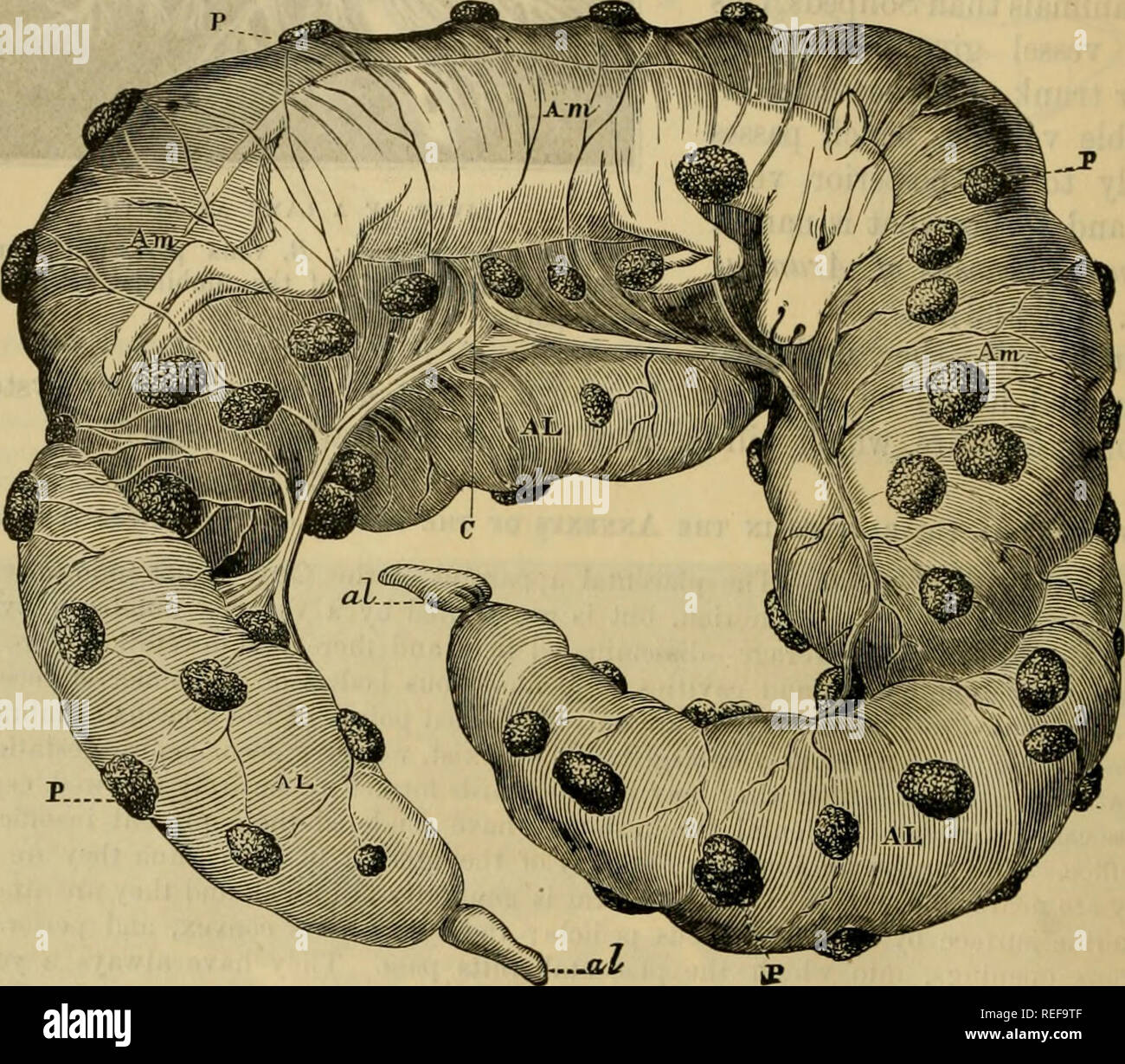
. The comparative anatomy of the domesticated animals. Veterinary anatomy. 1028 EMBRYOLOGY. chorion. This sac, which is an expansion of the urachus, is always reversed on one of the sides of tlie amnion ; its two braiicbes are sacculated on their surface like the large intestine ami the greatest forms a cul-de. sac. or conical diverticulum. * The extremities of the allantoid coraua appear to have pierced the cliorion ; they form a point covered with a yellow, mucus substance, and separated from the rest of the membrane by a circular constricticu. This part is beneath the chorion, like the rema

Image details
Contributor:
The Book Worm / Alamy Stock PhotoImage ID:
REF9TFFile size:
7.2 MB (570.9 KB Compressed download)Releases:
Model - no | Property - noDo I need a release?Dimensions:
1689 x 1480 px | 28.6 x 25.1 cm | 11.3 x 9.9 inches | 150dpiMore information:
This image is a public domain image, which means either that copyright has expired in the image or the copyright holder has waived their copyright. Alamy charges you a fee for access to the high resolution copy of the image.
This image could have imperfections as it’s either historical or reportage.
. The comparative anatomy of the domesticated animals. Veterinary anatomy. 1028 EMBRYOLOGY. chorion. This sac, which is an expansion of the urachus, is always reversed on one of the sides of tlie amnion ; its two braiicbes are sacculated on their surface like the large intestine ami the greatest forms a cul-de. sac. or conical diverticulum. * The extremities of the allantoid coraua appear to have pierced the cliorion ; they form a point covered with a yellow, mucus substance, and separated from the rest of the membrane by a circular constricticu. This part is beneath the chorion, like the remainder of the membrane, only the vessels do not extend beyond the constriction; so that the elements of the chorion and allantois here undergo a kind of murtiticati(jn. The allantoid infundibulum is encircled by a vascular network that accompanies it throughout tue umbilical cord. The epithelium of the allantois is every wliere colourable by iodine reagents, in Kuniiuants. At times the hippomanes is Ibund floating in the liquid it contains. Jmntow.—Altogether like that of Solipeds, tiiis membrane is readily resolved into two Fig. 564.. FCETUS OP THE SHEEP, FREED FROM ITS CONNECTION WITH THE UTERUS. AL AL, Allantois slightly inflated, seen beneath the chorion; Am Am Am, amnion slightly dis- tended with fluid underneath the chorioa ; P, P, P, placentae on the surface of the chorion ; C, umbilical chord ; al, al, extremities of the allantoidean cornua, looking as if protruding through the chorion. laminae, and presents on its inner surface a great number of little, yellowisii-white, epidermic patches, more especially visible on the amniotic covering of the cord. The epithelium is only stained by iodine at these patches, or villi. These productions are surrounded at their base by a girdle of glycogenic cells. In the foetus of the Cow, at a late stage of gestation, the amniotic fluid is not abundant, and becomes white and viscid; in one instance we found it stringy, like a solution