. The comparative anatomy of the domesticated animals. Veterinary anatomy. 66 THE BONES. shows the continuation of the foramen. These three faces Fig. 38.. POSTERIOR ASPECT OF HORSE'S SKULL. , Occipital tuberosity; 2, fora- men magnum; 3, 3, occipital condyles ; 4, 4, styloid pro- cesses ; 5, 5, petrous bone; 6, basilar process; 7, pterygoid fissure of the sphenoid bone ; 8, foramen lacerum ; 9, 9, supra- condyloid, or anterior mastoid process ; 10, 10, articular emi- nence, or temporal condyle; 11, body of sphenoid bone; 12, ptery- goid process ; 13, ethmoid bone ; 14, temporal bone and sphen
Image details
Contributor:
The Book Worm / Alamy Stock PhotoImage ID:
REFE7TFile size:
7.2 MB (361.7 KB Compressed download)Releases:
Model - no | Property - noDo I need a release?Dimensions:
999 x 2503 px | 8.5 x 21.2 cm | 3.3 x 8.3 inches | 300dpiMore information:
This image is a public domain image, which means either that copyright has expired in the image or the copyright holder has waived their copyright. Alamy charges you a fee for access to the high resolution copy of the image.
This image could have imperfections as it’s either historical or reportage.
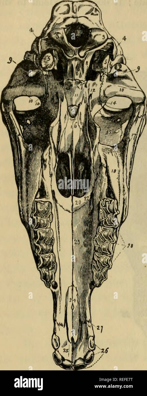
. The comparative anatomy of the domesticated animals. Veterinary anatomy. 66 THE BONES. shows the continuation of the foramen. These three faces Fig. 38.. POSTERIOR ASPECT OF HORSE'S SKULL. , Occipital tuberosity; 2, fora- men magnum; 3, 3, occipital condyles ; 4, 4, styloid pro- cesses ; 5, 5, petrous bone; 6, basilar process; 7, pterygoid fissure of the sphenoid bone ; 8, foramen lacerum ; 9, 9, supra- condyloid, or anterior mastoid process ; 10, 10, articular emi- nence, or temporal condyle; 11, body of sphenoid bone; 12, ptery- goid process ; 13, ethmoid bone ; 14, temporal bone and sphenoidal sutuie; 15, lachrymal bone; 16, vomer; 17, malar bone; 18, maxillary tuberosity ; 19, pos- terior nares, or guttural opening of the nose; 20, palatine bone; 21, palatine styloid process ; 22, palato-maxillary foramen; 23, palatine process of superior maxillary bone, with suture ; 24, ditto of premaxillary bone; 25, premaxillary bone; 26, upper incisor teeth ; 27, point of junc- tion of the premaxillary with the supermaxillary bone ; 28, up- per molar teeth (young mouth). palatine groove, which terminates in the incisive are separated by as many borders : two internah hmitiug the corresponding face before and be- hind ; and an external, separating the labial from the buccal face. The latter only merits notice. It is very thick, and is divided into two parts : an inferior, which describes a curved line, con- cavity upwards, and is excavated by three alveoli for the reception of the incisor teeth ; another, the superior, is straight, vertical, and somewhat sharp, and forms part of the dental interspace. It is limited above, near the base of the external process, by a cavity for the formation of the alveolus of the canine tooth. Processes.—These are distinguished as external and intei'iial. The first, the longest and strongest, is flattened on both sides ; its external face is smooth, and continued with that of the thick por- tion of the bone ; its internal face is covered