. The comparative anatomy of the domesticated animals. Veterinary anatomy. TEE MOUTH, 361 "6. Teeth.—The teeth of the Dog are forty-two in number: twelve incisors, four canines, and twenty-six molars, " The incisors, six in each side of the jaws, are more developed in the superior than in the inferior maxilla, and are divided, as in the Horse, into pincers, intermediates, and corner incisors; the last being much stronger than the preceding, and these again stronger than the pincers. *' Their free part presents, in the virgin tooth, three tubercles: a middle, which is the strongest, a
Image details
Contributor:
Library Book Collection / Alamy Stock PhotoImage ID:
RPXX9RFile size:
7.1 MB (248.2 KB Compressed download)Releases:
Model - no | Property - noDo I need a release?Dimensions:
1655 x 1510 px | 28 x 25.6 cm | 11 x 10.1 inches | 150dpiMore information:
This image is a public domain image, which means either that copyright has expired in the image or the copyright holder has waived their copyright. Alamy charges you a fee for access to the high resolution copy of the image.
This image could have imperfections as it’s either historical or reportage.
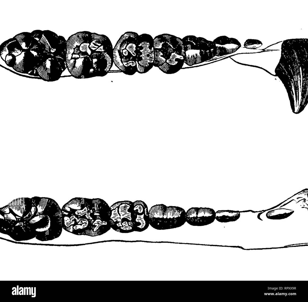
. The comparative anatomy of the domesticated animals. Veterinary anatomy. TEE MOUTH, 361 "6. Teeth.—The teeth of the Dog are forty-two in number: twelve incisors, four canines, and twenty-six molars, " The incisors, six in each side of the jaws, are more developed in the superior than in the inferior maxilla, and are divided, as in the Horse, into pincers, intermediates, and corner incisors; the last being much stronger than the preceding, and these again stronger than the pincers. *' Their free part presents, in the virgin tooth, three tubercles: a middle, which is the strongest, and two lateral; these, together, are not unlike a trefoil or the upper part of a fleur-de-lis, especially those in the upper jaw. On the internal face is remarked a table or slope, somewhat resembling that of the Ox and Sheep, and separated from the root by a very distinct border whose extremities mark the lateral lobes. This table is of no advantage in ascertaining the age. *'The root, very developed, flattened on both sides, and separated from the fi»e Fig. 166.. Fig. 167. GENERAL AND LATERAL VIEW OF THE DOG S TEETH. portion by a well-defined neck, is solidly encased in a deep alveolus. Its internal cavity is very promptly obliterated. " When tiie tooth is submitted to wear, the middle lobe is the first to disappear; so that it no longer resembles a trefoil (fig. 167). " The caducous incisors are much smaller and -more pointed than the permanent ones; yet, like them, they show lateral lobes. At the period of their eruption these teeth are somewhat widely apart. *'The fangs, or canine teeth, two in each jaw, are very strong, elongated organs, conical in form, curved backwards and outwards, and placed immediately after the incisors. *'The upper fangs are the thickest, and have a small space between them and the comer incisors, in which the inferior canines are lodged. " These teeth are deciduous, like the incisors, and are distinguished from the replacing ones