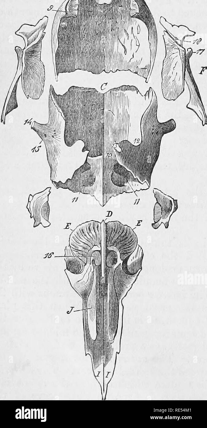
. The comparative anatomy of the domesticated animals. Veterinary anatomy. tuberosities placed on each side of the perpendiciilar lamina, and offering for study a middle portion, a base, and a summit. Bach of these is formed by an assemblage of numerous, extremely thin, osseous plates, curved into small and very fragile convolutions. These, elongated from above to below, become longer as they are more anterior; they are attached by their superior extremities to the transverse plate which separates the cranium from the nasal cavities, and by one of their borders to a thin leaf of bone which env

Image details
Contributor:
The Book Worm / Alamy Stock PhotoImage ID:
RE54M1File size:
7.2 MB (342.8 KB Compressed download)Releases:
Model - no | Property - noDo I need a release?Dimensions:
1139 x 2195 px | 19.3 x 37.2 cm | 7.6 x 14.6 inches | 150dpiMore information:
This image is a public domain image, which means either that copyright has expired in the image or the copyright holder has waived their copyright. Alamy charges you a fee for access to the high resolution copy of the image.
This image could have imperfections as it’s either historical or reportage.
. The comparative anatomy of the domesticated animals. Veterinary anatomy. tuberosities placed on each side of the perpendiciilar lamina, and offering for study a middle portion, a base, and a summit. Bach of these is formed by an assemblage of numerous, extremely thin, osseous plates, curved into small and very fragile convolutions. These, elongated from above to below, become longer as they are more anterior; they are attached by their superior extremities to the transverse plate which separates the cranium from the nasal cavities, and by one of their borders to a thin leaf of bone which envelops the lateral masses out- wardly. They have received the name of the ethmoidal volutes (or cells). Middle portion.—This should be studied externally and inter- nally. The external surface of each ethmoidal mass is divided into two sections: an internal, making part of the nasal cavities; the other, external, concurs in form- ing the walls of the f I'ontal and maxillary sinuses. The first, the least extensive, is almost plane; parallel to the perpendicular la- mina, it is isolated from it by the narrow space which forms the bottom of the nasal cavities; it ji;rcsents several openings which S(3parate the most superficial cells, and join the internal canals to be hereafter noticed. The second, A, Occipital bone.—1, Condyle; 2, Con- dyloid foramen; 3, Styloid process; 4, Summit o, f basilar process.—B, Parie- tal bone.—8, Parietal protuberance; 9, Channel whicli concurs to form the parieto-temporal canal. — c, Frontal bone.—10, Transyerse crests separating the cranial from the facial portion of the bone; 11, Frontal sinuses; 12, Notch on the lateral border occupied by the wing of the sphenoid bonp.; 13, Notch for the formation of the oi-bital foramen; 14, Summit of the orbital process; 15, Supraorbital foramen.— D, Perpendicular lamma of the ethmoid bone.—E, E, Lateral masses of (the eth- moid bone.—16, The great ethmoid cell. —P, Squamous portion of the^temp