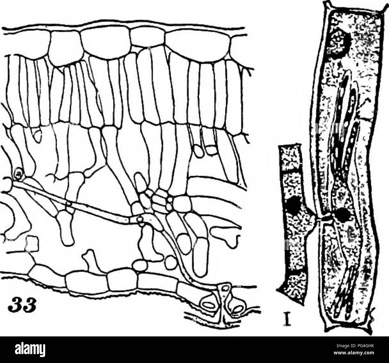
. The fungi which cause plant disease . Plant diseases; Fungi. THE FUNGI WHICH CAUSE PLANT DISEASE 171. Fig. 122.â^I, E. graminis, showing branching haustoria. 33, Phyllactinia, intercellular hyphs. After Smith. Erysiphaceae (p. 170) "â "⢠"â¢" This family on account of its abundance everywhere, its sim- plicity of structure, and its possession of typical ascigerous and conidial stages forms a favorite type for introductory study of the Ascomycetes. Its members are easy of recognition, form- ing a coating of white conidia, conidiophores and mycelium upon the surface of its
Image details
Contributor:
Central Historic Books / Alamy Stock PhotoImage ID:
PG4GHKFile size:
7.1 MB (313.5 KB Compressed download)Releases:
Model - no | Property - noDo I need a release?Dimensions:
1699 x 1470 px | 28.8 x 24.9 cm | 11.3 x 9.8 inches | 150dpiMore information:
This image is a public domain image, which means either that copyright has expired in the image or the copyright holder has waived their copyright. Alamy charges you a fee for access to the high resolution copy of the image.
This image could have imperfections as it’s either historical or reportage.
. The fungi which cause plant disease . Plant diseases; Fungi. THE FUNGI WHICH CAUSE PLANT DISEASE 171. Fig. 122.â^I, E. graminis, showing branching haustoria. 33, Phyllactinia, intercellular hyphs. After Smith. Erysiphaceae (p. 170) "â "⢠"â¢" This family on account of its abundance everywhere, its sim- plicity of structure, and its possession of typical ascigerous and conidial stages forms a favorite type for introductory study of the Ascomycetes. Its members are easy of recognition, form- ing a coating of white conidia, conidiophores and mycelium upon the surface of its hosts and giving them an appear- ance much as though they had been lightly dusted with flour. Later in the season the white patches are more or less liberally sprinkled with the black perithecia leading to the conmion name powdery mildew. An important list of the economic forms and their hosts has been published by Halsted." The mycelium except in Phyllactinia is entirely superficial. It is usually quite hyaline and is branched, septate and its cells uninucleate. It fastens to the host and penetrates its epidermal cells by uninucleate haustoria which by their various lobings aid in specific characterization. Figs. 122, 123. Haustoria may be grouped in three general classes; (1) those arising directly from the lower surface of the mycelium; (2) those arising at the side of the mycelium as small semicircular processes; (3) arising from more or less deeply-lobed lateral swellings of the mycelium. The relation of the haustoria to the host cells has been extensively studied by Smith.⢠The conidia arise in basipetal succession on simple scattered conidiophores (Fig. 129); are hyaline, oval or barrel-shaped, smooth, 1-celled. Neger has shown that they vary greatly in size with nutrition conditions.*" Conidia germinate readily at once in dry air, better in himiid air, producing from one to three germ tubes. Haustoria are. Please note that these images are extracted