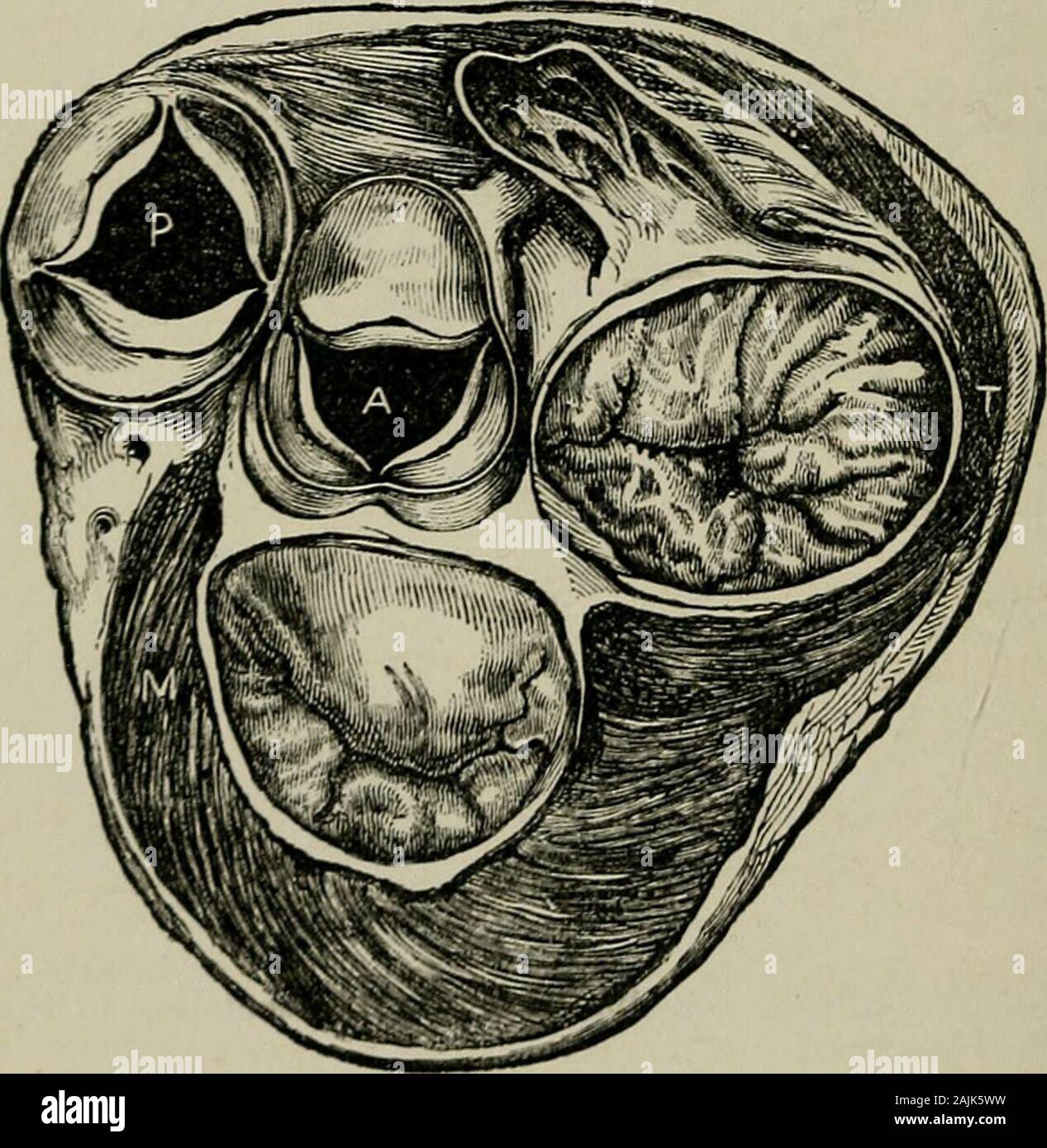
The physical signs of cardiac disease, for the use of clinical students . oughthe auricles, and note the arrangement of the four orifices(vide Fig. III.). The separation of the pulmonary valves fromthe corresponding auriculo-ventricular set, is somewhat surprisingat first sight; but a moments consideration will tell us that thisis due to the conus arteriosus (infundibulum) of the right ven-tricle, which passes in front of the aortic orifice. We shall find 4 ANATOMICAL CONSIDERATIONS. later that aortic regurgitation muriuurs are very well heard overthe stenium, especially in its middle and lowe

Image details
Contributor:
The Reading Room / Alamy Stock PhotoImage ID:
2AJK5WWFile size:
7.2 MB (449.3 KB Compressed download)Releases:
Model - no | Property - noDo I need a release?Dimensions:
1561 x 1601 px | 26.4 x 27.1 cm | 10.4 x 10.7 inches | 150dpiMore information:
This image is a public domain image, which means either that copyright has expired in the image or the copyright holder has waived their copyright. Alamy charges you a fee for access to the high resolution copy of the image.
This image could have imperfections as it’s either historical or reportage.
The physical signs of cardiac disease, for the use of clinical students . oughthe auricles, and note the arrangement of the four orifices(vide Fig. III.). The separation of the pulmonary valves fromthe corresponding auriculo-ventricular set, is somewhat surprisingat first sight; but a moments consideration will tell us that thisis due to the conus arteriosus (infundibulum) of the right ven-tricle, which passes in front of the aortic orifice. We shall find 4 ANATOMICAL CONSIDERATIONS. later that aortic regurgitation muriuurs are very well heard overthe stenium, especially in its middle and lowest thirds; and therelation of pjirts which we have just described probably explain*the phenomenon. During the diastole, the pulmonary valvesbeing competent, there is no current of blood in the in-fundibulum, and it is the portion of the right ventricle inclosest relation with the sternum : an aortic regurgitationmurmur will thus be readily transmitted to the surfacetluough the infundibulum, and once having reached the sternumit will be readily conducted along the bone.. Fig. III. (from Heatlis Practical Anatomy) sliows tlie relative position of thedifferent cardiac orifices. It will be evident why aortic regurgitation murmurs are sowell heard over the sternum, as they liave only to pass through the infundibulumof tlie right ventricle, where, during the diastole, there is no movement of blood.Having once reached the sternum, such murmurs are well carried along the bone. The arrangement of the cusps of the arterial valves—twoanterior and one posterior in tlie pulmonary artery, twoposterior and one anterior in the aorta—is worthy of note.The orifices of the coronary arteries open from behind the ANATOMICAL CONSIDERATIONS. 5 interior and left posterior aortic cusps. Immediately behind thetwo posterior cusps of the aorta is situated the large anteriorflap of the mitral valve, which is embraced by the sliortsemilunar posterior flap of the same. The close proximity ofthe two sets