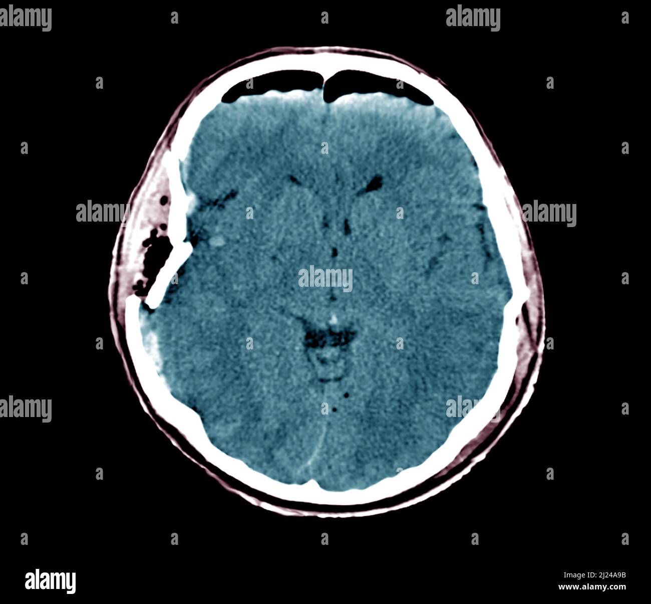
Traumatic brain injury, CT scan
RMID:Image ID:2J24A9B
Image details
Contributor:
Science Photo Library / Alamy Stock PhotoImage ID:
2J24A9BFile size:
38.7 MB (462 KB Compressed download)Releases:
Model - no | Property - noDo I need a release?Dimensions:
3969 x 3406 px | 33.6 x 28.8 cm | 13.2 x 11.4 inches | 300dpiPhotographer:
ZEPHYR/SCIENCE PHOTO LIBRARYMore information:
Computed tomography (CT) scan in axial section of the brain of a 30 year old male patient with traumatic brain injury after a car accident. The CT shows a skull fracture of the right temporoparietal wall (left). There is a wound of the dura mater with presence of a frontal aeric bubble (top). The CT also shows wound with external and internal hemorrhagic contusion (brain bruises). The patient also has a diffuse internal cerebral oedema associated with a slight displacement of the ventricles.