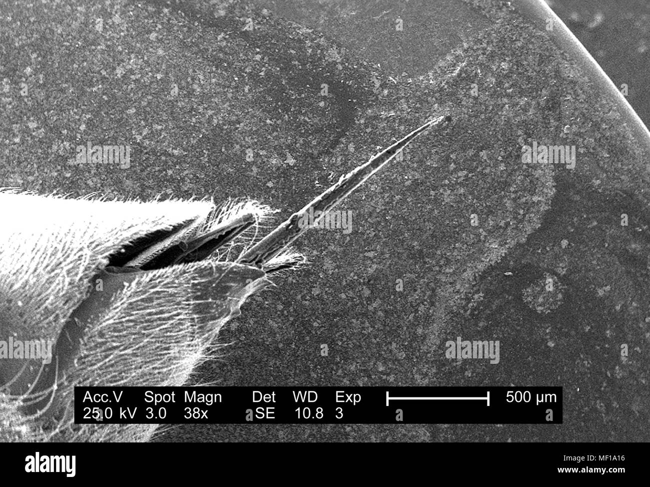
Ultrastructural morphologic details of a bee stinger apparatus depicted in the low magnified scanning electron microscopic (SEM) image, 2005. Image courtesy Centers for Disease Control (CDC) / Janice Haney Carr, Oren Mayer. ()
RMID:Image ID:MF1A16
Image details
Contributor:
Gado Images / Alamy Stock PhotoImage ID:
MF1A16File size:
54.6 MB (1.7 MB Compressed download)Releases:
Model - no | Property - noDo I need a release?Dimensions:
5300 x 3602 px | 44.9 x 30.5 cm | 17.7 x 12 inches | 300dpiDate taken:
1 January 2005Location:
United StatesPhotographer:
Smith Collection/GadoMore information:
This image could have imperfections as it’s either historical or reportage.
Ultrastructural morphologic details of a bee stinger apparatus depicted in the low magnified scanning electron microscopic (SEM) image, 2005. Image courtesy Centers for Disease Control (CDC) / Janice Haney Carr, Oren Mayer. ()