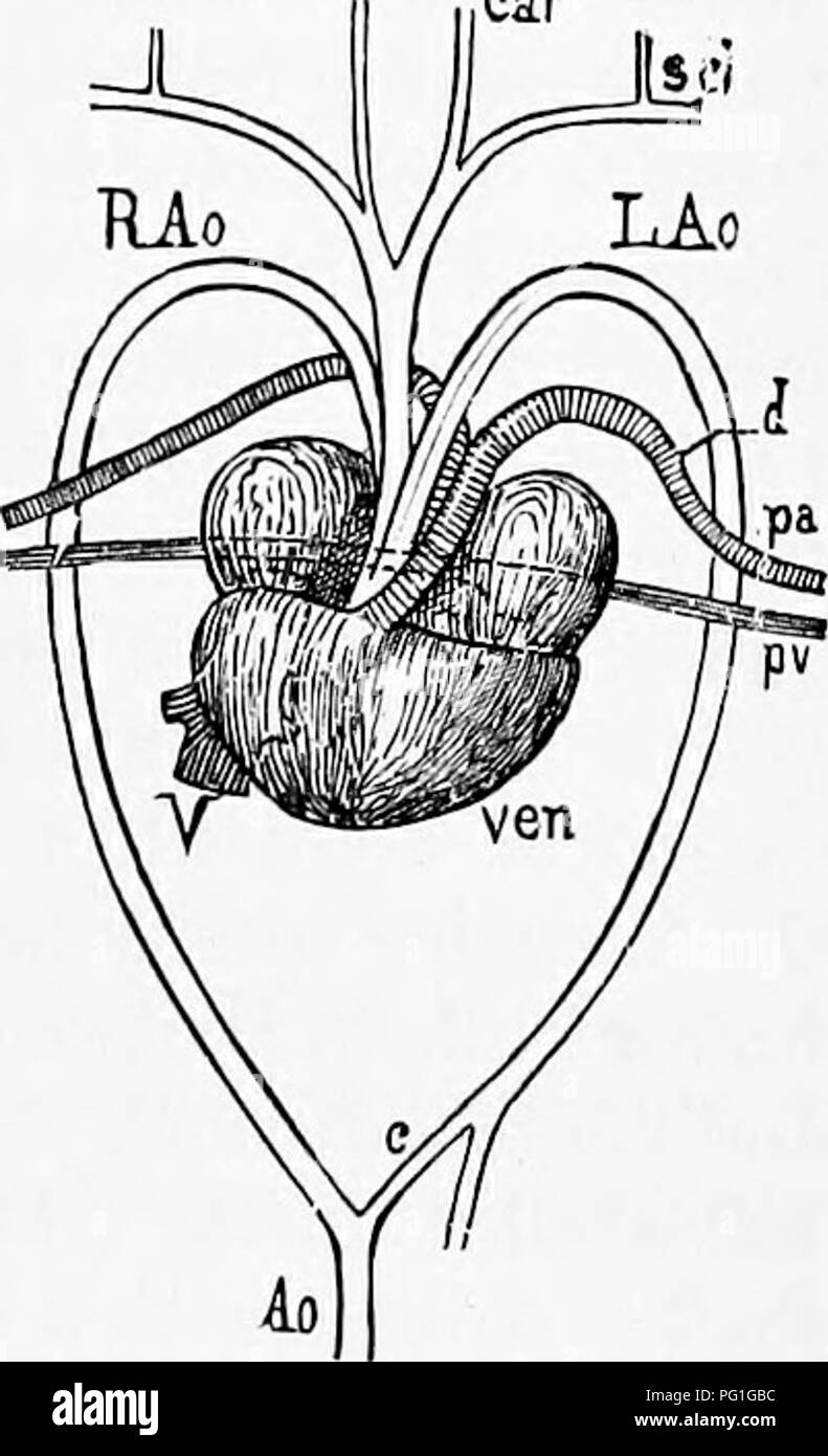
. Zoology : for students and general readers . Zoology. AWATOMY OF THE TURTLE. 509 mesentery, and contains three oval eggs, one of wliicli is lettered Eg. The oviduct can be followed to its anterior end which is much pigmented and has a terminal opening. The cut-end of the left oviduct {ovd) shows the folds of the lining mucous membrane. The ovary (o) is likewise suspended by a thin membrane, the mesovarium, and is equally developed on both sides in a complete specimen. It is easily recognized by the numerous bulging yellow spheres, of all sizes, which are the egg-yolks in various stages of de

Image details
Contributor:
Central Historic Books / Alamy Stock PhotoImage ID:
PG1GBCFile size:
7.1 MB (249 KB Compressed download)Releases:
Model - no | Property - noDo I need a release?Dimensions:
1233 x 2025 px | 20.9 x 34.3 cm | 8.2 x 13.5 inches | 150dpiMore information:
This image is a public domain image, which means either that copyright has expired in the image or the copyright holder has waived their copyright. Alamy charges you a fee for access to the high resolution copy of the image.
This image could have imperfections as it’s either historical or reportage.
. Zoology : for students and general readers . Zoology. AWATOMY OF THE TURTLE. 509 mesentery, and contains three oval eggs, one of wliicli is lettered Eg. The oviduct can be followed to its anterior end which is much pigmented and has a terminal opening. The cut-end of the left oviduct {ovd) shows the folds of the lining mucous membrane. The ovary (o) is likewise suspended by a thin membrane, the mesovarium, and is equally developed on both sides in a complete specimen. It is easily recognized by the numerous bulging yellow spheres, of all sizes, which are the egg-yolks in various stages of development. The heart of the turtle (Fig. 447) will repay careful dis- section. A small round body lies just in front of it; this is A 01 the thyroid gl nCar and, usually considered the equivalent through its real nature is still un- certain. The heart itself (Fig. 447) consists of two auricles and one ventricle (ven), with an imper- fect internal septum. It receives the veins upon its dorsal surface, and gives off the arterial trunks from its ventral side. The two auricles are equal in size ; together they a little more than equal the ventricle. The arterial vessels arise together a little to the right, and are most conveniently described as three in number : 1st. The right aorta {R Ao) arising on the left; 2d. The left aorta on the right {L Ao) ; the two cross near their origin and curve upwards and back- wards, to reunite posteriorly just in front of the retractor muscles, their union forming the single median descending aorta; 3d. The pulmonary aorta {pa), which soon divides into a branch for each lung. The left aorta gives off a branch {d) which persists as a mere cord, the remnant of the ductus arteriosus, wliicli originally united the aorta witli the pulmonary artery. The right aorta gives off an innominate branch, that soon divides, and from each division springs. Fig. 447.—Ventral purface of the heart of the Turtle, Cliryf:e- Tnys picl.a. Bissected and drawn by C. S.