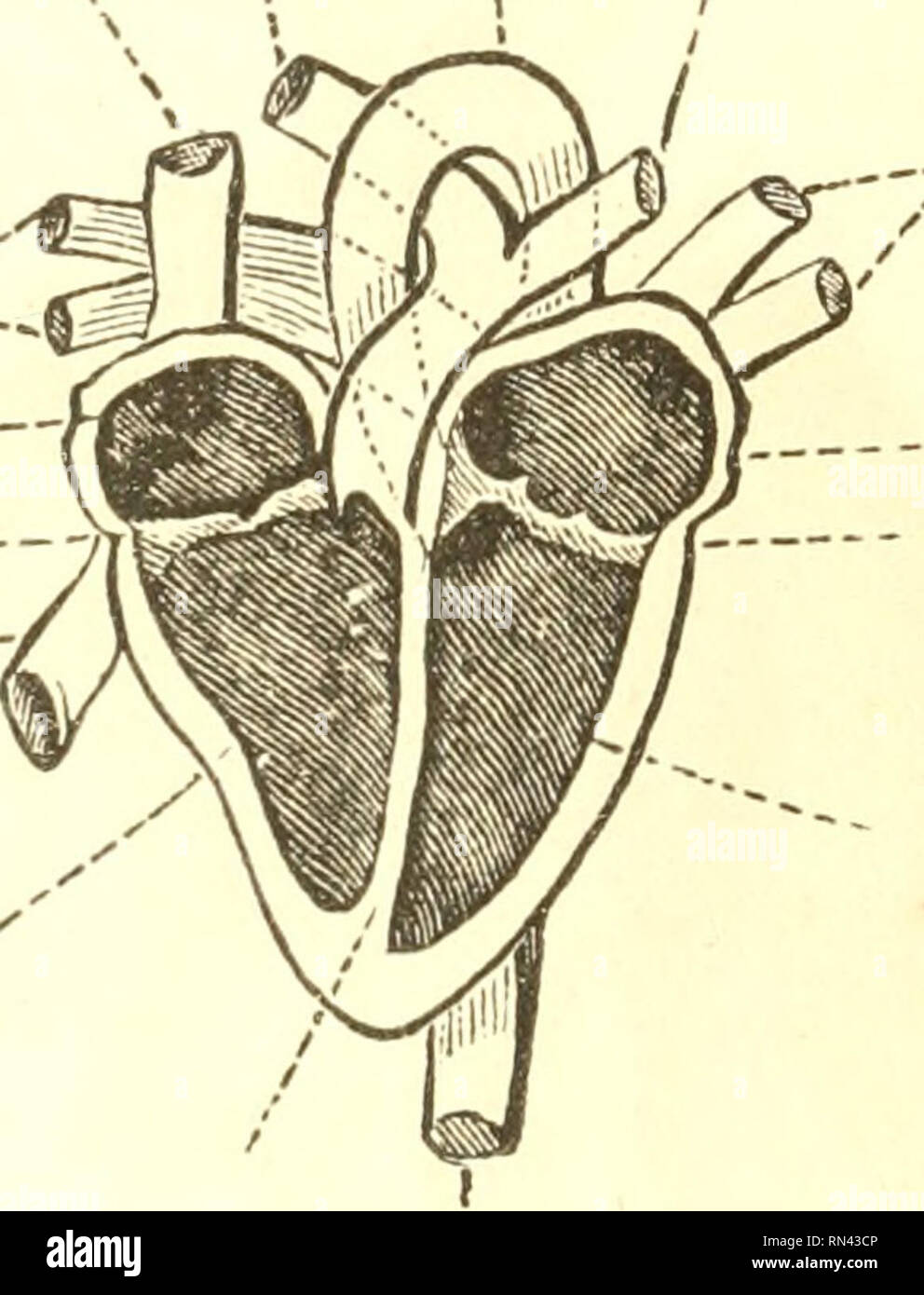
. Animal physiology. Physiology, Comparative. 224 STRUCTURE OF THE HEART. of which the upper one is termed the auricle, and the lower the ventricle. Thus we have the right and left auricles, and the right and left ventricles. Each auricle communicates witli its corresponding ventricle, by an aperture in the Superior Pulm. Pulmonary vena cava art. Aorta artery Pulmonary veins »<! Right auricle Tricuspid valves Inferior vena cava "' Right ventricle. Pulmonary veins Left auricle Mitral valve Left ventricle Partition Aorta Fig. 123.—Ideal Section of the Human Heart. transverse partition,

Image details
Contributor:
Library Book Collection / Alamy Stock PhotoImage ID:
RN43CPFile size:
7.1 MB (252 KB Compressed download)Releases:
Model - no | Property - noDo I need a release?Dimensions:
1381 x 1809 px | 23.4 x 30.6 cm | 9.2 x 12.1 inches | 150dpiMore information:
This image is a public domain image, which means either that copyright has expired in the image or the copyright holder has waived their copyright. Alamy charges you a fee for access to the high resolution copy of the image.
This image could have imperfections as it’s either historical or reportage.
. Animal physiology. Physiology, Comparative. 224 STRUCTURE OF THE HEART. of which the upper one is termed the auricle, and the lower the ventricle. Thus we have the right and left auricles, and the right and left ventricles. Each auricle communicates witli its corresponding ventricle, by an aperture in the Superior Pulm. Pulmonary vena cava art. Aorta artery Pulmonary veins »<!_ Right auricle Tricuspid valves Inferior vena cava "' Right ventricle. Pulmonary veins Left auricle Mitral valve Left ventricle Partition Aorta Fig. 123.—Ideal Section of the Human Heart. transverse partition, which is guarded by a valve. The walls of the ventricles are much tliicker than those of the auricles; and for this evident reason, —that the ventricles have to propel the blood, by their contraction, through a system of remote vessels; whilst the auricles have only to transmit the fluid tliat has been poured into them by the veins, into the ventricles, which dilate themselves to receive it. The difference in the thickness of the walls of the left and the right ventiicles is explainable on the same principle; for the left ventricle has to send the blood, by its contractile power, through the remotest parts of the body; whilst the right has only to transmit it through the lungs, which, being much nearer, require a far less amoimt of force for the circulation of the blood through them. 258. The arterial system of the greater circulation entirely springs from one large trunk, which is called the aorta (see figs. 122-124); this originates in the left ventricle, and is the only vessel which passes forth from that cavity. It first ascends towards the bottom of the nock; then forms what is termed the arch, a sudden curve, which gives it a downward. Please note that these images are extracted from scanned page images that may have been digitally enhanced for readability - coloration and appearance of these illustrations may not perfectly resemble the original work.. Carpenter, Will