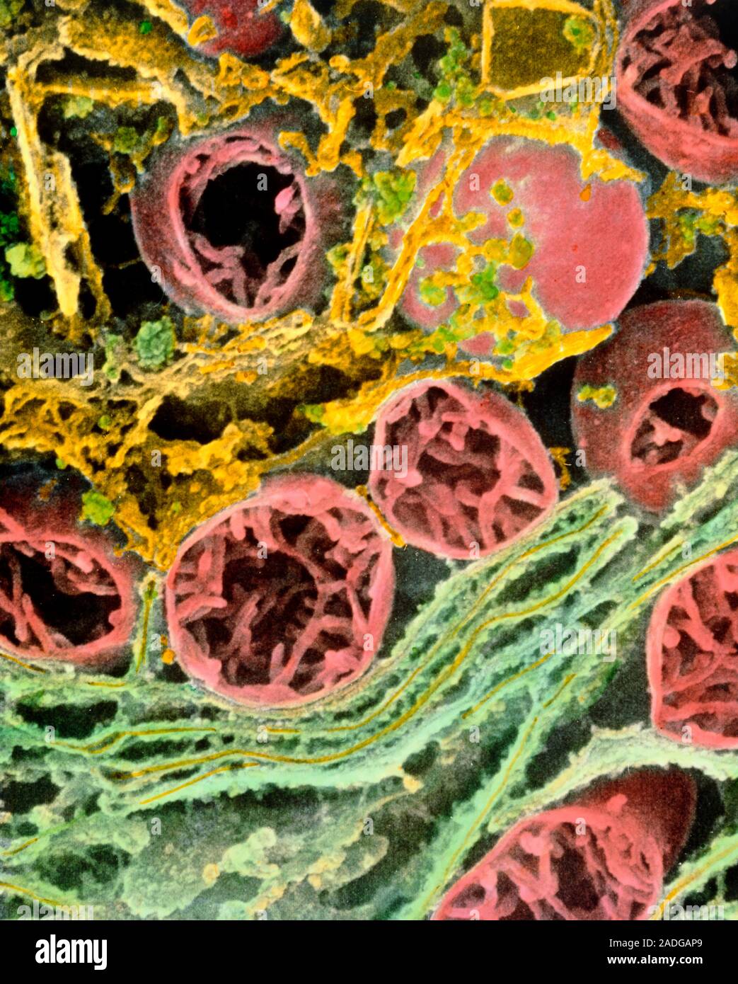
Mitochondria. Coloured Scanning Electron Micrograph (SEM) of mitochondria and rough endoplasmic reticulum in a liver cell. The internal structure of a
RMID:Image ID:2ADGAP9
Image details
Contributor:
Science Photo Library / Alamy Stock PhotoImage ID:
2ADGAP9File size:
57 MB (2.5 MB Compressed download)Releases:
Model - no | Property - noDo I need a release?Dimensions:
4000 x 4983 px | 33.9 x 42.2 cm | 13.3 x 16.6 inches | 300dpiDate taken:
6 January 1995More information:
Mitochondria. Coloured Scanning Electron Micrograph (SEM) of mitochondria and rough endoplasmic reticulum in a liver cell. The internal structure of a hepatocyte or liver cell is crowded with organelles, due to the metabolic activities of the cell. Mitochondria (pink) are sectioned through to show folds or cristae; they are sites of cell respiration and store energy. Rough endoplasmic reticulum (green) forms pathways in the cell; it is studded with ribosomes (dots, sites of protein synthesis). Liver cells detoxify waste products in blood, store glycogen and synthesize proteins. Magnification: x28, 000 at 6x7cm size. x36, 000 at 4x5ins