···
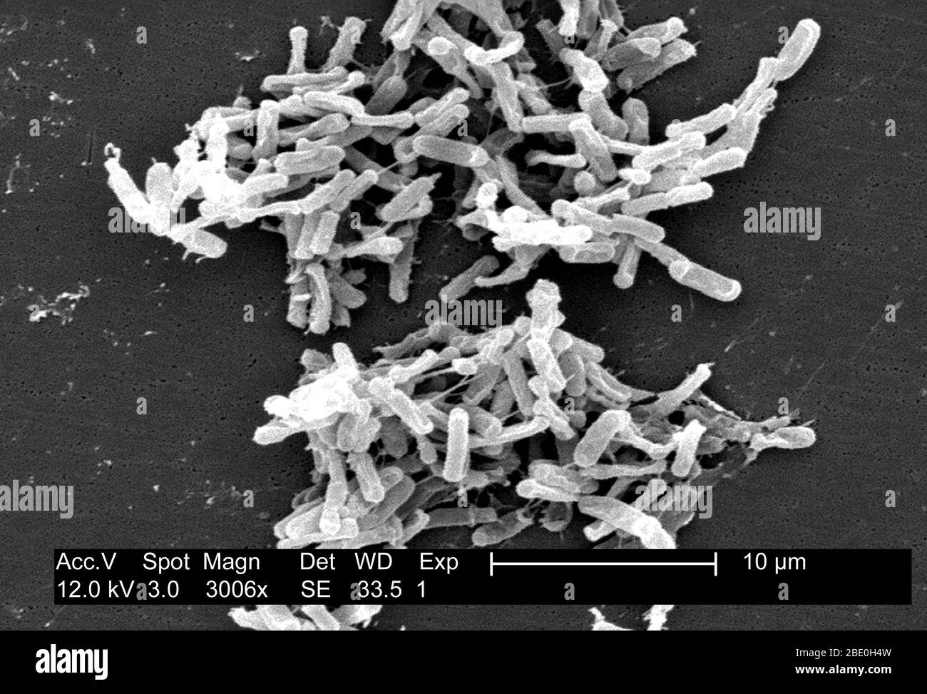
Scanning Electron Micrograph (SEM) showing Gram-positive Clostridium difficile bacteria. These C. difficile organisms were cultured from a stool sample obtained during an outbreak of gastrointestinal illness, and extracted using a .1µm filter. C. difficile causes diarrhea, and more serious intestinal conditions such as colitis. Image details File size:
15.6 MB (711.8 KB Compressed download)
Open your image file to the full size using image processing software.
Dimensions:
2835 x 1927 px | 24 x 16.3 cm | 9.5 x 6.4 inches | 300dpi
Date taken:
1 November 2011
More information:
This image could have imperfections as it’s either historical or reportage.
Search stock photos by tags
Similar stock images Human lip. Coloured scanning electron micrograph (SEM) of a human lip, showing sweat gland openings on the drier external lip surface. These openings (pores) release sweat onto the surface of the skin. This has the effect of cooling the body, as the sweat's evaporation carries away heat. Magnification x300 when printed 10cm wide. Stock Photo https://www.alamy.com/image-license-details/?v=1 https://www.alamy.com/human-lip-coloured-scanning-electron-micrograph-sem-of-a-human-lip-showing-sweat-gland-openings-on-the-drier-external-lip-surface-these-openings-pores-release-sweat-onto-the-surface-of-the-skin-this-has-the-effect-of-cooling-the-body-as-the-sweats-evaporation-carries-away-heat-magnification-x300-when-printed-10cm-wide-image359042804.html RF 2BT3PN8 – Human lip. Coloured scanning electron micrograph (SEM) of a human lip, showing sweat gland openings on the drier external lip surface. These openings (pores) release sweat onto the surface of the skin. This has the effect of cooling the body, as the sweat's evaporation carries away heat. Magnification x300 when printed 10cm wide. Scanning Electron Micrograph (SEM) showing silicon beads on the head of a pin. Stock Photo https://www.alamy.com/image-license-details/?v=1 https://www.alamy.com/scanning-electron-micrograph-sem-showing-silicon-beads-on-the-head-of-a-pin-image352826116.html RM 2BE0H8M – Scanning Electron Micrograph (SEM) showing silicon beads on the head of a pin. Scanning electron microscope (SEM) micrograph showing spider's silk, including thread, hydrogel and nano-fibril silk types, at a magnification of 1500x, 2016. Stock Photo https://www.alamy.com/image-license-details/?v=1 https://www.alamy.com/stock-photo-scanning-electron-microscope-sem-micrograph-showing-spiders-silk-including-170361191.html RM KW4HC7 – Scanning electron microscope (SEM) micrograph showing spider's silk, including thread, hydrogel and nano-fibril silk types, at a magnification of 1500x, 2016. Scanning Electron Micrograph (SEM) showing Legionella pneumophila on biofilm; magnification: 3200X. This organism is the causative agent of Legionnaires' Disease. Stock Photo https://www.alamy.com/image-license-details/?v=1 https://www.alamy.com/scanning-electron-micrograph-sem-showing-legionella-pneumophila-on-biofilm-magnification-3200x-this-organism-is-the-causative-agent-of-legionnaires-disease-image352826001.html RM 2BE0H4H – Scanning Electron Micrograph (SEM) showing Legionella pneumophila on biofilm; magnification: 3200X. This organism is the causative agent of Legionnaires' Disease. Ant head. Coloured scanning electron micrograph (SEM) of the head of an ant (family Formicidae). showing its large compound eyes (blue) and jaws. Magnification: x50 when printed 10 centimetres wide. Stock Photo https://www.alamy.com/image-license-details/?v=1 https://www.alamy.com/ant-head-coloured-scanning-electron-micrograph-sem-of-the-head-of-an-ant-family-formicidae-showing-its-large-compound-eyes-blue-and-jaws-magnification-x50-when-printed-10-centimetres-wide-image359042735.html RF 2BT3PJR – Ant head. Coloured scanning electron micrograph (SEM) of the head of an ant (family Formicidae). showing its large compound eyes (blue) and jaws. Magnification: x50 when printed 10 centimetres wide. Scanning Electron Micrograph (SEM) showing sigma-phase crystallization in steel. Magnification: 500X at 8x10'. Nomarski interference contrast method. Stock Photo https://www.alamy.com/image-license-details/?v=1 https://www.alamy.com/scanning-electron-micrograph-sem-showing-sigma-phase-crystallization-in-steel-magnification-500x-at-8x10-nomarski-interference-contrast-method-image352826119.html RM 2BE0H8R – Scanning Electron Micrograph (SEM) showing sigma-phase crystallization in steel. Magnification: 500X at 8x10'. Nomarski interference contrast method. Colorized scanning electron micrograph showing carbapenem-resistant Klebsiella pneumoniae interacting with a human neutrophil. Stock Photo https://www.alamy.com/image-license-details/?v=1 https://www.alamy.com/colorized-scanning-electron-micrograph-showing-carbapenem-resistant-klebsiella-pneumoniae-interacting-with-a-human-neutrophil-image388135132.html RF 2DFD290 – Colorized scanning electron micrograph showing carbapenem-resistant Klebsiella pneumoniae interacting with a human neutrophil. Tendon, coloured scanning electron micrograph (SEM), showing bundles of collagen fibres. The parallel alignment of the fibres make tendons inelastic but flexible. Tendons attach muscle to bone. Magnification: x5000 when printed at 10 centimetres wide Stock Photo https://www.alamy.com/image-license-details/?v=1 https://www.alamy.com/tendon-coloured-scanning-electron-micrograph-sem-showing-bundles-of-collagen-fibres-the-parallel-alignment-of-the-fibres-make-tendons-inelastic-but-flexible-tendons-attach-muscle-to-bone-magnification-x5000-when-printed-at-10-centimetres-wide-image359042864.html RF 2BT3PRC – Tendon, coloured scanning electron micrograph (SEM), showing bundles of collagen fibres. The parallel alignment of the fibres make tendons inelastic but flexible. Tendons attach muscle to bone. Magnification: x5000 when printed at 10 centimetres wide Colorized scanning electron micrograph showing carbapenem-resistnt Klebsiella pneumoniae interacting with a human neutrophil. Stock Photo https://www.alamy.com/image-license-details/?v=1 https://www.alamy.com/colorized-scanning-electron-micrograph-showing-carbapenem-resistnt-klebsiella-pneumoniae-interacting-with-a-human-neutrophil-image388135147.html RF 2DFD29F – Colorized scanning electron micrograph showing carbapenem-resistnt Klebsiella pneumoniae interacting with a human neutrophil. Scanning Electron Micrograph (SEM) showing Gram-positive Clostridium difficile bacteria. These C. difficile organisms were cultured from a stool sample obtained during an outbreak of gastrointestinal illness, and extracted using a .1µm filter. C. difficile causes diarrhea, and more serious intestinal conditions such as colitis. Stock Photo https://www.alamy.com/image-license-details/?v=1 https://www.alamy.com/scanning-electron-micrograph-sem-showing-gram-positive-clostridium-difficile-bacteria-these-c-difficile-organisms-were-cultured-from-a-stool-sample-obtained-during-an-outbreak-of-gastrointestinal-illness-and-extracted-using-a-1m-filter-c-difficile-causes-diarrhea-and-more-serious-intestinal-conditions-such-as-colitis-image352825996.html RM 2BE0H4C – Scanning Electron Micrograph (SEM) showing Gram-positive Clostridium difficile bacteria. These C. difficile organisms were cultured from a stool sample obtained during an outbreak of gastrointestinal illness, and extracted using a .1µm filter. C. difficile causes diarrhea, and more serious intestinal conditions such as colitis. Tendon, coloured scanning electron micrograph (SEM), showing bundles of collagen fibres. The parallel alignment of the fibres make tendons inelastic but flexible. Tendons attach muscle to bone. Magnification: x5000 when printed at 10 centimetres wide Stock Photo https://www.alamy.com/image-license-details/?v=1 https://www.alamy.com/tendon-coloured-scanning-electron-micrograph-sem-showing-bundles-of-collagen-fibres-the-parallel-alignment-of-the-fibres-make-tendons-inelastic-but-flexible-tendons-attach-muscle-to-bone-magnification-x5000-when-printed-at-10-centimetres-wide-image359042761.html RF 2BT3PKN – Tendon, coloured scanning electron micrograph (SEM), showing bundles of collagen fibres. The parallel alignment of the fibres make tendons inelastic but flexible. Tendons attach muscle to bone. Magnification: x5000 when printed at 10 centimetres wide Colorized scanning electron micrograph showing carbapenem-resistant Klebsiella pneumoniae interacting with a human neutrophil. Stock Photo https://www.alamy.com/image-license-details/?v=1 https://www.alamy.com/colorized-scanning-electron-micrograph-showing-carbapenem-resistant-klebsiella-pneumoniae-interacting-with-a-human-neutrophil-image388135168.html RF 2DFD2A8 – Colorized scanning electron micrograph showing carbapenem-resistant Klebsiella pneumoniae interacting with a human neutrophil. 