Quick filters:
Embryonic development Stock Photos and Images
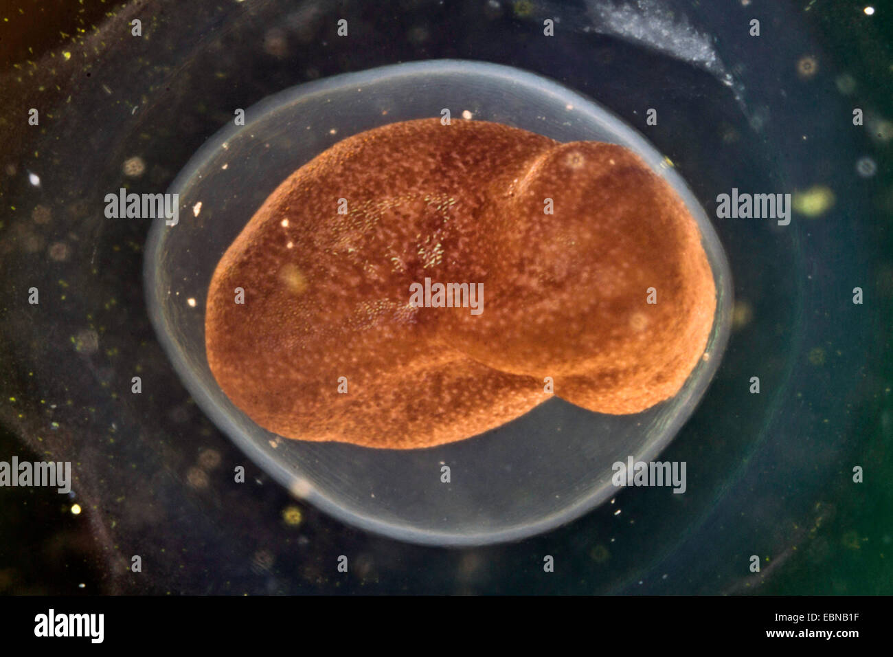 moor frog (Rana arvalis), egg with embryonic development Stock Photohttps://www.alamy.com/image-license-details/?v=1https://www.alamy.com/stock-photo-moor-frog-rana-arvalis-egg-with-embryonic-development-76072347.html
RMEBNB1F–moor frog (Rana arvalis), egg with embryonic development
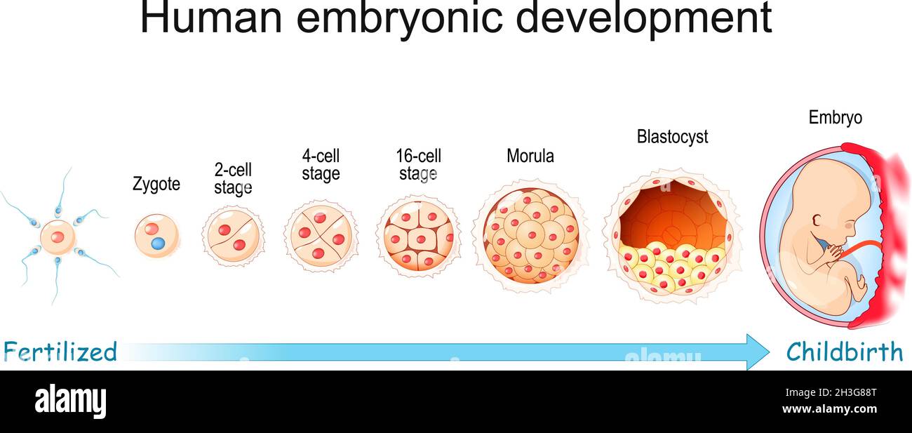 Human embryonic development. From Fertilization to Childbirth. Zygote, Morula, Blastocyst and Embryo stage. Vector poster for education and medical Stock Vectorhttps://www.alamy.com/image-license-details/?v=1https://www.alamy.com/human-embryonic-development-from-fertilization-to-childbirth-zygote-morula-blastocyst-and-embryo-stage-vector-poster-for-education-and-medical-image449671288.html
RF2H3G88T–Human embryonic development. From Fertilization to Childbirth. Zygote, Morula, Blastocyst and Embryo stage. Vector poster for education and medical
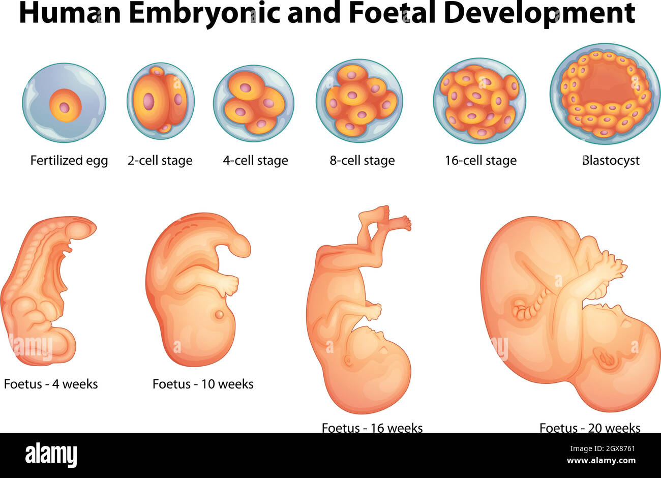 Stages in human embryonic development Stock Vectorhttps://www.alamy.com/image-license-details/?v=1https://www.alamy.com/stages-in-human-embryonic-development-image446421529.html
RF2GX8761–Stages in human embryonic development
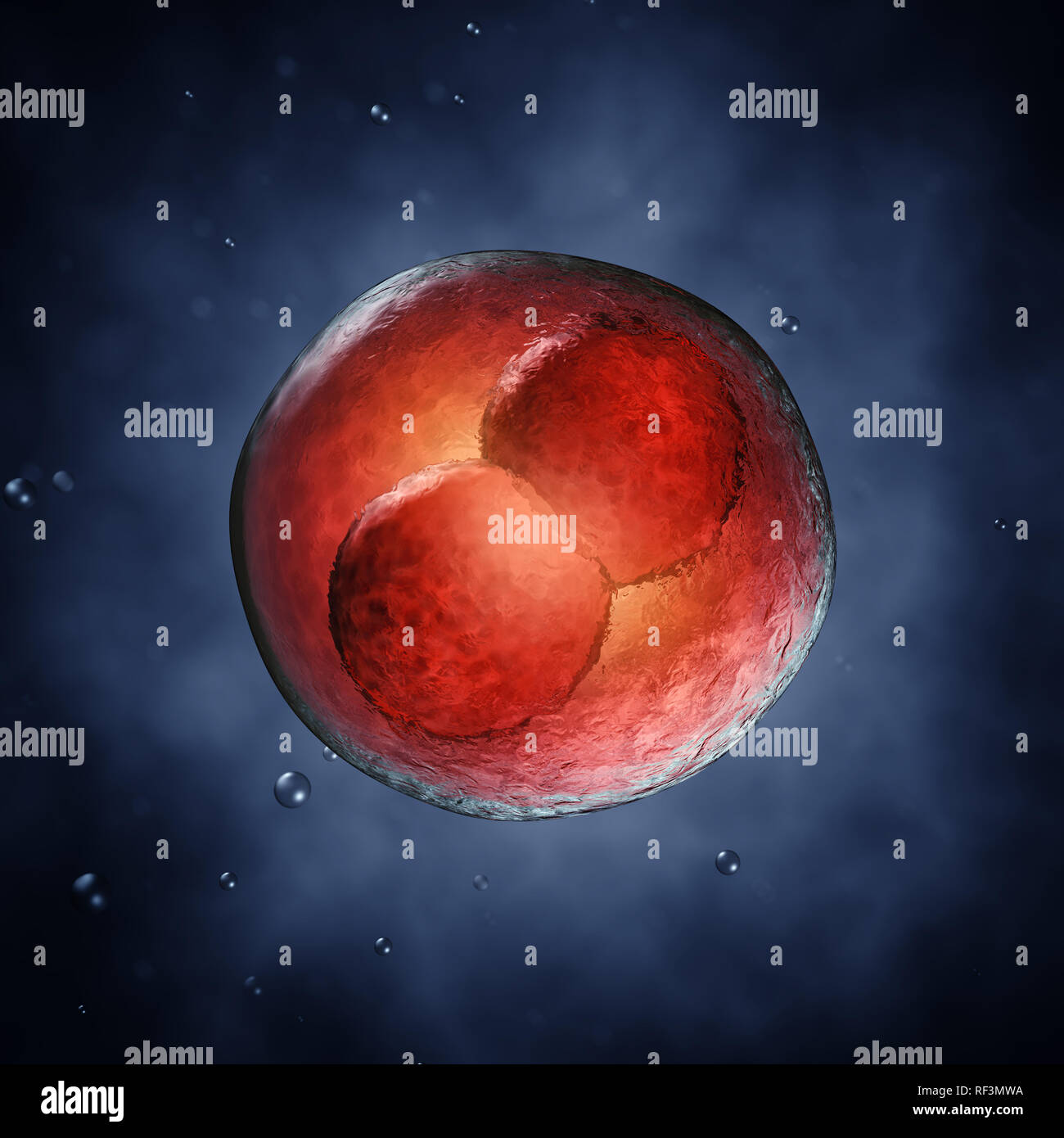 Two-cell embryo, Embryonic development Stock Photohttps://www.alamy.com/image-license-details/?v=1https://www.alamy.com/two-cell-embryo-embryonic-development-image233036870.html
RFRF3MWA–Two-cell embryo, Embryonic development
 Embryonic development Stock Photohttps://www.alamy.com/image-license-details/?v=1https://www.alamy.com/stock-photo-embryonic-development-13228213.html
RFACTM9X–Embryonic development
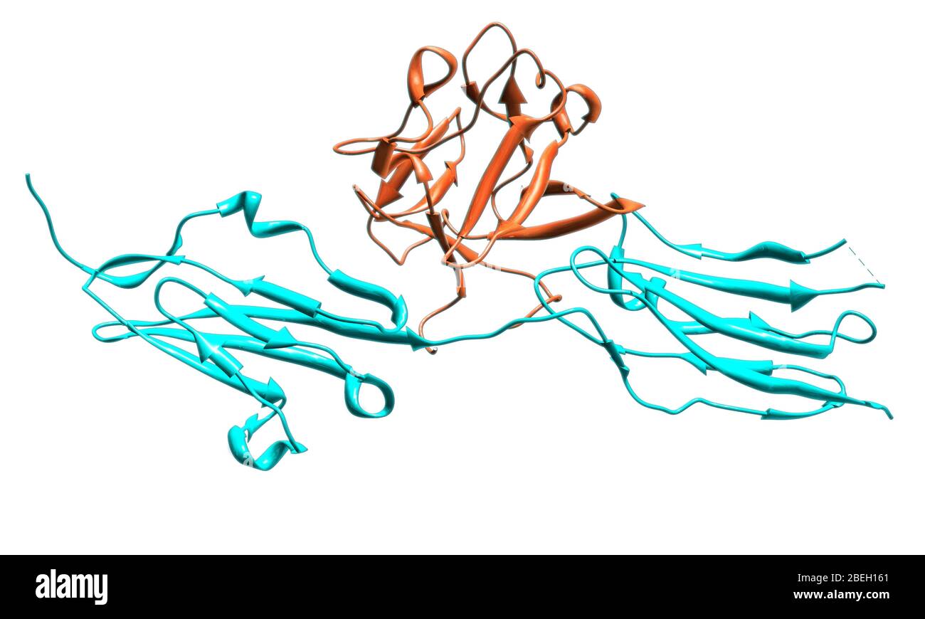 A molecular model of fibroblast growth factor receptor 2 (FGFR2), a protein which plays an important role in embryonic development and tissue repair by mediating cell devision, growth and differentiation. Fibroblast growth factor 1 (orange) is bound at the junction between the two FGFR2 units (blue). Mutations in the FGFR2 gene are associated with a variety of medical conditions, many of which involve abnormal bone development and cancer. Stock Photohttps://www.alamy.com/image-license-details/?v=1https://www.alamy.com/a-molecular-model-of-fibroblast-growth-factor-receptor-2-fgfr2-a-protein-which-plays-an-important-role-in-embryonic-development-and-tissue-repair-by-mediating-cell-devision-growth-and-differentiation-fibroblast-growth-factor-1-orange-is-bound-at-the-junction-between-the-two-fgfr2-units-blue-mutations-in-the-fgfr2-gene-are-associated-with-a-variety-of-medical-conditions-many-of-which-involve-abnormal-bone-development-and-cancer-image353186681.html
RM2BEH161–A molecular model of fibroblast growth factor receptor 2 (FGFR2), a protein which plays an important role in embryonic development and tissue repair by mediating cell devision, growth and differentiation. Fibroblast growth factor 1 (orange) is bound at the junction between the two FGFR2 units (blue). Mutations in the FGFR2 gene are associated with a variety of medical conditions, many of which involve abnormal bone development and cancer.
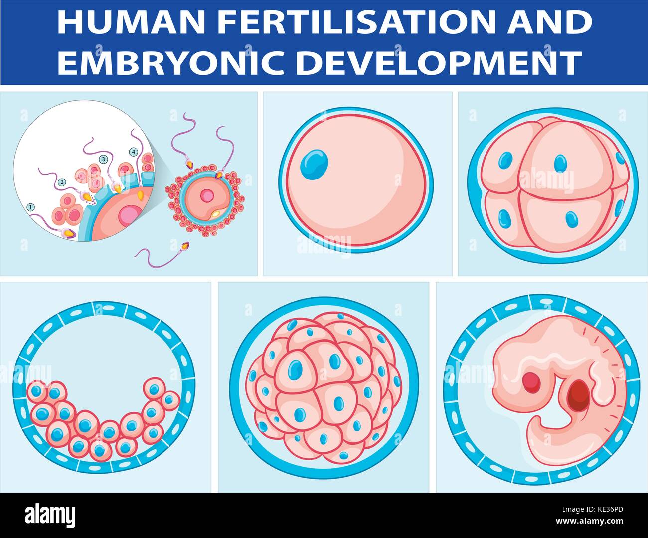 Diagram showing human fertilisation and embryonic development illustration Stock Vectorhttps://www.alamy.com/image-license-details/?v=1https://www.alamy.com/stock-image-diagram-showing-human-fertilisation-and-embryonic-development-illustration-163569685.html
RFKE36PD–Diagram showing human fertilisation and embryonic development illustration
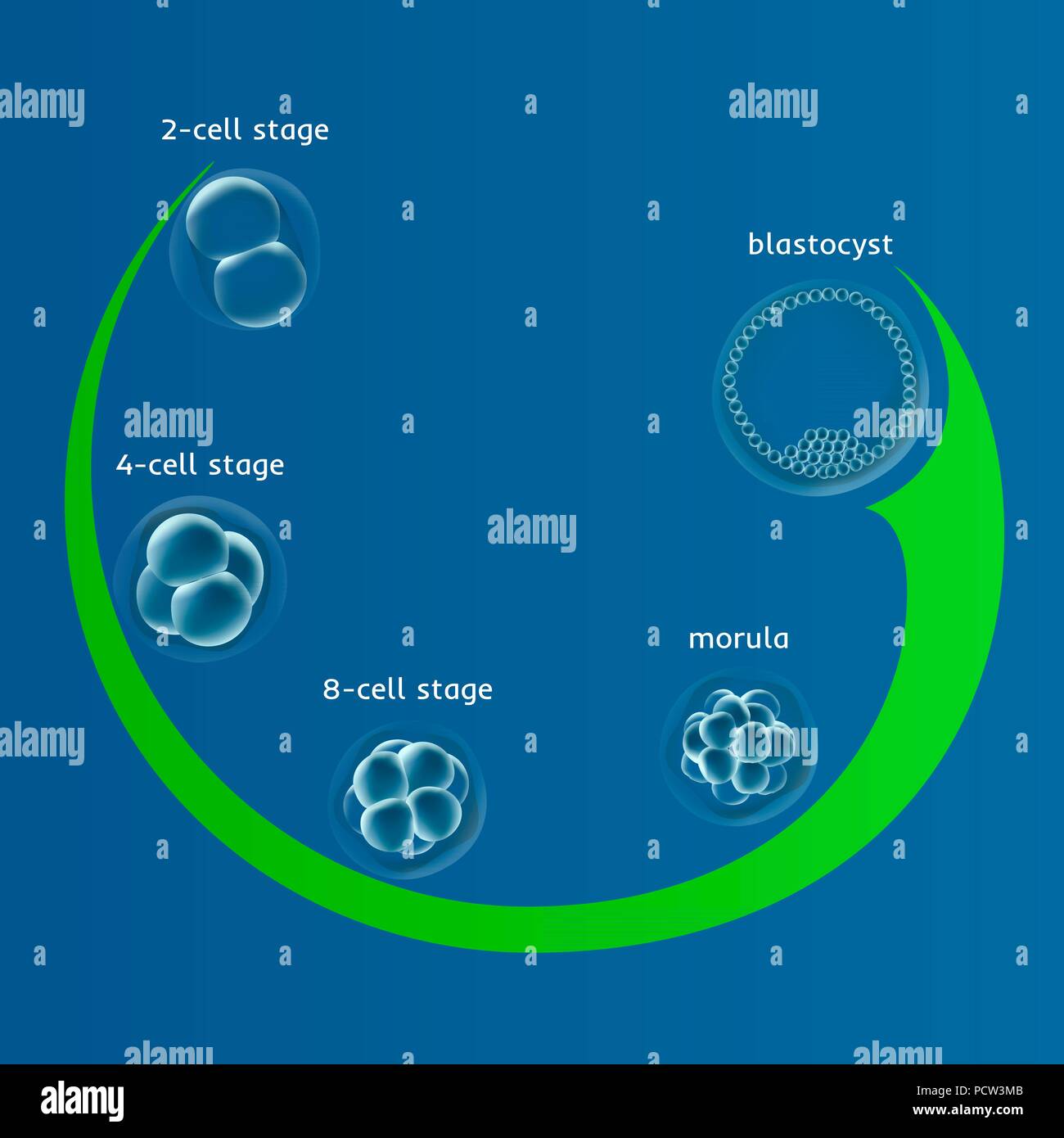 Human embryonic development, illustration. Stock Photohttps://www.alamy.com/image-license-details/?v=1https://www.alamy.com/human-embryonic-development-illustration-image214452011.html
RFPCW3MB–Human embryonic development, illustration.
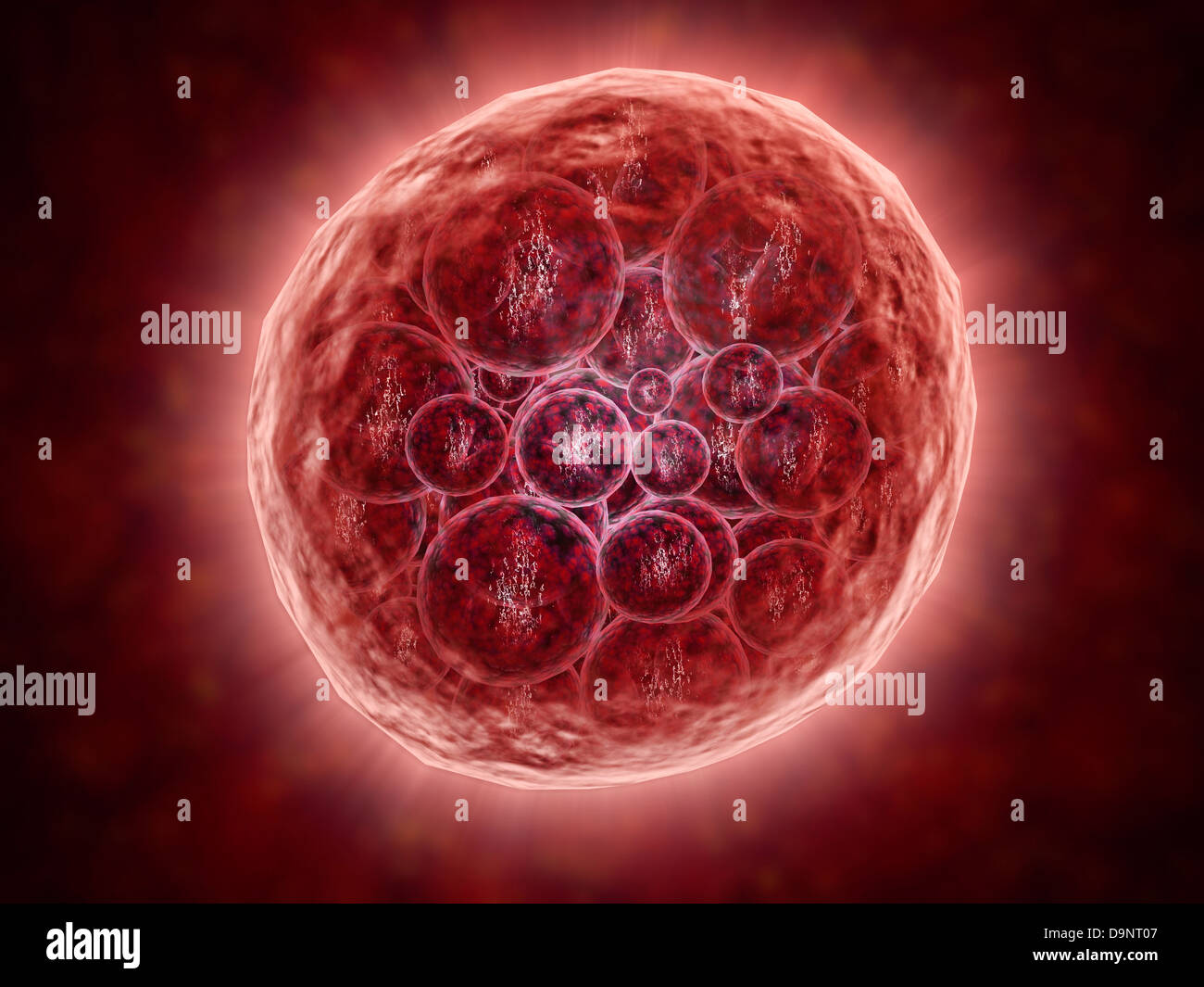 Cluster of blastomeres forming a developing morula (early stage of embryonic development). Stock Photohttps://www.alamy.com/image-license-details/?v=1https://www.alamy.com/stock-photo-cluster-of-blastomeres-forming-a-developing-morula-early-stage-of-57642823.html
RFD9NT07–Cluster of blastomeres forming a developing morula (early stage of embryonic development).
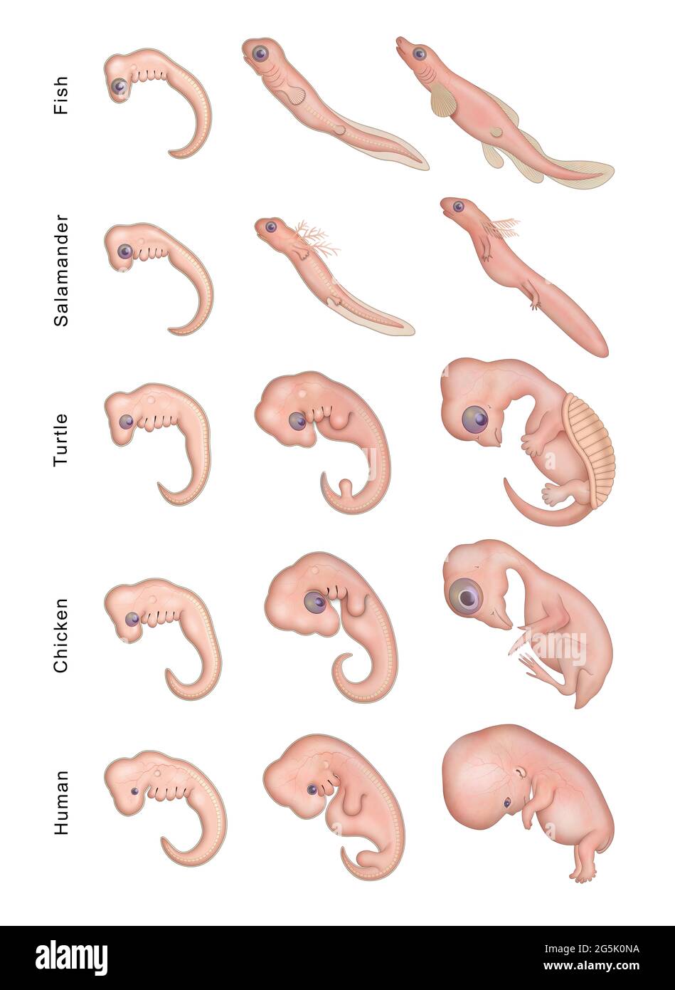 Different Stages Early Embryonic Development Stock Photohttps://www.alamy.com/image-license-details/?v=1https://www.alamy.com/different-stages-early-embryonic-development-image433750166.html
RF2G5K0NA–Different Stages Early Embryonic Development
 Cluster of blastomeres forming a developing morula (early stage of embryonic development) Stock Photohttps://www.alamy.com/image-license-details/?v=1https://www.alamy.com/cluster-of-blastomeres-forming-a-developing-morula-early-stage-of-embryonic-development-image619197427.html
RM2XYATHR–Cluster of blastomeres forming a developing morula (early stage of embryonic development)
 Human fertilization and embryonic development. Digital illustration, 3D render. Stock Photohttps://www.alamy.com/image-license-details/?v=1https://www.alamy.com/human-fertilization-and-embryonic-development-digital-illustration-3d-render-image624589070.html
RF2Y84DME–Human fertilization and embryonic development. Digital illustration, 3D render.
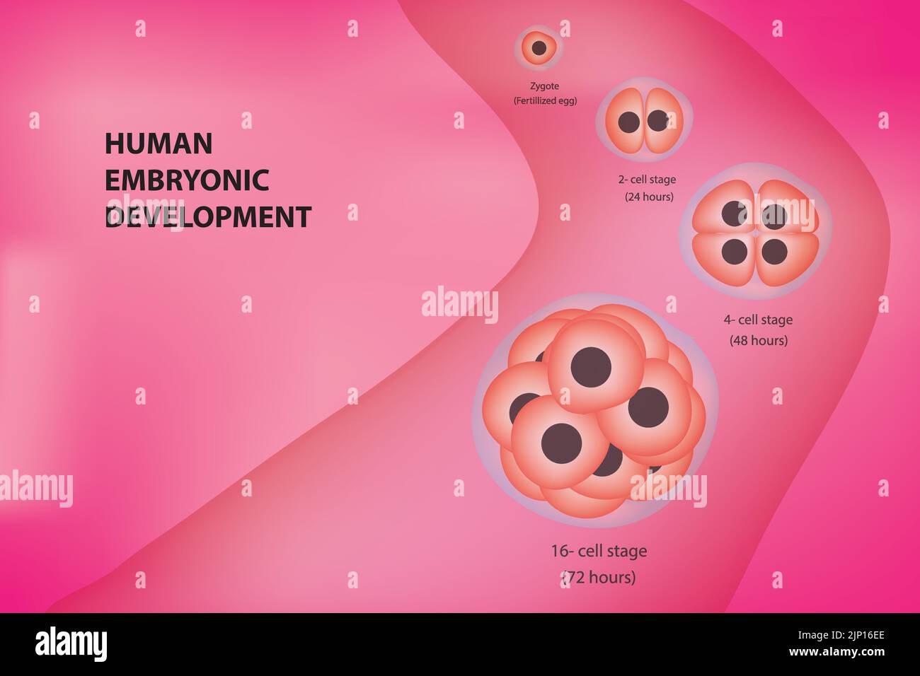 Human embryonic development cell stages Stock Vectorhttps://www.alamy.com/image-license-details/?v=1https://www.alamy.com/human-embryonic-development-cell-stages-image478229430.html
RF2JP16EE–Human embryonic development cell stages
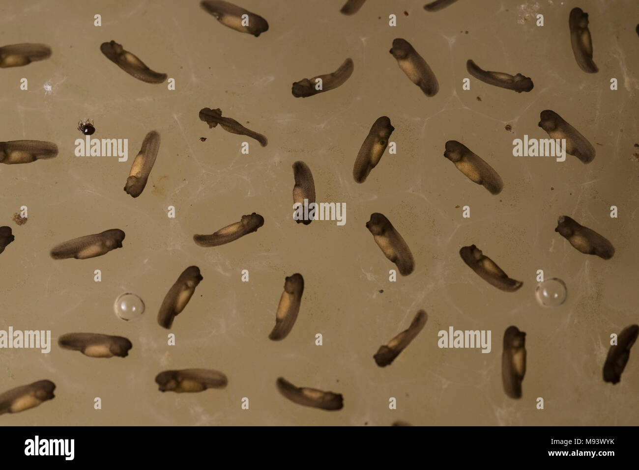 A mass of tadpoles growing in a puddle in Peru. Stock Photohttps://www.alamy.com/image-license-details/?v=1https://www.alamy.com/a-mass-of-tadpoles-growing-in-a-puddle-in-peru-image177721815.html
RFM93WYK–A mass of tadpoles growing in a puddle in Peru.
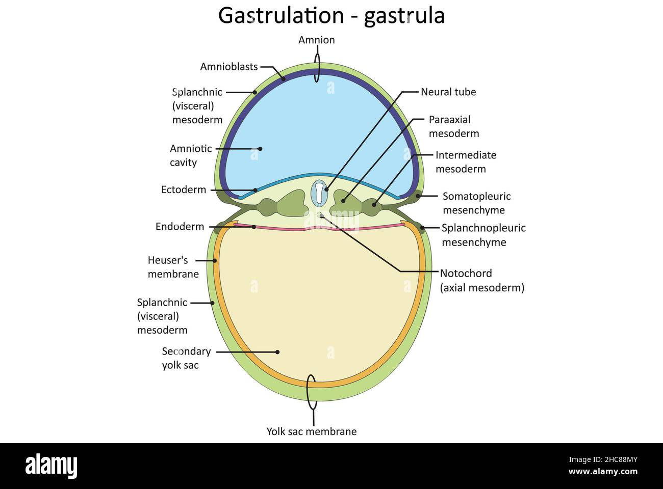 Gastrulation, Gastrula, tri layer stage of embryonic development, endoderm, ectoderm, mesoderm. Stock Photohttps://www.alamy.com/image-license-details/?v=1https://www.alamy.com/gastrulation-gastrula-tri-layer-stage-of-embryonic-development-endoderm-ectoderm-mesoderm-image455027915.html
RF2HC88MY–Gastrulation, Gastrula, tri layer stage of embryonic development, endoderm, ectoderm, mesoderm.
 Marram grass and Sand Couch, forming an embryonic dune, the first stage of dune development Stock Photohttps://www.alamy.com/image-license-details/?v=1https://www.alamy.com/stock-image-marram-grass-and-sand-couch-forming-an-embryonic-dune-the-first-stage-160282738.html
RFK8NE7E–Marram grass and Sand Couch, forming an embryonic dune, the first stage of dune development
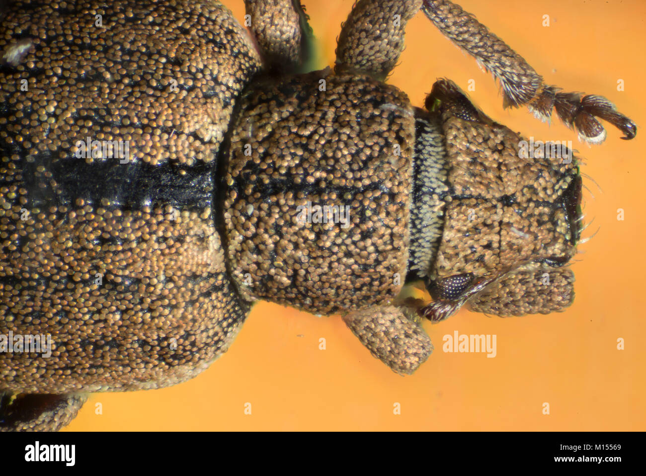 Beetles are a group of insects that form the order Coleoptera, in the superorder Endopterygota. Their front pair of wings is hardened into wing-cases, Stock Photohttps://www.alamy.com/image-license-details/?v=1https://www.alamy.com/stock-photo-beetles-are-a-group-of-insects-that-form-the-order-coleoptera-in-the-172832193.html
RFM15569–Beetles are a group of insects that form the order Coleoptera, in the superorder Endopterygota. Their front pair of wings is hardened into wing-cases,
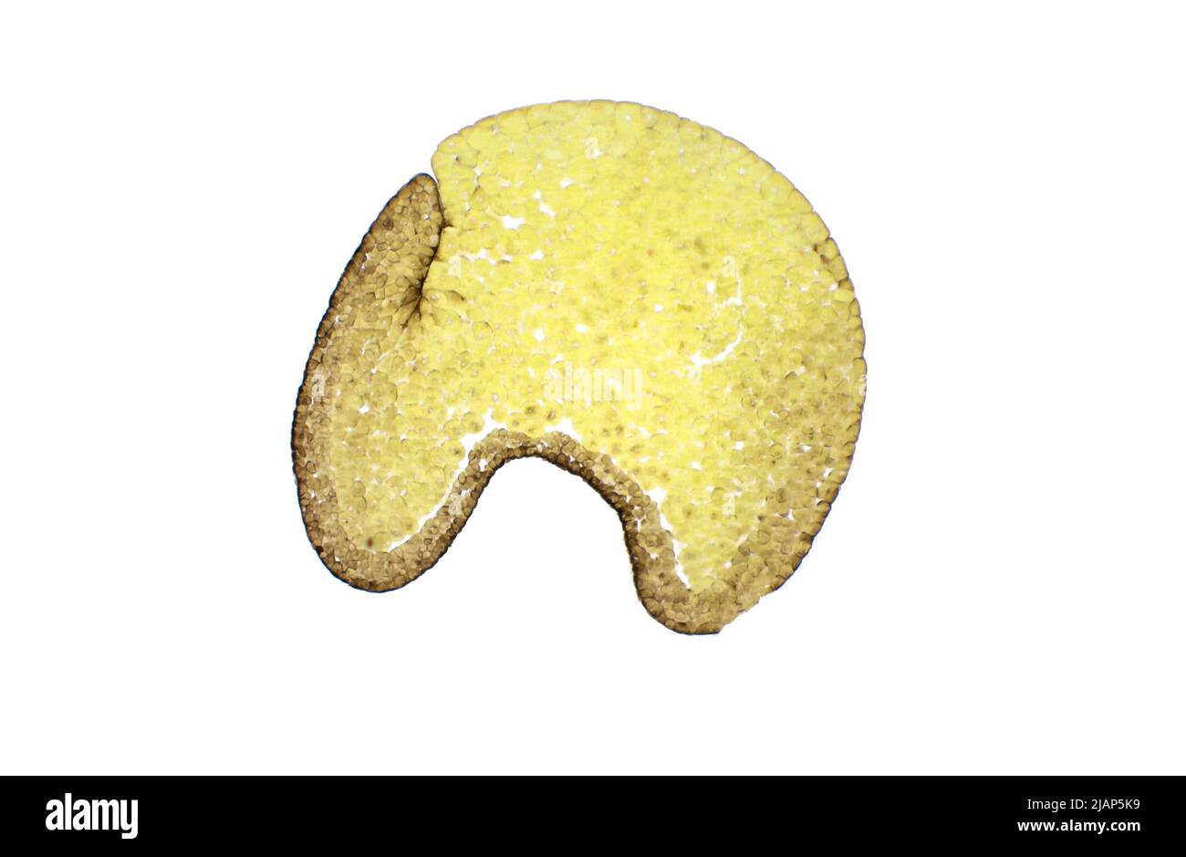 Gastrulation in the Frog. Frog embryo (Rana), gastrula stage, light micrograph. Van Gieson's stain. Magnification: x40. Stock Photohttps://www.alamy.com/image-license-details/?v=1https://www.alamy.com/gastrulation-in-the-frog-frog-embryo-rana-gastrula-stage-light-micrograph-van-giesons-stain-magnification-x40-image471313901.html
RF2JAP5K9–Gastrulation in the Frog. Frog embryo (Rana), gastrula stage, light micrograph. Van Gieson's stain. Magnification: x40.
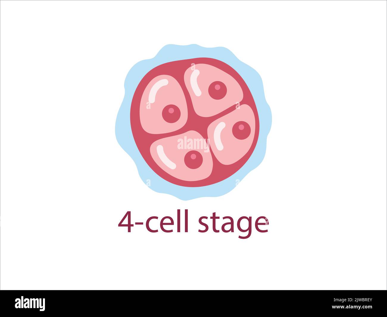 Zygote 4-cell stage. Human embryonic development. vector medical illustration. Stock Vectorhttps://www.alamy.com/image-license-details/?v=1https://www.alamy.com/zygote-4-cell-stage-human-embryonic-development-vector-medical-illustration-image480306259.html
RF2JWBREY–Zygote 4-cell stage. Human embryonic development. vector medical illustration.
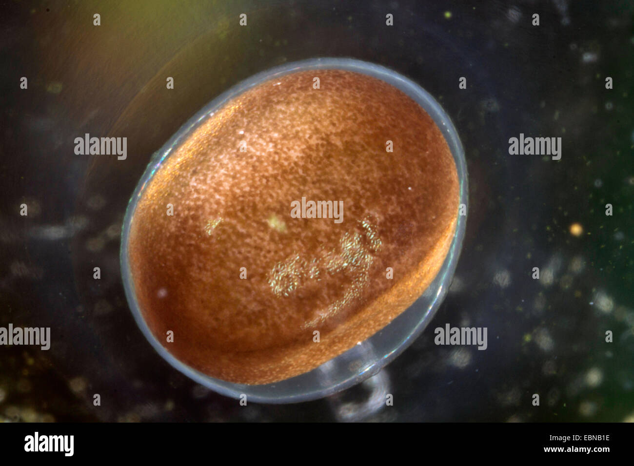 moor frog (Rana arvalis), egg with embryonic development Stock Photohttps://www.alamy.com/image-license-details/?v=1https://www.alamy.com/stock-photo-moor-frog-rana-arvalis-egg-with-embryonic-development-76072346.html
RMEBNB1E–moor frog (Rana arvalis), egg with embryonic development
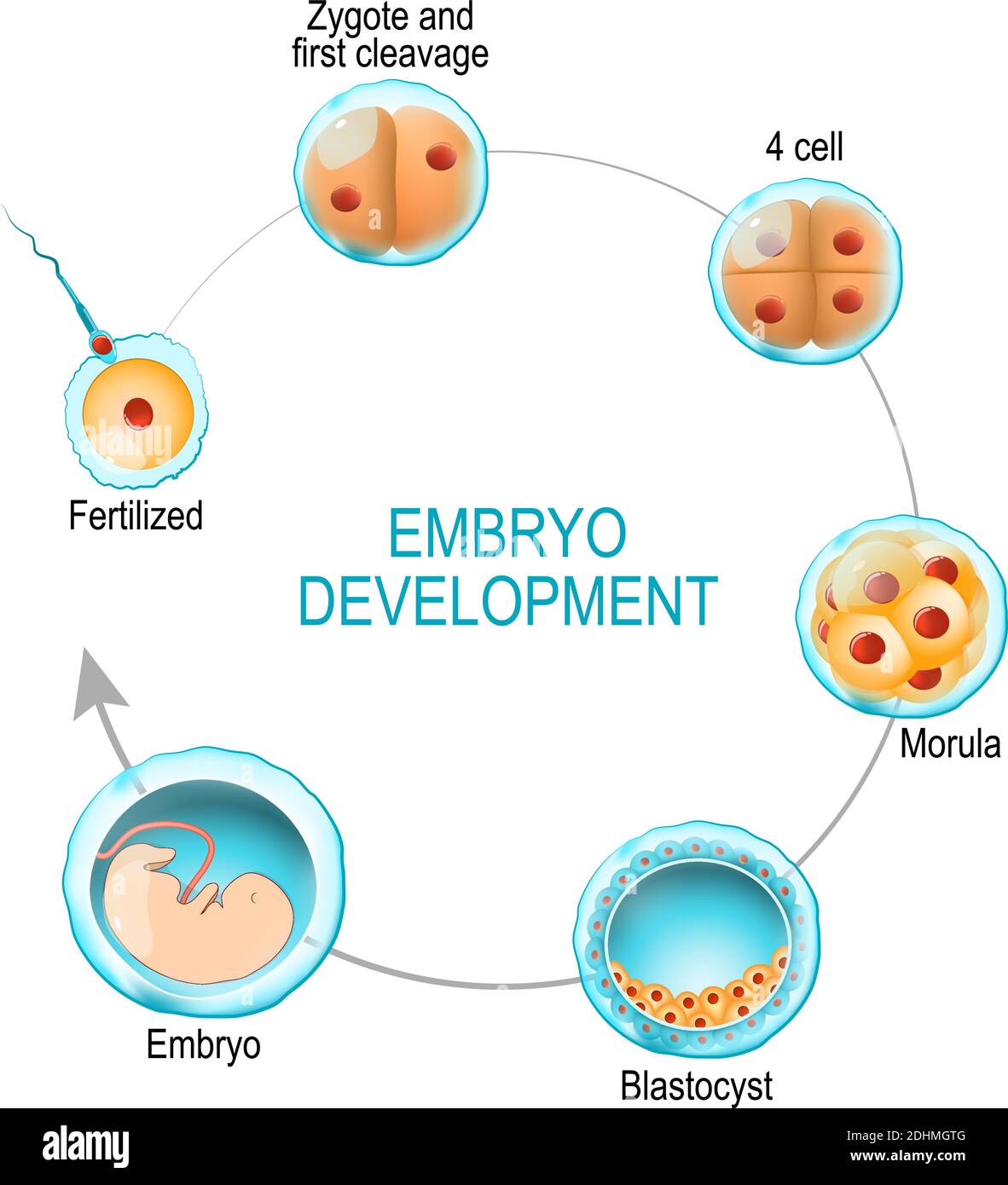 embryo development. from fertilization to zygote, morula and Blastocyst. vector diagram for medical, educational and scientific use Stock Vectorhttps://www.alamy.com/image-license-details/?v=1https://www.alamy.com/embryo-development-from-fertilization-to-zygote-morula-and-blastocyst-vector-diagram-for-medical-educational-and-scientific-use-image389529520.html
RF2DHMGTG–embryo development. from fertilization to zygote, morula and Blastocyst. vector diagram for medical, educational and scientific use
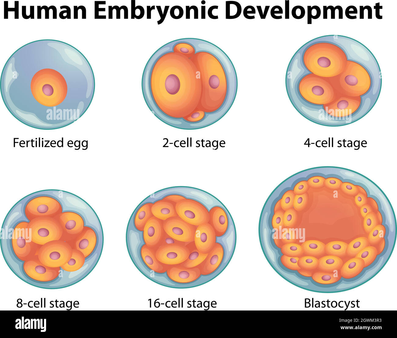 Stages in human embryonic development Stock Vectorhttps://www.alamy.com/image-license-details/?v=1https://www.alamy.com/stages-in-human-embryonic-development-image446067639.html
RF2GWM3R3–Stages in human embryonic development
 Early stages of embryonic development (embryogenesis) isolated on white. Fertilized egg, 2-cell,4-cell and morula. Cell division (cleavage) and embryo Stock Photohttps://www.alamy.com/image-license-details/?v=1https://www.alamy.com/early-stages-of-embryonic-development-embryogenesis-isolated-on-white-fertilized-egg-2-cell4-cell-and-morula-cell-division-cleavage-and-embryo-image424645135.html
RF2FJT75K–Early stages of embryonic development (embryogenesis) isolated on white. Fertilized egg, 2-cell,4-cell and morula. Cell division (cleavage) and embryo
 Embryonic development Stock Photohttps://www.alamy.com/image-license-details/?v=1https://www.alamy.com/stock-photo-embryonic-development-13248691.html
RFACXW8M–Embryonic development
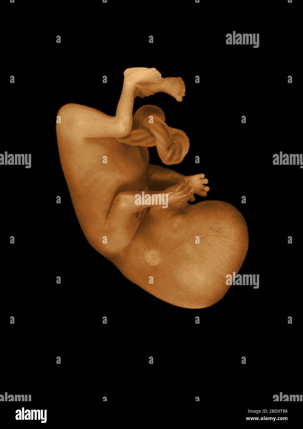 13 week old human fetus Stock Photohttps://www.alamy.com/image-license-details/?v=1https://www.alamy.com/13-week-old-human-fetus-image352787688.html
RM2BDXT88–13 week old human fetus
 Rehovot, Israel. 04th Aug, 2022. Jacob (Yaqub) Hanna, Professor of Molecular Genetics, is pictured at his laboratory at Weizmann Institute of Science in Rehovot. Yagub has led a team of scientists at the Weizmann Institute that could develop the world's first 'synthetic embryos', made of stem cells from mice without the need for sperms, eggs or fertilisation. The embryo-like structures are expected to drive deeper understanding of embryonic development and could be used to grow individual organs for transplanting. Credit: Ilia Yefimovich/dpa/Alamy Live News Stock Photohttps://www.alamy.com/image-license-details/?v=1https://www.alamy.com/rehovot-israel-04th-aug-2022-jacob-yaqub-hanna-professor-of-molecular-genetics-is-pictured-at-his-laboratory-at-weizmann-institute-of-science-in-rehovot-yagub-has-led-a-team-of-scientists-at-the-weizmann-institute-that-could-develop-the-worlds-first-synthetic-embryos-made-of-stem-cells-from-mice-without-the-need-for-sperms-eggs-or-fertilisation-the-embryo-like-structures-are-expected-to-drive-deeper-understanding-of-embryonic-development-and-could-be-used-to-grow-individual-organs-for-transplanting-credit-ilia-yefimovichdpaalamy-live-news-image477044158.html
RM2JM36KA–Rehovot, Israel. 04th Aug, 2022. Jacob (Yaqub) Hanna, Professor of Molecular Genetics, is pictured at his laboratory at Weizmann Institute of Science in Rehovot. Yagub has led a team of scientists at the Weizmann Institute that could develop the world's first 'synthetic embryos', made of stem cells from mice without the need for sperms, eggs or fertilisation. The embryo-like structures are expected to drive deeper understanding of embryonic development and could be used to grow individual organs for transplanting. Credit: Ilia Yefimovich/dpa/Alamy Live News
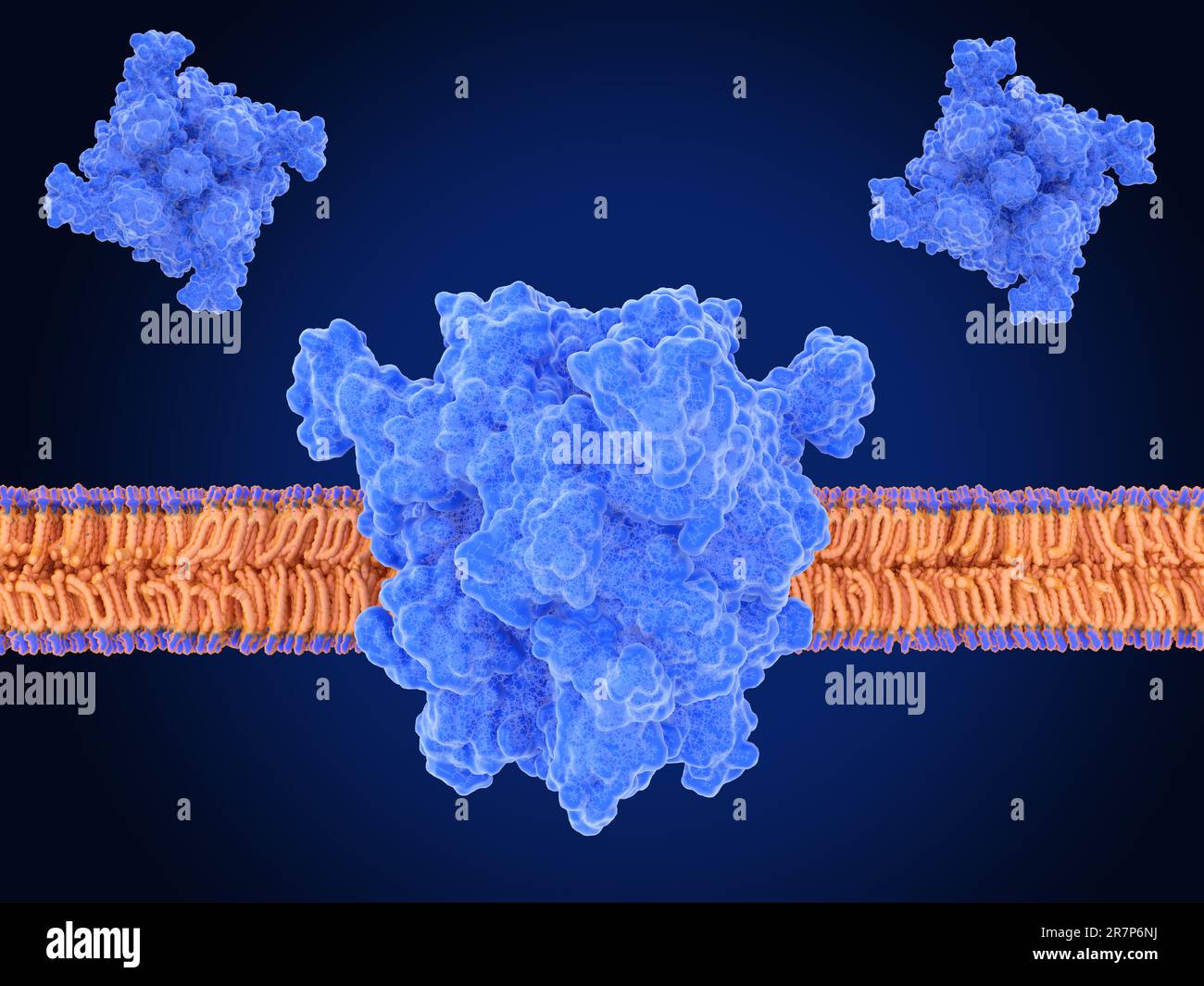 Illustration of the transient receptor potential cation channel, subfamily M, member 7 (TRPM7) in its open (top left) and closed (top right) configurations and in a cell membrane (centre). TRPM7 regulates the uptake of divalent cations, such as calcium and magnesium into a cell. It plays an essential role in embryonic development, immune response, cell mobility, proliferation, and differentiation. TRPM7 is implicated in neuronal and cardiovascular disorders, tumor progression and has emerged as a new drug target. Stock Photohttps://www.alamy.com/image-license-details/?v=1https://www.alamy.com/illustration-of-the-transient-receptor-potential-cation-channel-subfamily-m-member-7-trpm7-in-its-open-top-left-and-closed-top-right-configurations-and-in-a-cell-membrane-centre-trpm7-regulates-the-uptake-of-divalent-cations-such-as-calcium-and-magnesium-into-a-cell-it-plays-an-essential-role-in-embryonic-development-immune-response-cell-mobility-proliferation-and-differentiation-trpm7-is-implicated-in-neuronal-and-cardiovascular-disorders-tumor-progression-and-has-emerged-as-a-new-drug-target-image555522622.html
RF2R7P6NJ–Illustration of the transient receptor potential cation channel, subfamily M, member 7 (TRPM7) in its open (top left) and closed (top right) configurations and in a cell membrane (centre). TRPM7 regulates the uptake of divalent cations, such as calcium and magnesium into a cell. It plays an essential role in embryonic development, immune response, cell mobility, proliferation, and differentiation. TRPM7 is implicated in neuronal and cardiovascular disorders, tumor progression and has emerged as a new drug target.
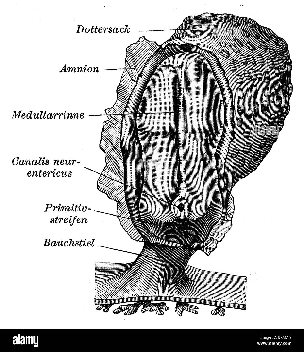 Human embryonic system Stock Photohttps://www.alamy.com/image-license-details/?v=1https://www.alamy.com/stock-photo-human-embryonic-system-29124563.html
RMBKAMJY–Human embryonic system
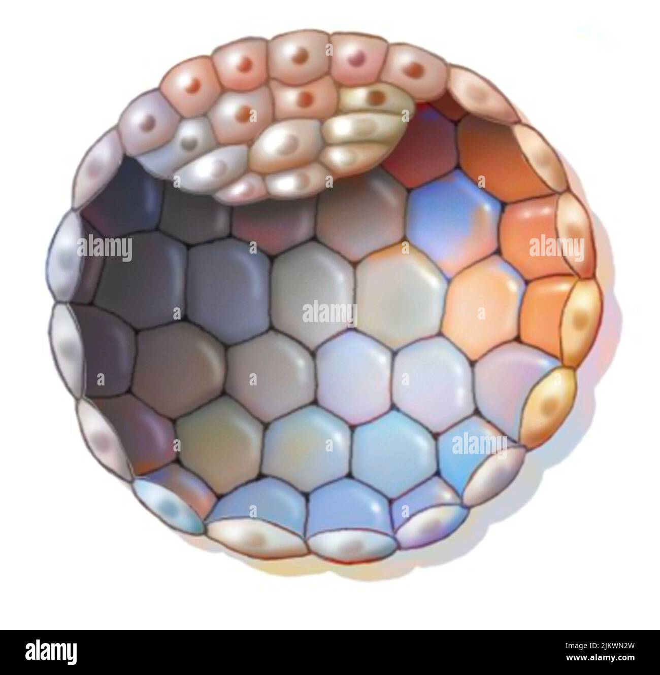 Human blastocyst characterized by the formation of a cluster of embryonic cells. Stock Photohttps://www.alamy.com/image-license-details/?v=1https://www.alamy.com/human-blastocyst-characterized-by-the-formation-of-a-cluster-of-embryonic-cells-image476923745.html
RF2JKWN2W–Human blastocyst characterized by the formation of a cluster of embryonic cells.
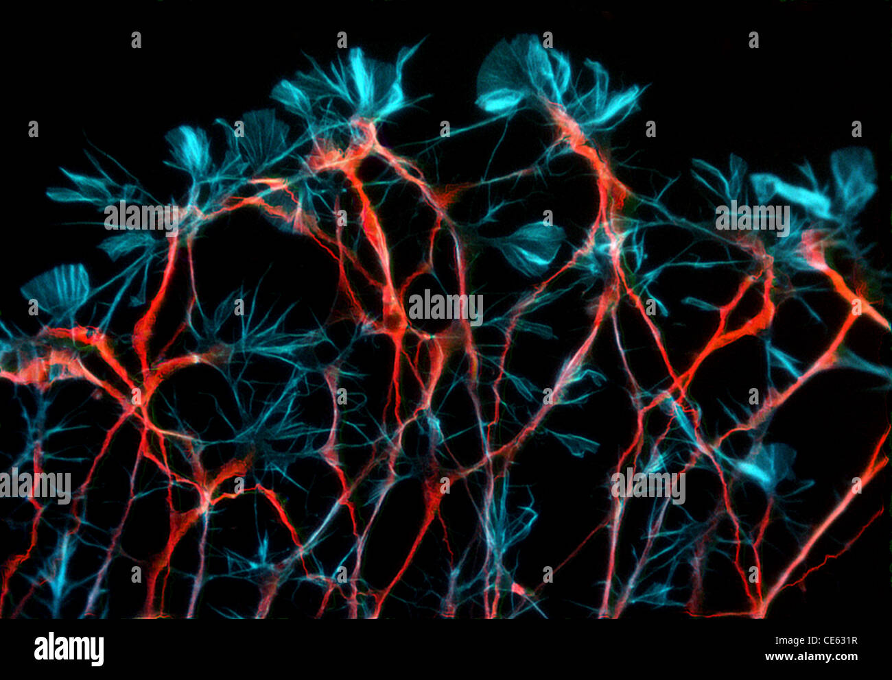 Developing rat sensory neurons. Stock Photohttps://www.alamy.com/image-license-details/?v=1https://www.alamy.com/stock-photo-developing-rat-sensory-neurons-43160035.html
RMCE631R–Developing rat sensory neurons.
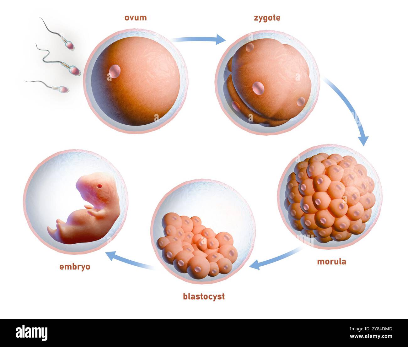 Human fertilization and embryonic development. Digital illustration, 3D render. Stock Photohttps://www.alamy.com/image-license-details/?v=1https://www.alamy.com/human-fertilization-and-embryonic-development-digital-illustration-3d-render-image624589069.html
RF2Y84DMD–Human fertilization and embryonic development. Digital illustration, 3D render.
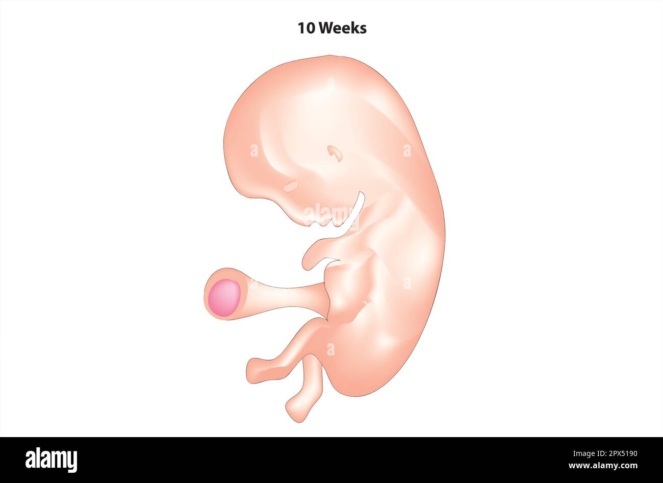 Anatomy of 10 weeks fetus Stock Vectorhttps://www.alamy.com/image-license-details/?v=1https://www.alamy.com/anatomy-of-10-weeks-fetus-image549613260.html
RF2PX5190–Anatomy of 10 weeks fetus
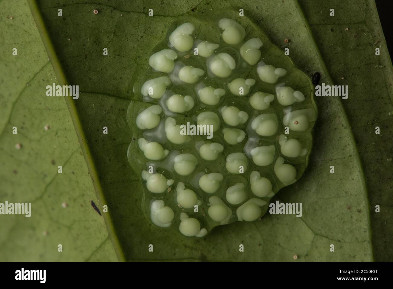 The eggs of a glass frog develop on the underside of a leaf in a rainforest in Ecuador. Stock Photohttps://www.alamy.com/image-license-details/?v=1https://www.alamy.com/the-eggs-of-a-glass-frog-develop-on-the-underside-of-a-leaf-in-a-rainforest-in-ecuador-image364502876.html
RM2C50F3T–The eggs of a glass frog develop on the underside of a leaf in a rainforest in Ecuador.
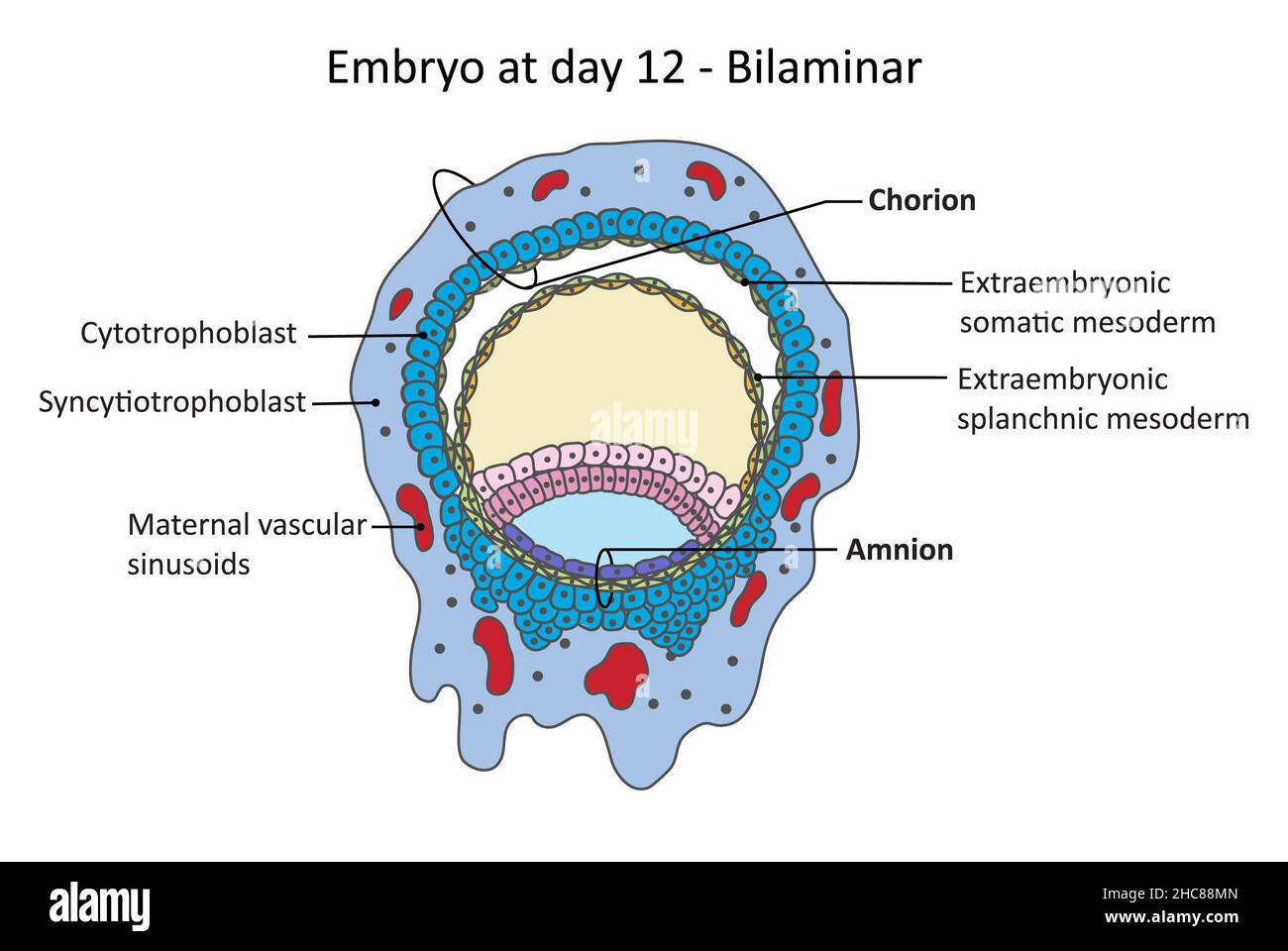 Embry at the day 12, pre-embryonic development, chorionic cavity, amniotic cavity Stock Photohttps://www.alamy.com/image-license-details/?v=1https://www.alamy.com/embry-at-the-day-12-pre-embryonic-development-chorionic-cavity-amniotic-cavity-image455027909.html
RF2HC88MN–Embry at the day 12, pre-embryonic development, chorionic cavity, amniotic cavity
 Sand Couch, forming an embryonic dune, the first stage of dune development Stock Photohttps://www.alamy.com/image-license-details/?v=1https://www.alamy.com/sand-couch-forming-an-embryonic-dune-the-first-stage-of-dune-development-image157915867.html
RFK4WK8B–Sand Couch, forming an embryonic dune, the first stage of dune development
 Wilde animal in Sweden, Europe Stock Photohttps://www.alamy.com/image-license-details/?v=1https://www.alamy.com/stock-photo-wilde-animal-in-sweden-europe-56398654.html
RMD7N51J–Wilde animal in Sweden, Europe
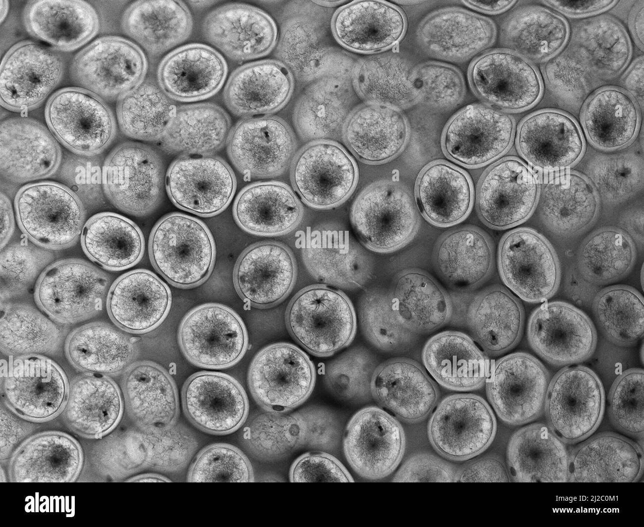 Ascaris (Ascaris megalocephala) embryos in the uterus. Fertilization in Ascaris (Ascaris megalocephala). Stock Photohttps://www.alamy.com/image-license-details/?v=1https://www.alamy.com/ascaris-ascaris-megalocephala-embryos-in-the-uterus-fertilization-in-ascaris-ascaris-megalocephala-image466173233.html
RF2J2C0M1–Ascaris (Ascaris megalocephala) embryos in the uterus. Fertilization in Ascaris (Ascaris megalocephala).
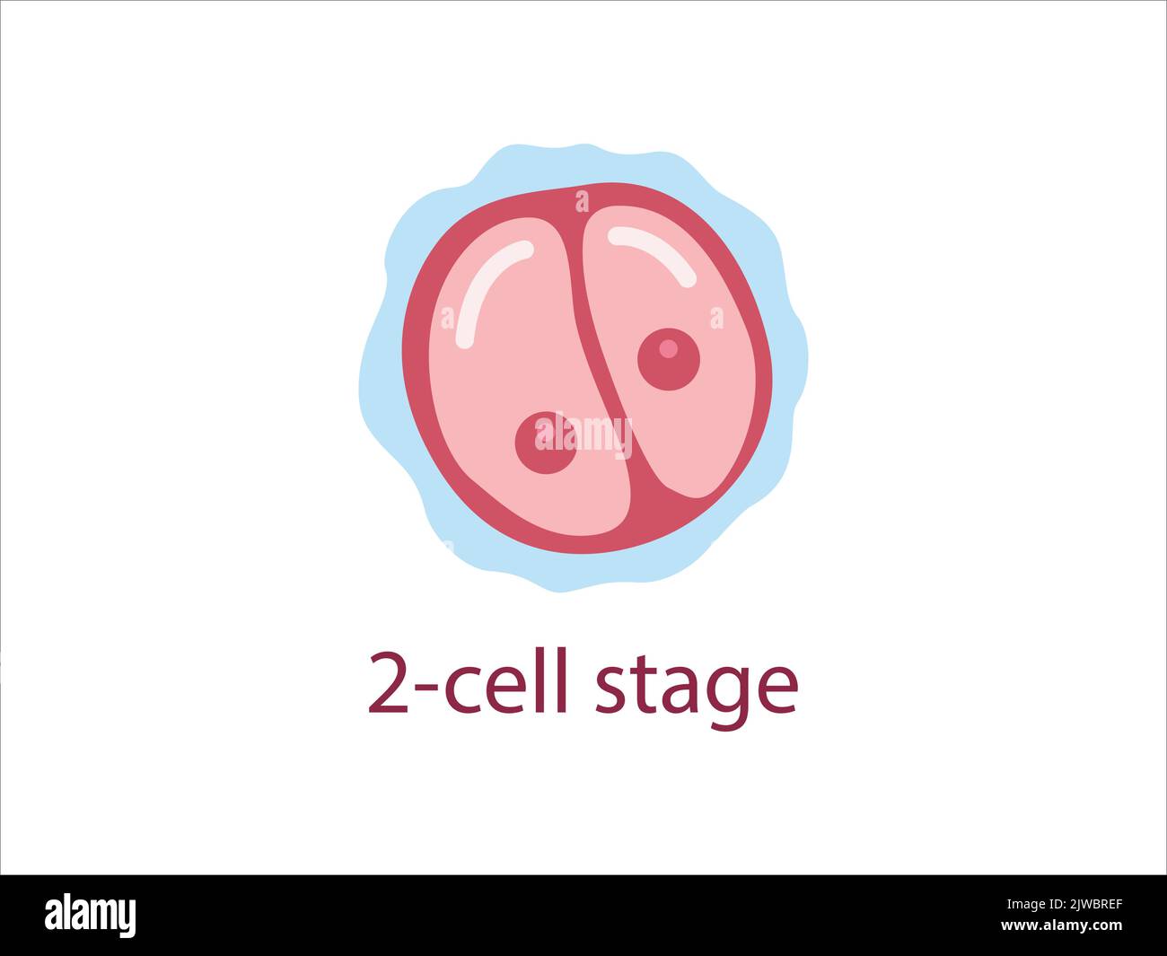 Zygote 2-cell stage. Human embryonic development. Vector medical illustration. Stock Vectorhttps://www.alamy.com/image-license-details/?v=1https://www.alamy.com/zygote-2-cell-stage-human-embryonic-development-vector-medical-illustration-image480306247.html
RF2JWBREF–Zygote 2-cell stage. Human embryonic development. Vector medical illustration.
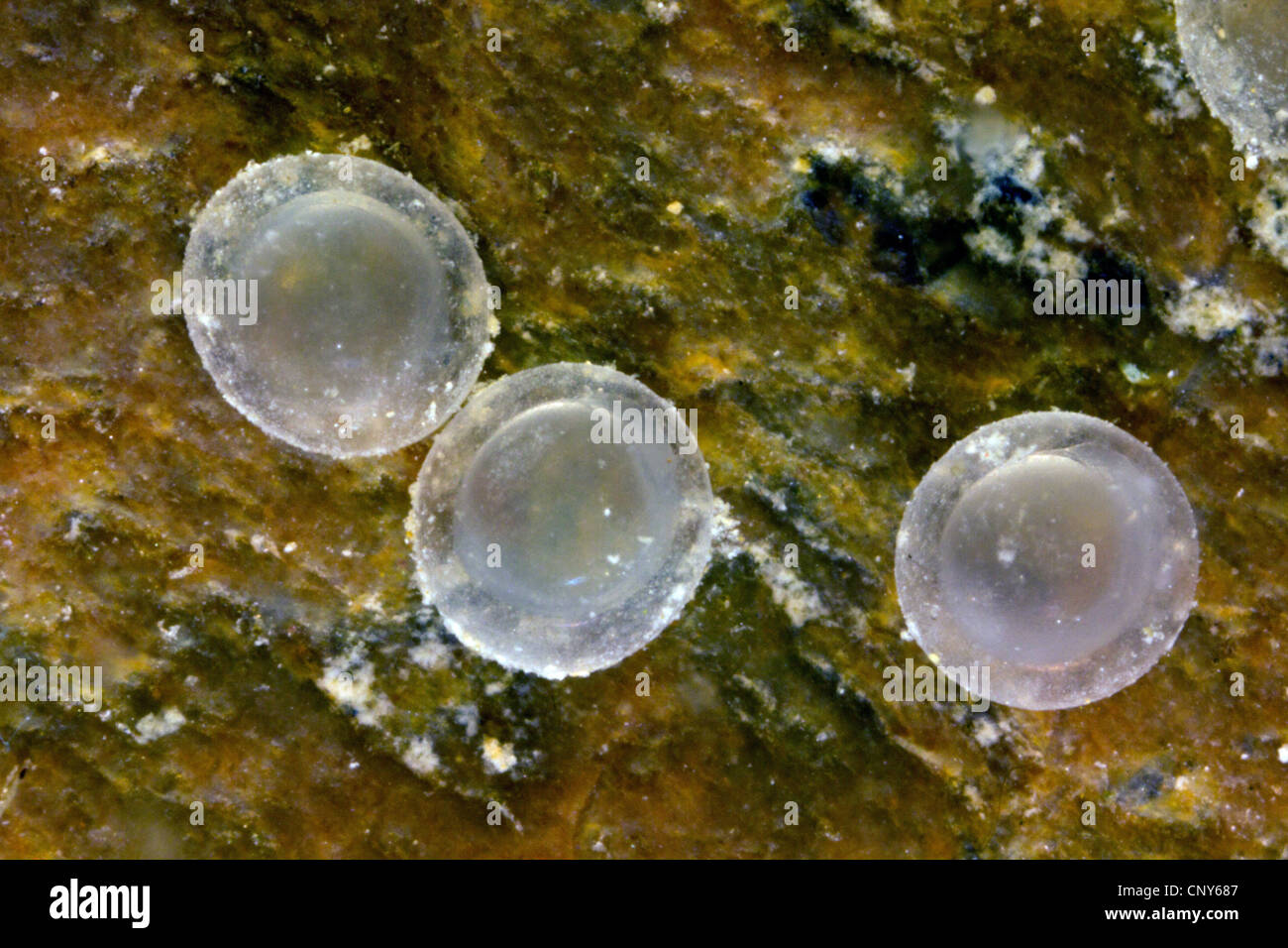 Italian bleak (Alburnus albidus), eggs with beginning embryonic development, Germany, Bavaria, Lake Chiemsee Stock Photohttps://www.alamy.com/image-license-details/?v=1https://www.alamy.com/stock-photo-italian-bleak-alburnus-albidus-eggs-with-beginning-embryonic-development-47926151.html
RMCNY687–Italian bleak (Alburnus albidus), eggs with beginning embryonic development, Germany, Bavaria, Lake Chiemsee
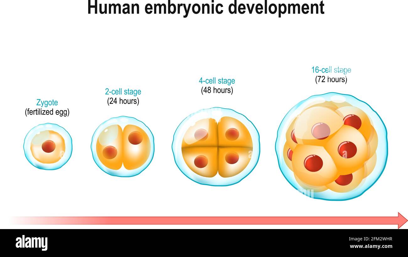 Human embryonic development. From Zygote (fertilized egg) to 16-cell stage. three day after fertilization. cell division and prenatal development Stock Vectorhttps://www.alamy.com/image-license-details/?v=1https://www.alamy.com/human-embryonic-development-from-zygote-fertilized-egg-to-16-cell-stage-three-day-after-fertilization-cell-division-and-prenatal-development-image425405955.html
RF2FM2WHR–Human embryonic development. From Zygote (fertilized egg) to 16-cell stage. three day after fertilization. cell division and prenatal development
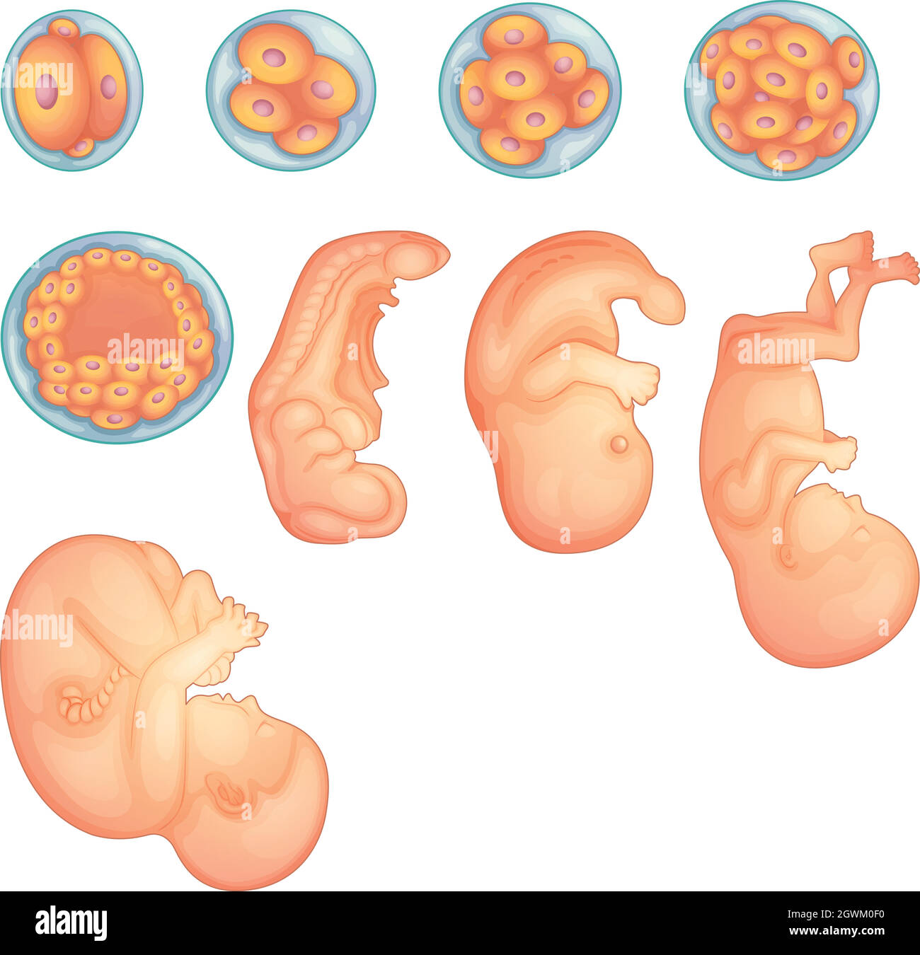 Stages in human embryonic development Stock Vectorhttps://www.alamy.com/image-license-details/?v=1https://www.alamy.com/stages-in-human-embryonic-development-image446065060.html
RF2GWM0F0–Stages in human embryonic development
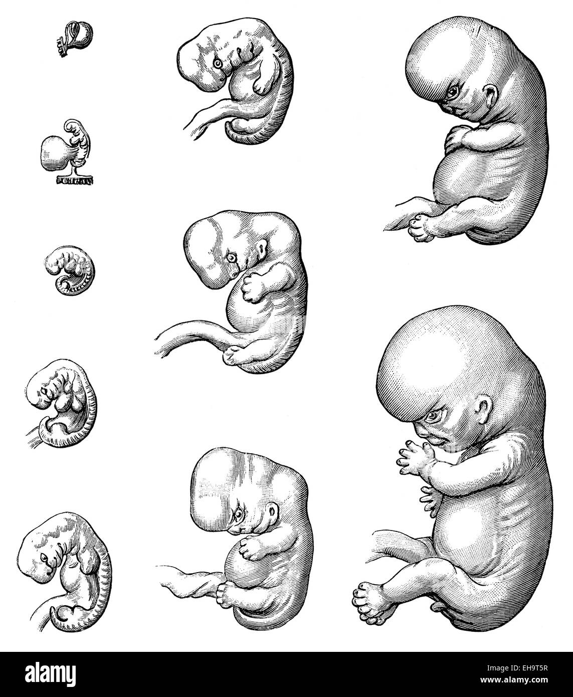 Development, the embryo up to the 9th week, 19th century, Stock Photohttps://www.alamy.com/image-license-details/?v=1https://www.alamy.com/stock-photo-development-the-embryo-up-to-the-9th-week-19th-century-79507171.html
RMEH9T5R–Development, the embryo up to the 9th week, 19th century,
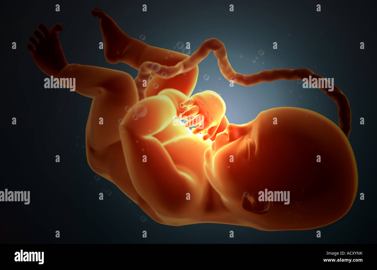 Embryonic development Stock Photohttps://www.alamy.com/image-license-details/?v=1https://www.alamy.com/stock-photo-embryonic-development-13249518.html
RFACXYNK–Embryonic development
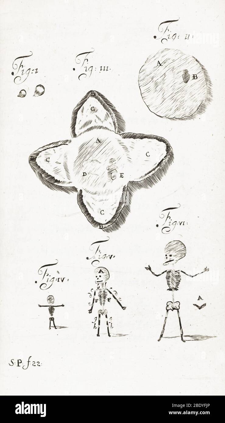 Human Embryo Development, 1685 Stock Photohttps://www.alamy.com/image-license-details/?v=1https://www.alamy.com/human-embryo-development-1685-image352802878.html
RM2BDYFJP–Human Embryo Development, 1685
 Rehovot, Israel. 04th Aug, 2022. Jacob (Yaqub) Hanna, Professor of Molecular Genetics, is pictured at his laboratory at Weizmann Institute of Science in Rehovot. Yaqub has led a team of scientists at the Weizmann Institute that could develop the world's first 'synthetic embryos', made of stem cells from mice without the need for sperms, eggs or fertilisation. The embryo-like structures are expected to drive deeper understanding of embryonic development and could be used to grow individual organs for transplanting. Credit: Ilia Yefimovich/dpa/Alamy Live News Stock Photohttps://www.alamy.com/image-license-details/?v=1https://www.alamy.com/rehovot-israel-04th-aug-2022-jacob-yaqub-hanna-professor-of-molecular-genetics-is-pictured-at-his-laboratory-at-weizmann-institute-of-science-in-rehovot-yaqub-has-led-a-team-of-scientists-at-the-weizmann-institute-that-could-develop-the-worlds-first-synthetic-embryos-made-of-stem-cells-from-mice-without-the-need-for-sperms-eggs-or-fertilisation-the-embryo-like-structures-are-expected-to-drive-deeper-understanding-of-embryonic-development-and-could-be-used-to-grow-individual-organs-for-transplanting-credit-ilia-yefimovichdpaalamy-live-news-image477044157.html
RM2JM36K9–Rehovot, Israel. 04th Aug, 2022. Jacob (Yaqub) Hanna, Professor of Molecular Genetics, is pictured at his laboratory at Weizmann Institute of Science in Rehovot. Yaqub has led a team of scientists at the Weizmann Institute that could develop the world's first 'synthetic embryos', made of stem cells from mice without the need for sperms, eggs or fertilisation. The embryo-like structures are expected to drive deeper understanding of embryonic development and could be used to grow individual organs for transplanting. Credit: Ilia Yefimovich/dpa/Alamy Live News
 Computed tomography (CT) scan of a chest from above showing an azygos lobe in the left lung (segmented area centre-left). An azygos lobe is an anatomical variant in the upper lobe of the lung in which a segment of the lung is separated by a deep groove called an azygos fissure, which consists of the azygos vein and pleura (thin tissue). It is a rare variation, occurring in around 0.3% of people during embryonic development. It causes no symptoms, but may make certain surgical procedures more difficult. Stock Photohttps://www.alamy.com/image-license-details/?v=1https://www.alamy.com/computed-tomography-ct-scan-of-a-chest-from-above-showing-an-azygos-lobe-in-the-left-lung-segmented-area-centre-left-an-azygos-lobe-is-an-anatomical-variant-in-the-upper-lobe-of-the-lung-in-which-a-segment-of-the-lung-is-separated-by-a-deep-groove-called-an-azygos-fissure-which-consists-of-the-azygos-vein-and-pleura-thin-tissue-it-is-a-rare-variation-occurring-in-around-03-of-people-during-embryonic-development-it-causes-no-symptoms-but-may-make-certain-surgical-procedures-more-difficult-image612233850.html
RF2XG1JEJ–Computed tomography (CT) scan of a chest from above showing an azygos lobe in the left lung (segmented area centre-left). An azygos lobe is an anatomical variant in the upper lobe of the lung in which a segment of the lung is separated by a deep groove called an azygos fissure, which consists of the azygos vein and pleura (thin tissue). It is a rare variation, occurring in around 0.3% of people during embryonic development. It causes no symptoms, but may make certain surgical procedures more difficult.
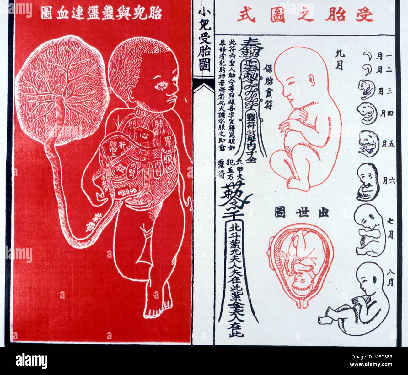 Physiognomy. Chinese print showing the stages of embryonic growth in the womb. From a Chinese annual calendar manual. Stock Photohttps://www.alamy.com/image-license-details/?v=1https://www.alamy.com/physiognomy-chinese-print-showing-the-stages-of-embryonic-growth-in-the-womb-from-a-chinese-annual-calendar-manual-image179152937.html
RMMBD3B5–Physiognomy. Chinese print showing the stages of embryonic growth in the womb. From a Chinese annual calendar manual.
 Stem cell research, nuclear transfer of embryonic stem cells used in cloning for medical research Stock Photohttps://www.alamy.com/image-license-details/?v=1https://www.alamy.com/stock-photo-stem-cell-research-nuclear-transfer-of-embryonic-stem-cells-used-in-81331135.html
RFEM8XKB–Stem cell research, nuclear transfer of embryonic stem cells used in cloning for medical research
RF2EDF1BA–Pregnancy woman stages vector illustration with flat silhouettes of pregnant women and realistic human embryonic development icons
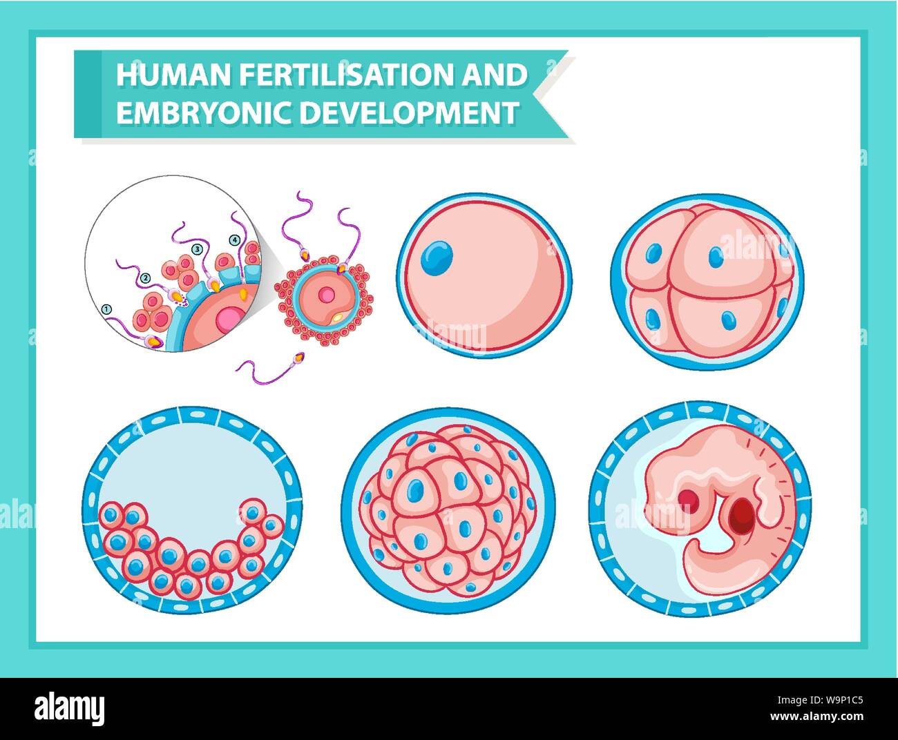 Scientific medical illustration of embryonic development illustration Stock Vectorhttps://www.alamy.com/image-license-details/?v=1https://www.alamy.com/scientific-medical-illustration-of-embryonic-development-illustration-image264171493.html
RFW9P1C5–Scientific medical illustration of embryonic development illustration
 Lyon, France. 06th Oct, 2023. Sample of bedbugs at different stages of embryonic development until adulthood, study on bedbugs, at the biometrics and evolutionary biology laboratory, Lyon, France on October 5, 2023. The French environment ministry said bedbugs have made a resurgence since disappearing in the 1950s due to international travel and increased resistance to pesticides. Photo by Thibaut Durand/ABACAPRESS.COM Credit: Abaca Press/Alamy Live News Stock Photohttps://www.alamy.com/image-license-details/?v=1https://www.alamy.com/lyon-france-06th-oct-2023-sample-of-bedbugs-at-different-stages-of-embryonic-development-until-adulthood-study-on-bedbugs-at-the-biometrics-and-evolutionary-biology-laboratory-lyon-france-on-october-5-2023-the-french-environment-ministry-said-bedbugs-have-made-a-resurgence-since-disappearing-in-the-1950s-due-to-international-travel-and-increased-resistance-to-pesticides-photo-by-thibaut-durandabacapresscom-credit-abaca-pressalamy-live-news-image568558953.html
RM2T102MW–Lyon, France. 06th Oct, 2023. Sample of bedbugs at different stages of embryonic development until adulthood, study on bedbugs, at the biometrics and evolutionary biology laboratory, Lyon, France on October 5, 2023. The French environment ministry said bedbugs have made a resurgence since disappearing in the 1950s due to international travel and increased resistance to pesticides. Photo by Thibaut Durand/ABACAPRESS.COM Credit: Abaca Press/Alamy Live News
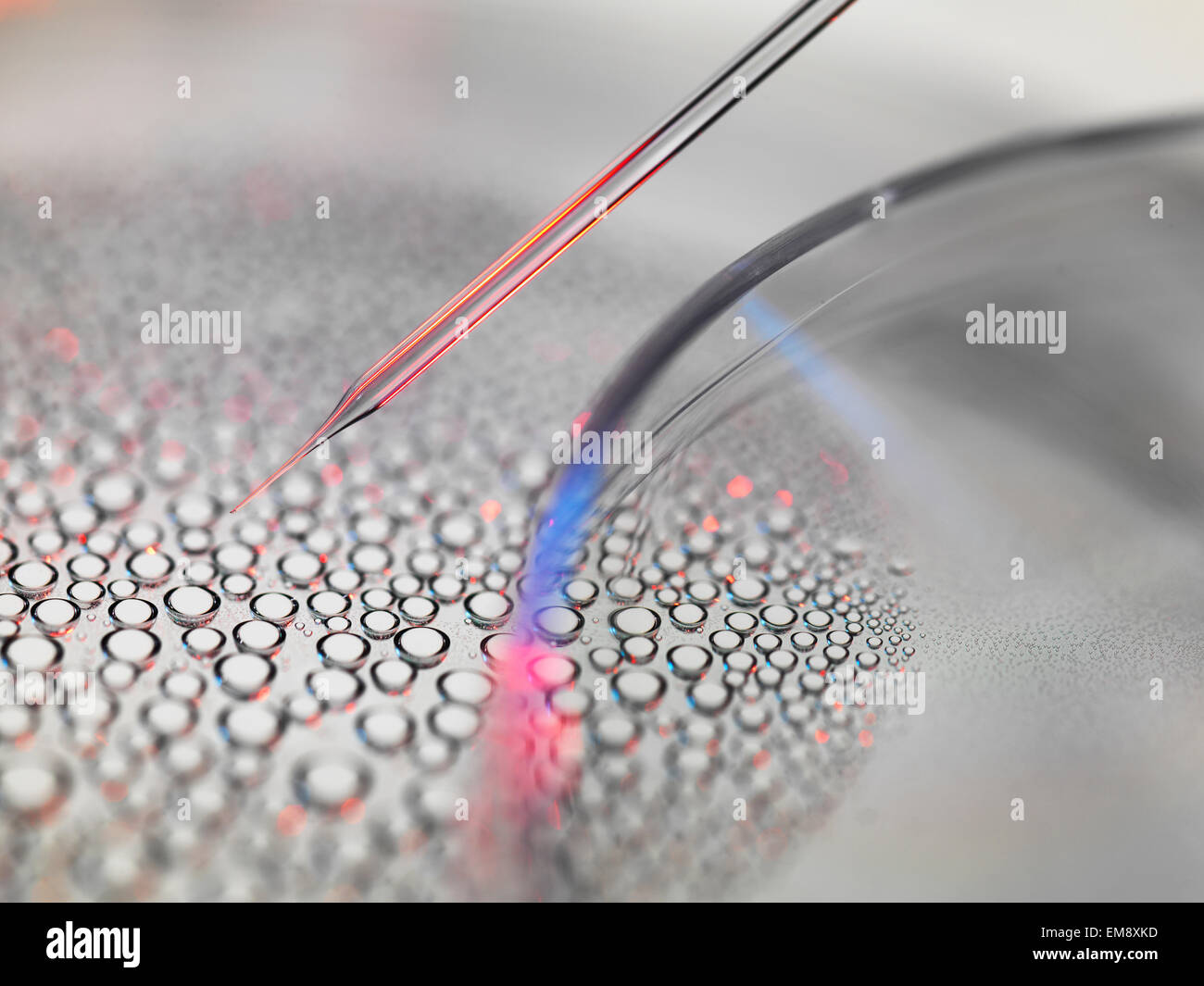 Stem cell research, nuclear transfer of embryonic stem cells from petri dish used in cloning for medical research Stock Photohttps://www.alamy.com/image-license-details/?v=1https://www.alamy.com/stock-photo-stem-cell-research-nuclear-transfer-of-embryonic-stem-cells-from-petri-81331137.html
RFEM8XKD–Stem cell research, nuclear transfer of embryonic stem cells from petri dish used in cloning for medical research
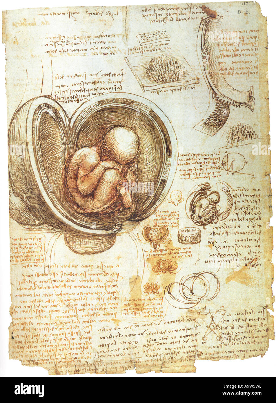 Anatomical studies of embryonic development by Leonardo da Vinci Stock Photohttps://www.alamy.com/image-license-details/?v=1https://www.alamy.com/anatomical-studies-of-embryonic-development-by-leonardo-da-vinci-image7109981.html
RMA9W5WE–Anatomical studies of embryonic development by Leonardo da Vinci
 Sand Couch, forming an embryonic dune, the first stage of dune development Stock Photohttps://www.alamy.com/image-license-details/?v=1https://www.alamy.com/sand-couch-forming-an-embryonic-dune-the-first-stage-of-dune-development-image155988024.html
RFK1NT8T–Sand Couch, forming an embryonic dune, the first stage of dune development
 Wilde animal in Sweden, Europe Stock Photohttps://www.alamy.com/image-license-details/?v=1https://www.alamy.com/stock-photo-wilde-animal-in-sweden-europe-48776522.html
RMCR9XXJ–Wilde animal in Sweden, Europe
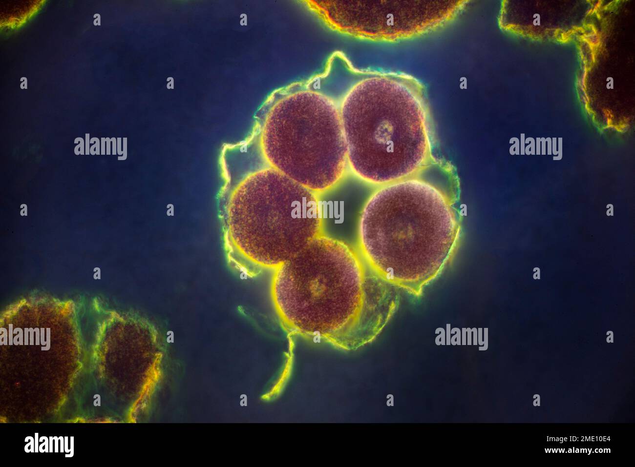
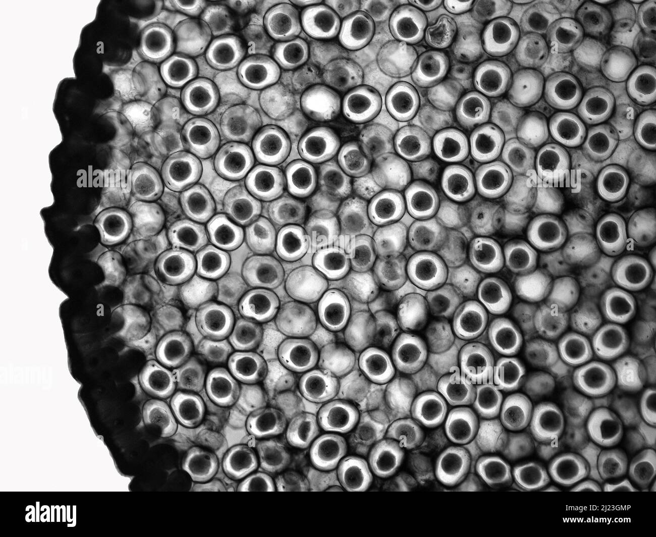 Fragmentation eggs of horse roundworm (Ascaris megalocephala). Light micrograph. Syncarion stage. Stock Photohttps://www.alamy.com/image-license-details/?v=1https://www.alamy.com/fragmentation-eggs-of-horse-roundworm-ascaris-megalocephala-light-micrograph-syncarion-stage-image465988230.html
RF2J23GMP–Fragmentation eggs of horse roundworm (Ascaris megalocephala). Light micrograph. Syncarion stage.
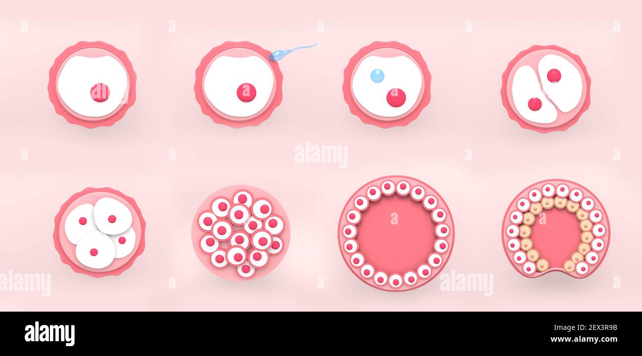 The stages of segmentation of a fertilized ovum. Human embryonic development. 3d rendering medical illustration. Stock Photohttps://www.alamy.com/image-license-details/?v=1https://www.alamy.com/the-stages-of-segmentation-of-a-fertilized-ovum-human-embryonic-development-3d-rendering-medical-illustration-image411903671.html
RF2EX3R9B–The stages of segmentation of a fertilized ovum. Human embryonic development. 3d rendering medical illustration.
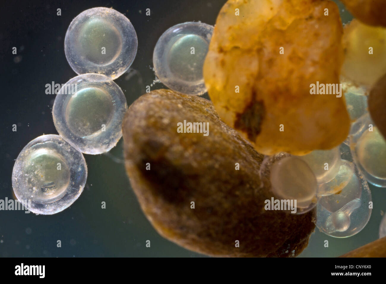 bleak (alburnus alburnus), eggs at pebble stones with beginning embryonic development, Germany, Bavaria, Lake Chiemsee Stock Photohttps://www.alamy.com/image-license-details/?v=1https://www.alamy.com/stock-photo-bleak-alburnus-alburnus-eggs-at-pebble-stones-with-beginning-embryonic-47926648.html
RMCNY6X0–bleak (alburnus alburnus), eggs at pebble stones with beginning embryonic development, Germany, Bavaria, Lake Chiemsee
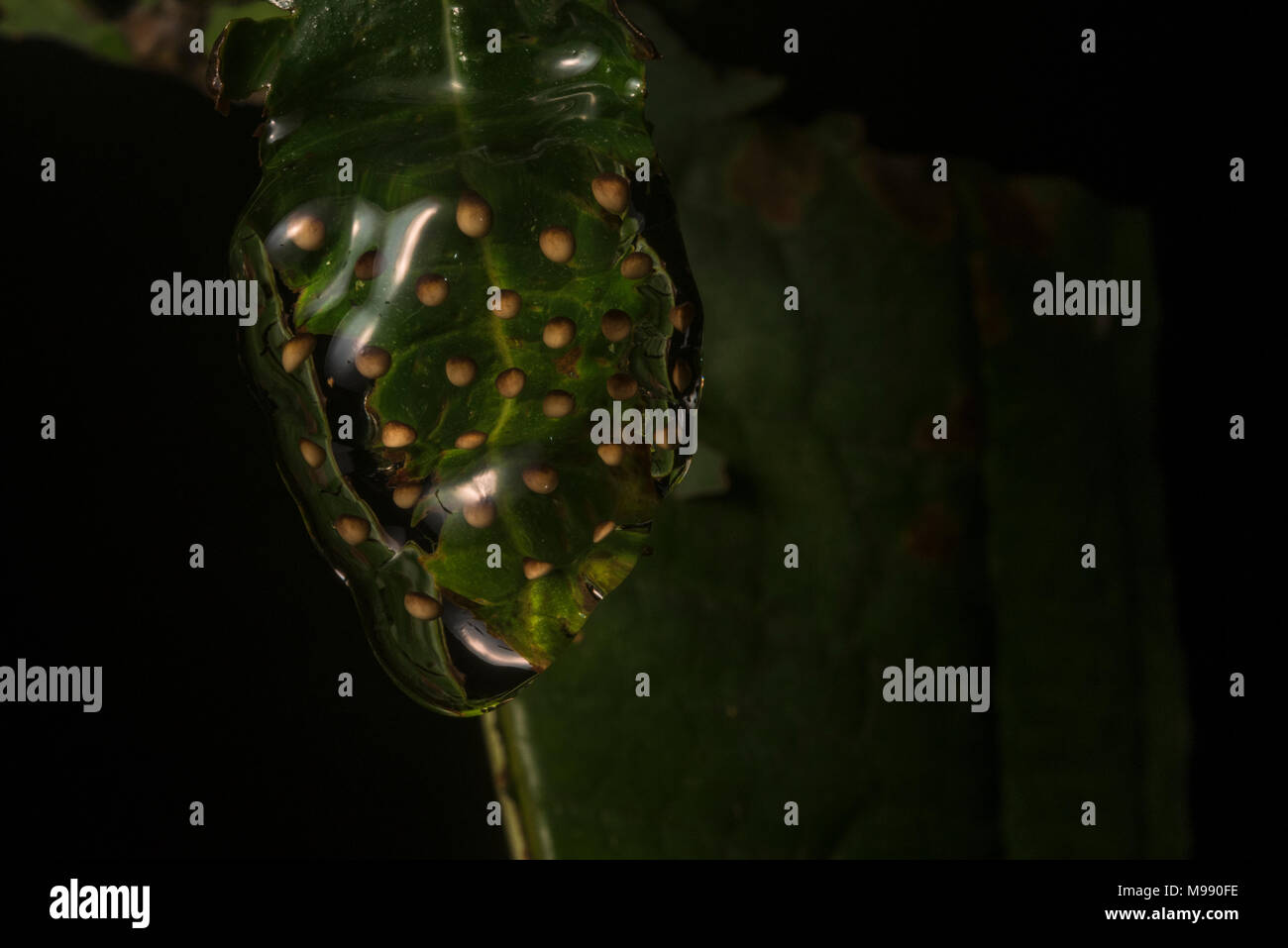 A egg mass from a Dendropsophus treefrog, they hang their eggs from the leaves above the water which fall into the pond when they hatch. Stock Photohttps://www.alamy.com/image-license-details/?v=1https://www.alamy.com/a-egg-mass-from-a-dendropsophus-treefrog-they-hang-their-eggs-from-the-leaves-above-the-water-which-fall-into-the-pond-when-they-hatch-image177833586.html
RMM990FE–A egg mass from a Dendropsophus treefrog, they hang their eggs from the leaves above the water which fall into the pond when they hatch.
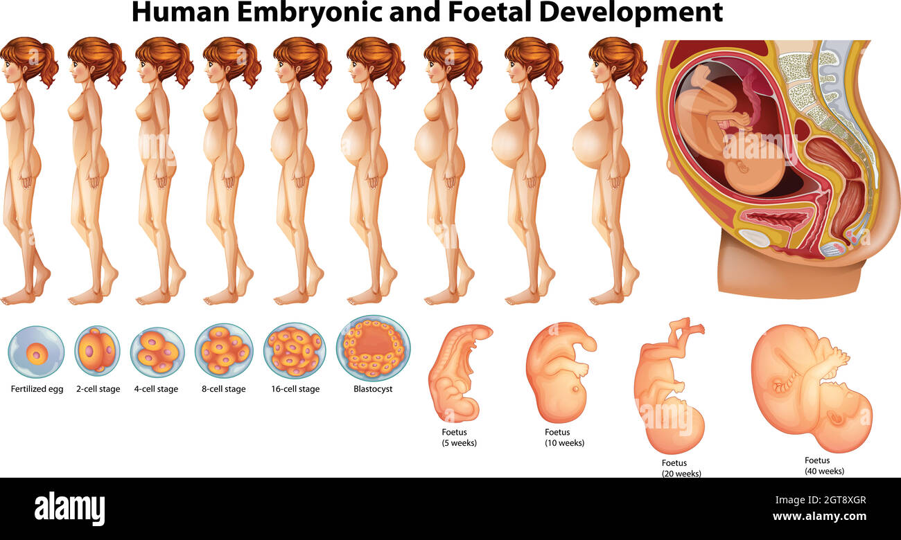 Vector of Human Embryonic and Foetal Development Stock Vectorhttps://www.alamy.com/image-license-details/?v=1https://www.alamy.com/vector-of-human-embryonic-and-foetal-development-image445207415.html
RF2GT8XGR–Vector of Human Embryonic and Foetal Development
 Starfish emryo development stages Stock Photohttps://www.alamy.com/image-license-details/?v=1https://www.alamy.com/starfish-emryo-development-stages-image507728955.html
RM2ME11CY–Starfish emryo development stages
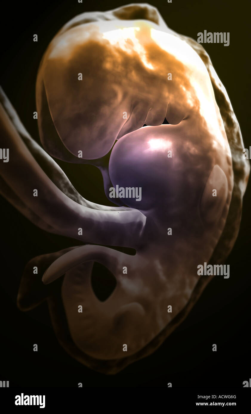 Embryonic development Stock Photohttps://www.alamy.com/image-license-details/?v=1https://www.alamy.com/stock-photo-embryonic-development-13236231.html
RFACWG6G–Embryonic development
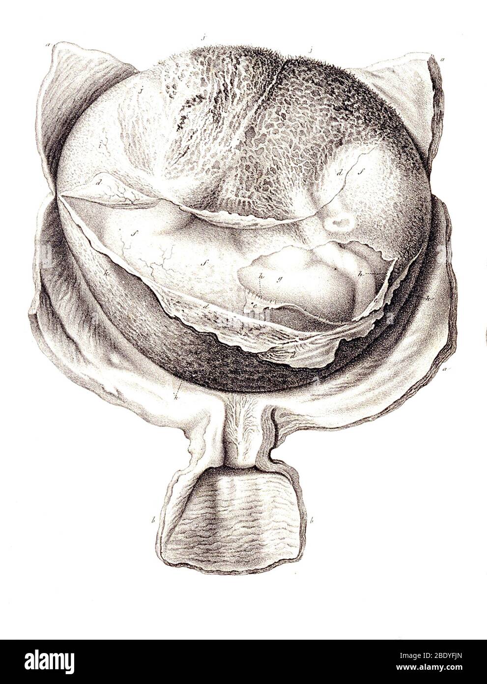 Development of the Fetus, c. 1840 Stock Photohttps://www.alamy.com/image-license-details/?v=1https://www.alamy.com/development-of-the-fetus-c-1840-image352802877.html
RM2BDYFJN–Development of the Fetus, c. 1840
 Rehovot, Israel. 04th Aug, 2022. Lab workers are pictured in the laboratory of Professor Jacob (Yaqub) Hanna, at Weizmann Institute of Science in Rehovot. Yaqub has led a team of scientists at the Weizmann Institute that could develop the world's first 'synthetic embryos', made of stem cells from mice without the need for sperms, eggs or fertilisation. The embryo-like structures are expected to drive deeper understanding of embryonic development and could be used to grow individual organs for transplanting. Credit: Ilia Yefimovich/dpa/Alamy Live News Stock Photohttps://www.alamy.com/image-license-details/?v=1https://www.alamy.com/rehovot-israel-04th-aug-2022-lab-workers-are-pictured-in-the-laboratory-of-professor-jacob-yaqub-hanna-at-weizmann-institute-of-science-in-rehovot-yaqub-has-led-a-team-of-scientists-at-the-weizmann-institute-that-could-develop-the-worlds-first-synthetic-embryos-made-of-stem-cells-from-mice-without-the-need-for-sperms-eggs-or-fertilisation-the-embryo-like-structures-are-expected-to-drive-deeper-understanding-of-embryonic-development-and-could-be-used-to-grow-individual-organs-for-transplanting-credit-ilia-yefimovichdpaalamy-live-news-image477044944.html
RM2JM37KC–Rehovot, Israel. 04th Aug, 2022. Lab workers are pictured in the laboratory of Professor Jacob (Yaqub) Hanna, at Weizmann Institute of Science in Rehovot. Yaqub has led a team of scientists at the Weizmann Institute that could develop the world's first 'synthetic embryos', made of stem cells from mice without the need for sperms, eggs or fertilisation. The embryo-like structures are expected to drive deeper understanding of embryonic development and could be used to grow individual organs for transplanting. Credit: Ilia Yefimovich/dpa/Alamy Live News
 Coloured computed tomography (CT) scan of a chest from above showing an azygos lobe in the left lung (segmented area centre-left). An azygos lobe is an anatomical variant in the upper lobe of the lung in which a segment of the lung is separated by a deep groove called an azygos fissure, which consists of the azygos vein and pleura (thin tissue). It is a rare variation, occurring in around 0.3% of people during embryonic development. It causes no symptoms, but may make certain surgical procedures more difficult. Stock Photohttps://www.alamy.com/image-license-details/?v=1https://www.alamy.com/coloured-computed-tomography-ct-scan-of-a-chest-from-above-showing-an-azygos-lobe-in-the-left-lung-segmented-area-centre-left-an-azygos-lobe-is-an-anatomical-variant-in-the-upper-lobe-of-the-lung-in-which-a-segment-of-the-lung-is-separated-by-a-deep-groove-called-an-azygos-fissure-which-consists-of-the-azygos-vein-and-pleura-thin-tissue-it-is-a-rare-variation-occurring-in-around-03-of-people-during-embryonic-development-it-causes-no-symptoms-but-may-make-certain-surgical-procedures-more-difficult-image612233854.html
RF2XG1JEP–Coloured computed tomography (CT) scan of a chest from above showing an azygos lobe in the left lung (segmented area centre-left). An azygos lobe is an anatomical variant in the upper lobe of the lung in which a segment of the lung is separated by a deep groove called an azygos fissure, which consists of the azygos vein and pleura (thin tissue). It is a rare variation, occurring in around 0.3% of people during embryonic development. It causes no symptoms, but may make certain surgical procedures more difficult.
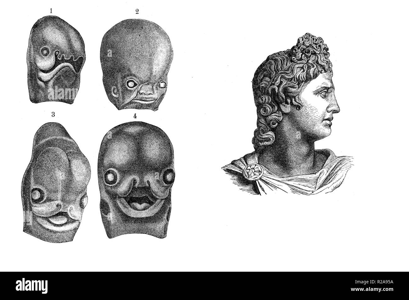 Vintage illustration, human fetus face development from 3d week to 3d month, comparison with Apollo god classical profile Stock Photohttps://www.alamy.com/image-license-details/?v=1https://www.alamy.com/vintage-illustration-human-fetus-face-development-from-3d-week-to-3d-month-comparison-with-apollo-god-classical-profile-image225190822.html
RFR2A95A–Vintage illustration, human fetus face development from 3d week to 3d month, comparison with Apollo god classical profile
 Embryological development of the renal (urinary) system, segmentation of the nephric duct Stock Photohttps://www.alamy.com/image-license-details/?v=1https://www.alamy.com/embryological-development-of-the-renal-urinary-system-segmentation-of-the-nephric-duct-image455241010.html
RF2HCJ0FE–Embryological development of the renal (urinary) system, segmentation of the nephric duct
RF2EDDW5E–Set of isolated decorative icons showing stages of human embryonic development with period clarification in months realistic vector illustration
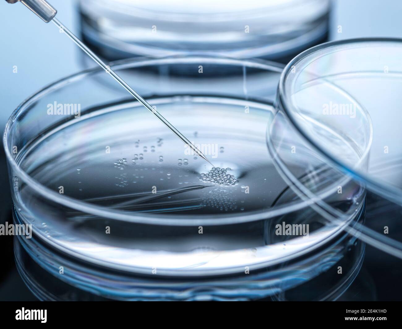 Nuclear transfer being carried out on embryonic stem cells Stock Photohttps://www.alamy.com/image-license-details/?v=1https://www.alamy.com/nuclear-transfer-being-carried-out-on-embryonic-stem-cells-image398715449.html
RF2E4K1HD–Nuclear transfer being carried out on embryonic stem cells
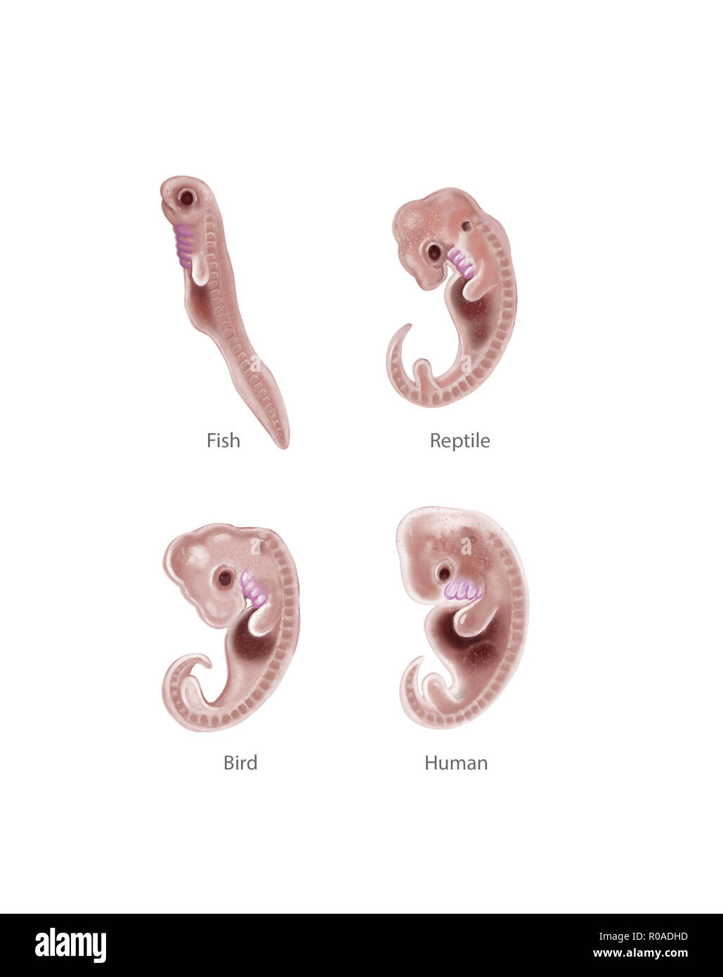 Digital illustration of 4 species embryo Stock Photohttps://www.alamy.com/image-license-details/?v=1https://www.alamy.com/digital-illustration-of-4-species-embryo-image223964985.html
RFR0ADHD–Digital illustration of 4 species embryo
 Tadpoles of the European Common Frog Rana temporaria in early embryonic stages of development shot from inside the gelatinous sheath of frogspawn Stock Photohttps://www.alamy.com/image-license-details/?v=1https://www.alamy.com/tadpoles-of-the-european-common-frog-rana-temporaria-in-early-embryonic-stages-of-development-shot-from-inside-the-gelatinous-sheath-of-frogspawn-image463789874.html
RM2HXFCM2–Tadpoles of the European Common Frog Rana temporaria in early embryonic stages of development shot from inside the gelatinous sheath of frogspawn
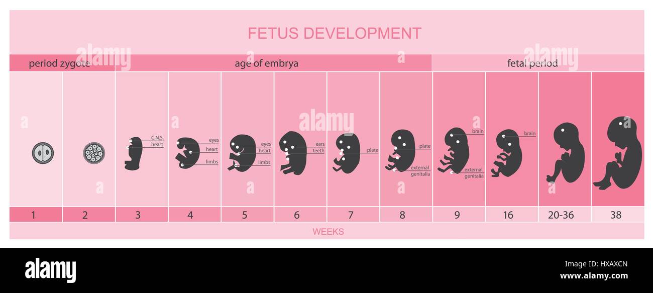 Fetus development, vetor Stock Vectorhttps://www.alamy.com/image-license-details/?v=1https://www.alamy.com/stock-photo-fetus-development-vetor-136693893.html
RFHXAXCN–Fetus development, vetor
 Embryo dune development: Sand couch grass on a very small dune on a stormy beach Stock Photohttps://www.alamy.com/image-license-details/?v=1https://www.alamy.com/embryo-dune-development-sand-couch-grass-on-a-very-small-dune-on-a-stormy-beach-image226191490.html
RFR3YWFE–Embryo dune development: Sand couch grass on a very small dune on a stormy beach
 A developing Fern leaf, unfurling, Cambridge, England, UK. Stock Photohttps://www.alamy.com/image-license-details/?v=1https://www.alamy.com/stock-photo-a-developing-fern-leaf-unfurling-cambridge-england-uk-92287743.html
RFFA41XR–A developing Fern leaf, unfurling, Cambridge, England, UK.
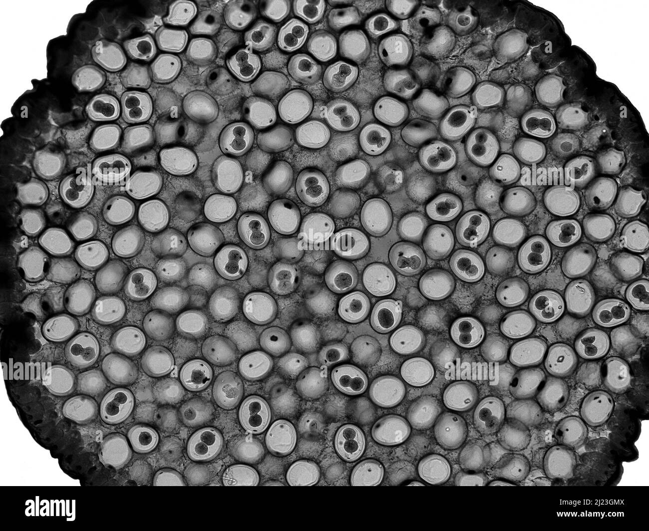 Fragmentation eggs of horse roundworm (Ascaris megalocephala). Light micrograph. Syncarion stage. Stock Photohttps://www.alamy.com/image-license-details/?v=1https://www.alamy.com/fragmentation-eggs-of-horse-roundworm-ascaris-megalocephala-light-micrograph-syncarion-stage-image465988234.html
RF2J23GMX–Fragmentation eggs of horse roundworm (Ascaris megalocephala). Light micrograph. Syncarion stage.
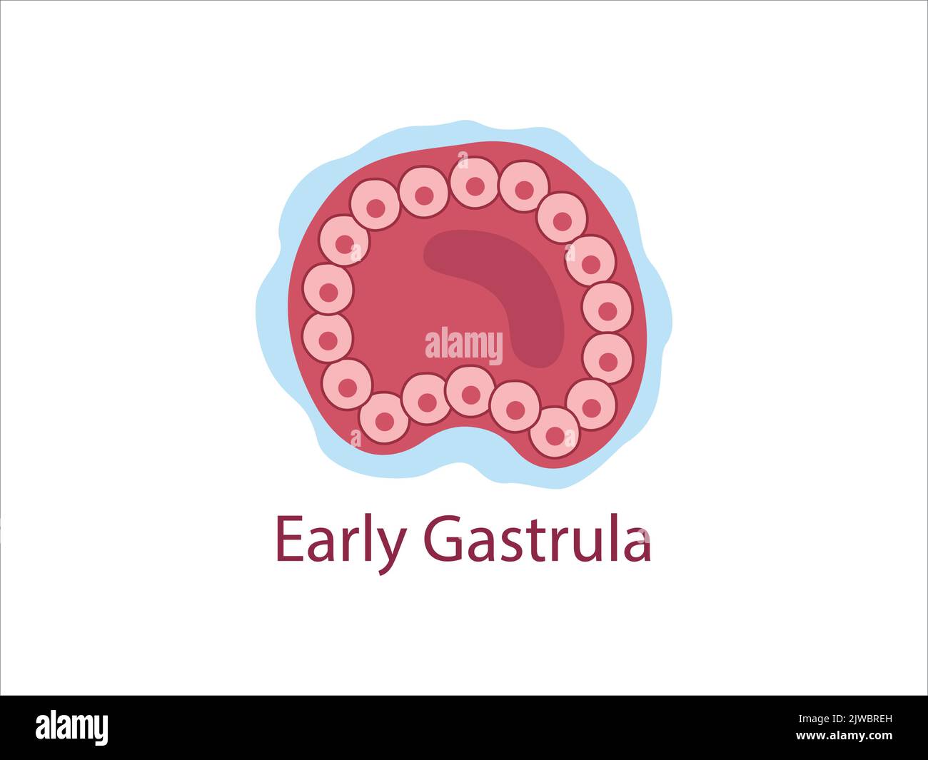 Gastrula. The cells of endoderm and ectoderm. The stage of segmentation of a fertilized ovum. Human embryonic development. Vector medical illustratio Stock Vectorhttps://www.alamy.com/image-license-details/?v=1https://www.alamy.com/gastrula-the-cells-of-endoderm-and-ectoderm-the-stage-of-segmentation-of-a-fertilized-ovum-human-embryonic-development-vector-medical-illustratio-image480306249.html
RF2JWBREH–Gastrula. The cells of endoderm and ectoderm. The stage of segmentation of a fertilized ovum. Human embryonic development. Vector medical illustratio
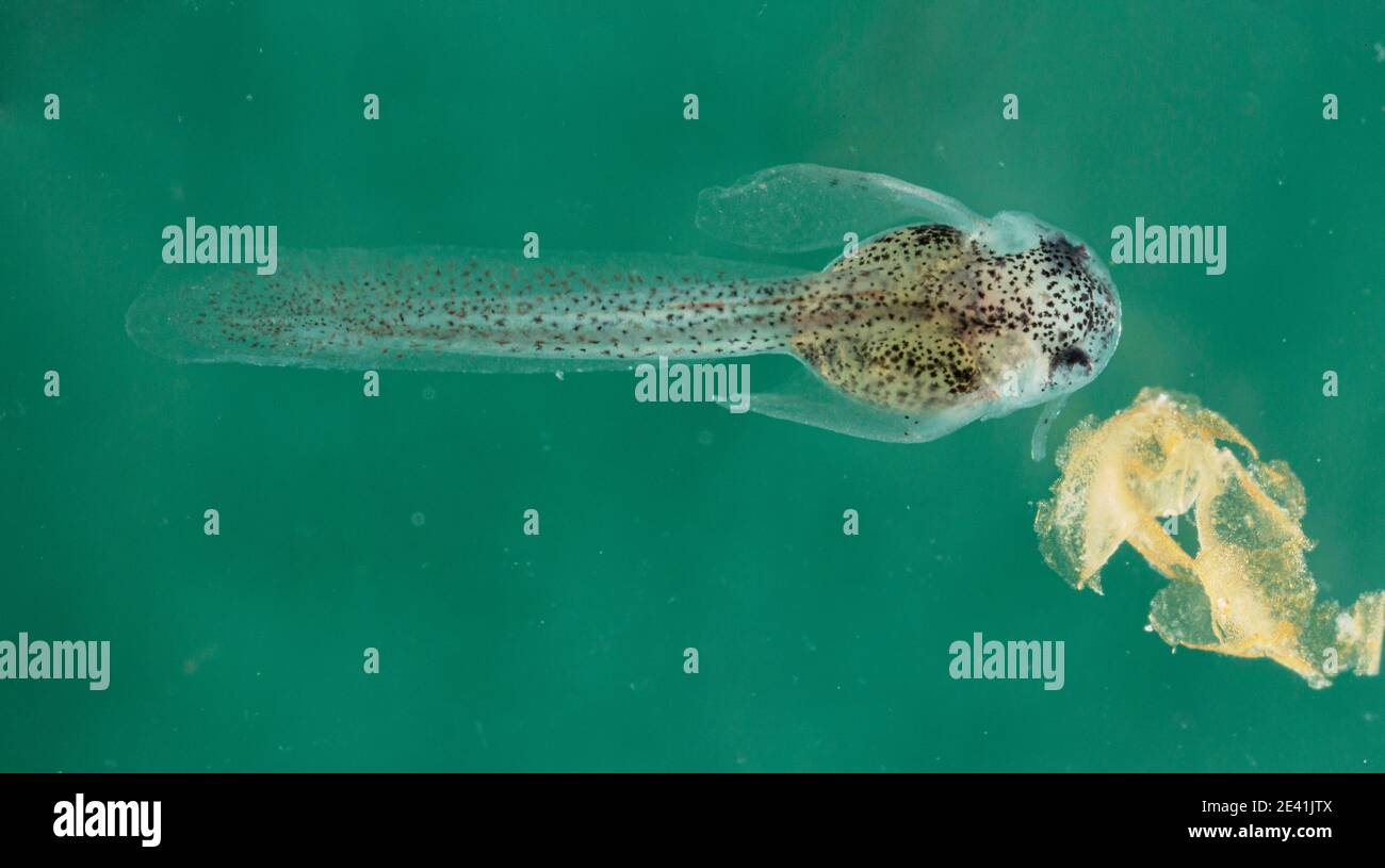 Panda cory (Corydoras panda), larva with remains of the embryonic membrane Stock Photohttps://www.alamy.com/image-license-details/?v=1https://www.alamy.com/panda-cory-corydoras-panda-larva-with-remains-of-the-embryonic-membrane-image398333850.html
RM2E41JTX–Panda cory (Corydoras panda), larva with remains of the embryonic membrane
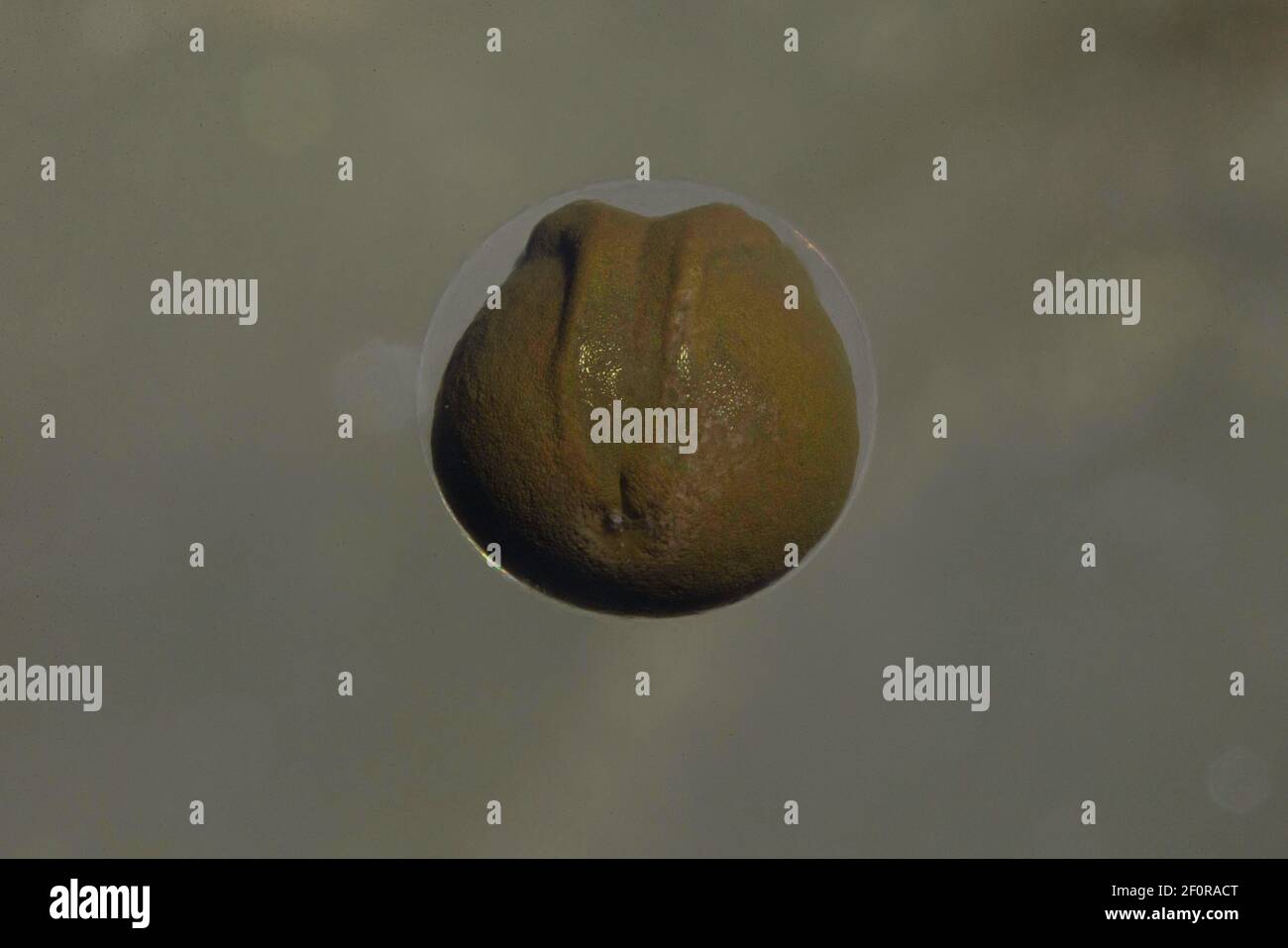 Common frog (Rana temporaria), early stage of embryo development with visible neural crest in gelatinous egg case, Thuringia, Germany Stock Photohttps://www.alamy.com/image-license-details/?v=1https://www.alamy.com/common-frog-rana-temporaria-early-stage-of-embryo-development-with-visible-neural-crest-in-gelatinous-egg-case-thuringia-germany-image413561928.html
RF2F0RACT–Common frog (Rana temporaria), early stage of embryo development with visible neural crest in gelatinous egg case, Thuringia, Germany
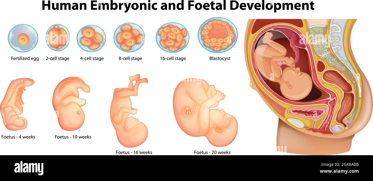 Diagram showing human embryonic and foetal development Stock Vectorhttps://www.alamy.com/image-license-details/?v=1https://www.alamy.com/diagram-showing-human-embryonic-and-foetal-development-image446423723.html
RF2GX8A0B–Diagram showing human embryonic and foetal development
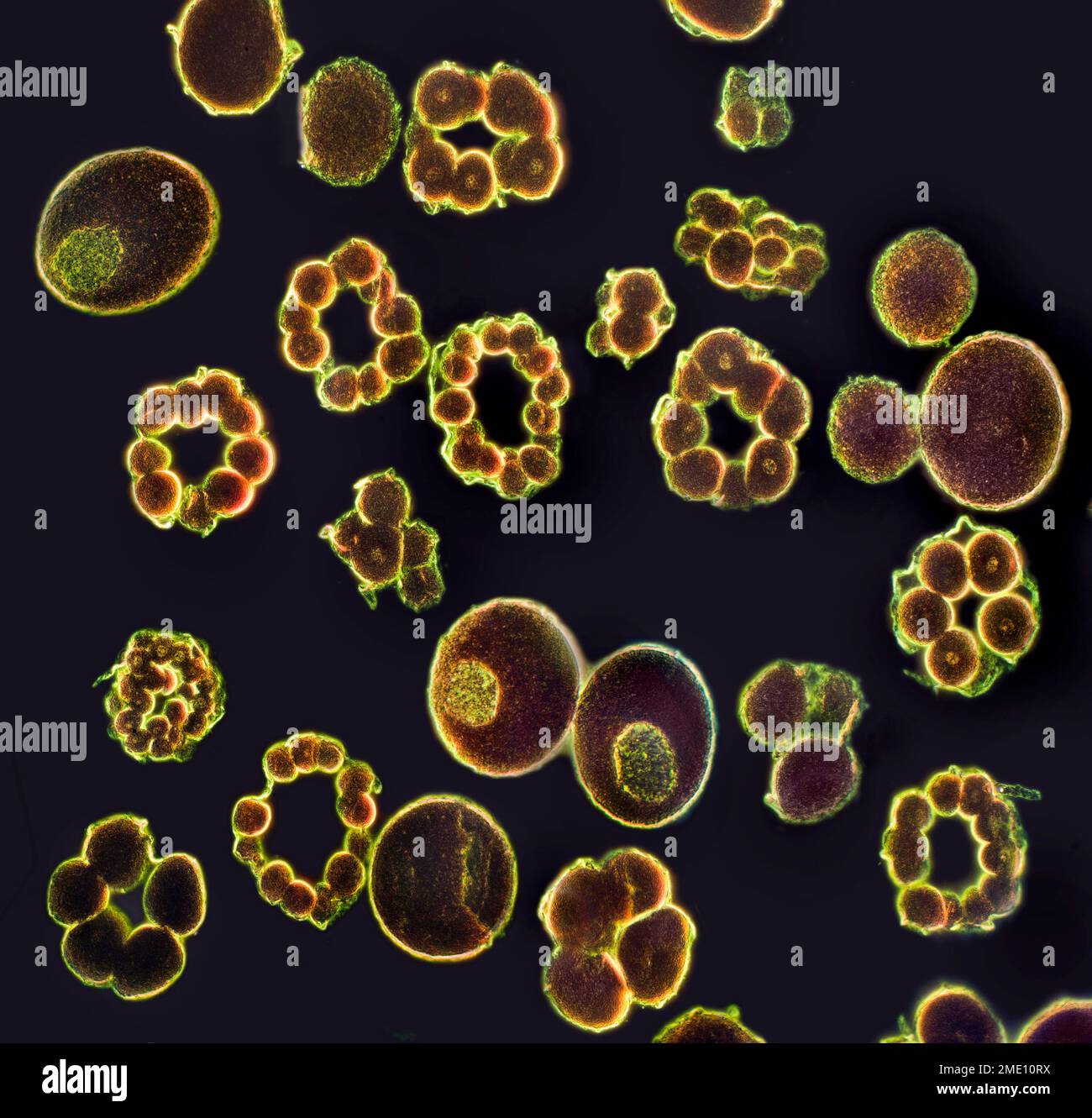 Starfish emryo development stages Stock Photohttps://www.alamy.com/image-license-details/?v=1https://www.alamy.com/starfish-emryo-development-stages-image507728478.html
RM2ME10RX–Starfish emryo development stages
 Embryonic development Stock Photohttps://www.alamy.com/image-license-details/?v=1https://www.alamy.com/stock-photo-embryonic-development-13263991.html
RFAD0ERM–Embryonic development
 Development of the Fetus, c. 1840 Stock Photohttps://www.alamy.com/image-license-details/?v=1https://www.alamy.com/development-of-the-fetus-c-1840-image352802872.html
RM2BDYFJG–Development of the Fetus, c. 1840
 Rehovot, Israel. 04th Aug, 2022. Jacob (Yaqub) Hanna, Professor of Molecular Genetics, works on embryos samples in his laboratory at Weizmann Institute of Science in Rehovot. Yaqub has led a team of scientists at the Weizmann Institute that could develop the world's first 'synthetic embryos', made of stem cells from mice without the need for sperms, eggs or fertilisation. The embryo-like structures are expected to drive deeper understanding of embryonic development and could be used to grow individual organs for transplanting. Credit: Ilia Yefimovich/dpa/Alamy Live News Stock Photohttps://www.alamy.com/image-license-details/?v=1https://www.alamy.com/rehovot-israel-04th-aug-2022-jacob-yaqub-hanna-professor-of-molecular-genetics-works-on-embryos-samples-in-his-laboratory-at-weizmann-institute-of-science-in-rehovot-yaqub-has-led-a-team-of-scientists-at-the-weizmann-institute-that-could-develop-the-worlds-first-synthetic-embryos-made-of-stem-cells-from-mice-without-the-need-for-sperms-eggs-or-fertilisation-the-embryo-like-structures-are-expected-to-drive-deeper-understanding-of-embryonic-development-and-could-be-used-to-grow-individual-organs-for-transplanting-credit-ilia-yefimovichdpaalamy-live-news-image477044895.html
RM2JM37HK–Rehovot, Israel. 04th Aug, 2022. Jacob (Yaqub) Hanna, Professor of Molecular Genetics, works on embryos samples in his laboratory at Weizmann Institute of Science in Rehovot. Yaqub has led a team of scientists at the Weizmann Institute that could develop the world's first 'synthetic embryos', made of stem cells from mice without the need for sperms, eggs or fertilisation. The embryo-like structures are expected to drive deeper understanding of embryonic development and could be used to grow individual organs for transplanting. Credit: Ilia Yefimovich/dpa/Alamy Live News
 Coloured computed tomography (CT) scan of a chest from above showing an azygos lobe in the left lung (segmented area centre-left). An azygos lobe is an anatomical variant in the upper lobe of the lung in which a segment of the lung is separated by a deep groove called an azygos fissure, which consists of the azygos vein and pleura (thin tissue). It is a rare variation, occurring in around 0.3% of people during embryonic development. It causes no symptoms, but may make certain surgical procedures more difficult. Stock Photohttps://www.alamy.com/image-license-details/?v=1https://www.alamy.com/coloured-computed-tomography-ct-scan-of-a-chest-from-above-showing-an-azygos-lobe-in-the-left-lung-segmented-area-centre-left-an-azygos-lobe-is-an-anatomical-variant-in-the-upper-lobe-of-the-lung-in-which-a-segment-of-the-lung-is-separated-by-a-deep-groove-called-an-azygos-fissure-which-consists-of-the-azygos-vein-and-pleura-thin-tissue-it-is-a-rare-variation-occurring-in-around-03-of-people-during-embryonic-development-it-causes-no-symptoms-but-may-make-certain-surgical-procedures-more-difficult-image612233858.html
RF2XG1JEX–Coloured computed tomography (CT) scan of a chest from above showing an azygos lobe in the left lung (segmented area centre-left). An azygos lobe is an anatomical variant in the upper lobe of the lung in which a segment of the lung is separated by a deep groove called an azygos fissure, which consists of the azygos vein and pleura (thin tissue). It is a rare variation, occurring in around 0.3% of people during embryonic development. It causes no symptoms, but may make certain surgical procedures more difficult.
RFT0DGAA–Division of human cells, embryonic development line icon.
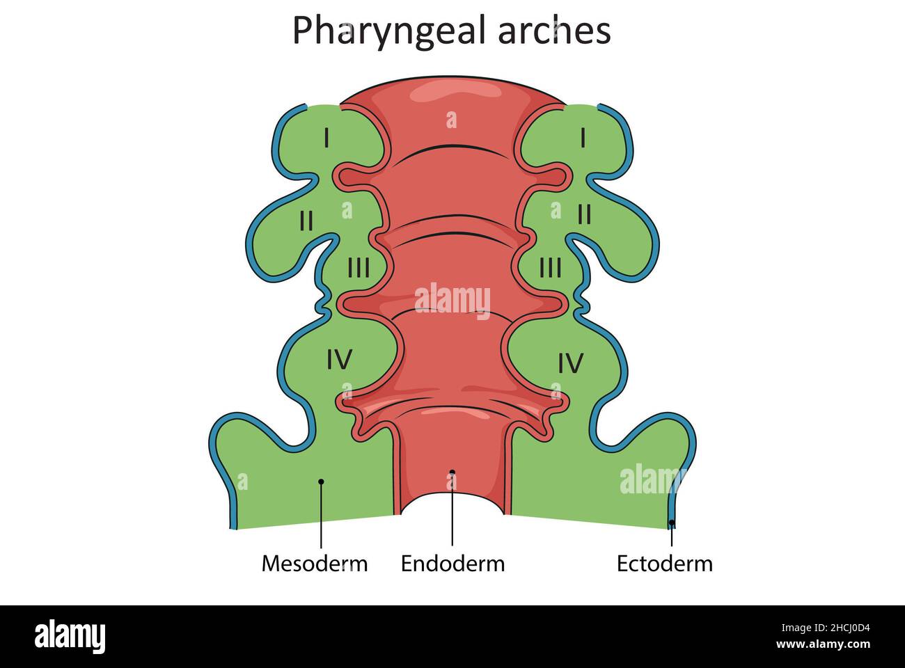 Pharyngeal arches development and embryology Stock Photohttps://www.alamy.com/image-license-details/?v=1https://www.alamy.com/pharyngeal-arches-development-and-embryology-image455240944.html
RF2HCJ0D4–Pharyngeal arches development and embryology
 Bird egg held over a candling electric light. The wider egg end exposed to the light reveals the sharp demarcation between the air space, the shell an Stock Photohttps://www.alamy.com/image-license-details/?v=1https://www.alamy.com/stock-photo-bird-egg-held-over-a-candling-electric-light-the-wider-egg-end-exposed-127027259.html
RMHAJGFR–Bird egg held over a candling electric light. The wider egg end exposed to the light reveals the sharp demarcation between the air space, the shell an
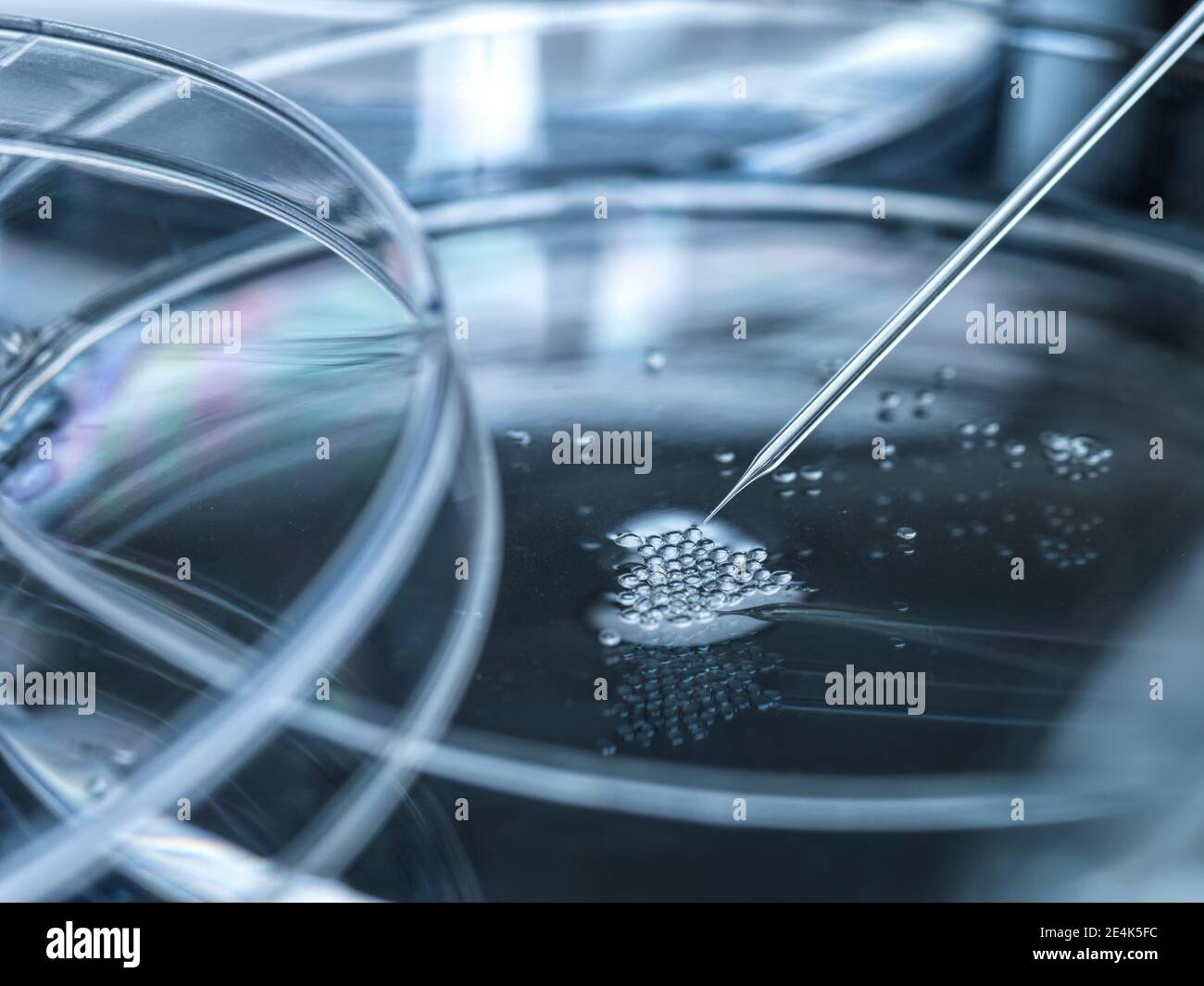 Petri dish with embryonic stem cells used in cloning and genetic modification Stock Photohttps://www.alamy.com/image-license-details/?v=1https://www.alamy.com/petri-dish-with-embryonic-stem-cells-used-in-cloning-and-genetic-modification-image398718528.html
RF2E4K5FC–Petri dish with embryonic stem cells used in cloning and genetic modification
 This illustration presents a detailed study of amphibian anatomy, featuring various stages of embryonic development. The top section displays rounded forms, possibly indicative of early-stage embryos, highlighting their spherical shapes with distinct markings. Below, the images progress to more defined structures, where features such as limbs, eyes, and mouth begin to emerge. Each segment is meticulously labeled with numerical indicators, providing a scientific reference for each developmental phase. The overall arrangement emphasizes the complexity and gradual transformation of amphibian life Stock Photohttps://www.alamy.com/image-license-details/?v=1https://www.alamy.com/this-illustration-presents-a-detailed-study-of-amphibian-anatomy-featuring-various-stages-of-embryonic-development-the-top-section-displays-rounded-forms-possibly-indicative-of-early-stage-embryos-highlighting-their-spherical-shapes-with-distinct-markings-below-the-images-progress-to-more-defined-structures-where-features-such-as-limbs-eyes-and-mouth-begin-to-emerge-each-segment-is-meticulously-labeled-with-numerical-indicators-providing-a-scientific-reference-for-each-developmental-phase-the-overall-arrangement-emphasizes-the-complexity-and-gradual-transformation-of-amphibian-life-image631096262.html
RM2YJMWM6–This illustration presents a detailed study of amphibian anatomy, featuring various stages of embryonic development. The top section displays rounded forms, possibly indicative of early-stage embryos, highlighting their spherical shapes with distinct markings. Below, the images progress to more defined structures, where features such as limbs, eyes, and mouth begin to emerge. Each segment is meticulously labeled with numerical indicators, providing a scientific reference for each developmental phase. The overall arrangement emphasizes the complexity and gradual transformation of amphibian life
 Tadpoles of the European Common Frog Rana temporaria in early embryonic stages of development shot from inside the gelatinous sheath of frogspawn Stock Photohttps://www.alamy.com/image-license-details/?v=1https://www.alamy.com/tadpoles-of-the-european-common-frog-rana-temporaria-in-early-embryonic-stages-of-development-shot-from-inside-the-gelatinous-sheath-of-frogspawn-image463789879.html
RM2HXFCM7–Tadpoles of the European Common Frog Rana temporaria in early embryonic stages of development shot from inside the gelatinous sheath of frogspawn
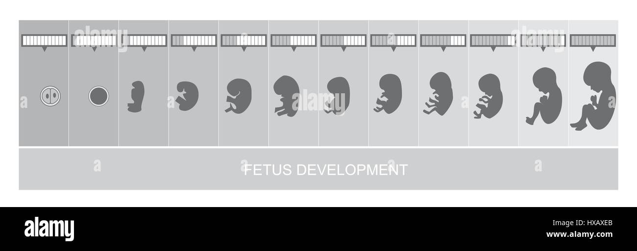 Fetus stages development, vetor Stock Vectorhttps://www.alamy.com/image-license-details/?v=1https://www.alamy.com/stock-photo-fetus-stages-development-vetor-136693939.html
RFHXAXEB–Fetus stages development, vetor
 Embryo dune development: Sand couch grass on a very small dune on a stormy beach Stock Photohttps://www.alamy.com/image-license-details/?v=1https://www.alamy.com/embryo-dune-development-sand-couch-grass-on-a-very-small-dune-on-a-stormy-beach-image226185215.html
RFR3YHFB–Embryo dune development: Sand couch grass on a very small dune on a stormy beach
 Anatomical studies of embryonic development by Leonardo da Vinci Stock Photohttps://www.alamy.com/image-license-details/?v=1https://www.alamy.com/anatomical-studies-of-embryonic-development-by-leonardo-da-vinci-image7394509.html
RMABP3TE–Anatomical studies of embryonic development by Leonardo da Vinci
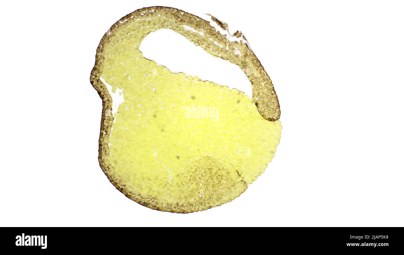 Gastrulation. Frog development. Medium gastrula stage. The frog been used as a model organism for the study of gastrulation. Van Gieson's stain. Stock Photohttps://www.alamy.com/image-license-details/?v=1https://www.alamy.com/gastrulation-frog-development-medium-gastrula-stage-the-frog-been-used-as-a-model-organism-for-the-study-of-gastrulation-van-giesons-stain-image471313900.html
RF2JAP5K8–Gastrulation. Frog development. Medium gastrula stage. The frog been used as a model organism for the study of gastrulation. Van Gieson's stain.
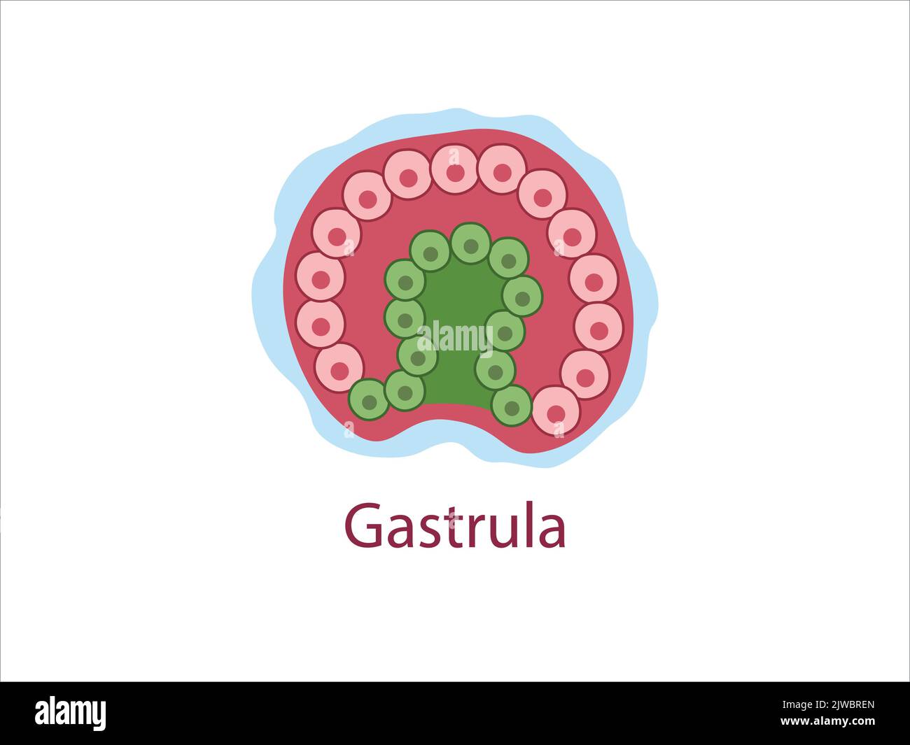 Gastrula. The cells of endoderm and ectoderm. The stage of segmentation of a fertilized ovum. Human embryonic development. Vector medical illustratio Stock Vectorhttps://www.alamy.com/image-license-details/?v=1https://www.alamy.com/gastrula-the-cells-of-endoderm-and-ectoderm-the-stage-of-segmentation-of-a-fertilized-ovum-human-embryonic-development-vector-medical-illustratio-image480306253.html
RF2JWBREN–Gastrula. The cells of endoderm and ectoderm. The stage of segmentation of a fertilized ovum. Human embryonic development. Vector medical illustratio
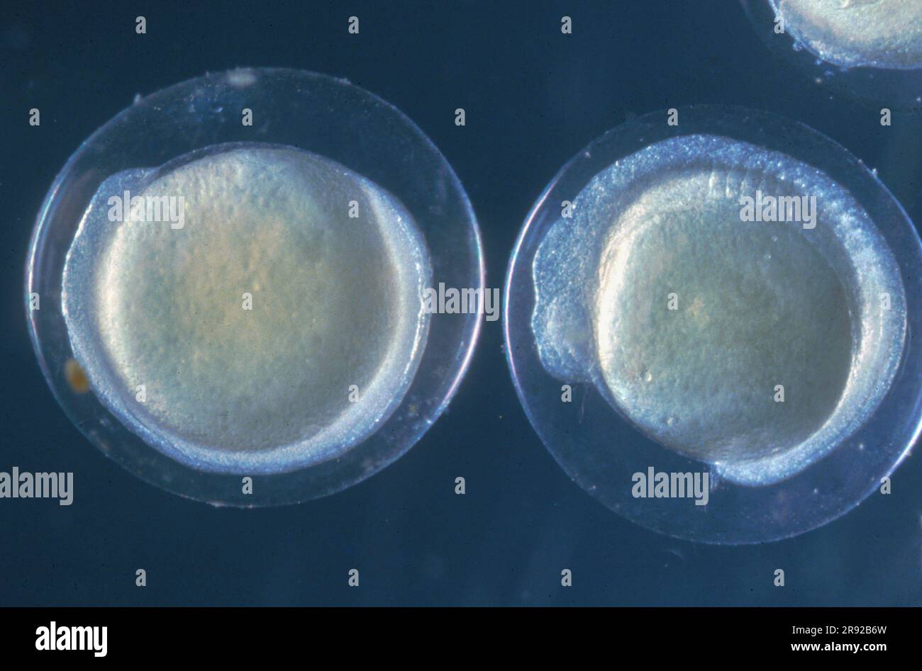 minnow, Eurasian minnow (Phoxinus phoxinus), eggs with incipient embryonic development Stock Photohttps://www.alamy.com/image-license-details/?v=1https://www.alamy.com/minnow-eurasian-minnow-phoxinus-phoxinus-eggs-with-incipient-embryonic-development-image556316401.html
RM2R92B6W–minnow, Eurasian minnow (Phoxinus phoxinus), eggs with incipient embryonic development
 Twins - two unborn babies are in uterus of mother. Prenatal development of fetus and foetus. Vector illustration. Stock Photohttps://www.alamy.com/image-license-details/?v=1https://www.alamy.com/twins-two-unborn-babies-are-in-uterus-of-mother-prenatal-development-of-fetus-and-foetus-vector-illustration-image392875419.html
RF2DR50GY–Twins - two unborn babies are in uterus of mother. Prenatal development of fetus and foetus. Vector illustration.
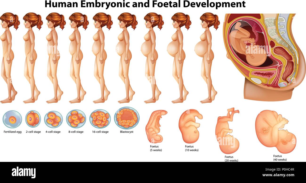 Vector of Human Embryonic and Foetal Development illustration Stock Vectorhttps://www.alamy.com/image-license-details/?v=1https://www.alamy.com/vector-of-human-embryonic-and-foetal-development-illustration-image206907143.html
RFP0HC4R–Vector of Human Embryonic and Foetal Development illustration
