Quick filters:
External iliac vein Stock Photos and Images
 ILIAC, ANGIOGRAPHY Stock Photohttps://www.alamy.com/image-license-details/?v=1https://www.alamy.com/stock-photo-iliac-angiography-49255851.html
RMCT3P9F–ILIAC, ANGIOGRAPHY
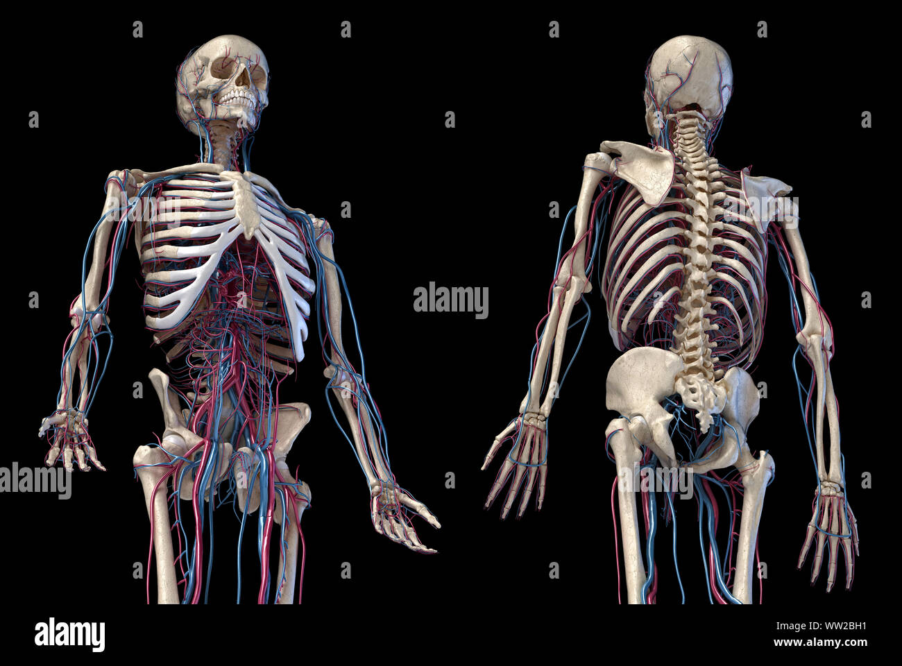 Human anatomy, 3d illustration of the skeleton with cardiovascular system. Perspective view of 3/4 upper part, front and back sides. On black backgrou Stock Photohttps://www.alamy.com/image-license-details/?v=1https://www.alamy.com/human-anatomy-3d-illustration-of-the-skeleton-with-cardiovascular-system-perspective-view-of-34-upper-part-front-and-back-sides-on-black-backgrou-image273574925.html
RFWW2BH1–Human anatomy, 3d illustration of the skeleton with cardiovascular system. Perspective view of 3/4 upper part, front and back sides. On black backgrou
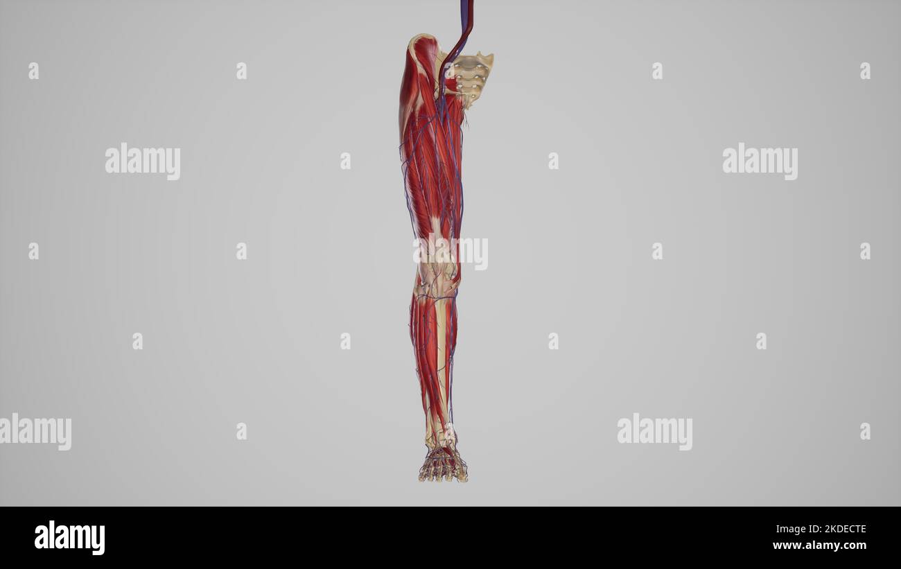 lower limb with muscles, blood vessels Stock Photohttps://www.alamy.com/image-license-details/?v=1https://www.alamy.com/lower-limb-with-muscles-blood-vessels-image490198254.html
RF2KDECTE–lower limb with muscles, blood vessels
 Pelvis anatomy, artwork Stock Photohttps://www.alamy.com/image-license-details/?v=1https://www.alamy.com/stock-photo-pelvis-anatomy-artwork-55444249.html
RFD65KKN–Pelvis anatomy, artwork
 Antique Medical Illustration of Foot with Tumors circa 1881 Stock Photohttps://www.alamy.com/image-license-details/?v=1https://www.alamy.com/stock-photo-antique-medical-illustration-of-foot-with-tumors-circa-1881-37200591.html
RFC4EHMF–Antique Medical Illustration of Foot with Tumors circa 1881
 Female cardiovascular system, artwork Stock Photohttps://www.alamy.com/image-license-details/?v=1https://www.alamy.com/stock-photo-female-cardiovascular-system-artwork-55445369.html
RFD65N3N–Female cardiovascular system, artwork
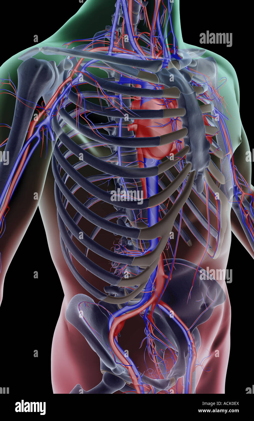 The blood supply of the trunk Stock Photohttps://www.alamy.com/image-license-details/?v=1https://www.alamy.com/stock-photo-the-blood-supply-of-the-trunk-13174513.html
RFACK0EX–The blood supply of the trunk
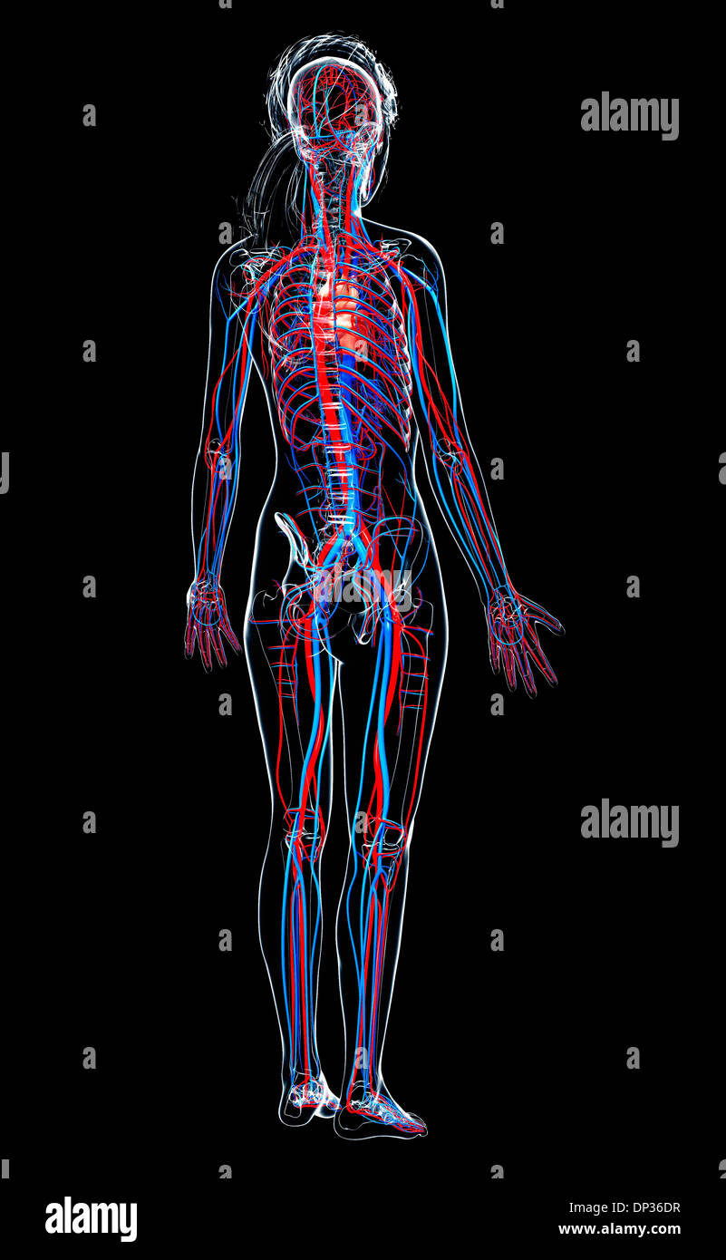 Female cardiovascular system, artwork Stock Photohttps://www.alamy.com/image-license-details/?v=1https://www.alamy.com/female-cardiovascular-system-artwork-image65224483.html
RFDP36DR–Female cardiovascular system, artwork
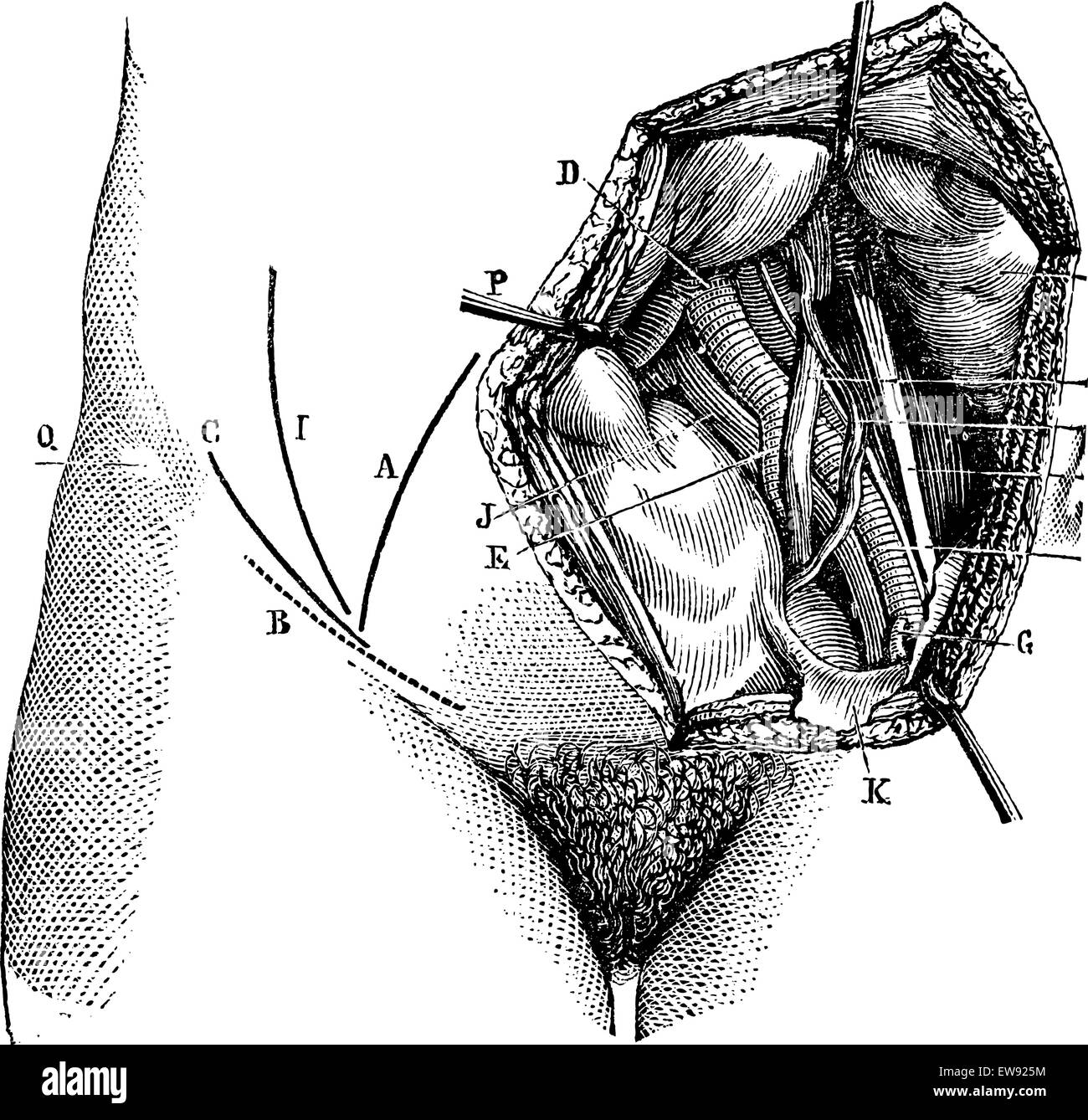 Fig. 618. Iliac artery and its branches, vintage engraved illustration. Magasin Pittoresque 1875. Stock Vectorhttps://www.alamy.com/image-license-details/?v=1https://www.alamy.com/stock-photo-fig-618-iliac-artery-and-its-branches-vintage-engraved-illustration-84407168.html
RFEW925M–Fig. 618. Iliac artery and its branches, vintage engraved illustration. Magasin Pittoresque 1875.
 Pelvis anatomy, artwork Stock Photohttps://www.alamy.com/image-license-details/?v=1https://www.alamy.com/stock-photo-pelvis-anatomy-artwork-55445090.html
RFD65MNP–Pelvis anatomy, artwork
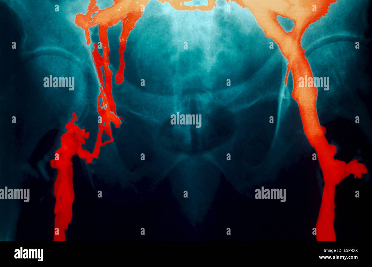 Angiography (X-ray of the veines) showing a phlebitis with recent thrombosis on the external iliac vein. Stock Photohttps://www.alamy.com/image-license-details/?v=1https://www.alamy.com/stock-photo-angiography-x-ray-of-the-veines-showing-a-phlebitis-with-recent-thrombosis-72416482.html
RME5PRXX–Angiography (X-ray of the veines) showing a phlebitis with recent thrombosis on the external iliac vein.
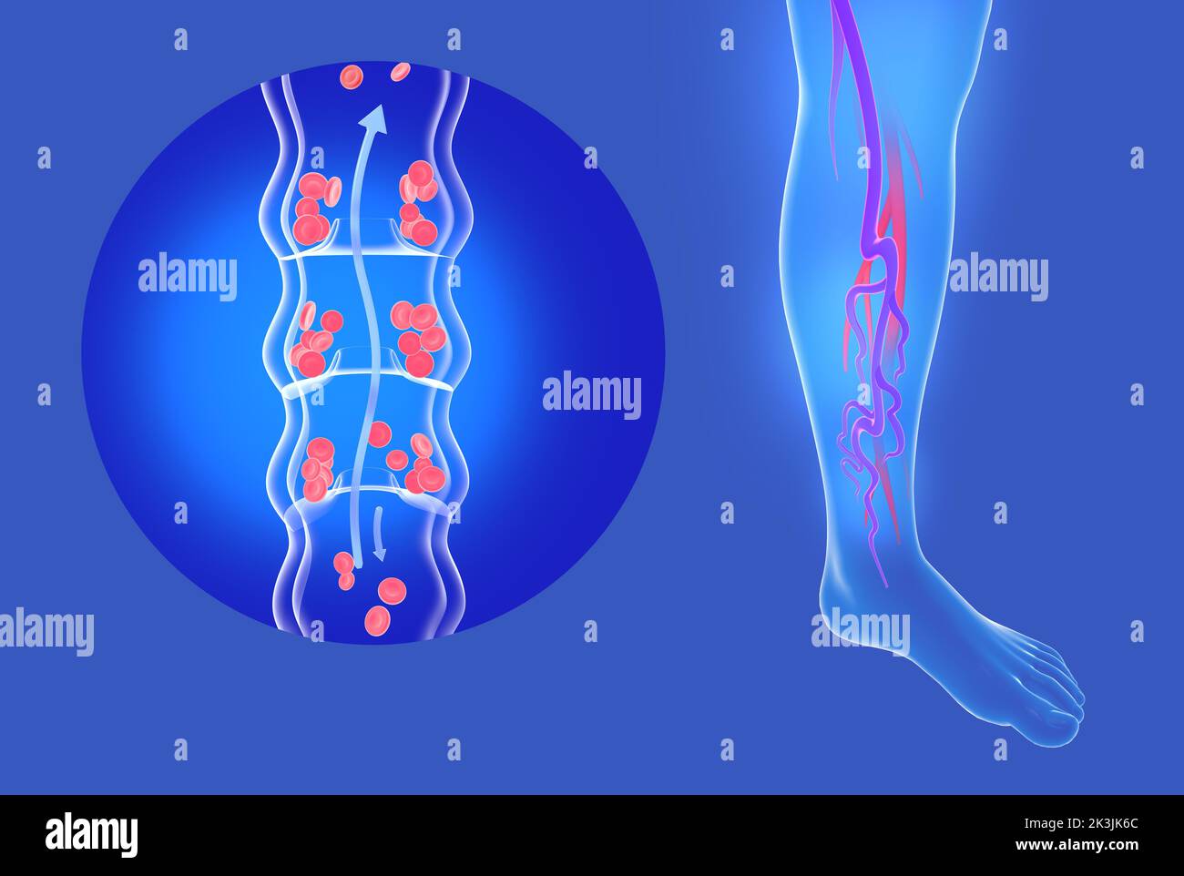 Anatomical 3d illustration of incompetent vein and varicose veins in legs. Transparent circulatory system of veins. Stock Photohttps://www.alamy.com/image-license-details/?v=1https://www.alamy.com/anatomical-3d-illustration-of-incompetent-vein-and-varicose-veins-in-legs-transparent-circulatory-system-of-veins-image484144484.html
RF2K3JK6C–Anatomical 3d illustration of incompetent vein and varicose veins in legs. Transparent circulatory system of veins.
 circumflex iliac vein is formed by the union of the venae comitantes of the deep iliac circumflex artery, and joins the external iliac vein Stock Photohttps://www.alamy.com/image-license-details/?v=1https://www.alamy.com/circumflex-iliac-vein-is-formed-by-the-union-of-the-venae-comitantes-of-the-deep-iliac-circumflex-artery-and-joins-the-external-iliac-vein-image596592416.html
RF2WJH3M0–circumflex iliac vein is formed by the union of the venae comitantes of the deep iliac circumflex artery, and joins the external iliac vein
 Anatomical 3d illustration of two images, diseased incompetent vein and healthy vein. Graphic representation of the blood circulatory system. Stock Photohttps://www.alamy.com/image-license-details/?v=1https://www.alamy.com/anatomical-3d-illustration-of-two-images-diseased-incompetent-vein-and-healthy-vein-graphic-representation-of-the-blood-circulatory-system-image484144494.html
RF2K3JK6P–Anatomical 3d illustration of two images, diseased incompetent vein and healthy vein. Graphic representation of the blood circulatory system.
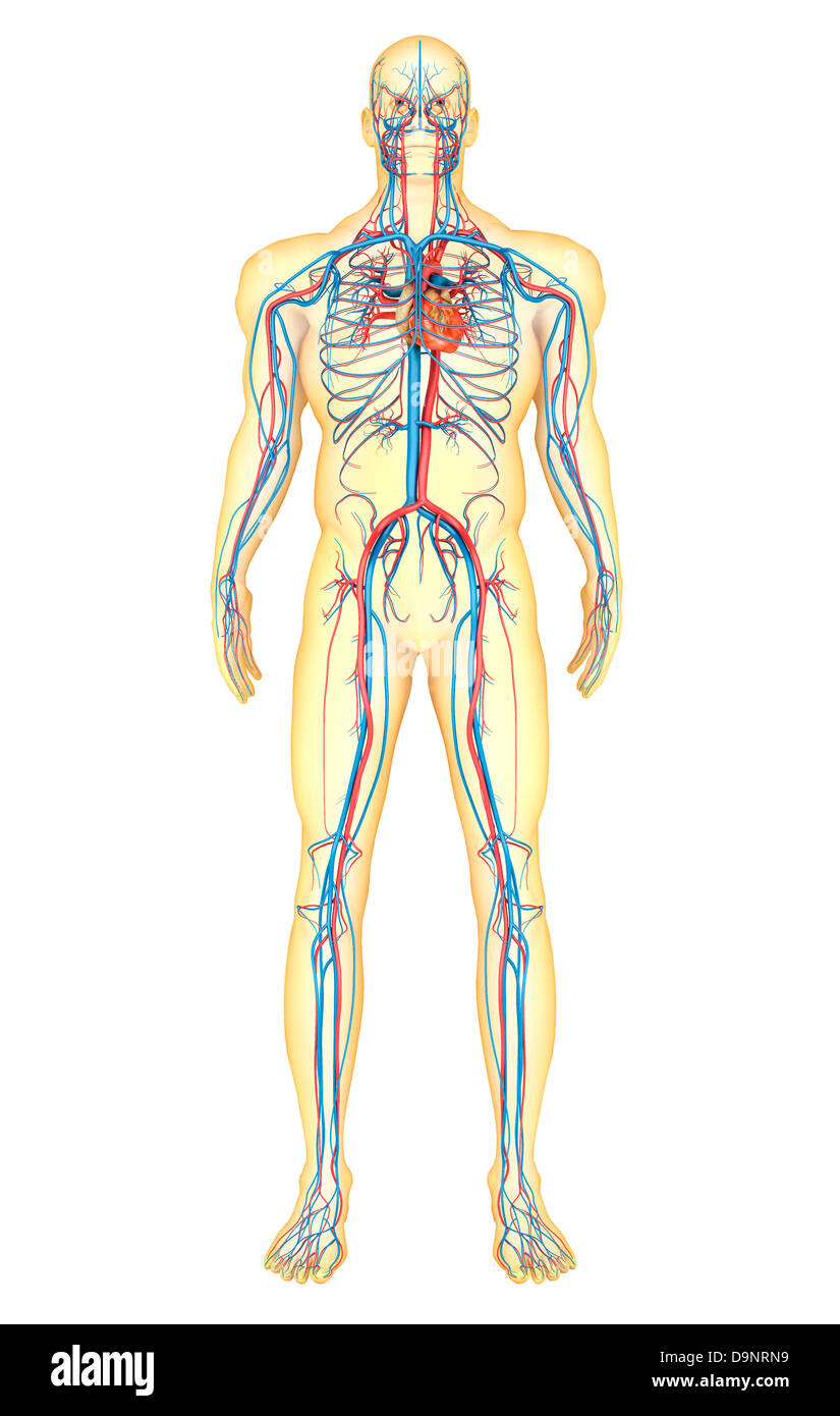 Anatomy of human body and circulatory system, front view. Stock Photohttps://www.alamy.com/image-license-details/?v=1https://www.alamy.com/stock-photo-anatomy-of-human-body-and-circulatory-system-front-view-57642629.html
RFD9NRN9–Anatomy of human body and circulatory system, front view.
 Anatomical 3d illustration of two images, diseased incompetent vein next to healthy vein. Transparent image of the blood circulatory system. Stock Photohttps://www.alamy.com/image-license-details/?v=1https://www.alamy.com/anatomical-3d-illustration-of-two-images-diseased-incompetent-vein-next-to-healthy-vein-transparent-image-of-the-blood-circulatory-system-image484144506.html
RF2K3JK76–Anatomical 3d illustration of two images, diseased incompetent vein next to healthy vein. Transparent image of the blood circulatory system.
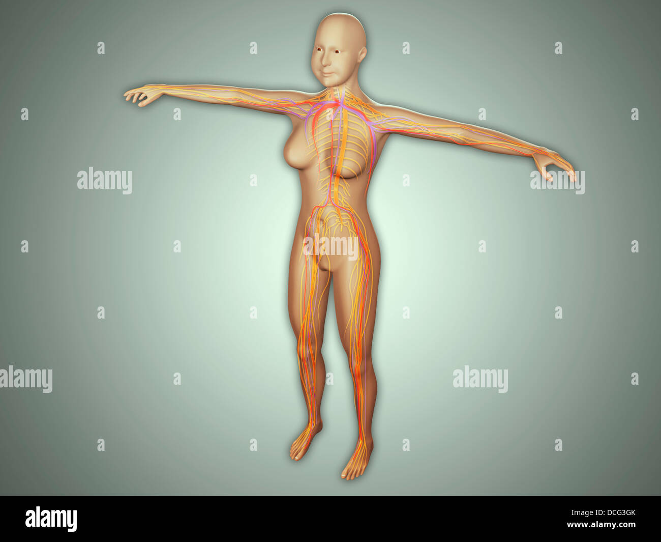 Anatomy of female body with arteries, veins and nervous system. Stock Photohttps://www.alamy.com/image-license-details/?v=1https://www.alamy.com/stock-photo-anatomy-of-female-body-with-arteries-veins-and-nervous-system-59361027.html
RFDCG3GK–Anatomy of female body with arteries, veins and nervous system.
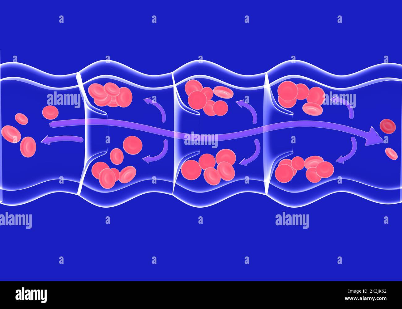 Anatomical 3d illustration of blood circulation in an incompetent diseased vein. Transparent image of glass on a blue background. Stock Photohttps://www.alamy.com/image-license-details/?v=1https://www.alamy.com/anatomical-3d-illustration-of-blood-circulation-in-an-incompetent-diseased-vein-transparent-image-of-glass-on-a-blue-background-image484144474.html
RF2K3JK62–Anatomical 3d illustration of blood circulation in an incompetent diseased vein. Transparent image of glass on a blue background.
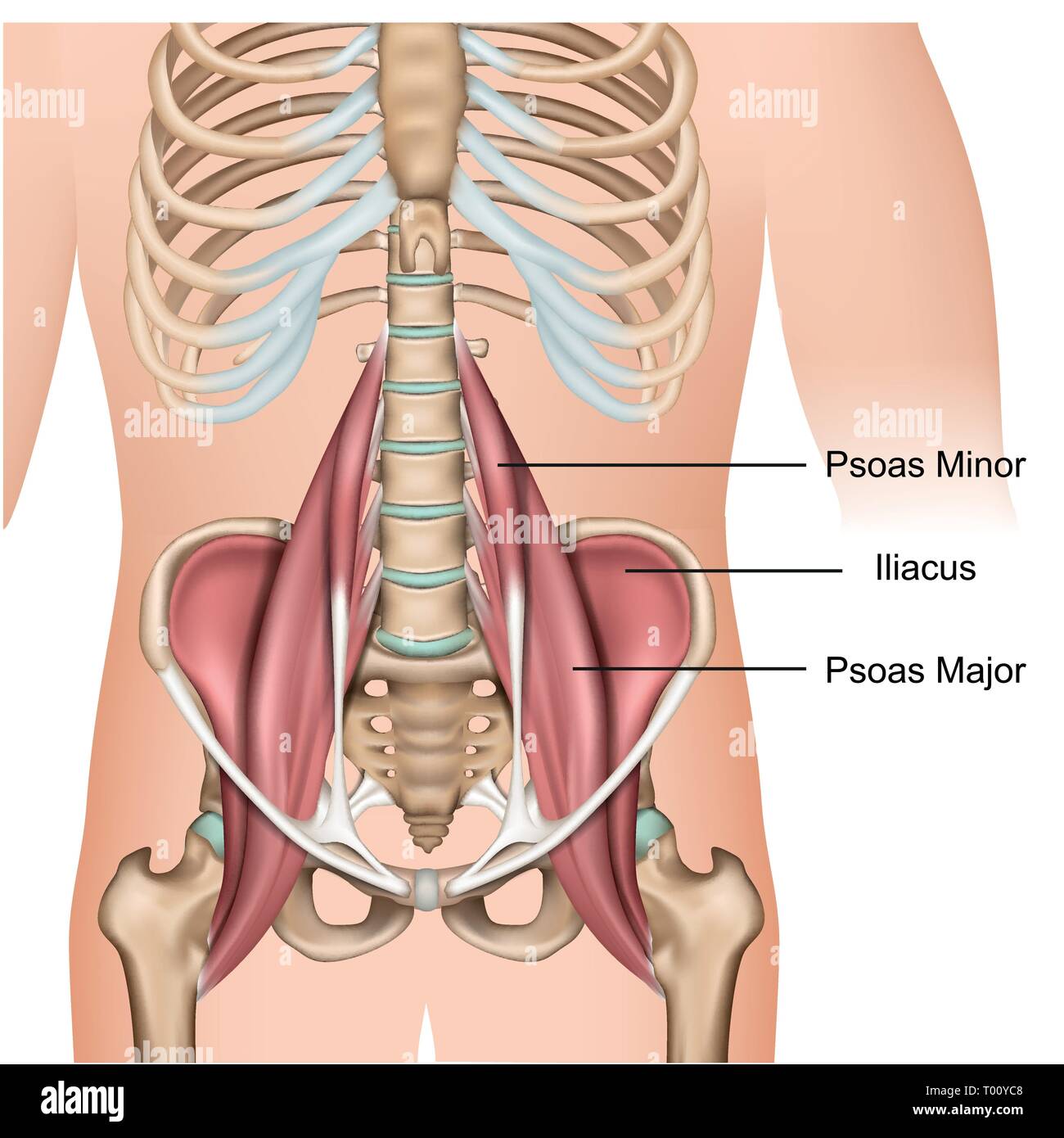 Psoas major muscle anatomy 3d medical vector illustration on white background Stock Vectorhttps://www.alamy.com/image-license-details/?v=1https://www.alamy.com/psoas-major-muscle-anatomy-3d-medical-vector-illustration-on-white-background-image240966664.html
RFT00YC8–Psoas major muscle anatomy 3d medical vector illustration on white background
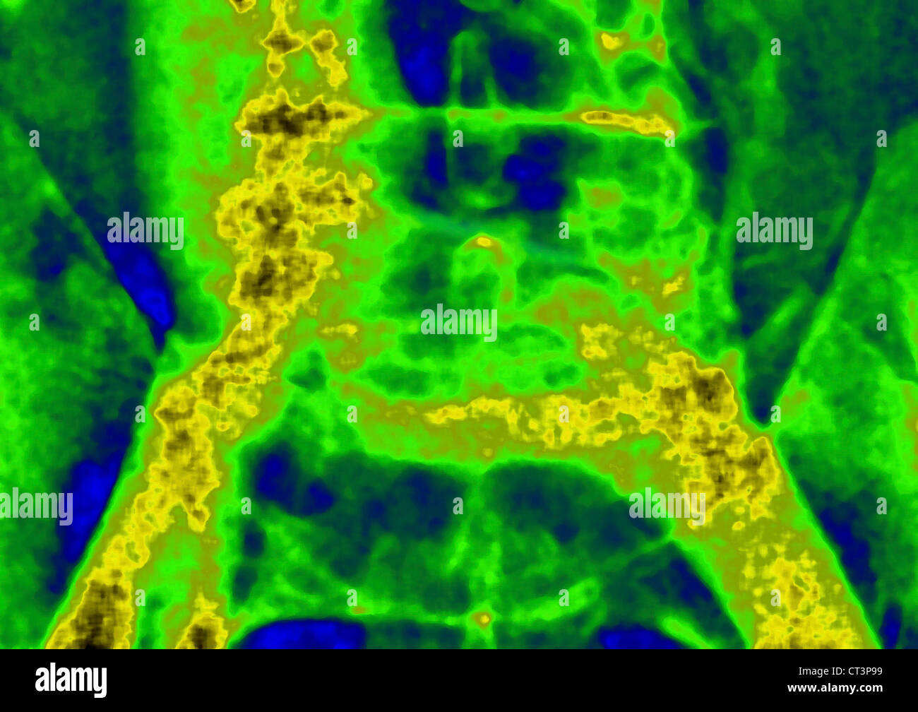 ILIAC, ANGIOGRAPHY Stock Photohttps://www.alamy.com/image-license-details/?v=1https://www.alamy.com/stock-photo-iliac-angiography-49255845.html
RMCT3P99–ILIAC, ANGIOGRAPHY
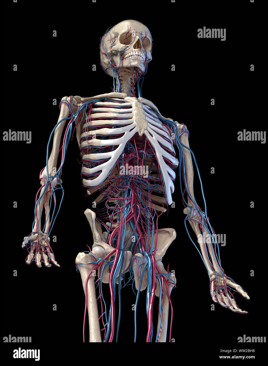 Human anatomy, 3d illustration of the skeleton with cardiovascular system. Perspective view of 3/4 upper part, front side. On black background. Stock Photohttps://www.alamy.com/image-license-details/?v=1https://www.alamy.com/human-anatomy-3d-illustration-of-the-skeleton-with-cardiovascular-system-perspective-view-of-34-upper-part-front-side-on-black-background-image273574932.html
RFWW2BH8–Human anatomy, 3d illustration of the skeleton with cardiovascular system. Perspective view of 3/4 upper part, front side. On black background.
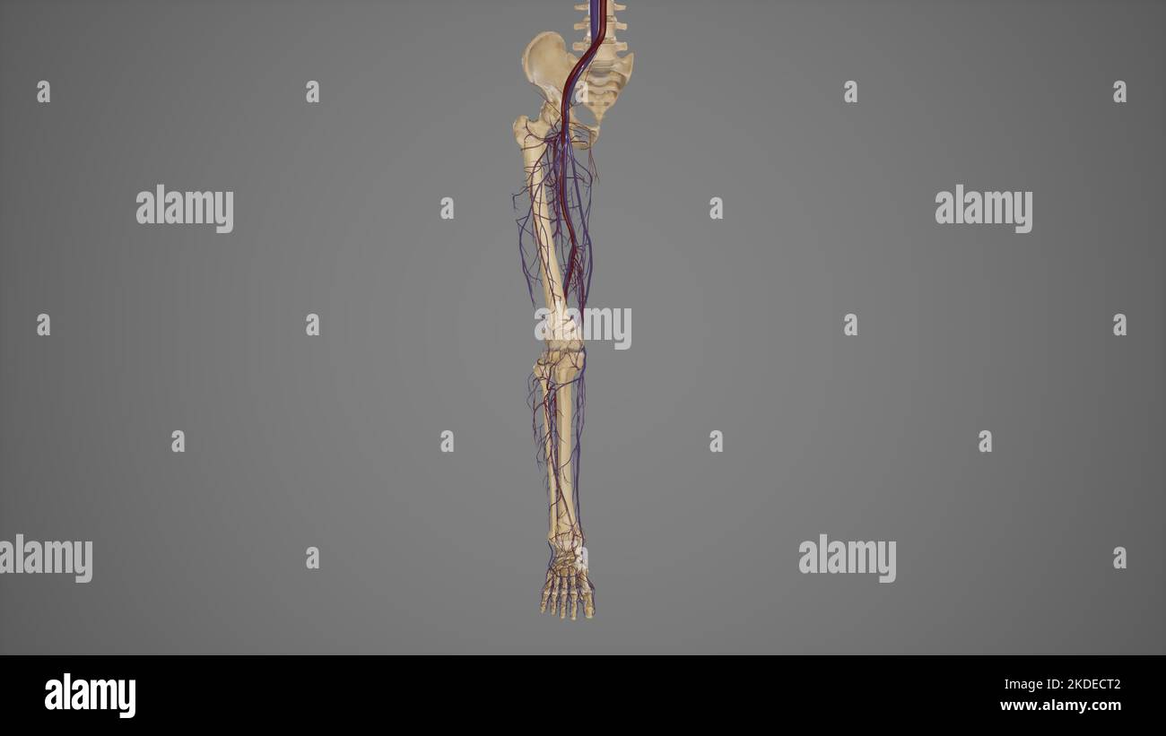 Lower limb with blood vessels anterior view Stock Photohttps://www.alamy.com/image-license-details/?v=1https://www.alamy.com/lower-limb-with-blood-vessels-anterior-view-image490198242.html
RF2KDECT2–Lower limb with blood vessels anterior view
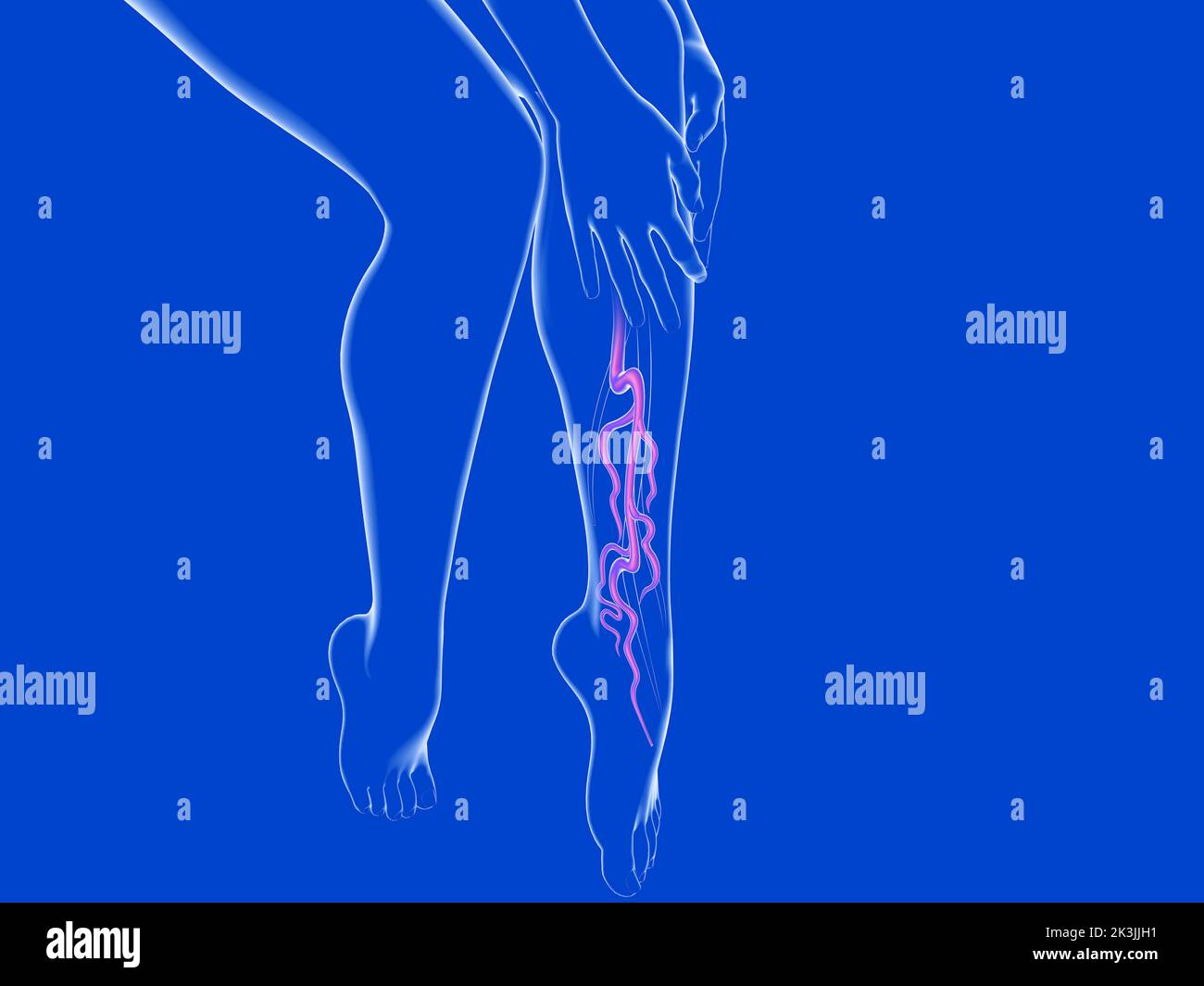 3d illustration of the varicose vein system. Transparent glass anatomy of female legs. Stock Photohttps://www.alamy.com/image-license-details/?v=1https://www.alamy.com/3d-illustration-of-the-varicose-vein-system-transparent-glass-anatomy-of-female-legs-image484143997.html
RF2K3JJH1–3d illustration of the varicose vein system. Transparent glass anatomy of female legs.
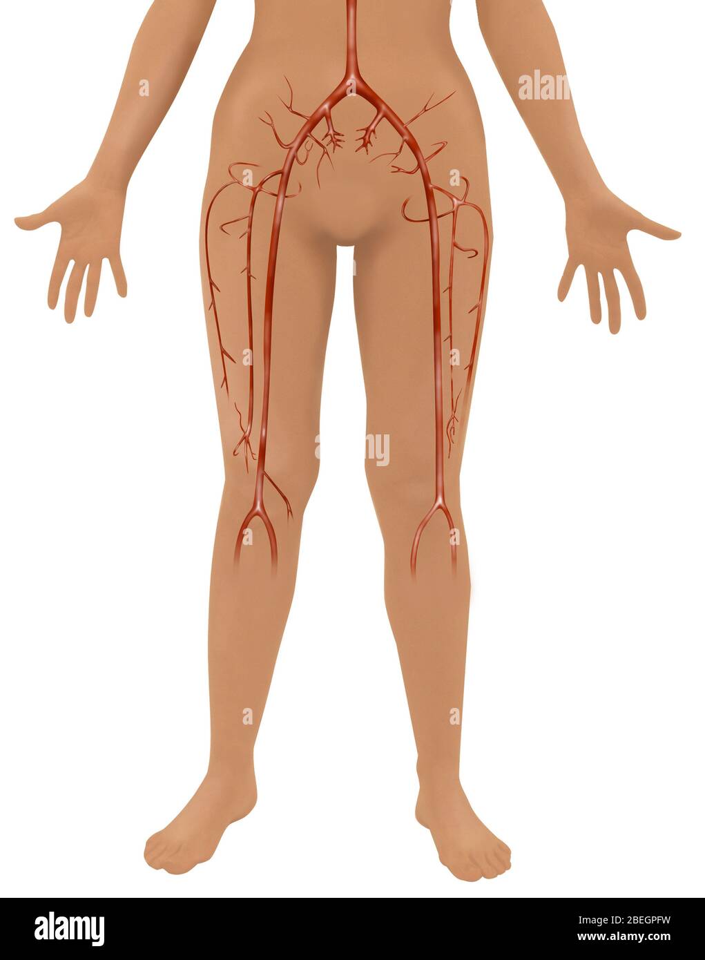 Arteries Stock Photohttps://www.alamy.com/image-license-details/?v=1https://www.alamy.com/arteries-image353181469.html
RF2BEGPFW–Arteries
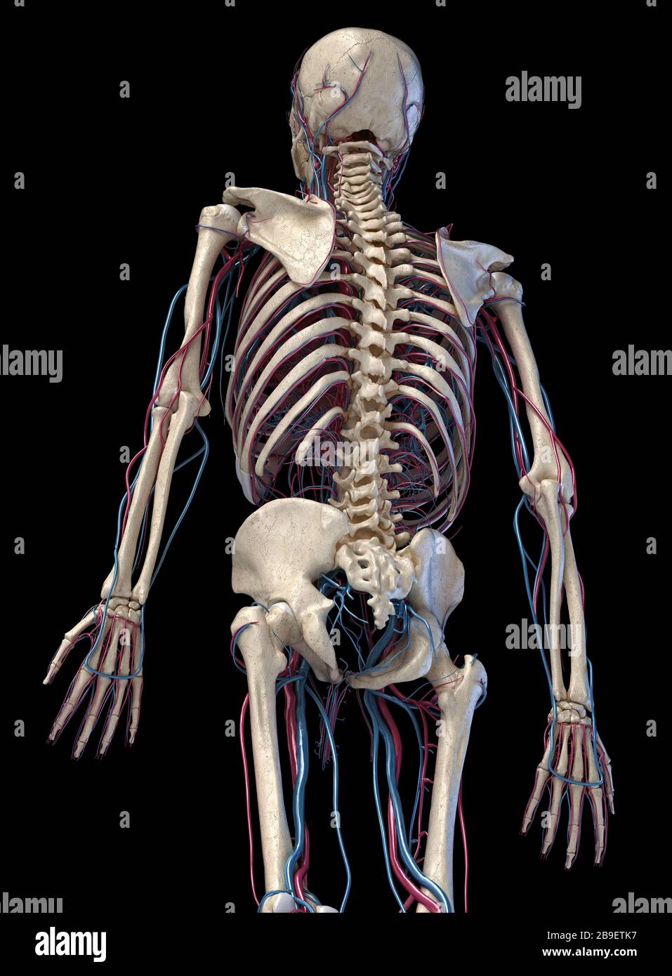 3/4 upper body rear view of human skeletal and vascular systems, black background. Stock Photohttps://www.alamy.com/image-license-details/?v=1https://www.alamy.com/34-upper-body-rear-view-of-human-skeletal-and-vascular-systems-black-background-image350065947.html
RF2B9ETK7–3/4 upper body rear view of human skeletal and vascular systems, black background.
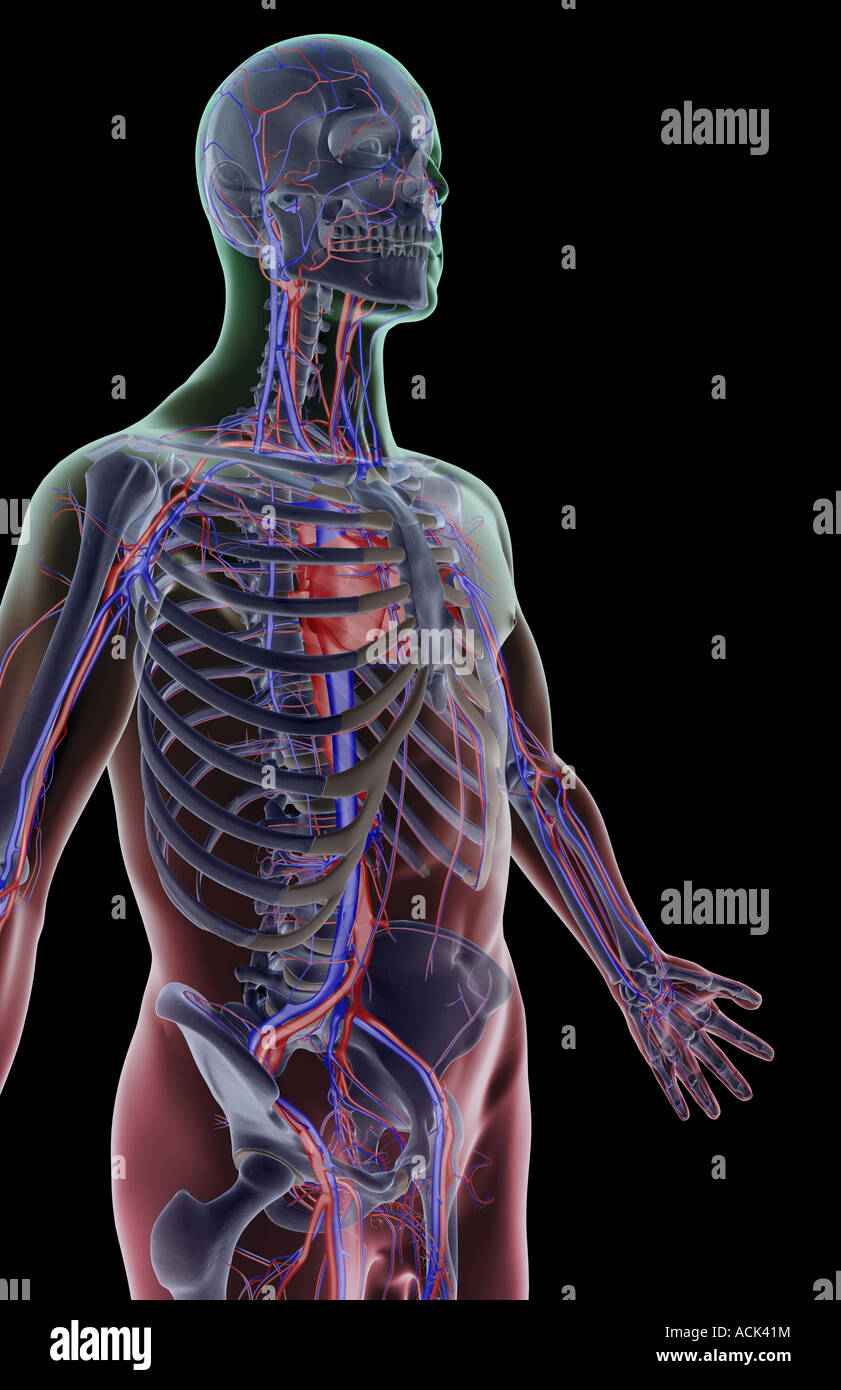 The blood supply of the upper body Stock Photohttps://www.alamy.com/image-license-details/?v=1https://www.alamy.com/stock-photo-the-blood-supply-of-the-upper-body-13175695.html
RFACK41M–The blood supply of the upper body
 . Tumours, innocent and malignant; their clinical characters and appropriate treatment. con-cerned, fairly symmetrical. Otto Weber recorded a remark-able case of numerous symmetrical exostoses of the longbones of the upper and lower limbs, the ribs, and scapulain a man 25 years old. A chondro-sarcoma arose in the righthip-bone and attained enormous proportions. It perforatedthe left external iliac vein, and pieces of the tumour, detachedas emboli, lodged in the pulmonary artery. Exostoses.—It has been customary to describe all kinds 38 CONNECTIVE-TISSUE TUMOURS of tumours composed of bone, or Stock Photohttps://www.alamy.com/image-license-details/?v=1https://www.alamy.com/tumours-innocent-and-malignant-their-clinical-characters-and-appropriate-treatment-con-cerned-fairly-symmetrical-otto-weber-recorded-a-remark-able-case-of-numerous-symmetrical-exostoses-of-the-longbones-of-the-upper-and-lower-limbs-the-ribs-and-scapulain-a-man-25-years-old-a-chondro-sarcoma-arose-in-the-righthip-bone-and-attained-enormous-proportions-it-perforatedthe-left-external-iliac-vein-and-pieces-of-the-tumour-detachedas-emboli-lodged-in-the-pulmonary-artery-exostosesit-has-been-customary-to-describe-all-kinds-38-connective-tissue-tumours-of-tumours-composed-of-bone-or-image336976526.html
RM2AG6GYX–. Tumours, innocent and malignant; their clinical characters and appropriate treatment. con-cerned, fairly symmetrical. Otto Weber recorded a remark-able case of numerous symmetrical exostoses of the longbones of the upper and lower limbs, the ribs, and scapulain a man 25 years old. A chondro-sarcoma arose in the righthip-bone and attained enormous proportions. It perforatedthe left external iliac vein, and pieces of the tumour, detachedas emboli, lodged in the pulmonary artery. Exostoses.—It has been customary to describe all kinds 38 CONNECTIVE-TISSUE TUMOURS of tumours composed of bone, or
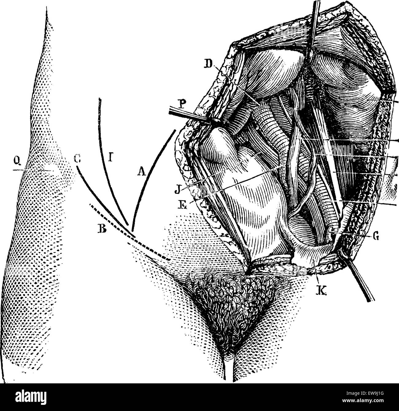 Fig. 618. Iliac artery and its branches, vintage engraved illustration. Magasin Pittoresque 1875. Stock Vectorhttps://www.alamy.com/image-license-details/?v=1https://www.alamy.com/stock-photo-fig-618-iliac-artery-and-its-branches-vintage-engraved-illustration-84419596.html
RFEW9J1G–Fig. 618. Iliac artery and its branches, vintage engraved illustration. Magasin Pittoresque 1875.
 Female cardiovascular system, artwork Stock Photohttps://www.alamy.com/image-license-details/?v=1https://www.alamy.com/stock-photo-female-cardiovascular-system-artwork-55416013.html
RFD64BK9–Female cardiovascular system, artwork
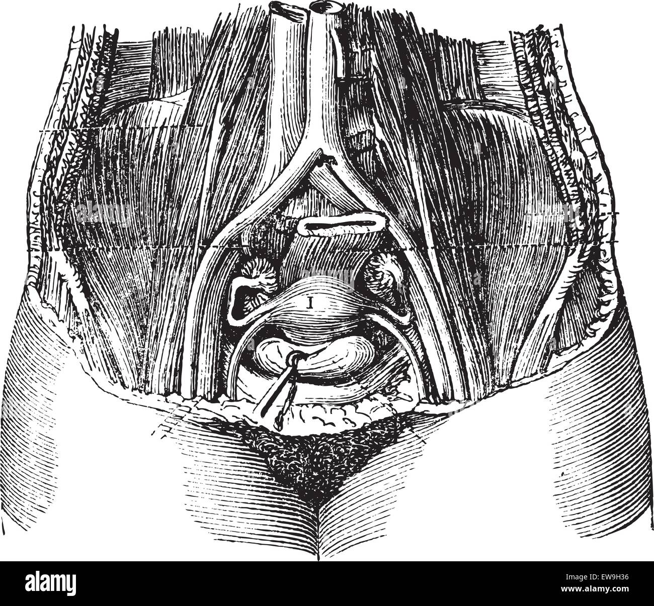 Fig. 162. Pool of women, with its soft parts, seen from top to bottom and front to back, vintage engraved illustration. Magasin Stock Vectorhttps://www.alamy.com/image-license-details/?v=1https://www.alamy.com/stock-photo-fig-162-pool-of-women-with-its-soft-parts-seen-from-top-to-bottom-84418858.html
RFEW9H36–Fig. 162. Pool of women, with its soft parts, seen from top to bottom and front to back, vintage engraved illustration. Magasin
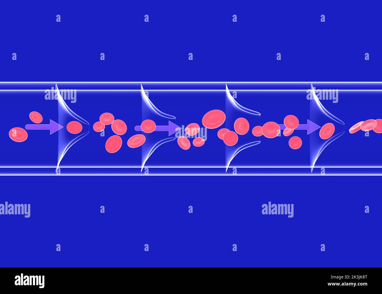 Anatomical 3d illustration of blood circulation in a healthy vein. Transparent image of glass on a blue background. Stock Photohttps://www.alamy.com/image-license-details/?v=1https://www.alamy.com/anatomical-3d-illustration-of-blood-circulation-in-a-healthy-vein-transparent-image-of-glass-on-a-blue-background-image484144552.html
RF2K3JK8T–Anatomical 3d illustration of blood circulation in a healthy vein. Transparent image of glass on a blue background.
 circumflex iliac vein is formed by the union of the venae comitantes of the deep iliac circumflex artery, and joins the external iliac vein Stock Photohttps://www.alamy.com/image-license-details/?v=1https://www.alamy.com/circumflex-iliac-vein-is-formed-by-the-union-of-the-venae-comitantes-of-the-deep-iliac-circumflex-artery-and-joins-the-external-iliac-vein-image596588495.html
RF2WJGXKY–circumflex iliac vein is formed by the union of the venae comitantes of the deep iliac circumflex artery, and joins the external iliac vein
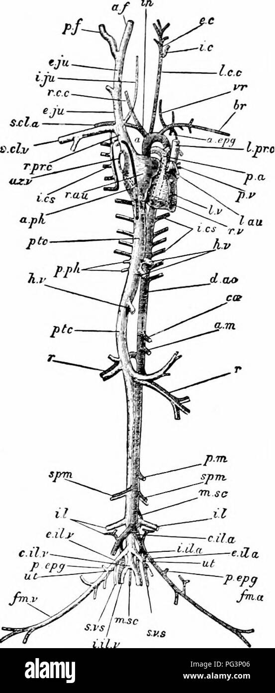 . A manual of zoology. . l.lt.t Fig. 307.—LepuS Cuniculus The vascular system. The heart is somewhat dis- placed towards the left of the subject: the arteries of the right and the veins of the left side are in great measure removed a, arch of the aorta; a. epg, internal mammary artery; a. ft anterior facia! vein: a. m, anterior mesenteric artery; a. p/i, anterior phrenic vein; az. v, azygos vein; Br, branchial artery; c. il. a, common iliac artery; etc, cceliac artery ; d. no, dorsal aorta; e. e, external carotid artery; e. il. a, external iliac artery; e. il. v, external iliac vein: e. jn, ex Stock Photohttps://www.alamy.com/image-license-details/?v=1https://www.alamy.com/a-manual-of-zoology-lltt-fig-307lepus-cuniculus-the-vascular-system-the-heart-is-somewhat-dis-placed-towards-the-left-of-the-subject-the-arteries-of-the-right-and-the-veins-of-the-left-side-are-in-great-measure-removed-a-arch-of-the-aorta-a-epg-internal-mammary-artery-a-ft-anterior-facia!-vein-a-m-anterior-mesenteric-artery-a-pi-anterior-phrenic-vein-az-v-azygos-vein-br-branchial-artery-c-il-a-common-iliac-artery-etc-cceliac-artery-d-no-dorsal-aorta-e-e-external-carotid-artery-e-il-a-external-iliac-artery-e-il-v-external-iliac-vein-e-jn-ex-image216442022.html
RMPG3P06–. A manual of zoology. . l.lt.t Fig. 307.—LepuS Cuniculus The vascular system. The heart is somewhat dis- placed towards the left of the subject: the arteries of the right and the veins of the left side are in great measure removed a, arch of the aorta; a. epg, internal mammary artery; a. ft anterior facia! vein: a. m, anterior mesenteric artery; a. p/i, anterior phrenic vein; az. v, azygos vein; Br, branchial artery; c. il. a, common iliac artery; etc, cceliac artery ; d. no, dorsal aorta; e. e, external carotid artery; e. il. a, external iliac artery; e. il. v, external iliac vein: e. jn, ex
 . Elementary anatomy and physiology : for colleges, academies, and other schools . A Front View of the Femoral, Iliac, and Aortic Lymphatic Vessels and Glands. 1, Sa- phena Magna Vein. 2, External Iliac Artery and Vein. 8, Primitive Iliac Artery and Vein. 4, The Aorta. 5, Ascending Vena Cava. 6, 7, Lymphatics which are alongside of the Saphena Vein on the Thigh. 8, Lower Set of Inguinal Lymphatic Glands which receive these Vessels. 9, Superior Set of Inguinal Lymphatic Glands which receive theso Vessels. 10, The Chain of Lymphatics in Front of the External Iliac Vessels. 11, Lym- phatics which Stock Photohttps://www.alamy.com/image-license-details/?v=1https://www.alamy.com/elementary-anatomy-and-physiology-for-colleges-academies-and-other-schools-a-front-view-of-the-femoral-iliac-and-aortic-lymphatic-vessels-and-glands-1-sa-phena-magna-vein-2-external-iliac-artery-and-vein-8-primitive-iliac-artery-and-vein-4-the-aorta-5-ascending-vena-cava-6-7-lymphatics-which-are-alongside-of-the-saphena-vein-on-the-thigh-8-lower-set-of-inguinal-lymphatic-glands-which-receive-these-vessels-9-superior-set-of-inguinal-lymphatic-glands-which-receive-theso-vessels-10-the-chain-of-lymphatics-in-front-of-the-external-iliac-vessels-11-lym-phatics-which-image178409579.html
RMMA776K–. Elementary anatomy and physiology : for colleges, academies, and other schools . A Front View of the Femoral, Iliac, and Aortic Lymphatic Vessels and Glands. 1, Sa- phena Magna Vein. 2, External Iliac Artery and Vein. 8, Primitive Iliac Artery and Vein. 4, The Aorta. 5, Ascending Vena Cava. 6, 7, Lymphatics which are alongside of the Saphena Vein on the Thigh. 8, Lower Set of Inguinal Lymphatic Glands which receive these Vessels. 9, Superior Set of Inguinal Lymphatic Glands which receive theso Vessels. 10, The Chain of Lymphatics in Front of the External Iliac Vessels. 11, Lym- phatics which
 THROMBOPHLEBITIS, ANGIOGRAPHY Stock Photohttps://www.alamy.com/image-license-details/?v=1https://www.alamy.com/stock-photo-thrombophlebitis-angiography-49255815.html
RMCT3P87–THROMBOPHLEBITIS, ANGIOGRAPHY
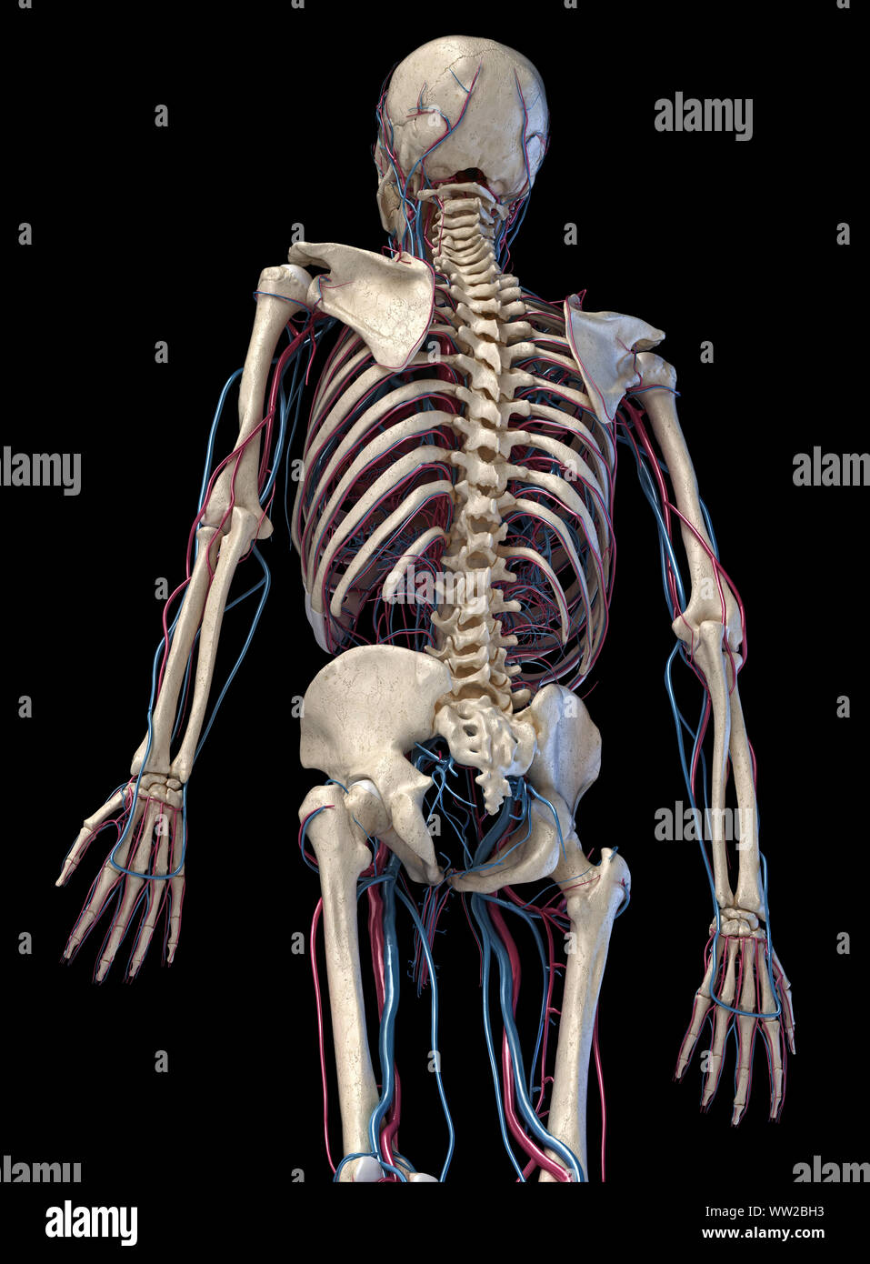 Human anatomy, 3d illustration of the skeleton with cardiovascular system. Perspective view of 3/4 upper part, back side. On black background. Stock Photohttps://www.alamy.com/image-license-details/?v=1https://www.alamy.com/human-anatomy-3d-illustration-of-the-skeleton-with-cardiovascular-system-perspective-view-of-34-upper-part-back-side-on-black-background-image273574927.html
RFWW2BH3–Human anatomy, 3d illustration of the skeleton with cardiovascular system. Perspective view of 3/4 upper part, back side. On black background.
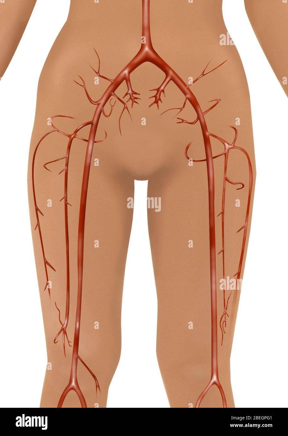 Arteries Stock Photohttps://www.alamy.com/image-license-details/?v=1https://www.alamy.com/arteries-image353181473.html
RF2BEGPG1–Arteries
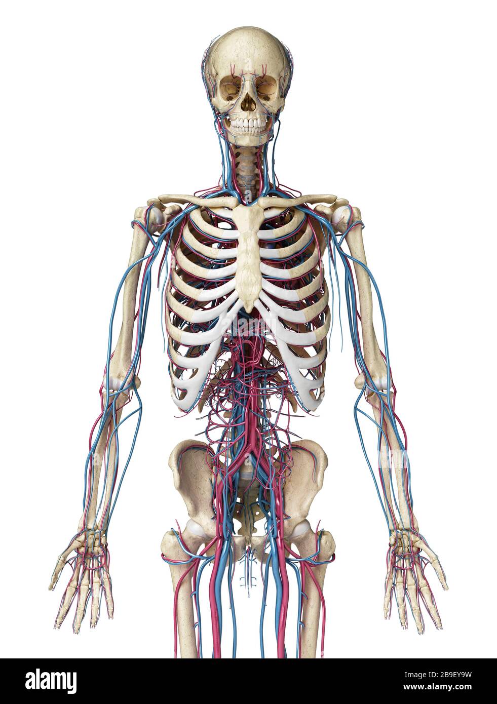 Upper body front view of human skeleton with veins and arteries, black background. Stock Photohttps://www.alamy.com/image-license-details/?v=1https://www.alamy.com/upper-body-front-view-of-human-skeleton-with-veins-and-arteries-black-background-image350068037.html
RF2B9EY9W–Upper body front view of human skeleton with veins and arteries, black background.
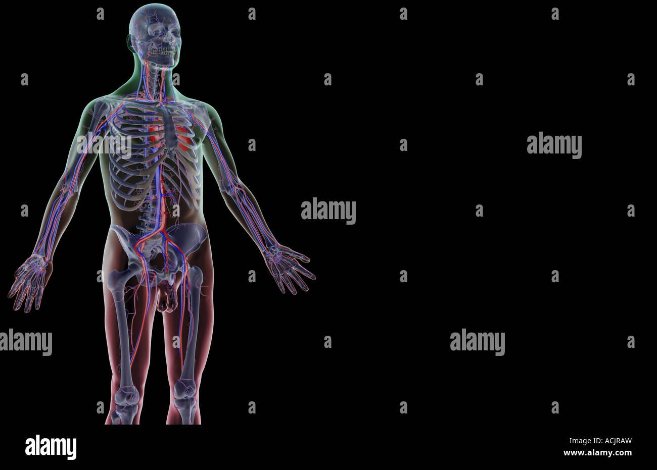 The blood supply of the upper body Stock Photohttps://www.alamy.com/image-license-details/?v=1https://www.alamy.com/stock-photo-the-blood-supply-of-the-upper-body-13172784.html
RFACJRAW–The blood supply of the upper body
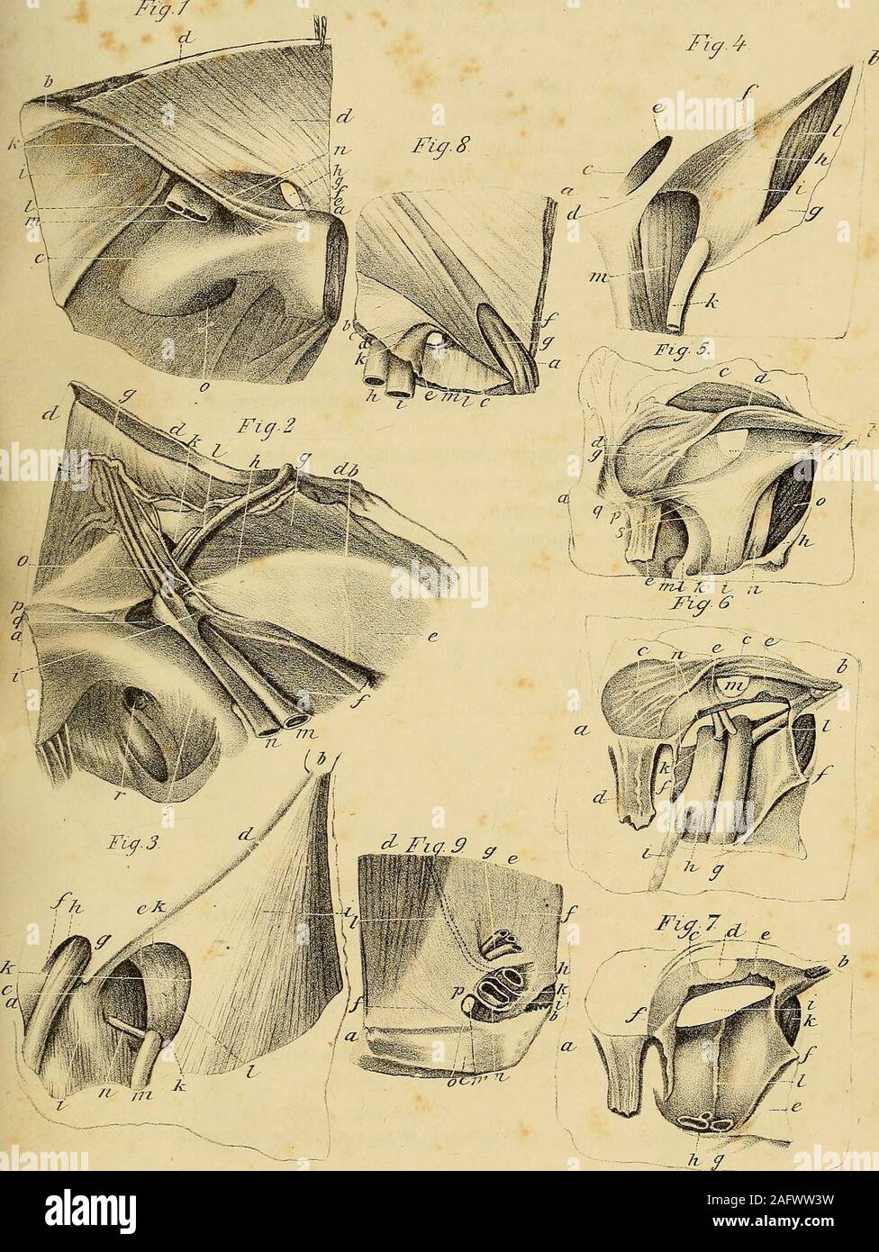 . The anatomy and surgical treatment of abdominal hernia. them to the edge of the crural sheath.k. Internal abdominal ring, or upper aperture of the inguinal canal./. Spermatic cord passing through that aperture. m. External iliac artery.n. External iliac vein.o. Epigastric artery and vein.p. Third insertion of the external oblique into the pubes, covered, however, by the fascia transversalis.q. The space by which the crural hernia descends, the finger having passed into it before the drawing was made to push down the fascia which extends over it.r. The thyroideal foramen. Fig. 3. An anterior Stock Photohttps://www.alamy.com/image-license-details/?v=1https://www.alamy.com/the-anatomy-and-surgical-treatment-of-abdominal-hernia-them-to-the-edge-of-the-crural-sheathk-internal-abdominal-ring-or-upper-aperture-of-the-inguinal-canal-spermatic-cord-passing-through-that-aperture-m-external-iliac-arteryn-external-iliac-veino-epigastric-artery-and-veinp-third-insertion-of-the-external-oblique-into-the-pubes-covered-however-by-the-fascia-transversalisq-the-space-by-which-the-crural-hernia-descends-the-finger-having-passed-into-it-before-the-drawing-was-made-to-push-down-the-fascia-which-extends-over-itr-the-thyroideal-foramen-fig-3-an-anterior-image336785341.html
RM2AFWW3W–. The anatomy and surgical treatment of abdominal hernia. them to the edge of the crural sheath.k. Internal abdominal ring, or upper aperture of the inguinal canal./. Spermatic cord passing through that aperture. m. External iliac artery.n. External iliac vein.o. Epigastric artery and vein.p. Third insertion of the external oblique into the pubes, covered, however, by the fascia transversalis.q. The space by which the crural hernia descends, the finger having passed into it before the drawing was made to push down the fascia which extends over it.r. The thyroideal foramen. Fig. 3. An anterior
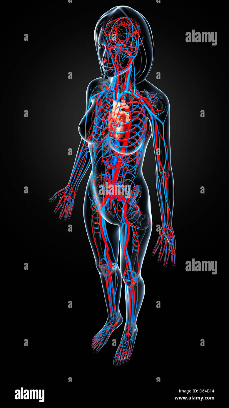 Female cardiovascular system, artwork Stock Photohttps://www.alamy.com/image-license-details/?v=1https://www.alamy.com/stock-photo-female-cardiovascular-system-artwork-55415504.html
RFD64B14–Female cardiovascular system, artwork
 Fig. 162. Pool of women, with its soft parts, seen from top to bottom and front to back, vintage engraved illustration. Magasin Stock Vectorhttps://www.alamy.com/image-license-details/?v=1https://www.alamy.com/stock-photo-fig-162-pool-of-women-with-its-soft-parts-seen-from-top-to-bottom-84406344.html
RFEW9148–Fig. 162. Pool of women, with its soft parts, seen from top to bottom and front to back, vintage engraved illustration. Magasin
 Anatomical 3d animation of a diseased and incompetent vertical vein. Valves malfunction accumulating red blood cells. Transparent image of glass. Stock Photohttps://www.alamy.com/image-license-details/?v=1https://www.alamy.com/anatomical-3d-animation-of-a-diseased-and-incompetent-vertical-vein-valves-malfunction-accumulating-red-blood-cells-transparent-image-of-glass-image611431194.html
RF2XEN2MA–Anatomical 3d animation of a diseased and incompetent vertical vein. Valves malfunction accumulating red blood cells. Transparent image of glass.
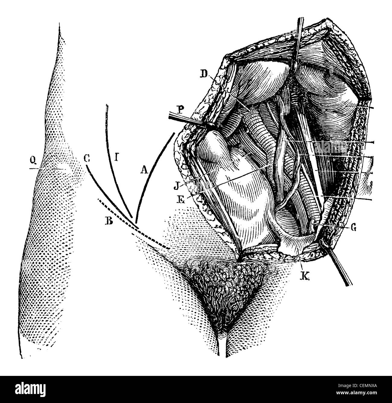 Fig. 618. Iliac artery and its branches, vintage engraved illustration. Magasin Pittoresque 1875. Stock Photohttps://www.alamy.com/image-license-details/?v=1https://www.alamy.com/stock-photo-fig-618-iliac-artery-and-its-branches-vintage-engraved-illustration-43482162.html
RFCEMNXA–Fig. 618. Iliac artery and its branches, vintage engraved illustration. Magasin Pittoresque 1875.
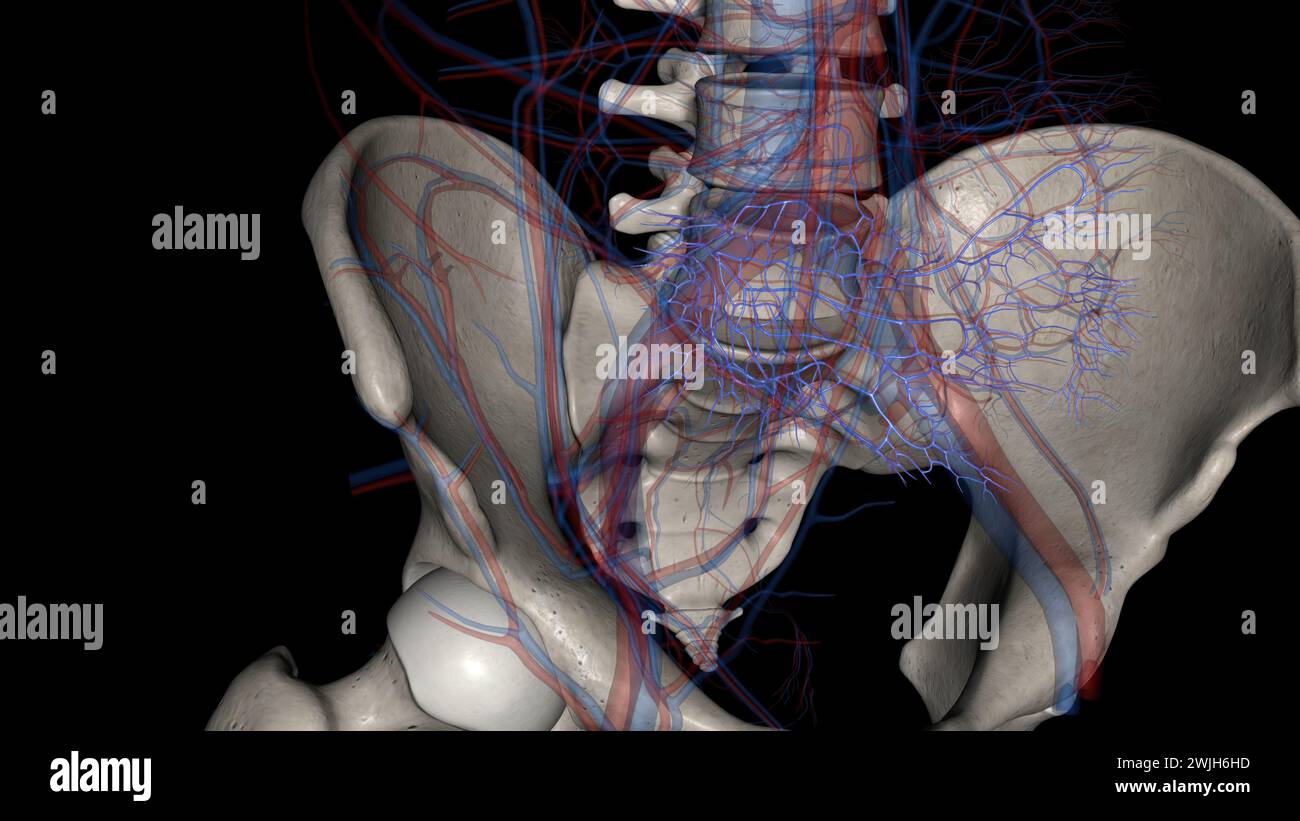 The common iliac vein is formed by the unification of the internal and external iliac veins 3d illustration Stock Photohttps://www.alamy.com/image-license-details/?v=1https://www.alamy.com/the-common-iliac-vein-is-formed-by-the-unification-of-the-internal-and-external-iliac-veins-3d-illustration-image596594697.html
RF2WJH6HD–The common iliac vein is formed by the unification of the internal and external iliac veins 3d illustration
 . Cunningham's Text-book of anatomy. Anatomy. 988 THE VASCULAE SYSTEM. — Superficial epigastric vein Superficial circumflex —" iliac vein Superficial external pudendal vein Femoral vein Great saphenous vein Lateral superficial femoral vein Medial superficial femoral vein artery, whilst just before its termination it crosses the lateral side of the hypo- gastric artery, and separates that vessel from the medial border of the psoas major muscle. In its whole course the vein lies anterior to the obturator nerve. It is usually provided with one bicuspid valve ; sometimes there are two, but bo Stock Photohttps://www.alamy.com/image-license-details/?v=1https://www.alamy.com/cunninghams-text-book-of-anatomy-anatomy-988-the-vasculae-system-superficial-epigastric-vein-superficial-circumflex-quot-iliac-vein-superficial-external-pudendal-vein-femoral-vein-great-saphenous-vein-lateral-superficial-femoral-vein-medial-superficial-femoral-vein-artery-whilst-just-before-its-termination-it-crosses-the-lateral-side-of-the-hypo-gastric-artery-and-separates-that-vessel-from-the-medial-border-of-the-psoas-major-muscle-in-its-whole-course-the-vein-lies-anterior-to-the-obturator-nerve-it-is-usually-provided-with-one-bicuspid-valve-sometimes-there-are-two-but-bo-image216334416.html
RMPFXTN4–. Cunningham's Text-book of anatomy. Anatomy. 988 THE VASCULAE SYSTEM. — Superficial epigastric vein Superficial circumflex —" iliac vein Superficial external pudendal vein Femoral vein Great saphenous vein Lateral superficial femoral vein Medial superficial femoral vein artery, whilst just before its termination it crosses the lateral side of the hypo- gastric artery, and separates that vessel from the medial border of the psoas major muscle. In its whole course the vein lies anterior to the obturator nerve. It is usually provided with one bicuspid valve ; sometimes there are two, but bo
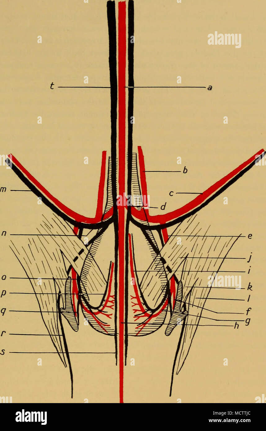 . Fig. 8. Arteries and veins of the genital region in the Fin whale. Diagrammatic. a. Dorsal aorta. b. Hypogastric artery. c. Epigastric artery. d. Common iliac artery. e. Genital artery. /. Artery to pelvic musculature (external iliac of Murie). g. Internal iliac of Murie. h. Caudal artery. i. Pudic artery. j. Caudal attachment of rectus abdominis muscle. k. Iliac attachment of rectus abdominis muscle. /. Superficial attachment of ditto. m. Epigastric vein. n. Common iliac vein. o. Genital vein. p. Pudic vein. q. Vein from pelvic musculature (external iliac of Murie). r. Lumbar vein, s. Cauda Stock Photohttps://www.alamy.com/image-license-details/?v=1https://www.alamy.com/fig-8-arteries-and-veins-of-the-genital-region-in-the-fin-whale-diagrammatic-a-dorsal-aorta-b-hypogastric-artery-c-epigastric-artery-d-common-iliac-artery-e-genital-artery-artery-to-pelvic-musculature-external-iliac-of-murie-g-internal-iliac-of-murie-h-caudal-artery-i-pudic-artery-j-caudal-attachment-of-rectus-abdominis-muscle-k-iliac-attachment-of-rectus-abdominis-muscle-superficial-attachment-of-ditto-m-epigastric-vein-n-common-iliac-vein-o-genital-vein-p-pudic-vein-q-vein-from-pelvic-musculature-external-iliac-of-murie-r-lumbar-vein-s-cauda-image180025732.html
RMMCTTJC–. Fig. 8. Arteries and veins of the genital region in the Fin whale. Diagrammatic. a. Dorsal aorta. b. Hypogastric artery. c. Epigastric artery. d. Common iliac artery. e. Genital artery. /. Artery to pelvic musculature (external iliac of Murie). g. Internal iliac of Murie. h. Caudal artery. i. Pudic artery. j. Caudal attachment of rectus abdominis muscle. k. Iliac attachment of rectus abdominis muscle. /. Superficial attachment of ditto. m. Epigastric vein. n. Common iliac vein. o. Genital vein. p. Pudic vein. q. Vein from pelvic musculature (external iliac of Murie). r. Lumbar vein, s. Cauda
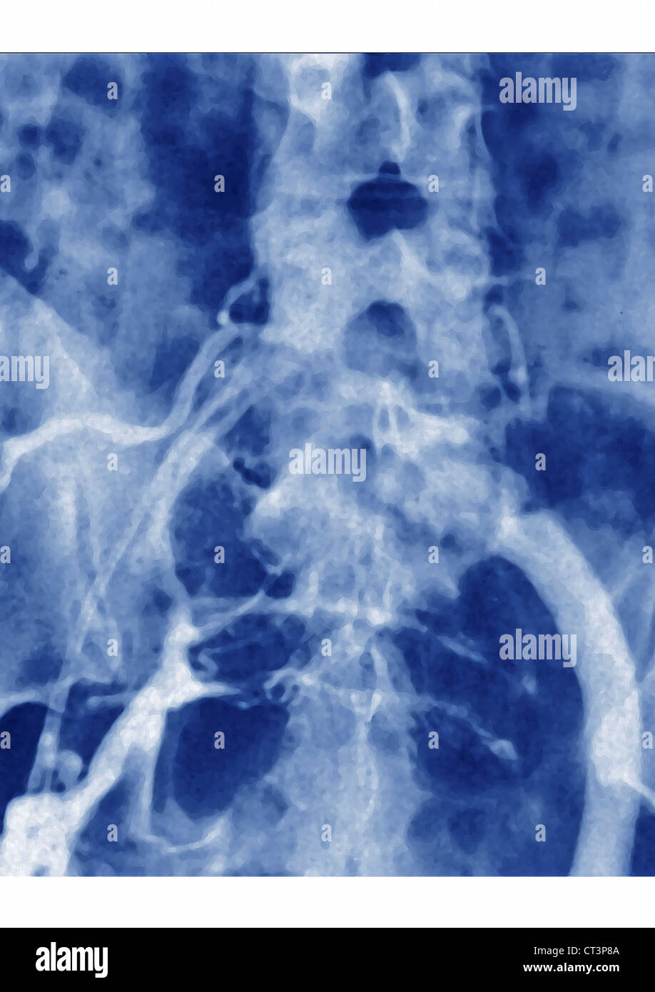 THROMBOPHLEBITIS, ANGIOGRAPHY Stock Photohttps://www.alamy.com/image-license-details/?v=1https://www.alamy.com/stock-photo-thrombophlebitis-angiography-49255818.html
RMCT3P8A–THROMBOPHLEBITIS, ANGIOGRAPHY
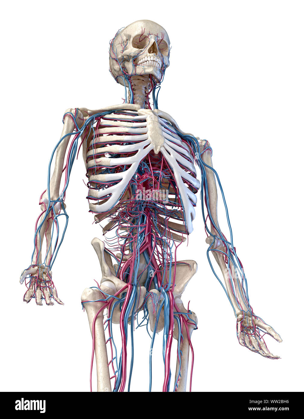 Human anatomy, 3d illustration of the skeleton with cardiovascular system. Perspective view of 3/4 upper part, front side. On white background. Stock Photohttps://www.alamy.com/image-license-details/?v=1https://www.alamy.com/human-anatomy-3d-illustration-of-the-skeleton-with-cardiovascular-system-perspective-view-of-34-upper-part-front-side-on-white-background-image273574930.html
RFWW2BH6–Human anatomy, 3d illustration of the skeleton with cardiovascular system. Perspective view of 3/4 upper part, front side. On white background.
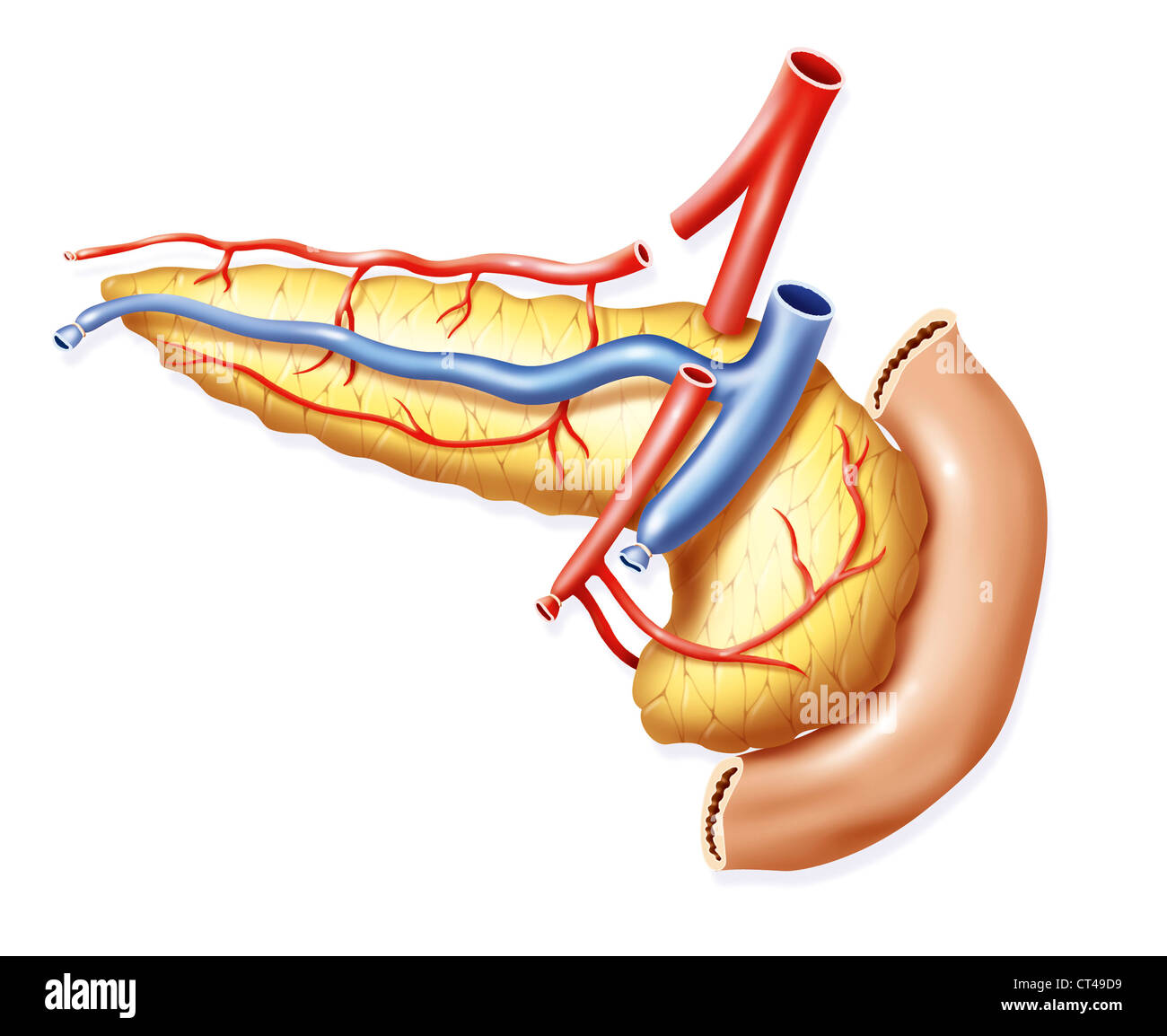 PANCREAS TRANSPLANT, DRAWING Stock Photohttps://www.alamy.com/image-license-details/?v=1https://www.alamy.com/stock-photo-pancreas-transplant-drawing-49267717.html
RMCT49D9–PANCREAS TRANSPLANT, DRAWING
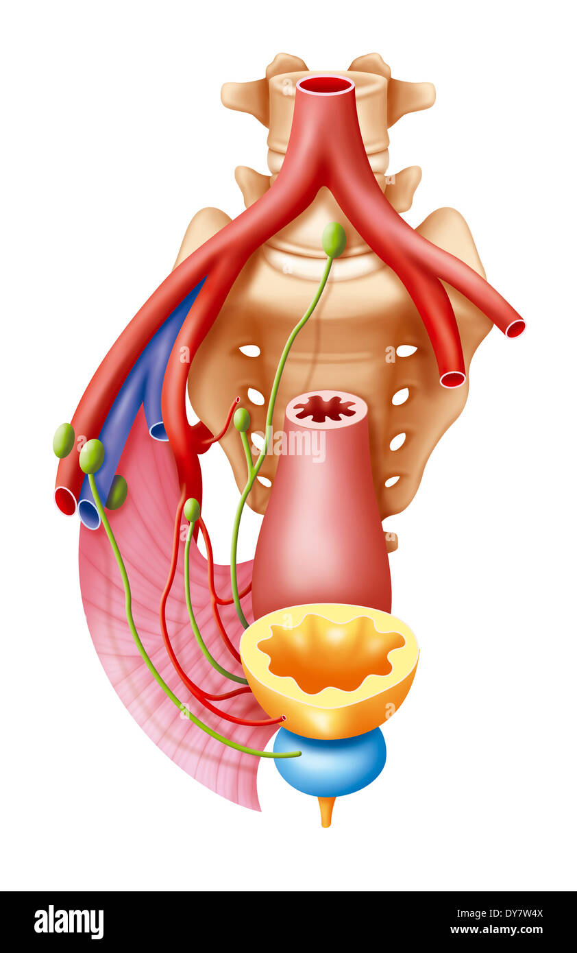 Anatomy Stock Photohttps://www.alamy.com/image-license-details/?v=1https://www.alamy.com/anatomy-image68400218.html
RMDY7W4X–Anatomy
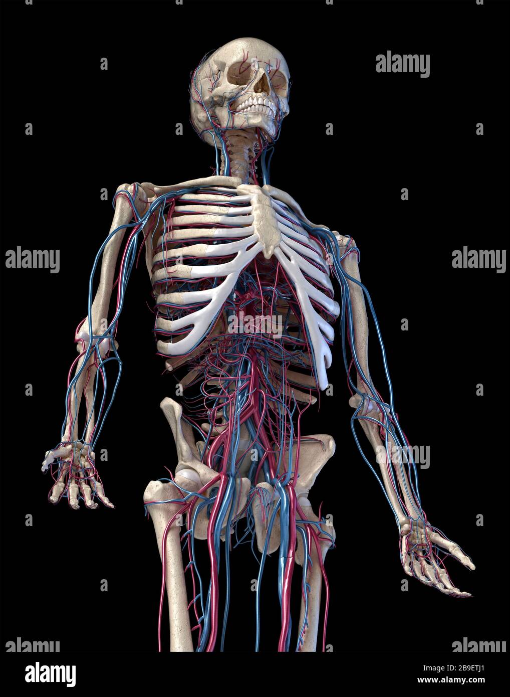 3/4 upper body front view of human skeletal and vascular systems, black background. Stock Photohttps://www.alamy.com/image-license-details/?v=1https://www.alamy.com/34-upper-body-front-view-of-human-skeletal-and-vascular-systems-black-background-image350065913.html
RF2B9ETJ1–3/4 upper body front view of human skeletal and vascular systems, black background.
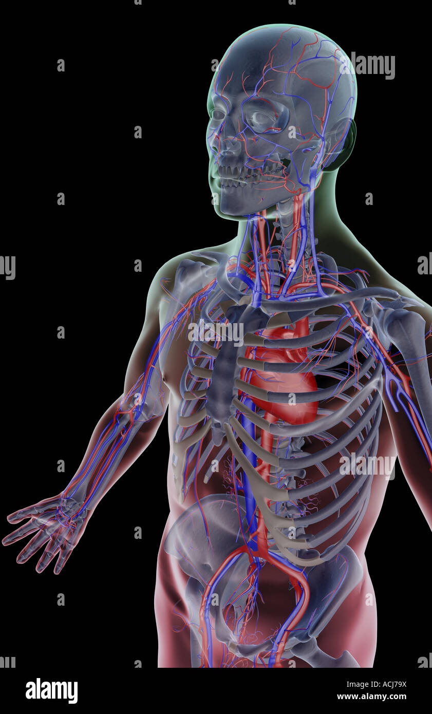 The blood supply of the upper body Stock Photohttps://www.alamy.com/image-license-details/?v=1https://www.alamy.com/stock-photo-the-blood-supply-of-the-upper-body-13167397.html
RFACJ79X–The blood supply of the upper body
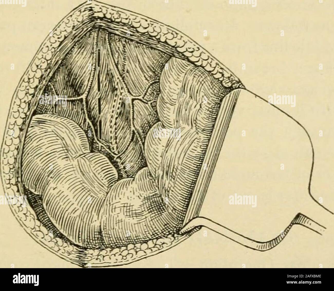 . Manual of operative surgery. At the upper angle of the perito-neal wound lies the common iliac, at the centre of the wound lies the bifurcation,at the lower angle lie the external and internal iliacs, side by side, still coveredby a sheath of fascia. Step 4.—Incise the fascial sheath secundum artem; denude the internal iliac;pass an aneurysm needle from without inwards, closely hugging the artery soas to avoid injury to the external iliac vein, and apply a ligature % inch from theorigin of the vessel, i.e., at a point a very little below the brim of the true pelvis.Do not forget that the int Stock Photohttps://www.alamy.com/image-license-details/?v=1https://www.alamy.com/manual-of-operative-surgery-at-the-upper-angle-of-the-perito-neal-wound-lies-the-common-iliac-at-the-centre-of-the-wound-lies-the-bifurcationat-the-lower-angle-lie-the-external-and-internal-iliacs-side-by-side-still-coveredby-a-sheath-of-fascia-step-4incise-the-fascial-sheath-secundum-artem-denude-the-internal-iliacpass-an-aneurysm-needle-from-without-inwards-closely-hugging-the-artery-soas-to-avoid-injury-to-the-external-iliac-vein-and-apply-a-ligature-inch-from-theorigin-of-the-vessel-ie-at-a-point-a-very-little-below-the-brim-of-the-true-pelvisdo-not-forget-that-the-int-image336796782.html
RM2AFXBME–. Manual of operative surgery. At the upper angle of the perito-neal wound lies the common iliac, at the centre of the wound lies the bifurcation,at the lower angle lie the external and internal iliacs, side by side, still coveredby a sheath of fascia. Step 4.—Incise the fascial sheath secundum artem; denude the internal iliac;pass an aneurysm needle from without inwards, closely hugging the artery soas to avoid injury to the external iliac vein, and apply a ligature % inch from theorigin of the vessel, i.e., at a point a very little below the brim of the true pelvis.Do not forget that the int
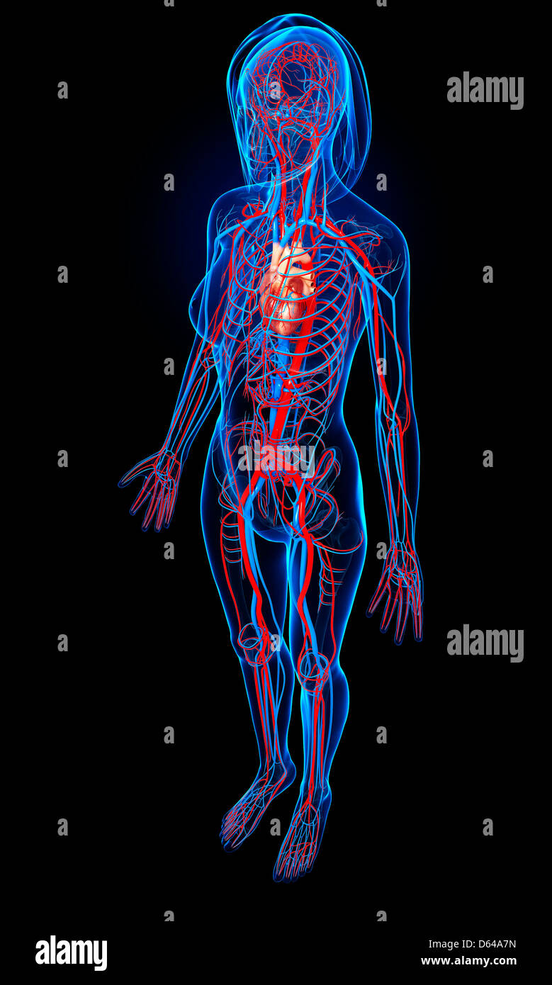 Female cardiovascular system, artwork Stock Photohttps://www.alamy.com/image-license-details/?v=1https://www.alamy.com/stock-photo-female-cardiovascular-system-artwork-55414905.html
RFD64A7N–Female cardiovascular system, artwork
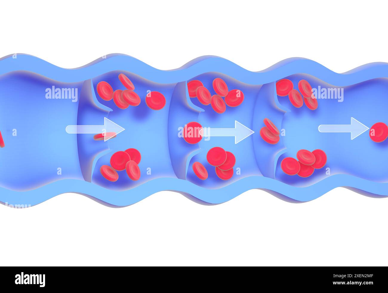 Anatomical 3d animation of a diseased and incompetent vein. Valves malfunction, accumulating red blood cells in the walls, causing varicose veins. Stock Photohttps://www.alamy.com/image-license-details/?v=1https://www.alamy.com/anatomical-3d-animation-of-a-diseased-and-incompetent-vein-valves-malfunction-accumulating-red-blood-cells-in-the-walls-causing-varicose-veins-image611431199.html
RF2XEN2MF–Anatomical 3d animation of a diseased and incompetent vein. Valves malfunction, accumulating red blood cells in the walls, causing varicose veins.
 Fig. 162. Pool of women, with its soft parts, seen from top to bottom and front to back, vintage engraved illustration. Magasin Pittoresque 1875. Stock Photohttps://www.alamy.com/image-license-details/?v=1https://www.alamy.com/stock-photo-fig-162-pool-of-women-with-its-soft-parts-seen-from-top-to-bottom-43483004.html
RFCEMR0C–Fig. 162. Pool of women, with its soft parts, seen from top to bottom and front to back, vintage engraved illustration. Magasin Pittoresque 1875.
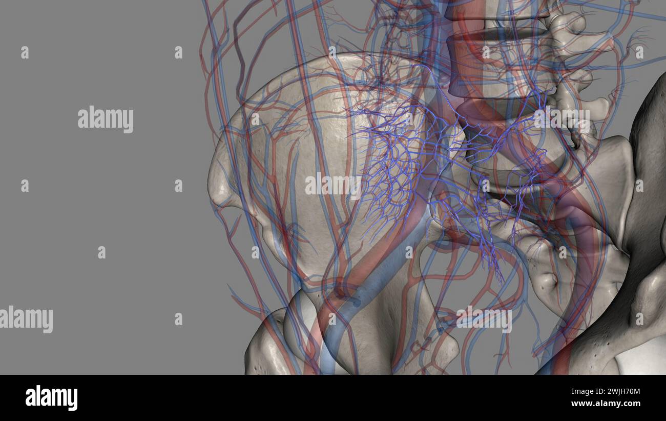 The common iliac vein is formed by the unification of the internal and external iliac veins 3d illustration Stock Photohttps://www.alamy.com/image-license-details/?v=1https://www.alamy.com/the-common-iliac-vein-is-formed-by-the-unification-of-the-internal-and-external-iliac-veins-3d-illustration-image596595012.html
RF2WJH70M–The common iliac vein is formed by the unification of the internal and external iliac veins 3d illustration
![. The elements of embryology . Embryology. XII.] VERTEBRAL VEINS. 411 inferior (Fig. 139, il). These vessels, whose development has not been adequately investigated, form the common. Diagram or the Chief Venous Trunks op Man. (Prom Gegenbaur.) cs. coronary sinus ; s. subclavian vein; ji. internal jugular ; je. external jugular ; az. azygos vein ; ha. hemiazygos vein ; c. dotted lino shewing previous position of cardinal veins; ci. vena cava inferior ; r. renal veins ; il. iliac ; hy. hypogas- tric veins ; h. hepatic veins. The dotted lines shew the position of embryonic vessels aborted in the Stock Photo . The elements of embryology . Embryology. XII.] VERTEBRAL VEINS. 411 inferior (Fig. 139, il). These vessels, whose development has not been adequately investigated, form the common. Diagram or the Chief Venous Trunks op Man. (Prom Gegenbaur.) cs. coronary sinus ; s. subclavian vein; ji. internal jugular ; je. external jugular ; az. azygos vein ; ha. hemiazygos vein ; c. dotted lino shewing previous position of cardinal veins; ci. vena cava inferior ; r. renal veins ; il. iliac ; hy. hypogas- tric veins ; h. hepatic veins. The dotted lines shew the position of embryonic vessels aborted in the Stock Photo](https://c8.alamy.com/comp/PG3T85/the-elements-of-embryology-embryology-xii-vertebral-veins-411-inferior-fig-139-il-these-vessels-whose-development-has-not-been-adequately-investigated-form-the-common-diagram-or-the-chief-venous-trunks-op-man-prom-gegenbaur-cs-coronary-sinus-s-subclavian-vein-ji-internal-jugular-je-external-jugular-az-azygos-vein-ha-hemiazygos-vein-c-dotted-lino-shewing-previous-position-of-cardinal-veins-ci-vena-cava-inferior-r-renal-veins-il-iliac-hy-hypogas-tric-veins-h-hepatic-veins-the-dotted-lines-shew-the-position-of-embryonic-vessels-aborted-in-the-PG3T85.jpg) . The elements of embryology . Embryology. XII.] VERTEBRAL VEINS. 411 inferior (Fig. 139, il). These vessels, whose development has not been adequately investigated, form the common. Diagram or the Chief Venous Trunks op Man. (Prom Gegenbaur.) cs. coronary sinus ; s. subclavian vein; ji. internal jugular ; je. external jugular ; az. azygos vein ; ha. hemiazygos vein ; c. dotted lino shewing previous position of cardinal veins; ci. vena cava inferior ; r. renal veins ; il. iliac ; hy. hypogas- tric veins ; h. hepatic veins. The dotted lines shew the position of embryonic vessels aborted in the Stock Photohttps://www.alamy.com/image-license-details/?v=1https://www.alamy.com/the-elements-of-embryology-embryology-xii-vertebral-veins-411-inferior-fig-139-il-these-vessels-whose-development-has-not-been-adequately-investigated-form-the-common-diagram-or-the-chief-venous-trunks-op-man-prom-gegenbaur-cs-coronary-sinus-s-subclavian-vein-ji-internal-jugular-je-external-jugular-az-azygos-vein-ha-hemiazygos-vein-c-dotted-lino-shewing-previous-position-of-cardinal-veins-ci-vena-cava-inferior-r-renal-veins-il-iliac-hy-hypogas-tric-veins-h-hepatic-veins-the-dotted-lines-shew-the-position-of-embryonic-vessels-aborted-in-the-image216443813.html
. The elements of embryology . Embryology. XII.] VERTEBRAL VEINS. 411 inferior (Fig. 139, il). These vessels, whose development has not been adequately investigated, form the common. Diagram or the Chief Venous Trunks op Man. (Prom Gegenbaur.) cs. coronary sinus ; s. subclavian vein; ji. internal jugular ; je. external jugular ; az. azygos vein ; ha. hemiazygos vein ; c. dotted lino shewing previous position of cardinal veins; ci. vena cava inferior ; r. renal veins ; il. iliac ; hy. hypogas- tric veins ; h. hepatic veins. The dotted lines shew the position of embryonic vessels aborted in the Stock Photohttps://www.alamy.com/image-license-details/?v=1https://www.alamy.com/the-elements-of-embryology-embryology-xii-vertebral-veins-411-inferior-fig-139-il-these-vessels-whose-development-has-not-been-adequately-investigated-form-the-common-diagram-or-the-chief-venous-trunks-op-man-prom-gegenbaur-cs-coronary-sinus-s-subclavian-vein-ji-internal-jugular-je-external-jugular-az-azygos-vein-ha-hemiazygos-vein-c-dotted-lino-shewing-previous-position-of-cardinal-veins-ci-vena-cava-inferior-r-renal-veins-il-iliac-hy-hypogas-tric-veins-h-hepatic-veins-the-dotted-lines-shew-the-position-of-embryonic-vessels-aborted-in-the-image216443813.htmlRMPG3T85–. The elements of embryology . Embryology. XII.] VERTEBRAL VEINS. 411 inferior (Fig. 139, il). These vessels, whose development has not been adequately investigated, form the common. Diagram or the Chief Venous Trunks op Man. (Prom Gegenbaur.) cs. coronary sinus ; s. subclavian vein; ji. internal jugular ; je. external jugular ; az. azygos vein ; ha. hemiazygos vein ; c. dotted lino shewing previous position of cardinal veins; ci. vena cava inferior ; r. renal veins ; il. iliac ; hy. hypogas- tric veins ; h. hepatic veins. The dotted lines shew the position of embryonic vessels aborted in the
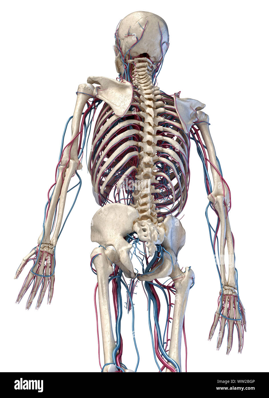 Human anatomy, 3d illustration of the skeleton with cardiovascular system. Perspective view of 3/4 upper part, back side. On white background. Stock Photohttps://www.alamy.com/image-license-details/?v=1https://www.alamy.com/human-anatomy-3d-illustration-of-the-skeleton-with-cardiovascular-system-perspective-view-of-34-upper-part-back-side-on-white-background-image273574918.html
RFWW2BGP–Human anatomy, 3d illustration of the skeleton with cardiovascular system. Perspective view of 3/4 upper part, back side. On white background.
 Anatomy Stock Photohttps://www.alamy.com/image-license-details/?v=1https://www.alamy.com/anatomy-image68400216.html
RMDY7W4T–Anatomy
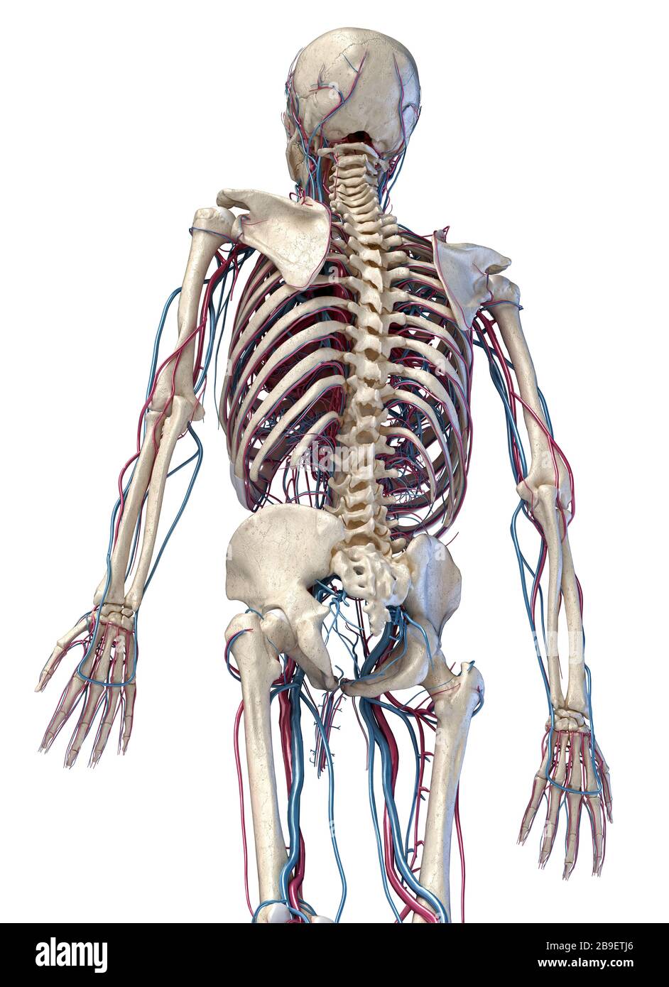 3/4 upper body rear view of human skeletal and vascular systems, white background. Stock Photohttps://www.alamy.com/image-license-details/?v=1https://www.alamy.com/34-upper-body-rear-view-of-human-skeletal-and-vascular-systems-white-background-image350065918.html
RF2B9ETJ6–3/4 upper body rear view of human skeletal and vascular systems, white background.
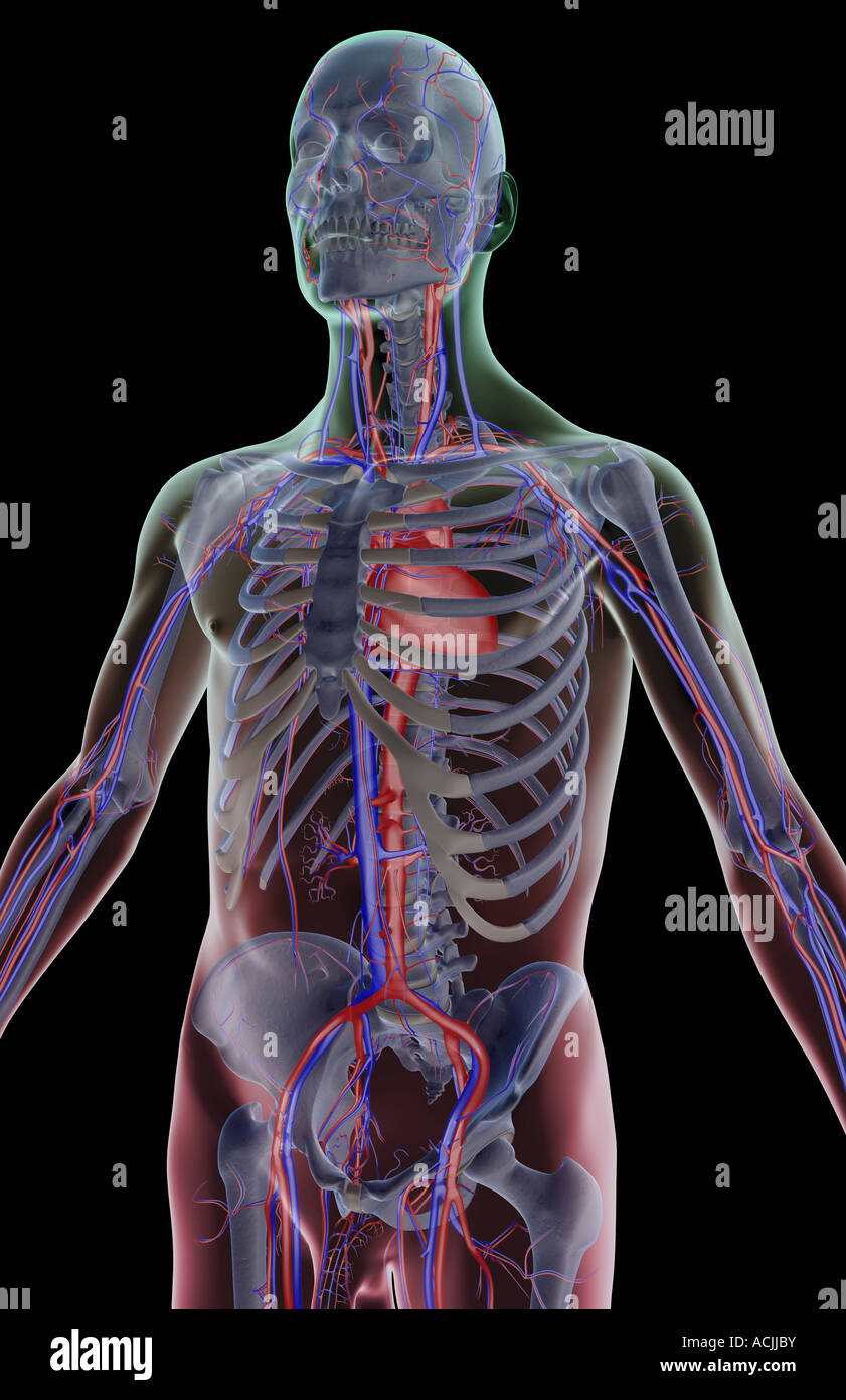 The blood supply of the upper body Stock Photohttps://www.alamy.com/image-license-details/?v=1https://www.alamy.com/stock-photo-the-blood-supply-of-the-upper-body-13171118.html
RFACJJBY–The blood supply of the upper body
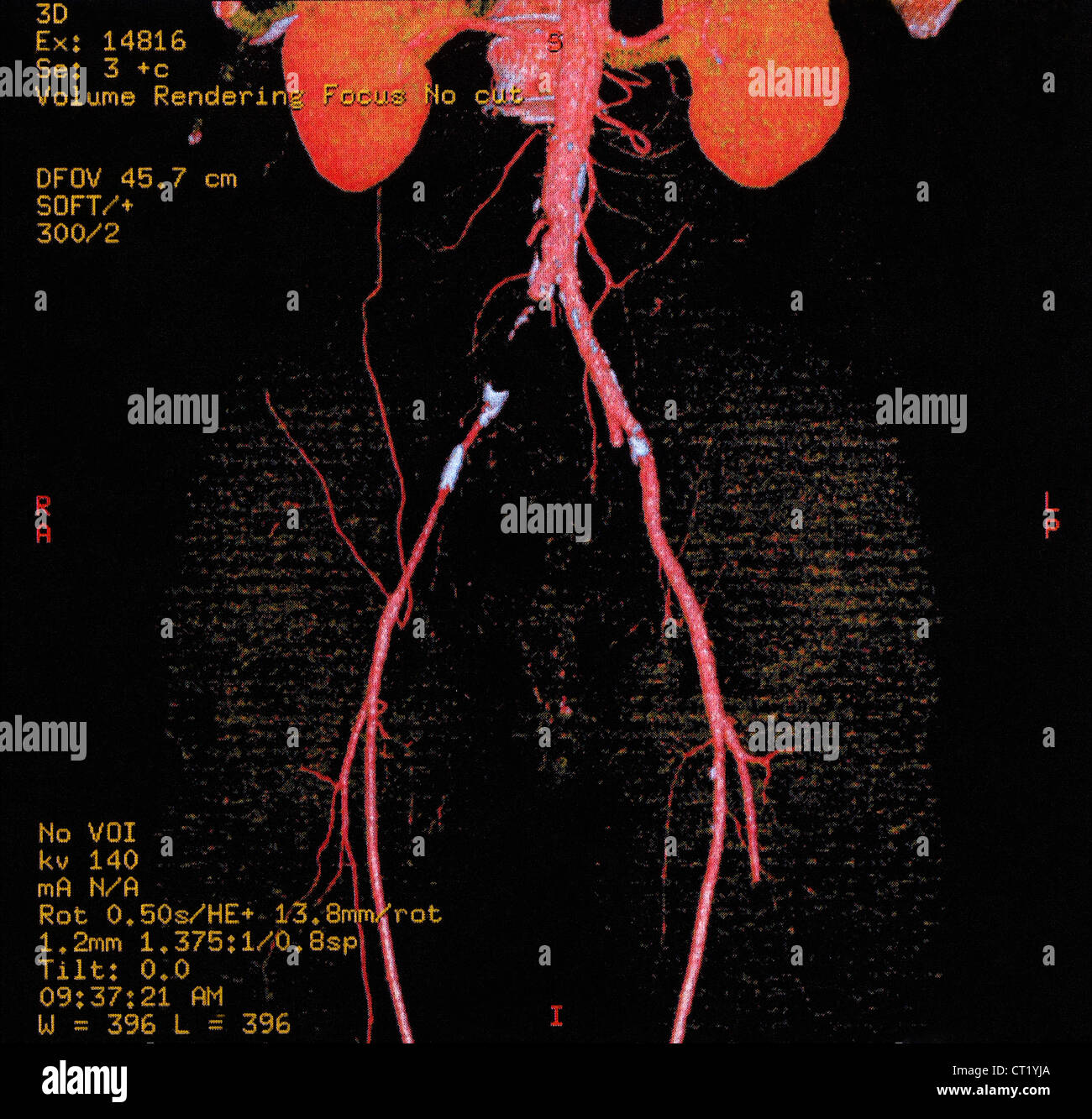 ILIAC THROMBOSIS, 3D SCAN Stock Photohttps://www.alamy.com/image-license-details/?v=1https://www.alamy.com/stock-photo-iliac-thrombosis-3d-scan-49216114.html
RMCT1YJA–ILIAC THROMBOSIS, 3D SCAN
 A system of human anatomy, general and special . ject to the production of phlebolites. * The veins of the trunk and neck. 1. The superior vena cava. 2. The left venainnominata. 3. The right vena innominata. 4. The right subclavian vein. 5. The in.ternal jugular vein. 6. The external jugular. 7. The anterior jugular. 8. The inferiorvena cava. 9. The external iliac vein. 10. The internal iliac vein. 11. The commoniliac veins ; the small vein between these is the vena sacra media. 12, 12. Lumbar veins.13. The right spermatic vein. 14. The left spermatic, opening into the left renal vein.15. The Stock Photohttps://www.alamy.com/image-license-details/?v=1https://www.alamy.com/a-system-of-human-anatomy-general-and-special-ject-to-the-production-of-phlebolites-the-veins-of-the-trunk-and-neck-1-the-superior-vena-cava-2-the-left-venainnominata-3-the-right-vena-innominata-4-the-right-subclavian-vein-5-the-internal-jugular-vein-6-the-external-jugular-7-the-anterior-jugular-8-the-inferiorvena-cava-9-the-external-iliac-vein-10-the-internal-iliac-vein-11-the-commoniliac-veins-the-small-vein-between-these-is-the-vena-sacra-media-12-12-lumbar-veins13-the-right-spermatic-vein-14-the-left-spermatic-opening-into-the-left-renal-vein15-the-image342720131.html
RM2AWG70K–A system of human anatomy, general and special . ject to the production of phlebolites. * The veins of the trunk and neck. 1. The superior vena cava. 2. The left venainnominata. 3. The right vena innominata. 4. The right subclavian vein. 5. The in.ternal jugular vein. 6. The external jugular. 7. The anterior jugular. 8. The inferiorvena cava. 9. The external iliac vein. 10. The internal iliac vein. 11. The commoniliac veins ; the small vein between these is the vena sacra media. 12, 12. Lumbar veins.13. The right spermatic vein. 14. The left spermatic, opening into the left renal vein.15. The
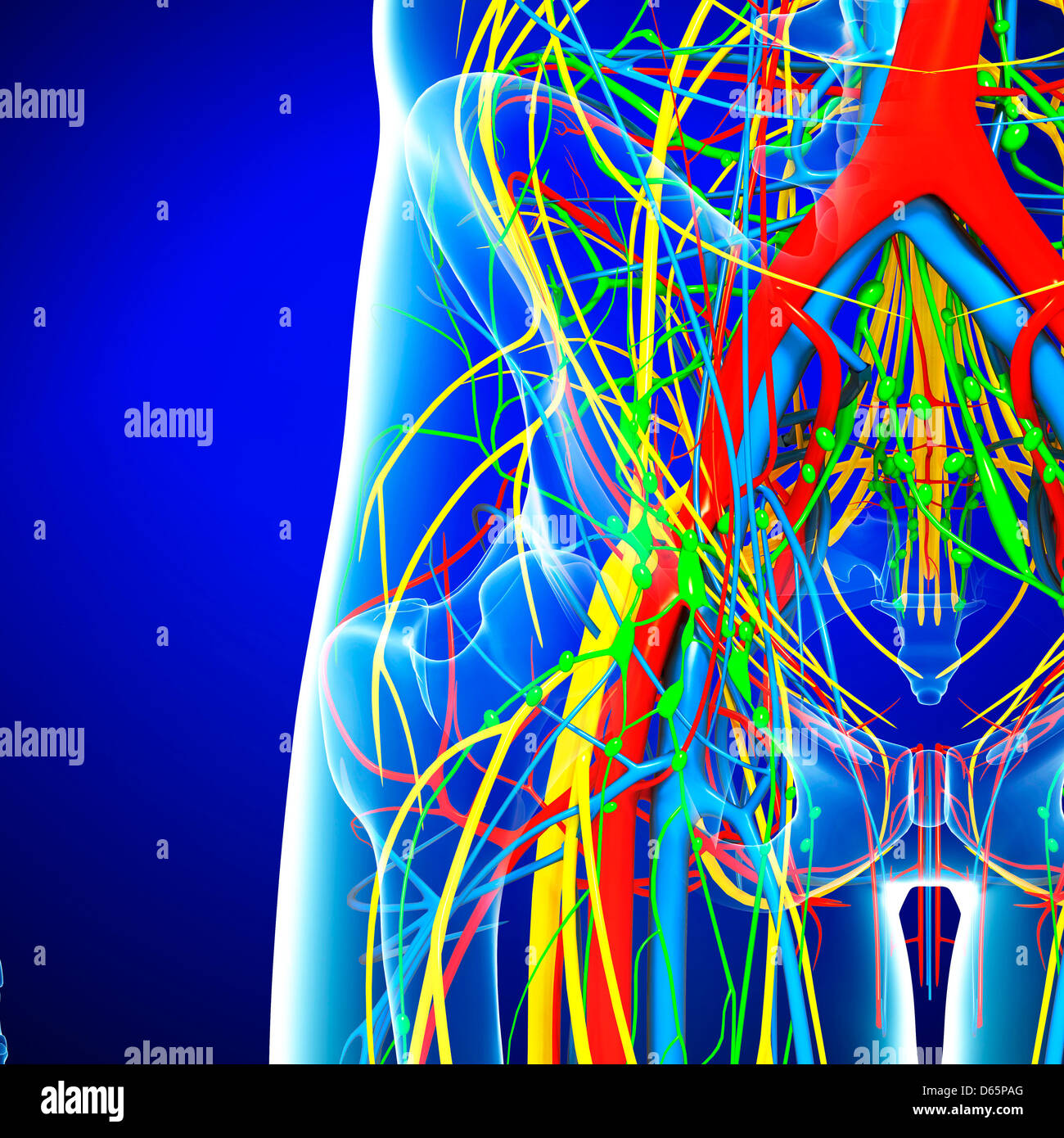 Pelvis anatomy, artwork Stock Photohttps://www.alamy.com/image-license-details/?v=1https://www.alamy.com/stock-photo-pelvis-anatomy-artwork-55446344.html
RFD65PAG–Pelvis anatomy, artwork
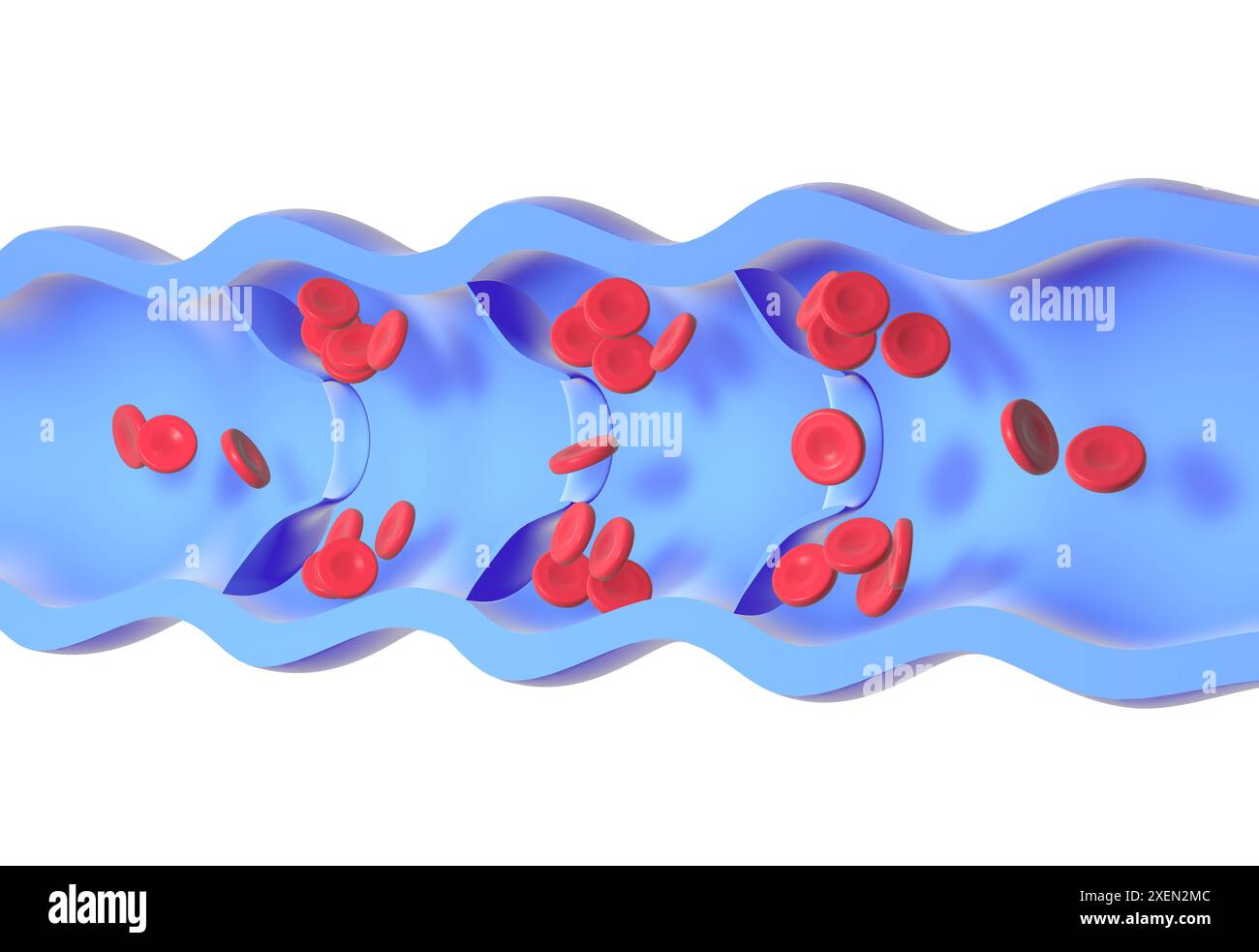 Anatomical 3d animation of a diseased and incompetent vein. Valves malfunction, accumulating red blood cells in the walls, causing varicose veins. Stock Photohttps://www.alamy.com/image-license-details/?v=1https://www.alamy.com/anatomical-3d-animation-of-a-diseased-and-incompetent-vein-valves-malfunction-accumulating-red-blood-cells-in-the-walls-causing-varicose-veins-image611431196.html
RF2XEN2MC–Anatomical 3d animation of a diseased and incompetent vein. Valves malfunction, accumulating red blood cells in the walls, causing varicose veins.
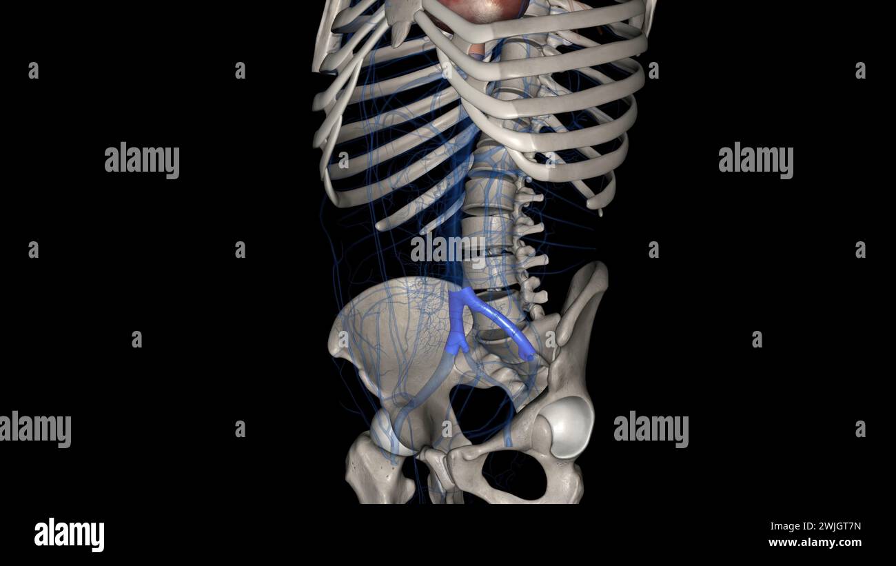 The common iliac vein is formed by the unification of the internal and external iliac veins 3d illustration Stock Photohttps://www.alamy.com/image-license-details/?v=1https://www.alamy.com/the-common-iliac-vein-is-formed-by-the-unification-of-the-internal-and-external-iliac-veins-3d-illustration-image596586585.html
RF2WJGT7N–The common iliac vein is formed by the unification of the internal and external iliac veins 3d illustration
 . The surgical anatomy of the horse ... Horses. -18. Plate XXIII.—Obtukatok and Anterior Crural Nerves I. External iliac artery. 2. E.xternal iliac vein. j. Obturator externus. 4. Filaments of obturator nerve. 5. Obturator foramen. 6. Cotyloid cavity. 7, Obturator internus. 8. Obturator nerve. 9. Bladder (distended). 10. Internal iliac artery. 1 i Recium. 12. Posterior aorta. 13 Circumflex-iliac artery. 14. Psoas parvus, cut through to expose anterior crural nerve. 15. Psoas magnus. i5. Anterior crural nerve. 17. Vastus internus. 18. Rectus femoris.. Please note that these images are extracted Stock Photohttps://www.alamy.com/image-license-details/?v=1https://www.alamy.com/the-surgical-anatomy-of-the-horse-horses-18-plate-xxiiiobtukatok-and-anterior-crural-nerves-i-external-iliac-artery-2-external-iliac-vein-j-obturator-externus-4-filaments-of-obturator-nerve-5-obturator-foramen-6-cotyloid-cavity-7-obturator-internus-8-obturator-nerve-9-bladder-distended-10-internal-iliac-artery-1-i-recium-12-posterior-aorta-13-circumflex-iliac-artery-14-psoas-parvus-cut-through-to-expose-anterior-crural-nerve-15-psoas-magnus-i5-anterior-crural-nerve-17-vastus-internus-18-rectus-femoris-please-note-that-these-images-are-extracted-image216394405.html
RMPG1H7H–. The surgical anatomy of the horse ... Horses. -18. Plate XXIII.—Obtukatok and Anterior Crural Nerves I. External iliac artery. 2. E.xternal iliac vein. j. Obturator externus. 4. Filaments of obturator nerve. 5. Obturator foramen. 6. Cotyloid cavity. 7, Obturator internus. 8. Obturator nerve. 9. Bladder (distended). 10. Internal iliac artery. 1 i Recium. 12. Posterior aorta. 13 Circumflex-iliac artery. 14. Psoas parvus, cut through to expose anterior crural nerve. 15. Psoas magnus. i5. Anterior crural nerve. 17. Vastus internus. 18. Rectus femoris.. Please note that these images are extracted
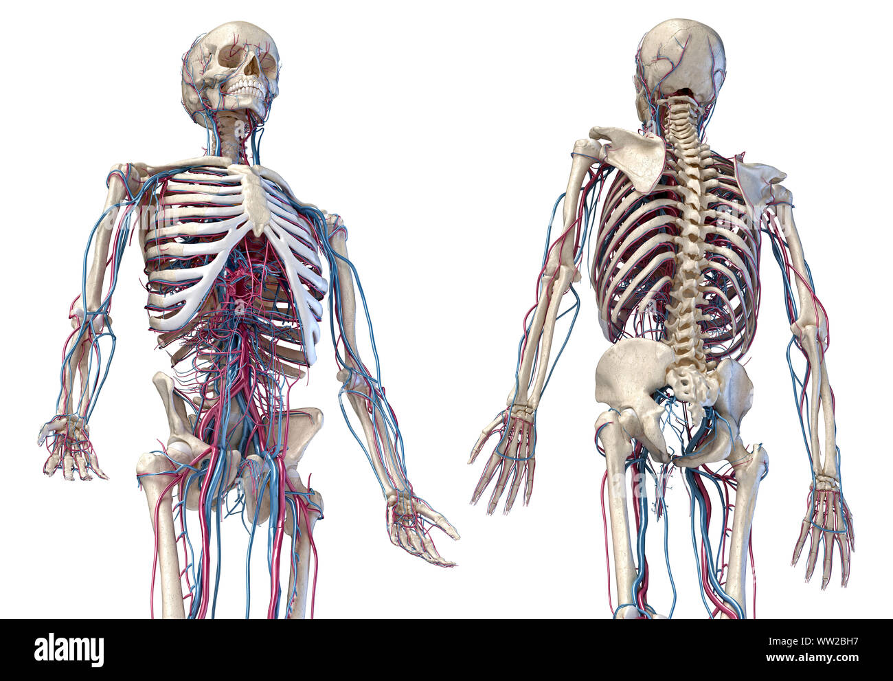 Human anatomy, 3d illustration of the skeleton with cardiovascular system. Perspective view of 3/4 upper part, front and back sides. On white backgrou Stock Photohttps://www.alamy.com/image-license-details/?v=1https://www.alamy.com/human-anatomy-3d-illustration-of-the-skeleton-with-cardiovascular-system-perspective-view-of-34-upper-part-front-and-back-sides-on-white-backgrou-image273574931.html
RFWW2BH7–Human anatomy, 3d illustration of the skeleton with cardiovascular system. Perspective view of 3/4 upper part, front and back sides. On white backgrou
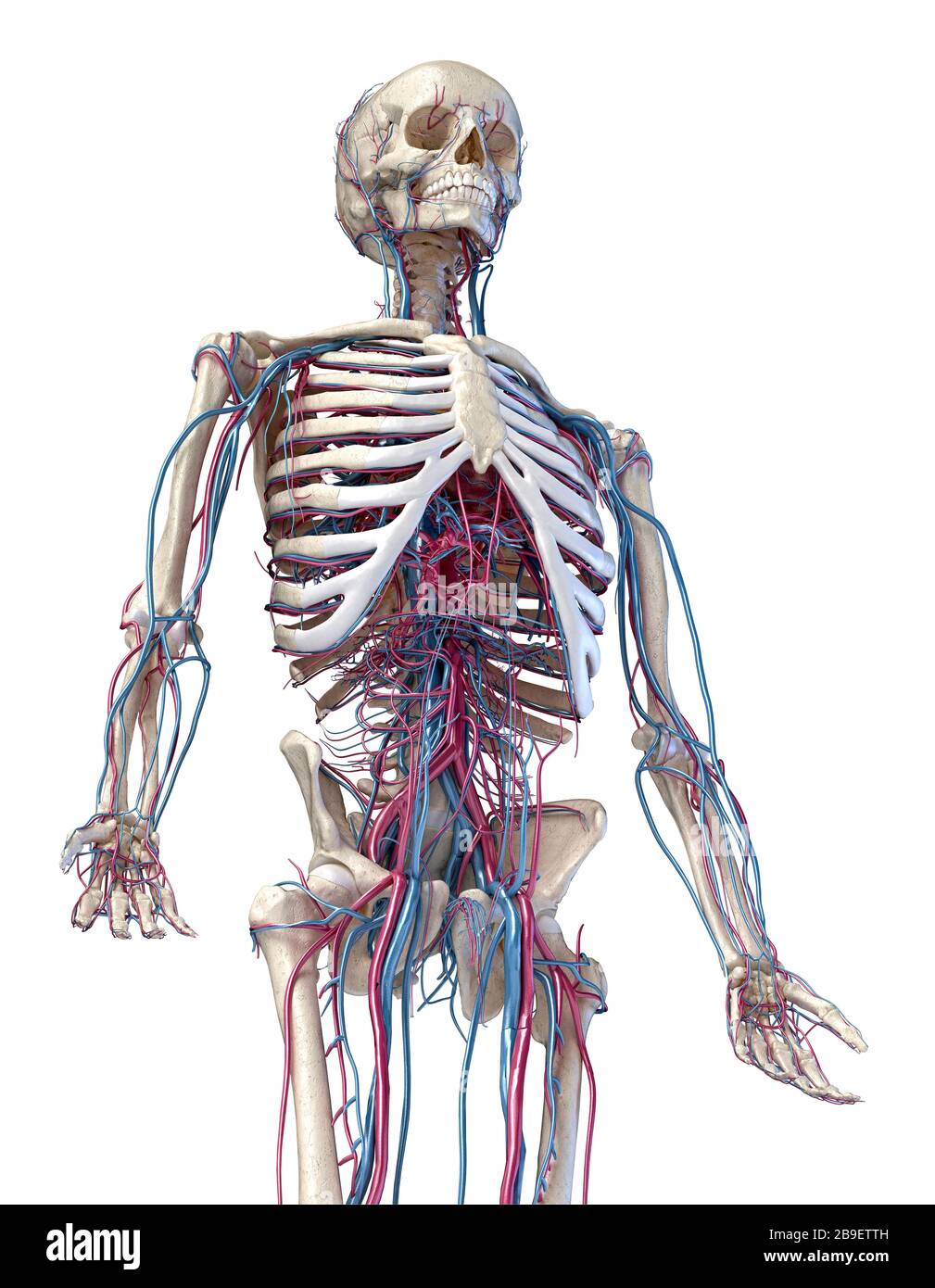 3/4 upper body front view of human skeletal and vascular systems, white background. Stock Photohttps://www.alamy.com/image-license-details/?v=1https://www.alamy.com/34-upper-body-front-view-of-human-skeletal-and-vascular-systems-white-background-image350066097.html
RF2B9ETTH–3/4 upper body front view of human skeletal and vascular systems, white background.
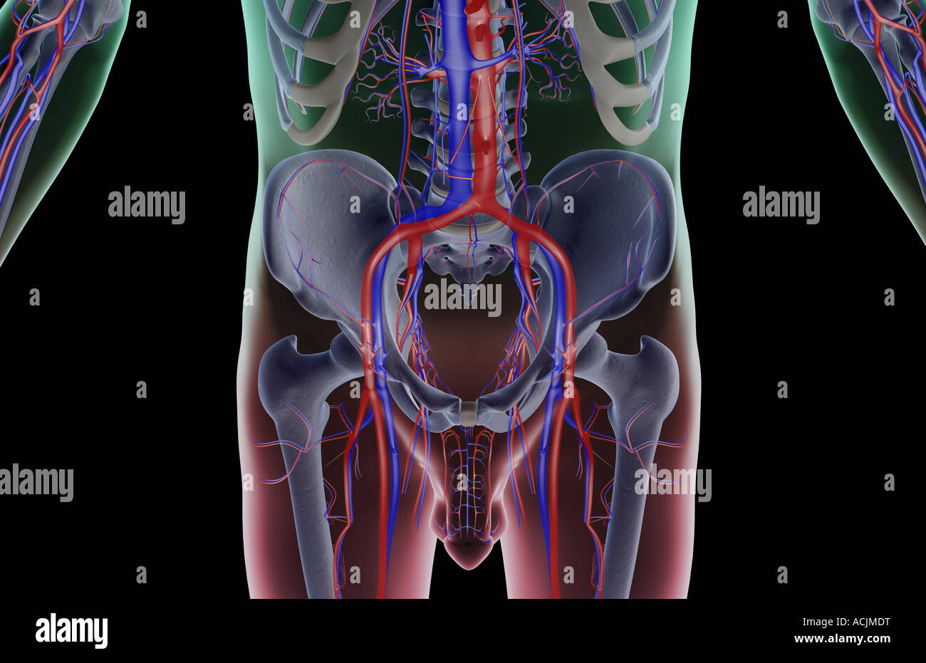 The blood supply of the lower body Stock Photohttps://www.alamy.com/image-license-details/?v=1https://www.alamy.com/stock-photo-the-blood-supply-of-the-lower-body-13171811.html
RFACJMDT–The blood supply of the lower body
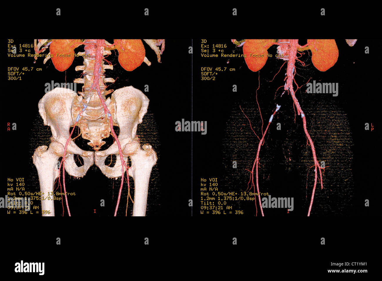 ILIAC THROMBOSIS, 3D SCAN Stock Photohttps://www.alamy.com/image-license-details/?v=1https://www.alamy.com/stock-photo-iliac-thrombosis-3d-scan-49216161.html
RMCT1YM1–ILIAC THROMBOSIS, 3D SCAN
 A text-book of clinical anatomy : for students and practitioners . Fig. 88.—Cross-section of pelvis at level of sacro-iliac joints. S, Sacrum. I, Ilium.SC, Iliac colon (sigmoid flexure). C, Cecum, and beginning of ascending colon, i,Placed to inner side of each sacro-iliac joint. 2, Ureters. 3, Mesosigmoid, showing greatlength of latter. 4, Pelvic colon not cut, seen from above. 5, Omentum. 6, Externaliliac artery. 7, External iliac vein. 8, Iliopsoas muscle. The white dot above it is theanterior crural nerve. 9, 10, and 11, Gluteal muscles. 283. Fig. 89.—Cross-section of female pelvis at leve Stock Photohttps://www.alamy.com/image-license-details/?v=1https://www.alamy.com/a-text-book-of-clinical-anatomy-for-students-and-practitioners-fig-88cross-section-of-pelvis-at-level-of-sacro-iliac-joints-s-sacrum-i-iliumsc-iliac-colon-sigmoid-flexure-c-cecum-and-beginning-of-ascending-colon-iplaced-to-inner-side-of-each-sacro-iliac-joint-2-ureters-3-mesosigmoid-showing-greatlength-of-latter-4-pelvic-colon-not-cut-seen-from-above-5-omentum-6-externaliliac-artery-7-external-iliac-vein-8-iliopsoas-muscle-the-white-dot-above-it-is-theanterior-crural-nerve-9-10-and-11-gluteal-muscles-283-fig-89cross-section-of-female-pelvis-at-leve-image340214921.html
RM2ANE3GW–A text-book of clinical anatomy : for students and practitioners . Fig. 88.—Cross-section of pelvis at level of sacro-iliac joints. S, Sacrum. I, Ilium.SC, Iliac colon (sigmoid flexure). C, Cecum, and beginning of ascending colon, i,Placed to inner side of each sacro-iliac joint. 2, Ureters. 3, Mesosigmoid, showing greatlength of latter. 4, Pelvic colon not cut, seen from above. 5, Omentum. 6, Externaliliac artery. 7, External iliac vein. 8, Iliopsoas muscle. The white dot above it is theanterior crural nerve. 9, 10, and 11, Gluteal muscles. 283. Fig. 89.—Cross-section of female pelvis at leve
 Female cardiovascular system, artwork Stock Photohttps://www.alamy.com/image-license-details/?v=1https://www.alamy.com/stock-photo-female-cardiovascular-system-artwork-55447842.html
RFD65T82–Female cardiovascular system, artwork
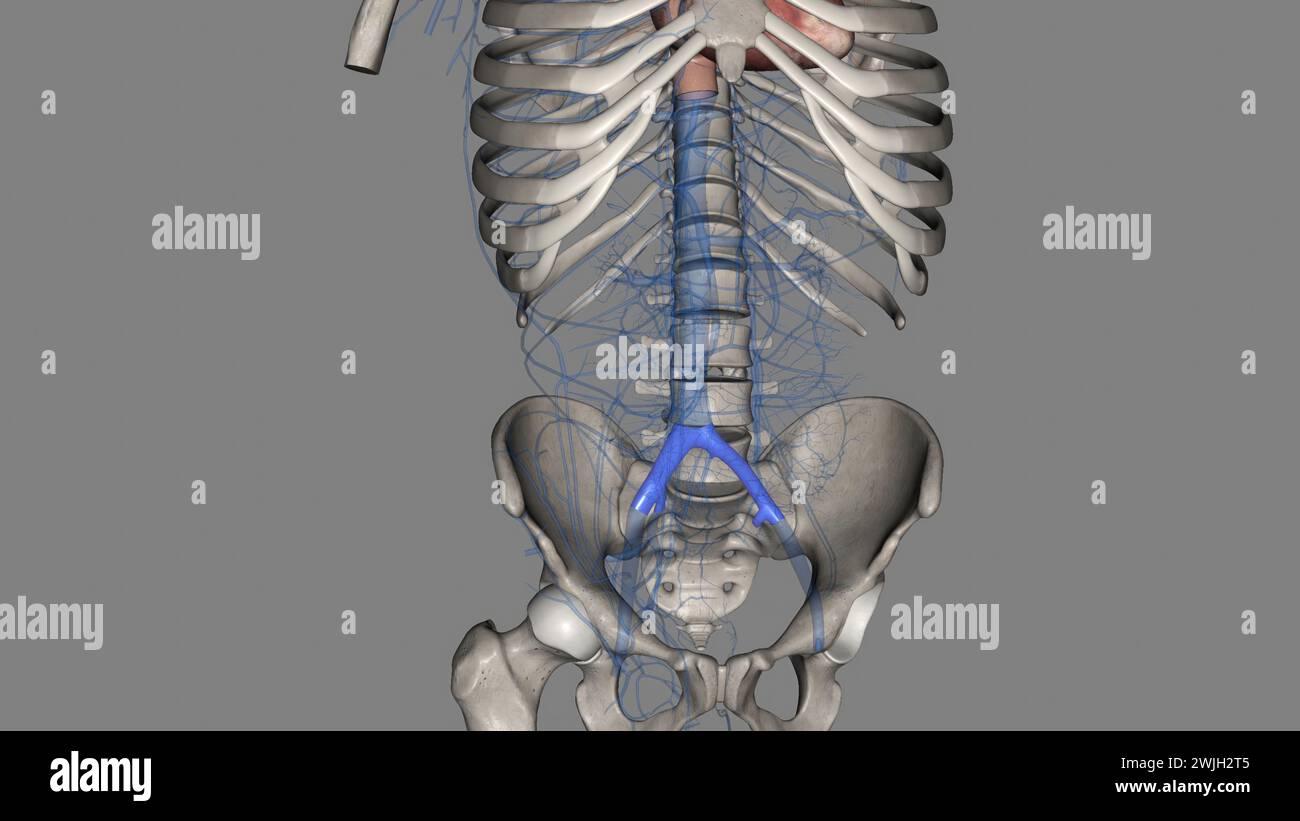 The common iliac vein is formed by the unification of the internal and external iliac veins 3d illustration Stock Photohttps://www.alamy.com/image-license-details/?v=1https://www.alamy.com/the-common-iliac-vein-is-formed-by-the-unification-of-the-internal-and-external-iliac-veins-3d-illustration-image596591749.html
RF2WJH2T5–The common iliac vein is formed by the unification of the internal and external iliac veins 3d illustration
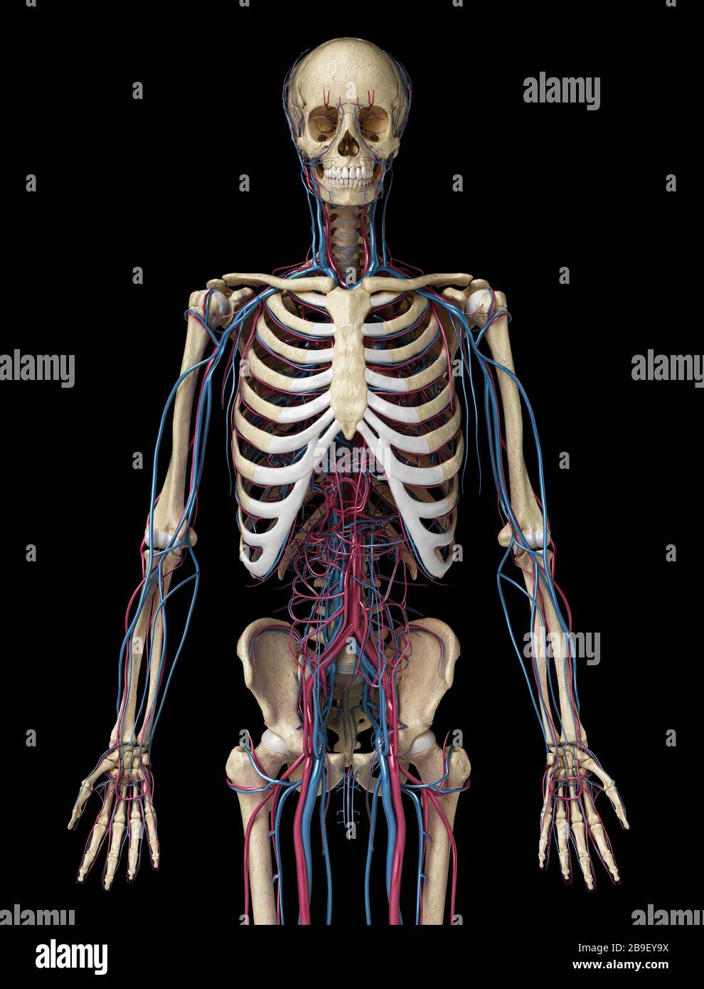 Upper body front view of human skeleton with veins and arteries, black background. Stock Photohttps://www.alamy.com/image-license-details/?v=1https://www.alamy.com/upper-body-front-view-of-human-skeleton-with-veins-and-arteries-black-background-image350068038.html
RF2B9EY9X–Upper body front view of human skeleton with veins and arteries, black background.
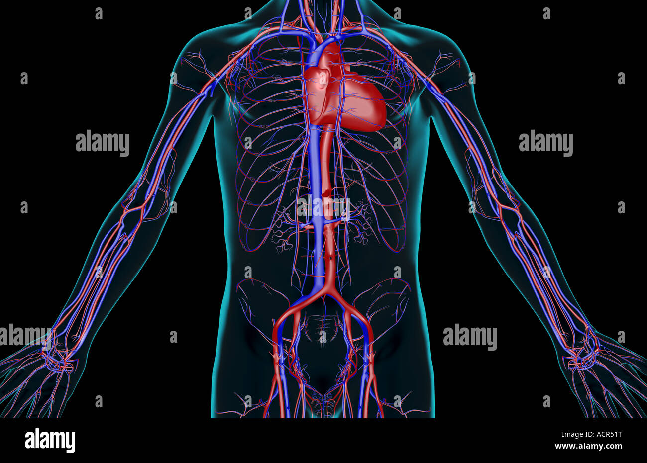 The blood supply of the trunk Stock Photohttps://www.alamy.com/image-license-details/?v=1https://www.alamy.com/stock-photo-the-blood-supply-of-the-trunk-13213667.html
RFACR51T–The blood supply of the trunk
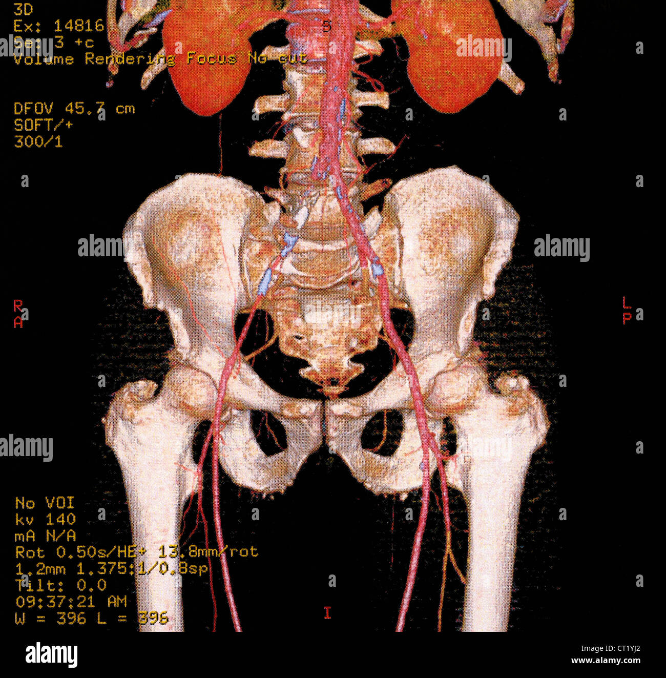 ILIAC THROMBOSIS, 3D SCAN Stock Photohttps://www.alamy.com/image-license-details/?v=1https://www.alamy.com/stock-photo-iliac-thrombosis-3d-scan-49216106.html
RMCT1YJ2–ILIAC THROMBOSIS, 3D SCAN
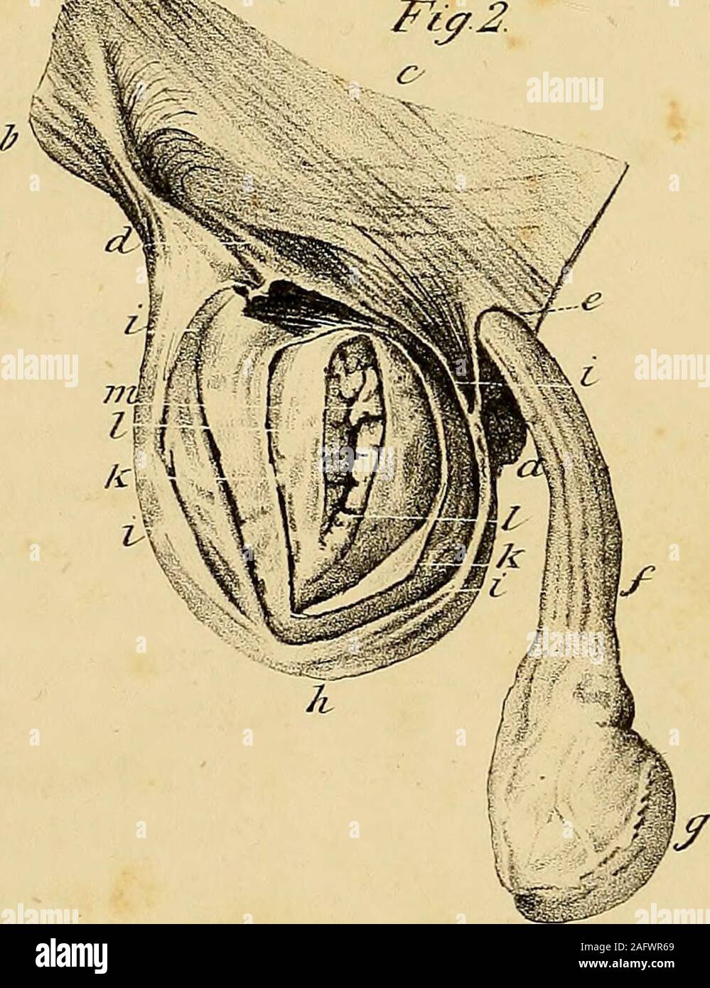 . The anatomy and surgical treatment of abdominal hernia. ?X 7*FreneA,, t£eZ StftcZcLLTS XtOt- EXPLANATION OF PLATE XVL 405 Fig= 3. Posterior view of figure 1. a. Symphysis pubis. b. Spinous process of the ilium. c. Ilium cut through. d d d. Rectus and other abdominal muscles, e. Linea semilunaris. /. Posterior edge of the crural arch. g. Fascia iliaca. h. The iliacus internus muscle. i i. Fascia transversalis. k. Internal abdominal ring. Z. Spermatic cord passing through the internal ring. m. External iliac artery. n. External iliac vein, o. Epigastric artery and vein. p. Sac of crural hernia Stock Photohttps://www.alamy.com/image-license-details/?v=1https://www.alamy.com/the-anatomy-and-surgical-treatment-of-abdominal-hernia-x-7frenea-tez-stftczcllts-xtot-explanation-of-plate-xvl-405-fig=-3-posterior-view-of-figure-1-a-symphysis-pubis-b-spinous-process-of-the-ilium-c-ilium-cut-through-d-d-d-rectus-and-other-abdominal-muscles-e-linea-semilunaris-posterior-edge-of-the-crural-arch-g-fascia-iliaca-h-the-iliacus-internus-muscle-i-i-fascia-transversalis-k-internal-abdominal-ring-z-spermatic-cord-passing-through-the-internal-ring-m-external-iliac-artery-n-external-iliac-vein-o-epigastric-artery-and-vein-p-sac-of-crural-hernia-image336783841.html
RM2AFWR69–. The anatomy and surgical treatment of abdominal hernia. ?X 7*FreneA,, t£eZ StftcZcLLTS XtOt- EXPLANATION OF PLATE XVL 405 Fig= 3. Posterior view of figure 1. a. Symphysis pubis. b. Spinous process of the ilium. c. Ilium cut through. d d d. Rectus and other abdominal muscles, e. Linea semilunaris. /. Posterior edge of the crural arch. g. Fascia iliaca. h. The iliacus internus muscle. i i. Fascia transversalis. k. Internal abdominal ring. Z. Spermatic cord passing through the internal ring. m. External iliac artery. n. External iliac vein, o. Epigastric artery and vein. p. Sac of crural hernia
 Pelvis anatomy, artwork Stock Photohttps://www.alamy.com/image-license-details/?v=1https://www.alamy.com/stock-photo-pelvis-anatomy-artwork-55416055.html
RFD64BMR–Pelvis anatomy, artwork
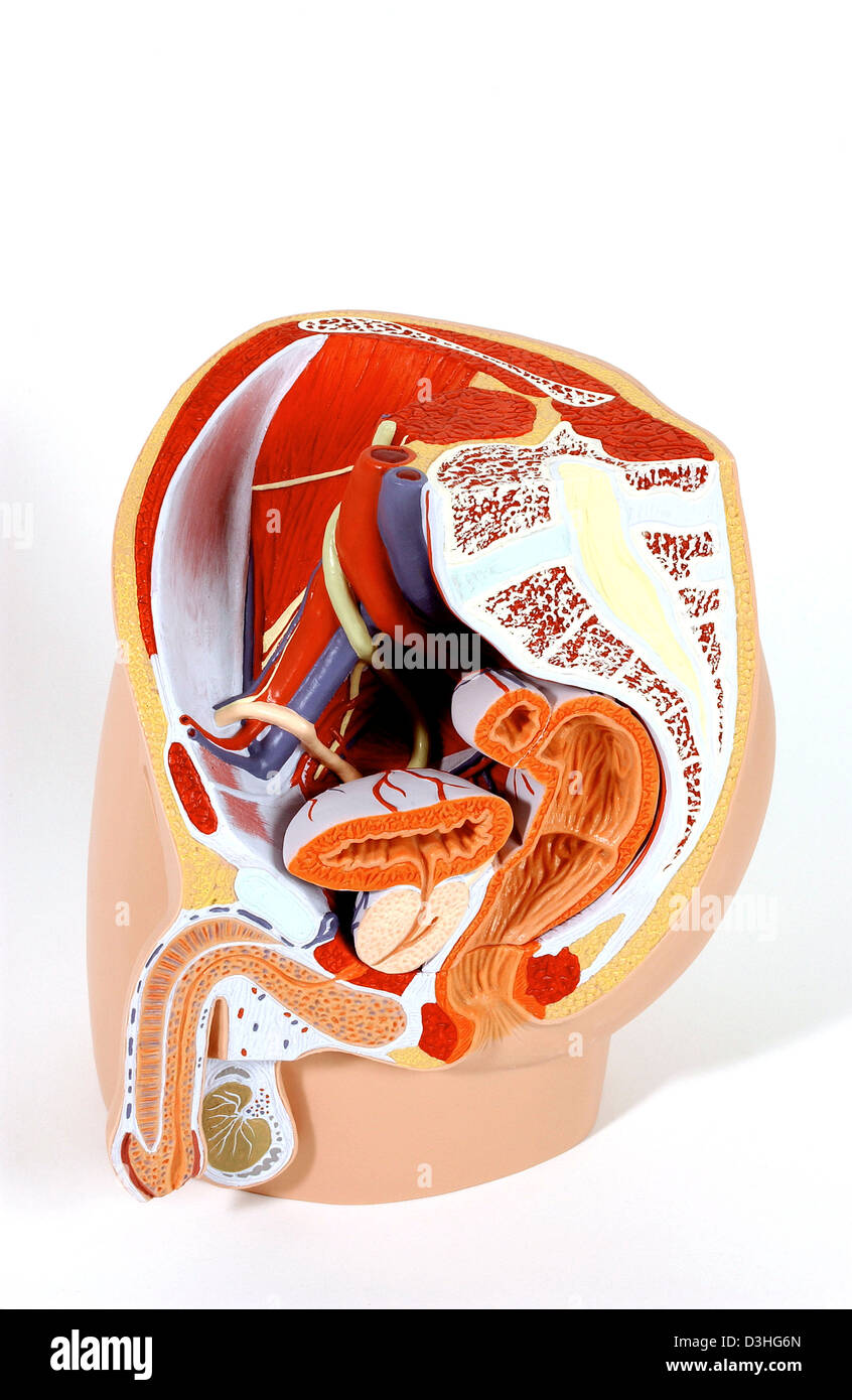 ANATOMY, MALE GENITALIA Stock Photohttps://www.alamy.com/image-license-details/?v=1https://www.alamy.com/stock-photo-anatomy-male-genitalia-53860989.html
RMD3HG6N–ANATOMY, MALE GENITALIA
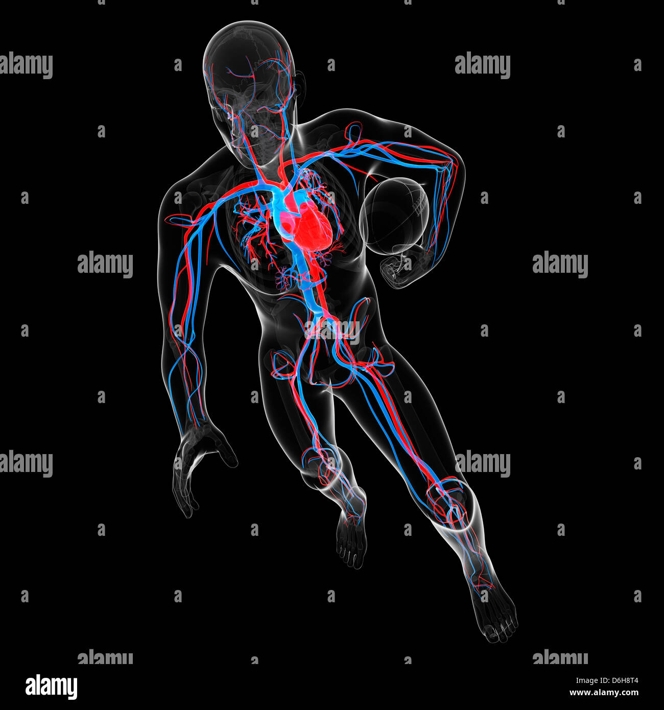 Cardiovascular system, artwork Stock Photohttps://www.alamy.com/image-license-details/?v=1https://www.alamy.com/stock-photo-cardiovascular-system-artwork-55699172.html
RFD6H8T4–Cardiovascular system, artwork
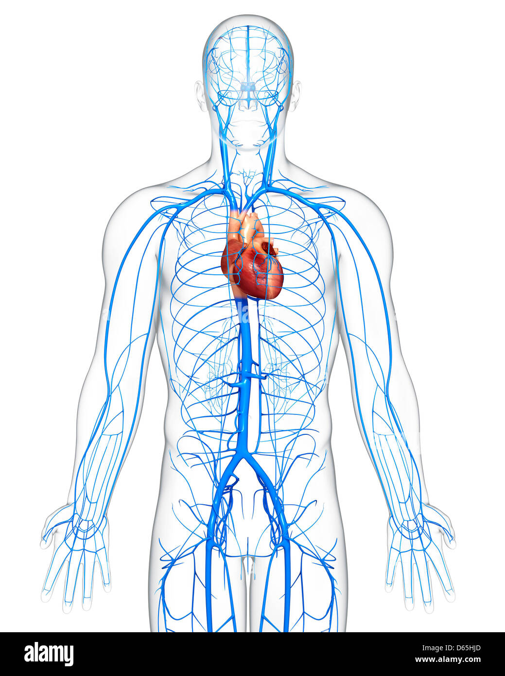 Human veins, artwork Stock Photohttps://www.alamy.com/image-license-details/?v=1https://www.alamy.com/stock-photo-human-veins-artwork-55442645.html
RFD65HJD–Human veins, artwork
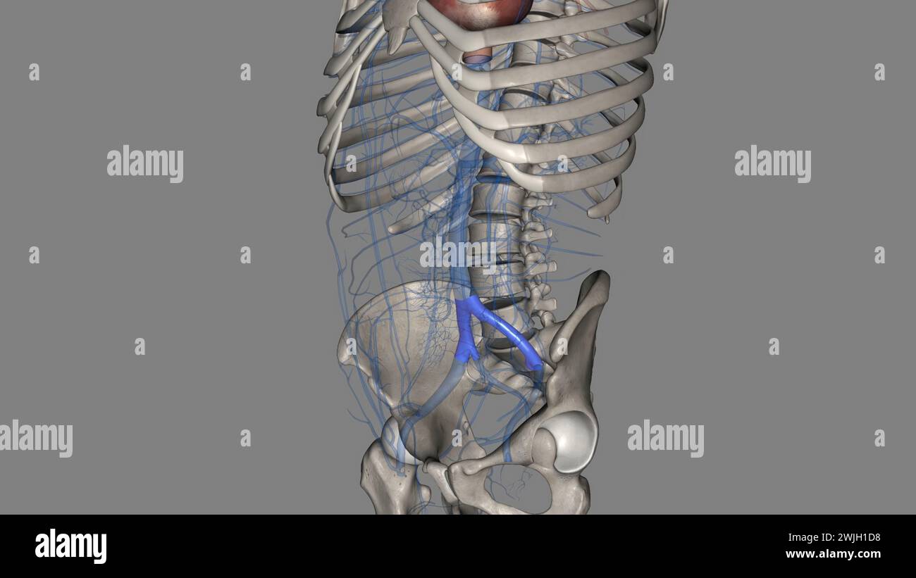 The common iliac vein is formed by the unification of the internal and external iliac veins 3d illustration Stock Photohttps://www.alamy.com/image-license-details/?v=1https://www.alamy.com/the-common-iliac-vein-is-formed-by-the-unification-of-the-internal-and-external-iliac-veins-3d-illustration-image596590660.html
RF2WJH1D8–The common iliac vein is formed by the unification of the internal and external iliac veins 3d illustration
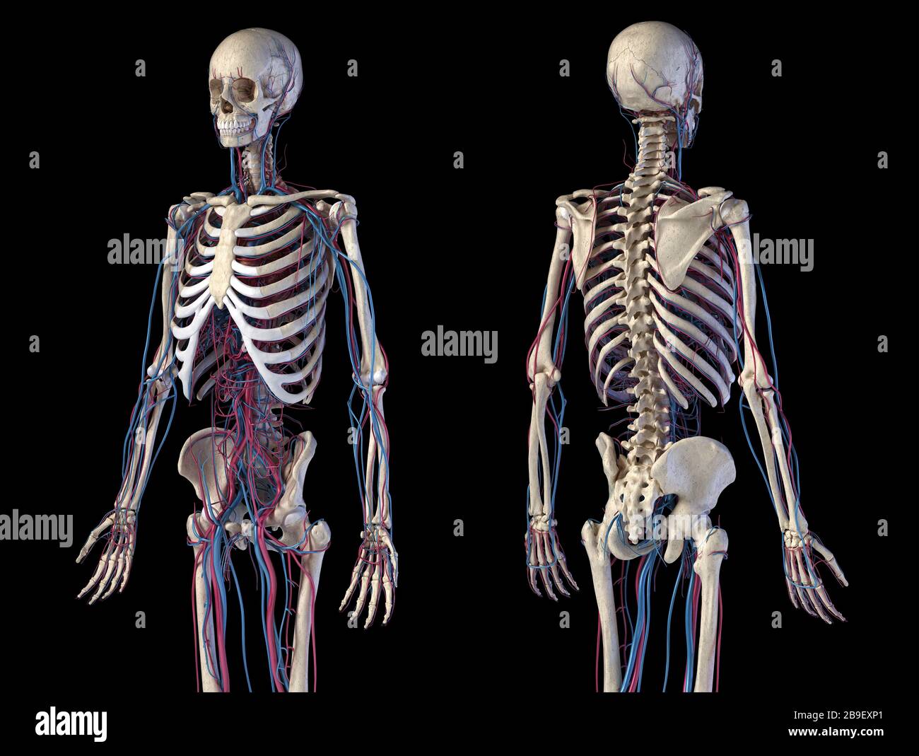 Front and back view of human skeletal and cardiovascular systems, black background. Stock Photohttps://www.alamy.com/image-license-details/?v=1https://www.alamy.com/front-and-back-view-of-human-skeletal-and-cardiovascular-systems-black-background-image350067593.html
RF2B9EXP1–Front and back view of human skeletal and cardiovascular systems, black background.
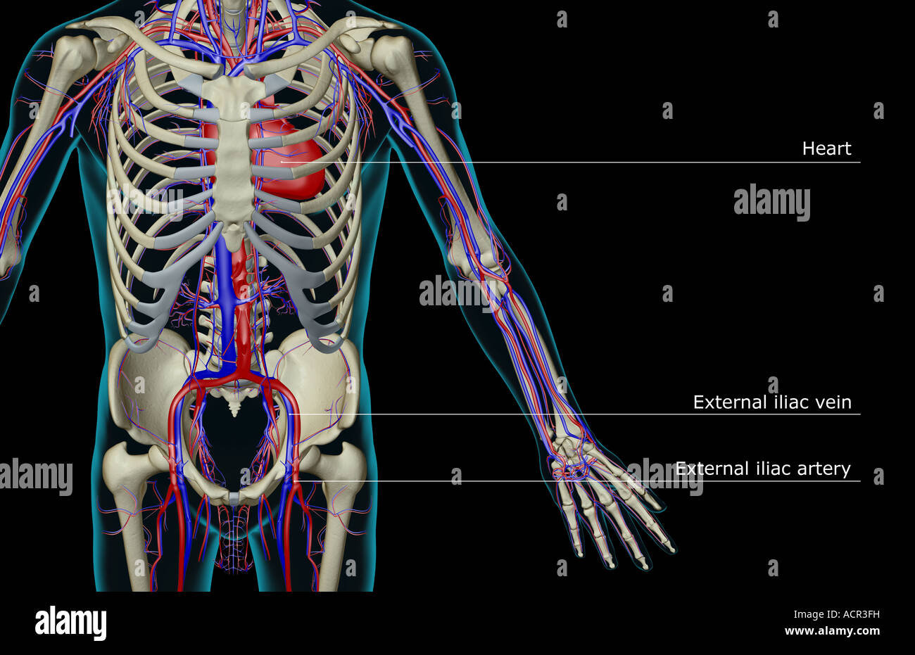 The blood supply of the trunk Stock Photohttps://www.alamy.com/image-license-details/?v=1https://www.alamy.com/stock-photo-the-blood-supply-of-the-trunk-13213156.html
RFACR3FH–The blood supply of the trunk
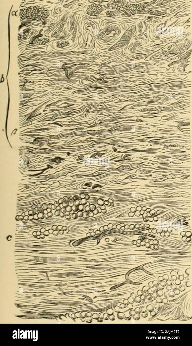 A system of gynecology . le; d, internal obturator muscle; c, e, psoasmuscle; /, linea alba ; ;/. r;. ureters; ft, obturator nerve ; i, internal inguinal ring: 1, abdom-inal aorta: 2, inferior mesenteric artery ; 3,3, common iliac arteries : 1. external iliac artery;5,vena cava; 6, renal veins; 7,7, common iliac veins; 8, external iliac vein: 9, internaliliac artery: 10, gluteal; 11, ileo-lumbar; 12, sciatic; 18, pudic; it. obturator: IS, epigastricveins; 17. uterine veins; is, vagino-vesical venous cete; 19, spermatic veins: 20, bulb ofovary; 21, vein to round ligament; JJ, Fallopian veins. n Stock Photohttps://www.alamy.com/image-license-details/?v=1https://www.alamy.com/a-system-of-gynecology-le-d-internal-obturator-muscle-c-e-psoasmuscle-linea-alba-r-ureters-ft-obturator-nerve-i-internal-inguinal-ring-1-abdom-inal-aorta-2-inferior-mesenteric-artery-33-common-iliac-arteries-1-external-iliac-artery5vena-cava-6-renal-veins-77-common-iliac-veins-8-external-iliac-vein-9-internaliliac-artery-10-gluteal-11-ileo-lumbar-12-sciatic-18-pudic-it-obturator-is-epigastricveins-17-uterine-veins-is-vagino-vesical-venous-cete-19-spermatic-veins-20-bulb-ofovary-21-vein-to-round-ligament-jj-fallopian-veins-n-image338502089.html
RM2AJM2T9–A system of gynecology . le; d, internal obturator muscle; c, e, psoasmuscle; /, linea alba ; ;/. r;. ureters; ft, obturator nerve ; i, internal inguinal ring: 1, abdom-inal aorta: 2, inferior mesenteric artery ; 3,3, common iliac arteries : 1. external iliac artery;5,vena cava; 6, renal veins; 7,7, common iliac veins; 8, external iliac vein: 9, internaliliac artery: 10, gluteal; 11, ileo-lumbar; 12, sciatic; 18, pudic; it. obturator: IS, epigastricveins; 17. uterine veins; is, vagino-vesical venous cete; 19, spermatic veins: 20, bulb ofovary; 21, vein to round ligament; JJ, Fallopian veins. n
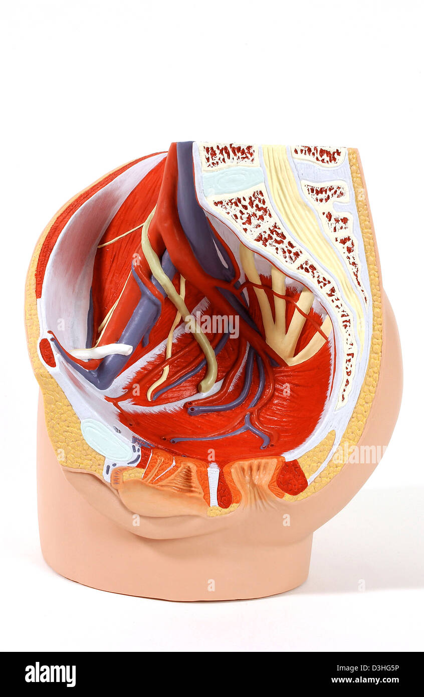 ANATOMY, FEMALE GENITALIA Stock Photohttps://www.alamy.com/image-license-details/?v=1https://www.alamy.com/stock-photo-anatomy-female-genitalia-53860962.html
RMD3HG5P–ANATOMY, FEMALE GENITALIA
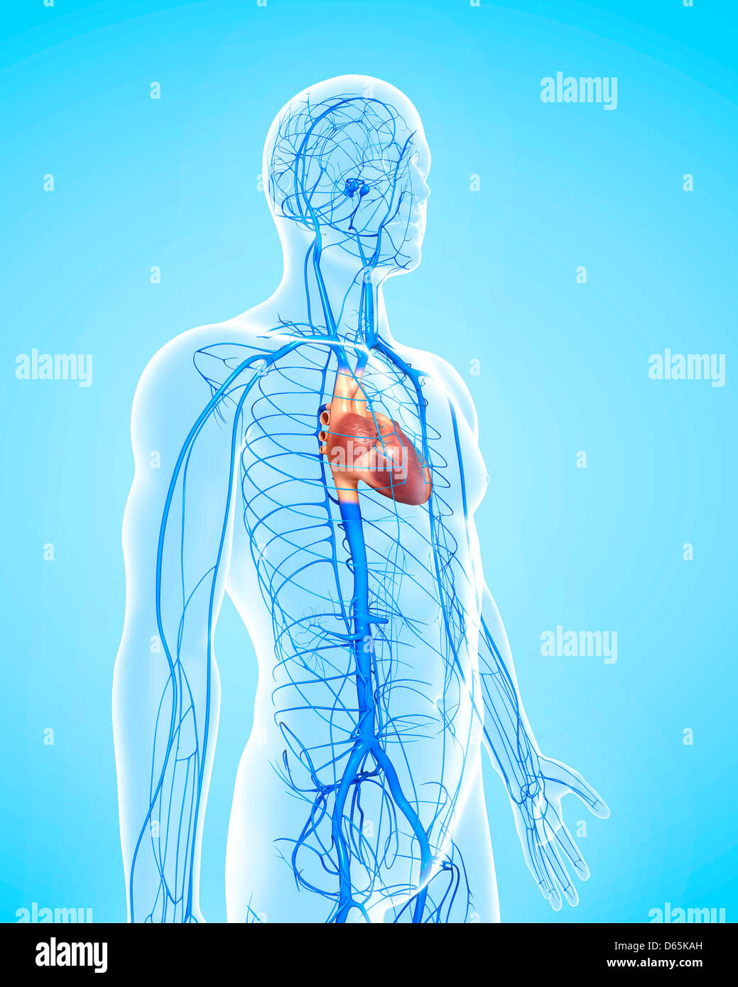 Human veins, artwork Stock Photohttps://www.alamy.com/image-license-details/?v=1https://www.alamy.com/stock-photo-human-veins-artwork-55443993.html
RFD65KAH–Human veins, artwork
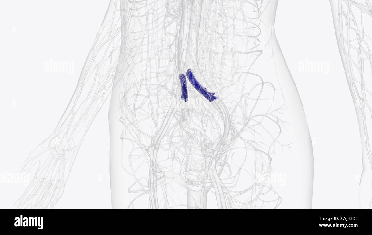 The common iliac vein is formed by the unification of the internal and external iliac veins 3d illustration Stock Photohttps://www.alamy.com/image-license-details/?v=1https://www.alamy.com/the-common-iliac-vein-is-formed-by-the-unification-of-the-internal-and-external-iliac-veins-3d-illustration-image596592225.html
RF2WJH3D5–The common iliac vein is formed by the unification of the internal and external iliac veins 3d illustration
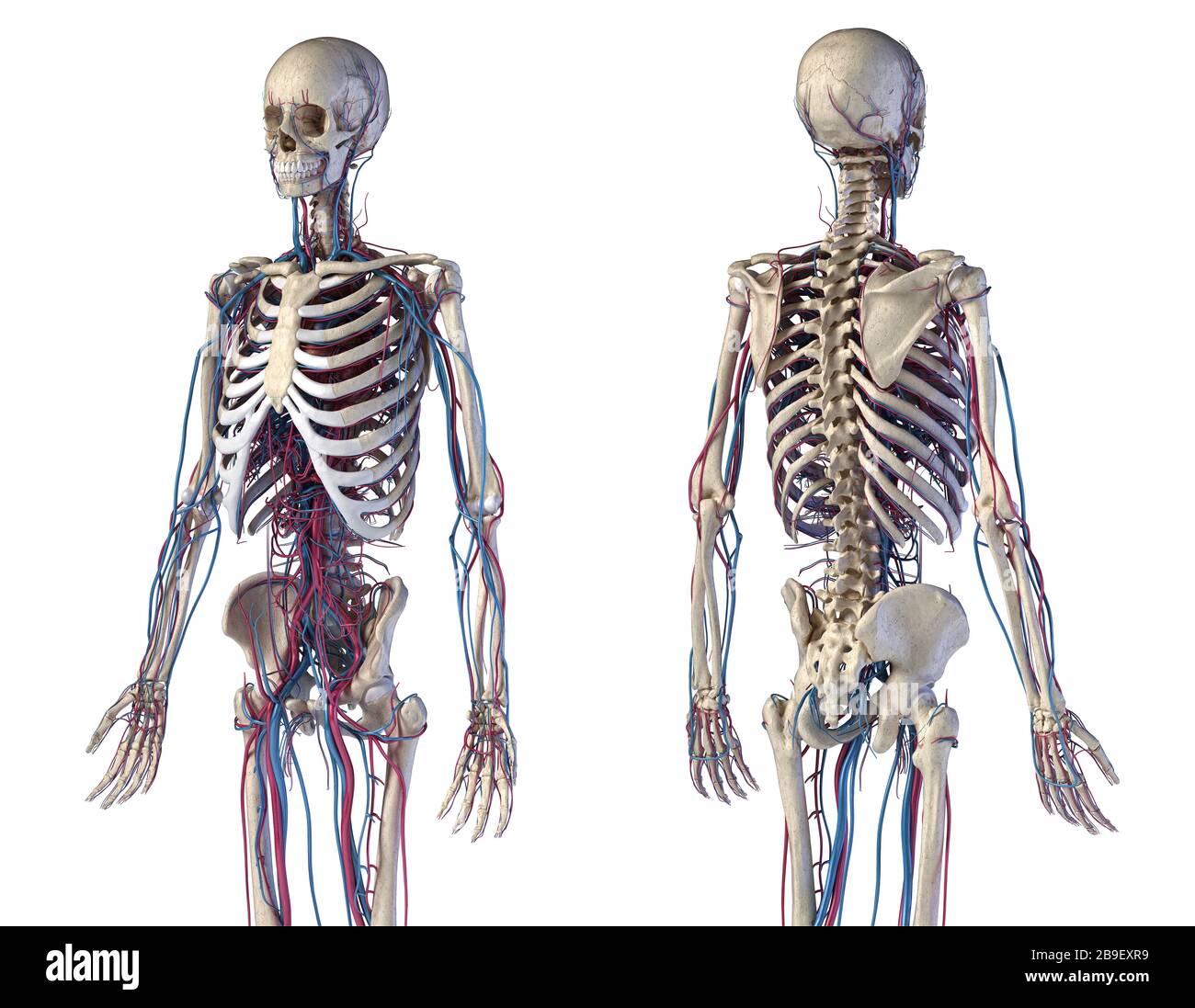 Front and back view of human skeletal and cardiovascular systems, white background. Stock Photohttps://www.alamy.com/image-license-details/?v=1https://www.alamy.com/front-and-back-view-of-human-skeletal-and-cardiovascular-systems-white-background-image350067629.html
RF2B9EXR9–Front and back view of human skeletal and cardiovascular systems, white background.
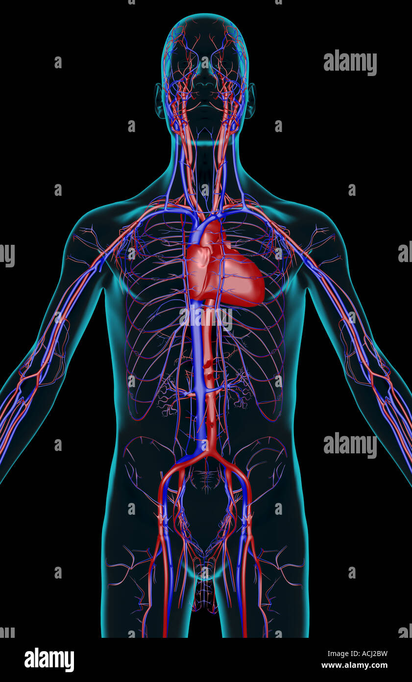 The blood supply of the upper body Stock Photohttps://www.alamy.com/image-license-details/?v=1https://www.alamy.com/stock-photo-the-blood-supply-of-the-upper-body-13165740.html
RFACJ2BW–The blood supply of the upper body
 . The anatomy and surgical treatment of hernia. / .4% , -V^ 7 5M»1. OPERATIVE MEASURES FOR STRANGULATED FEMORAL HERNLA. 141 /, k. Fascia propria of the sac laid open. h. The iliacus internus muscle. /, /. Hernial sac opened. /, /. Fascia transversalis. in. Omentum seen within the sac. k. Internal abdominal ring. /. Spermatic cord passing through the inter-nal rina;. Figure Posterior view of Fig. i. m. External iliac artery. a. Symphysis pubis. «. External iliac vein. b. Spinous process of the ilium. o. Epigastric artery and vein. c. Ilium cut through. /. Sac of the crural hernia.(/, d, d. Re Stock Photohttps://www.alamy.com/image-license-details/?v=1https://www.alamy.com/the-anatomy-and-surgical-treatment-of-hernia-4-v-7-5m1-operative-measures-for-strangulated-femoral-hernla-141-k-fascia-propria-of-the-sac-laid-open-h-the-iliacus-internus-muscle-hernial-sac-opened-fascia-transversalis-in-omentum-seen-within-the-sac-k-internal-abdominal-ring-spermatic-cord-passing-through-the-inter-nal-rina-figure-posterior-view-of-fig-i-m-external-iliac-artery-a-symphysis-pubis-external-iliac-vein-b-spinous-process-of-the-ilium-o-epigastric-artery-and-vein-c-ilium-cut-through-sac-of-the-crural-hernia-d-d-re-image370331375.html
RM2CEE1CF–. The anatomy and surgical treatment of hernia. / .4% , -V^ 7 5M»1. OPERATIVE MEASURES FOR STRANGULATED FEMORAL HERNLA. 141 /, k. Fascia propria of the sac laid open. h. The iliacus internus muscle. /, /. Hernial sac opened. /, /. Fascia transversalis. in. Omentum seen within the sac. k. Internal abdominal ring. /. Spermatic cord passing through the inter-nal rina;. Figure Posterior view of Fig. i. m. External iliac artery. a. Symphysis pubis. «. External iliac vein. b. Spinous process of the ilium. o. Epigastric artery and vein. c. Ilium cut through. /. Sac of the crural hernia.(/, d, d. Re
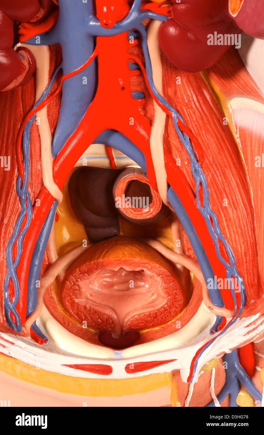 ANATOMY, PELVIS Stock Photohttps://www.alamy.com/image-license-details/?v=1https://www.alamy.com/stock-photo-anatomy-pelvis-53861004.html
RMD3HG78–ANATOMY, PELVIS
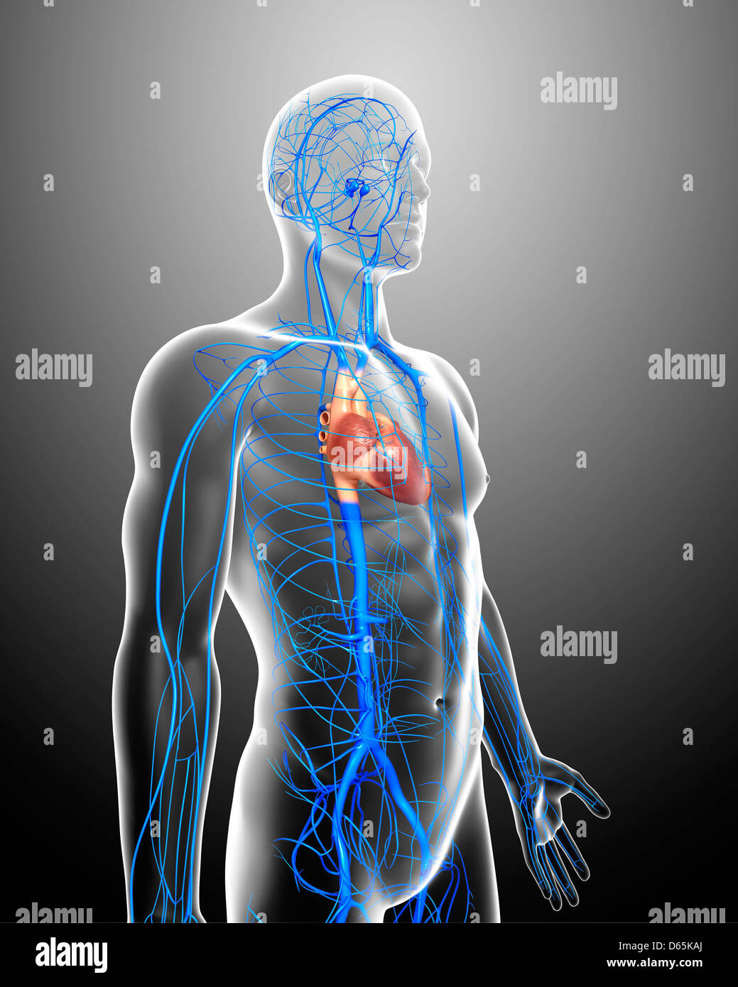 Human veins, artwork Stock Photohttps://www.alamy.com/image-license-details/?v=1https://www.alamy.com/stock-photo-human-veins-artwork-55443994.html
RFD65KAJ–Human veins, artwork
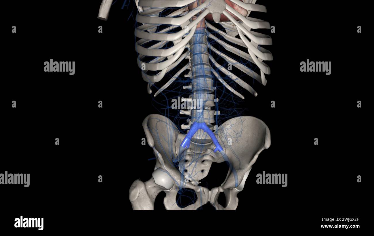 The common iliac vein is formed by the unification of the internal and external iliac veins 3d illustration Stock Photohttps://www.alamy.com/image-license-details/?v=1https://www.alamy.com/the-common-iliac-vein-is-formed-by-the-unification-of-the-internal-and-external-iliac-veins-3d-illustration-image596588009.html
RF2WJGX2H–The common iliac vein is formed by the unification of the internal and external iliac veins 3d illustration
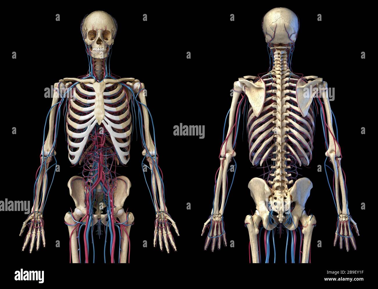 Front and back view of human skeleton with veins and arteries, black background. Stock Photohttps://www.alamy.com/image-license-details/?v=1https://www.alamy.com/front-and-back-view-of-human-skeleton-with-veins-and-arteries-black-background-image350067803.html
RF2B9EY1F–Front and back view of human skeleton with veins and arteries, black background.
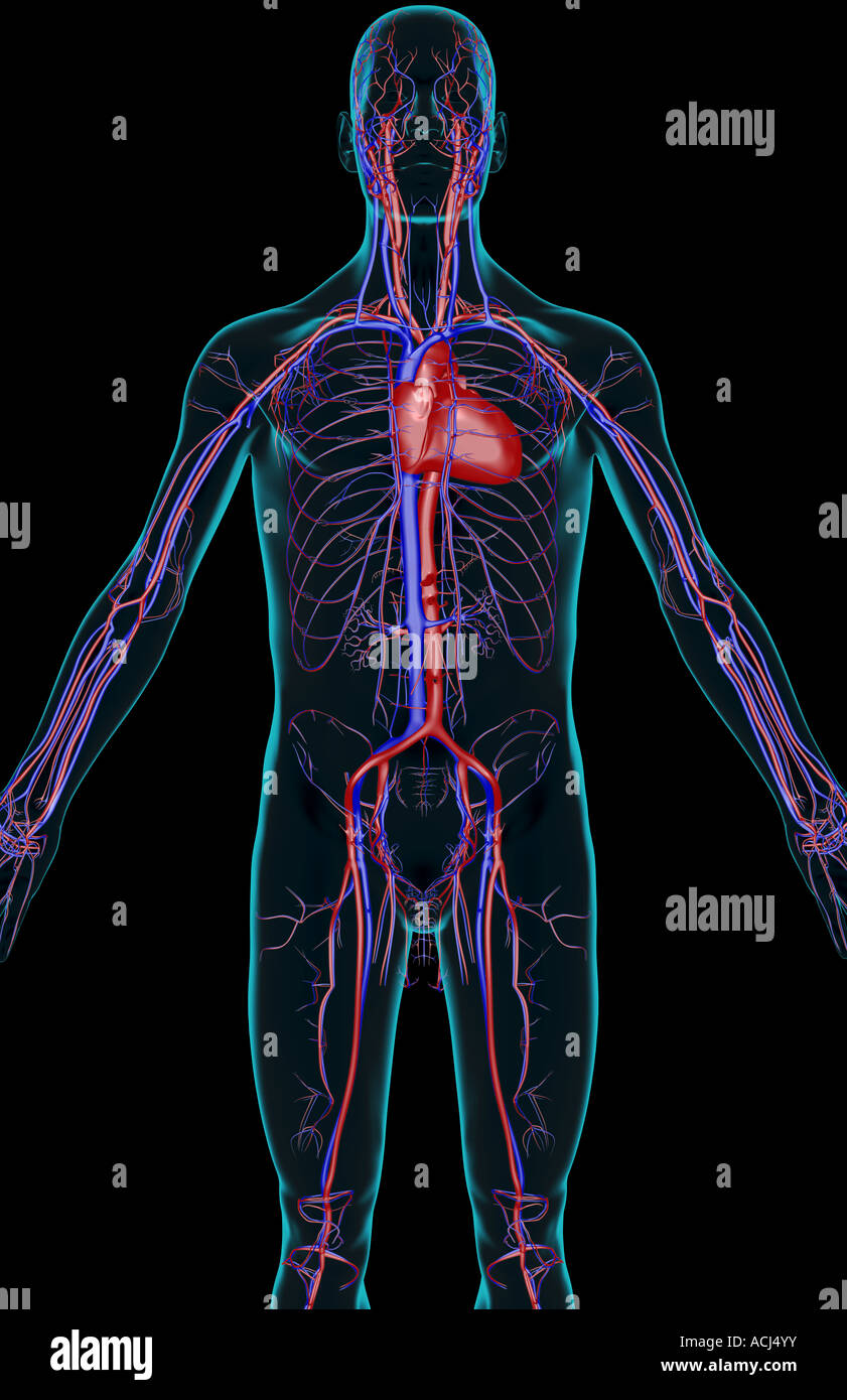 The blood supply of the upper body Stock Photohttps://www.alamy.com/image-license-details/?v=1https://www.alamy.com/stock-photo-the-blood-supply-of-the-upper-body-13166606.html
RFACJ4YY–The blood supply of the upper body
 . Anatomy, descriptive and applied. Anatomy. THE BEEP VETNS OF THE LOWER EXTREMITY 74.3 and joins the external iliac vein about three-quarters of an inch above Poupart's ligament. The pubic vein communicates with the obturator em in the obturator fora- men, and ascends on the back of the pubis to terminate in the external iliac -ein. The internal iliac vein {v. hypoc/astrica) commences near the upper part of the great sacrosciatic foramen, passes upward behind and slightly to the inner side of the internal iliac artery, and at the brim of the pelvis joins with the external iliac to form the Stock Photohttps://www.alamy.com/image-license-details/?v=1https://www.alamy.com/anatomy-descriptive-and-applied-anatomy-the-beep-vetns-of-the-lower-extremity-743-and-joins-the-external-iliac-vein-about-three-quarters-of-an-inch-above-pouparts-ligament-the-pubic-vein-communicates-with-the-obturator-em-in-the-obturator-fora-men-and-ascends-on-the-back-of-the-pubis-to-terminate-in-the-external-iliac-ein-the-internal-iliac-vein-v-hypocastrica-commences-near-the-upper-part-of-the-great-sacrosciatic-foramen-passes-upward-behind-and-slightly-to-the-inner-side-of-the-internal-iliac-artery-and-at-the-brim-of-the-pelvis-joins-with-the-external-iliac-to-form-the-image236773020.html
RMRN5XB8–. Anatomy, descriptive and applied. Anatomy. THE BEEP VETNS OF THE LOWER EXTREMITY 74.3 and joins the external iliac vein about three-quarters of an inch above Poupart's ligament. The pubic vein communicates with the obturator em in the obturator fora- men, and ascends on the back of the pubis to terminate in the external iliac -ein. The internal iliac vein {v. hypoc/astrica) commences near the upper part of the great sacrosciatic foramen, passes upward behind and slightly to the inner side of the internal iliac artery, and at the brim of the pelvis joins with the external iliac to form the