Quick filters:
Maxillary jaw Stock Photos and Images
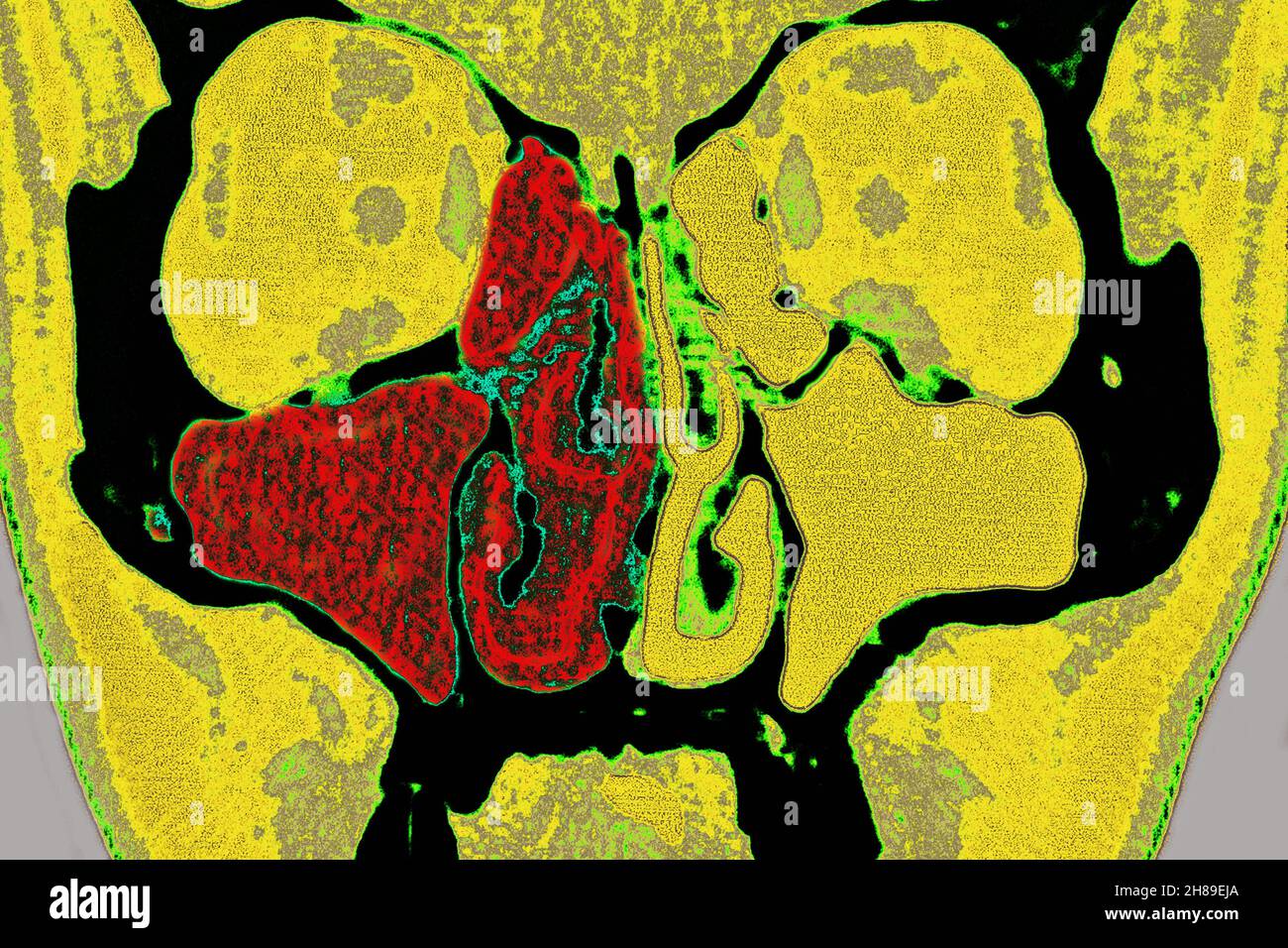 Sinusitis Stock Photohttps://www.alamy.com/image-license-details/?v=1https://www.alamy.com/sinusitis-image452595874.html
RM2H89EJA–Sinusitis
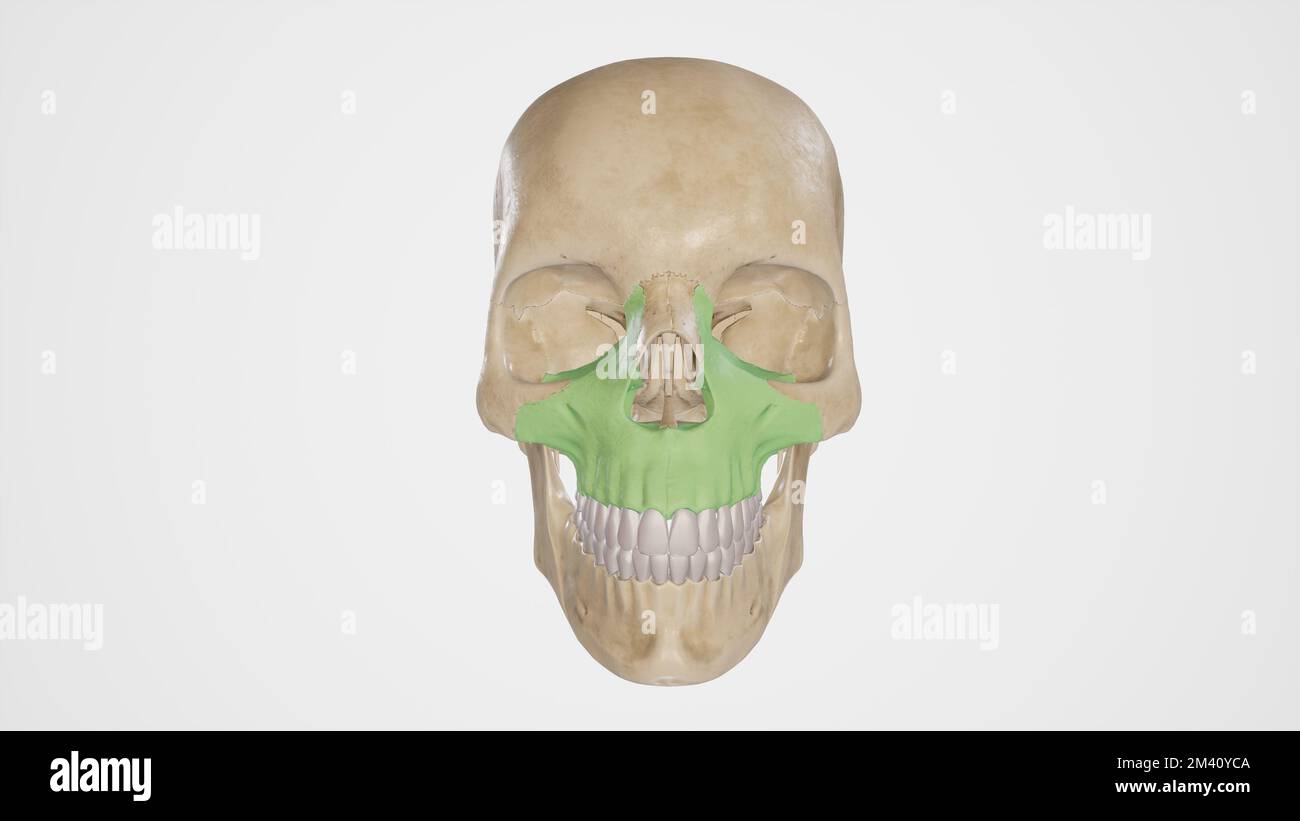 Maxillary Bone Stock Photohttps://www.alamy.com/image-license-details/?v=1https://www.alamy.com/maxillary-bone-image501580810.html
RF2M40YCA–Maxillary Bone
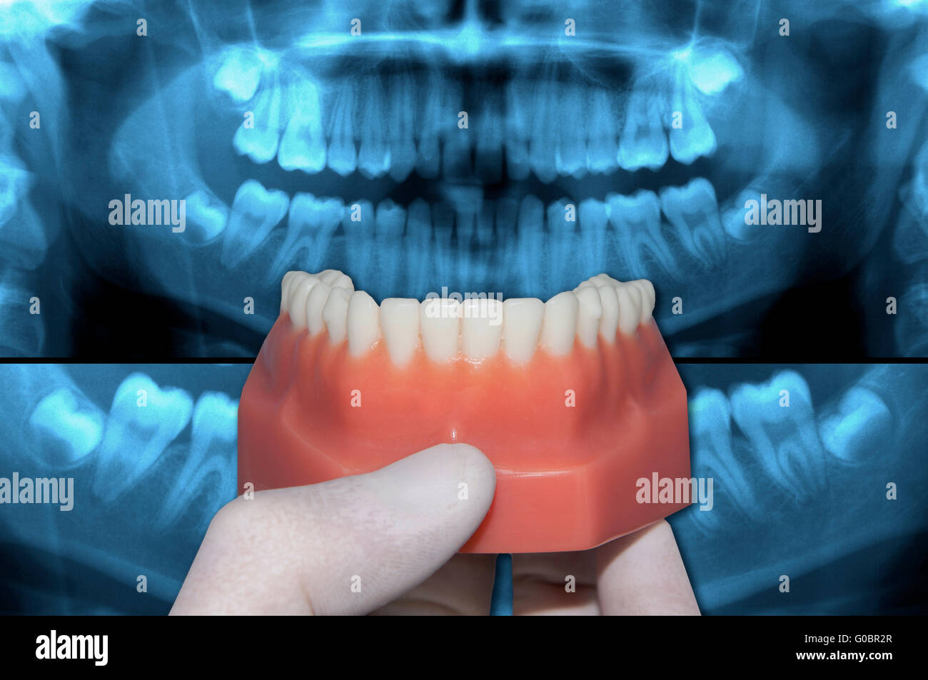 dentist show inferior arch over x-ray teeth Stock Photohttps://www.alamy.com/image-license-details/?v=1https://www.alamy.com/stock-photo-dentist-show-inferior-arch-over-x-ray-teeth-103521791.html
RFG0BR2R–dentist show inferior arch over x-ray teeth
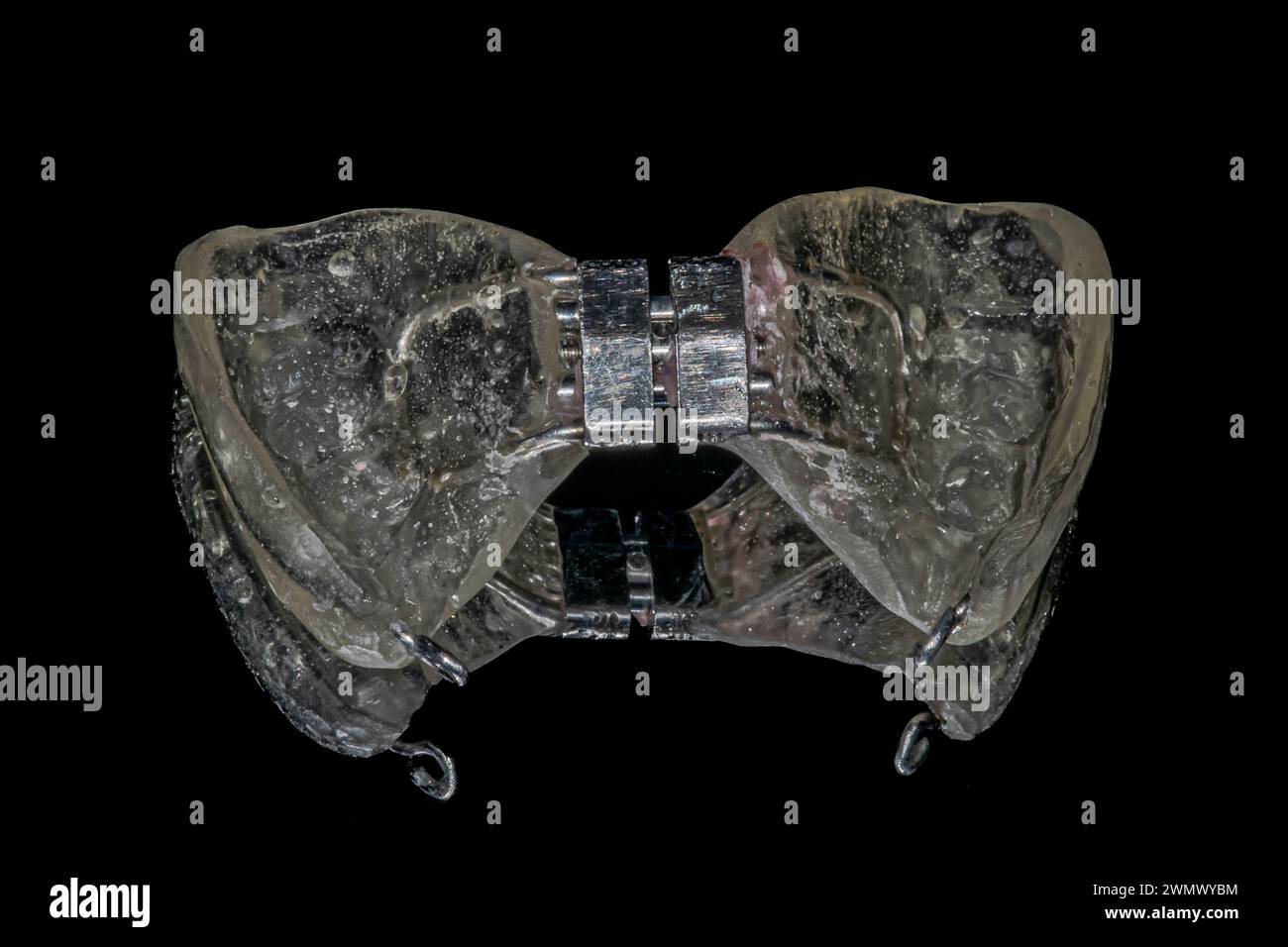 Palatal expander orthodontics device to widen the maxilla, made of acrylic resin, metal hooks and a check actuator. Black background mirror reflection Stock Photohttps://www.alamy.com/image-license-details/?v=1https://www.alamy.com/palatal-expander-orthodontics-device-to-widen-the-maxilla-made-of-acrylic-resin-metal-hooks-and-a-check-actuator-black-background-mirror-reflection-image598015928.html
RF2WMWYBM–Palatal expander orthodontics device to widen the maxilla, made of acrylic resin, metal hooks and a check actuator. Black background mirror reflection
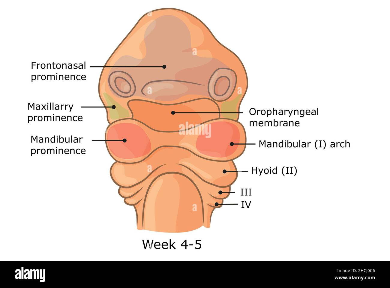 Development of external structures of human face week 4 - 5. Stock Photohttps://www.alamy.com/image-license-details/?v=1https://www.alamy.com/development-of-external-structures-of-human-face-week-4-5-image455240918.html
RF2HCJ0C6–Development of external structures of human face week 4 - 5.
 Skull with open jaw, illustration. Stock Photohttps://www.alamy.com/image-license-details/?v=1https://www.alamy.com/skull-with-open-jaw-illustration-image367367028.html
RF2C9K0B0–Skull with open jaw, illustration.
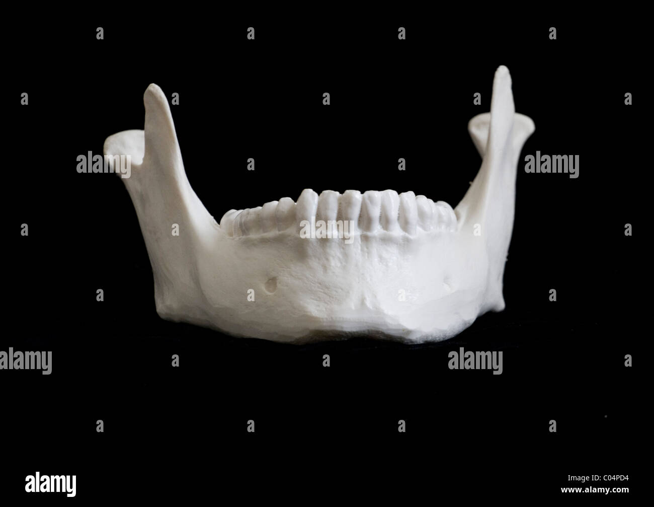 Human Skeletal Model, Mandible, Jaw bone, front view, Stock Photohttps://www.alamy.com/image-license-details/?v=1https://www.alamy.com/stock-photo-human-skeletal-model-mandible-jaw-bone-front-view-34526160.html
RFC04PD4–Human Skeletal Model, Mandible, Jaw bone, front view,
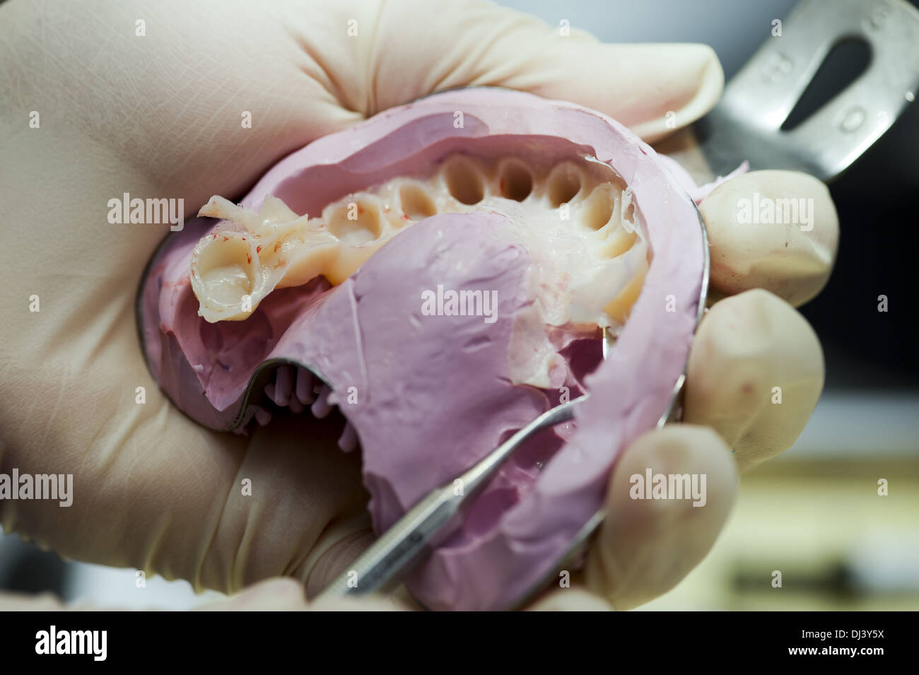 Temporary removal of a maxillary impression Stock Photohttps://www.alamy.com/image-license-details/?v=1https://www.alamy.com/temporary-removal-of-a-maxillary-impression-image62782102.html
RMDJ3Y5X–Temporary removal of a maxillary impression
 Temporary removal of a maxillary impression Stock Photohttps://www.alamy.com/image-license-details/?v=1https://www.alamy.com/stock-photo-temporary-removal-of-a-maxillary-impression-76324897.html
RMEC4W55–Temporary removal of a maxillary impression
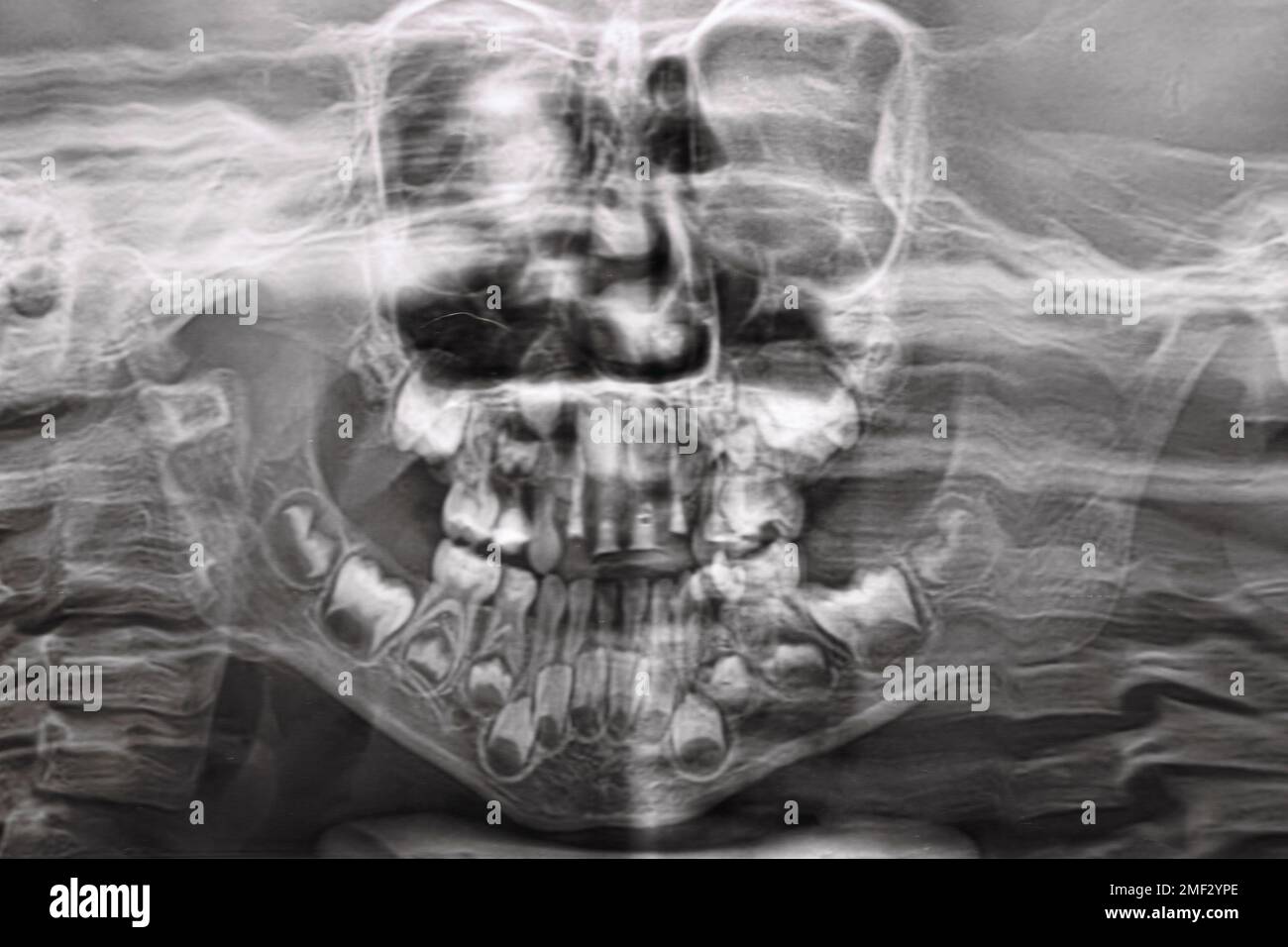 A panoramic X-ray shows several embedded and impacted teeth on both the upper and lower jaw of children's teeth Stock Photohttps://www.alamy.com/image-license-details/?v=1https://www.alamy.com/a-panoramic-x-ray-shows-several-embedded-and-impacted-teeth-on-both-the-upper-and-lower-jaw-of-childrens-teeth-image508386214.html
RF2MF2YPE–A panoramic X-ray shows several embedded and impacted teeth on both the upper and lower jaw of children's teeth
 Mesial bite profile before and after orthodontic treatment. Human with malocclusion, lower jaw extended forward, bite correction by braces. Vector Stock Vectorhttps://www.alamy.com/image-license-details/?v=1https://www.alamy.com/mesial-bite-profile-before-and-after-orthodontic-treatment-human-with-malocclusion-lower-jaw-extended-forward-bite-correction-by-braces-vector-image366914184.html
RF2C8XAP0–Mesial bite profile before and after orthodontic treatment. Human with malocclusion, lower jaw extended forward, bite correction by braces. Vector
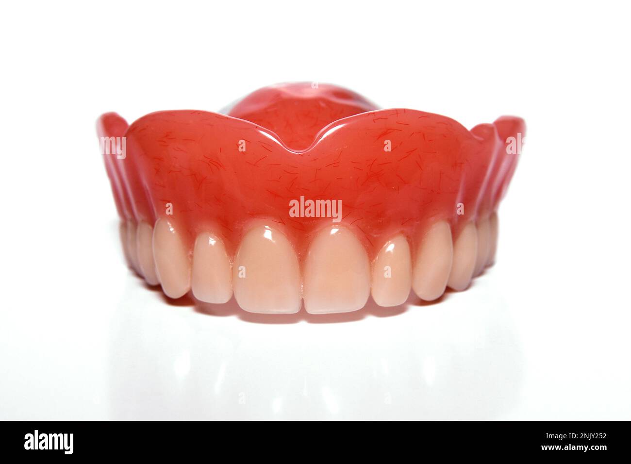 denture, upper jaw Stock Photohttps://www.alamy.com/image-license-details/?v=1https://www.alamy.com/denture-upper-jaw-image527969262.html
RM2NJY252–denture, upper jaw
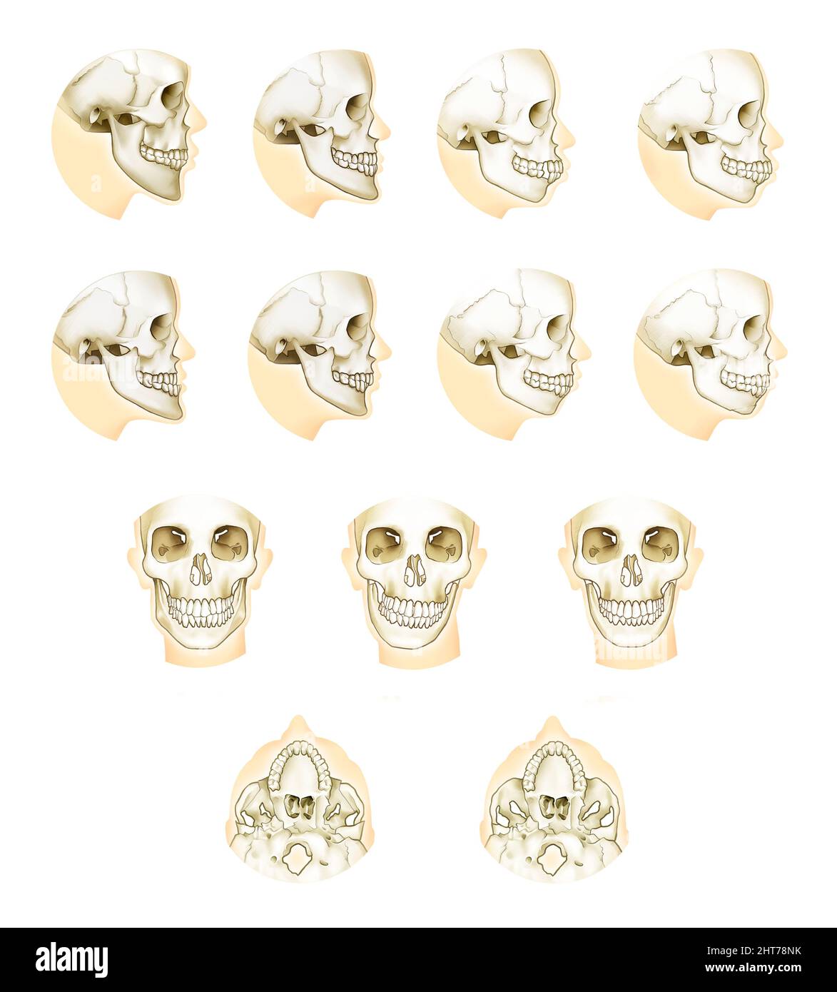 Realistic illustration of orthodontic double jaw surgery anatomy Stock Photohttps://www.alamy.com/image-license-details/?v=1https://www.alamy.com/realistic-illustration-of-orthodontic-double-jaw-surgery-anatomy-image462381855.html
RF2HT78NK–Realistic illustration of orthodontic double jaw surgery anatomy
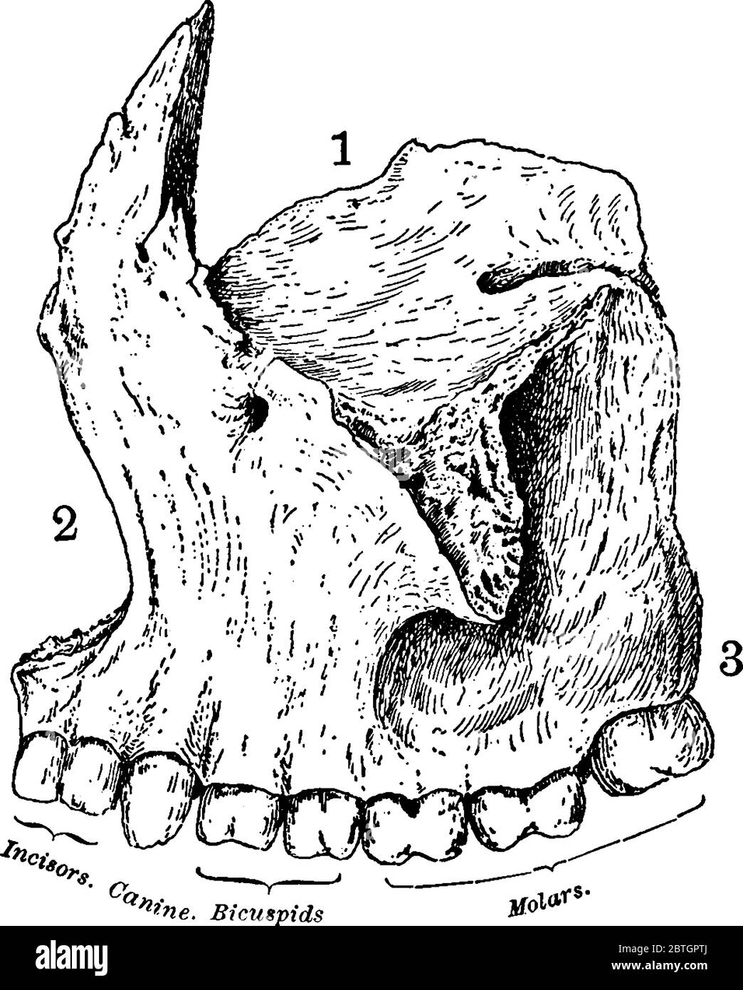 Superior maxillary bone together with its fellow on the opposite side, forms the whole of the upper jaw, with its parts labelled, vintage line drawing Stock Vectorhttps://www.alamy.com/image-license-details/?v=1https://www.alamy.com/superior-maxillary-bone-together-with-its-fellow-on-the-opposite-side-forms-the-whole-of-the-upper-jaw-with-its-parts-labelled-vintage-line-drawing-image359328274.html
RF2BTGPTJ–Superior maxillary bone together with its fellow on the opposite side, forms the whole of the upper jaw, with its parts labelled, vintage line drawing
 Dental roots in maxillary sinus. Medical illustration in flat style. Stock Vectorhttps://www.alamy.com/image-license-details/?v=1https://www.alamy.com/dental-roots-in-maxillary-sinus-medical-illustration-in-flat-style-image502009683.html
RF2M4MED7–Dental roots in maxillary sinus. Medical illustration in flat style.
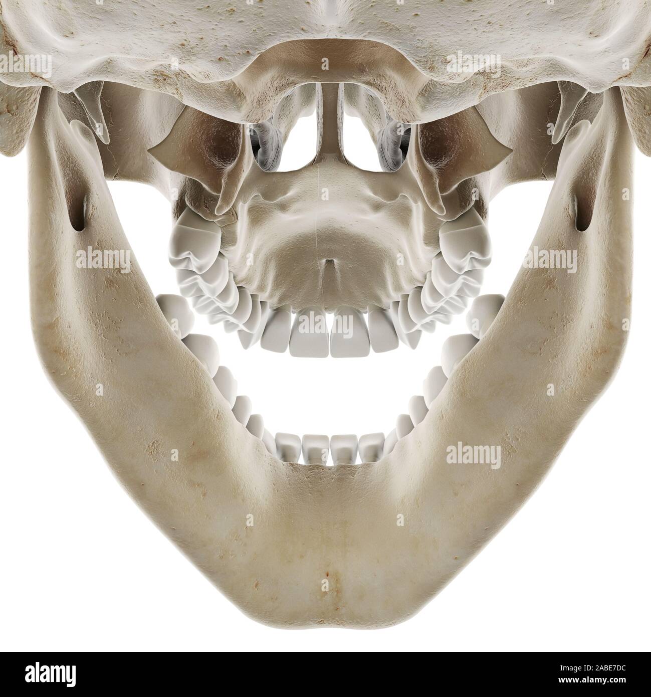 3d rendered medically accurate illustration of the skull with open jaw Stock Photohttps://www.alamy.com/image-license-details/?v=1https://www.alamy.com/3d-rendered-medically-accurate-illustration-of-the-skull-with-open-jaw-image334071400.html
RF2ABE7DC–3d rendered medically accurate illustration of the skull with open jaw
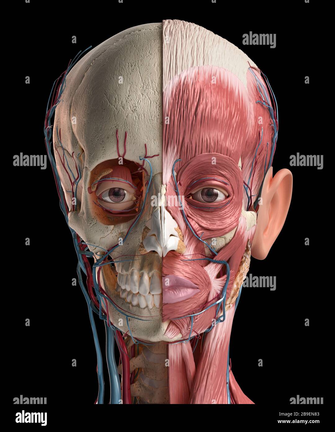 Human head anatomy of skull, facial muscles, veins and arteries, black background. Stock Photohttps://www.alamy.com/image-license-details/?v=1https://www.alamy.com/human-head-anatomy-of-skull-facial-muscles-veins-and-arteries-black-background-image350063283.html
RF2B9EN83–Human head anatomy of skull, facial muscles, veins and arteries, black background.
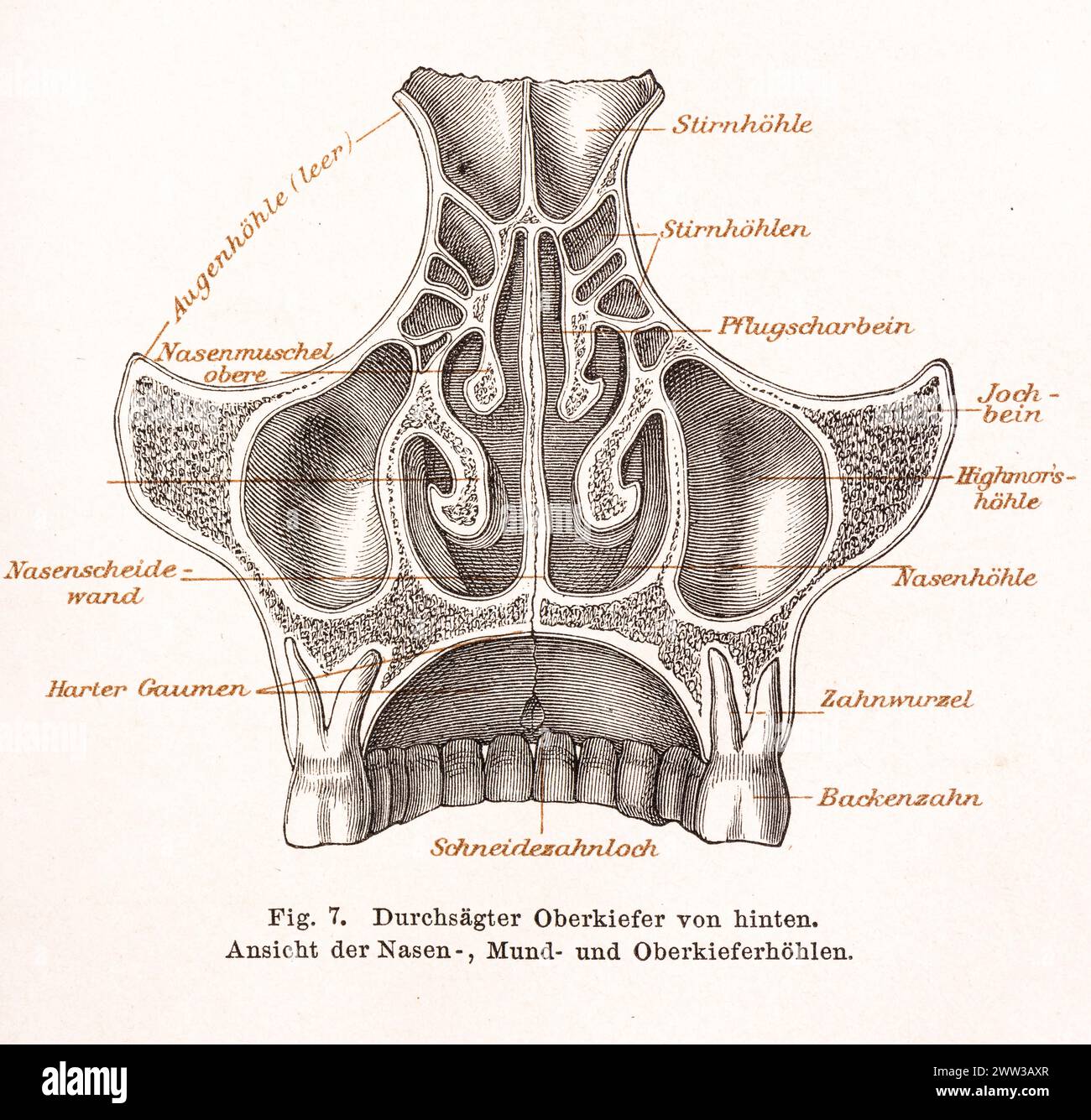 Medicine, anatomy, illustration, drawing, detailed representation of the sawed-through upper jaw and the nasal cavities, oral cavity and maxillary Stock Photohttps://www.alamy.com/image-license-details/?v=1https://www.alamy.com/medicine-anatomy-illustration-drawing-detailed-representation-of-the-sawed-through-upper-jaw-and-the-nasal-cavities-oral-cavity-and-maxillary-image600593359.html
RF2WW3AXR–Medicine, anatomy, illustration, drawing, detailed representation of the sawed-through upper jaw and the nasal cavities, oral cavity and maxillary
 X-ray of a skull of the person. Radiography X ray dislocated jaw broken bones nasopharynx high quality Stock Photohttps://www.alamy.com/image-license-details/?v=1https://www.alamy.com/x-ray-of-a-skull-of-the-person-radiography-x-ray-dislocated-jaw-broken-bones-nasopharynx-high-quality-image620886960.html
RF2Y23RJ8–X-ray of a skull of the person. Radiography X ray dislocated jaw broken bones nasopharynx high quality
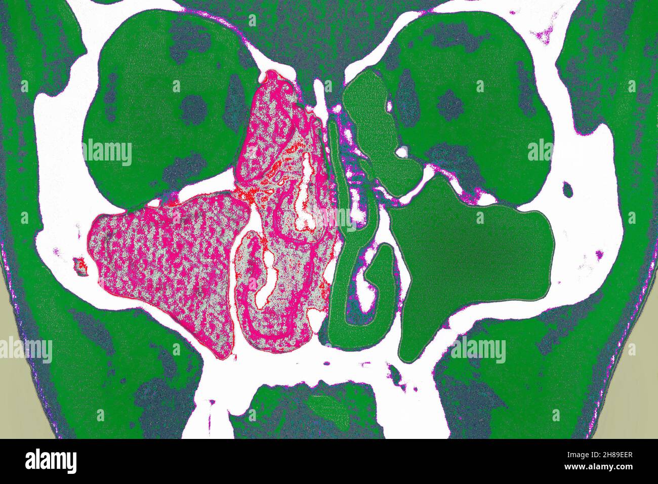 Sinusitis Stock Photohttps://www.alamy.com/image-license-details/?v=1https://www.alamy.com/sinusitis-image452595775.html
RM2H89EER–Sinusitis
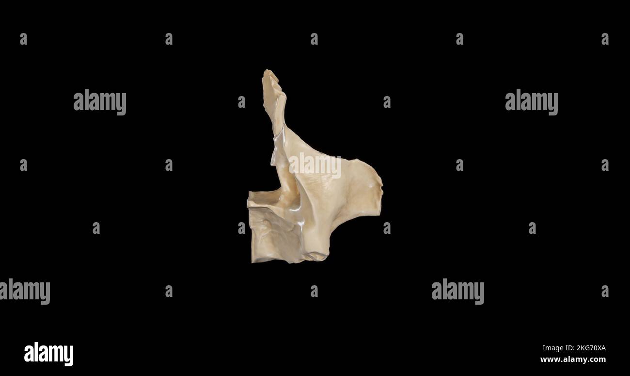 Posterior view of Right Maxilla Stock Photohttps://www.alamy.com/image-license-details/?v=1https://www.alamy.com/posterior-view-of-right-maxilla-image491879202.html
RF2KG70XA–Posterior view of Right Maxilla
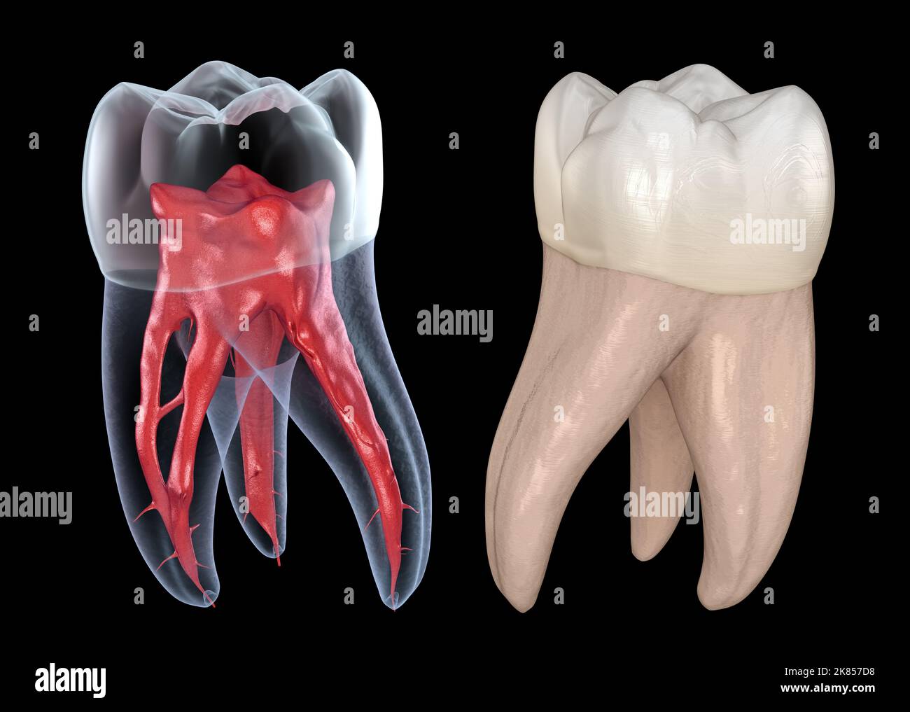 Dental root anatomy - First maxillary molar tooth. Medically accurate dental 3D illustration Stock Photohttps://www.alamy.com/image-license-details/?v=1https://www.alamy.com/dental-root-anatomy-first-maxillary-molar-tooth-medically-accurate-dental-3d-illustration-image486923172.html
RF2K857D8–Dental root anatomy - First maxillary molar tooth. Medically accurate dental 3D illustration
 Palatal expander orthodontics device to widen the maxilla, made of acrylic resin, metal hooks and a check actuator. Black background mirror reflection Stock Photohttps://www.alamy.com/image-license-details/?v=1https://www.alamy.com/palatal-expander-orthodontics-device-to-widen-the-maxilla-made-of-acrylic-resin-metal-hooks-and-a-check-actuator-black-background-mirror-reflection-image598016235.html
RF2WMWYPK–Palatal expander orthodontics device to widen the maxilla, made of acrylic resin, metal hooks and a check actuator. Black background mirror reflection
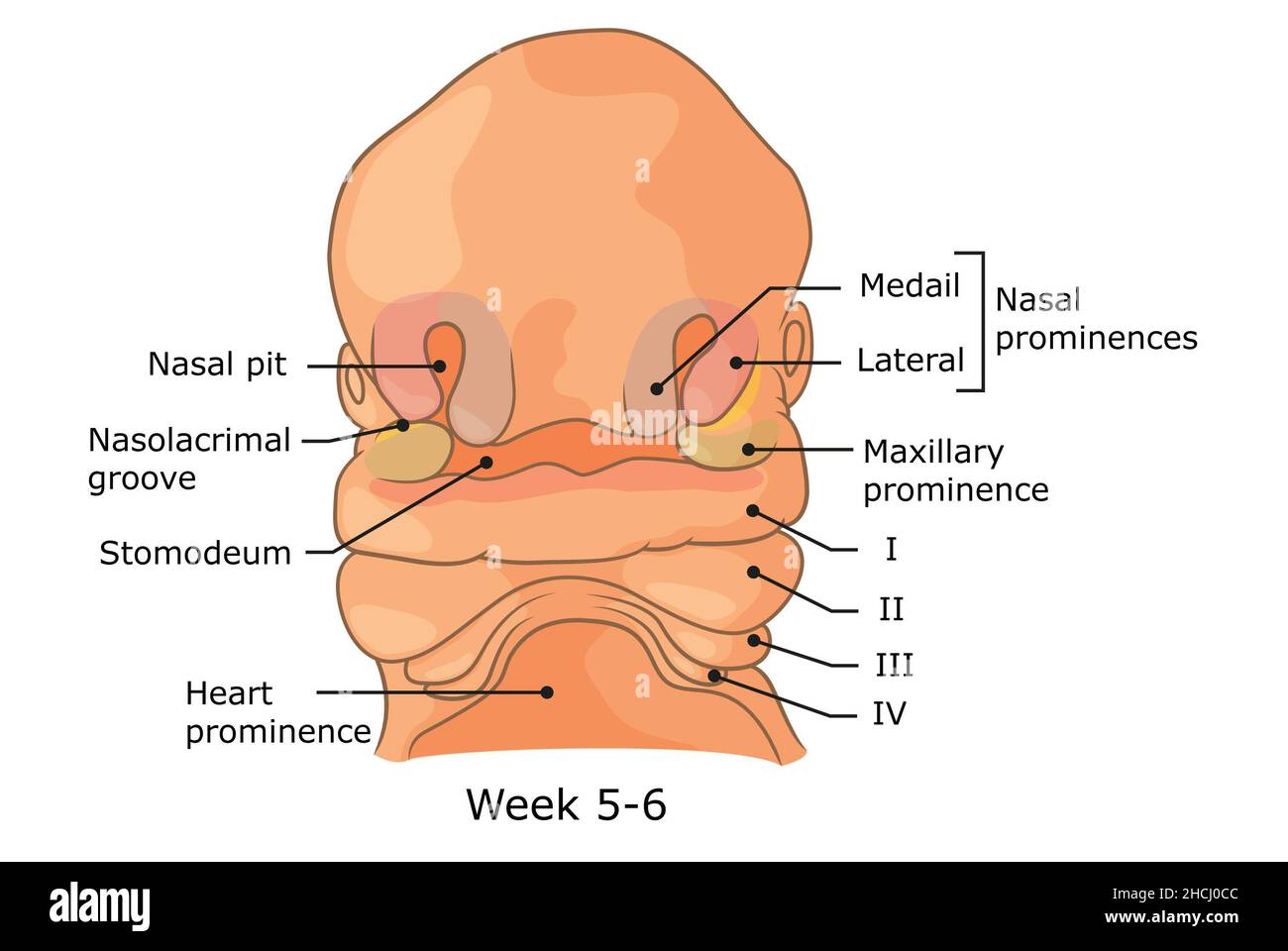 Development of external structures of human face week 5 - 6. Stock Photohttps://www.alamy.com/image-license-details/?v=1https://www.alamy.com/development-of-external-structures-of-human-face-week-5-6-image455240924.html
RF2HCJ0CC–Development of external structures of human face week 5 - 6.
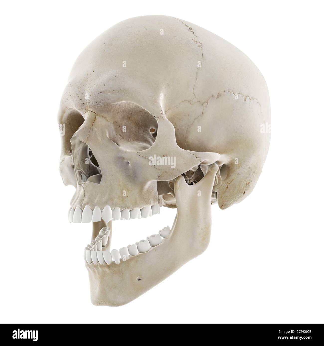 Skull with open jaw, illustration. Stock Photohttps://www.alamy.com/image-license-details/?v=1https://www.alamy.com/skull-with-open-jaw-illustration-image367367067.html
RF2C9K0CB–Skull with open jaw, illustration.
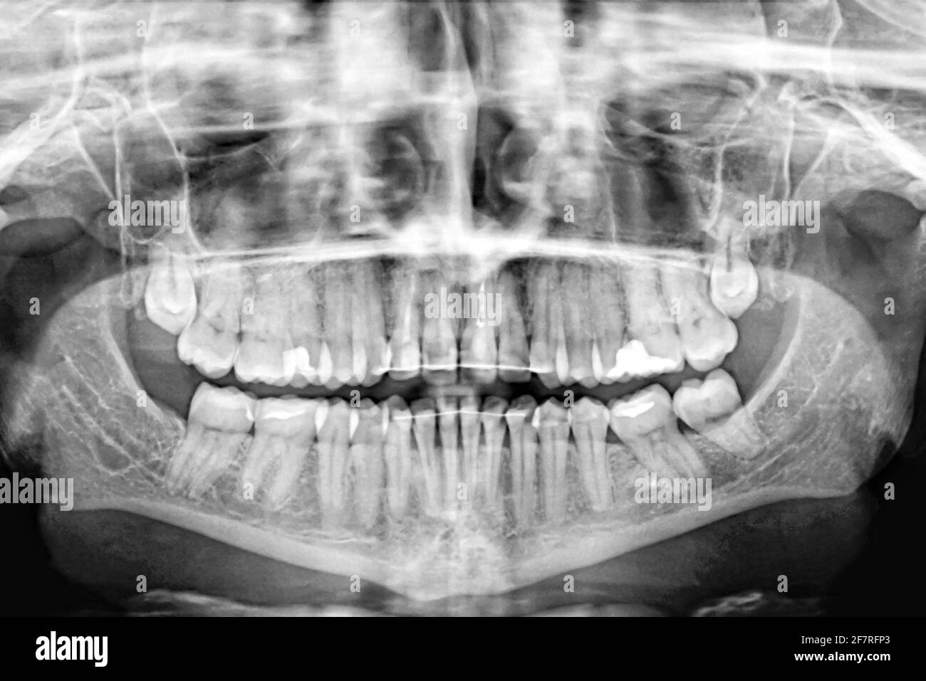 Panoramic X-ray scan of humans teeth.Examination and treatment. Dental care.Banner. Stock Photohttps://www.alamy.com/image-license-details/?v=1https://www.alamy.com/panoramic-x-ray-scan-of-humans-teethexamination-and-treatment-dental-carebanner-image417868699.html
RF2F7RFP3–Panoramic X-ray scan of humans teeth.Examination and treatment. Dental care.Banner.
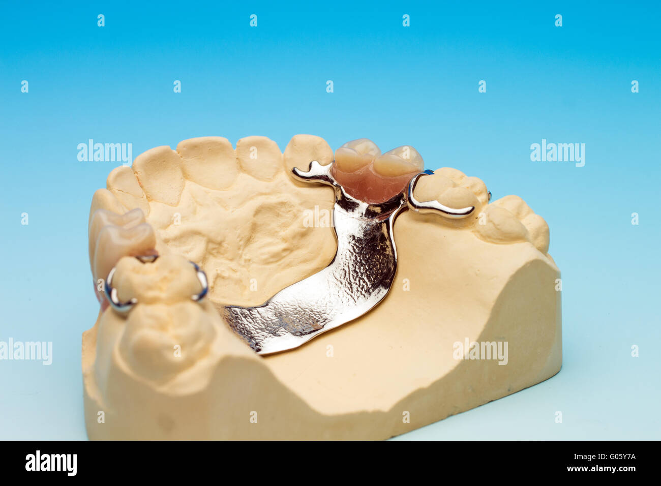 Maxillary dental bridge Stock Photohttps://www.alamy.com/image-license-details/?v=1https://www.alamy.com/stock-photo-maxillary-dental-bridge-103393342.html
RMG05Y7A–Maxillary dental bridge
 Anatomy. Lower Jaw Bones. Temporo-Maxillary Articulation 1880 old print Stock Photohttps://www.alamy.com/image-license-details/?v=1https://www.alamy.com/anatomy-lower-jaw-bones-temporo-maxillary-articulation-1880-old-print-image623228915.html
RF2Y5XERF–Anatomy. Lower Jaw Bones. Temporo-Maxillary Articulation 1880 old print
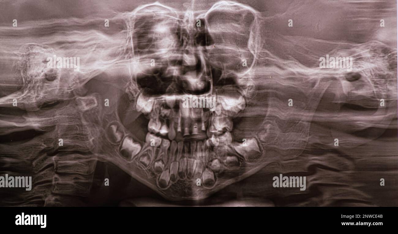 A panoramic X-ray shows several embedded and impacted teeth on both the upper and lower jaw of children's teeth Stock Photohttps://www.alamy.com/image-license-details/?v=1https://www.alamy.com/a-panoramic-x-ray-shows-several-embedded-and-impacted-teeth-on-both-the-upper-and-lower-jaw-of-childrens-teeth-image531951963.html
RF2NWCE4B–A panoramic X-ray shows several embedded and impacted teeth on both the upper and lower jaw of children's teeth
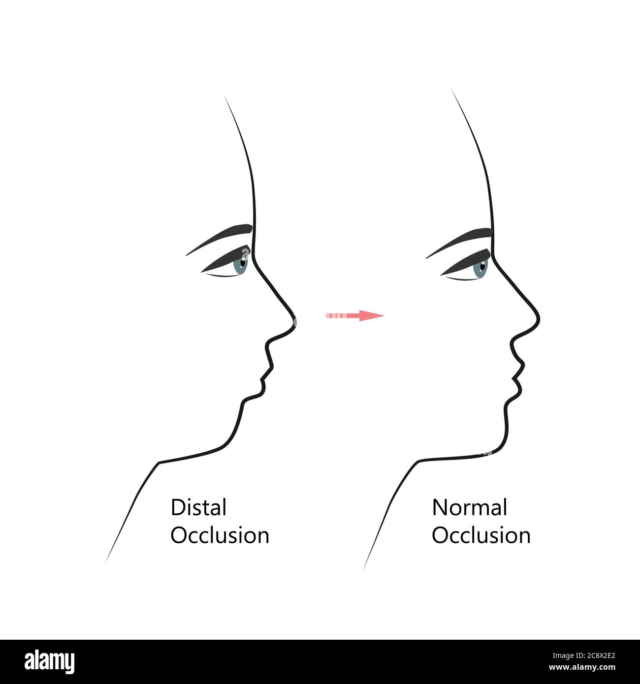 Distal bite profile before and after orthodontic treatment. Human with malocclusion, lower jaw pushing back, bite correction by braces. Vector Stock Vectorhttps://www.alamy.com/image-license-details/?v=1https://www.alamy.com/distal-bite-profile-before-and-after-orthodontic-treatment-human-with-malocclusion-lower-jaw-pushing-back-bite-correction-by-braces-vector-image366907690.html
RF2C8X2E2–Distal bite profile before and after orthodontic treatment. Human with malocclusion, lower jaw pushing back, bite correction by braces. Vector
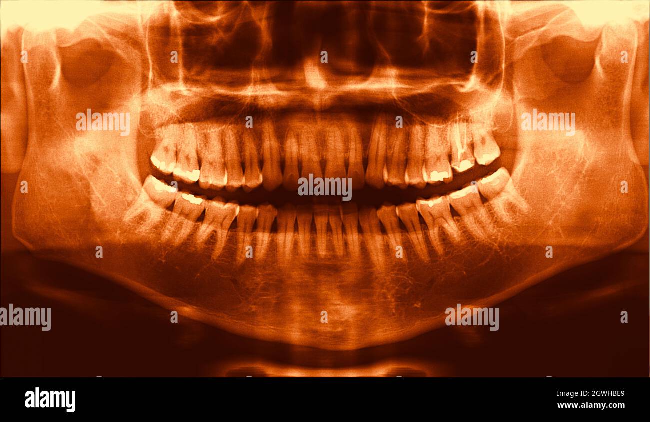 full arch wide maxillary male dental panoramic X-Ray radiography Stock Photohttps://www.alamy.com/image-license-details/?v=1https://www.alamy.com/full-arch-wide-maxillary-male-dental-panoramic-x-ray-radiography-image446007809.html
RF2GWHBE9–full arch wide maxillary male dental panoramic X-Ray radiography
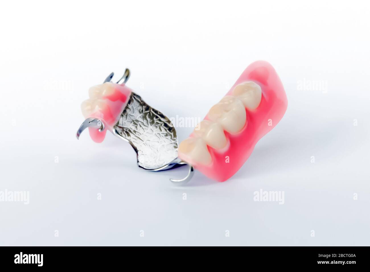 clasp prosthesis with clam fixation on the upper jaw isolated on a white background Stock Photohttps://www.alamy.com/image-license-details/?v=1https://www.alamy.com/clasp-prosthesis-with-clam-fixation-on-the-upper-jaw-isolated-on-a-white-background-image352122634.html
RF2BCTG0A–clasp prosthesis with clam fixation on the upper jaw isolated on a white background
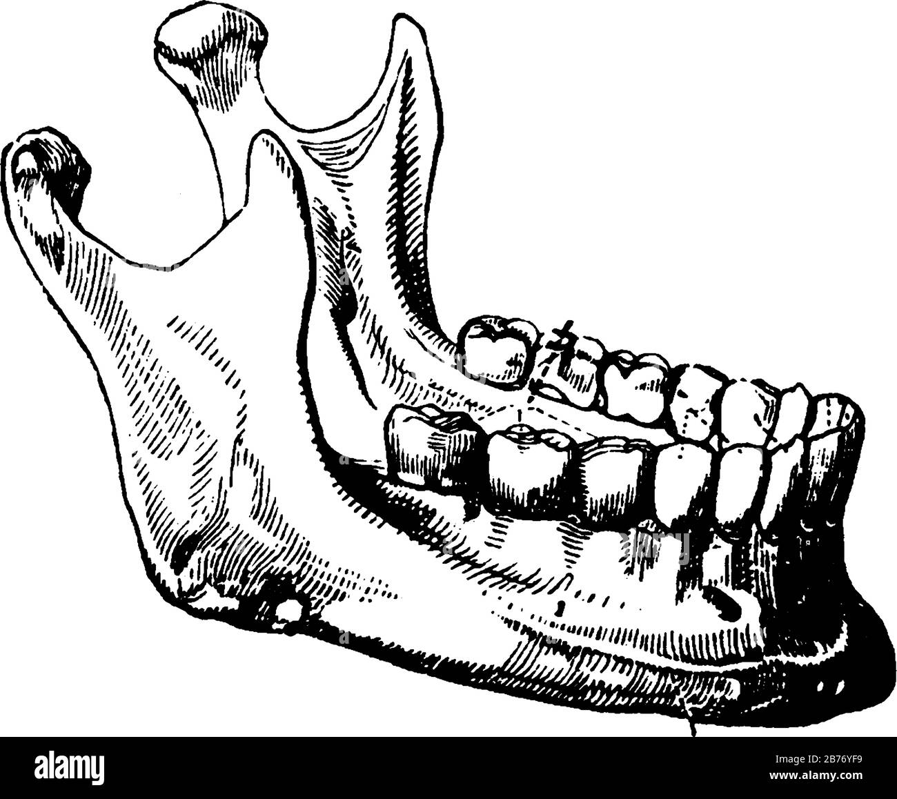 The term, mandibular, is given to teeth in the lower jaw. Adult mouth has 32 teeth, the middlemost four teeth on the lower jaws, vintage line drawing Stock Vectorhttps://www.alamy.com/image-license-details/?v=1https://www.alamy.com/the-term-mandibular-is-given-to-teeth-in-the-lower-jaw-adult-mouth-has-32-teeth-the-middlemost-four-teeth-on-the-lower-jaws-vintage-line-drawing-image348663261.html
RF2B76YF9–The term, mandibular, is given to teeth in the lower jaw. Adult mouth has 32 teeth, the middlemost four teeth on the lower jaws, vintage line drawing
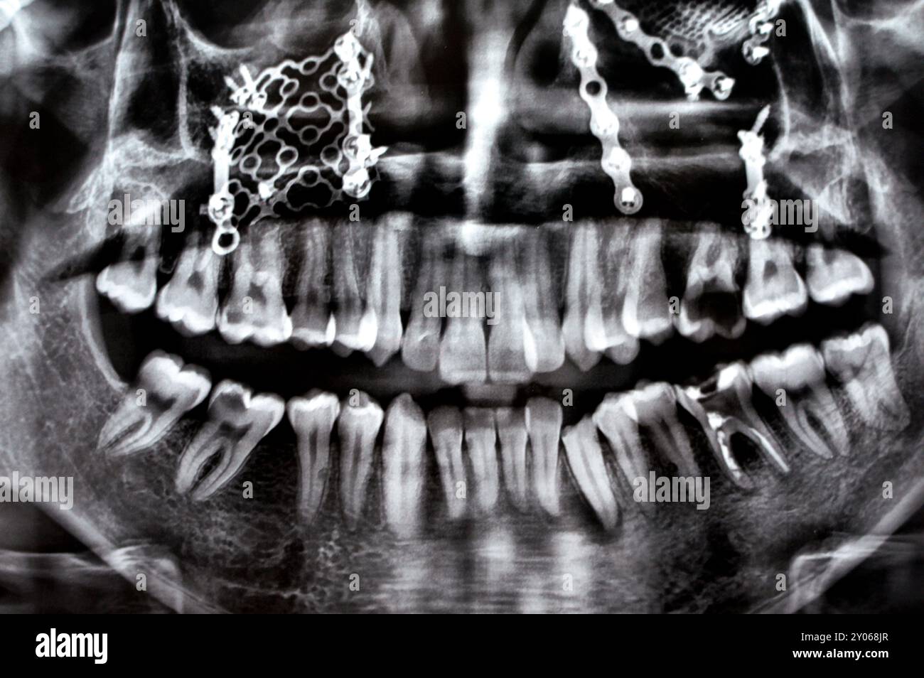 Panoramic X Ray shows multiple plates, screws for maxillary fixation of a fractured maxilla, titanium mesh on right side, decayed UL6, LL6 failed root Stock Photohttps://www.alamy.com/image-license-details/?v=1https://www.alamy.com/panoramic-x-ray-shows-multiple-plates-screws-for-maxillary-fixation-of-a-fractured-maxilla-titanium-mesh-on-right-side-decayed-ul6-ll6-failed-root-image619711759.html
RF2Y068JR–Panoramic X Ray shows multiple plates, screws for maxillary fixation of a fractured maxilla, titanium mesh on right side, decayed UL6, LL6 failed root
 3d rendered medically accurate illustration of the skull with open jaw Stock Photohttps://www.alamy.com/image-license-details/?v=1https://www.alamy.com/3d-rendered-medically-accurate-illustration-of-the-skull-with-open-jaw-image334071384.html
RF2ABE7CT–3d rendered medically accurate illustration of the skull with open jaw
 Human head anatomy of skull, facial muscles, veins and arteries, white background. Stock Photohttps://www.alamy.com/image-license-details/?v=1https://www.alamy.com/human-head-anatomy-of-skull-facial-muscles-veins-and-arteries-white-background-image350063558.html
RF2B9ENHX–Human head anatomy of skull, facial muscles, veins and arteries, white background.
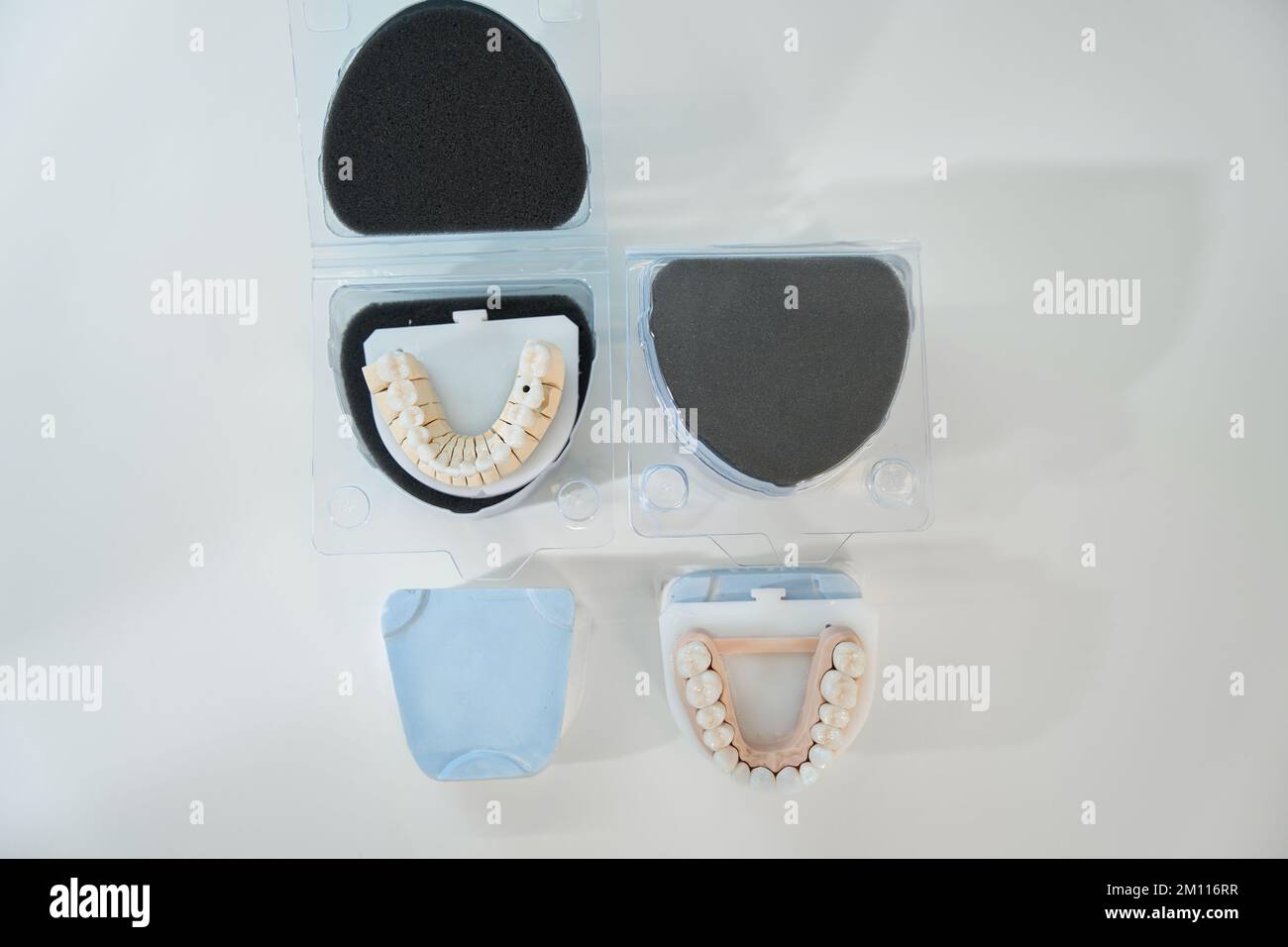 Maxillary and mandibular dentition designed for medical students and patients Stock Photohttps://www.alamy.com/image-license-details/?v=1https://www.alamy.com/maxillary-and-mandibular-dentition-designed-for-medical-students-and-patients-image499742651.html
RF2M116RR–Maxillary and mandibular dentition designed for medical students and patients
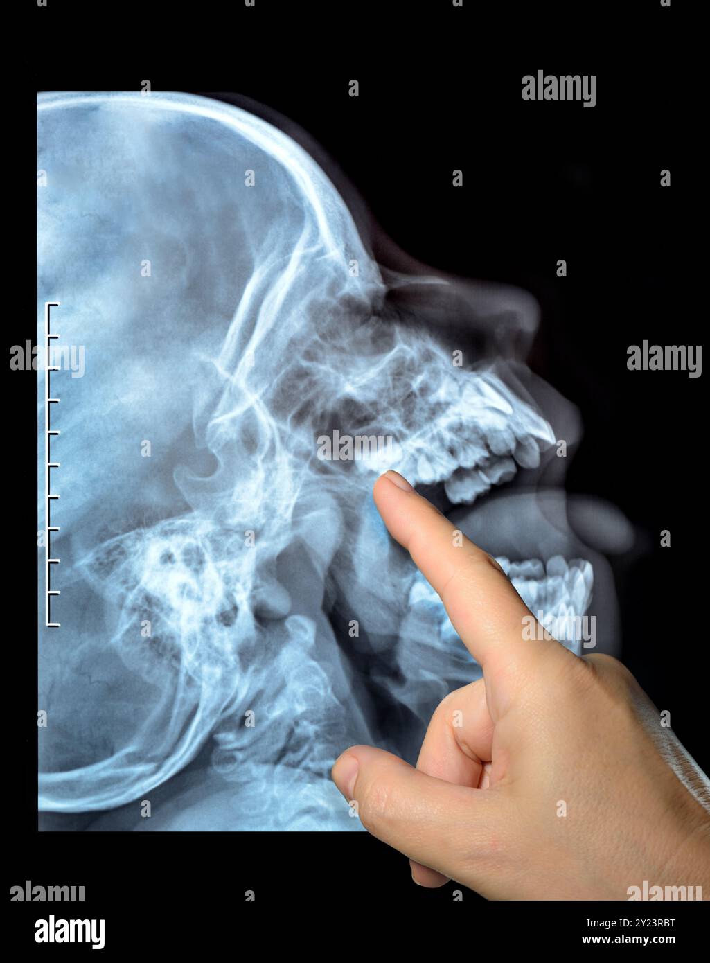 X-ray of a skull of the person. Radiography X ray dislocated jaw broken bones nasopharynx high quality hand finger points Stock Photohttps://www.alamy.com/image-license-details/?v=1https://www.alamy.com/x-ray-of-a-skull-of-the-person-radiography-x-ray-dislocated-jaw-broken-bones-nasopharynx-high-quality-hand-finger-points-image620886780.html
RF2Y23RBT–X-ray of a skull of the person. Radiography X ray dislocated jaw broken bones nasopharynx high quality hand finger points
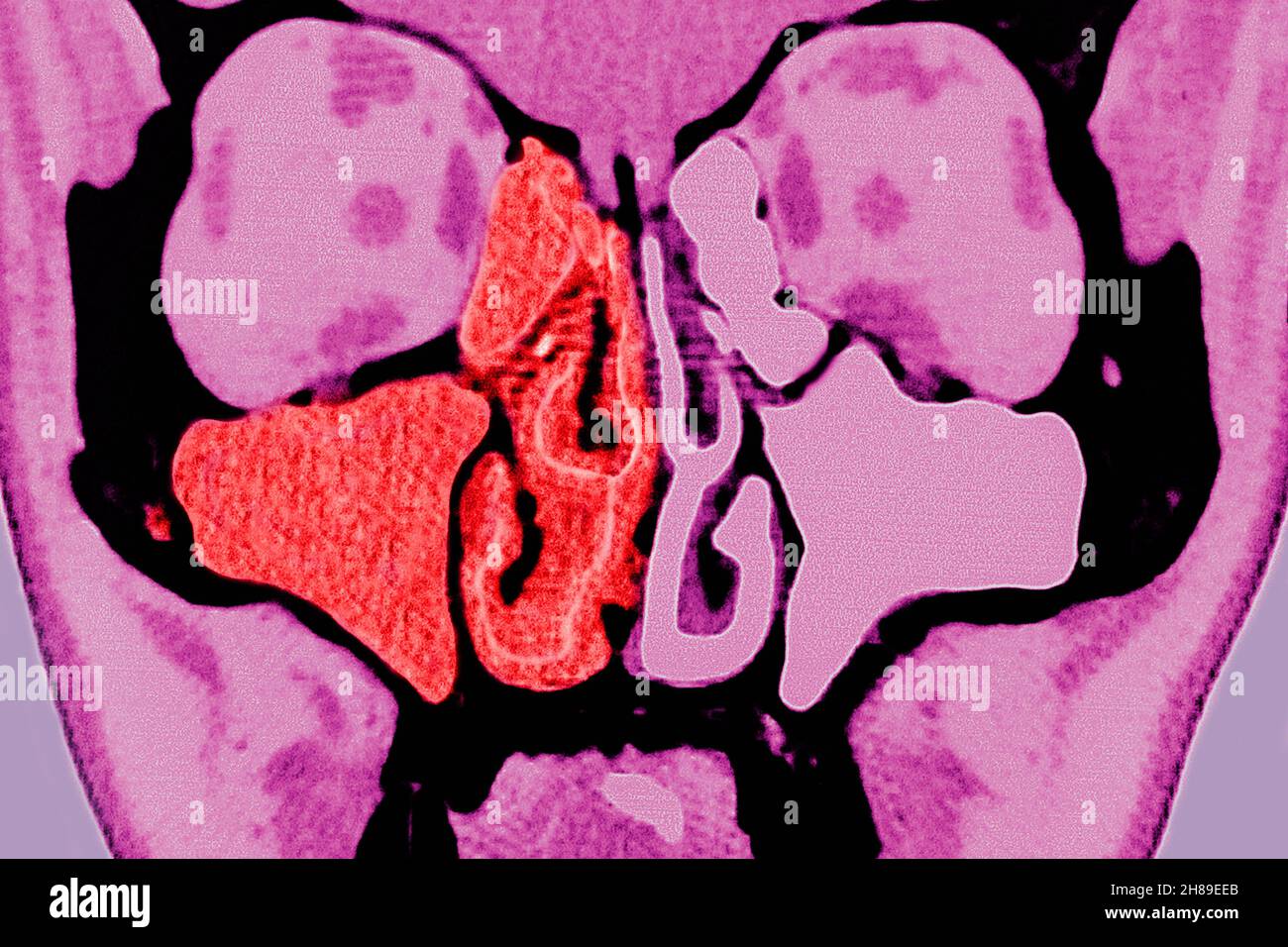 Sinusitis Stock Photohttps://www.alamy.com/image-license-details/?v=1https://www.alamy.com/sinusitis-image452595763.html
RM2H89EEB–Sinusitis
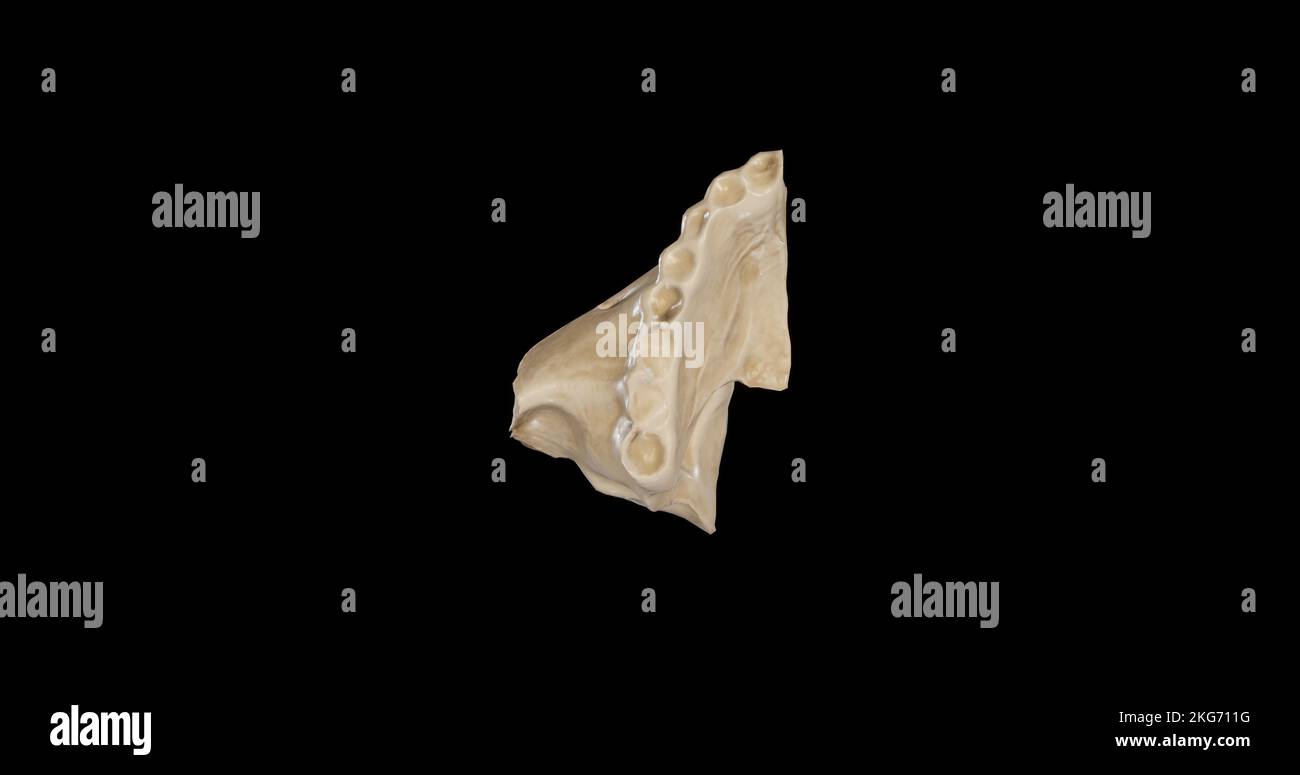 Inferior view of Right Maxilla Stock Photohttps://www.alamy.com/image-license-details/?v=1https://www.alamy.com/inferior-view-of-right-maxilla-image491879292.html
RF2KG711G–Inferior view of Right Maxilla
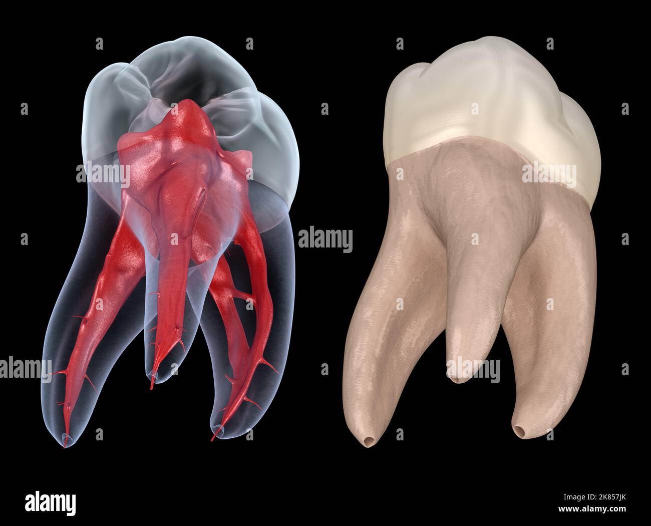 Dental root anatomy - First maxillary molar tooth. Medically accurate dental 3D illustration Stock Photohttps://www.alamy.com/image-license-details/?v=1https://www.alamy.com/dental-root-anatomy-first-maxillary-molar-tooth-medically-accurate-dental-3d-illustration-image486923323.html
RF2K857JK–Dental root anatomy - First maxillary molar tooth. Medically accurate dental 3D illustration
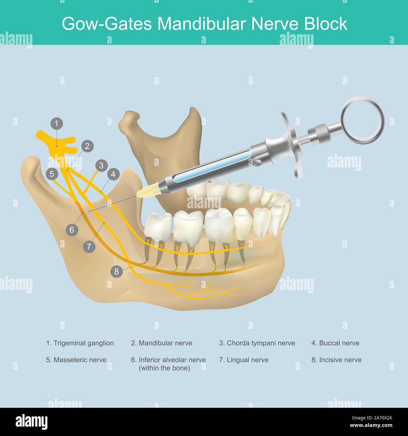 Mandibular Nerve Block. Illustration reference to Dentist the lower jaw area aesthetic just injection, to stop the pain from teeth nerves. Stock Vectorhttps://www.alamy.com/image-license-details/?v=1https://www.alamy.com/mandibular-nerve-block-illustration-reference-to-dentist-the-lower-jaw-area-aesthetic-just-injection-to-stop-the-pain-from-teeth-nerves-image331320043.html
RF2A70X2K–Mandibular Nerve Block. Illustration reference to Dentist the lower jaw area aesthetic just injection, to stop the pain from teeth nerves.
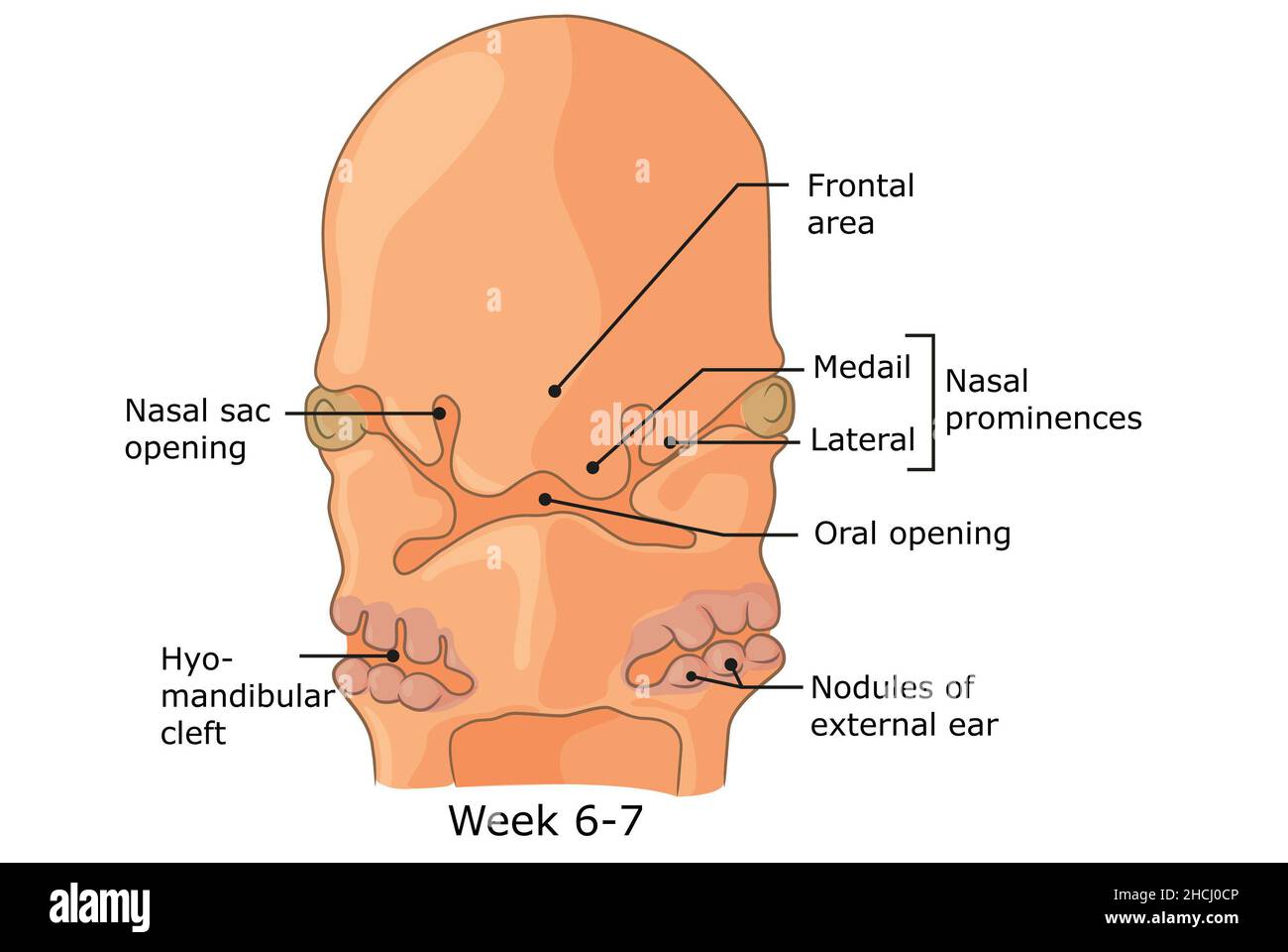 Development of external structures of human face week 6 - 7. Stock Photohttps://www.alamy.com/image-license-details/?v=1https://www.alamy.com/development-of-external-structures-of-human-face-week-6-7-image455240934.html
RF2HCJ0CP–Development of external structures of human face week 6 - 7.
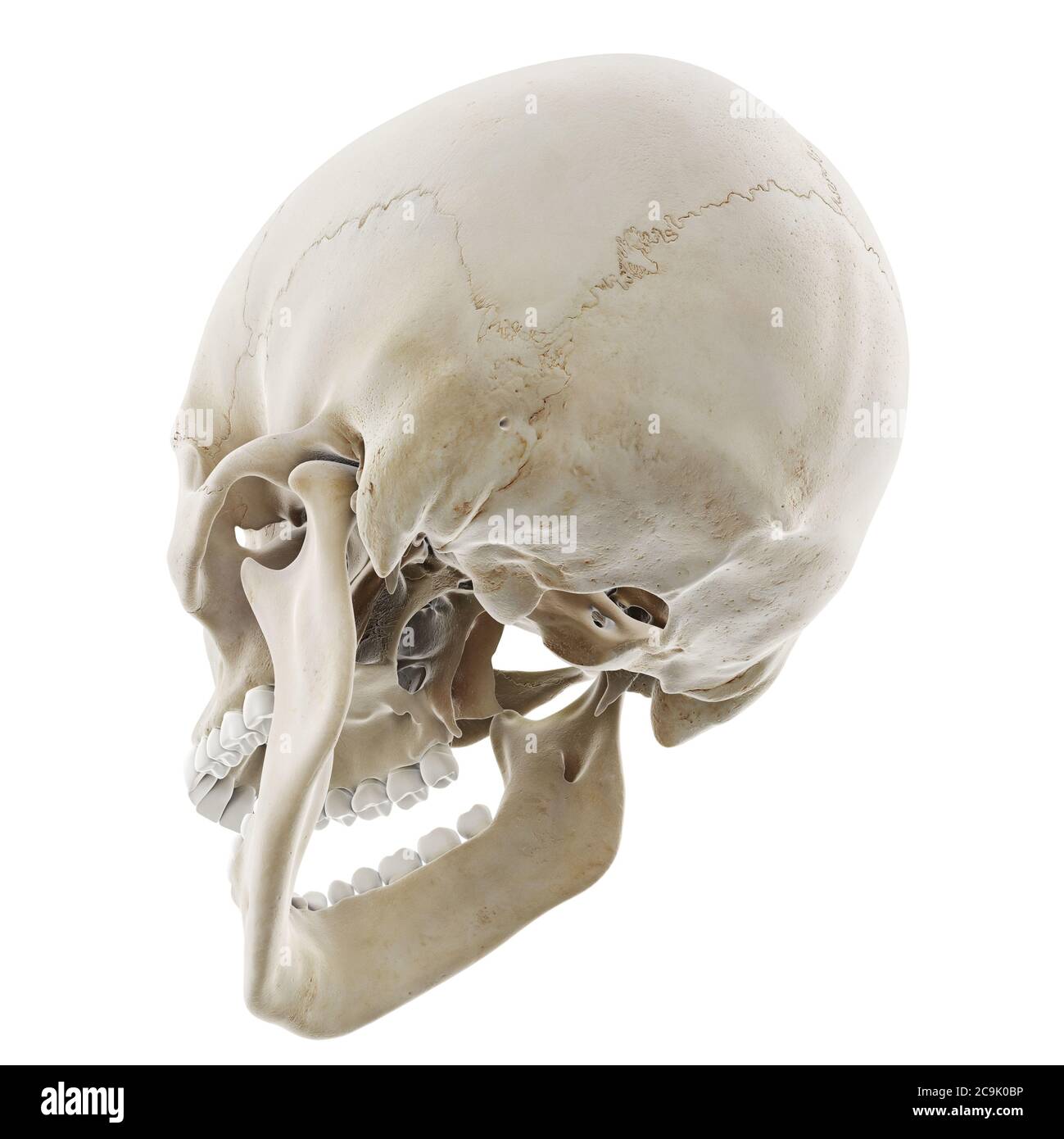 Skull with open jaw, illustration. Stock Photohttps://www.alamy.com/image-license-details/?v=1https://www.alamy.com/skull-with-open-jaw-illustration-image367367050.html
RF2C9K0BP–Skull with open jaw, illustration.
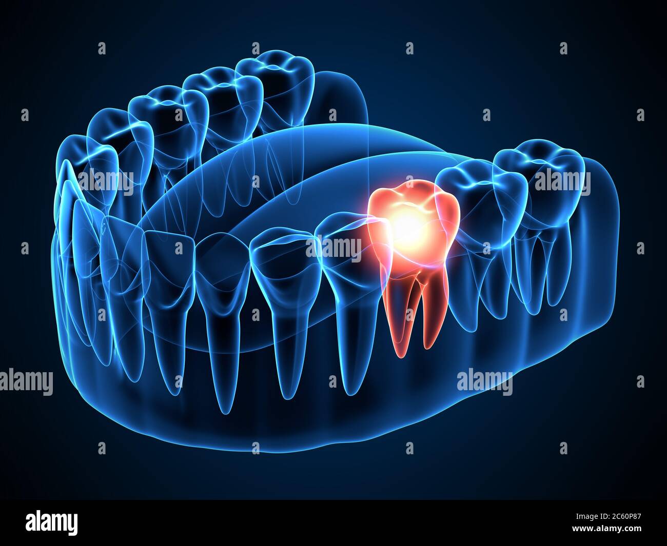 3d render of jaw x-ray with aching molar tooth. Toothache concept. Stock Photohttps://www.alamy.com/image-license-details/?v=1https://www.alamy.com/3d-render-of-jaw-x-ray-with-aching-molar-tooth-toothache-concept-image365123143.html
RF2C60P87–3d render of jaw x-ray with aching molar tooth. Toothache concept.
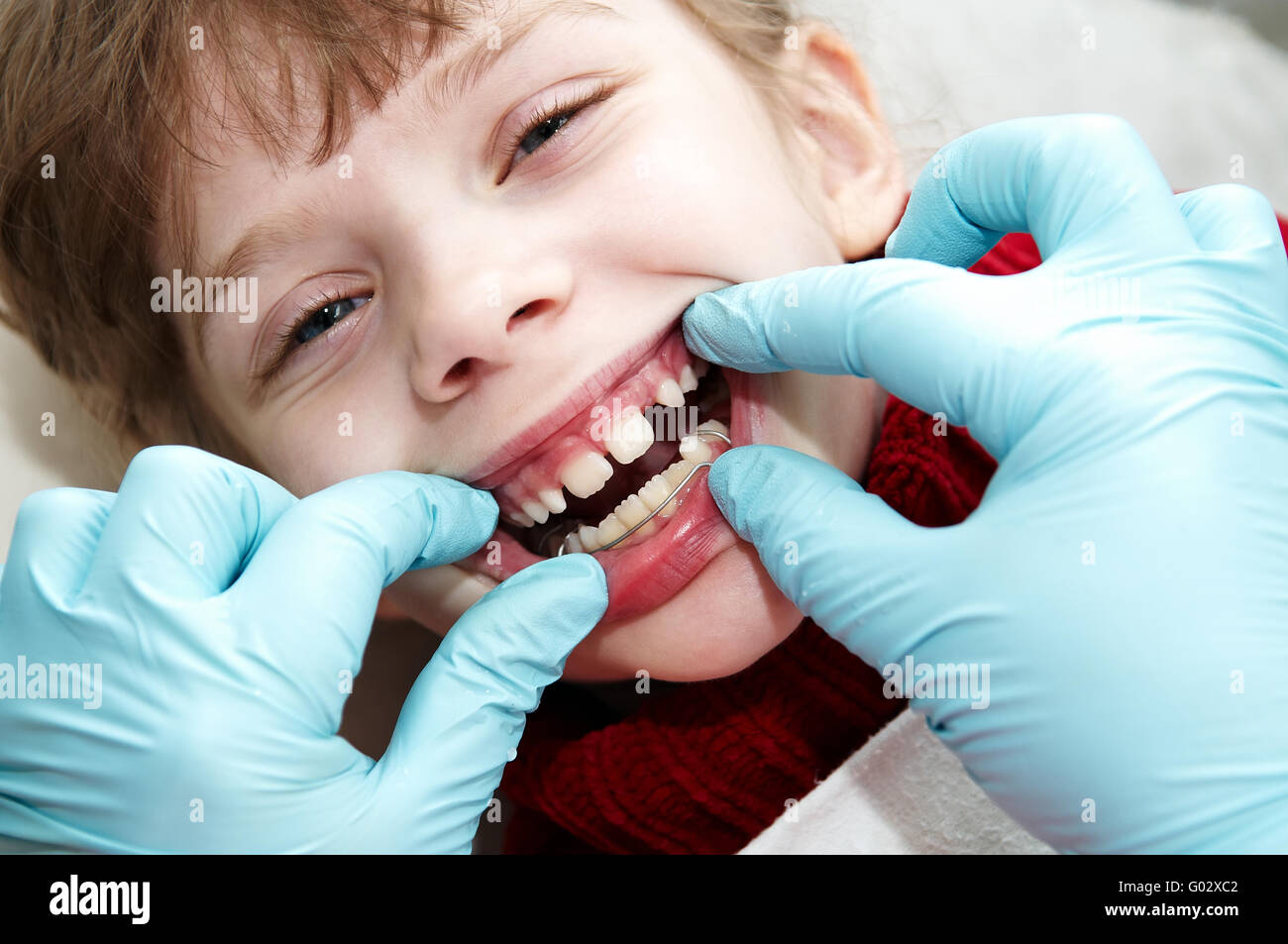 at dentist medic orthodontic doctor examination Stock Photohttps://www.alamy.com/image-license-details/?v=1https://www.alamy.com/stock-photo-at-dentist-medic-orthodontic-doctor-examination-103326834.html
RFG02XC2–at dentist medic orthodontic doctor examination
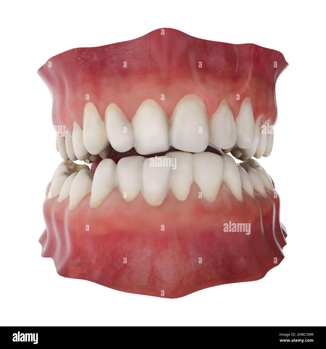 Jaw with abnormal teeth position. Orthodontic treatment concept. Realistic 3D illustration Stock Photohttps://www.alamy.com/image-license-details/?v=1https://www.alamy.com/jaw-with-abnormal-teeth-position-orthodontic-treatment-concept-realistic-3d-illustration-image454497283.html
RF2HBC3WR–Jaw with abnormal teeth position. Orthodontic treatment concept. Realistic 3D illustration
 A panoramic X-ray shows several embedded and impacted teeth on both the upper and lower jaw of children's teeth Stock Photohttps://www.alamy.com/image-license-details/?v=1https://www.alamy.com/a-panoramic-x-ray-shows-several-embedded-and-impacted-teeth-on-both-the-upper-and-lower-jaw-of-childrens-teeth-image531951915.html
RF2NWCE2K–A panoramic X-ray shows several embedded and impacted teeth on both the upper and lower jaw of children's teeth
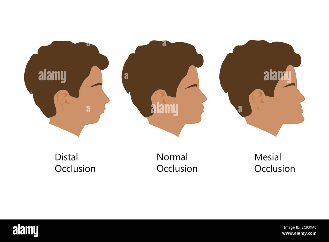 Guy with Distal, Normal, and Mesial bite profile, vector illustration. Overbite or underbite before and after orthodontic treatment. Human with Stock Vectorhttps://www.alamy.com/image-license-details/?v=1https://www.alamy.com/guy-with-distal-normal-and-mesial-bite-profile-vector-illustration-overbite-or-underbite-before-and-after-orthodontic-treatment-human-with-image367036152.html
RF2C93XA0–Guy with Distal, Normal, and Mesial bite profile, vector illustration. Overbite or underbite before and after orthodontic treatment. Human with
 A closeup photograph of insertion of dental implants into a patient's upper jaw. Stock Photohttps://www.alamy.com/image-license-details/?v=1https://www.alamy.com/a-closeup-photograph-of-insertion-of-dental-implants-into-a-patients-upper-jaw-image365499838.html
RF2C6HXNJ–A closeup photograph of insertion of dental implants into a patient's upper jaw.
 Teeth mould dentist plaster 3D impression denture dentures lower jaw Cutout cut out white background isolated copy space Orthodo Stock Photohttps://www.alamy.com/image-license-details/?v=1https://www.alamy.com/stock-photo-teeth-mould-dentist-plaster-3d-impression-denture-dentures-lower-jaw-92299427.html
RMFA4GT3–Teeth mould dentist plaster 3D impression denture dentures lower jaw Cutout cut out white background isolated copy space Orthodo
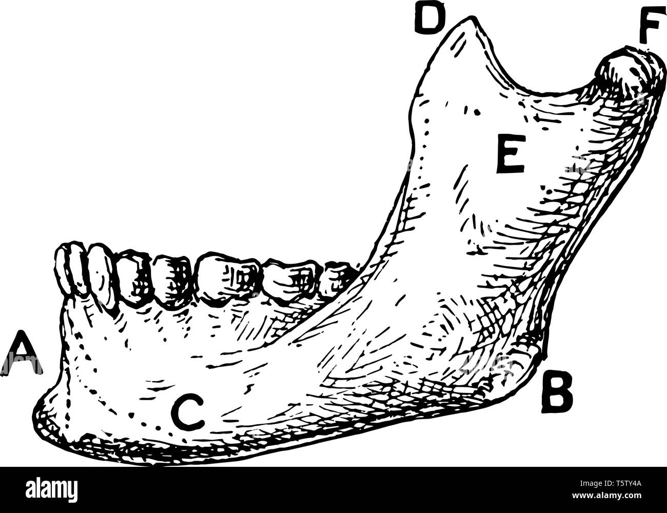 symphysis menti and angle of jaw vintage line drawing or engraving illustration. Stock Vectorhttps://www.alamy.com/image-license-details/?v=1https://www.alamy.com/symphysis-menti-and-angle-of-jaw-vintage-line-drawing-or-engraving-illustration-image244566570.html
RFT5TY4A–symphysis menti and angle of jaw vintage line drawing or engraving illustration.
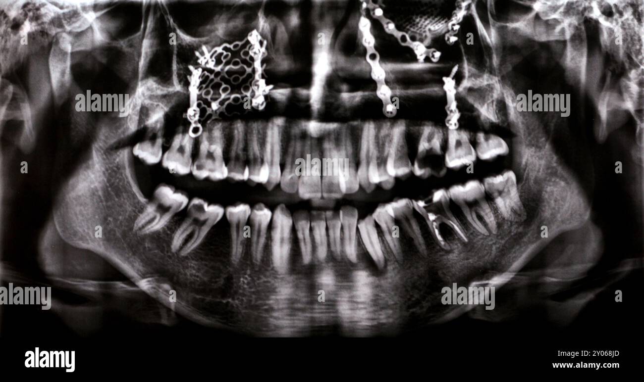 Panoramic X Ray shows multiple plates, screws for maxillary fixation of a fractured maxilla, titanium mesh on right side, decayed UL6, LL6 failed root Stock Photohttps://www.alamy.com/image-license-details/?v=1https://www.alamy.com/panoramic-x-ray-shows-multiple-plates-screws-for-maxillary-fixation-of-a-fractured-maxilla-titanium-mesh-on-right-side-decayed-ul6-ll6-failed-root-image619711749.html
RF2Y068JD–Panoramic X Ray shows multiple plates, screws for maxillary fixation of a fractured maxilla, titanium mesh on right side, decayed UL6, LL6 failed root
 3d rendered medically accurate illustration of the skull with open jaw Stock Photohttps://www.alamy.com/image-license-details/?v=1https://www.alamy.com/3d-rendered-medically-accurate-illustration-of-the-skull-with-open-jaw-image334050055.html
RF2ABD873–3d rendered medically accurate illustration of the skull with open jaw
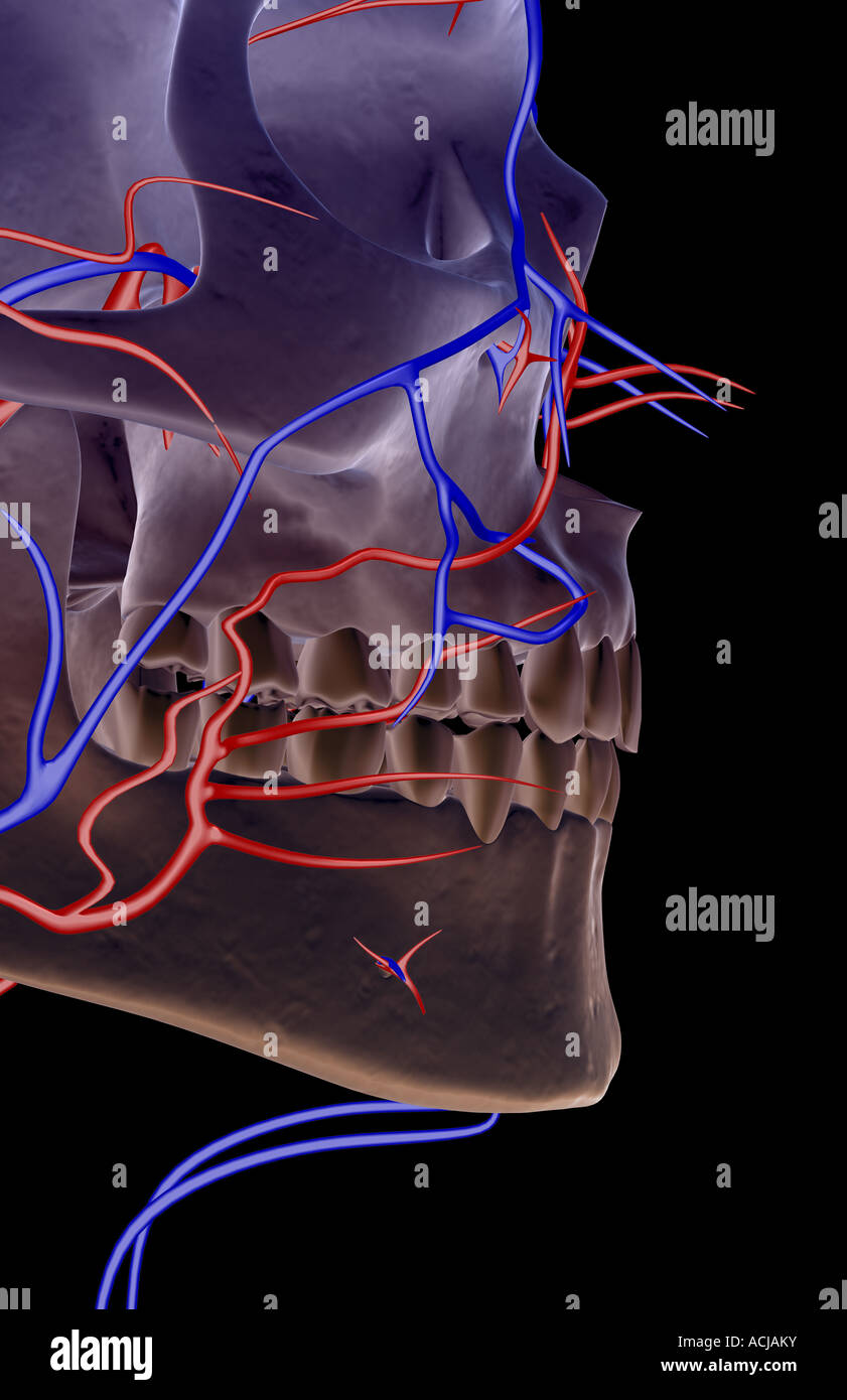 The blood supply of the jaw Stock Photohttps://www.alamy.com/image-license-details/?v=1https://www.alamy.com/stock-photo-the-blood-supply-of-the-jaw-13168526.html
RFACJAKY–The blood supply of the jaw
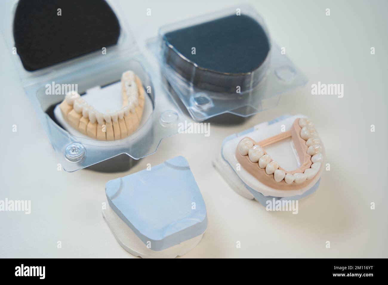 Artificial mandibular and maxillary dentition used for patient education Stock Photohttps://www.alamy.com/image-license-details/?v=1https://www.alamy.com/artificial-mandibular-and-maxillary-dentition-used-for-patient-education-image499742764.html
RF2M116YT–Artificial mandibular and maxillary dentition used for patient education
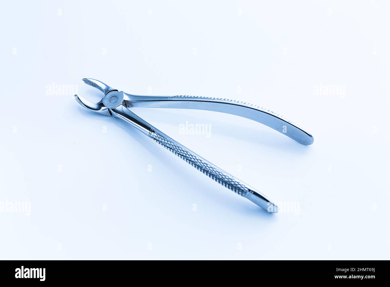 Maxillary wisdom tooth extraction for a dentist Stock Photohttps://www.alamy.com/image-license-details/?v=1https://www.alamy.com/maxillary-wisdom-tooth-extraction-for-a-dentist-image460294510.html
RF2HMT69J–Maxillary wisdom tooth extraction for a dentist
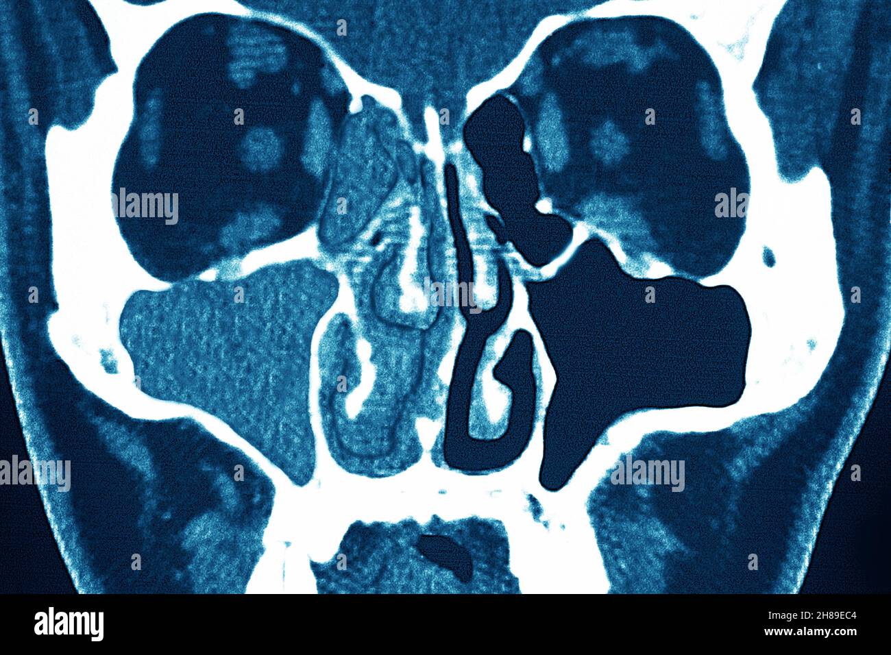 Sinusitis Stock Photohttps://www.alamy.com/image-license-details/?v=1https://www.alamy.com/sinusitis-image452595700.html
RM2H89EC4–Sinusitis
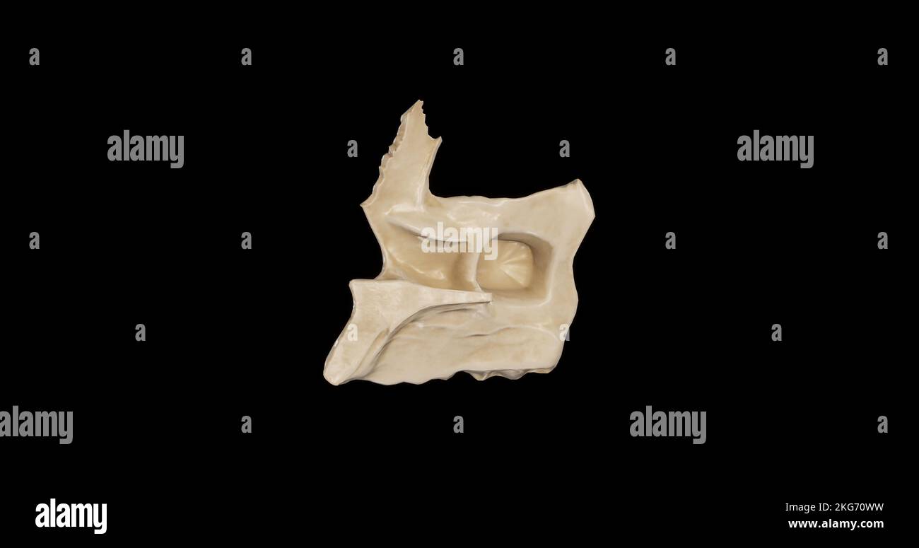 Medial view of Right Maxilla Stock Photohttps://www.alamy.com/image-license-details/?v=1https://www.alamy.com/medial-view-of-right-maxilla-image491879189.html
RF2KG70WW–Medial view of Right Maxilla
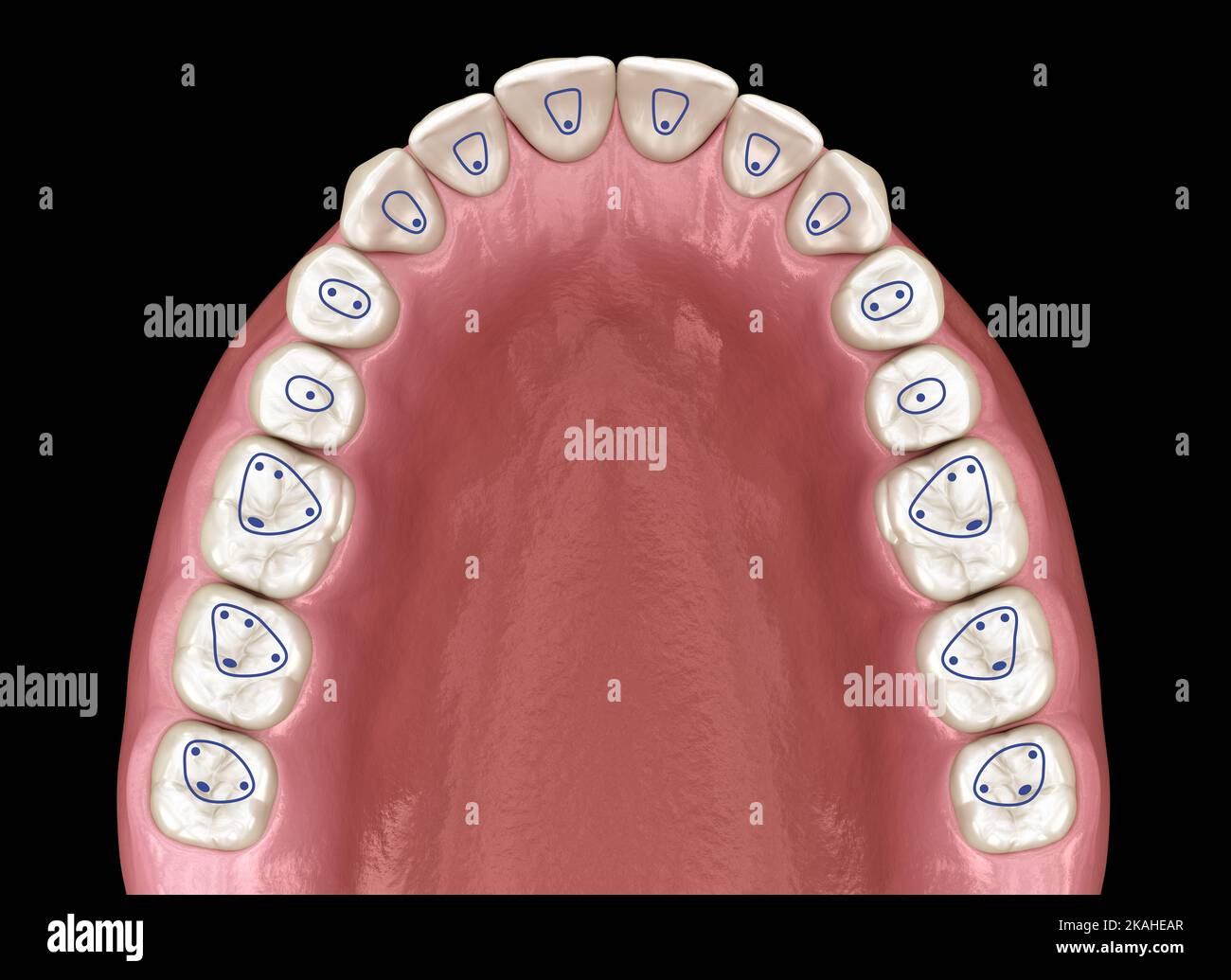 Root canal orifice locations plan of maxillary jaw. 3D illustration Stock Photohttps://www.alamy.com/image-license-details/?v=1https://www.alamy.com/root-canal-orifice-locations-plan-of-maxillary-jaw-3d-illustration-image488421327.html
RF2KAHEAR–Root canal orifice locations plan of maxillary jaw. 3D illustration
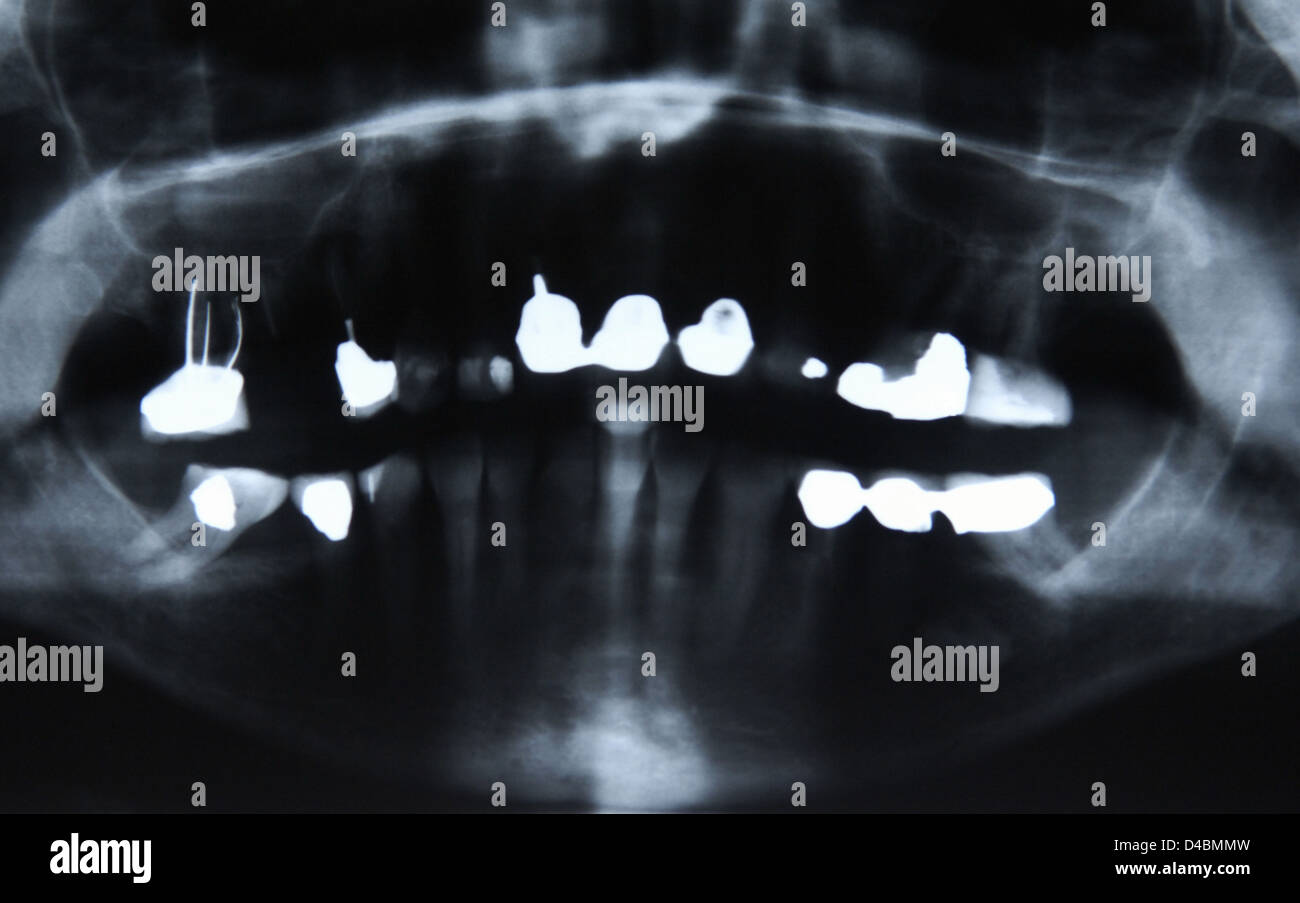 Dentistry X-ray image of female jaw and teeth Stock Photohttps://www.alamy.com/image-license-details/?v=1https://www.alamy.com/stock-photo-dentistry-x-ray-image-of-female-jaw-and-teeth-54347465.html
RMD4BMMW–Dentistry X-ray image of female jaw and teeth
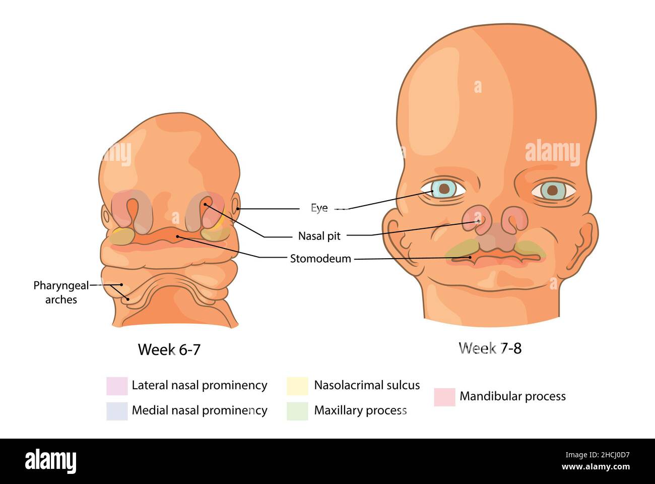 Embryological development of the face weeks 6-8 Stock Photohttps://www.alamy.com/image-license-details/?v=1https://www.alamy.com/embryological-development-of-the-face-weeks-6-8-image455240947.html
RF2HCJ0D7–Embryological development of the face weeks 6-8
 Skull with open jaw, illustration. Stock Photohttps://www.alamy.com/image-license-details/?v=1https://www.alamy.com/skull-with-open-jaw-illustration-image367367003.html
RF2C9K0A3–Skull with open jaw, illustration.
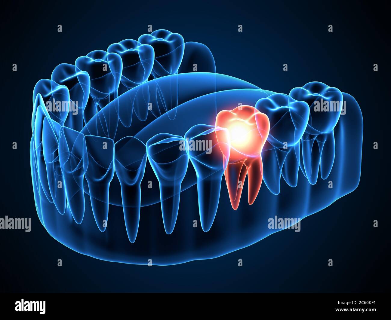 3d render of jaw x-ray with aching molar tooth. Toothache concept. Stock Photohttps://www.alamy.com/image-license-details/?v=1https://www.alamy.com/3d-render-of-jaw-x-ray-with-aching-molar-tooth-toothache-concept-image365120981.html
RF2C60KF1–3d render of jaw x-ray with aching molar tooth. Toothache concept.
 at dentist medic orthodontic doctor examination Stock Photohttps://www.alamy.com/image-license-details/?v=1https://www.alamy.com/stock-photo-at-dentist-medic-orthodontic-doctor-examination-103326812.html
RFG02XB8–at dentist medic orthodontic doctor examination
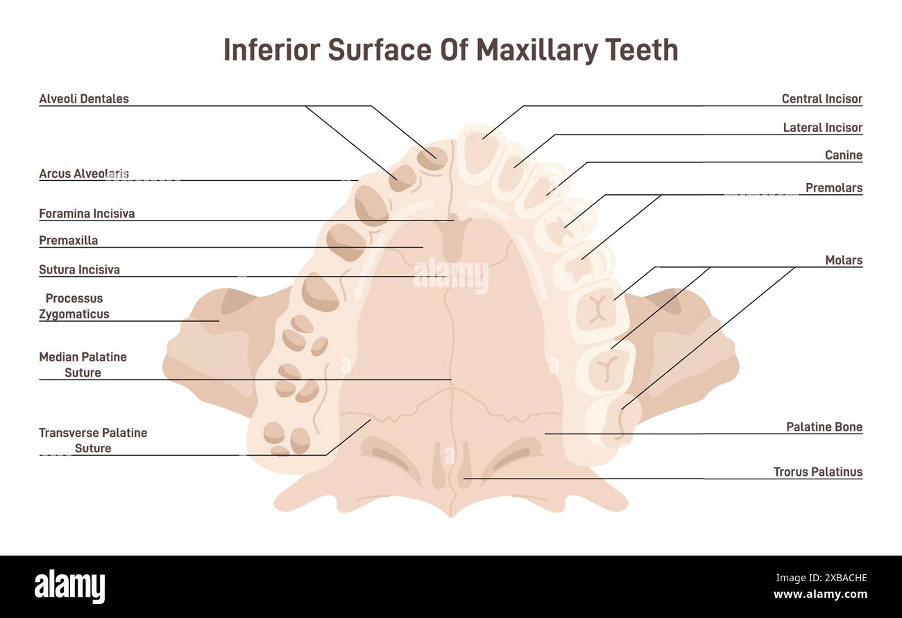 Maxillary anatomy. Inferior surface of upper jaw skeletal structure with teeth. Dentistry infographic. Flat vector illustration Stock Vectorhttps://www.alamy.com/image-license-details/?v=1https://www.alamy.com/maxillary-anatomy-inferior-surface-of-upper-jaw-skeletal-structure-with-teeth-dentistry-infographic-flat-vector-illustration-image609353514.html
RF2XBACHE–Maxillary anatomy. Inferior surface of upper jaw skeletal structure with teeth. Dentistry infographic. Flat vector illustration
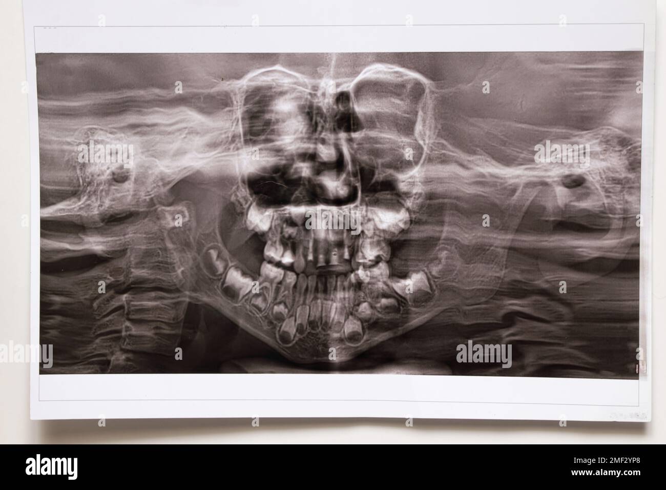 A panoramic X-ray shows several embedded and impacted teeth on both the upper and lower jaw of children's teeth Stock Photohttps://www.alamy.com/image-license-details/?v=1https://www.alamy.com/a-panoramic-x-ray-shows-several-embedded-and-impacted-teeth-on-both-the-upper-and-lower-jaw-of-childrens-teeth-image508386208.html
RF2MF2YP8–A panoramic X-ray shows several embedded and impacted teeth on both the upper and lower jaw of children's teeth
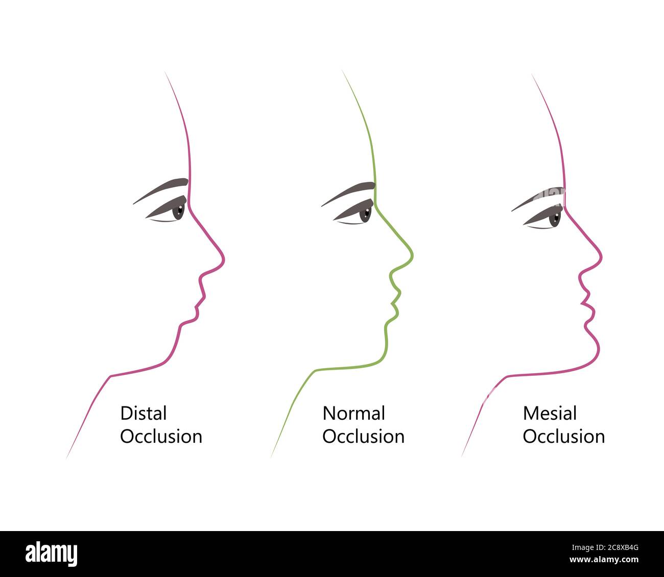 distal, Normal, and Mesial bite profile, vector illustration. Overbite or underbite before and after orthodontic treatment. Human with malocclusion Stock Vectorhttps://www.alamy.com/image-license-details/?v=1https://www.alamy.com/distal-normal-and-mesial-bite-profile-vector-illustration-overbite-or-underbite-before-and-after-orthodontic-treatment-human-with-malocclusion-image366914480.html
RF2C8XB4G–distal, Normal, and Mesial bite profile, vector illustration. Overbite or underbite before and after orthodontic treatment. Human with malocclusion
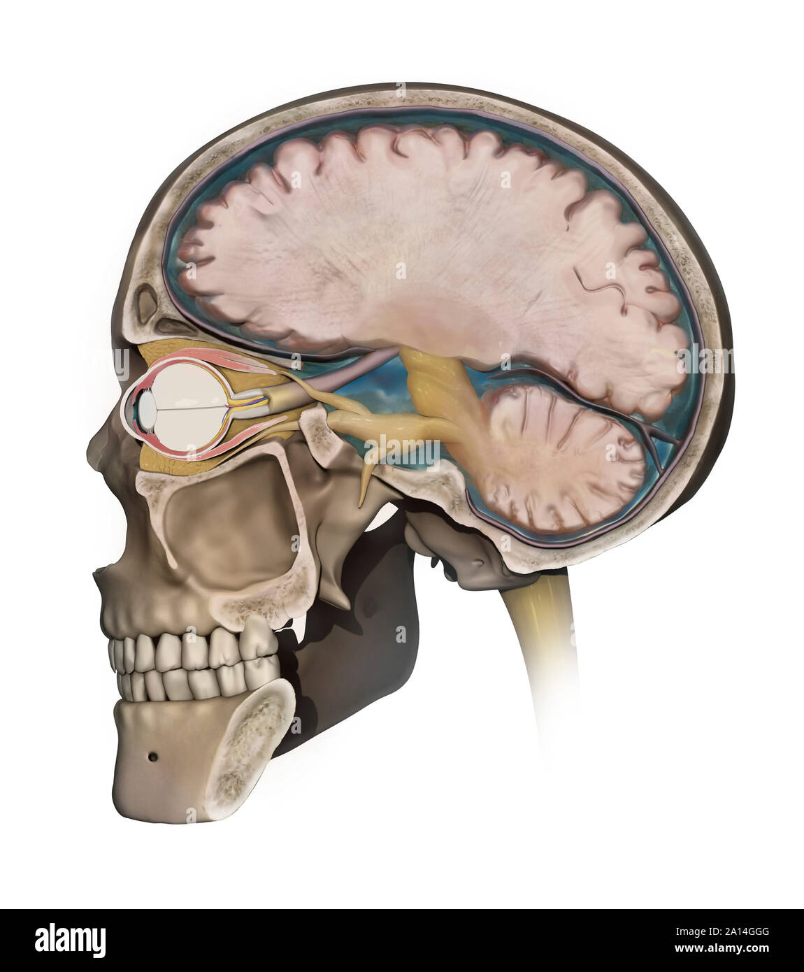 Medical illustration depicting the anatomy of a sagittal section of the human cranium. Stock Photohttps://www.alamy.com/image-license-details/?v=1https://www.alamy.com/medical-illustration-depicting-the-anatomy-of-a-sagittal-section-of-the-human-cranium-image327712464.html
RF2A14GGG–Medical illustration depicting the anatomy of a sagittal section of the human cranium.
 Teeth mould dentist plaster impression 3D denture dentures lower jaw Cutout cut out white background isolated copy space Stock Photohttps://www.alamy.com/image-license-details/?v=1https://www.alamy.com/stock-photo-teeth-mould-dentist-plaster-impression-3d-denture-dentures-lower-jaw-92299428.html
RMFA4GT4–Teeth mould dentist plaster impression 3D denture dentures lower jaw Cutout cut out white background isolated copy space
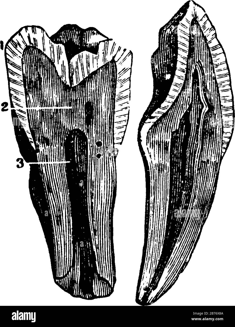 The internal view of a tooth cut through from the top or crown to the tips of the root, with the parts labelled, 1, enamel; 2, dentine; 3, pulp, vinta Stock Vectorhttps://www.alamy.com/image-license-details/?v=1https://www.alamy.com/the-internal-view-of-a-tooth-cut-through-from-the-top-or-crown-to-the-tips-of-the-root-with-the-parts-labelled-1-enamel-2-dentine-3-pulp-vinta-image348662282.html
RF2B76X8A–The internal view of a tooth cut through from the top or crown to the tips of the root, with the parts labelled, 1, enamel; 2, dentine; 3, pulp, vinta
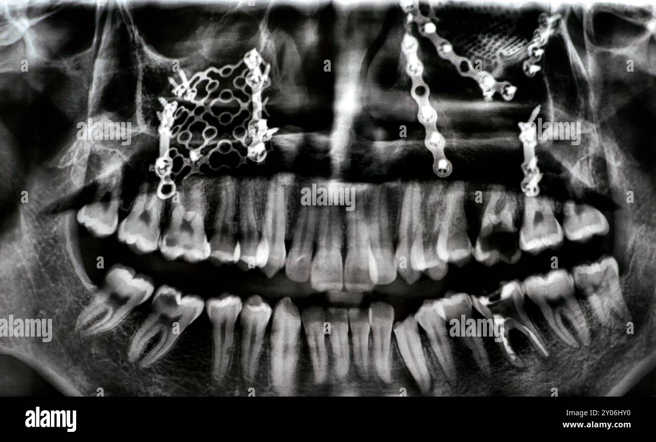 Panoramic X Ray shows multiple plates, screws for maxillary fixation of a fractured maxilla, titanium mesh on right side, decayed UL6, LL6 failed root Stock Photohttps://www.alamy.com/image-license-details/?v=1https://www.alamy.com/panoramic-x-ray-shows-multiple-plates-screws-for-maxillary-fixation-of-a-fractured-maxilla-titanium-mesh-on-right-side-decayed-ul6-ll6-failed-root-image619719044.html
RF2Y06HY0–Panoramic X Ray shows multiple plates, screws for maxillary fixation of a fractured maxilla, titanium mesh on right side, decayed UL6, LL6 failed root
 3d rendered medically accurate illustration of the skull with open jaw Stock Photohttps://www.alamy.com/image-license-details/?v=1https://www.alamy.com/3d-rendered-medically-accurate-illustration-of-the-skull-with-open-jaw-image334050053.html
RF2ABD871–3d rendered medically accurate illustration of the skull with open jaw
 The blood supply of the jaw Stock Photohttps://www.alamy.com/image-license-details/?v=1https://www.alamy.com/stock-photo-the-blood-supply-of-the-jaw-13174915.html
RFACK1MM–The blood supply of the jaw
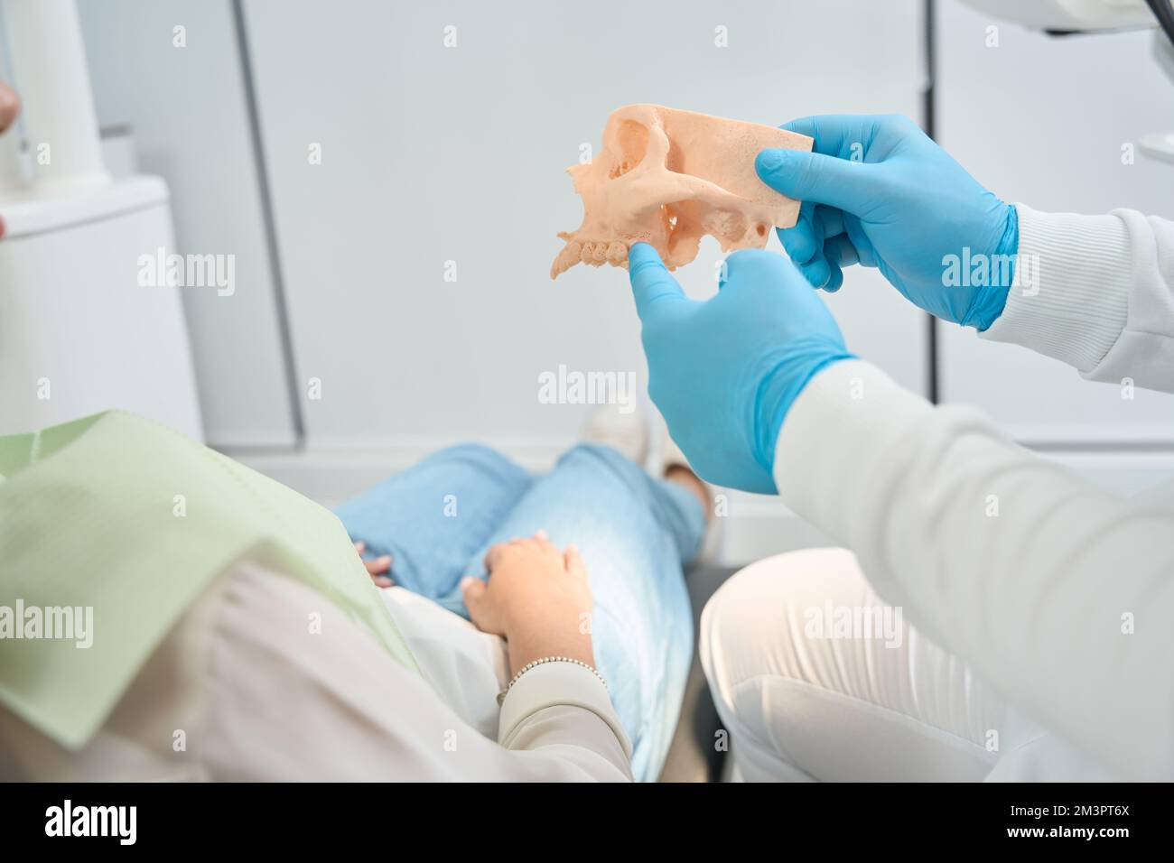 Stomatologist is telling about upper jawbone density to client Stock Photohttps://www.alamy.com/image-license-details/?v=1https://www.alamy.com/stomatologist-is-telling-about-upper-jawbone-density-to-client-image501446594.html
RF2M3PT6X–Stomatologist is telling about upper jawbone density to client
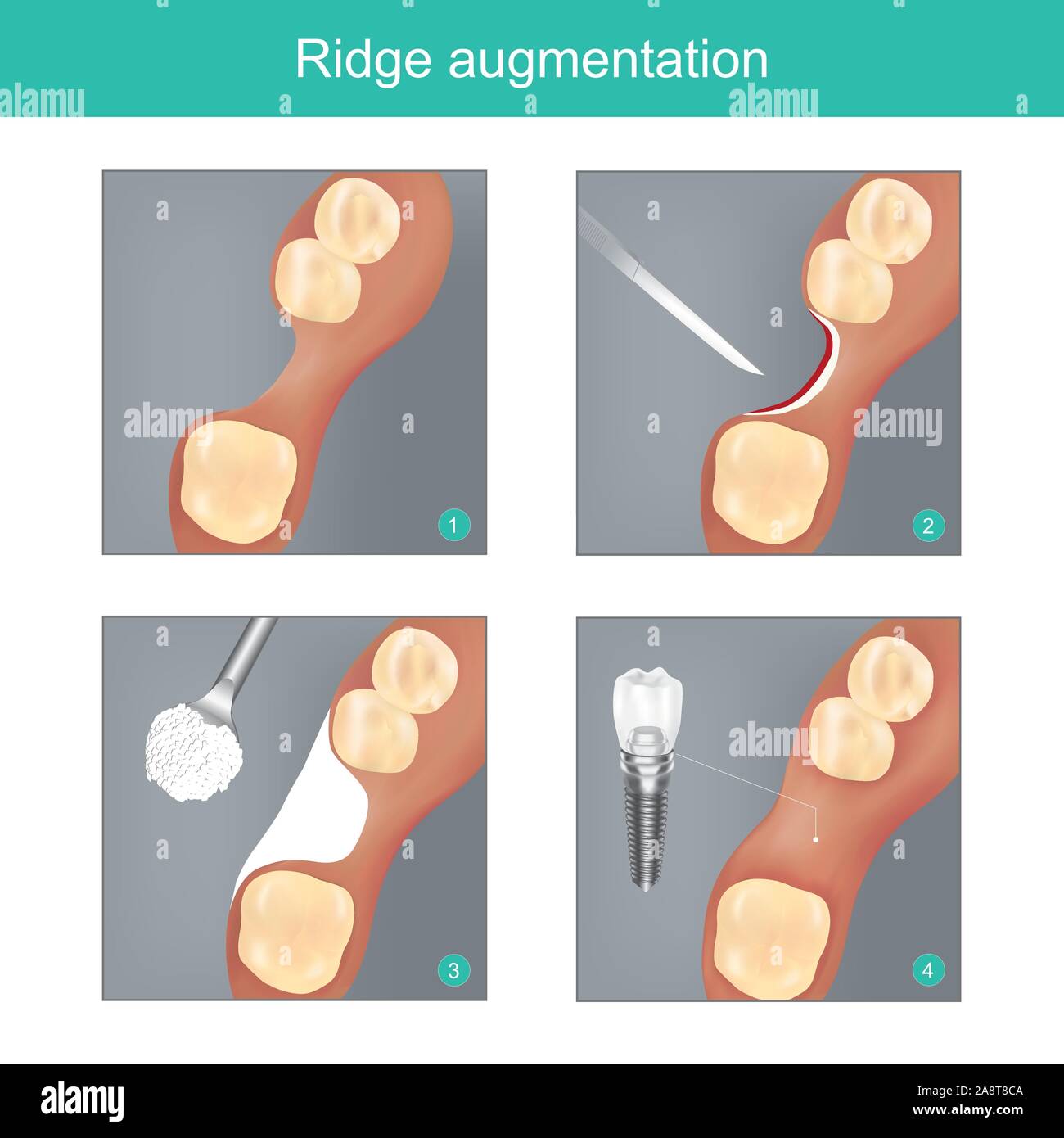 Ridge augmentation. The method dental used materials synthetic or human bone for Replace the Jawbone In missing, and to Prepare dental implant work to Stock Vectorhttps://www.alamy.com/image-license-details/?v=1https://www.alamy.com/ridge-augmentation-the-method-dental-used-materials-synthetic-or-human-bone-for-replace-the-jawbone-in-missing-and-to-prepare-dental-implant-work-to-image332447706.html
RF2A8T8CA–Ridge augmentation. The method dental used materials synthetic or human bone for Replace the Jawbone In missing, and to Prepare dental implant work to
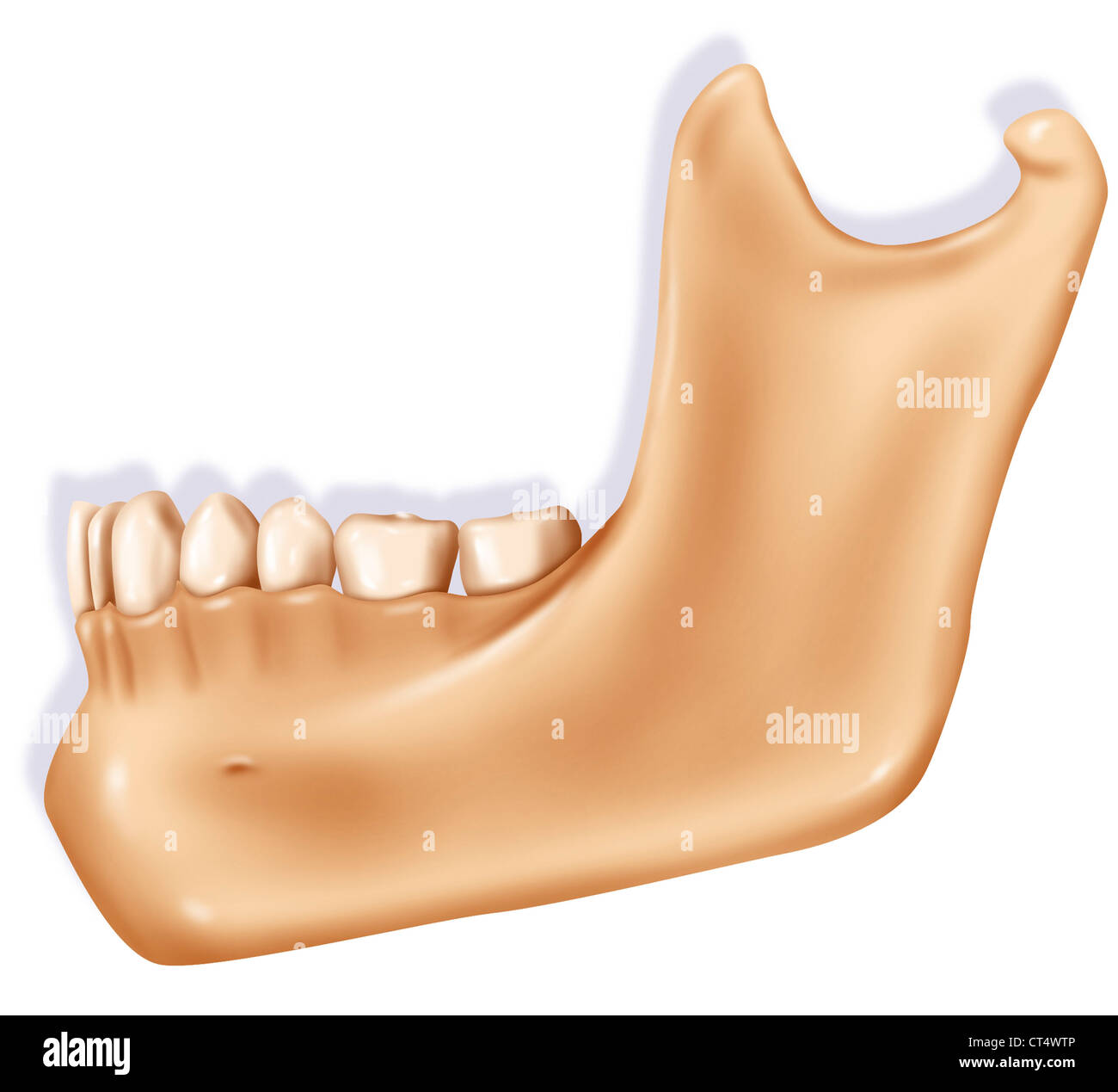 JAW, ILLUSTRATION Stock Photohttps://www.alamy.com/image-license-details/?v=1https://www.alamy.com/stock-photo-jaw-illustration-49280582.html
RMCT4WTP–JAW, ILLUSTRATION
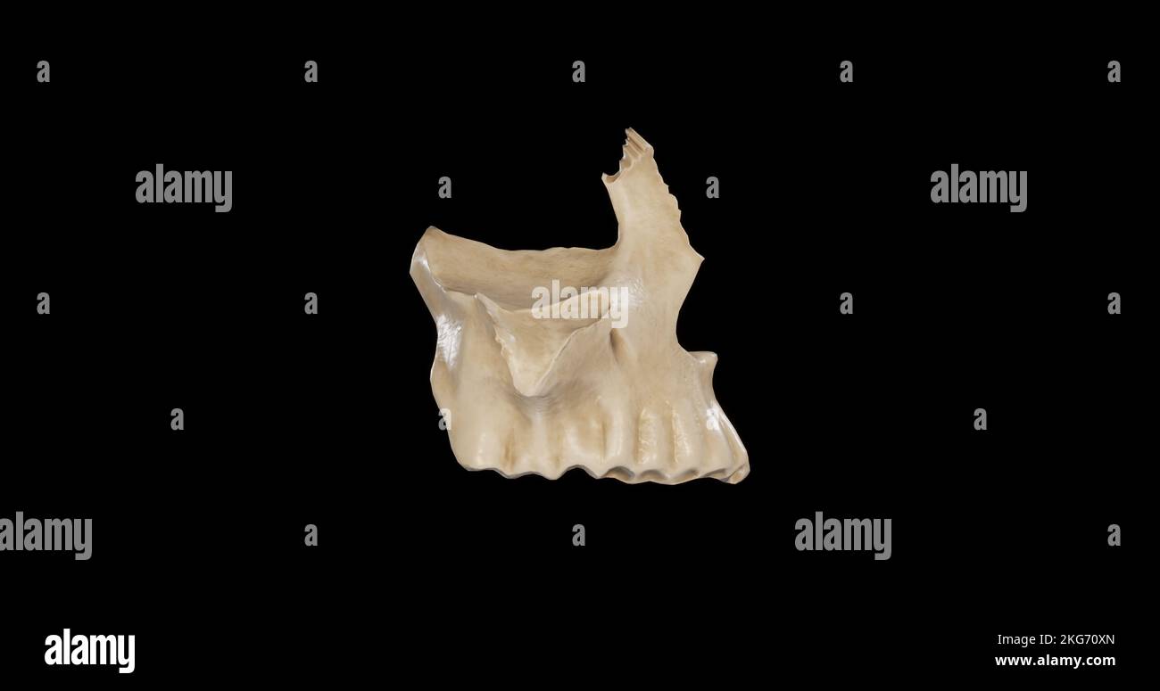 Lateral view of Right Maxilla Stock Photohttps://www.alamy.com/image-license-details/?v=1https://www.alamy.com/lateral-view-of-right-maxilla-image491879213.html
RF2KG70XN–Lateral view of Right Maxilla
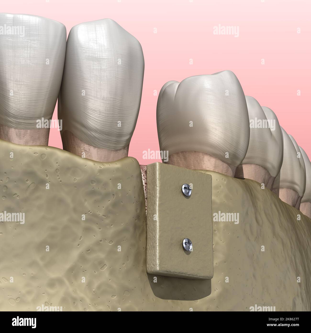 Augmentation Surgery - Bone transplantation, preparing for implantation. 3D illustration Stock Photohttps://www.alamy.com/image-license-details/?v=1https://www.alamy.com/augmentation-surgery-bone-transplantation-preparing-for-implantation-3d-illustration-image486941052.html
RF2K8627T–Augmentation Surgery - Bone transplantation, preparing for implantation. 3D illustration
 Dentistry X-ray image of female jaw and teeth Stock Photohttps://www.alamy.com/image-license-details/?v=1https://www.alamy.com/stock-photo-dentistry-x-ray-image-of-female-jaw-and-teeth-54347484.html
RMD4BMNG–Dentistry X-ray image of female jaw and teeth
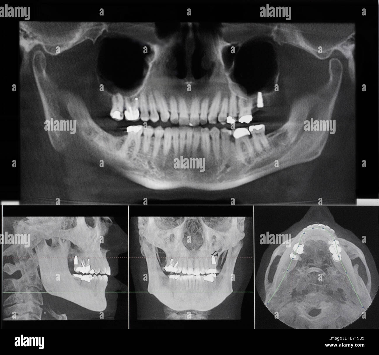 Teeth x-ray Stock Photohttps://www.alamy.com/image-license-details/?v=1https://www.alamy.com/stock-photo-teeth-x-ray-33835401.html
RMBY19B5–Teeth x-ray
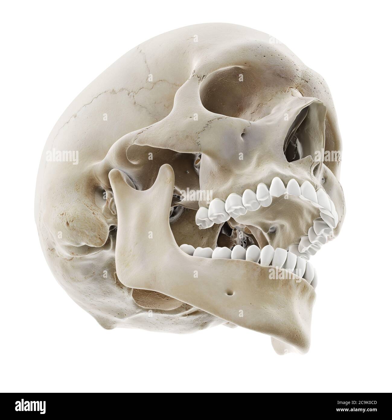 Skull with open jaw, illustration. Stock Photohttps://www.alamy.com/image-license-details/?v=1https://www.alamy.com/skull-with-open-jaw-illustration-image367367069.html
RF2C9K0CD–Skull with open jaw, illustration.
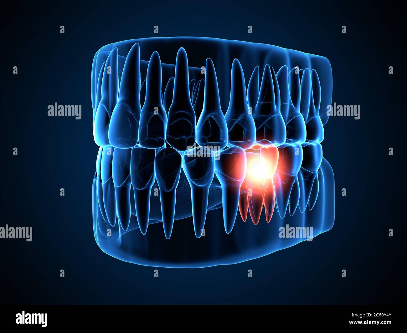 3d render of jaw x-ray with aching molar tooth. Toothache concept. Stock Photohttps://www.alamy.com/image-license-details/?v=1https://www.alamy.com/3d-render-of-jaw-x-ray-with-aching-molar-tooth-toothache-concept-image365126971.html
RF2C60Y4Y–3d render of jaw x-ray with aching molar tooth. Toothache concept.
 at dentist medic orthodontic doctor examination Stock Photohttps://www.alamy.com/image-license-details/?v=1https://www.alamy.com/stock-photo-at-dentist-medic-orthodontic-doctor-examination-103327096.html
RFG02XNC–at dentist medic orthodontic doctor examination
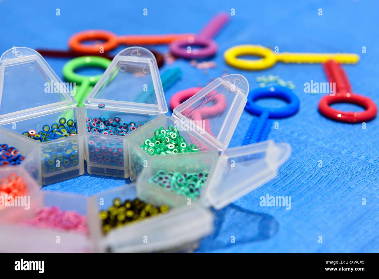 Colored Elastic Rubber Bands for Teeth Metal Braces. Close Up Stock Photohttps://www.alamy.com/image-license-details/?v=1https://www.alamy.com/colored-elastic-rubber-bands-for-teeth-metal-braces-close-up-image567271773.html
RF2RXWCX5–Colored Elastic Rubber Bands for Teeth Metal Braces. Close Up
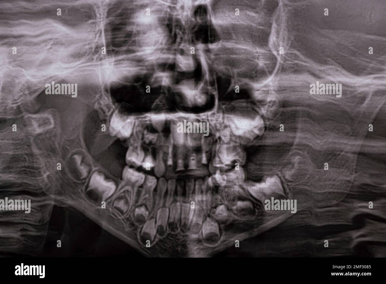 A panoramic X-ray shows several embedded and impacted teeth on both the upper and lower jaw of children's teeth Stock Photohttps://www.alamy.com/image-license-details/?v=1https://www.alamy.com/a-panoramic-x-ray-shows-several-embedded-and-impacted-teeth-on-both-the-upper-and-lower-jaw-of-childrens-teeth-image508386597.html
RF2MF3085–A panoramic X-ray shows several embedded and impacted teeth on both the upper and lower jaw of children's teeth
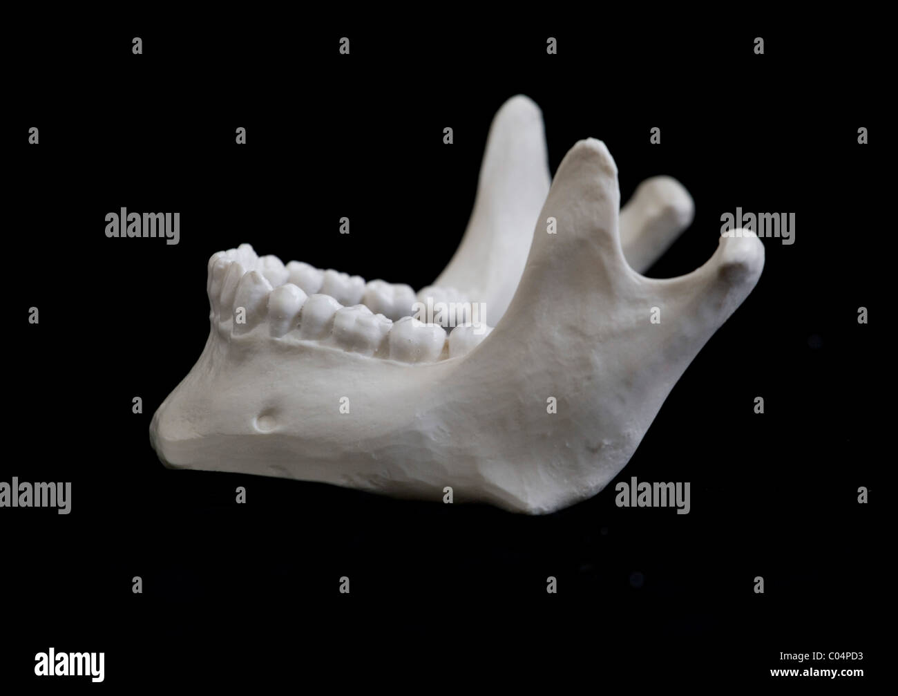 Human Skeletal Model, Mandible. Stock Photohttps://www.alamy.com/image-license-details/?v=1https://www.alamy.com/stock-photo-human-skeletal-model-mandible-34526159.html
RFC04PD3–Human Skeletal Model, Mandible.
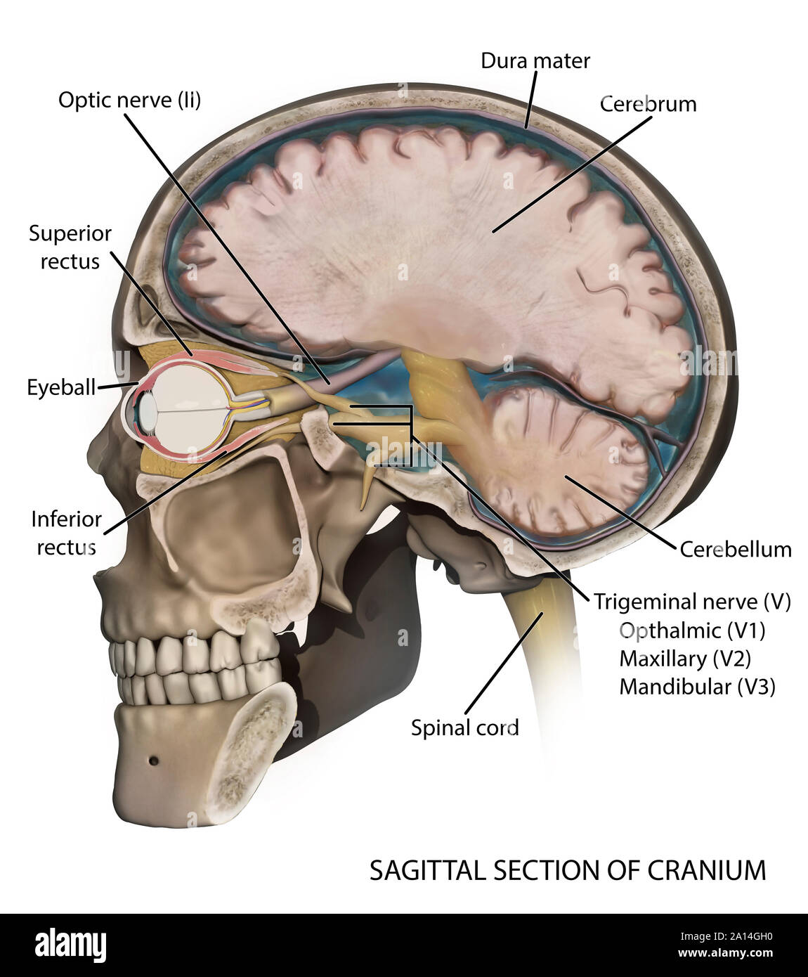 Medical illustration depicting the anatomy of a sagittal section of the human cranium. Stock Photohttps://www.alamy.com/image-license-details/?v=1https://www.alamy.com/medical-illustration-depicting-the-anatomy-of-a-sagittal-section-of-the-human-cranium-image327712476.html
RF2A14GH0–Medical illustration depicting the anatomy of a sagittal section of the human cranium.
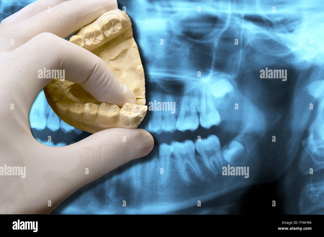 dentist showing molar tooth over panoramic dental radiography Stock Photohttps://www.alamy.com/image-license-details/?v=1https://www.alamy.com/stock-photo-dentist-showing-molar-tooth-over-panoramic-dental-radiography-102836754.html
RFFY8H96–dentist showing molar tooth over panoramic dental radiography
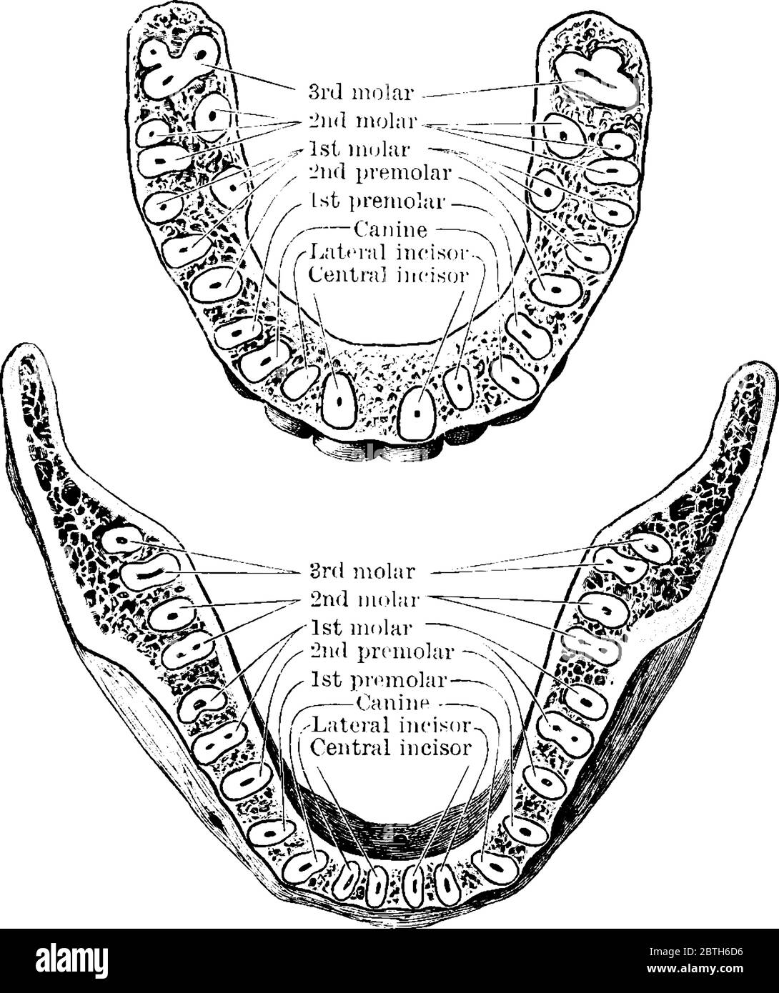 The horizontal section through both the upper and lower jaws, showing the roots of the teeth, vintage line drawing or engraving illustration. Stock Vectorhttps://www.alamy.com/image-license-details/?v=1https://www.alamy.com/the-horizontal-section-through-both-the-upper-and-lower-jaws-showing-the-roots-of-the-teeth-vintage-line-drawing-or-engraving-illustration-image359337362.html
RF2BTH6D6–The horizontal section through both the upper and lower jaws, showing the roots of the teeth, vintage line drawing or engraving illustration.
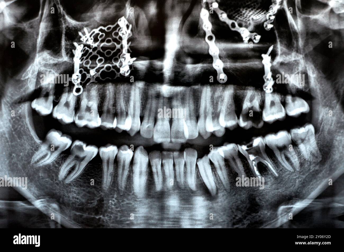 Panoramic X Ray shows multiple plates, screws for maxillary fixation of a fractured maxilla, titanium mesh on right side, decayed UL6, LL6 failed root Stock Photohttps://www.alamy.com/image-license-details/?v=1https://www.alamy.com/panoramic-x-ray-shows-multiple-plates-screws-for-maxillary-fixation-of-a-fractured-maxilla-titanium-mesh-on-right-side-decayed-ul6-ll6-failed-root-image619726197.html
RF2Y06Y2D–Panoramic X Ray shows multiple plates, screws for maxillary fixation of a fractured maxilla, titanium mesh on right side, decayed UL6, LL6 failed root
 3d rendered medically accurate illustration of the skull with open jaw Stock Photohttps://www.alamy.com/image-license-details/?v=1https://www.alamy.com/3d-rendered-medically-accurate-illustration-of-the-skull-with-open-jaw-image334050122.html
RF2ABD89E–3d rendered medically accurate illustration of the skull with open jaw
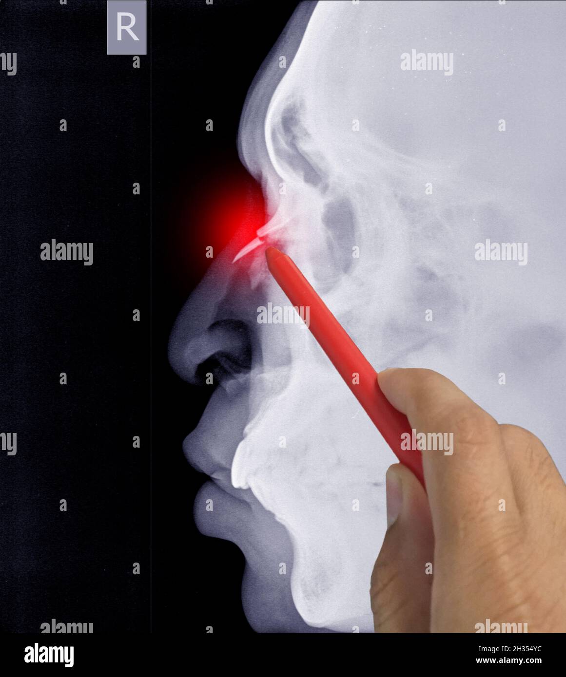 Close up X-ray Nasal bone Lateral showing fracture nasal bone, Doctor holding a red pen point, symptoms medical healthcare concept. Stock Photohttps://www.alamy.com/image-license-details/?v=1https://www.alamy.com/close-up-x-ray-nasal-bone-lateral-showing-fracture-nasal-bone-doctor-holding-a-red-pen-point-symptoms-medical-healthcare-concept-image449427200.html
RF2H354YC–Close up X-ray Nasal bone Lateral showing fracture nasal bone, Doctor holding a red pen point, symptoms medical healthcare concept.
 Stomatologist explaining anatomy of cheekbone to client Stock Photohttps://www.alamy.com/image-license-details/?v=1https://www.alamy.com/stomatologist-explaining-anatomy-of-cheekbone-to-client-image501446612.html
RF2M3PT7G–Stomatologist explaining anatomy of cheekbone to client
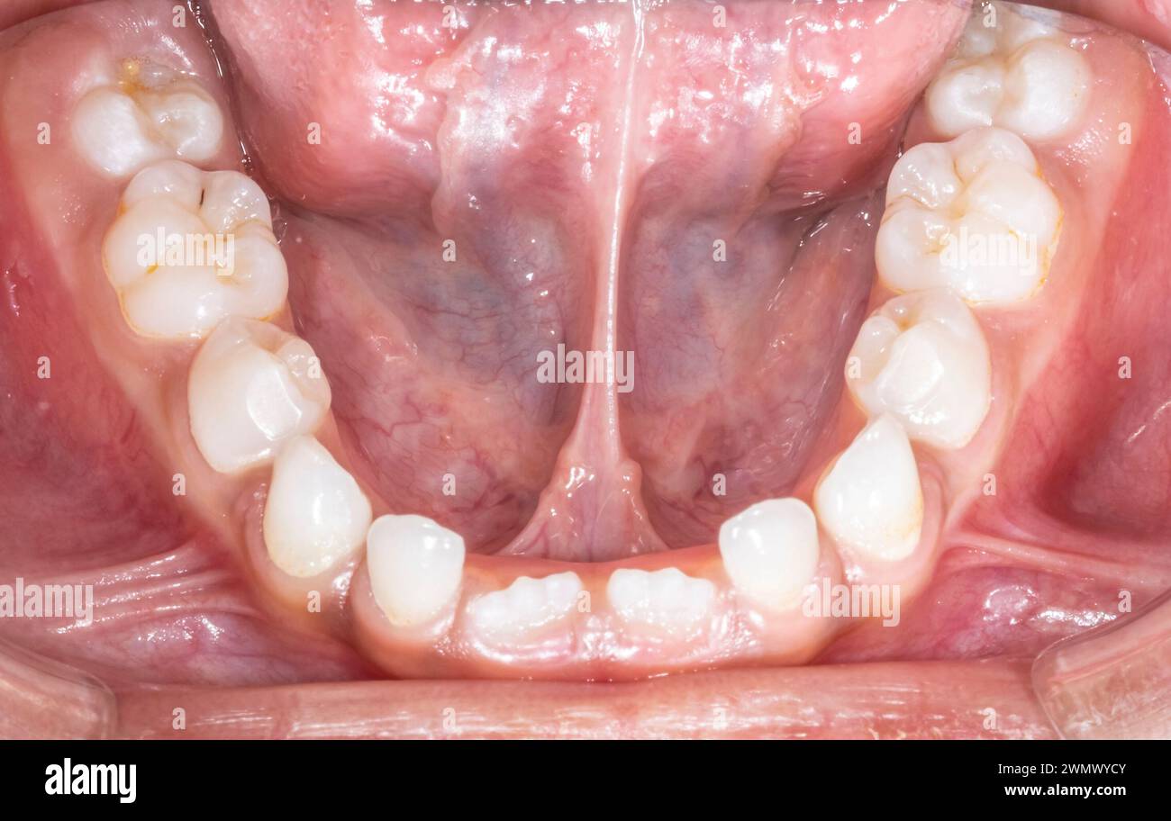 Mandibular arch occlusal view of a child milky dentition and permanent incisors erupting, tongue lifted with voluminous lingual frenum. Stock Photohttps://www.alamy.com/image-license-details/?v=1https://www.alamy.com/mandibular-arch-occlusal-view-of-a-child-milky-dentition-and-permanent-incisors-erupting-tongue-lifted-with-voluminous-lingual-frenum-image598015963.html
RF2WMWYCY–Mandibular arch occlusal view of a child milky dentition and permanent incisors erupting, tongue lifted with voluminous lingual frenum.
 FRACTURED JAW Stock Photohttps://www.alamy.com/image-license-details/?v=1https://www.alamy.com/stock-photo-fractured-jaw-49254266.html
RMCT3M8X–FRACTURED JAW
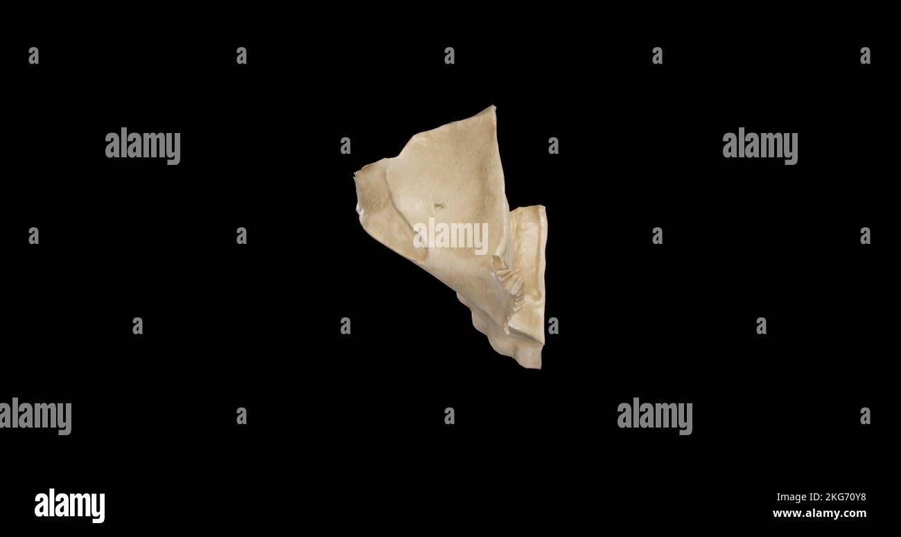 Superior view of Right Maxilla Stock Photohttps://www.alamy.com/image-license-details/?v=1https://www.alamy.com/superior-view-of-right-maxilla-image491879228.html
RF2KG70Y8–Superior view of Right Maxilla
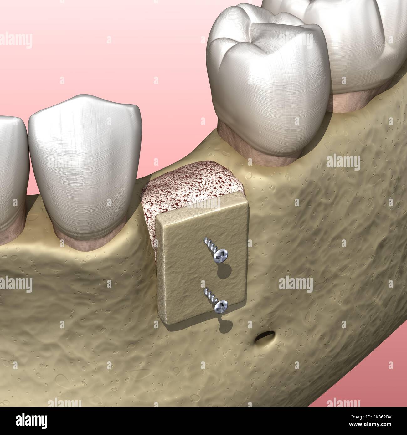 Augmentation Surgery - Bone transplantation, preparing for implantation. 3D illustration Stock Photohttps://www.alamy.com/image-license-details/?v=1https://www.alamy.com/augmentation-surgery-bone-transplantation-preparing-for-implantation-3d-illustration-image486941166.html
RF2K862BX–Augmentation Surgery - Bone transplantation, preparing for implantation. 3D illustration
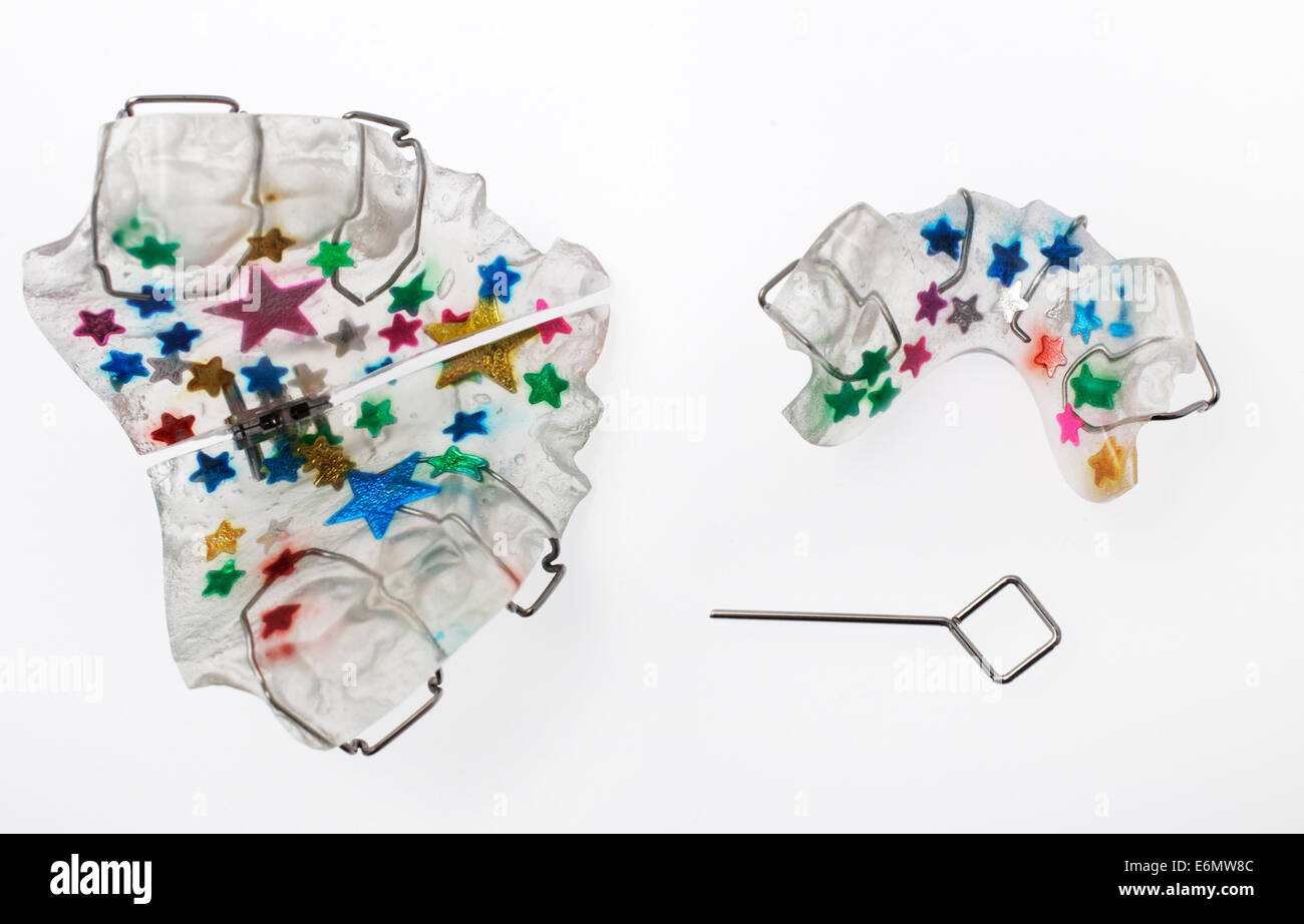 Multicolored upper and lower orthodontic appliance called twin block with a key used for adjusting the expansion screw Stock Photohttps://www.alamy.com/image-license-details/?v=1https://www.alamy.com/stock-photo-multicolored-upper-and-lower-orthodontic-appliance-called-twin-block-72988284.html
RFE6MW8C–Multicolored upper and lower orthodontic appliance called twin block with a key used for adjusting the expansion screw
