Quick filters:
Nasopalatine Stock Photos and Images
 One person is answering question about dental care. She is at risk for nasopalatine cyst. Stock Photohttps://www.alamy.com/image-license-details/?v=1https://www.alamy.com/one-person-is-answering-question-about-dental-care-she-is-at-risk-for-nasopalatine-cyst-image538592024.html
RF2P86YHC–One person is answering question about dental care. She is at risk for nasopalatine cyst.
 Applied anatomy and oral surgery for dental students . nasi, angular. The branches of the internal maxillary artery are asfollows: {a) Maxillary portion: Tympanic, middlemeningeal, small meningeal, and inferior dental. (6)Pterygoid portion: Deep temporal, pterygoid, masseteric,and buccal. (c) Sphenomaxillary portion: Alveolarto the upper teeth, infra-orbital, descending palatine,vidian, pterygopalatine, and nasopalatine. The vertebral arteries are given off from the subclavianarteries, and pass upward one on either side of the neck,through the foramina in the transverse processes of thecervica Stock Photohttps://www.alamy.com/image-license-details/?v=1https://www.alamy.com/applied-anatomy-and-oral-surgery-for-dental-students-nasi-angular-the-branches-of-the-internal-maxillary-artery-are-asfollows-a-maxillary-portion-tympanic-middlemeningeal-small-meningeal-and-inferior-dental-6pterygoid-portion-deep-temporal-pterygoid-massetericand-buccal-c-sphenomaxillary-portion-alveolarto-the-upper-teeth-infra-orbital-descending-palatinevidian-pterygopalatine-and-nasopalatine-the-vertebral-arteries-are-given-off-from-the-subclavianarteries-and-pass-upward-one-on-either-side-of-the-neckthrough-the-foramina-in-the-transverse-processes-of-thecervica-image343235448.html
RM2AXBM8T–Applied anatomy and oral surgery for dental students . nasi, angular. The branches of the internal maxillary artery are asfollows: {a) Maxillary portion: Tympanic, middlemeningeal, small meningeal, and inferior dental. (6)Pterygoid portion: Deep temporal, pterygoid, masseteric,and buccal. (c) Sphenomaxillary portion: Alveolarto the upper teeth, infra-orbital, descending palatine,vidian, pterygopalatine, and nasopalatine. The vertebral arteries are given off from the subclavianarteries, and pass upward one on either side of the neck,through the foramina in the transverse processes of thecervica
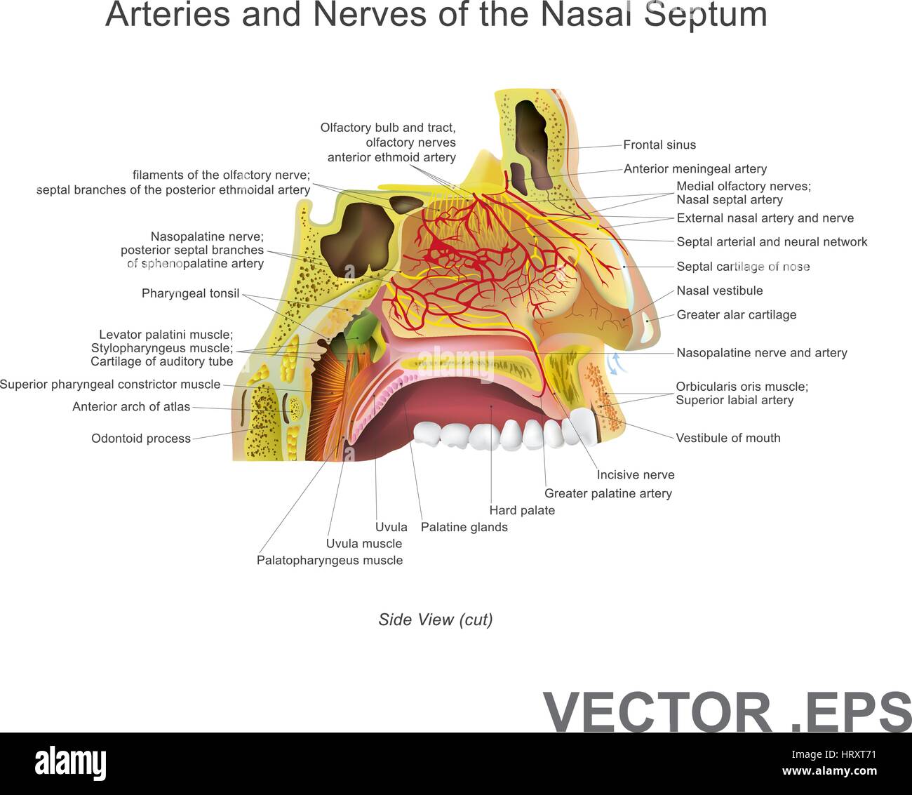 The nasal cavity (or nasal fossa) is a large air filled space above and behind the nose in the middle of the face. Each cavity is the continuation of Stock Vectorhttps://www.alamy.com/image-license-details/?v=1https://www.alamy.com/stock-photo-the-nasal-cavity-or-nasal-fossa-is-a-large-air-filled-space-above-135199429.html
RFHRXT71–The nasal cavity (or nasal fossa) is a large air filled space above and behind the nose in the middle of the face. Each cavity is the continuation of
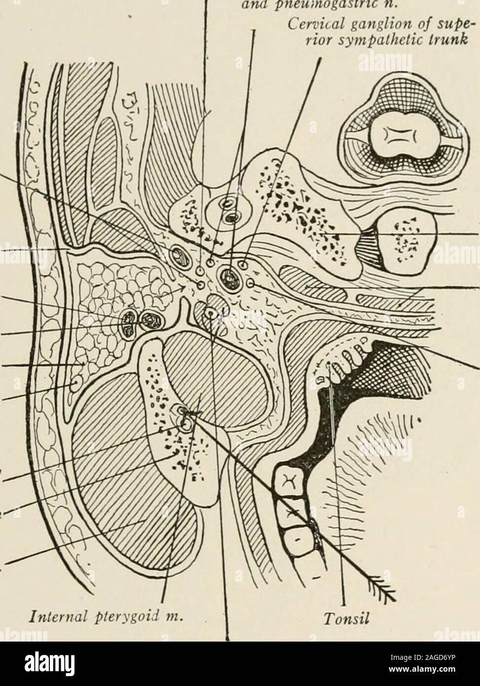 . Local and regional anesthesia; with chapters on spinal, epidural, paravertebral, and parasacral analgesia, and other applications of local and regional anesthesia to the surgery of the eye, ear, nose and throat, and to dental practice. 66.) These can be easily reachedby injections over the opening of the anterior and posterior palatinecanals, the anterior just behind the incisor teeth in the middle line THE HEAD, SCALP, CRANIUM, BRAIN, AND FACE ;69 (nasopalatine nerve) and the posterior just to the inner side of thelast molar tooth (anterior palatine), where the hard palate joins thealveolar Stock Photohttps://www.alamy.com/image-license-details/?v=1https://www.alamy.com/local-and-regional-anesthesia-with-chapters-on-spinal-epidural-paravertebral-and-parasacral-analgesia-and-other-applications-of-local-and-regional-anesthesia-to-the-surgery-of-the-eye-ear-nose-and-throat-and-to-dental-practice-66-these-can-be-easily-reachedby-injections-over-the-opening-of-the-anterior-and-posterior-palatinecanals-the-anterior-just-behind-the-incisor-teeth-in-the-middle-line-the-head-scalp-cranium-brain-and-face-69-nasopalatine-nerve-and-the-posterior-just-to-the-inner-side-of-thelast-molar-tooth-anterior-palatine-where-the-hard-palate-joins-thealveolar-image337122346.html
RM2AGD6YP–. Local and regional anesthesia; with chapters on spinal, epidural, paravertebral, and parasacral analgesia, and other applications of local and regional anesthesia to the surgery of the eye, ear, nose and throat, and to dental practice. 66.) These can be easily reachedby injections over the opening of the anterior and posterior palatinecanals, the anterior just behind the incisor teeth in the middle line THE HEAD, SCALP, CRANIUM, BRAIN, AND FACE ;69 (nasopalatine nerve) and the posterior just to the inner side of thelast molar tooth (anterior palatine), where the hard palate joins thealveolar
 . Local and regional anesthesia : with chapters on spinal, epidural, paravertebral, and parasacral analgesia, and on other applications of local and regional anesthesia to the surgery of the eye, ear, nose and throat, and to dental practice. Fig- 243-—Innervation of nasal septum. Fig. 244.—Innervation of lateral nasal (Braun.) wall: /, Olfactory nerve; //, nasal nerve; ///, nasopalatine nerve. (Braun.). Fig. 245.—Points of anesthesia for Killian regional method. (Braun.) 596 LOCAL ANESTHESIA The middle turbinate is usually much simpler, and is treated in muchthe same manner. The nasofrontal du Stock Photohttps://www.alamy.com/image-license-details/?v=1https://www.alamy.com/local-and-regional-anesthesia-with-chapters-on-spinal-epidural-paravertebral-and-parasacral-analgesia-and-on-other-applications-of-local-and-regional-anesthesia-to-the-surgery-of-the-eye-ear-nose-and-throat-and-to-dental-practice-fig-243-innervation-of-nasal-septum-fig-244innervation-of-lateral-nasal-braun-wall-olfactory-nerve-nasal-nerve-nasopalatine-nerve-braun-fig-245points-of-anesthesia-for-killian-regional-method-braun-596-local-anesthesia-the-middle-turbinate-is-usually-much-simpler-and-is-treated-in-muchthe-same-manner-the-nasofrontal-du-image336626236.html
RM2AFJJ5G–. Local and regional anesthesia : with chapters on spinal, epidural, paravertebral, and parasacral analgesia, and on other applications of local and regional anesthesia to the surgery of the eye, ear, nose and throat, and to dental practice. Fig- 243-—Innervation of nasal septum. Fig. 244.—Innervation of lateral nasal (Braun.) wall: /, Olfactory nerve; //, nasal nerve; ///, nasopalatine nerve. (Braun.). Fig. 245.—Points of anesthesia for Killian regional method. (Braun.) 596 LOCAL ANESTHESIA The middle turbinate is usually much simpler, and is treated in muchthe same manner. The nasofrontal du
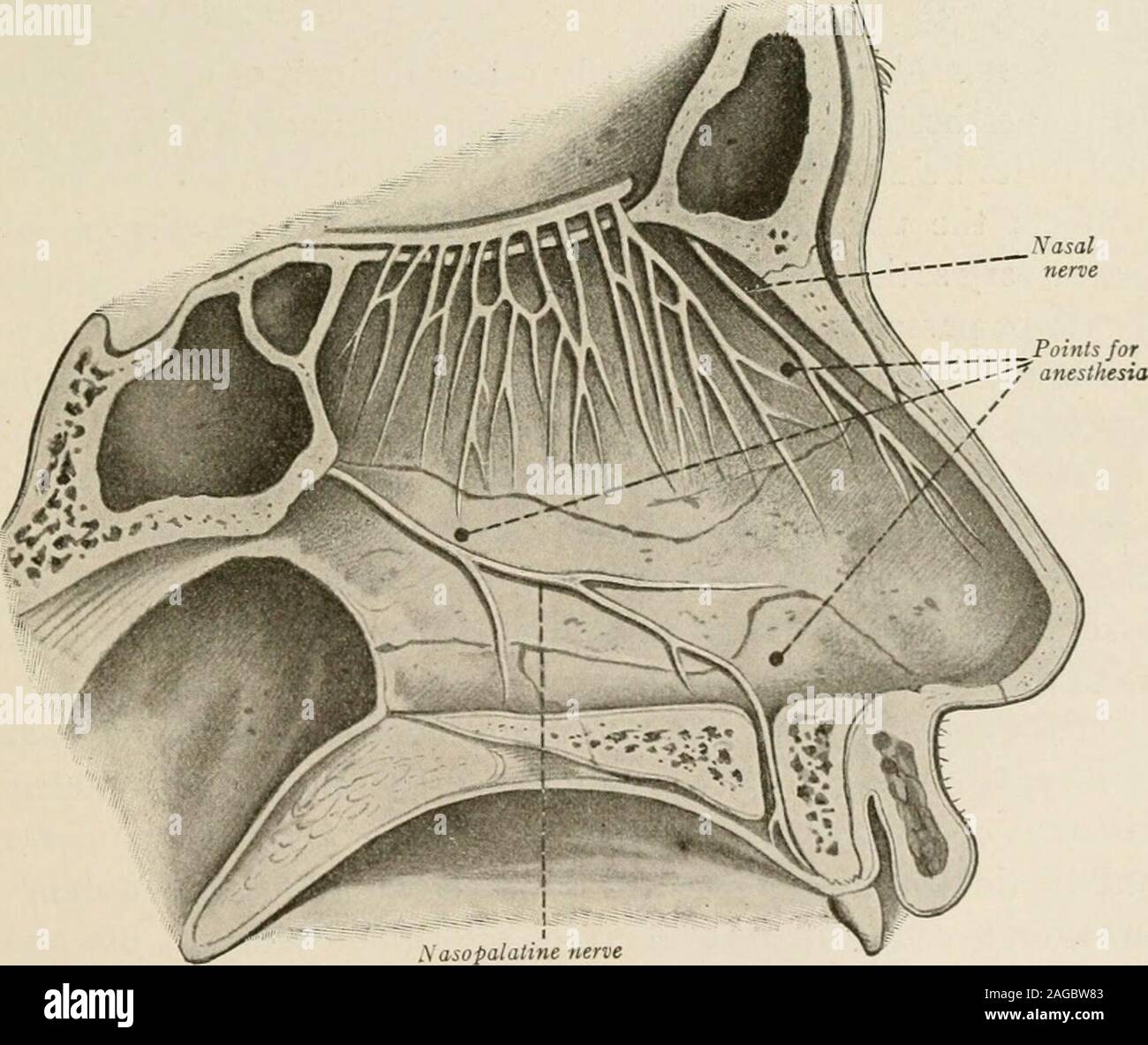 . Local and regional anesthesia; with chapters on spinal, epidural, paravertebral, and parasacral analgesia, and other applications of local and regional anesthesia to the surgery of the eye, ear, nose and throat, and to dental practice. Fig. 248.—Innervation of nasal septum. Fig. 249.—Innervation of lateral nasal (Braun.) wall: /, Olfactory nerve; II, nasal nerve J ///, nasopalatine nerve. (Braun.). Fig. 250.—Points of anesthesia for Killian regional method. (Braun.) 41 642 LOCAL ANESTHESIA croaching upon the surrounding parts; however, the application ofthe cocain-adrenahn solution soon prod Stock Photohttps://www.alamy.com/image-license-details/?v=1https://www.alamy.com/local-and-regional-anesthesia-with-chapters-on-spinal-epidural-paravertebral-and-parasacral-analgesia-and-other-applications-of-local-and-regional-anesthesia-to-the-surgery-of-the-eye-ear-nose-and-throat-and-to-dental-practice-fig-248innervation-of-nasal-septum-fig-249innervation-of-lateral-nasal-braun-wall-olfactory-nerve-ii-nasal-nerve-j-nasopalatine-nerve-braun-fig-250points-of-anesthesia-for-killian-regional-method-braun-41-642-local-anesthesia-croaching-upon-the-surrounding-parts-however-the-application-ofthe-cocain-adrenahn-solution-soon-prod-image337092787.html
RM2AGBW83–. Local and regional anesthesia; with chapters on spinal, epidural, paravertebral, and parasacral analgesia, and other applications of local and regional anesthesia to the surgery of the eye, ear, nose and throat, and to dental practice. Fig. 248.—Innervation of nasal septum. Fig. 249.—Innervation of lateral nasal (Braun.) wall: /, Olfactory nerve; II, nasal nerve J ///, nasopalatine nerve. (Braun.). Fig. 250.—Points of anesthesia for Killian regional method. (Braun.) 41 642 LOCAL ANESTHESIA croaching upon the surrounding parts; however, the application ofthe cocain-adrenahn solution soon prod
 . Regional anesthesia : its technic and clinical application . Fig. 54.—The sphenopalatine ganglion and palatine nerves. inosculate with the terminal branches of the nasopalatine nerve (Fig.55). It supplies the posterior half of the hard palate and its mucousmembrane. In the palatine canal the anterior palatine nerve givesoff branches to the mucous membrane of the turbinates, except theanterior portion of the inferior turbinate. (b) The posterior palatine nerves run parallel to the anterior palatinenerve and supply the mucous membrane of the soft palate and thetonsil. (c) The nasopalatine nerv Stock Photohttps://www.alamy.com/image-license-details/?v=1https://www.alamy.com/regional-anesthesia-its-technic-and-clinical-application-fig-54the-sphenopalatine-ganglion-and-palatine-nerves-inosculate-with-the-terminal-branches-of-the-nasopalatine-nerve-fig55-it-supplies-the-posterior-half-of-the-hard-palate-and-its-mucousmembrane-in-the-palatine-canal-the-anterior-palatine-nerve-givesoff-branches-to-the-mucous-membrane-of-the-turbinates-except-theanterior-portion-of-the-inferior-turbinate-b-the-posterior-palatine-nerves-run-parallel-to-the-anterior-palatinenerve-and-supply-the-mucous-membrane-of-the-soft-palate-and-thetonsil-c-the-nasopalatine-nerv-image370065887.html
RM2CE1XPR–. Regional anesthesia : its technic and clinical application . Fig. 54.—The sphenopalatine ganglion and palatine nerves. inosculate with the terminal branches of the nasopalatine nerve (Fig.55). It supplies the posterior half of the hard palate and its mucousmembrane. In the palatine canal the anterior palatine nerve givesoff branches to the mucous membrane of the turbinates, except theanterior portion of the inferior turbinate. (b) The posterior palatine nerves run parallel to the anterior palatinenerve and supply the mucous membrane of the soft palate and thetonsil. (c) The nasopalatine nerv
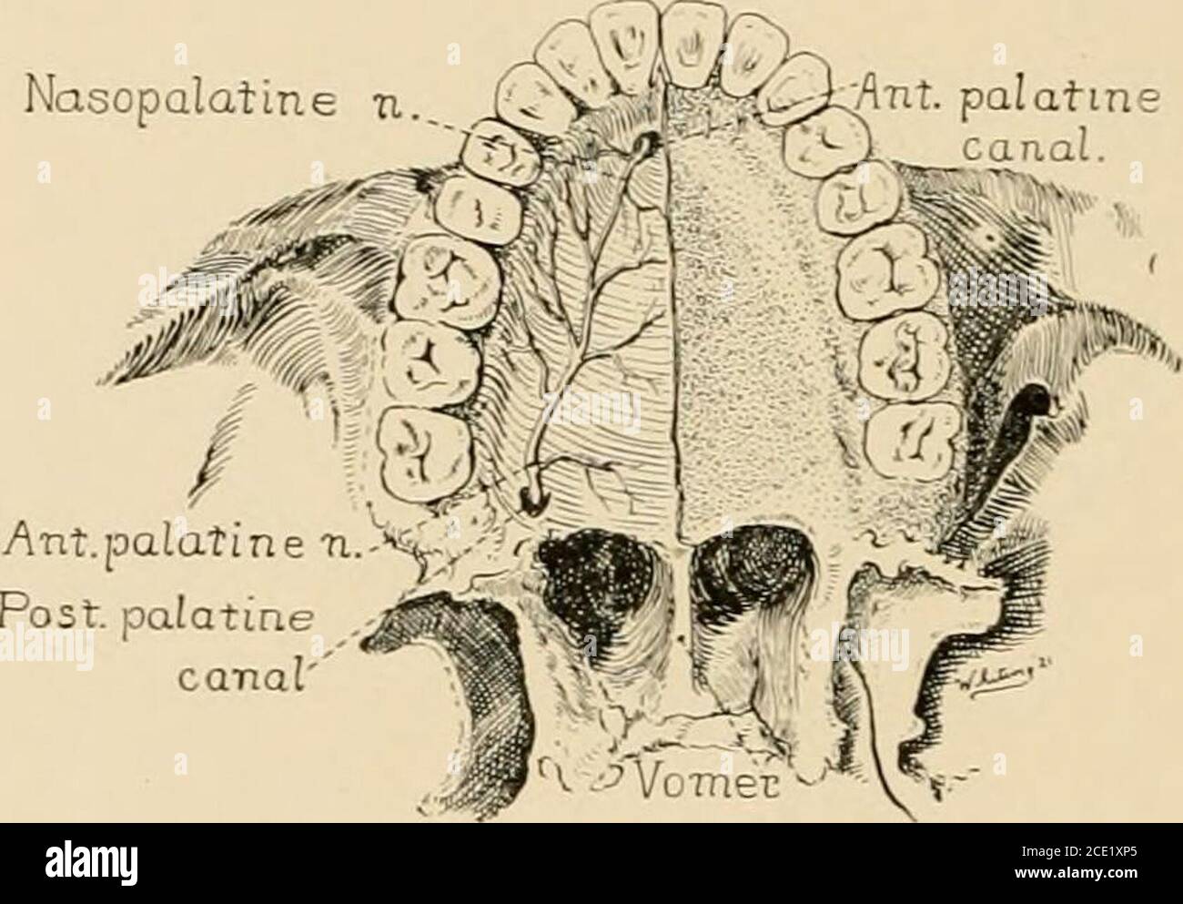 . Regional anesthesia : its technic and clinical application . embrane of the turbinates, except theanterior portion of the inferior turbinate. (b) The posterior palatine nerves run parallel to the anterior palatinenerve and supply the mucous membrane of the soft palate and thetonsil. (c) The nasopalatine nerve enters the nasal fossa through the 84 REGIONAL ANESTHESIA sphenopalatine foramen, runs obliquely downward and forward in agroove in the vomer and septal cartilage, passes through the anteriorpalatine canal and reaches the hard palate, where it anastomoses withthe anterior palatine nerve Stock Photohttps://www.alamy.com/image-license-details/?v=1https://www.alamy.com/regional-anesthesia-its-technic-and-clinical-application-embrane-of-the-turbinates-except-theanterior-portion-of-the-inferior-turbinate-b-the-posterior-palatine-nerves-run-parallel-to-the-anterior-palatinenerve-and-supply-the-mucous-membrane-of-the-soft-palate-and-thetonsil-c-the-nasopalatine-nerve-enters-the-nasal-fossa-through-the-84-regional-anesthesia-sphenopalatine-foramen-runs-obliquely-downward-and-forward-in-agroove-in-the-vomer-and-septal-cartilage-passes-through-the-anteriorpalatine-canal-and-reaches-the-hard-palate-where-it-anastomoses-withthe-anterior-palatine-nerve-image370065869.html
RM2CE1XP5–. Regional anesthesia : its technic and clinical application . embrane of the turbinates, except theanterior portion of the inferior turbinate. (b) The posterior palatine nerves run parallel to the anterior palatinenerve and supply the mucous membrane of the soft palate and thetonsil. (c) The nasopalatine nerve enters the nasal fossa through the 84 REGIONAL ANESTHESIA sphenopalatine foramen, runs obliquely downward and forward in agroove in the vomer and septal cartilage, passes through the anteriorpalatine canal and reaches the hard palate, where it anastomoses withthe anterior palatine nerve
![. On the anatomy of vertebrates [electronic resource] . al membrane is chiefly supplied by thenasopalatine nerve, b. The turbinal or labyrinthic olfactorynerves are more numerous, rather smaller, and more plainlyanastomotic in their course over the upper and middle turbinals,lying in grooves of the former, and extended chiefly upon theinner and lower front of the midturbinal; a few combine withthat part of the nasopalatine which supplies the lower part of themiddle turbinal. The lower turbinal is almost exclusively sup-plied by a branch of the (nasopalatine. The main charac-teristic of the hum Stock Photo . On the anatomy of vertebrates [electronic resource] . al membrane is chiefly supplied by thenasopalatine nerve, b. The turbinal or labyrinthic olfactorynerves are more numerous, rather smaller, and more plainlyanastomotic in their course over the upper and middle turbinals,lying in grooves of the former, and extended chiefly upon theinner and lower front of the midturbinal; a few combine withthat part of the nasopalatine which supplies the lower part of themiddle turbinal. The lower turbinal is almost exclusively sup-plied by a branch of the (nasopalatine. The main charac-teristic of the hum Stock Photo](https://c8.alamy.com/comp/2CP89Y2/on-the-anatomy-of-vertebrates-electronic-resource-al-membrane-is-chiefly-supplied-by-thenasopalatine-nerve-b-the-turbinal-or-labyrinthic-olfactorynerves-are-more-numerous-rather-smaller-and-more-plainlyanastomotic-in-their-course-over-the-upper-and-middle-turbinalslying-in-grooves-of-the-former-and-extended-chiefly-upon-theinner-and-lower-front-of-the-midturbinal-a-few-combine-withthat-part-of-the-nasopalatine-which-supplies-the-lower-part-of-themiddle-turbinal-the-lower-turbinal-is-almost-exclusively-sup-plied-by-a-branch-of-the-nasopalatine-the-main-charac-teristic-of-the-hum-2CP89Y2.jpg) . On the anatomy of vertebrates [electronic resource] . al membrane is chiefly supplied by thenasopalatine nerve, b. The turbinal or labyrinthic olfactorynerves are more numerous, rather smaller, and more plainlyanastomotic in their course over the upper and middle turbinals,lying in grooves of the former, and extended chiefly upon theinner and lower front of the midturbinal; a few combine withthat part of the nasopalatine which supplies the lower part of themiddle turbinal. The lower turbinal is almost exclusively sup-plied by a branch of the (nasopalatine. The main charac-teristic of the hum Stock Photohttps://www.alamy.com/image-license-details/?v=1https://www.alamy.com/on-the-anatomy-of-vertebrates-electronic-resource-al-membrane-is-chiefly-supplied-by-thenasopalatine-nerve-b-the-turbinal-or-labyrinthic-olfactorynerves-are-more-numerous-rather-smaller-and-more-plainlyanastomotic-in-their-course-over-the-upper-and-middle-turbinalslying-in-grooves-of-the-former-and-extended-chiefly-upon-theinner-and-lower-front-of-the-midturbinal-a-few-combine-withthat-part-of-the-nasopalatine-which-supplies-the-lower-part-of-themiddle-turbinal-the-lower-turbinal-is-almost-exclusively-sup-plied-by-a-branch-of-the-nasopalatine-the-main-charac-teristic-of-the-hum-image375123590.html
. On the anatomy of vertebrates [electronic resource] . al membrane is chiefly supplied by thenasopalatine nerve, b. The turbinal or labyrinthic olfactorynerves are more numerous, rather smaller, and more plainlyanastomotic in their course over the upper and middle turbinals,lying in grooves of the former, and extended chiefly upon theinner and lower front of the midturbinal; a few combine withthat part of the nasopalatine which supplies the lower part of themiddle turbinal. The lower turbinal is almost exclusively sup-plied by a branch of the (nasopalatine. The main charac-teristic of the hum Stock Photohttps://www.alamy.com/image-license-details/?v=1https://www.alamy.com/on-the-anatomy-of-vertebrates-electronic-resource-al-membrane-is-chiefly-supplied-by-thenasopalatine-nerve-b-the-turbinal-or-labyrinthic-olfactorynerves-are-more-numerous-rather-smaller-and-more-plainlyanastomotic-in-their-course-over-the-upper-and-middle-turbinalslying-in-grooves-of-the-former-and-extended-chiefly-upon-theinner-and-lower-front-of-the-midturbinal-a-few-combine-withthat-part-of-the-nasopalatine-which-supplies-the-lower-part-of-themiddle-turbinal-the-lower-turbinal-is-almost-exclusively-sup-plied-by-a-branch-of-the-nasopalatine-the-main-charac-teristic-of-the-hum-image375123590.htmlRM2CP89Y2–. On the anatomy of vertebrates [electronic resource] . al membrane is chiefly supplied by thenasopalatine nerve, b. The turbinal or labyrinthic olfactorynerves are more numerous, rather smaller, and more plainlyanastomotic in their course over the upper and middle turbinals,lying in grooves of the former, and extended chiefly upon theinner and lower front of the midturbinal; a few combine withthat part of the nasopalatine which supplies the lower part of themiddle turbinal. The lower turbinal is almost exclusively sup-plied by a branch of the (nasopalatine. The main charac-teristic of the hum
 . Regional anesthesia : its technic and clinical application . Nasopalatine N. Fig. lOS.—Sensory nerve supply of the nasal cavities (the septum). The nasal cavities are supplied by the terminal branches of theanterior ethmoidal or nasal nerve (branch of the ophthalmic nerve) OPERATIONS ON THE HEAD I33 and the posterior nasal and nasopalatine nerves, given off by the maxil-lary nerve through the palatine ganglion (Figs. 104 and 105). The antrum of Highmore is innervated by the maxillary nerve alone;the frontal sinuses, by the anterior ethmoidal or nasal nerve and frontalnerve (branches of the o Stock Photohttps://www.alamy.com/image-license-details/?v=1https://www.alamy.com/regional-anesthesia-its-technic-and-clinical-application-nasopalatine-n-fig-lossensory-nerve-supply-of-the-nasal-cavities-the-septum-the-nasal-cavities-are-supplied-by-the-terminal-branches-of-theanterior-ethmoidal-or-nasal-nerve-branch-of-the-ophthalmic-nerve-operations-on-the-head-i33-and-the-posterior-nasal-and-nasopalatine-nerves-given-off-by-the-maxil-lary-nerve-through-the-palatine-ganglion-figs-104-and-105-the-antrum-of-highmore-is-innervated-by-the-maxillary-nerve-alonethe-frontal-sinuses-by-the-anterior-ethmoidal-or-nasal-nerve-and-frontalnerve-branches-of-the-o-image370064403.html
RM2CE1TWR–. Regional anesthesia : its technic and clinical application . Nasopalatine N. Fig. lOS.—Sensory nerve supply of the nasal cavities (the septum). The nasal cavities are supplied by the terminal branches of theanterior ethmoidal or nasal nerve (branch of the ophthalmic nerve) OPERATIONS ON THE HEAD I33 and the posterior nasal and nasopalatine nerves, given off by the maxil-lary nerve through the palatine ganglion (Figs. 104 and 105). The antrum of Highmore is innervated by the maxillary nerve alone;the frontal sinuses, by the anterior ethmoidal or nasal nerve and frontalnerve (branches of the o
 . Regional anesthesia : its technic and clinical application . x-t xxasal N-Ex-t -nasal N- V Fig. 104.—Sensory nerve supply of the nasal cavities (the lateral wall).. Nasopalatine N. Fig. lOS.—Sensory nerve supply of the nasal cavities (the septum). The nasal cavities are supplied by the terminal branches of theanterior ethmoidal or nasal nerve (branch of the ophthalmic nerve) OPERATIONS ON THE HEAD I33 and the posterior nasal and nasopalatine nerves, given off by the maxil-lary nerve through the palatine ganglion (Figs. 104 and 105). The antrum of Highmore is innervated by the maxillary nerve Stock Photohttps://www.alamy.com/image-license-details/?v=1https://www.alamy.com/regional-anesthesia-its-technic-and-clinical-application-x-t-xxasal-n-ex-t-nasal-n-v-fig-104sensory-nerve-supply-of-the-nasal-cavities-the-lateral-wall-nasopalatine-n-fig-lossensory-nerve-supply-of-the-nasal-cavities-the-septum-the-nasal-cavities-are-supplied-by-the-terminal-branches-of-theanterior-ethmoidal-or-nasal-nerve-branch-of-the-ophthalmic-nerve-operations-on-the-head-i33-and-the-posterior-nasal-and-nasopalatine-nerves-given-off-by-the-maxil-lary-nerve-through-the-palatine-ganglion-figs-104-and-105-the-antrum-of-highmore-is-innervated-by-the-maxillary-nerve-image370064377.html
RM2CE1TTW–. Regional anesthesia : its technic and clinical application . x-t xxasal N-Ex-t -nasal N- V Fig. 104.—Sensory nerve supply of the nasal cavities (the lateral wall).. Nasopalatine N. Fig. lOS.—Sensory nerve supply of the nasal cavities (the septum). The nasal cavities are supplied by the terminal branches of theanterior ethmoidal or nasal nerve (branch of the ophthalmic nerve) OPERATIONS ON THE HEAD I33 and the posterior nasal and nasopalatine nerves, given off by the maxil-lary nerve through the palatine ganglion (Figs. 104 and 105). The antrum of Highmore is innervated by the maxillary nerve
 . Regional anesthesia : its technic and clinical application . Fig. 136.—Areas of nerve supply of the palate: N.p.n., Nasopalatine nerve (in-cisor, canine, and bicuspid regions); A.p.n., anterior palatine nerve (molar region;An., anastomosis. l66 REGIONAL ANESTHESIA palatine block (page 84) associated with the injection of 1 c.c. of the2 per cent, solution beneath the frenum of the upper lip. The anes-thesia of the incisors alone is realized by submucous injections made inthe labial fold at the apex of the teeth, and by the anterior palatineblock (page 84). Lower Teeth.—^All the lower teeth ar Stock Photohttps://www.alamy.com/image-license-details/?v=1https://www.alamy.com/regional-anesthesia-its-technic-and-clinical-application-fig-136areas-of-nerve-supply-of-the-palate-npn-nasopalatine-nerve-in-cisor-canine-and-bicuspid-regions-apn-anterior-palatine-nerve-molar-regionan-anastomosis-l66-regional-anesthesia-palatine-block-page-84-associated-with-the-injection-of-1-cc-of-the2-per-cent-solution-beneath-the-frenum-of-the-upper-lip-the-anes-thesia-of-the-incisors-alone-is-realized-by-submucous-injections-made-inthe-labial-fold-at-the-apex-of-the-teeth-and-by-the-anterior-palatineblock-page-84-lower-teethall-the-lower-teeth-ar-image370063448.html
RM2CE1RKM–. Regional anesthesia : its technic and clinical application . Fig. 136.—Areas of nerve supply of the palate: N.p.n., Nasopalatine nerve (in-cisor, canine, and bicuspid regions); A.p.n., anterior palatine nerve (molar region;An., anastomosis. l66 REGIONAL ANESTHESIA palatine block (page 84) associated with the injection of 1 c.c. of the2 per cent, solution beneath the frenum of the upper lip. The anes-thesia of the incisors alone is realized by submucous injections made inthe labial fold at the apex of the teeth, and by the anterior palatineblock (page 84). Lower Teeth.—^All the lower teeth ar
 . Regional anesthesia : its technic and clinical application . , 7--rO u- Fig. 135.—Areas of the nerve supply of the maxilla: A.s.d.n., Anterior superiordental nerve (incisor and canine regions); M.s.d.n., middle superior dental nerve (bi-cuspid region); P.s.d.n., posterior superior dental nerve (molar region).. Fig. 136.—Areas of nerve supply of the palate: N.p.n., Nasopalatine nerve (in-cisor, canine, and bicuspid regions); A.p.n., anterior palatine nerve (molar region;An., anastomosis. l66 REGIONAL ANESTHESIA palatine block (page 84) associated with the injection of 1 c.c. of the2 per cent, Stock Photohttps://www.alamy.com/image-license-details/?v=1https://www.alamy.com/regional-anesthesia-its-technic-and-clinical-application-7-ro-u-fig-135areas-of-the-nerve-supply-of-the-maxilla-asdn-anterior-superiordental-nerve-incisor-and-canine-regions-msdn-middle-superior-dental-nerve-bi-cuspid-region-psdn-posterior-superior-dental-nerve-molar-region-fig-136areas-of-nerve-supply-of-the-palate-npn-nasopalatine-nerve-in-cisor-canine-and-bicuspid-regions-apn-anterior-palatine-nerve-molar-regionan-anastomosis-l66-regional-anesthesia-palatine-block-page-84-associated-with-the-injection-of-1-cc-of-the2-per-cent-image370063421.html
RM2CE1RJN–. Regional anesthesia : its technic and clinical application . , 7--rO u- Fig. 135.—Areas of the nerve supply of the maxilla: A.s.d.n., Anterior superiordental nerve (incisor and canine regions); M.s.d.n., middle superior dental nerve (bi-cuspid region); P.s.d.n., posterior superior dental nerve (molar region).. Fig. 136.—Areas of nerve supply of the palate: N.p.n., Nasopalatine nerve (in-cisor, canine, and bicuspid regions); A.p.n., anterior palatine nerve (molar region;An., anastomosis. l66 REGIONAL ANESTHESIA palatine block (page 84) associated with the injection of 1 c.c. of the2 per cent,
 . Practical anatomy of the rabbit : an elementary laboratory textbook in mammalian anatomy . Rabbits; Anatomy, Comparative. The Head and Neck. 253 nected with the sphenopalatine ganglion. Nasal rami pass to the mucous membrane of the nose, and the nasopalatine nerve enters the nasal region, traversing the surface of the septum and reaching the anterior portion of the palate through the incisive foramina. The nerve of the pterygoid canal (n. canalis pterygoidei) is a slender cord which passes backward along the orbital wall from the posterodorsal angle of the gang- lion. It lies on the medial s Stock Photohttps://www.alamy.com/image-license-details/?v=1https://www.alamy.com/practical-anatomy-of-the-rabbit-an-elementary-laboratory-textbook-in-mammalian-anatomy-rabbits-anatomy-comparative-the-head-and-neck-253-nected-with-the-sphenopalatine-ganglion-nasal-rami-pass-to-the-mucous-membrane-of-the-nose-and-the-nasopalatine-nerve-enters-the-nasal-region-traversing-the-surface-of-the-septum-and-reaching-the-anterior-portion-of-the-palate-through-the-incisive-foramina-the-nerve-of-the-pterygoid-canal-n-canalis-pterygoidei-is-a-slender-cord-which-passes-backward-along-the-orbital-wall-from-the-posterodorsal-angle-of-the-gang-lion-it-lies-on-the-medial-s-image232135499.html
RMRDJK5F–. Practical anatomy of the rabbit : an elementary laboratory textbook in mammalian anatomy . Rabbits; Anatomy, Comparative. The Head and Neck. 253 nected with the sphenopalatine ganglion. Nasal rami pass to the mucous membrane of the nose, and the nasopalatine nerve enters the nasal region, traversing the surface of the septum and reaching the anterior portion of the palate through the incisive foramina. The nerve of the pterygoid canal (n. canalis pterygoidei) is a slender cord which passes backward along the orbital wall from the posterodorsal angle of the gang- lion. It lies on the medial s
 . Anatomy, descriptive and applied. Anatomy. THE SKULL AS A WHOLE 129 middle and posterior palatine nerves from the sphenopalatine (Meckel's) ganglion, and marked by the commencement of a ridge which runs transversely inward, Anterior palatine fossa. Transmits left nasopalatine nerve. Transmits anterior palatine vessel. Transmits right nasopalatine nerve. Accessory palatine foramina. ?Sphenoid process of palate. Pterygopalatine canal.. Fig. 97.—Base of the skuU. E.xternal surface. 9. Please note that these images are extracted from scanned page images that may have been digitally enhanced for Stock Photohttps://www.alamy.com/image-license-details/?v=1https://www.alamy.com/anatomy-descriptive-and-applied-anatomy-the-skull-as-a-whole-129-middle-and-posterior-palatine-nerves-from-the-sphenopalatine-meckels-ganglion-and-marked-by-the-commencement-of-a-ridge-which-runs-transversely-inward-anterior-palatine-fossa-transmits-left-nasopalatine-nerve-transmits-anterior-palatine-vessel-transmits-right-nasopalatine-nerve-accessory-palatine-foramina-sphenoid-process-of-palate-pterygopalatine-canal-fig-97base-of-the-skuu-external-surface-9-please-note-that-these-images-are-extracted-from-scanned-page-images-that-may-have-been-digitally-enhanced-for-image236762672.html
RMRN5D5M–. Anatomy, descriptive and applied. Anatomy. THE SKULL AS A WHOLE 129 middle and posterior palatine nerves from the sphenopalatine (Meckel's) ganglion, and marked by the commencement of a ridge which runs transversely inward, Anterior palatine fossa. Transmits left nasopalatine nerve. Transmits anterior palatine vessel. Transmits right nasopalatine nerve. Accessory palatine foramina. ?Sphenoid process of palate. Pterygopalatine canal.. Fig. 97.—Base of the skuU. E.xternal surface. 9. Please note that these images are extracted from scanned page images that may have been digitally enhanced for
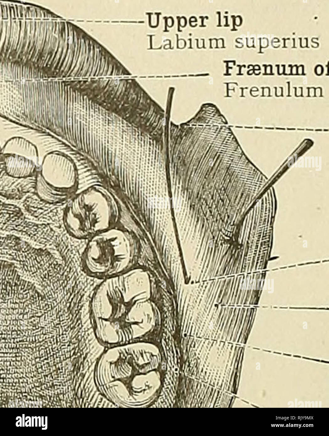 . An atlas of human anatomy for students and physicians. Anatomy. 416 CEPHALIC AND CERVICAL PORTIONS OF THE DIGESTIVE ORGANS Papilla palatina, or incisive pad' Papilla incisiva Unilateral remnant of the.^^ nasopalatine canal' Transverse rugae of the hard palate^ ^ Plicae palatinae transverscE â¢'' Longitudinal ridge or raphe of the palate Raphe palati Hard palate Palatum durum' Orifices of the palatine glands Soft palate, or velum pendulum palati Palatum molle (Ve'um. palatinum) The uvula Uvula palatina Pharyngeal cavity Cavum pharyngis Upper lip Labium superms Frsenum of the upper lip Frenu Stock Photohttps://www.alamy.com/image-license-details/?v=1https://www.alamy.com/an-atlas-of-human-anatomy-for-students-and-physicians-anatomy-416-cephalic-and-cervical-portions-of-the-digestive-organs-papilla-palatina-or-incisive-pad-papilla-incisiva-unilateral-remnant-of-the-nasopalatine-canal-transverse-rugae-of-the-hard-palate-plicae-palatinae-transversce-longitudinal-ridge-or-raphe-of-the-palate-raphe-palati-hard-palate-palatum-durum-orifices-of-the-palatine-glands-soft-palate-or-velum-pendulum-palati-palatum-molle-veum-palatinum-the-uvula-uvula-palatina-pharyngeal-cavity-cavum-pharyngis-upper-lip-labium-superms-frsenum-of-the-upper-lip-frenu-image235398938.html
RMRJY9MX–. An atlas of human anatomy for students and physicians. Anatomy. 416 CEPHALIC AND CERVICAL PORTIONS OF THE DIGESTIVE ORGANS Papilla palatina, or incisive pad' Papilla incisiva Unilateral remnant of the.^^ nasopalatine canal' Transverse rugae of the hard palate^ ^ Plicae palatinae transverscE â¢'' Longitudinal ridge or raphe of the palate Raphe palati Hard palate Palatum durum' Orifices of the palatine glands Soft palate, or velum pendulum palati Palatum molle (Ve'um. palatinum) The uvula Uvula palatina Pharyngeal cavity Cavum pharyngis Upper lip Labium superms Frsenum of the upper lip Frenu
 . Anatomy, descriptive and applied. Anatomy. THE NASAL FOSS^ 1083 The nasal duct opens into the anterior part of the inferior meatus, the opening being frequently overlapped by a fold of mucous membrane.' The Inner Wall (Fig. 799).—The inner wall or septum is frequently more or less deflected from the mesal plane (Fig. 799), thus limiting the size of one fossa and increasing that of the other. Ridges or spurs of bone growing outward from the septum are also sometimes present. Immediately over the incisie foramen at the lower edge of the cartilage of the septum a depression, the nasopalatine r Stock Photohttps://www.alamy.com/image-license-details/?v=1https://www.alamy.com/anatomy-descriptive-and-applied-anatomy-the-nasal-foss-1083-the-nasal-duct-opens-into-the-anterior-part-of-the-inferior-meatus-the-opening-being-frequently-overlapped-by-a-fold-of-mucous-membrane-the-inner-wall-fig-799the-inner-wall-or-septum-is-frequently-more-or-less-deflected-from-the-mesal-plane-fig-799-thus-limiting-the-size-of-one-fossa-and-increasing-that-of-the-other-ridges-or-spurs-of-bone-growing-outward-from-the-septum-are-also-sometimes-present-immediately-over-the-incisie-foramen-at-the-lower-edge-of-the-cartilage-of-the-septum-a-depression-the-nasopalatine-r-image236769112.html
RMRN5NBM–. Anatomy, descriptive and applied. Anatomy. THE NASAL FOSS^ 1083 The nasal duct opens into the anterior part of the inferior meatus, the opening being frequently overlapped by a fold of mucous membrane.' The Inner Wall (Fig. 799).—The inner wall or septum is frequently more or less deflected from the mesal plane (Fig. 799), thus limiting the size of one fossa and increasing that of the other. Ridges or spurs of bone growing outward from the septum are also sometimes present. Immediately over the incisie foramen at the lower edge of the cartilage of the septum a depression, the nasopalatine r
 . Anatomy in a nutshell : a treatise on human anatomy in its relation to osteopathy. Human anatomy; Osteopathic medicine; Osteopathic Medicine; Anatomy. PLATE CCXXXVI. ANTERIOR PALATINE FOSSA TRANSMITS LEFT NASO- PALATINE NERVE TRANSMITS ANTERIOR PALATINE VESSEL TRANSMITS RIGHT NASOPALATINE NERVE ACCESSORY PALATINE FORAMINA TENSOR PALATI POSTERIOR NASAL SPINE AZYGOS UVULAE HAMULAR PROCESS SPHENOID PROCESS OF PALATE PTERYGOPALATINE CANAL BASILAR PROCESS TENSOR TYMPANI RECTU! PHARYNGEAL SPINE FOR. SITUATION OF SUPERIOR CONSTRICTOR EUSTACHIAN TUBE AND CANAL FOR TENSOR TYMPANI LEVATOR PALATI —— Stock Photohttps://www.alamy.com/image-license-details/?v=1https://www.alamy.com/anatomy-in-a-nutshell-a-treatise-on-human-anatomy-in-its-relation-to-osteopathy-human-anatomy-osteopathic-medicine-osteopathic-medicine-anatomy-plate-ccxxxvi-anterior-palatine-fossa-transmits-left-naso-palatine-nerve-transmits-anterior-palatine-vessel-transmits-right-nasopalatine-nerve-accessory-palatine-foramina-tensor-palati-posterior-nasal-spine-azygos-uvulae-hamular-process-sphenoid-process-of-palate-pterygopalatine-canal-basilar-process-tensor-tympani-rectu!-pharyngeal-spine-for-situation-of-superior-constrictor-eustachian-tube-and-canal-for-tensor-tympani-levator-palati-image236802523.html
RMRN780Y–. Anatomy in a nutshell : a treatise on human anatomy in its relation to osteopathy. Human anatomy; Osteopathic medicine; Osteopathic Medicine; Anatomy. PLATE CCXXXVI. ANTERIOR PALATINE FOSSA TRANSMITS LEFT NASO- PALATINE NERVE TRANSMITS ANTERIOR PALATINE VESSEL TRANSMITS RIGHT NASOPALATINE NERVE ACCESSORY PALATINE FORAMINA TENSOR PALATI POSTERIOR NASAL SPINE AZYGOS UVULAE HAMULAR PROCESS SPHENOID PROCESS OF PALATE PTERYGOPALATINE CANAL BASILAR PROCESS TENSOR TYMPANI RECTU! PHARYNGEAL SPINE FOR. SITUATION OF SUPERIOR CONSTRICTOR EUSTACHIAN TUBE AND CANAL FOR TENSOR TYMPANI LEVATOR PALATI ——
 . Practical anatomy of the rabbit : an elementary laboratory textbook in mammalian anatomy . Rabbits; Anatomy, Comparative. DESIGNATIONS FOR PLATE III. I. Nasal-bone. ' 2. Levator alae nasi muscle. 3. Nasal septum. 4. Nasoturbinal cartilage. S- Maxilloturbinal (concha inferior). 6. Nasal fossa. 7. Nasolacrimal duct. 8. Vomeronasal organ and cartilage. g. Premaxilla. 10. Small upper incisor. 11. Large upper incisor. 12. Nasopalatine ducts. 13. Oral cavity. 14. Tongue. 15. Vibrissae. 16. Caninus muscle. 17. Terminals of superior maxillary nerve. 18. Buccal glands. 19. Buccinator muscle. 20. Term Stock Photohttps://www.alamy.com/image-license-details/?v=1https://www.alamy.com/practical-anatomy-of-the-rabbit-an-elementary-laboratory-textbook-in-mammalian-anatomy-rabbits-anatomy-comparative-designations-for-plate-iii-i-nasal-bone-2-levator-alae-nasi-muscle-3-nasal-septum-4-nasoturbinal-cartilage-s-maxilloturbinal-concha-inferior-6-nasal-fossa-7-nasolacrimal-duct-8-vomeronasal-organ-and-cartilage-g-premaxilla-10-small-upper-incisor-11-large-upper-incisor-12-nasopalatine-ducts-13-oral-cavity-14-tongue-15-vibrissae-16-caninus-muscle-17-terminals-of-superior-maxillary-nerve-18-buccal-glands-19-buccinator-muscle-20-term-image232135641.html
RMRDJKAH–. Practical anatomy of the rabbit : an elementary laboratory textbook in mammalian anatomy . Rabbits; Anatomy, Comparative. DESIGNATIONS FOR PLATE III. I. Nasal-bone. ' 2. Levator alae nasi muscle. 3. Nasal septum. 4. Nasoturbinal cartilage. S- Maxilloturbinal (concha inferior). 6. Nasal fossa. 7. Nasolacrimal duct. 8. Vomeronasal organ and cartilage. g. Premaxilla. 10. Small upper incisor. 11. Large upper incisor. 12. Nasopalatine ducts. 13. Oral cavity. 14. Tongue. 15. Vibrissae. 16. Caninus muscle. 17. Terminals of superior maxillary nerve. 18. Buccal glands. 19. Buccinator muscle. 20. Term
 . Bensley's Practical anatomy of the rabbit : an elementary laboratory text-book in mammalian anatomy. Rabbits -- Anatomy. DESIGNATIONS FOR PLATE III 1. Nasal bone. 14. 2. Levator alae nasi muscle. 15. 3. Nasal septum. 16. 4. Nasoturbinal cartilage. 17. 5. Maxilloturbinal (concha inferior). 18. 6. Nasal fossa. • 19. 7, Nasolacrimal duct. 20. S. Vomeronasal organ and cartilage. 21. 9. Premaxilla. 22. 10. Small upper incisor. Ts. 11. Large upper incisor. 24. 12. Nasopalatine ducts. 13. Oral cavity. 25. 14. Tongue. Vibrissae. Caninus muscle. Terminals of superior maxillary nerve. 18. Buccal gland Stock Photohttps://www.alamy.com/image-license-details/?v=1https://www.alamy.com/bensleys-practical-anatomy-of-the-rabbit-an-elementary-laboratory-text-book-in-mammalian-anatomy-rabbits-anatomy-designations-for-plate-iii-1-nasal-bone-14-2-levator-alae-nasi-muscle-15-3-nasal-septum-16-4-nasoturbinal-cartilage-17-5-maxilloturbinal-concha-inferior-18-6-nasal-fossa-19-7-nasolacrimal-duct-20-s-vomeronasal-organ-and-cartilage-21-9-premaxilla-22-10-small-upper-incisor-ts-11-large-upper-incisor-24-12-nasopalatine-ducts-13-oral-cavity-25-14-tongue-vibrissae-caninus-muscle-terminals-of-superior-maxillary-nerve-18-buccal-gland-image234749594.html
RMRHWNE2–. Bensley's Practical anatomy of the rabbit : an elementary laboratory text-book in mammalian anatomy. Rabbits -- Anatomy. DESIGNATIONS FOR PLATE III 1. Nasal bone. 14. 2. Levator alae nasi muscle. 15. 3. Nasal septum. 16. 4. Nasoturbinal cartilage. 17. 5. Maxilloturbinal (concha inferior). 18. 6. Nasal fossa. • 19. 7, Nasolacrimal duct. 20. S. Vomeronasal organ and cartilage. 21. 9. Premaxilla. 22. 10. Small upper incisor. Ts. 11. Large upper incisor. 24. 12. Nasopalatine ducts. 13. Oral cavity. 25. 14. Tongue. Vibrissae. Caninus muscle. Terminals of superior maxillary nerve. 18. Buccal gland