Nitella Black & White Stock Photos
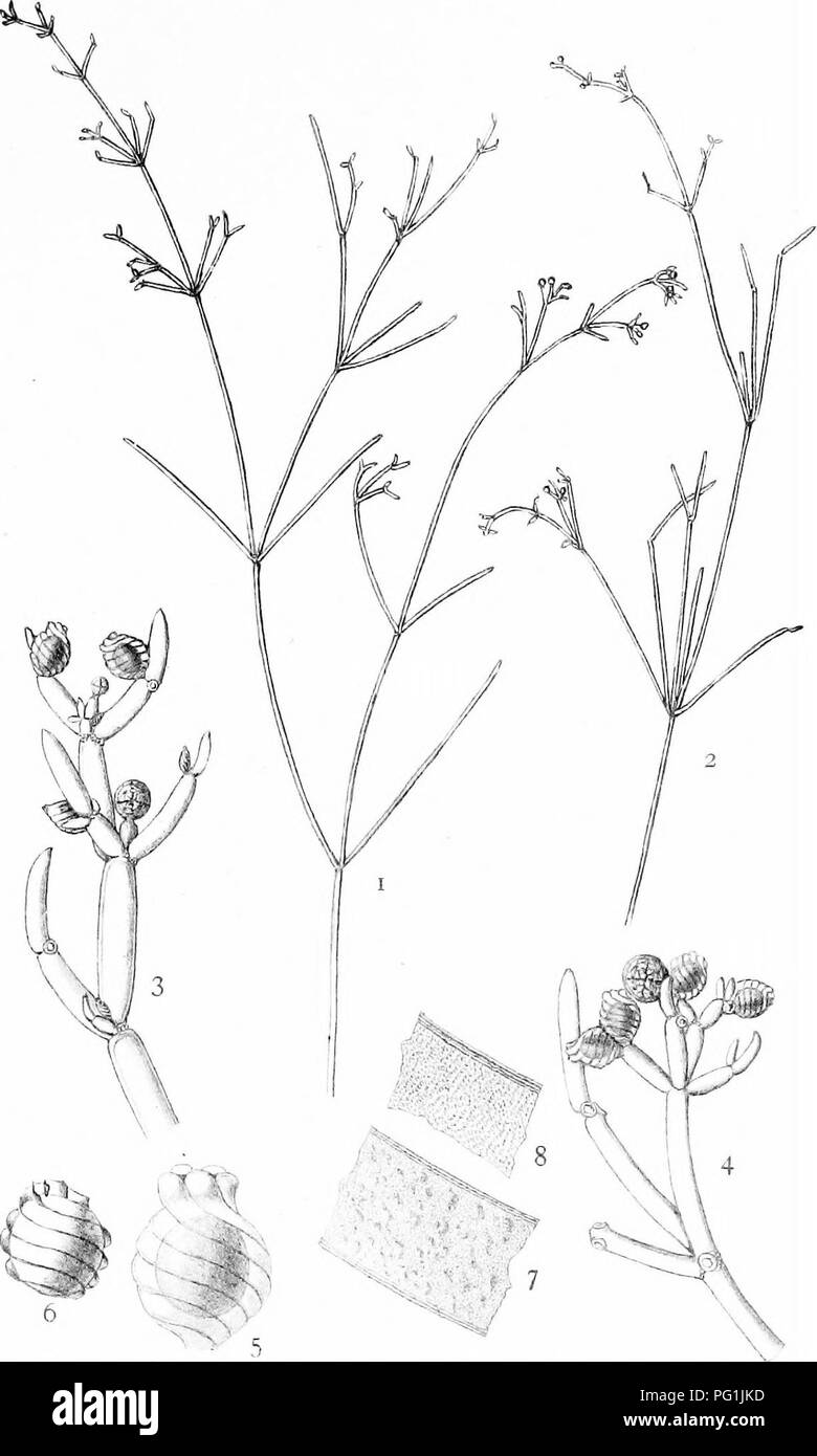
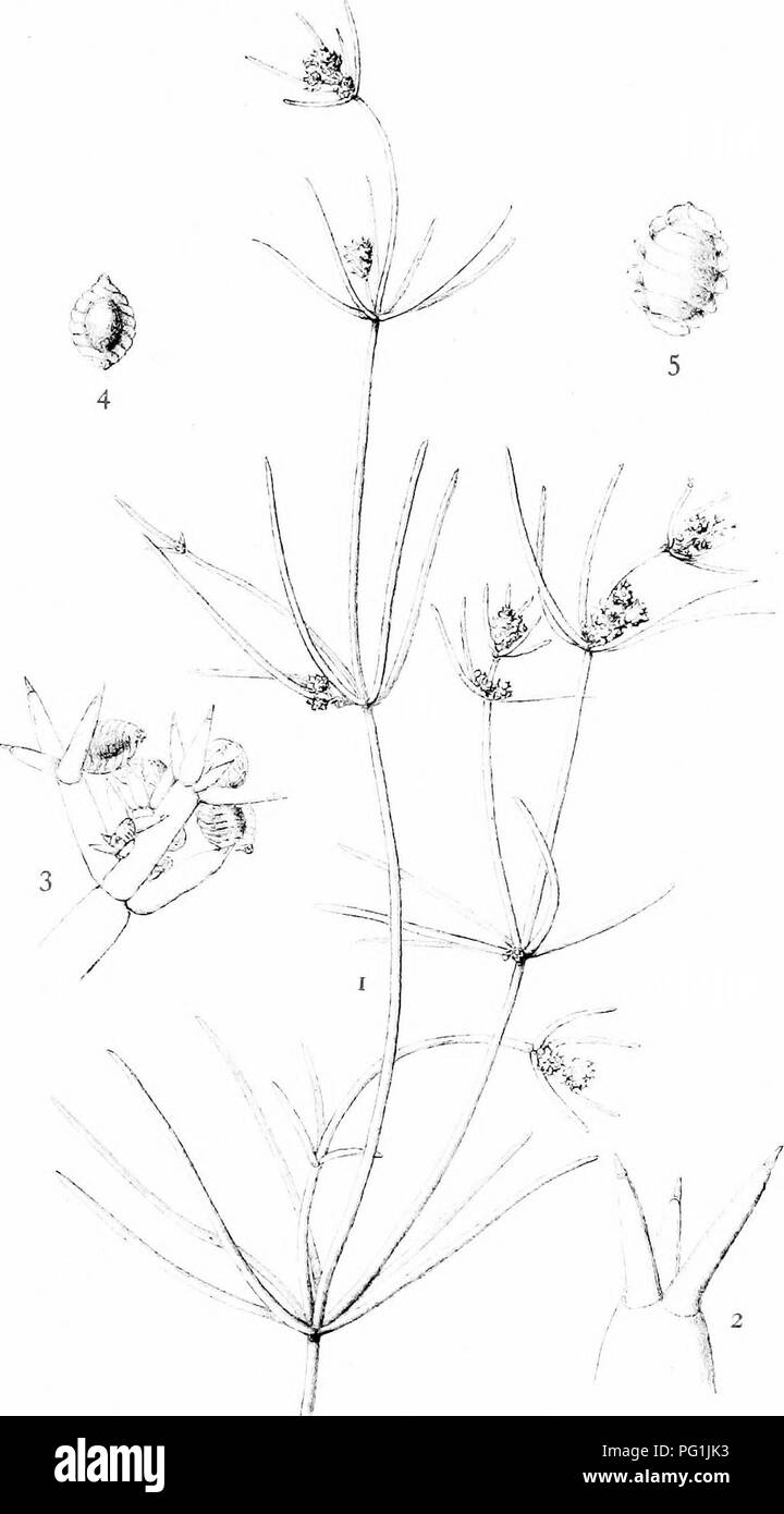 . The British Charophyta. Characeae. PLATE XI. NITELLA TRANSLUCENS. Please note that these images are extracted from scanned page images that may have been digitally enhanced for readability - coloration and appearance of these illustrations may not perfectly resemble the original work.. Groves, James; Bullock-Webster, George Russell, 1858- joint author. London, The Ray society Stock Photohttps://www.alamy.com/image-license-details/?v=1https://www.alamy.com/the-british-charophyta-characeae-plate-xi-nitella-translucens-please-note-that-these-images-are-extracted-from-scanned-page-images-that-may-have-been-digitally-enhanced-for-readability-coloration-and-appearance-of-these-illustrations-may-not-perfectly-resemble-the-original-work-groves-james-bullock-webster-george-russell-1858-joint-author-london-the-ray-society-image216395511.html
RMPG1JK3–. The British Charophyta. Characeae. PLATE XI. NITELLA TRANSLUCENS. Please note that these images are extracted from scanned page images that may have been digitally enhanced for readability - coloration and appearance of these illustrations may not perfectly resemble the original work.. Groves, James; Bullock-Webster, George Russell, 1858- joint author. London, The Ray society
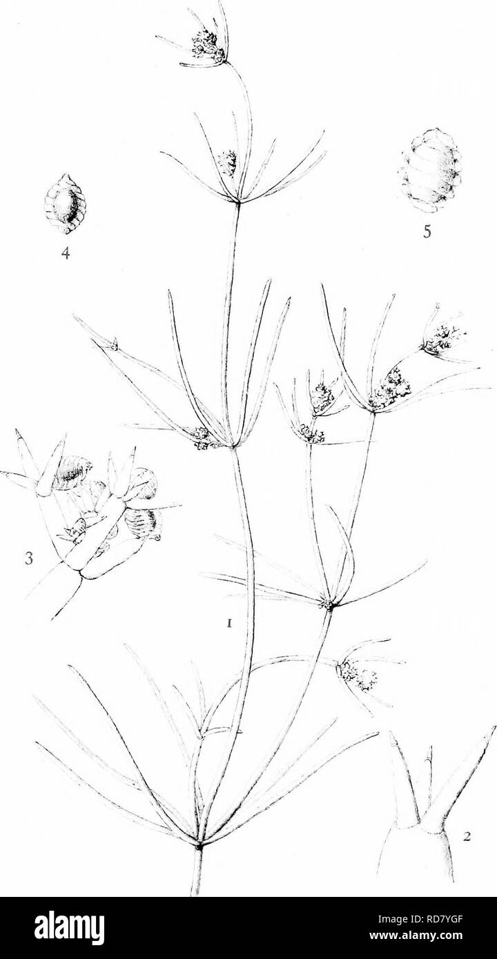 . The British Charophyta. Characeae. PLATE XI. NITELLA TRANSLUCENS. Please note that these images are extracted from scanned page images that may have been digitally enhanced for readability - coloration and appearance of these illustrations may not perfectly resemble the original work.. Groves, James; Bullock-Webster, George Russell, 1858- joint author. London, The Ray society Stock Photohttps://www.alamy.com/image-license-details/?v=1https://www.alamy.com/the-british-charophyta-characeae-plate-xi-nitella-translucens-please-note-that-these-images-are-extracted-from-scanned-page-images-that-may-have-been-digitally-enhanced-for-readability-coloration-and-appearance-of-these-illustrations-may-not-perfectly-resemble-the-original-work-groves-james-bullock-webster-george-russell-1858-joint-author-london-the-ray-society-image231900607.html
RMRD7YGF–. The British Charophyta. Characeae. PLATE XI. NITELLA TRANSLUCENS. Please note that these images are extracted from scanned page images that may have been digitally enhanced for readability - coloration and appearance of these illustrations may not perfectly resemble the original work.. Groves, James; Bullock-Webster, George Russell, 1858- joint author. London, The Ray society
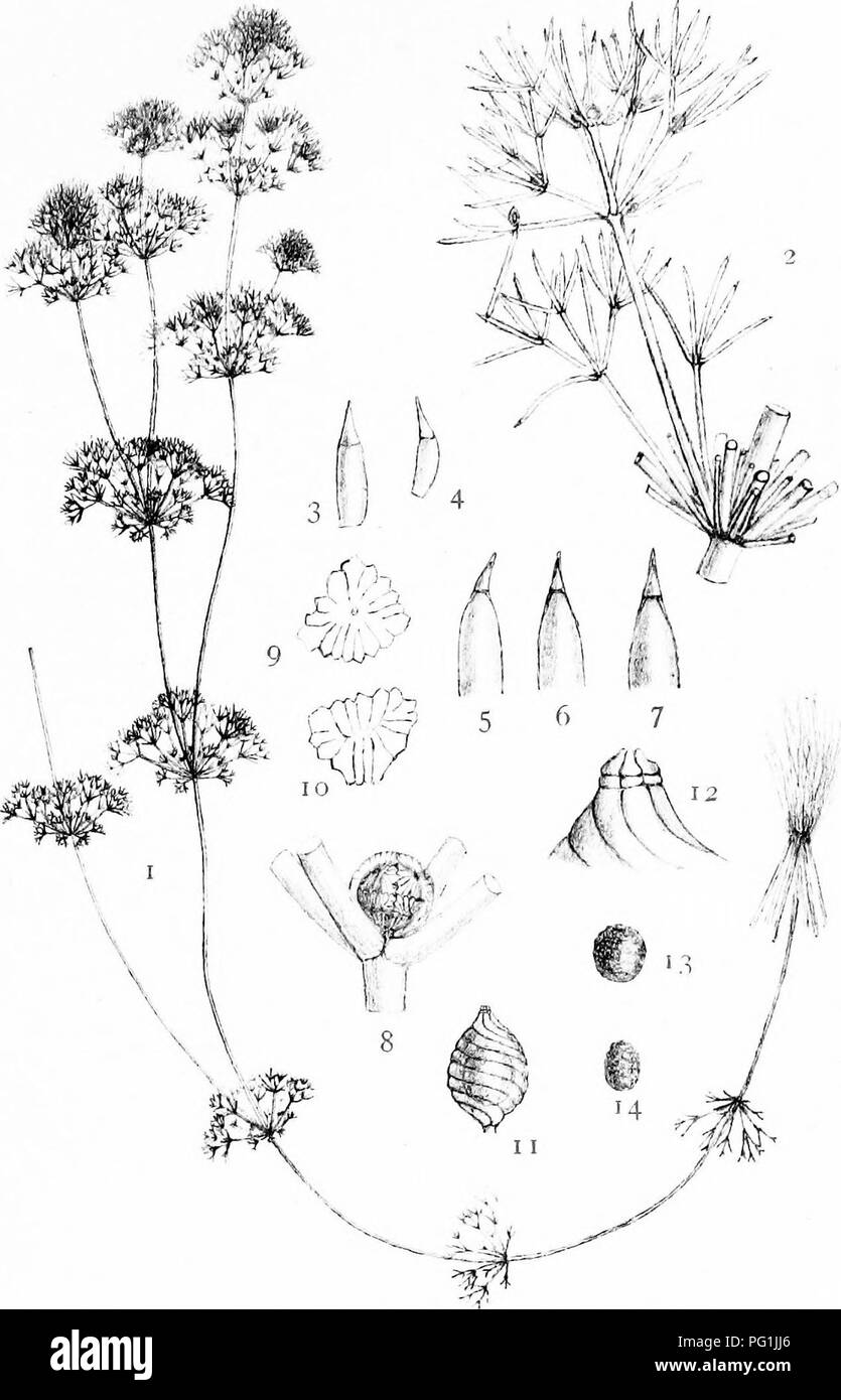 . The British Charophyta. Characeae. PLATE XVI. M. (?rorp, del NITELLA HYALINA. Please note that these images are extracted from scanned page images that may have been digitally enhanced for readability - coloration and appearance of these illustrations may not perfectly resemble the original work.. Groves, James; Bullock-Webster, George Russell, 1858- joint author. London, The Ray society Stock Photohttps://www.alamy.com/image-license-details/?v=1https://www.alamy.com/the-british-charophyta-characeae-plate-xvi-m-rorp-del-nitella-hyalina-please-note-that-these-images-are-extracted-from-scanned-page-images-that-may-have-been-digitally-enhanced-for-readability-coloration-and-appearance-of-these-illustrations-may-not-perfectly-resemble-the-original-work-groves-james-bullock-webster-george-russell-1858-joint-author-london-the-ray-society-image216395486.html
RMPG1JJ6–. The British Charophyta. Characeae. PLATE XVI. M. (?rorp, del NITELLA HYALINA. Please note that these images are extracted from scanned page images that may have been digitally enhanced for readability - coloration and appearance of these illustrations may not perfectly resemble the original work.. Groves, James; Bullock-Webster, George Russell, 1858- joint author. London, The Ray society
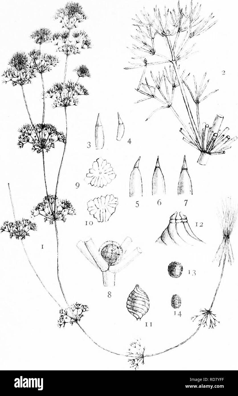 . The British Charophyta. Characeae. PLATE XVI. M. (?rorp, del NITELLA HYALINA. Please note that these images are extracted from scanned page images that may have been digitally enhanced for readability - coloration and appearance of these illustrations may not perfectly resemble the original work.. Groves, James; Bullock-Webster, George Russell, 1858- joint author. London, The Ray society Stock Photohttps://www.alamy.com/image-license-details/?v=1https://www.alamy.com/the-british-charophyta-characeae-plate-xvi-m-rorp-del-nitella-hyalina-please-note-that-these-images-are-extracted-from-scanned-page-images-that-may-have-been-digitally-enhanced-for-readability-coloration-and-appearance-of-these-illustrations-may-not-perfectly-resemble-the-original-work-groves-james-bullock-webster-george-russell-1858-joint-author-london-the-ray-society-image231900579.html
RMRD7YFF–. The British Charophyta. Characeae. PLATE XVI. M. (?rorp, del NITELLA HYALINA. Please note that these images are extracted from scanned page images that may have been digitally enhanced for readability - coloration and appearance of these illustrations may not perfectly resemble the original work.. Groves, James; Bullock-Webster, George Russell, 1858- joint author. London, The Ray society
 . The British Charophyta. Characeae. PLATE IX. M. n. ,(;. ,f. ;,-„-. ,,,, NITELLA SPANIOCLEMA. Please note that these images are extracted from scanned page images that may have been digitally enhanced for readability - coloration and appearance of these illustrations may not perfectly resemble the original work.. Groves, James; Bullock-Webster, George Russell, 1858- joint author. London, The Ray society Stock Photohttps://www.alamy.com/image-license-details/?v=1https://www.alamy.com/the-british-charophyta-characeae-plate-ix-m-n-f-nitella-spanioclema-please-note-that-these-images-are-extracted-from-scanned-page-images-that-may-have-been-digitally-enhanced-for-readability-coloration-and-appearance-of-these-illustrations-may-not-perfectly-resemble-the-original-work-groves-james-bullock-webster-george-russell-1858-joint-author-london-the-ray-society-image216395521.html
RMPG1JKD–. The British Charophyta. Characeae. PLATE IX. M. n. ,(;. ,f. ;,-„-. ,,,, NITELLA SPANIOCLEMA. Please note that these images are extracted from scanned page images that may have been digitally enhanced for readability - coloration and appearance of these illustrations may not perfectly resemble the original work.. Groves, James; Bullock-Webster, George Russell, 1858- joint author. London, The Ray society
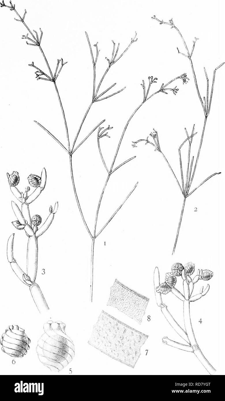 . The British Charophyta. Characeae. PLATE IX. M. n. ,(;. ,f. ;,-„-. ,,,, NITELLA SPANIOCLEMA. Please note that these images are extracted from scanned page images that may have been digitally enhanced for readability - coloration and appearance of these illustrations may not perfectly resemble the original work.. Groves, James; Bullock-Webster, George Russell, 1858- joint author. London, The Ray society Stock Photohttps://www.alamy.com/image-license-details/?v=1https://www.alamy.com/the-british-charophyta-characeae-plate-ix-m-n-f-nitella-spanioclema-please-note-that-these-images-are-extracted-from-scanned-page-images-that-may-have-been-digitally-enhanced-for-readability-coloration-and-appearance-of-these-illustrations-may-not-perfectly-resemble-the-original-work-groves-james-bullock-webster-george-russell-1858-joint-author-london-the-ray-society-image231900616.html
RMRD7YGT–. The British Charophyta. Characeae. PLATE IX. M. n. ,(;. ,f. ;,-„-. ,,,, NITELLA SPANIOCLEMA. Please note that these images are extracted from scanned page images that may have been digitally enhanced for readability - coloration and appearance of these illustrations may not perfectly resemble the original work.. Groves, James; Bullock-Webster, George Russell, 1858- joint author. London, The Ray society
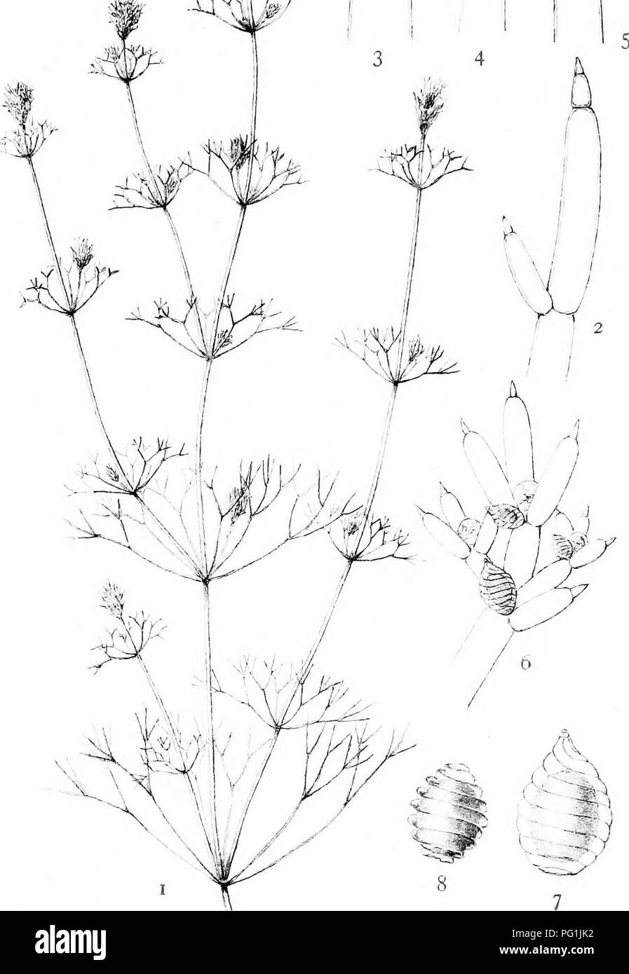 . The British Charophyta. Characeae. PLATE XII /1 '"A. NITELLA MUCRONATA. Please note that these images are extracted from scanned page images that may have been digitally enhanced for readability - coloration and appearance of these illustrations may not perfectly resemble the original work.. Groves, James; Bullock-Webster, George Russell, 1858- joint author. London, The Ray society Stock Photohttps://www.alamy.com/image-license-details/?v=1https://www.alamy.com/the-british-charophyta-characeae-plate-xii-1-quota-nitella-mucronata-please-note-that-these-images-are-extracted-from-scanned-page-images-that-may-have-been-digitally-enhanced-for-readability-coloration-and-appearance-of-these-illustrations-may-not-perfectly-resemble-the-original-work-groves-james-bullock-webster-george-russell-1858-joint-author-london-the-ray-society-image216395510.html
RMPG1JK2–. The British Charophyta. Characeae. PLATE XII /1 '"A. NITELLA MUCRONATA. Please note that these images are extracted from scanned page images that may have been digitally enhanced for readability - coloration and appearance of these illustrations may not perfectly resemble the original work.. Groves, James; Bullock-Webster, George Russell, 1858- joint author. London, The Ray society
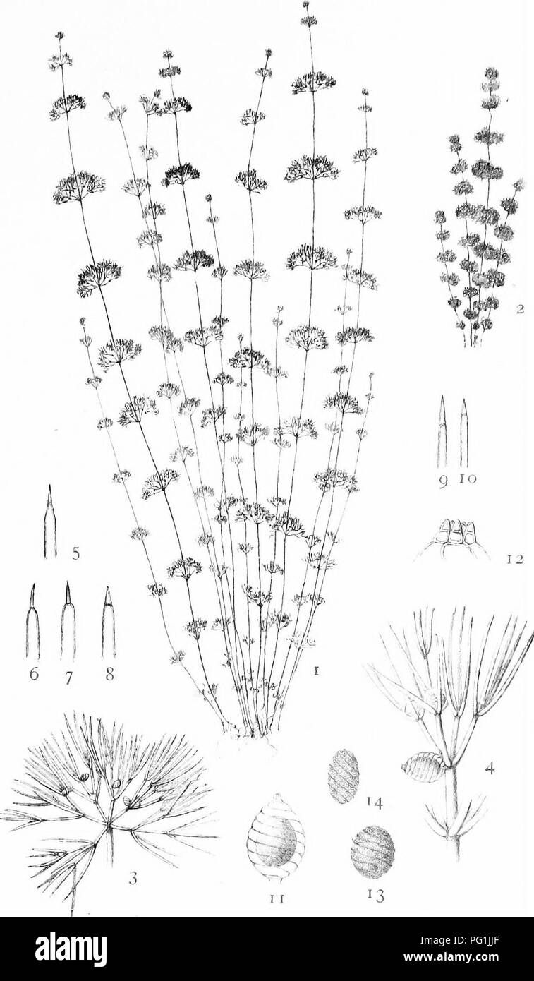
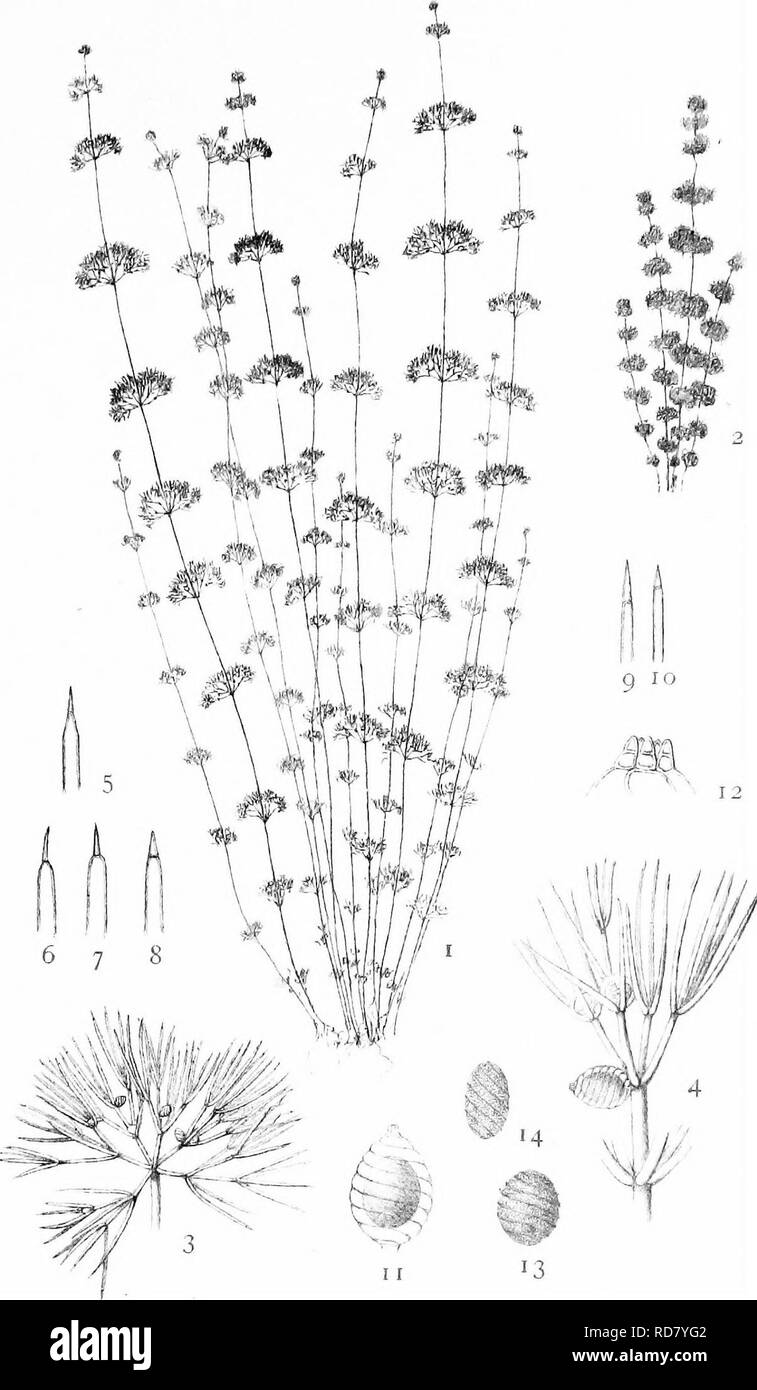 . The British Charophyta. Characeae. PLATE XIV. 6 7 8 )^kif^, 11 13 ,i;. Bri.|,..<rfe(. NITELLA TENUISSIMA. Please note that these images are extracted from scanned page images that may have been digitally enhanced for readability - coloration and appearance of these illustrations may not perfectly resemble the original work.. Groves, James; Bullock-Webster, George Russell, 1858- joint author. London, The Ray society Stock Photohttps://www.alamy.com/image-license-details/?v=1https://www.alamy.com/the-british-charophyta-characeae-plate-xiv-6-7-8-kif-11-13-i-briltrfe-nitella-tenuissima-please-note-that-these-images-are-extracted-from-scanned-page-images-that-may-have-been-digitally-enhanced-for-readability-coloration-and-appearance-of-these-illustrations-may-not-perfectly-resemble-the-original-work-groves-james-bullock-webster-george-russell-1858-joint-author-london-the-ray-society-image231900594.html
RMRD7YG2–. The British Charophyta. Characeae. PLATE XIV. 6 7 8 )^kif^, 11 13 ,i;. Bri.|,..<rfe(. NITELLA TENUISSIMA. Please note that these images are extracted from scanned page images that may have been digitally enhanced for readability - coloration and appearance of these illustrations may not perfectly resemble the original work.. Groves, James; Bullock-Webster, George Russell, 1858- joint author. London, The Ray society
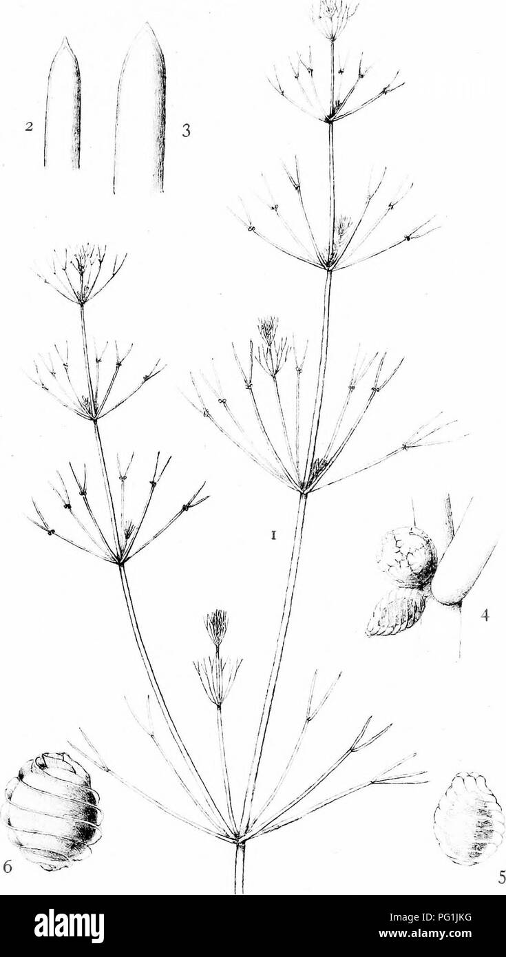 . The British Charophyta. Characeae. PLATE VIII. St. Groves del. NITELLA FLEXILIS. Please note that these images are extracted from scanned page images that may have been digitally enhanced for readability - coloration and appearance of these illustrations may not perfectly resemble the original work.. Groves, James; Bullock-Webster, George Russell, 1858- joint author. London, The Ray society Stock Photohttps://www.alamy.com/image-license-details/?v=1https://www.alamy.com/the-british-charophyta-characeae-plate-viii-st-groves-del-nitella-flexilis-please-note-that-these-images-are-extracted-from-scanned-page-images-that-may-have-been-digitally-enhanced-for-readability-coloration-and-appearance-of-these-illustrations-may-not-perfectly-resemble-the-original-work-groves-james-bullock-webster-george-russell-1858-joint-author-london-the-ray-society-image216395524.html
RMPG1JKG–. The British Charophyta. Characeae. PLATE VIII. St. Groves del. NITELLA FLEXILIS. Please note that these images are extracted from scanned page images that may have been digitally enhanced for readability - coloration and appearance of these illustrations may not perfectly resemble the original work.. Groves, James; Bullock-Webster, George Russell, 1858- joint author. London, The Ray society
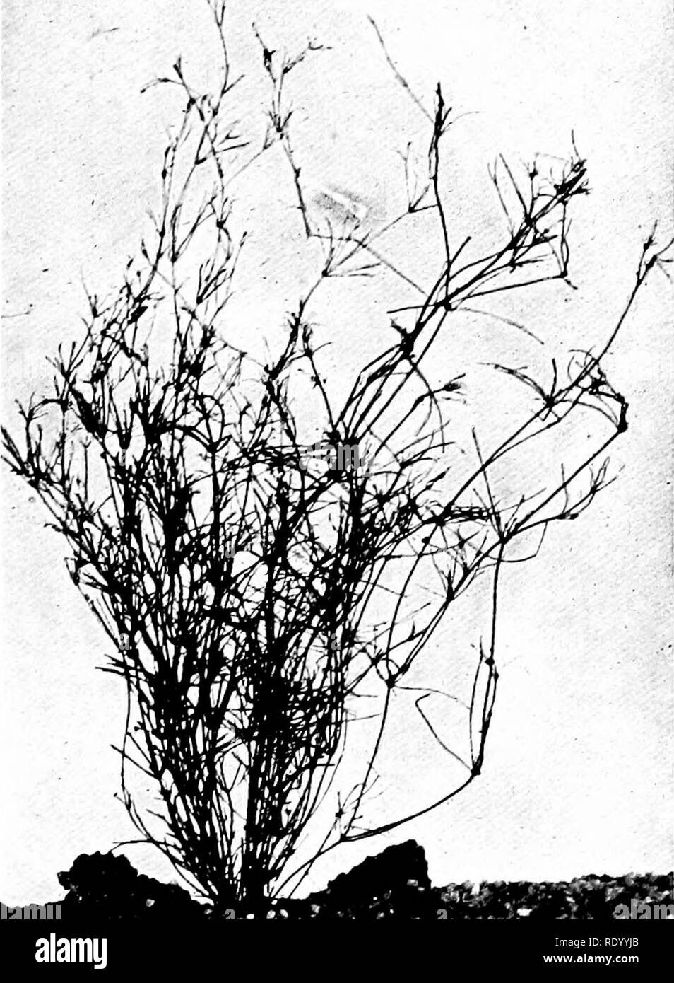 . The freshwater aquarium and its inhabitants; a guide for the amateur aquarist. Aquariums. Aquarium Plants 65. Fig. 29. Small-fruited Nitella, Nitella micro- carpa.. Please note that these images are extracted from scanned page images that may have been digitally enhanced for readability - coloration and appearance of these illustrations may not perfectly resemble the original work.. Eggeling, Otto, 1848-; Ehrenberg, Frederick, 1849- joint author. New York, H. Holt and Company Stock Photohttps://www.alamy.com/image-license-details/?v=1https://www.alamy.com/the-freshwater-aquarium-and-its-inhabitants-a-guide-for-the-amateur-aquarist-aquariums-aquarium-plants-65-fig-29-small-fruited-nitella-nitella-micro-carpa-please-note-that-these-images-are-extracted-from-scanned-page-images-that-may-have-been-digitally-enhanced-for-readability-coloration-and-appearance-of-these-illustrations-may-not-perfectly-resemble-the-original-work-eggeling-otto-1848-ehrenberg-frederick-1849-joint-author-new-york-h-holt-and-company-image232339699.html
RMRDYYJB–. The freshwater aquarium and its inhabitants; a guide for the amateur aquarist. Aquariums. Aquarium Plants 65. Fig. 29. Small-fruited Nitella, Nitella micro- carpa.. Please note that these images are extracted from scanned page images that may have been digitally enhanced for readability - coloration and appearance of these illustrations may not perfectly resemble the original work.. Eggeling, Otto, 1848-; Ehrenberg, Frederick, 1849- joint author. New York, H. Holt and Company
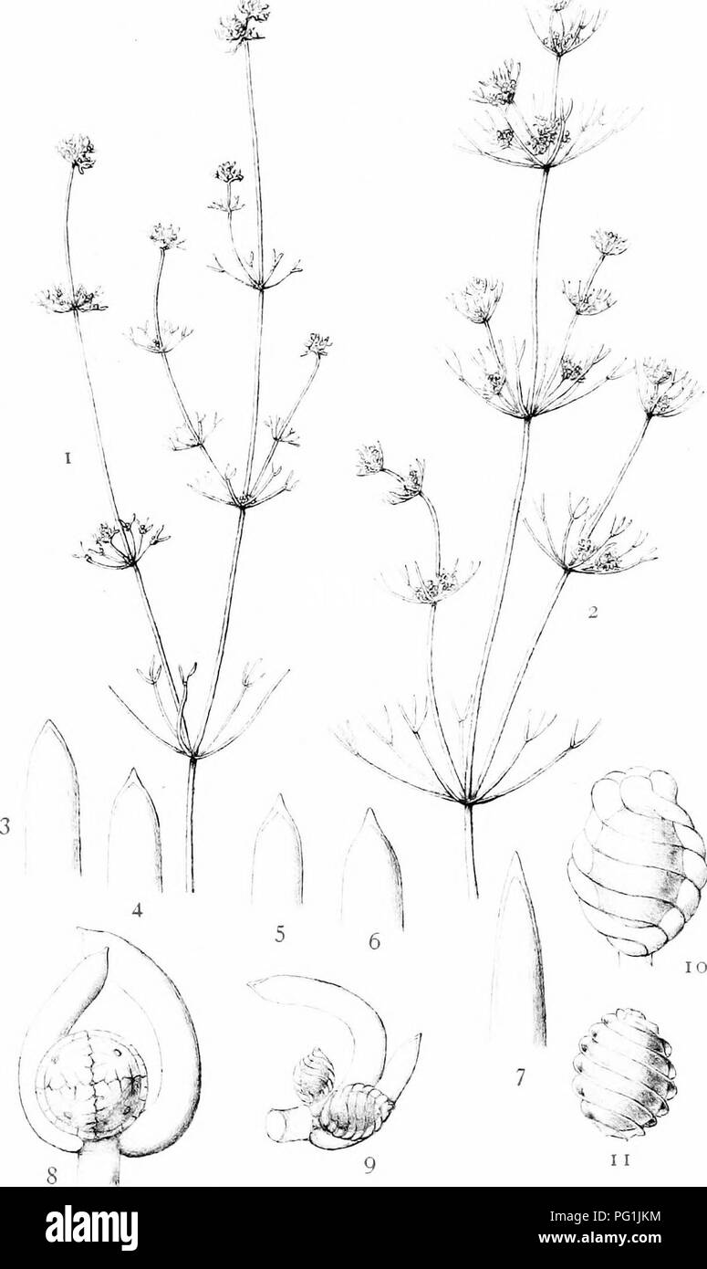 . The British Charophyta. Characeae. PLATE VII. ^f. droves (M. NITELLA OPACA. Please note that these images are extracted from scanned page images that may have been digitally enhanced for readability - coloration and appearance of these illustrations may not perfectly resemble the original work.. Groves, James; Bullock-Webster, George Russell, 1858- joint author. London, The Ray society Stock Photohttps://www.alamy.com/image-license-details/?v=1https://www.alamy.com/the-british-charophyta-characeae-plate-vii-f-droves-m-nitella-opaca-please-note-that-these-images-are-extracted-from-scanned-page-images-that-may-have-been-digitally-enhanced-for-readability-coloration-and-appearance-of-these-illustrations-may-not-perfectly-resemble-the-original-work-groves-james-bullock-webster-george-russell-1858-joint-author-london-the-ray-society-image216395528.html
RMPG1JKM–. The British Charophyta. Characeae. PLATE VII. ^f. droves (M. NITELLA OPACA. Please note that these images are extracted from scanned page images that may have been digitally enhanced for readability - coloration and appearance of these illustrations may not perfectly resemble the original work.. Groves, James; Bullock-Webster, George Russell, 1858- joint author. London, The Ray society
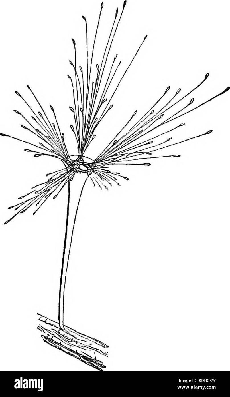 . Evenings at the microscope : or, researches among the minuter organs and forms of animal life. Microscopy; Microscopes; Medical microscopy. 464 EVENINGS AT THE MICKOSCOPE. of which is therefore appropriated to each new made animal. That the essential vitality of the creature resides in this nucleus is shown by another and highly curious mode of increase, namely, that which is effected by encystion. Let us search the live-box carefully, for amidst so great a profusion of Vorticelloe as we have. on this Nitella, it will go hard if we do not find some individuals in the encysted stage. Look at Stock Photohttps://www.alamy.com/image-license-details/?v=1https://www.alamy.com/evenings-at-the-microscope-or-researches-among-the-minuter-organs-and-forms-of-animal-life-microscopy-microscopes-medical-microscopy-464-evenings-at-the-mickoscope-of-which-is-therefore-appropriated-to-each-new-made-animal-that-the-essential-vitality-of-the-creature-resides-in-this-nucleus-is-shown-by-another-and-highly-curious-mode-of-increase-namely-that-which-is-effected-by-encystion-let-us-search-the-live-box-carefully-for-amidst-so-great-a-profusion-of-vorticelloe-as-we-have-on-this-nitella-it-will-go-hard-if-we-do-not-find-some-individuals-in-the-encysted-stage-look-at-image232108573.html
RMRDHCRW–. Evenings at the microscope : or, researches among the minuter organs and forms of animal life. Microscopy; Microscopes; Medical microscopy. 464 EVENINGS AT THE MICKOSCOPE. of which is therefore appropriated to each new made animal. That the essential vitality of the creature resides in this nucleus is shown by another and highly curious mode of increase, namely, that which is effected by encystion. Let us search the live-box carefully, for amidst so great a profusion of Vorticelloe as we have. on this Nitella, it will go hard if we do not find some individuals in the encysted stage. Look at
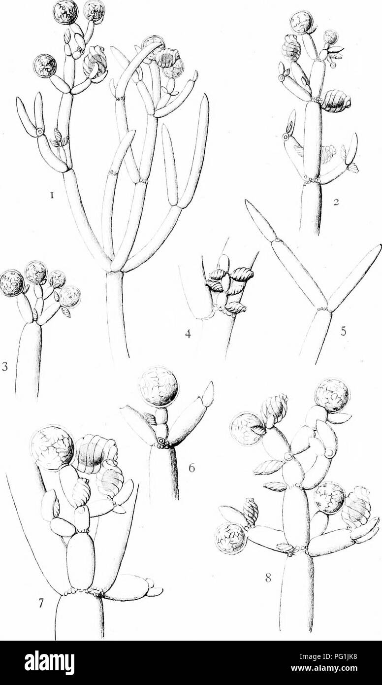 . The British Charophyta. Characeae. PLATE X. 0. ». B.-yr.Sr M.G.dtl. NITELLA SPANIOCLEMA. Please note that these images are extracted from scanned page images that may have been digitally enhanced for readability - coloration and appearance of these illustrations may not perfectly resemble the original work.. Groves, James; Bullock-Webster, George Russell, 1858- joint author. London, The Ray society Stock Photohttps://www.alamy.com/image-license-details/?v=1https://www.alamy.com/the-british-charophyta-characeae-plate-x-0-b-yrsr-mgdtl-nitella-spanioclema-please-note-that-these-images-are-extracted-from-scanned-page-images-that-may-have-been-digitally-enhanced-for-readability-coloration-and-appearance-of-these-illustrations-may-not-perfectly-resemble-the-original-work-groves-james-bullock-webster-george-russell-1858-joint-author-london-the-ray-society-image216395516.html
RMPG1JK8–. The British Charophyta. Characeae. PLATE X. 0. ». B.-yr.Sr M.G.dtl. NITELLA SPANIOCLEMA. Please note that these images are extracted from scanned page images that may have been digitally enhanced for readability - coloration and appearance of these illustrations may not perfectly resemble the original work.. Groves, James; Bullock-Webster, George Russell, 1858- joint author. London, The Ray society
 . A handbook of cryptogamic botany. Cryptogams. CHARACEJE 175 whorl, which subtend the branches., are arranged in a spiral line running round the stem; but this is not the case with the branches or secondary axes, where the members of contiguous whorls are superposed. The Characeae exhibit in an especially clear and beautiful manner the phenomenon oi cydosis, or rotation of the protoplasm (see fig. 163). The best objects for observation are the large internodal cells of Nitella (Ag.), the apical cells in the leaves of Chara, or some of those belonging to the reproductive organs, especially to Stock Photohttps://www.alamy.com/image-license-details/?v=1https://www.alamy.com/a-handbook-of-cryptogamic-botany-cryptogams-characeje-175-whorl-which-subtend-the-branches-are-arranged-in-a-spiral-line-running-round-the-stem-but-this-is-not-the-case-with-the-branches-or-secondary-axes-where-the-members-of-contiguous-whorls-are-superposed-the-characeae-exhibit-in-an-especially-clear-and-beautiful-manner-the-phenomenon-oi-cydosis-or-rotation-of-the-protoplasm-see-fig-163-the-best-objects-for-observation-are-the-large-internodal-cells-of-nitella-ag-the-apical-cells-in-the-leaves-of-chara-or-some-of-those-belonging-to-the-reproductive-organs-especially-to-image232421707.html
RMRE3M77–. A handbook of cryptogamic botany. Cryptogams. CHARACEJE 175 whorl, which subtend the branches., are arranged in a spiral line running round the stem; but this is not the case with the branches or secondary axes, where the members of contiguous whorls are superposed. The Characeae exhibit in an especially clear and beautiful manner the phenomenon oi cydosis, or rotation of the protoplasm (see fig. 163). The best objects for observation are the large internodal cells of Nitella (Ag.), the apical cells in the leaves of Chara, or some of those belonging to the reproductive organs, especially to
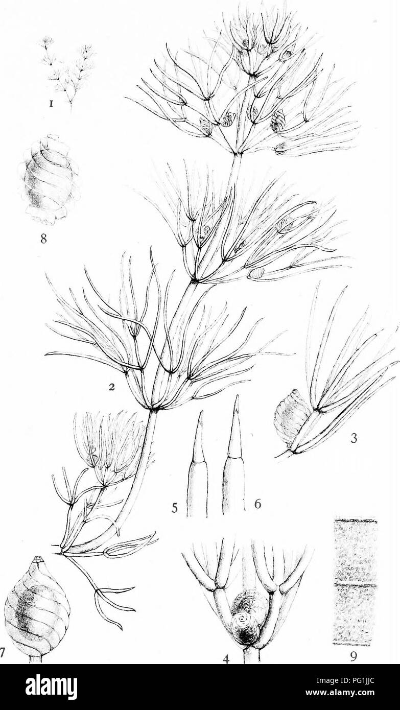 . The British Charophyta. Characeae. PLATE XV. Af. A- H. Grov'-f^ del. NITELLA BATRACHOSPERMA. Please note that these images are extracted from scanned page images that may have been digitally enhanced for readability - coloration and appearance of these illustrations may not perfectly resemble the original work.. Groves, James; Bullock-Webster, George Russell, 1858- joint author. London, The Ray society Stock Photohttps://www.alamy.com/image-license-details/?v=1https://www.alamy.com/the-british-charophyta-characeae-plate-xv-af-a-h-grov-f-del-nitella-batrachosperma-please-note-that-these-images-are-extracted-from-scanned-page-images-that-may-have-been-digitally-enhanced-for-readability-coloration-and-appearance-of-these-illustrations-may-not-perfectly-resemble-the-original-work-groves-james-bullock-webster-george-russell-1858-joint-author-london-the-ray-society-image216395492.html
RMPG1JJC–. The British Charophyta. Characeae. PLATE XV. Af. A- H. Grov'-f^ del. NITELLA BATRACHOSPERMA. Please note that these images are extracted from scanned page images that may have been digitally enhanced for readability - coloration and appearance of these illustrations may not perfectly resemble the original work.. Groves, James; Bullock-Webster, George Russell, 1858- joint author. London, The Ray society
 . A Manual of botany : being an introduction to the study of the structure, physiology, and classification of plants . Botany. 152 MOVEMENTS IN CELLSâEOTATION. times the plant consists of a single central cell; at other times there are several smaller ones surrounding it, which must be removed in order that the movements which occur in the central cell may be seen. Many of the species are incrusted with calcareous matter, and thus become opaque, while others, as Ohara or Nitella flexilis, have no incrustation, and are transparent. Those plants with unincrusted tubular, cells best exhibit movem Stock Photohttps://www.alamy.com/image-license-details/?v=1https://www.alamy.com/a-manual-of-botany-being-an-introduction-to-the-study-of-the-structure-physiology-and-classification-of-plants-botany-152-movements-in-cellseotation-times-the-plant-consists-of-a-single-central-cell-at-other-times-there-are-several-smaller-ones-surrounding-it-which-must-be-removed-in-order-that-the-movements-which-occur-in-the-central-cell-may-be-seen-many-of-the-species-are-incrusted-with-calcareous-matter-and-thus-become-opaque-while-others-as-ohara-or-nitella-flexilis-have-no-incrustation-and-are-transparent-those-plants-with-unincrusted-tubular-cells-best-exhibit-movem-image232114926.html
RMRDHMXP–. A Manual of botany : being an introduction to the study of the structure, physiology, and classification of plants . Botany. 152 MOVEMENTS IN CELLSâEOTATION. times the plant consists of a single central cell; at other times there are several smaller ones surrounding it, which must be removed in order that the movements which occur in the central cell may be seen. Many of the species are incrusted with calcareous matter, and thus become opaque, while others, as Ohara or Nitella flexilis, have no incrustation, and are transparent. Those plants with unincrusted tubular, cells best exhibit movem
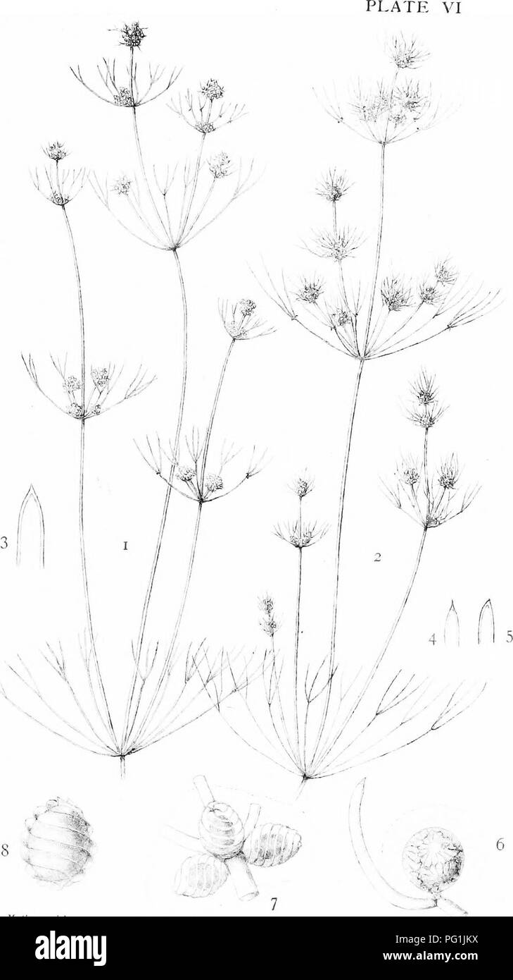 . The British Charophyta. Characeae. ,v '^ t^ ,1/ (Jn.c-.i ,1,1 -â ' 7 NITELLA CAPILLARIS. Please note that these images are extracted from scanned page images that may have been digitally enhanced for readability - coloration and appearance of these illustrations may not perfectly resemble the original work.. Groves, James; Bullock-Webster, George Russell, 1858- joint author. London, The Ray society Stock Photohttps://www.alamy.com/image-license-details/?v=1https://www.alamy.com/the-british-charophyta-characeae-v-t-1-jnc-i-11-7-nitella-capillaris-please-note-that-these-images-are-extracted-from-scanned-page-images-that-may-have-been-digitally-enhanced-for-readability-coloration-and-appearance-of-these-illustrations-may-not-perfectly-resemble-the-original-work-groves-james-bullock-webster-george-russell-1858-joint-author-london-the-ray-society-image216395534.html
RMPG1JKX–. The British Charophyta. Characeae. ,v '^ t^ ,1/ (Jn.c-.i ,1,1 -â ' 7 NITELLA CAPILLARIS. Please note that these images are extracted from scanned page images that may have been digitally enhanced for readability - coloration and appearance of these illustrations may not perfectly resemble the original work.. Groves, James; Bullock-Webster, George Russell, 1858- joint author. London, The Ray society
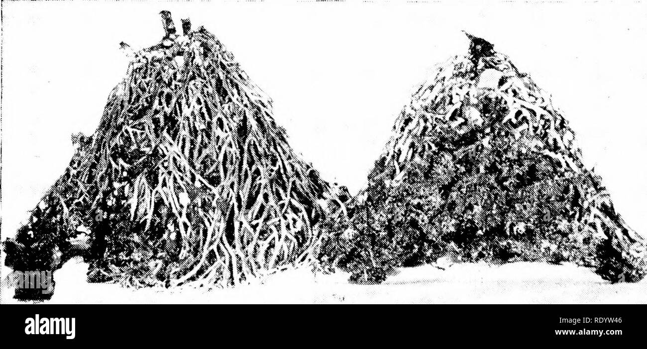 . Principles of modern biology. Biology. 233 - Multicellular Plants are syncytial, with several nuclei enclosed within each wall; and except lor the rhizoids, all parts of Nitella contain chloroplasts. These structural differentiations in Nitella indicate a corresponding functional speciali- zation, or division of labor, among the differ- ent parts of the plant. The rhizoids serve tor attachment, and being incapable of photo- synthesis, they depend upon the green parts for organic sustenance. The "leaves" are in a particularly favorable position for absorbing sunlight, and perform th Stock Photohttps://www.alamy.com/image-license-details/?v=1https://www.alamy.com/principles-of-modern-biology-biology-233-multicellular-plants-are-syncytial-with-several-nuclei-enclosed-within-each-wall-and-except-lor-the-rhizoids-all-parts-of-nitella-contain-chloroplasts-these-structural-differentiations-in-nitella-indicate-a-corresponding-functional-speciali-zation-or-division-of-labor-among-the-differ-ent-parts-of-the-plant-the-rhizoids-serve-tor-attachment-and-being-incapable-of-photo-synthesis-they-depend-upon-the-green-parts-for-organic-sustenance-the-quotleavesquot-are-in-a-particularly-favorable-position-for-absorbing-sunlight-and-perform-th-image232337734.html
RMRDYW46–. Principles of modern biology. Biology. 233 - Multicellular Plants are syncytial, with several nuclei enclosed within each wall; and except lor the rhizoids, all parts of Nitella contain chloroplasts. These structural differentiations in Nitella indicate a corresponding functional speciali- zation, or division of labor, among the differ- ent parts of the plant. The rhizoids serve tor attachment, and being incapable of photo- synthesis, they depend upon the green parts for organic sustenance. The "leaves" are in a particularly favorable position for absorbing sunlight, and perform th
 . The British Charophyta. Characeae. PLATE XIV. 6 7 8 )^kif^, 11 13 ,i;. Bri.|,..<rfe(. NITELLA TENUISSIMA. Please note that these images are extracted from scanned page images that may have been digitally enhanced for readability - coloration and appearance of these illustrations may not perfectly resemble the original work.. Groves, James; Bullock-Webster, George Russell, 1858- joint author. London, The Ray society Stock Photohttps://www.alamy.com/image-license-details/?v=1https://www.alamy.com/the-british-charophyta-characeae-plate-xiv-6-7-8-kif-11-13-i-briltrfe-nitella-tenuissima-please-note-that-these-images-are-extracted-from-scanned-page-images-that-may-have-been-digitally-enhanced-for-readability-coloration-and-appearance-of-these-illustrations-may-not-perfectly-resemble-the-original-work-groves-james-bullock-webster-george-russell-1858-joint-author-london-the-ray-society-image216395495.html
RMPG1JJF–. The British Charophyta. Characeae. PLATE XIV. 6 7 8 )^kif^, 11 13 ,i;. Bri.|,..<rfe(. NITELLA TENUISSIMA. Please note that these images are extracted from scanned page images that may have been digitally enhanced for readability - coloration and appearance of these illustrations may not perfectly resemble the original work.. Groves, James; Bullock-Webster, George Russell, 1858- joint author. London, The Ray society
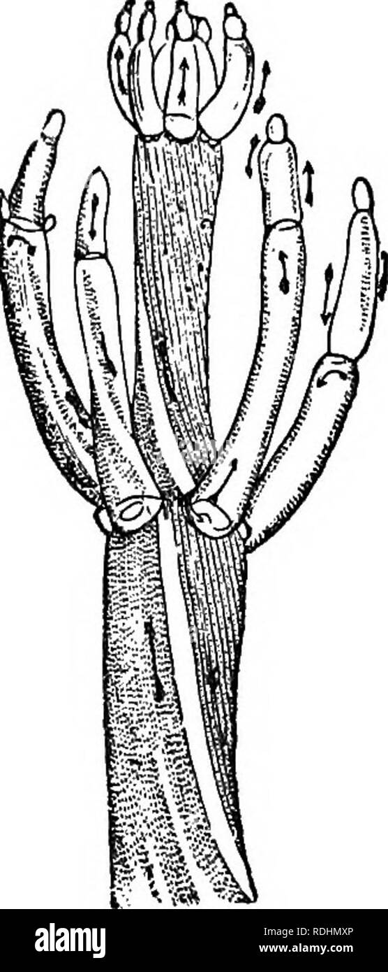
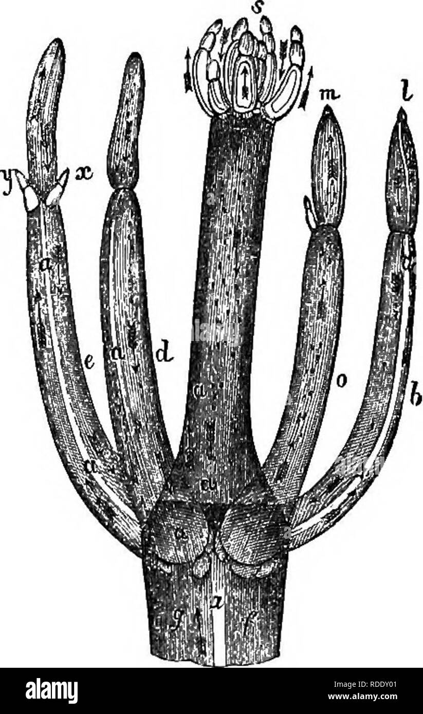 . A practical treatise on the use of the microscope, including the different methods of preparing and examining animal, vegetable, and mineral structures. Microscopes; Microscopy. Fig. 245. Fis. 246. arrows denote the direction of the movement, and the letters a a the colourless division of the joints which separate the ascending and descending currents; the circulation may even be Ti^itnessed in the whorl of young leaves at the top, s, and in all the other parts indicated by the arrows. Method of Viewing the Circulation.—If the Chara or Nitella be in abundance, a new piece may be selected eac Stock Photohttps://www.alamy.com/image-license-details/?v=1https://www.alamy.com/a-practical-treatise-on-the-use-of-the-microscope-including-the-different-methods-of-preparing-and-examining-animal-vegetable-and-mineral-structures-microscopes-microscopy-fig-245-fis-246-arrows-denote-the-direction-of-the-movement-and-the-letters-a-a-the-colourless-division-of-the-joints-which-separate-the-ascending-and-descending-currents-the-circulation-may-even-be-tiitnessed-in-the-whorl-of-young-leaves-at-the-top-s-and-in-all-the-other-parts-indicated-by-the-arrows-method-of-viewing-the-circulationif-the-chara-or-nitella-be-in-abundance-a-new-piece-may-be-selected-eac-image232031857.html
RMRDDY01–. A practical treatise on the use of the microscope, including the different methods of preparing and examining animal, vegetable, and mineral structures. Microscopes; Microscopy. Fig. 245. Fis. 246. arrows denote the direction of the movement, and the letters a a the colourless division of the joints which separate the ascending and descending currents; the circulation may even be Ti^itnessed in the whorl of young leaves at the top, s, and in all the other parts indicated by the arrows. Method of Viewing the Circulation.—If the Chara or Nitella be in abundance, a new piece may be selected eac
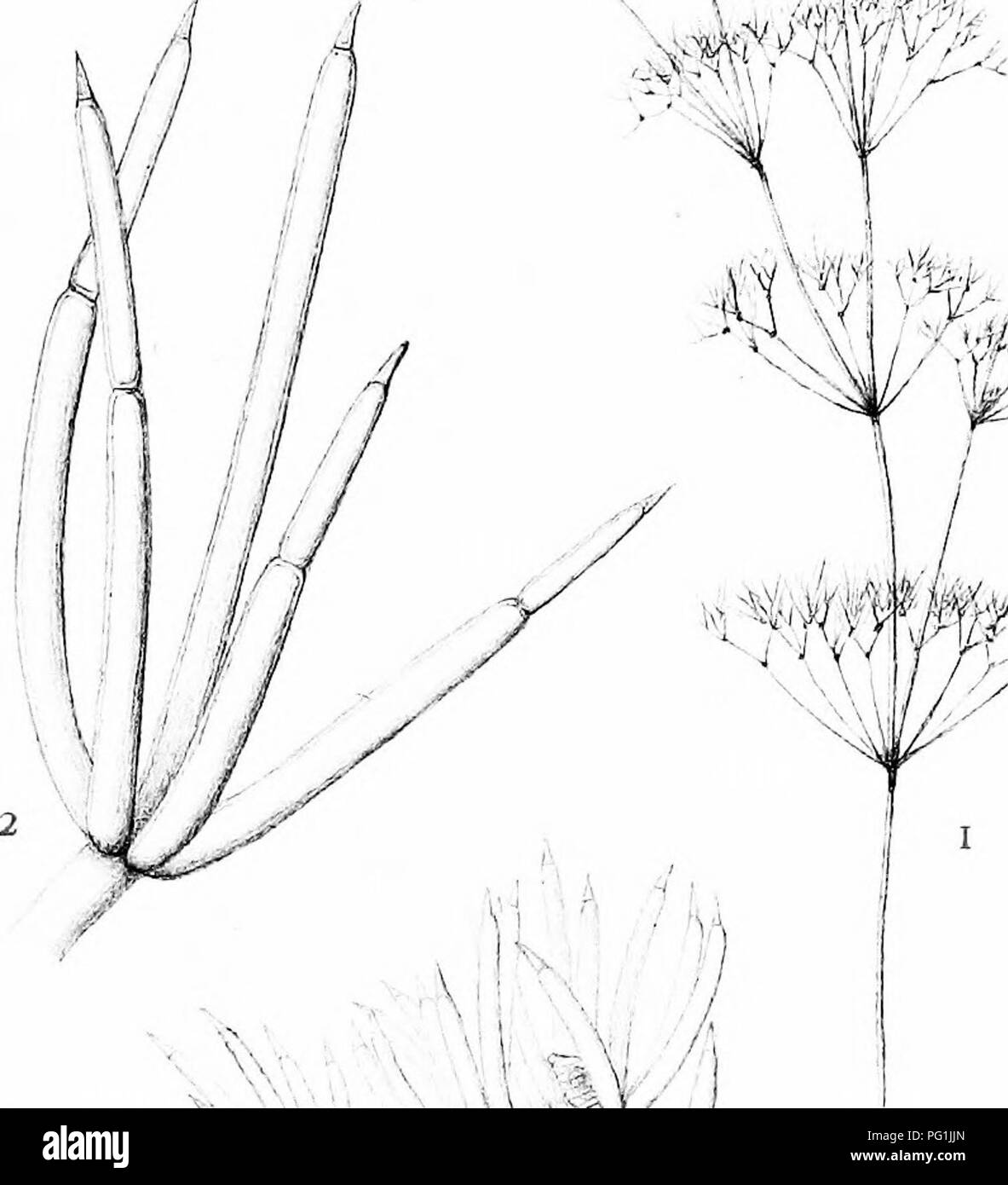
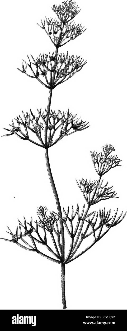 . The British Charophyta. Characeae. PLATE I.. NITELLA MUCRONATA VAR. GRACILLIMA. {muck enlarged) Cluirh-^ Whittingham A- (ir,gi;.<;, Ltd.. I.ith.. Please note that these images are extracted from scanned page images that may have been digitally enhanced for readability - coloration and appearance of these illustrations may not perfectly resemble the original work.. Groves, James; Bullock-Webster, George Russell, 1858- joint author. London, The Ray society Stock Photohttps://www.alamy.com/image-license-details/?v=1https://www.alamy.com/the-british-charophyta-characeae-plate-i-nitella-mucronata-var-gracillima-muck-enlarged-cluirh-whittingham-a-irgilt-ltd-iith-please-note-that-these-images-are-extracted-from-scanned-page-images-that-may-have-been-digitally-enhanced-for-readability-coloration-and-appearance-of-these-illustrations-may-not-perfectly-resemble-the-original-work-groves-james-bullock-webster-george-russell-1858-joint-author-london-the-ray-society-image216395773.html
RMPG1K0D–. The British Charophyta. Characeae. PLATE I.. NITELLA MUCRONATA VAR. GRACILLIMA. {muck enlarged) Cluirh-^ Whittingham A- (ir,gi;.<;, Ltd.. I.ith.. Please note that these images are extracted from scanned page images that may have been digitally enhanced for readability - coloration and appearance of these illustrations may not perfectly resemble the original work.. Groves, James; Bullock-Webster, George Russell, 1858- joint author. London, The Ray society
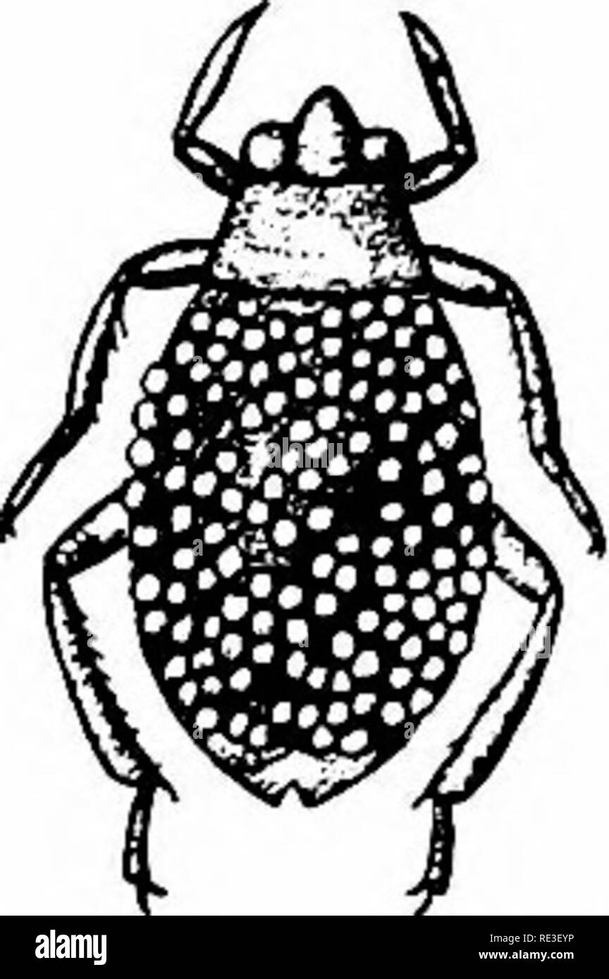 . Goldfish varieties and tropical aquarium fishes; a complete guide to aquaria and related subjects. Aquariums; Goldfish. Fig. 105. Water Scavenger Beetle (Life sice) it breathes at the surface from the mouth. Instead of long antennae they have palpi looking like club-shaped antennae. This beetle lives chiefly on decomposing vegetal and animal matter, although taking soft living plants such as Nitella. It has been claimed to be predaceous but there is doubt about their attacking fishes. They have been kept in aquaria with them without doing damage. On general principles, however, it is best to Stock Photohttps://www.alamy.com/image-license-details/?v=1https://www.alamy.com/goldfish-varieties-and-tropical-aquarium-fishes-a-complete-guide-to-aquaria-and-related-subjects-aquariums-goldfish-fig-105-water-scavenger-beetle-life-sice-it-breathes-at-the-surface-from-the-mouth-instead-of-long-antennae-they-have-palpi-looking-like-club-shaped-antennae-this-beetle-lives-chiefly-on-decomposing-vegetal-and-animal-matter-although-taking-soft-living-plants-such-as-nitella-it-has-been-claimed-to-be-predaceous-but-there-is-doubt-about-their-attacking-fishes-they-have-been-kept-in-aquaria-with-them-without-doing-damage-on-general-principles-however-it-is-best-to-image232417578.html
RMRE3EYP–. Goldfish varieties and tropical aquarium fishes; a complete guide to aquaria and related subjects. Aquariums; Goldfish. Fig. 105. Water Scavenger Beetle (Life sice) it breathes at the surface from the mouth. Instead of long antennae they have palpi looking like club-shaped antennae. This beetle lives chiefly on decomposing vegetal and animal matter, although taking soft living plants such as Nitella. It has been claimed to be predaceous but there is doubt about their attacking fishes. They have been kept in aquaria with them without doing damage. On general principles, however, it is best to
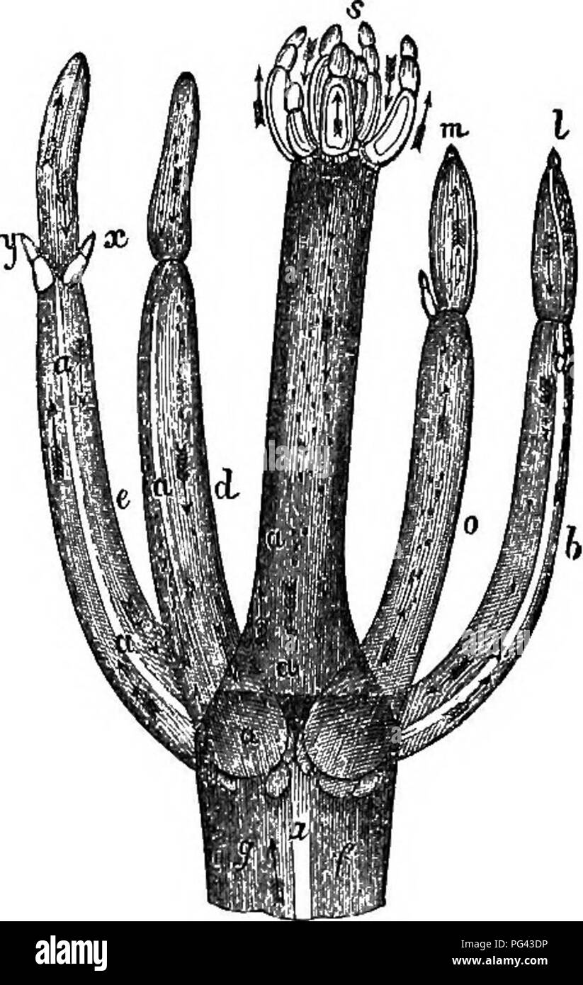 . A practical treatise on the use of the microscope, including the different methods of preparing and examining animal, vegetable, and mineral structures. Microscopes; Microscopy. Fig. 245. Fis. 246. arrows denote the direction of the movement, and the letters a a the colourless division of the joints which separate the ascending and descending currents; the circulation may even be Ti^itnessed in the whorl of young leaves at the top, s, and in all the other parts indicated by the arrows. Method of Viewing the Circulation.—If the Chara or Nitella be in abundance, a new piece may be selected eac Stock Photohttps://www.alamy.com/image-license-details/?v=1https://www.alamy.com/a-practical-treatise-on-the-use-of-the-microscope-including-the-different-methods-of-preparing-and-examining-animal-vegetable-and-mineral-structures-microscopes-microscopy-fig-245-fis-246-arrows-denote-the-direction-of-the-movement-and-the-letters-a-a-the-colourless-division-of-the-joints-which-separate-the-ascending-and-descending-currents-the-circulation-may-even-be-tiitnessed-in-the-whorl-of-young-leaves-at-the-top-s-and-in-all-the-other-parts-indicated-by-the-arrows-method-of-viewing-the-circulationif-the-chara-or-nitella-be-in-abundance-a-new-piece-may-be-selected-eac-image216449458.html
RMPG43DP–. A practical treatise on the use of the microscope, including the different methods of preparing and examining animal, vegetable, and mineral structures. Microscopes; Microscopy. Fig. 245. Fis. 246. arrows denote the direction of the movement, and the letters a a the colourless division of the joints which separate the ascending and descending currents; the circulation may even be Ti^itnessed in the whorl of young leaves at the top, s, and in all the other parts indicated by the arrows. Method of Viewing the Circulation.—If the Chara or Nitella be in abundance, a new piece may be selected eac
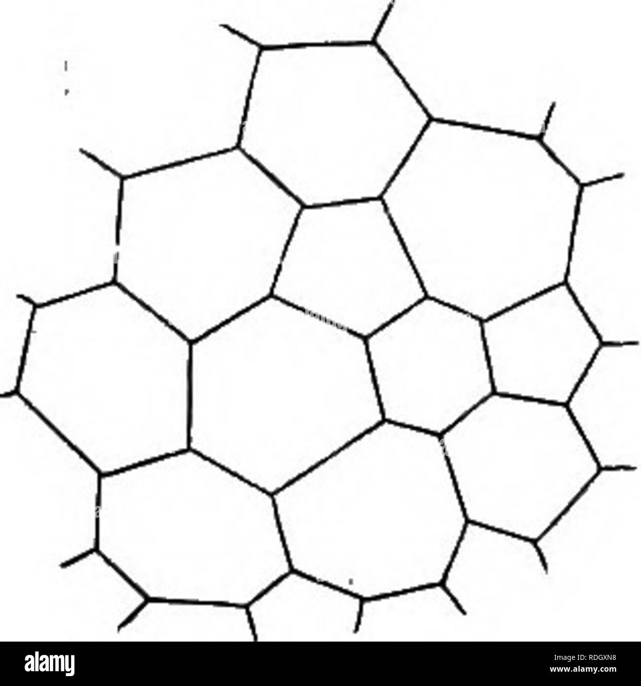 . An elementary text-book of botany, for the use of Japanese students. Botany. FORMATION OF CELLS. 107. Section S. KINDS OF THE CELL. The form and size of the cell vary greatly, the former being spherical, elliptical, cylindrical, cubical, prismatic, star-shaped, spindle-shaped, etc., and the latter being very large as in Nitella, or very minute as in Bacteria. Longi- tudinal rows of cells often become tulubar by the absorption of their transverse walls. The cells, which are not much longer than they are broad and have rounded or flattened surfaces, are said to be Parenchymatous (Fig. Fia. 122 Stock Photohttps://www.alamy.com/image-license-details/?v=1https://www.alamy.com/an-elementary-text-book-of-botany-for-the-use-of-japanese-students-botany-formation-of-cells-107-section-s-kinds-of-the-cell-the-form-and-size-of-the-cell-vary-greatly-the-former-being-spherical-elliptical-cylindrical-cubical-prismatic-star-shaped-spindle-shaped-etc-and-the-latter-being-very-large-as-in-nitella-or-very-minute-as-in-bacteria-longi-tudinal-rows-of-cells-often-become-tulubar-by-the-absorption-of-their-transverse-walls-the-cells-which-are-not-much-longer-than-they-are-broad-and-have-rounded-or-flattened-surfaces-are-said-to-be-parenchymatous-fig-fia-122-image232097524.html
RMRDGXN8–. An elementary text-book of botany, for the use of Japanese students. Botany. FORMATION OF CELLS. 107. Section S. KINDS OF THE CELL. The form and size of the cell vary greatly, the former being spherical, elliptical, cylindrical, cubical, prismatic, star-shaped, spindle-shaped, etc., and the latter being very large as in Nitella, or very minute as in Bacteria. Longi- tudinal rows of cells often become tulubar by the absorption of their transverse walls. The cells, which are not much longer than they are broad and have rounded or flattened surfaces, are said to be Parenchymatous (Fig. Fia. 122
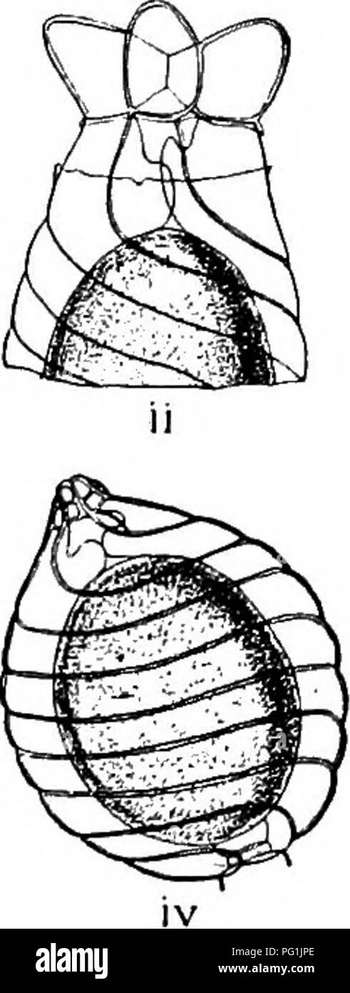 . The British Charophyta. Characeae. Fio. 23.—Process of fertilisation, i, ii, iii. Upper part of oogonium of Chara vulgaris. iv. Oogonium of Nitella tenuissima. i. Half- matured unfertilised oogonium showing upper and lower cavities formed by development of coronula and enveloping cells, ii. Fully matured unfertilised oogonium showing tumid growth at the head of enveloping cells, and transverse fracture of membrane caused by their extended growth, iii. Antherozoids penetrating interstices formed by contracted terminations of spiral cells, and reaching oospore through upper and lower cavities Stock Photohttps://www.alamy.com/image-license-details/?v=1https://www.alamy.com/the-british-charophyta-characeae-fio-23process-of-fertilisation-i-ii-iii-upper-part-of-oogonium-of-chara-vulgaris-iv-oogonium-of-nitella-tenuissima-i-half-matured-unfertilised-oogonium-showing-upper-and-lower-cavities-formed-by-development-of-coronula-and-enveloping-cells-ii-fully-matured-unfertilised-oogonium-showing-tumid-growth-at-the-head-of-enveloping-cells-and-transverse-fracture-of-membrane-caused-by-their-extended-growth-iii-antherozoids-penetrating-interstices-formed-by-contracted-terminations-of-spiral-cells-and-reaching-oospore-through-upper-and-lower-cavities-image216395606.html
RMPG1JPE–. The British Charophyta. Characeae. Fio. 23.—Process of fertilisation, i, ii, iii. Upper part of oogonium of Chara vulgaris. iv. Oogonium of Nitella tenuissima. i. Half- matured unfertilised oogonium showing upper and lower cavities formed by development of coronula and enveloping cells, ii. Fully matured unfertilised oogonium showing tumid growth at the head of enveloping cells, and transverse fracture of membrane caused by their extended growth, iii. Antherozoids penetrating interstices formed by contracted terminations of spiral cells, and reaching oospore through upper and lower cavities
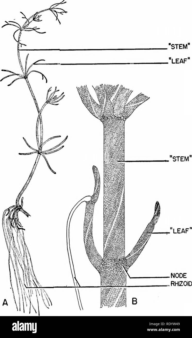 . Principles of modern biology. Biology. Nutrition of Multicellular Plants - 237 it lacks chlorophyll. The rhizoid cell is de- pendent on the other cells of the filament, and sugars produced by photosynthesis in the green cells are transferred to this colorless cell by diffusion through the intervening cell membranes. Among the most highly differentiated of fresh-water algae is Nitella (Fig. 13-2). This relatively large green alga may measure al- most a foot in length. Nitella exhibits a branching green "stem," which is attached to colorless rhizoids at the lower end, and which is su Stock Photohttps://www.alamy.com/image-license-details/?v=1https://www.alamy.com/principles-of-modern-biology-biology-nutrition-of-multicellular-plants-237-it-lacks-chlorophyll-the-rhizoid-cell-is-de-pendent-on-the-other-cells-of-the-filament-and-sugars-produced-by-photosynthesis-in-the-green-cells-are-transferred-to-this-colorless-cell-by-diffusion-through-the-intervening-cell-membranes-among-the-most-highly-differentiated-of-fresh-water-algae-is-nitella-fig-13-2-this-relatively-large-green-alga-may-measure-al-most-a-foot-in-length-nitella-exhibits-a-branching-green-quotstemquot-which-is-attached-to-colorless-rhizoids-at-the-lower-end-and-which-is-su-image232337737.html
RMRDYW49–. Principles of modern biology. Biology. Nutrition of Multicellular Plants - 237 it lacks chlorophyll. The rhizoid cell is de- pendent on the other cells of the filament, and sugars produced by photosynthesis in the green cells are transferred to this colorless cell by diffusion through the intervening cell membranes. Among the most highly differentiated of fresh-water algae is Nitella (Fig. 13-2). This relatively large green alga may measure al- most a foot in length. Nitella exhibits a branching green "stem," which is attached to colorless rhizoids at the lower end, and which is su
 . The British Charophyta. Characeae. PLATE XIII â J^â¢^^^^ -^^r %â 3 4 #'A ^f^lffe. JJ -411, :«^i% ''^:. if-'' ^#1^ â x.:^:,^ }/ V M ^j -' i; V 6 / "J^ X. .)/. i:rn,,sihl. NITELLA GRACILIS. Please note that these images are extracted from scanned page images that may have been digitally enhanced for readability - coloration and appearance of these illustrations may not perfectly resemble the original work.. Groves, James; Bullock-Webster, George Russell, 1858- joint author. London, The Ray society Stock Photohttps://www.alamy.com/image-license-details/?v=1https://www.alamy.com/the-british-charophyta-characeae-plate-xiii-j-r-3-4-a-flffe-jj-411-i-if-1-x-v-m-j-i-v-6-quotj-x-irnsihl-nitella-gracilis-please-note-that-these-images-are-extracted-from-scanned-page-images-that-may-have-been-digitally-enhanced-for-readability-coloration-and-appearance-of-these-illustrations-may-not-perfectly-resemble-the-original-work-groves-james-bullock-webster-george-russell-1858-joint-author-london-the-ray-society-image216395501.html
RMPG1JJN–. The British Charophyta. Characeae. PLATE XIII â J^â¢^^^^ -^^r %â 3 4 #'A ^f^lffe. JJ -411, :«^i% ''^:. if-'' ^#1^ â x.:^:,^ }/ V M ^j -' i; V 6 / "J^ X. .)/. i:rn,,sihl. NITELLA GRACILIS. Please note that these images are extracted from scanned page images that may have been digitally enhanced for readability - coloration and appearance of these illustrations may not perfectly resemble the original work.. Groves, James; Bullock-Webster, George Russell, 1858- joint author. London, The Ray society
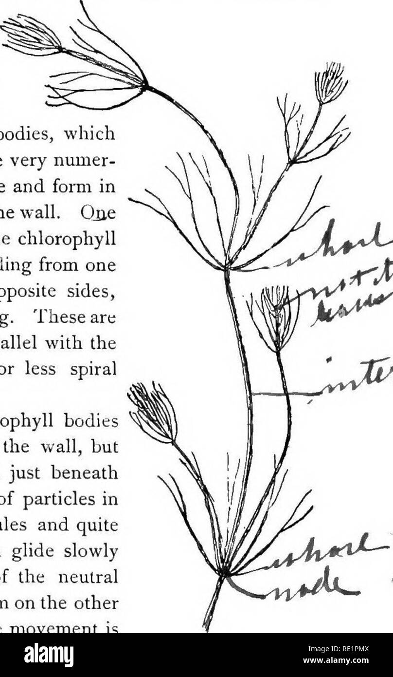 . Elementary botany. Botany. PROTOPLASM. node. These internodes are peculiar. They consist of but a single " cell," and are cylindrical, with closed ends. They are sometimes 5-10 cm. long. 19. Internode of nitella.—For the study of an internode of nitella, a small one, near the end, or the ends of one of the " leaves " is best suited, since it is more transparent. A small portion of the plant should be placed on the glass slip in water with the cover glass over a tuft of the branches near the growing end. Examined with the microscope the green chlorophyll bodies, which form Stock Photohttps://www.alamy.com/image-license-details/?v=1https://www.alamy.com/elementary-botany-botany-protoplasm-node-these-internodes-are-peculiar-they-consist-of-but-a-single-quot-cellquot-and-are-cylindrical-with-closed-ends-they-are-sometimes-5-10-cm-long-19-internode-of-nitellafor-the-study-of-an-internode-of-nitella-a-small-one-near-the-end-or-the-ends-of-one-of-the-quot-leaves-quot-is-best-suited-since-it-is-more-transparent-a-small-portion-of-the-plant-should-be-placed-on-the-glass-slip-in-water-with-the-cover-glass-over-a-tuft-of-the-branches-near-the-growing-end-examined-with-the-microscope-the-green-chlorophyll-bodies-which-form-image232379754.html
RMRE1PMX–. Elementary botany. Botany. PROTOPLASM. node. These internodes are peculiar. They consist of but a single " cell," and are cylindrical, with closed ends. They are sometimes 5-10 cm. long. 19. Internode of nitella.—For the study of an internode of nitella, a small one, near the end, or the ends of one of the " leaves " is best suited, since it is more transparent. A small portion of the plant should be placed on the glass slip in water with the cover glass over a tuft of the branches near the growing end. Examined with the microscope the green chlorophyll bodies, which form
 . Insect life; an introduction to nature study and a guide for teachers, students and others interested in out-of-door life. Entomology; Nature study. Fig. 292.—Duckweed. Watercress, Nasturtium officinale. Stoneworts, Chara and Nitella (several species of each). Frog-spittle or water-silk, Spirogira. A small quantity of duckweed, Lemna (Fig. 292), placed on the surface of the water adds to the beauty of an aquarium. When it is necessary to add water to an aqua- rium on account of loss by evaporation, rain wa- ter should be used to prevent an undue ac- cumulation of the mineral matter held in s Stock Photohttps://www.alamy.com/image-license-details/?v=1https://www.alamy.com/insect-life-an-introduction-to-nature-study-and-a-guide-for-teachers-students-and-others-interested-in-out-of-door-life-entomology-nature-study-fig-292duckweed-watercress-nasturtium-officinale-stoneworts-chara-and-nitella-several-species-of-each-frog-spittle-or-water-silk-spirogira-a-small-quantity-of-duckweed-lemna-fig-292-placed-on-the-surface-of-the-water-adds-to-the-beauty-of-an-aquarium-when-it-is-necessary-to-add-water-to-an-aqua-rium-on-account-of-loss-by-evaporation-rain-wa-ter-should-be-used-to-prevent-an-undue-ac-cumulation-of-the-mineral-matter-held-in-s-image216406826.html
RMPG2536–. Insect life; an introduction to nature study and a guide for teachers, students and others interested in out-of-door life. Entomology; Nature study. Fig. 292.—Duckweed. Watercress, Nasturtium officinale. Stoneworts, Chara and Nitella (several species of each). Frog-spittle or water-silk, Spirogira. A small quantity of duckweed, Lemna (Fig. 292), placed on the surface of the water adds to the beauty of an aquarium. When it is necessary to add water to an aqua- rium on account of loss by evaporation, rain wa- ter should be used to prevent an undue ac- cumulation of the mineral matter held in s
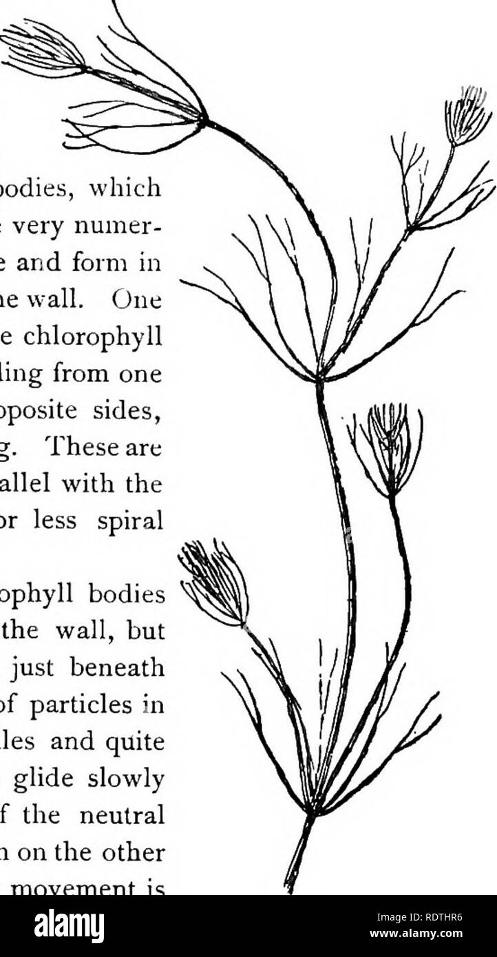 . Elementary botany. Botany. PROTOPLASM. 9 node. These internodes are peculiar. They consist of but a single " cell," and are cylindrical, with closed ends. They are sometimes 5-10 cm. long. 19. Internode of nitella.—For the study of an internode of nitella, a small one, near the end, or the ends of one of the " leaves " is best suited, since it is more transparent. A small portion of the plant should be placed on the glass slip in water with the cover glass over a tuft of the branches near the growing end. Examined with the microscope the green chlorophyll bodies, which fo Stock Photohttps://www.alamy.com/image-license-details/?v=1https://www.alamy.com/elementary-botany-botany-protoplasm-9-node-these-internodes-are-peculiar-they-consist-of-but-a-single-quot-cellquot-and-are-cylindrical-with-closed-ends-they-are-sometimes-5-10-cm-long-19-internode-of-nitellafor-the-study-of-an-internode-of-nitella-a-small-one-near-the-end-or-the-ends-of-one-of-the-quot-leaves-quot-is-best-suited-since-it-is-more-transparent-a-small-portion-of-the-plant-should-be-placed-on-the-glass-slip-in-water-with-the-cover-glass-over-a-tuft-of-the-branches-near-the-growing-end-examined-with-the-microscope-the-green-chlorophyll-bodies-which-fo-image232266138.html
RMRDTHR6–. Elementary botany. Botany. PROTOPLASM. 9 node. These internodes are peculiar. They consist of but a single " cell," and are cylindrical, with closed ends. They are sometimes 5-10 cm. long. 19. Internode of nitella.—For the study of an internode of nitella, a small one, near the end, or the ends of one of the " leaves " is best suited, since it is more transparent. A small portion of the plant should be placed on the glass slip in water with the cover glass over a tuft of the branches near the growing end. Examined with the microscope the green chlorophyll bodies, which fo
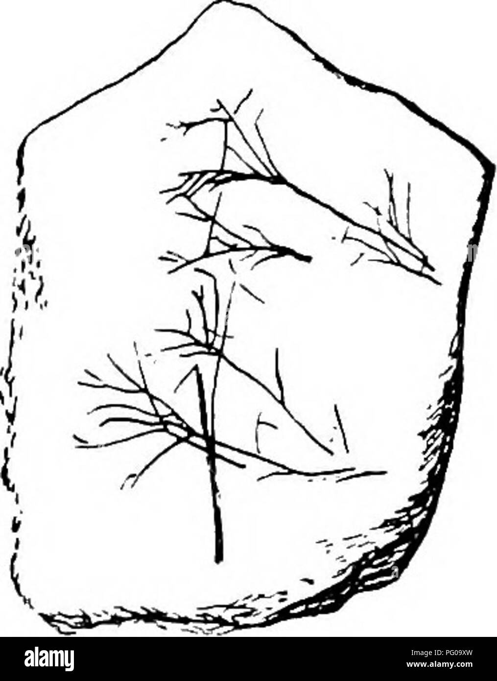 . The British Charophyta. Characeae. GEOLOGICAL HISTORY OF THE CHAROPHYTA. 73 ing to Karpinsky (5), p. 127, Muschelkalk is not found in the district named. Far down in the rocks of the PALEOZOIC era organ- isms occur which have been ascribed to the Charophyta. Silurian.—From a deposit of the Silurian period on the Kellerwald, remains closely resembling the vege- tative parts of a Nitella are figured by Potonie (15) (see Fig. 26), but in the absence of fruit it is not possible to determine their nature. Devonian.—In the Corniferous Limestone of Ohio, Meek, in 1873 (12), recorded the presence, i Stock Photohttps://www.alamy.com/image-license-details/?v=1https://www.alamy.com/the-british-charophyta-characeae-geological-history-of-the-charophyta-73-ing-to-karpinsky-5-p-127-muschelkalk-is-not-found-in-the-district-named-far-down-in-the-rocks-of-the-paleozoic-era-organ-isms-occur-which-have-been-ascribed-to-the-charophyta-silurianfrom-a-deposit-of-the-silurian-period-on-the-kellerwald-remains-closely-resembling-the-vege-tative-parts-of-a-nitella-are-figured-by-potonie-15-see-fig-26-but-in-the-absence-of-fruit-it-is-not-possible-to-determine-their-nature-devonianin-the-corniferous-limestone-of-ohio-meek-in-1873-12-recorded-the-presence-i-image216366721.html
RMPG09XW–. The British Charophyta. Characeae. GEOLOGICAL HISTORY OF THE CHAROPHYTA. 73 ing to Karpinsky (5), p. 127, Muschelkalk is not found in the district named. Far down in the rocks of the PALEOZOIC era organ- isms occur which have been ascribed to the Charophyta. Silurian.—From a deposit of the Silurian period on the Kellerwald, remains closely resembling the vege- tative parts of a Nitella are figured by Potonie (15) (see Fig. 26), but in the absence of fruit it is not possible to determine their nature. Devonian.—In the Corniferous Limestone of Ohio, Meek, in 1873 (12), recorded the presence, i
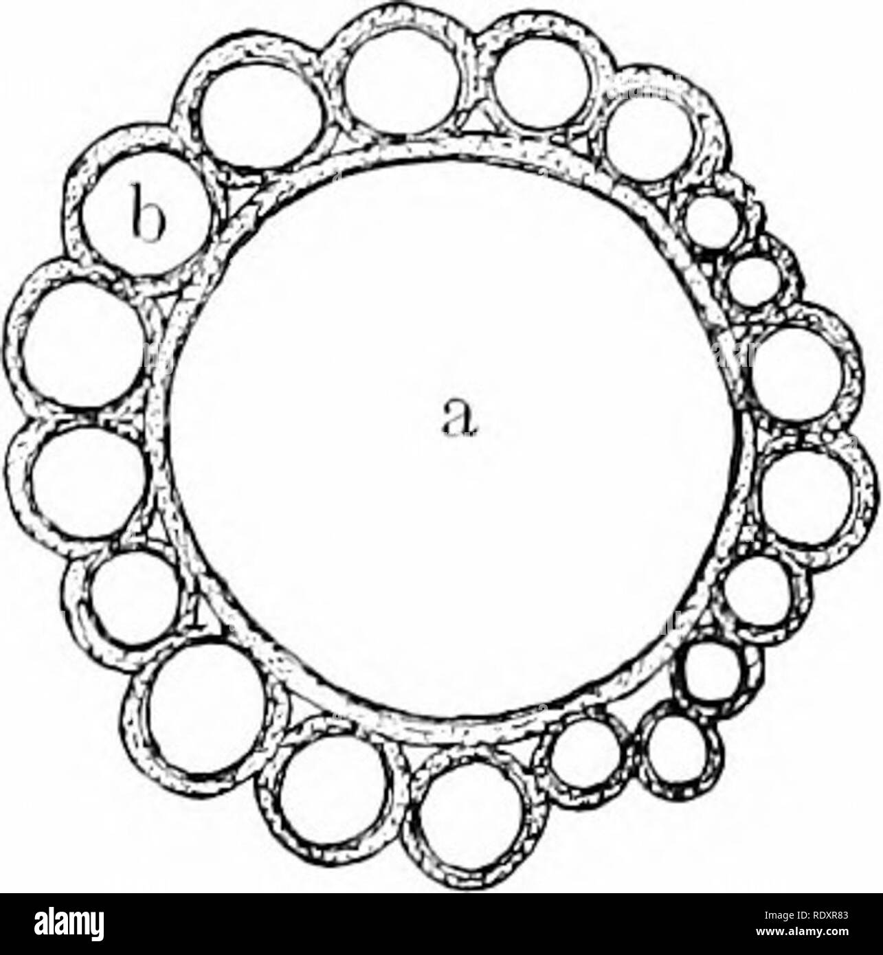 . Plant life, considered with special references to form and function. Plant physiology. 30 PLANT LIFE. 35. Cortex.—Nitella and C'hara are much alike, except that in Cliara the main axis and all its branches are composed of a row of large cells, surrounded by a jacket of smaller ones (fig. 37). The walls of these outer cells are ofteii much thickened, and incrusted with salts of lime to sucli an extent as to render the axis very brittle. Around the main axis the cell-jacket is of much complexity ; it becomes more smiple. Please note that these images are extracted from scanned page images that Stock Photohttps://www.alamy.com/image-license-details/?v=1https://www.alamy.com/plant-life-considered-with-special-references-to-form-and-function-plant-physiology-30-plant-life-35-cortexnitella-and-chara-are-much-alike-except-that-in-cliara-the-main-axis-and-all-its-branches-are-composed-of-a-row-of-large-cells-surrounded-by-a-jacket-of-smaller-ones-fig-37-the-walls-of-these-outer-cells-are-ofteii-much-thickened-and-incrusted-with-salts-of-lime-to-sucli-an-extent-as-to-render-the-axis-very-brittle-around-the-main-axis-the-cell-jacket-is-of-much-complexity-it-becomes-more-smiple-please-note-that-these-images-are-extracted-from-scanned-page-images-that-image232314323.html
RMRDXR83–. Plant life, considered with special references to form and function. Plant physiology. 30 PLANT LIFE. 35. Cortex.—Nitella and C'hara are much alike, except that in Cliara the main axis and all its branches are composed of a row of large cells, surrounded by a jacket of smaller ones (fig. 37). The walls of these outer cells are ofteii much thickened, and incrusted with salts of lime to sucli an extent as to render the axis very brittle. Around the main axis the cell-jacket is of much complexity ; it becomes more smiple. Please note that these images are extracted from scanned page images that
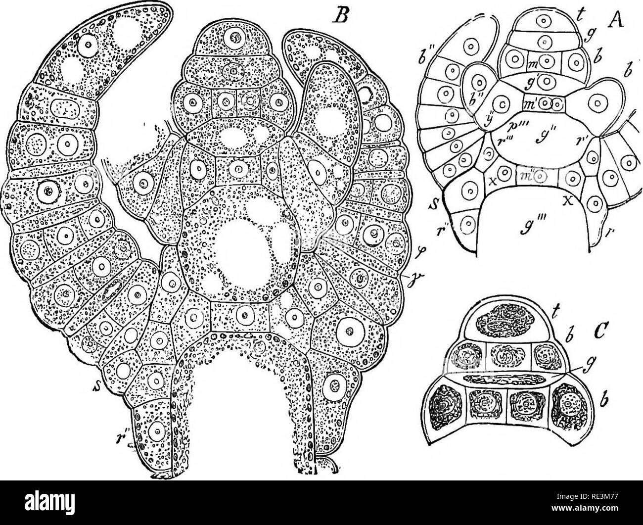
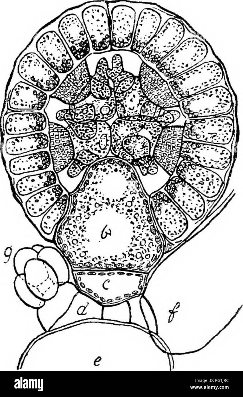 . The British Charophyta. Characeae. III. IV JTiQ. 18.—Development of antheridium of Nitella fiexilis (after Sachs), i. Early stage showing young antheridium divided into eight equal parts by the growth of two vertical walls, at right angles to one another, and a horizontal wall. ii. Further stage showing each eighth part divided into an exterior and interior portion, iii. Later stage showing interior portion again divided, making three layers, the outermost expanding tangentially to form the plates or shields, the middle growing radially to form the mauubria, and the inner- most forming the h Stock Photohttps://www.alamy.com/image-license-details/?v=1https://www.alamy.com/the-british-charophyta-characeae-iii-iv-jtiq-18development-of-antheridium-of-nitella-fiexilis-after-sachs-i-early-stage-showing-young-antheridium-divided-into-eight-equal-parts-by-the-growth-of-two-vertical-walls-at-right-angles-to-one-another-and-a-horizontal-wall-ii-further-stage-showing-each-eighth-part-divided-into-an-exterior-and-interior-portion-iii-later-stage-showing-interior-portion-again-divided-making-three-layers-the-outermost-expanding-tangentially-to-form-the-plates-or-shields-the-middle-growing-radially-to-form-the-mauubria-and-the-inner-most-forming-the-h-image216395632.html
RMPG1JRC–. The British Charophyta. Characeae. III. IV JTiQ. 18.—Development of antheridium of Nitella fiexilis (after Sachs), i. Early stage showing young antheridium divided into eight equal parts by the growth of two vertical walls, at right angles to one another, and a horizontal wall. ii. Further stage showing each eighth part divided into an exterior and interior portion, iii. Later stage showing interior portion again divided, making three layers, the outermost expanding tangentially to form the plates or shields, the middle growing radially to form the mauubria, and the inner- most forming the h
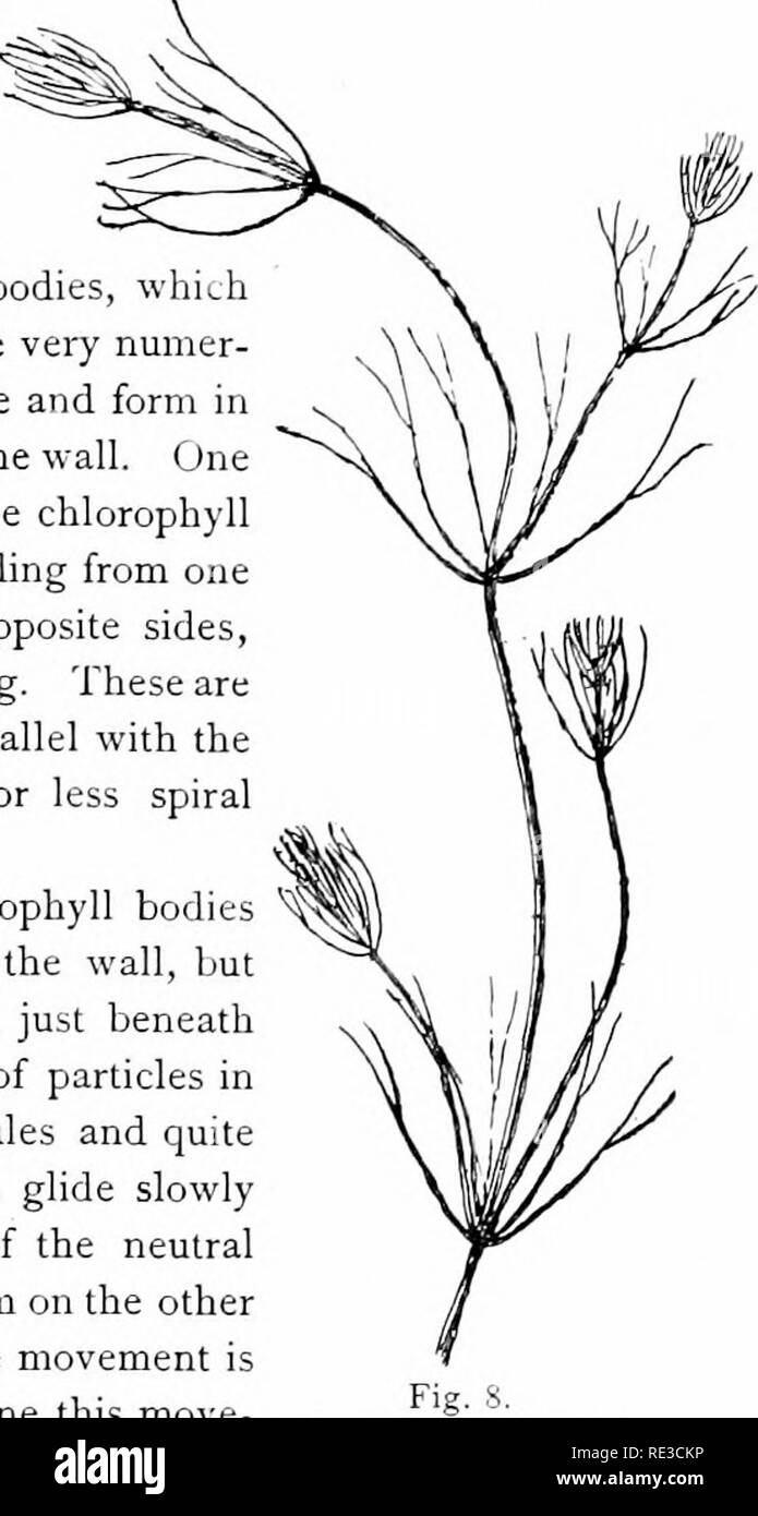 . Elementary botany. Botany. PROTOPLASM. 9 node. These internodes are peculiar. They consist of but a single "cell," and are cylindrical, with closed ends. They are sometimes 5-10 cm. long. 19. Internode of nitella.—For the study of an internode of nitella, a small one, near the end, or the ends of one of the " leaves" is best suited, since it is more transparent. A small portion of the plant should be placed on the glass slip in water with the cover glass over a tuft of the branches near the growing end. E.xamined with the microscope the green chlorophyll bodies, which for Stock Photohttps://www.alamy.com/image-license-details/?v=1https://www.alamy.com/elementary-botany-botany-protoplasm-9-node-these-internodes-are-peculiar-they-consist-of-but-a-single-quotcellquot-and-are-cylindrical-with-closed-ends-they-are-sometimes-5-10-cm-long-19-internode-of-nitellafor-the-study-of-an-internode-of-nitella-a-small-one-near-the-end-or-the-ends-of-one-of-the-quot-leavesquot-is-best-suited-since-it-is-more-transparent-a-small-portion-of-the-plant-should-be-placed-on-the-glass-slip-in-water-with-the-cover-glass-over-a-tuft-of-the-branches-near-the-growing-end-examined-with-the-microscope-the-green-chlorophyll-bodies-which-for-image232415786.html
RMRE3CKP–. Elementary botany. Botany. PROTOPLASM. 9 node. These internodes are peculiar. They consist of but a single "cell," and are cylindrical, with closed ends. They are sometimes 5-10 cm. long. 19. Internode of nitella.—For the study of an internode of nitella, a small one, near the end, or the ends of one of the " leaves" is best suited, since it is more transparent. A small portion of the plant should be placed on the glass slip in water with the cover glass over a tuft of the branches near the growing end. E.xamined with the microscope the green chlorophyll bodies, which for
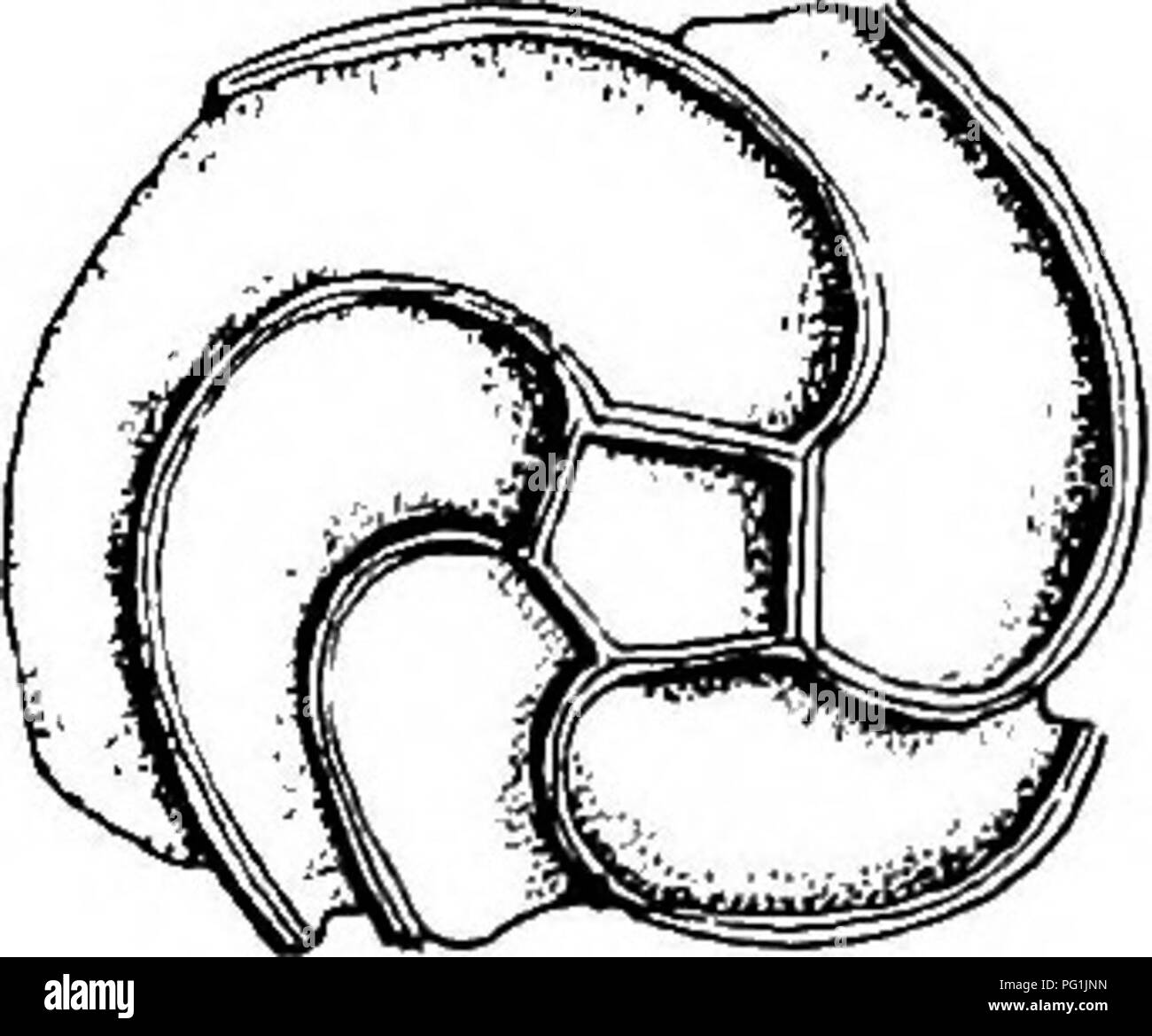 . The British Charophyta. Characeae. Fig. 25.^—Terminal disposition of ridges of oospore, i. View of apex {Nitella capillaris). ii. View of base (Cfeara 6oMica). ridges owe their origin to the overlying enveloping cells, the sutures on the posterior side of which become filled up as the membrane develops and hardens. The number visible varies according as the spiral-cells complete one, two, or three convolutions. In some species of Ghara the angles of asa c aws ^-^^ pentagonal base already referred to EUlu. CF6SL i- cj e/ . are prolonged downwards into claw-like processes which attain sometime Stock Photohttps://www.alamy.com/image-license-details/?v=1https://www.alamy.com/the-british-charophyta-characeae-fig-25terminal-disposition-of-ridges-of-oospore-i-view-of-apex-nitella-capillaris-ii-view-of-base-cfeara-6omica-ridges-owe-their-origin-to-the-overlying-enveloping-cells-the-sutures-on-the-posterior-side-of-which-become-filled-up-as-the-membrane-develops-and-hardens-the-number-visible-varies-according-as-the-spiral-cells-complete-one-two-or-three-convolutions-in-some-species-of-ghara-the-angles-of-asa-c-aws-pentagonal-base-already-referred-to-eulu-cf6sl-i-cj-e-are-prolonged-downwards-into-claw-like-processes-which-attain-sometime-image216395585.html
RMPG1JNN–. The British Charophyta. Characeae. Fig. 25.^—Terminal disposition of ridges of oospore, i. View of apex {Nitella capillaris). ii. View of base (Cfeara 6oMica). ridges owe their origin to the overlying enveloping cells, the sutures on the posterior side of which become filled up as the membrane develops and hardens. The number visible varies according as the spiral-cells complete one, two, or three convolutions. In some species of Ghara the angles of asa c aws ^-^^ pentagonal base already referred to EUlu. CF6SL i- cj e/ . are prolonged downwards into claw-like processes which attain sometime
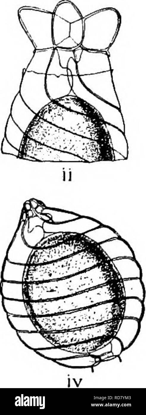 . The British Charophyta. Characeae. Fio. 23.—Process of fertilisation, i, ii, iii. Upper part of oogonium of Chara vulgaris. iv. Oogonium of Nitella tenuissima. i. Half- matured unfertilised oogonium showing upper and lower cavities formed by development of coronula and enveloping cells, ii. Fully matured unfertilised oogonium showing tumid growth at the head of enveloping cells, and transverse fracture of membrane caused by their extended growth, iii. Antherozoids penetrating interstices formed by contracted terminations of spiral cells, and reaching oospore through upper and lower cavities Stock Photohttps://www.alamy.com/image-license-details/?v=1https://www.alamy.com/the-british-charophyta-characeae-fio-23process-of-fertilisation-i-ii-iii-upper-part-of-oogonium-of-chara-vulgaris-iv-oogonium-of-nitella-tenuissima-i-half-matured-unfertilised-oogonium-showing-upper-and-lower-cavities-formed-by-development-of-coronula-and-enveloping-cells-ii-fully-matured-unfertilised-oogonium-showing-tumid-growth-at-the-head-of-enveloping-cells-and-transverse-fracture-of-membrane-caused-by-their-extended-growth-iii-antherozoids-penetrating-interstices-formed-by-contracted-terminations-of-spiral-cells-and-reaching-oospore-through-upper-and-lower-cavities-image231900707.html
RMRD7YM3–. The British Charophyta. Characeae. Fio. 23.—Process of fertilisation, i, ii, iii. Upper part of oogonium of Chara vulgaris. iv. Oogonium of Nitella tenuissima. i. Half- matured unfertilised oogonium showing upper and lower cavities formed by development of coronula and enveloping cells, ii. Fully matured unfertilised oogonium showing tumid growth at the head of enveloping cells, and transverse fracture of membrane caused by their extended growth, iii. Antherozoids penetrating interstices formed by contracted terminations of spiral cells, and reaching oospore through upper and lower cavities
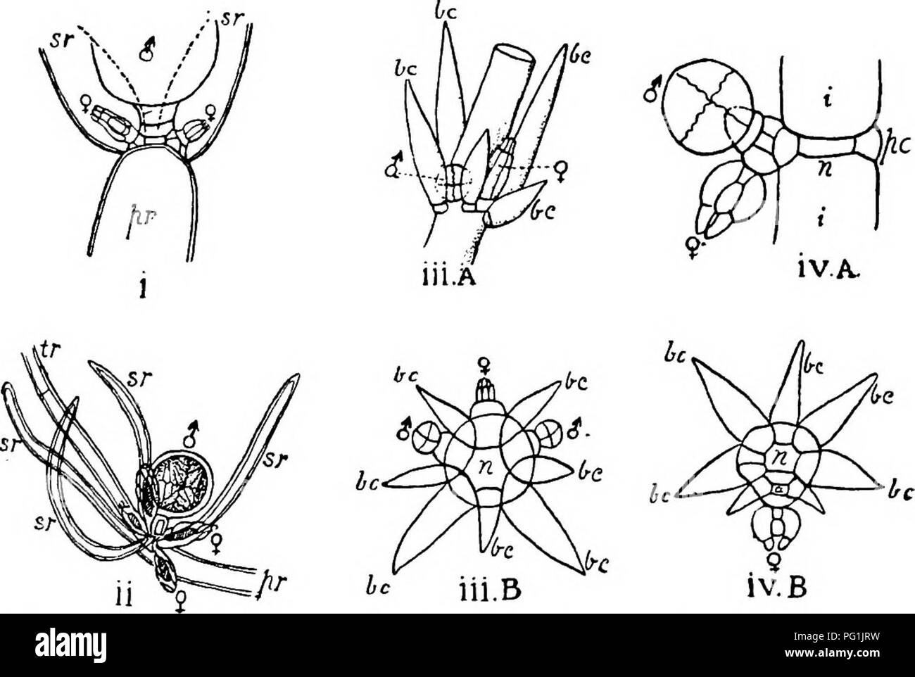 . The British Charophyta. Characeae. 44 BRITISH CHAROPHYTA. In Lychnothamnus and Nitellopsis the arrangement is apparently similar. In Lychnothamnus the oogonium, which is solitary, is produced separately from the central peripheral cell on the inner side of the branchlet, that is the side facing the stem or branch, and antheridia are normally produced from peripheral cells on either side of that producing the oogonium (Fig. 16 iii A and B). In Nitellopsis the only known. Pig. 16.—Position of reproductive organs in several genera. <J = antheridium, ? = oogonium in all figvires. i. Nitella f Stock Photohttps://www.alamy.com/image-license-details/?v=1https://www.alamy.com/the-british-charophyta-characeae-44-british-charophyta-in-lychnothamnus-and-nitellopsis-the-arrangement-is-apparently-similar-in-lychnothamnus-the-oogonium-which-is-solitary-is-produced-separately-from-the-central-peripheral-cell-on-the-inner-side-of-the-branchlet-that-is-the-side-facing-the-stem-or-branch-and-antheridia-are-normally-produced-from-peripheral-cells-on-either-side-of-that-producing-the-oogonium-fig-16-iii-a-and-b-in-nitellopsis-the-only-known-pig-16position-of-reproductive-organs-in-several-genera-ltj-=-antheridium-=-oogonium-in-all-figvires-i-nitella-f-image216395645.html
RMPG1JRW–. The British Charophyta. Characeae. 44 BRITISH CHAROPHYTA. In Lychnothamnus and Nitellopsis the arrangement is apparently similar. In Lychnothamnus the oogonium, which is solitary, is produced separately from the central peripheral cell on the inner side of the branchlet, that is the side facing the stem or branch, and antheridia are normally produced from peripheral cells on either side of that producing the oogonium (Fig. 16 iii A and B). In Nitellopsis the only known. Pig. 16.—Position of reproductive organs in several genera. <J = antheridium, ? = oogonium in all figvires. i. Nitella f
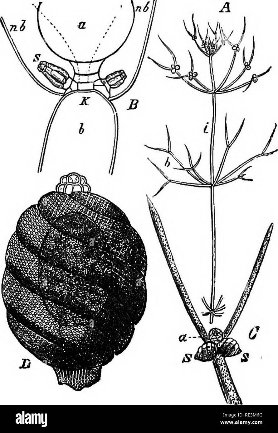 . A handbook of cryptogamic botany. Cryptogams. CHARACE.'E S79 The Characese are either monoecious 9r dicEcious, In the former case the male and female organs are formed in close juxtaposition on the same node, the archegone being somewhat below the antherid in Nitella, above it or by its side in Chara. The archegones, like the antherids, are metamorphosed leaves. When ready for fertilisation, the archegone has a, longer or shorter ovoid form, and is borne on a short pedicel-cell. In the interior is an axial row of cells enveloped by five tubes, which are at first straight, but are afterwards Stock Photohttps://www.alamy.com/image-license-details/?v=1https://www.alamy.com/a-handbook-of-cryptogamic-botany-cryptogams-characee-s79-the-characese-are-either-monoecious-9r-dicecious-in-the-former-case-the-male-and-female-organs-are-formed-in-close-juxtaposition-on-the-same-node-the-archegone-being-somewhat-below-the-antherid-in-nitella-above-it-or-by-its-side-in-chara-the-archegones-like-the-antherids-are-metamorphosed-leaves-when-ready-for-fertilisation-the-archegone-has-a-longer-or-shorter-ovoid-form-and-is-borne-on-a-short-pedicel-cell-in-the-interior-is-an-axial-row-of-cells-enveloped-by-five-tubes-which-are-at-first-straight-but-are-afterwards-image232421688.html
RMRE3M6G–. A handbook of cryptogamic botany. Cryptogams. CHARACE.'E S79 The Characese are either monoecious 9r dicEcious, In the former case the male and female organs are formed in close juxtaposition on the same node, the archegone being somewhat below the antherid in Nitella, above it or by its side in Chara. The archegones, like the antherids, are metamorphosed leaves. When ready for fertilisation, the archegone has a, longer or shorter ovoid form, and is borne on a short pedicel-cell. In the interior is an axial row of cells enveloped by five tubes, which are at first straight, but are afterwards
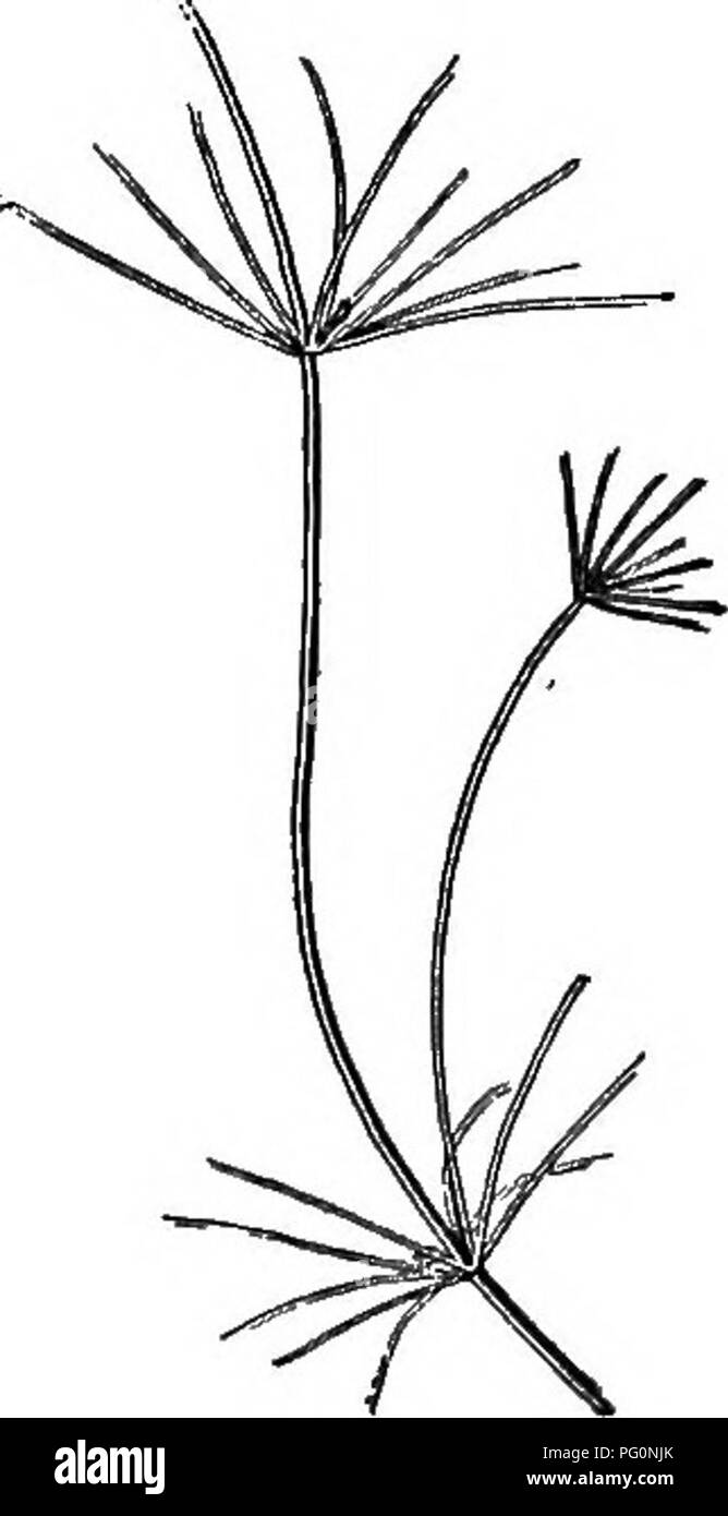 . Beginners' botany. Botany. Flo. 269. — Fucus. Fruiting branches at s, s. On the stem are-two air-bladders.. Fig. 270. — NiTELLA. Nitella.—This is a large branched and specialized fresh-water alga found in tufts attached to the bottom in shallow ponds (Fig. 270). Between the whorls of branches are long internodes consisting of a single cylindrical cell, which is oiie of the largest cells known in vegetable tissue. Under the microscope the walls of this cell are found to be lined with a layer of small stationary chloroplastids, within which layer the protoplasm, under favorable circumstances, Stock Photohttps://www.alamy.com/image-license-details/?v=1https://www.alamy.com/beginners-botany-botany-flo-269-fucus-fruiting-branches-at-s-s-on-the-stem-are-two-air-bladders-fig-270-nitella-nitellathis-is-a-large-branched-and-specialized-fresh-water-alga-found-in-tufts-attached-to-the-bottom-in-shallow-ponds-fig-270-between-the-whorls-of-branches-are-long-internodes-consisting-of-a-single-cylindrical-cell-which-is-oiie-of-the-largest-cells-known-in-vegetable-tissue-under-the-microscope-the-walls-of-this-cell-are-found-to-be-lined-with-a-layer-of-small-stationary-chloroplastids-within-which-layer-the-protoplasm-under-favorable-circumstances-image216375899.html
RMPG0NJK–. Beginners' botany. Botany. Flo. 269. — Fucus. Fruiting branches at s, s. On the stem are-two air-bladders.. Fig. 270. — NiTELLA. Nitella.—This is a large branched and specialized fresh-water alga found in tufts attached to the bottom in shallow ponds (Fig. 270). Between the whorls of branches are long internodes consisting of a single cylindrical cell, which is oiie of the largest cells known in vegetable tissue. Under the microscope the walls of this cell are found to be lined with a layer of small stationary chloroplastids, within which layer the protoplasm, under favorable circumstances,
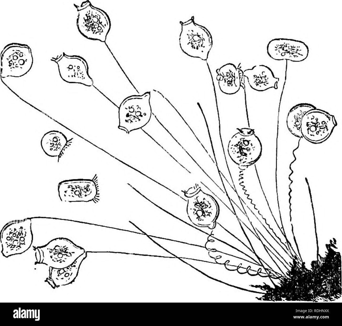 . Evenings at the microscope : or, Researches among the minuter organs and forms of animal life . Zoology; Microscopy; Microscopes. 402 EVENINGS AT THE MICROSCOPE. And now, having pretty well exhausted the contents of this live-box, let us try a dip from this other phial from another locality, equally productive, if I am not mistaken. Yes ; for, to begin, the stalks of Nitella here are fringed with populous colonies of the most attractive of all the Infusoria, the beautiful Vorticellm. The species is not the common bell-shaped one, but the smaller with pursed mouth, the little V. microstoma. L Stock Photohttps://www.alamy.com/image-license-details/?v=1https://www.alamy.com/evenings-at-the-microscope-or-researches-among-the-minuter-organs-and-forms-of-animal-life-zoology-microscopy-microscopes-402-evenings-at-the-microscope-and-now-having-pretty-well-exhausted-the-contents-of-this-live-box-let-us-try-a-dip-from-this-other-phial-from-another-locality-equally-productive-if-i-am-not-mistaken-yes-for-to-begin-the-stalks-of-nitella-here-are-fringed-with-populous-colonies-of-the-most-attractive-of-all-the-infusoria-the-beautiful-vorticellm-the-species-is-not-the-common-bell-shaped-one-but-the-smaller-with-pursed-mouth-the-little-v-microstoma-l-image232115714.html
RMRDHNXX–. Evenings at the microscope : or, Researches among the minuter organs and forms of animal life . Zoology; Microscopy; Microscopes. 402 EVENINGS AT THE MICROSCOPE. And now, having pretty well exhausted the contents of this live-box, let us try a dip from this other phial from another locality, equally productive, if I am not mistaken. Yes ; for, to begin, the stalks of Nitella here are fringed with populous colonies of the most attractive of all the Infusoria, the beautiful Vorticellm. The species is not the common bell-shaped one, but the smaller with pursed mouth, the little V. microstoma. L
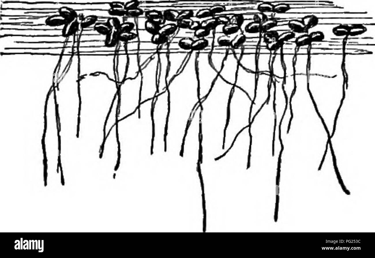 . Insect life; an introduction to nature study and a guide for teachers, students and others interested in out-of-door life. Entomology; Nature study. THE BREEDING OF INSECTS. 331. Fig. 292.—Duckweed. Watercress, Nasturtium officinale. Stoneworts, Chara and Nitella (several species of each). Frog-spittle or water-silk, Spirogira. A small quantity of duckweed, Lemna (Fig. 292), placed on the surface of the water adds to the beauty of an aquarium. When it is necessary to add water to an aqua- rium on account of loss by evaporation, rain wa- ter should be used to prevent an undue ac- cumulation o Stock Photohttps://www.alamy.com/image-license-details/?v=1https://www.alamy.com/insect-life-an-introduction-to-nature-study-and-a-guide-for-teachers-students-and-others-interested-in-out-of-door-life-entomology-nature-study-the-breeding-of-insects-331-fig-292duckweed-watercress-nasturtium-officinale-stoneworts-chara-and-nitella-several-species-of-each-frog-spittle-or-water-silk-spirogira-a-small-quantity-of-duckweed-lemna-fig-292-placed-on-the-surface-of-the-water-adds-to-the-beauty-of-an-aquarium-when-it-is-necessary-to-add-water-to-an-aqua-rium-on-account-of-loss-by-evaporation-rain-wa-ter-should-be-used-to-prevent-an-undue-ac-cumulation-o-image216406832.html
RMPG253C–. Insect life; an introduction to nature study and a guide for teachers, students and others interested in out-of-door life. Entomology; Nature study. THE BREEDING OF INSECTS. 331. Fig. 292.—Duckweed. Watercress, Nasturtium officinale. Stoneworts, Chara and Nitella (several species of each). Frog-spittle or water-silk, Spirogira. A small quantity of duckweed, Lemna (Fig. 292), placed on the surface of the water adds to the beauty of an aquarium. When it is necessary to add water to an aqua- rium on account of loss by evaporation, rain wa- ter should be used to prevent an undue ac- cumulation o
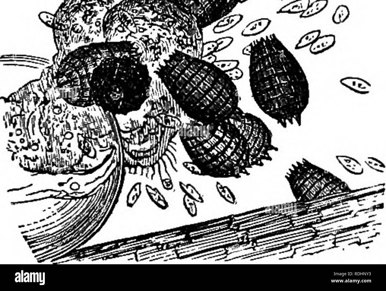 . Evenings at the microscope : or, Researches among the minuter organs and forms of animal life . Zoology; Microscopy; Microscopes. INFUSOEIA. 899 while I will tell you the tragical and lamentable history of just such an embryo as this, that was eaten up before it was born, under my own eye. One of the depredators was a very amusing animalcule, which is sufficiently scarce to make its occurrence a thing of interest, especially to a young microscopist, as I was at the time. A large egg of (as I believe) Euchlanis dilatata had been laid during the night on a leaf of Nitella, in the live-box. Whe Stock Photohttps://www.alamy.com/image-license-details/?v=1https://www.alamy.com/evenings-at-the-microscope-or-researches-among-the-minuter-organs-and-forms-of-animal-life-zoology-microscopy-microscopes-infusoeia-899-while-i-will-tell-you-the-tragical-and-lamentable-history-of-just-such-an-embryo-as-this-that-was-eaten-up-before-it-was-born-under-my-own-eye-one-of-the-depredators-was-a-very-amusing-animalcule-which-is-sufficiently-scarce-to-make-its-occurrence-a-thing-of-interest-especially-to-a-young-microscopist-as-i-was-at-the-time-a-large-egg-of-as-i-believe-euchlanis-dilatata-had-been-laid-during-the-night-on-a-leaf-of-nitella-in-the-live-box-whe-image232115719.html
RMRDHNY3–. Evenings at the microscope : or, Researches among the minuter organs and forms of animal life . Zoology; Microscopy; Microscopes. INFUSOEIA. 899 while I will tell you the tragical and lamentable history of just such an embryo as this, that was eaten up before it was born, under my own eye. One of the depredators was a very amusing animalcule, which is sufficiently scarce to make its occurrence a thing of interest, especially to a young microscopist, as I was at the time. A large egg of (as I believe) Euchlanis dilatata had been laid during the night on a leaf of Nitella, in the live-box. Whe
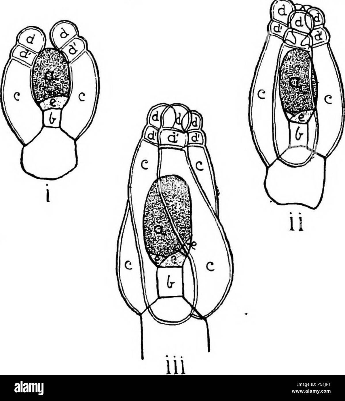 . The British Charophyta. Characeae. 52 BRITISH CHAEOPHYTA. ovoidal form. The lateral walls of the spiral cells subsequently break down, leaving the outer walls to form a single integument marked by spiral furrows along the lines of union. Incased and protected by the enveloping spiral cells the oosphere consists of a nucleated mass of protoplasm enclosed by a thin and extremely delicate. Pig. 22.—Development of oogonium of Nitella flexilis (after Sachs). o, oosphere; h, node-cell; c, enveloping cells; d, upper, and. d', lower cells of ooronula; e, turning cell. i. Early stages of the oogonium Stock Photohttps://www.alamy.com/image-license-details/?v=1https://www.alamy.com/the-british-charophyta-characeae-52-british-chaeophyta-ovoidal-form-the-lateral-walls-of-the-spiral-cells-subsequently-break-down-leaving-the-outer-walls-to-form-a-single-integument-marked-by-spiral-furrows-along-the-lines-of-union-incased-and-protected-by-the-enveloping-spiral-cells-the-oosphere-consists-of-a-nucleated-mass-of-protoplasm-enclosed-by-a-thin-and-extremely-delicate-pig-22development-of-oogonium-of-nitella-flexilis-after-sachs-o-oosphere-h-node-cell-c-enveloping-cells-d-upper-and-d-lower-cells-of-ooronula-e-turning-cell-i-early-stages-of-the-oogonium-image216395616.html
RMPG1JPT–. The British Charophyta. Characeae. 52 BRITISH CHAEOPHYTA. ovoidal form. The lateral walls of the spiral cells subsequently break down, leaving the outer walls to form a single integument marked by spiral furrows along the lines of union. Incased and protected by the enveloping spiral cells the oosphere consists of a nucleated mass of protoplasm enclosed by a thin and extremely delicate. Pig. 22.—Development of oogonium of Nitella flexilis (after Sachs). o, oosphere; h, node-cell; c, enveloping cells; d, upper, and. d', lower cells of ooronula; e, turning cell. i. Early stages of the oogonium
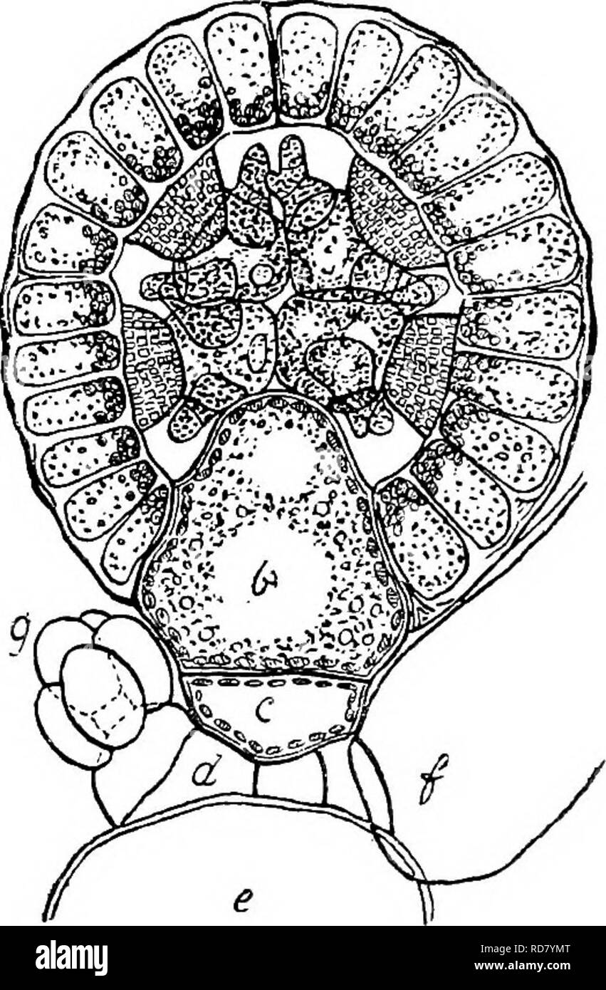 . The British Charophyta. Characeae. III. IV JTiQ. 18.—Development of antheridium of Nitella fiexilis (after Sachs), i. Early stage showing young antheridium divided into eight equal parts by the growth of two vertical walls, at right angles to one another, and a horizontal wall. ii. Further stage showing each eighth part divided into an exterior and interior portion, iii. Later stage showing interior portion again divided, making three layers, the outermost expanding tangentially to form the plates or shields, the middle growing radially to form the mauubria, and the inner- most forming the h Stock Photohttps://www.alamy.com/image-license-details/?v=1https://www.alamy.com/the-british-charophyta-characeae-iii-iv-jtiq-18development-of-antheridium-of-nitella-fiexilis-after-sachs-i-early-stage-showing-young-antheridium-divided-into-eight-equal-parts-by-the-growth-of-two-vertical-walls-at-right-angles-to-one-another-and-a-horizontal-wall-ii-further-stage-showing-each-eighth-part-divided-into-an-exterior-and-interior-portion-iii-later-stage-showing-interior-portion-again-divided-making-three-layers-the-outermost-expanding-tangentially-to-form-the-plates-or-shields-the-middle-growing-radially-to-form-the-mauubria-and-the-inner-most-forming-the-h-image231900728.html
RMRD7YMT–. The British Charophyta. Characeae. III. IV JTiQ. 18.—Development of antheridium of Nitella fiexilis (after Sachs), i. Early stage showing young antheridium divided into eight equal parts by the growth of two vertical walls, at right angles to one another, and a horizontal wall. ii. Further stage showing each eighth part divided into an exterior and interior portion, iii. Later stage showing interior portion again divided, making three layers, the outermost expanding tangentially to form the plates or shields, the middle growing radially to form the mauubria, and the inner- most forming the h
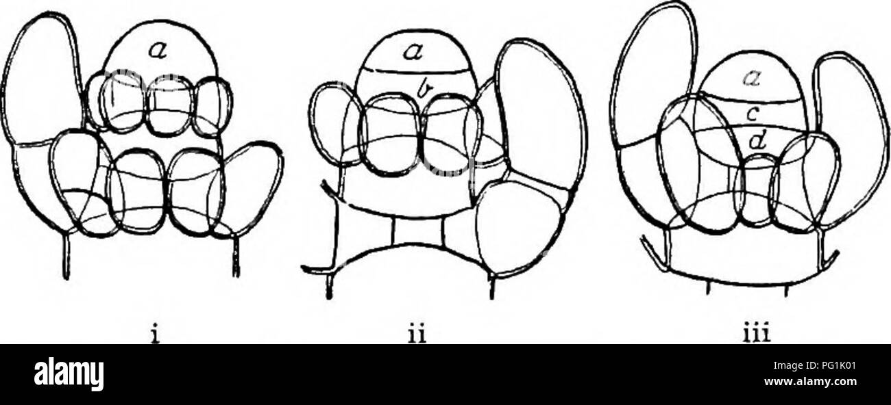 . The British Charophyta. Characeae. STRUaTUBE AND DEVELOPMENT. 23 stem-node, remaining quite short and undergoing division by means of longitudinal septa in the direc- tion of the axis, the lower (d) forming an internode, remaining undivided but lengthening considerably. In this way the stem of a Charophyte presents a. Fia. 3.—Yoimg shoot of Nitella in successive stages (after Giesen- hagen) ( x o. 150). In i the apical cell a is undivided; in ii a portion b has been cut off from its base by a transverse section ; in iii the lower cell b has been subdivided into a stem-node c and an internode Stock Photohttps://www.alamy.com/image-license-details/?v=1https://www.alamy.com/the-british-charophyta-characeae-struatube-and-development-23-stem-node-remaining-quite-short-and-undergoing-division-by-means-of-longitudinal-septa-in-the-direc-tion-of-the-axis-the-lower-d-forming-an-internode-remaining-undivided-but-lengthening-considerably-in-this-way-the-stem-of-a-charophyte-presents-a-fia-3yoimg-shoot-of-nitella-in-successive-stages-after-giesen-hagen-x-o-150-in-i-the-apical-cell-a-is-undivided-in-ii-a-portion-b-has-been-cut-off-from-its-base-by-a-transverse-section-in-iii-the-lower-cell-b-has-been-subdivided-into-a-stem-node-c-and-an-internode-image216395761.html
RMPG1K01–. The British Charophyta. Characeae. STRUaTUBE AND DEVELOPMENT. 23 stem-node, remaining quite short and undergoing division by means of longitudinal septa in the direc- tion of the axis, the lower (d) forming an internode, remaining undivided but lengthening considerably. In this way the stem of a Charophyte presents a. Fia. 3.—Yoimg shoot of Nitella in successive stages (after Giesen- hagen) ( x o. 150). In i the apical cell a is undivided; in ii a portion b has been cut off from its base by a transverse section ; in iii the lower cell b has been subdivided into a stem-node c and an internode
 . Principles of modern biology. Biology. 168 - The Cell. Fig. 9-8. One complete cell in a filament of the green alga, Spirogyra. The nu- cleus, with a distinct darkly stained nucleolus, lies at the center, suspended in place by delicate strands of cytoplasm. The chloroplast has the form of a spiral ribbon, on which numerous small stained bodies, the pyrenoids, are discernible in this re- touched photograph. (Copyright, General Biological Supply House, Inc.) such as Nitella (Fig. 13-2), are true multi- cellular organisms. Algae differ from the simple animals in that they all possess chlo- rophy Stock Photohttps://www.alamy.com/image-license-details/?v=1https://www.alamy.com/principles-of-modern-biology-biology-168-the-cell-fig-9-8-one-complete-cell-in-a-filament-of-the-green-alga-spirogyra-the-nu-cleus-with-a-distinct-darkly-stained-nucleolus-lies-at-the-center-suspended-in-place-by-delicate-strands-of-cytoplasm-the-chloroplast-has-the-form-of-a-spiral-ribbon-on-which-numerous-small-stained-bodies-the-pyrenoids-are-discernible-in-this-re-touched-photograph-copyright-general-biological-supply-house-inc-such-as-nitella-fig-13-2-are-true-multi-cellular-organisms-algae-differ-from-the-simple-animals-in-that-they-all-possess-chlo-rophy-image232317428.html
RMRDXY70–. Principles of modern biology. Biology. 168 - The Cell. Fig. 9-8. One complete cell in a filament of the green alga, Spirogyra. The nu- cleus, with a distinct darkly stained nucleolus, lies at the center, suspended in place by delicate strands of cytoplasm. The chloroplast has the form of a spiral ribbon, on which numerous small stained bodies, the pyrenoids, are discernible in this re- touched photograph. (Copyright, General Biological Supply House, Inc.) such as Nitella (Fig. 13-2), are true multi- cellular organisms. Algae differ from the simple animals in that they all possess chlo- rophy
 . Botany for high schools and colleges. Botany. OHARACEJE. 333 plant is composed of a jointed stem, which bears whorls of leaves at regular intervals. The stem is one-celled in trans- verse section, as in Nitella, or it has a large axial cell, which is surrounded by many long narrow ones, which form a cortical envelope, as in many species of Oliara. In some species the stem and leaves become incrusted with lime, giv- ing to them a good deal of hardness and brittleness. {a) The class is readily divisible into two orders — Nitelleae and Charese.* Order Nitellese.—In this order the stem and leave Stock Photohttps://www.alamy.com/image-license-details/?v=1https://www.alamy.com/botany-for-high-schools-and-colleges-botany-oharaceje-333-plant-is-composed-of-a-jointed-stem-which-bears-whorls-of-leaves-at-regular-intervals-the-stem-is-one-celled-in-trans-verse-section-as-in-nitella-or-it-has-a-large-axial-cell-which-is-surrounded-by-many-long-narrow-ones-which-form-a-cortical-envelope-as-in-many-species-of-oliara-in-some-species-the-stem-and-leaves-become-incrusted-with-lime-giv-ing-to-them-a-good-deal-of-hardness-and-brittleness-a-the-class-is-readily-divisible-into-two-orders-nitelleae-and-charese-order-nitellesein-this-order-the-stem-and-leave-image232281734.html
RMRDW9M6–. Botany for high schools and colleges. Botany. OHARACEJE. 333 plant is composed of a jointed stem, which bears whorls of leaves at regular intervals. The stem is one-celled in trans- verse section, as in Nitella, or it has a large axial cell, which is surrounded by many long narrow ones, which form a cortical envelope, as in many species of Oliara. In some species the stem and leaves become incrusted with lime, giv- ing to them a good deal of hardness and brittleness. {a) The class is readily divisible into two orders — Nitelleae and Charese.* Order Nitellese.—In this order the stem and leave
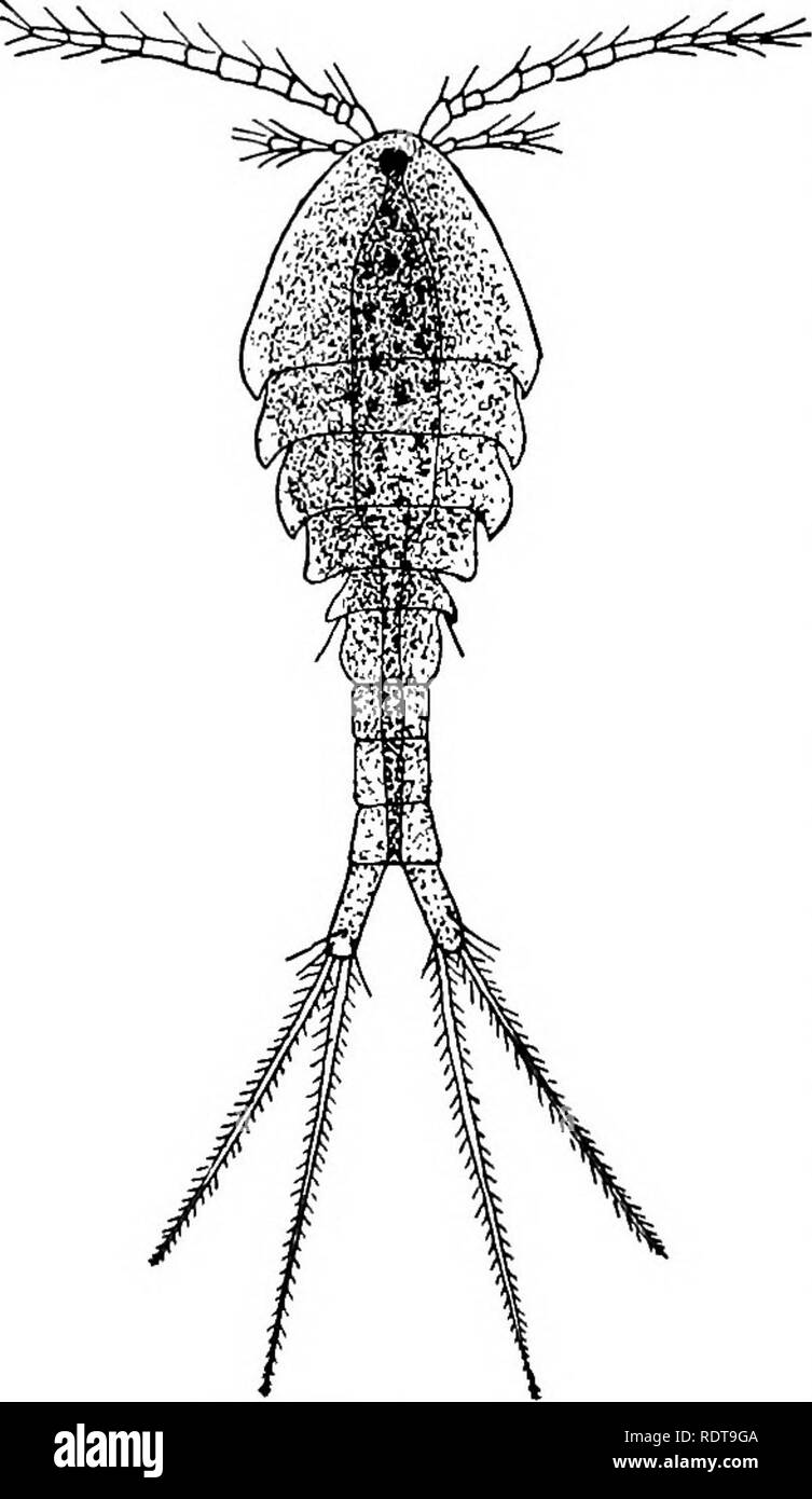 . A laboratory guide for beginners in zoology. Laboratory animals. xxn Introduction in one jar, get more of the material from the same place and treat it in the same way. The hydras may be trans- ferred to covered aquaria containing Vaucheria, Nitella, or similar plants, where under favorable conditions they will multiply and be available when wanted. With a dozen or more aquaria containing water plants in good condition in the laboratory, hydras are pretty sure to be present in some of them. Water snails should not be al- lowed in hydra jars. Cy- clops or other minute Crustacea are also likel Stock Photohttps://www.alamy.com/image-license-details/?v=1https://www.alamy.com/a-laboratory-guide-for-beginners-in-zoology-laboratory-animals-xxn-introduction-in-one-jar-get-more-of-the-material-from-the-same-place-and-treat-it-in-the-same-way-the-hydras-may-be-trans-ferred-to-covered-aquaria-containing-vaucheria-nitella-or-similar-plants-where-under-favorable-conditions-they-will-multiply-and-be-available-when-wanted-with-a-dozen-or-more-aquaria-containing-water-plants-in-good-condition-in-the-laboratory-hydras-are-pretty-sure-to-be-present-in-some-of-them-water-snails-should-not-be-al-lowed-in-hydra-jars-cy-clops-or-other-minute-crustacea-are-also-likel-image232259674.html
RMRDT9GA–. A laboratory guide for beginners in zoology. Laboratory animals. xxn Introduction in one jar, get more of the material from the same place and treat it in the same way. The hydras may be trans- ferred to covered aquaria containing Vaucheria, Nitella, or similar plants, where under favorable conditions they will multiply and be available when wanted. With a dozen or more aquaria containing water plants in good condition in the laboratory, hydras are pretty sure to be present in some of them. Water snails should not be al- lowed in hydra jars. Cy- clops or other minute Crustacea are also likel
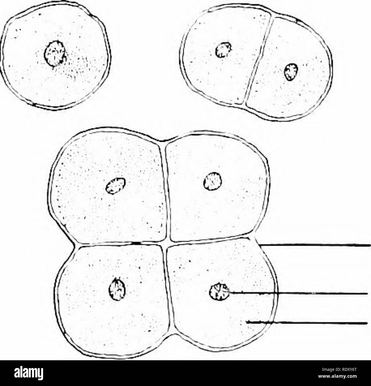 . Principles of modern biology. Biology. Fig. 9-8. One complete cell in a filament of the green alga, Spirogyra. The nu- cleus, with a distinct darkly stained nucleolus, lies at the center, suspended in place by delicate strands of cytoplasm. The chloroplast has the form of a spiral ribbon, on which numerous small stained bodies, the pyrenoids, are discernible in this re- touched photograph. (Copyright, General Biological Supply House, Inc.) such as Nitella (Fig. 13-2), are true multi- cellular organisms. Algae differ from the simple animals in that they all possess chlo- rophyll, and most hav Stock Photohttps://www.alamy.com/image-license-details/?v=1https://www.alamy.com/principles-of-modern-biology-biology-fig-9-8-one-complete-cell-in-a-filament-of-the-green-alga-spirogyra-the-nu-cleus-with-a-distinct-darkly-stained-nucleolus-lies-at-the-center-suspended-in-place-by-delicate-strands-of-cytoplasm-the-chloroplast-has-the-form-of-a-spiral-ribbon-on-which-numerous-small-stained-bodies-the-pyrenoids-are-discernible-in-this-re-touched-photograph-copyright-general-biological-supply-house-inc-such-as-nitella-fig-13-2-are-true-multi-cellular-organisms-algae-differ-from-the-simple-animals-in-that-they-all-possess-chlo-rophyll-and-most-hav-image232317424.html
RMRDXY6T–. Principles of modern biology. Biology. Fig. 9-8. One complete cell in a filament of the green alga, Spirogyra. The nu- cleus, with a distinct darkly stained nucleolus, lies at the center, suspended in place by delicate strands of cytoplasm. The chloroplast has the form of a spiral ribbon, on which numerous small stained bodies, the pyrenoids, are discernible in this re- touched photograph. (Copyright, General Biological Supply House, Inc.) such as Nitella (Fig. 13-2), are true multi- cellular organisms. Algae differ from the simple animals in that they all possess chlo- rophyll, and most hav
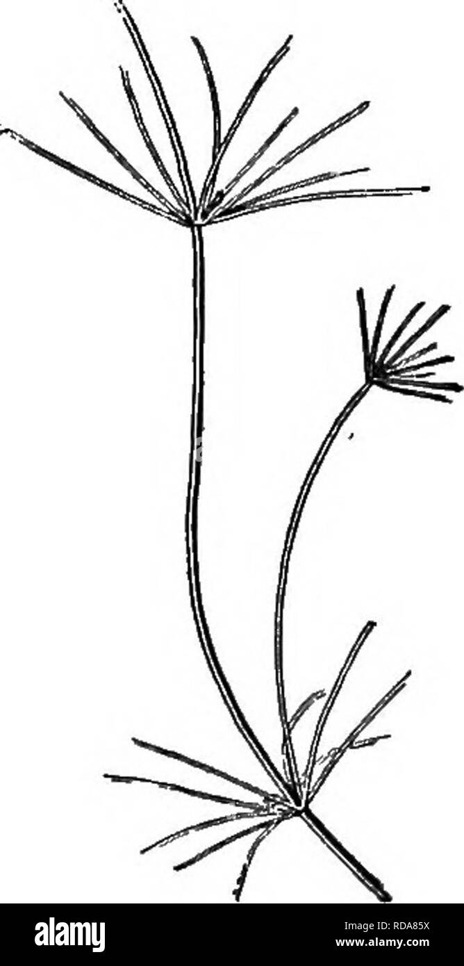 . Beginners' botany. Botany. Flo. 269. — Fucus. Fruiting branches at s, s. On the stem are-two air-bladders.. Fig. 270. — NiTELLA. Nitella.—This is a large branched and specialized fresh-water alga found in tufts attached to the bottom in shallow ponds (Fig. 270). Between the whorls of branches are long internodes consisting of a single cylindrical cell, which is oiie of the largest cells known in vegetable tissue. Under the microscope the walls of this cell are found to be lined with a layer of small stationary chloroplastids, within which layer the protoplasm, under favorable circumstances, Stock Photohttps://www.alamy.com/image-license-details/?v=1https://www.alamy.com/beginners-botany-botany-flo-269-fucus-fruiting-branches-at-s-s-on-the-stem-are-two-air-bladders-fig-270-nitella-nitellathis-is-a-large-branched-and-specialized-fresh-water-alga-found-in-tufts-attached-to-the-bottom-in-shallow-ponds-fig-270-between-the-whorls-of-branches-are-long-internodes-consisting-of-a-single-cylindrical-cell-which-is-oiie-of-the-largest-cells-known-in-vegetable-tissue-under-the-microscope-the-walls-of-this-cell-are-found-to-be-lined-with-a-layer-of-small-stationary-chloroplastids-within-which-layer-the-protoplasm-under-favorable-circumstances-image231951270.html
RMRDA85X–. Beginners' botany. Botany. Flo. 269. — Fucus. Fruiting branches at s, s. On the stem are-two air-bladders.. Fig. 270. — NiTELLA. Nitella.—This is a large branched and specialized fresh-water alga found in tufts attached to the bottom in shallow ponds (Fig. 270). Between the whorls of branches are long internodes consisting of a single cylindrical cell, which is oiie of the largest cells known in vegetable tissue. Under the microscope the walls of this cell are found to be lined with a layer of small stationary chloroplastids, within which layer the protoplasm, under favorable circumstances,
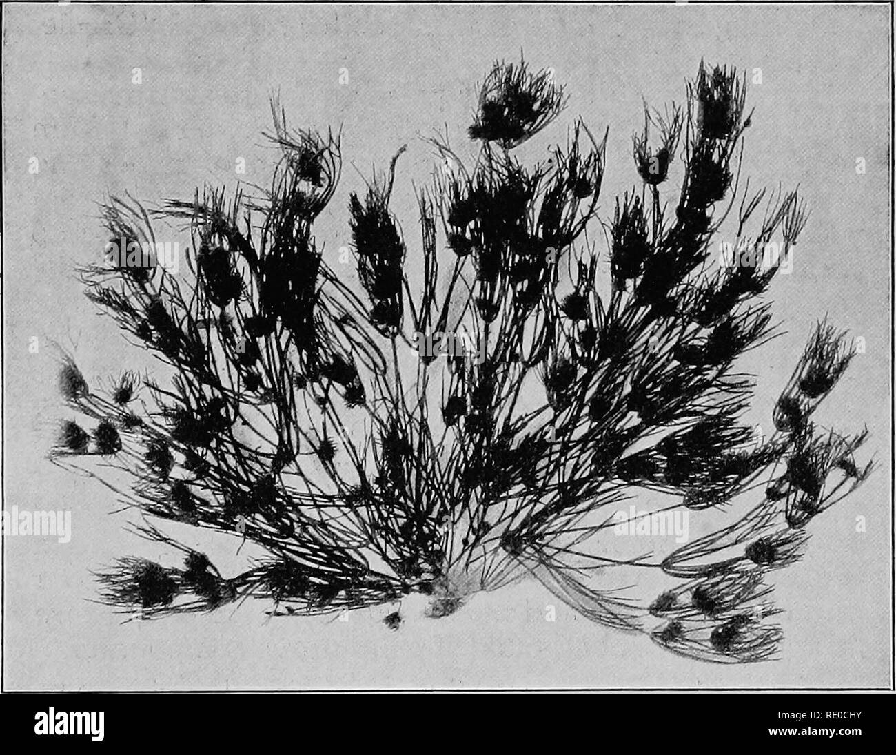 . The life of inland waters; an elementary text book of fresh-water biology for American students. Freshwater biology. The Stmeworts 137 The stoneworts (Chafacede)^—-TJiig group:is well repre- serited in freshwater by two oonimoii genera, well known to every biological laboratory student, Char a and Nitella. Both grow in protected shoals, and in the borders of clear lakes at depths below the heavy beating of the waves. Both are brittle and cannot withstand. Pig, 55. Nitella glomerulifera. wave action. Both prefer the waters that flow off from calcareous soils, and are oftenest found attached t Stock Photohttps://www.alamy.com/image-license-details/?v=1https://www.alamy.com/the-life-of-inland-waters-an-elementary-text-book-of-fresh-water-biology-for-american-students-freshwater-biology-the-stmeworts-137-the-stoneworts-chafacede-tjiig-groupis-well-repre-serited-in-freshwater-by-two-oonimoii-genera-well-known-to-every-biological-laboratory-student-char-a-and-nitella-both-grow-in-protected-shoals-and-in-the-borders-of-clear-lakes-at-depths-below-the-heavy-beating-of-the-waves-both-are-brittle-and-cannot-withstand-pig-55-nitella-glomerulifera-wave-action-both-prefer-the-waters-that-flow-off-from-calcareous-soils-and-are-oftenest-found-attached-t-image232349879.html
RMRE0CHY–. The life of inland waters; an elementary text book of fresh-water biology for American students. Freshwater biology. The Stmeworts 137 The stoneworts (Chafacede)^—-TJiig group:is well repre- serited in freshwater by two oonimoii genera, well known to every biological laboratory student, Char a and Nitella. Both grow in protected shoals, and in the borders of clear lakes at depths below the heavy beating of the waves. Both are brittle and cannot withstand. Pig, 55. Nitella glomerulifera. wave action. Both prefer the waters that flow off from calcareous soils, and are oftenest found attached t
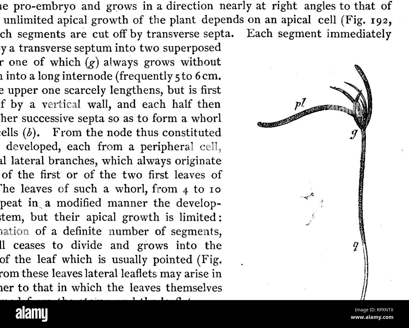 . Text-book of botany, morphological and physiological. Botany. 294 THALLOPHFTES. is wanting in Nitella. From the basal node of each leaf one distinct cortical lobe runs downwards, and one upwards1 (Fig. 192, r, r, r", and Fig. 194). In the middle of each internode therefore as many descending cortical lobes as there are. FIG. 192.—Longitudinal section through a bud of Ckara Jragiiis ; in A the contents of the cells have been left out; in B the fine-grained substance is protoplasm, the larger granules are chlorophyll; the formation of vacuoles is shown ; in C the contents of the cells hav Stock Photohttps://www.alamy.com/image-license-details/?v=1https://www.alamy.com/text-book-of-botany-morphological-and-physiological-botany-294-thallophftes-is-wanting-in-nitella-from-the-basal-node-of-each-leaf-one-distinct-cortical-lobe-runs-downwards-and-one-upwards1-fig-192-r-r-rquot-and-fig-194-in-the-middle-of-each-internode-therefore-as-many-descending-cortical-lobes-as-there-are-fig-192longitudinal-section-through-a-bud-of-ckara-jragiiis-in-a-the-contents-of-the-cells-have-been-left-out-in-b-the-fine-grained-substance-is-protoplasm-the-larger-granules-are-chlorophyll-the-formation-of-vacuoles-is-shown-in-c-the-contents-of-the-cells-hav-image237845130.html
RMRPXNTX–. Text-book of botany, morphological and physiological. Botany. 294 THALLOPHFTES. is wanting in Nitella. From the basal node of each leaf one distinct cortical lobe runs downwards, and one upwards1 (Fig. 192, r, r, r", and Fig. 194). In the middle of each internode therefore as many descending cortical lobes as there are. FIG. 192.—Longitudinal section through a bud of Ckara Jragiiis ; in A the contents of the cells have been left out; in B the fine-grained substance is protoplasm, the larger granules are chlorophyll; the formation of vacuoles is shown ; in C the contents of the cells hav
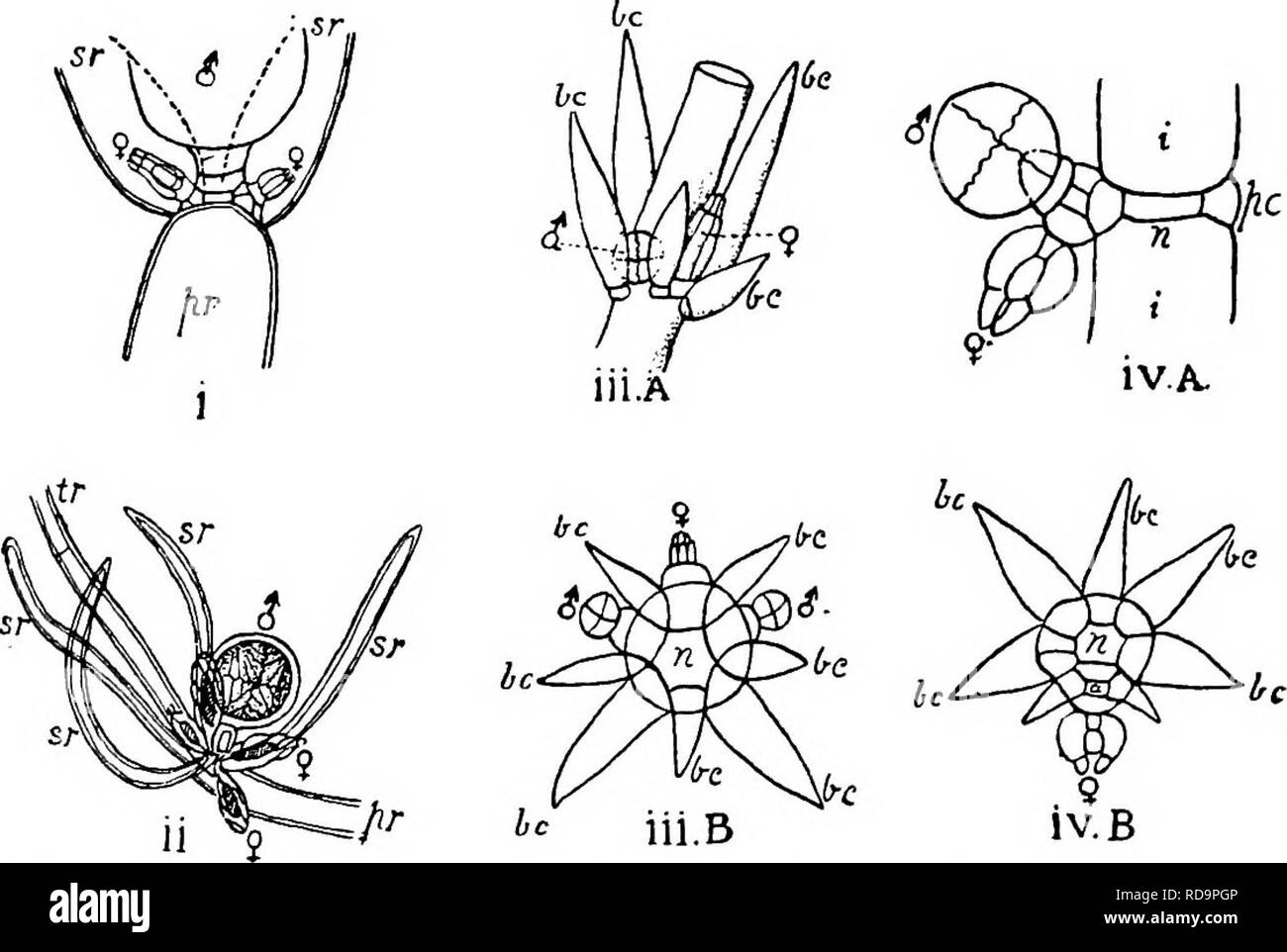 . The British Charophyta. Characeae. 44 BRITISH CHAROPHYTA. In Lychnothamnus and Nitellopsis the arrangement is apparently similar. In Lychnothamnus the oogonium, which is solitary, is produced separately from the central peripheral cell on the inner side of the branchlet, that is the side facing the stem or branch, and antheridia are normally produced from peripheral cells on either side of that producing the oogonium (Fig. 16 iii A and B). In Nitellopsis the only known. Pig. 16.—Position of reproductive organs in several genera. <J = antheridium, ? = oogonium in all figvires. i. Nitella f Stock Photohttps://www.alamy.com/image-license-details/?v=1https://www.alamy.com/the-british-charophyta-characeae-44-british-charophyta-in-lychnothamnus-and-nitellopsis-the-arrangement-is-apparently-similar-in-lychnothamnus-the-oogonium-which-is-solitary-is-produced-separately-from-the-central-peripheral-cell-on-the-inner-side-of-the-branchlet-that-is-the-side-facing-the-stem-or-branch-and-antheridia-are-normally-produced-from-peripheral-cells-on-either-side-of-that-producing-the-oogonium-fig-16-iii-a-and-b-in-nitellopsis-the-only-known-pig-16position-of-reproductive-organs-in-several-genera-ltj-=-antheridium-=-oogonium-in-all-figvires-i-nitella-f-image231940598.html
RMRD9PGP–. The British Charophyta. Characeae. 44 BRITISH CHAROPHYTA. In Lychnothamnus and Nitellopsis the arrangement is apparently similar. In Lychnothamnus the oogonium, which is solitary, is produced separately from the central peripheral cell on the inner side of the branchlet, that is the side facing the stem or branch, and antheridia are normally produced from peripheral cells on either side of that producing the oogonium (Fig. 16 iii A and B). In Nitellopsis the only known. Pig. 16.—Position of reproductive organs in several genera. <J = antheridium, ? = oogonium in all figvires. i. Nitella f
 . The British Charophyta. Characeae. Fig. 25.^—Terminal disposition of ridges of oospore, i. View of apex {Nitella capillaris). ii. View of base (Cfeara 6oMica). ridges owe their origin to the overlying enveloping cells, the sutures on the posterior side of which become filled up as the membrane develops and hardens. The number visible varies according as the spiral-cells complete one, two, or three convolutions. In some species of Ghara the angles of asa c aws ^-^^ pentagonal base already referred to EUlu. CF6SL i- cj e/ . are prolonged downwards into claw-like processes which attain sometime Stock Photohttps://www.alamy.com/image-license-details/?v=1https://www.alamy.com/the-british-charophyta-characeae-fig-25terminal-disposition-of-ridges-of-oospore-i-view-of-apex-nitella-capillaris-ii-view-of-base-cfeara-6omica-ridges-owe-their-origin-to-the-overlying-enveloping-cells-the-sutures-on-the-posterior-side-of-which-become-filled-up-as-the-membrane-develops-and-hardens-the-number-visible-varies-according-as-the-spiral-cells-complete-one-two-or-three-convolutions-in-some-species-of-ghara-the-angles-of-asa-c-aws-pentagonal-base-already-referred-to-eulu-cf6sl-i-cj-e-are-prolonged-downwards-into-claw-like-processes-which-attain-sometime-image231900689.html
RMRD7YKD–. The British Charophyta. Characeae. Fig. 25.^—Terminal disposition of ridges of oospore, i. View of apex {Nitella capillaris). ii. View of base (Cfeara 6oMica). ridges owe their origin to the overlying enveloping cells, the sutures on the posterior side of which become filled up as the membrane develops and hardens. The number visible varies according as the spiral-cells complete one, two, or three convolutions. In some species of Ghara the angles of asa c aws ^-^^ pentagonal base already referred to EUlu. CF6SL i- cj e/ . are prolonged downwards into claw-like processes which attain sometime
 . First lessons in zoology. Zoology. REHIRING ANIMALS AND MAKING COLLECTIONS 333 if the animals in it live naturally in quiet water. Among the more available plants for use in aquaria are the fol- lowing: " Waterweed, Elodea canadensis. " Bladderwort, Utriciilaria (several species). " Water-starwort, Callitriche (several species). "Watercress, Nasturtium officinale. " Stoneworts, Cliara and Nitella (several species of each). " Frog-spittle or water-silk,' Spirogyra. " A small quantity of duckweed, Lenina, placed on the surface of the water adds to the beauty Stock Photohttps://www.alamy.com/image-license-details/?v=1https://www.alamy.com/first-lessons-in-zoology-zoology-rehiring-animals-and-making-collections-333-if-the-animals-in-it-live-naturally-in-quiet-water-among-the-more-available-plants-for-use-in-aquaria-are-the-fol-lowing-quot-waterweed-elodea-canadensis-quot-bladderwort-utriciilaria-several-species-quot-water-starwort-callitriche-several-species-quotwatercress-nasturtium-officinale-quot-stoneworts-cliara-and-nitella-several-species-of-each-quot-frog-spittle-or-water-silk-spirogyra-quot-a-small-quantity-of-duckweed-lenina-placed-on-the-surface-of-the-water-adds-to-the-beauty-image232255246.html
RMRDT3X6–. First lessons in zoology. Zoology. REHIRING ANIMALS AND MAKING COLLECTIONS 333 if the animals in it live naturally in quiet water. Among the more available plants for use in aquaria are the fol- lowing: " Waterweed, Elodea canadensis. " Bladderwort, Utriciilaria (several species). " Water-starwort, Callitriche (several species). "Watercress, Nasturtium officinale. " Stoneworts, Cliara and Nitella (several species of each). " Frog-spittle or water-silk,' Spirogyra. " A small quantity of duckweed, Lenina, placed on the surface of the water adds to the beauty
 . Practical text-book of plant physiology. Plant physiology. MOVEMENTS OF PROTOPLASM 41 other hand an increase in the proportion of the oxygen does not accelerate the release of energy, or materially modify the per- formance of the organism except so far as it affects the pressure of the other atmospheric elements. 58. Streaming Movements of Protoplasm in the Absence of Oxygen. Mount a leaf of Philotria, Nitella, or a hair which shows a distinct streaming movement, on a cover-glass 2 cm. in diameter and invert over the opening of an Engelmann gas chamber. Seal the cover to the chamber by a sma Stock Photohttps://www.alamy.com/image-license-details/?v=1https://www.alamy.com/practical-text-book-of-plant-physiology-plant-physiology-movements-of-protoplasm-41-other-hand-an-increase-in-the-proportion-of-the-oxygen-does-not-accelerate-the-release-of-energy-or-materially-modify-the-per-formance-of-the-organism-except-so-far-as-it-affects-the-pressure-of-the-other-atmospheric-elements-58-streaming-movements-of-protoplasm-in-the-absence-of-oxygen-mount-a-leaf-of-philotria-nitella-or-a-hair-which-shows-a-distinct-streaming-movement-on-a-cover-glass-2-cm-in-diameter-and-invert-over-the-opening-of-an-engelmann-gas-chamber-seal-the-cover-to-the-chamber-by-a-sma-image232921333.html
RMREXDF1–. Practical text-book of plant physiology. Plant physiology. MOVEMENTS OF PROTOPLASM 41 other hand an increase in the proportion of the oxygen does not accelerate the release of energy, or materially modify the per- formance of the organism except so far as it affects the pressure of the other atmospheric elements. 58. Streaming Movements of Protoplasm in the Absence of Oxygen. Mount a leaf of Philotria, Nitella, or a hair which shows a distinct streaming movement, on a cover-glass 2 cm. in diameter and invert over the opening of an Engelmann gas chamber. Seal the cover to the chamber by a sma
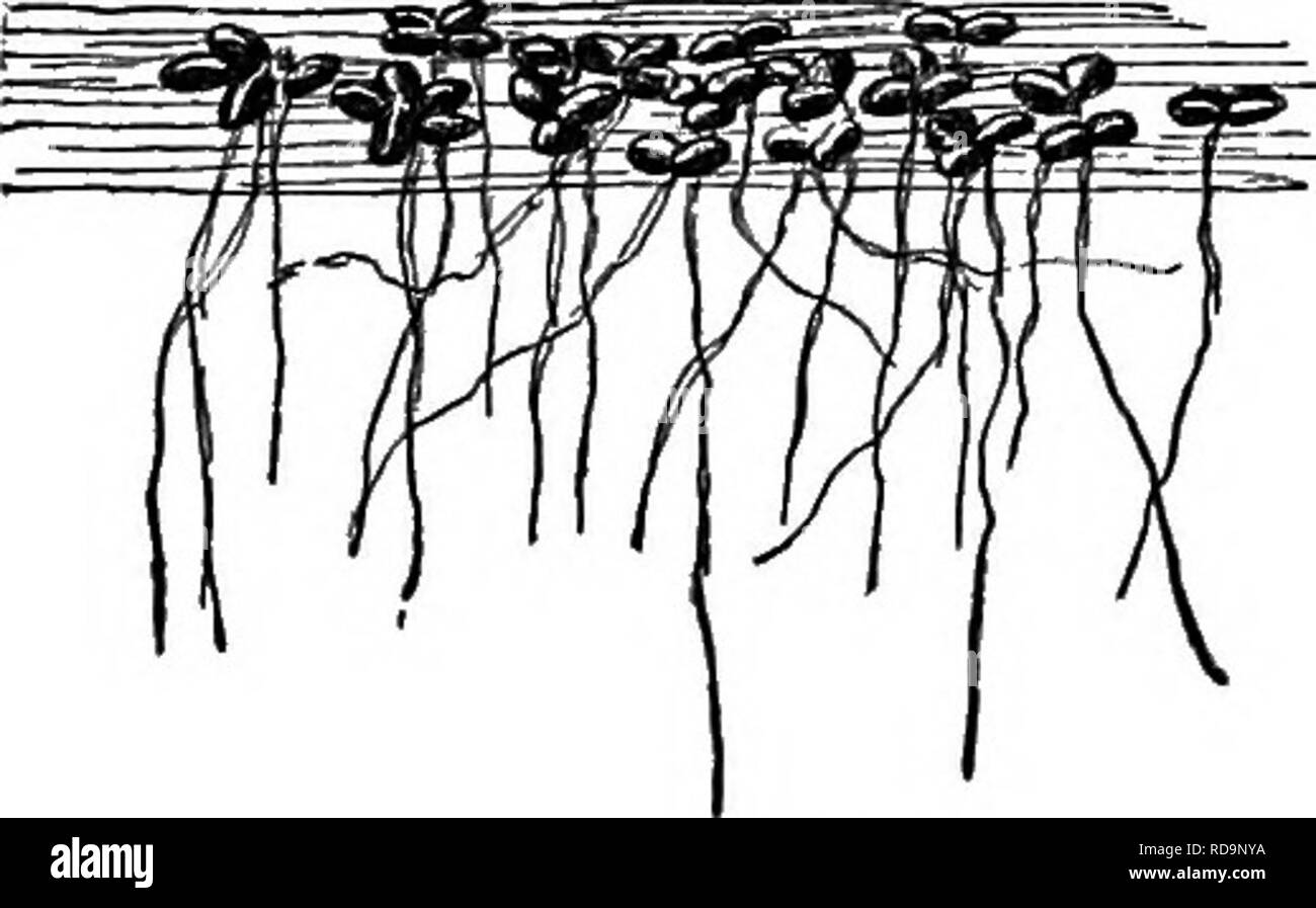 . Insect life; an introduction to nature study and a guide for teachers, students and others interested in out-of-door life. Entomology; Nature study. THE BREEDING OF INSECTS. 331. Fig. 292.—Duckweed. Watercress, Nasturtium officinale. Stoneworts, Chara and Nitella (several species of each). Frog-spittle or water-silk, Spirogira. A small quantity of duckweed, Lemna (Fig. 292), placed on the surface of the water adds to the beauty of an aquarium. When it is necessary to add water to an aqua- rium on account of loss by evaporation, rain wa- ter should be used to prevent an undue ac- cumulation o Stock Photohttps://www.alamy.com/image-license-details/?v=1https://www.alamy.com/insect-life-an-introduction-to-nature-study-and-a-guide-for-teachers-students-and-others-interested-in-out-of-door-life-entomology-nature-study-the-breeding-of-insects-331-fig-292duckweed-watercress-nasturtium-officinale-stoneworts-chara-and-nitella-several-species-of-each-frog-spittle-or-water-silk-spirogira-a-small-quantity-of-duckweed-lemna-fig-292-placed-on-the-surface-of-the-water-adds-to-the-beauty-of-an-aquarium-when-it-is-necessary-to-add-water-to-an-aqua-rium-on-account-of-loss-by-evaporation-rain-wa-ter-should-be-used-to-prevent-an-undue-ac-cumulation-o-image231940110.html
RMRD9NYA–. Insect life; an introduction to nature study and a guide for teachers, students and others interested in out-of-door life. Entomology; Nature study. THE BREEDING OF INSECTS. 331. Fig. 292.—Duckweed. Watercress, Nasturtium officinale. Stoneworts, Chara and Nitella (several species of each). Frog-spittle or water-silk, Spirogira. A small quantity of duckweed, Lemna (Fig. 292), placed on the surface of the water adds to the beauty of an aquarium. When it is necessary to add water to an aqua- rium on account of loss by evaporation, rain wa- ter should be used to prevent an undue ac- cumulation o
 . Insect life; an introduction to nature study and a guide for teachers, students and others interested in out-of-door life. Entomology; Nature study. Fig. 292.—Duckweed. Watercress, Nasturtium officinale. Stoneworts, Chara and Nitella (several species of each). Frog-spittle or water-silk, Spirogira. A small quantity of duckweed, Lemna (Fig. 292), placed on the surface of the water adds to the beauty of an aquarium. When it is necessary to add water to an aqua- rium on account of loss by evaporation, rain wa- ter should be used to prevent an undue ac- cumulation of the mineral matter held in s Stock Photohttps://www.alamy.com/image-license-details/?v=1https://www.alamy.com/insect-life-an-introduction-to-nature-study-and-a-guide-for-teachers-students-and-others-interested-in-out-of-door-life-entomology-nature-study-fig-292duckweed-watercress-nasturtium-officinale-stoneworts-chara-and-nitella-several-species-of-each-frog-spittle-or-water-silk-spirogira-a-small-quantity-of-duckweed-lemna-fig-292-placed-on-the-surface-of-the-water-adds-to-the-beauty-of-an-aquarium-when-it-is-necessary-to-add-water-to-an-aqua-rium-on-account-of-loss-by-evaporation-rain-wa-ter-should-be-used-to-prevent-an-undue-ac-cumulation-of-the-mineral-matter-held-in-s-image231940107.html
RMRD9NY7–. Insect life; an introduction to nature study and a guide for teachers, students and others interested in out-of-door life. Entomology; Nature study. Fig. 292.—Duckweed. Watercress, Nasturtium officinale. Stoneworts, Chara and Nitella (several species of each). Frog-spittle or water-silk, Spirogira. A small quantity of duckweed, Lemna (Fig. 292), placed on the surface of the water adds to the beauty of an aquarium. When it is necessary to add water to an aqua- rium on account of loss by evaporation, rain wa- ter should be used to prevent an undue ac- cumulation of the mineral matter held in s
 . The British Charophyta. Characeae. GEOLOGICAL HISTORY OF THE CHAROPHYTA. 73 ing to Karpinsky (5), p. 127, Muschelkalk is not found in the district named. Far down in the rocks of the PALEOZOIC era organ- isms occur which have been ascribed to the Charophyta. Silurian.—From a deposit of the Silurian period on the Kellerwald, remains closely resembling the vege- tative parts of a Nitella are figured by Potonie (15) (see Fig. 26), but in the absence of fruit it is not possible to determine their nature. Devonian.—In the Corniferous Limestone of Ohio, Meek, in 1873 (12), recorded the presence, i Stock Photohttps://www.alamy.com/image-license-details/?v=1https://www.alamy.com/the-british-charophyta-characeae-geological-history-of-the-charophyta-73-ing-to-karpinsky-5-p-127-muschelkalk-is-not-found-in-the-district-named-far-down-in-the-rocks-of-the-paleozoic-era-organ-isms-occur-which-have-been-ascribed-to-the-charophyta-silurianfrom-a-deposit-of-the-silurian-period-on-the-kellerwald-remains-closely-resembling-the-vege-tative-parts-of-a-nitella-are-figured-by-potonie-15-see-fig-26-but-in-the-absence-of-fruit-it-is-not-possible-to-determine-their-nature-devonianin-the-corniferous-limestone-of-ohio-meek-in-1873-12-recorded-the-presence-i-image232053121.html
RMRDEX3D–. The British Charophyta. Characeae. GEOLOGICAL HISTORY OF THE CHAROPHYTA. 73 ing to Karpinsky (5), p. 127, Muschelkalk is not found in the district named. Far down in the rocks of the PALEOZOIC era organ- isms occur which have been ascribed to the Charophyta. Silurian.—From a deposit of the Silurian period on the Kellerwald, remains closely resembling the vege- tative parts of a Nitella are figured by Potonie (15) (see Fig. 26), but in the absence of fruit it is not possible to determine their nature. Devonian.—In the Corniferous Limestone of Ohio, Meek, in 1873 (12), recorded the presence, i
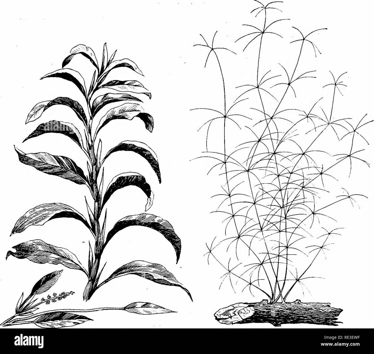 . Goldfish varieties and tropical aquarium fishes; a complete guide to aquaria and related subjects. Aquariums; Goldfish. TROPICAL AQUARIUM FISHES 179. Fig. 127. Potamogeton densus (Reduced one-half) Fig. 128. Nitella gracilis (Reduced one-third) POTAMOGETON In strong contrast to the foregoing dainty plants is Potamogeton densus, or pondweed. As will be seen from figure 127, these leaves are broad and robust. In color they are a bright green. This variety is said to be of European origin but is now common in ponds in the United States. If established in soil in flat pots it flourishes in a wel Stock Photohttps://www.alamy.com/image-license-details/?v=1https://www.alamy.com/goldfish-varieties-and-tropical-aquarium-fishes-a-complete-guide-to-aquaria-and-related-subjects-aquariums-goldfish-tropical-aquarium-fishes-179-fig-127-potamogeton-densus-reduced-one-half-fig-128-nitella-gracilis-reduced-one-third-potamogeton-in-strong-contrast-to-the-foregoing-dainty-plants-is-potamogeton-densus-or-pondweed-as-will-be-seen-from-figure-127-these-leaves-are-broad-and-robust-in-color-they-are-a-bright-green-this-variety-is-said-to-be-of-european-origin-but-is-now-common-in-ponds-in-the-united-states-if-established-in-soil-in-flat-pots-it-flourishes-in-a-wel-image232417515.html
RMRE3EWF–. Goldfish varieties and tropical aquarium fishes; a complete guide to aquaria and related subjects. Aquariums; Goldfish. TROPICAL AQUARIUM FISHES 179. Fig. 127. Potamogeton densus (Reduced one-half) Fig. 128. Nitella gracilis (Reduced one-third) POTAMOGETON In strong contrast to the foregoing dainty plants is Potamogeton densus, or pondweed. As will be seen from figure 127, these leaves are broad and robust. In color they are a bright green. This variety is said to be of European origin but is now common in ponds in the United States. If established in soil in flat pots it flourishes in a wel
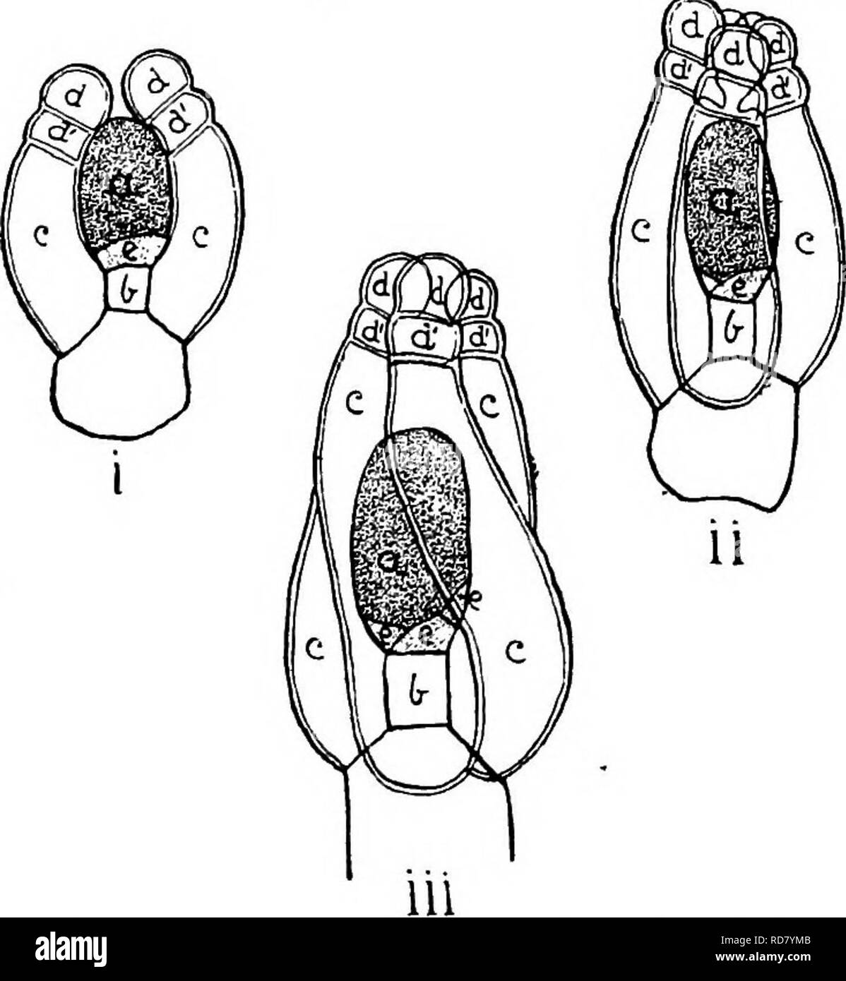 . The British Charophyta. Characeae. 52 BRITISH CHAEOPHYTA. ovoidal form. The lateral walls of the spiral cells subsequently break down, leaving the outer walls to form a single integument marked by spiral furrows along the lines of union. Incased and protected by the enveloping spiral cells the oosphere consists of a nucleated mass of protoplasm enclosed by a thin and extremely delicate. Pig. 22.—Development of oogonium of Nitella flexilis (after Sachs). o, oosphere; h, node-cell; c, enveloping cells; d, upper, and. d', lower cells of ooronula; e, turning cell. i. Early stages of the oogonium Stock Photohttps://www.alamy.com/image-license-details/?v=1https://www.alamy.com/the-british-charophyta-characeae-52-british-chaeophyta-ovoidal-form-the-lateral-walls-of-the-spiral-cells-subsequently-break-down-leaving-the-outer-walls-to-form-a-single-integument-marked-by-spiral-furrows-along-the-lines-of-union-incased-and-protected-by-the-enveloping-spiral-cells-the-oosphere-consists-of-a-nucleated-mass-of-protoplasm-enclosed-by-a-thin-and-extremely-delicate-pig-22development-of-oogonium-of-nitella-flexilis-after-sachs-o-oosphere-h-node-cell-c-enveloping-cells-d-upper-and-d-lower-cells-of-ooronula-e-turning-cell-i-early-stages-of-the-oogonium-image231900715.html
RMRD7YMB–. The British Charophyta. Characeae. 52 BRITISH CHAEOPHYTA. ovoidal form. The lateral walls of the spiral cells subsequently break down, leaving the outer walls to form a single integument marked by spiral furrows along the lines of union. Incased and protected by the enveloping spiral cells the oosphere consists of a nucleated mass of protoplasm enclosed by a thin and extremely delicate. Pig. 22.—Development of oogonium of Nitella flexilis (after Sachs). o, oosphere; h, node-cell; c, enveloping cells; d, upper, and. d', lower cells of ooronula; e, turning cell. i. Early stages of the oogonium
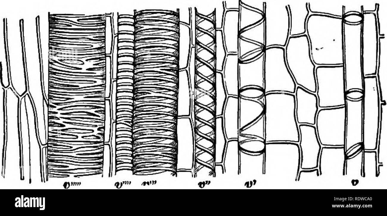 . The essentials of botany. Botany. 4 BOTANY. (e) Mount carefully in pure water a piece (2 to 4 centimetres) of one of the young " silks" of Indian corn. The movement is well seen in the long cells. Repeat the foregoing experiments. (/) The following may be taken also, viz.: the stamen hairs of Spiderwort, the epidermis of Live-for-ever leaf, fresh specimens of the Stoneworts (Chara and Nitella), Eel-grass, etc. 7. The Plant-Cell.âIn all common plants the protoplasm is found in little masses of definite shapes, each one en" closed in a little box (Fig. 1). The substance of these Stock Photohttps://www.alamy.com/image-license-details/?v=1https://www.alamy.com/the-essentials-of-botany-botany-4-botany-e-mount-carefully-in-pure-water-a-piece-2-to-4-centimetres-of-one-of-the-young-quot-silksquot-of-indian-corn-the-movement-is-well-seen-in-the-long-cells-repeat-the-foregoing-experiments-the-following-may-be-taken-also-viz-the-stamen-hairs-of-spiderwort-the-epidermis-of-live-for-ever-leaf-fresh-specimens-of-the-stoneworts-chara-and-nitella-eel-grass-etc-7-the-plant-cellin-all-common-plants-the-protoplasm-is-found-in-little-masses-of-definite-shapes-each-one-enquot-closed-in-a-little-box-fig-1-the-substance-of-these-image232283800.html
RMRDWCA0–. The essentials of botany. Botany. 4 BOTANY. (e) Mount carefully in pure water a piece (2 to 4 centimetres) of one of the young " silks" of Indian corn. The movement is well seen in the long cells. Repeat the foregoing experiments. (/) The following may be taken also, viz.: the stamen hairs of Spiderwort, the epidermis of Live-for-ever leaf, fresh specimens of the Stoneworts (Chara and Nitella), Eel-grass, etc. 7. The Plant-Cell.âIn all common plants the protoplasm is found in little masses of definite shapes, each one en" closed in a little box (Fig. 1). The substance of these
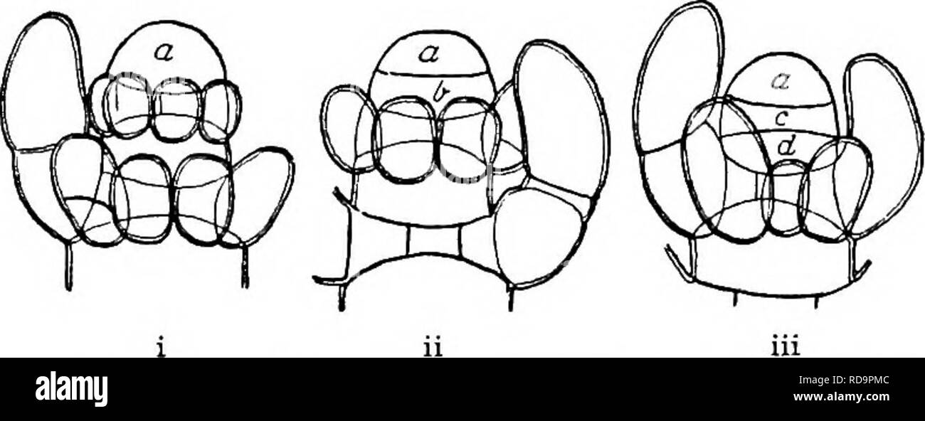 . The British Charophyta. Characeae. STRUaTUBE AND DEVELOPMENT. 23 stem-node, remaining quite short and undergoing division by means of longitudinal septa in the direc- tion of the axis, the lower (d) forming an internode, remaining undivided but lengthening considerably. In this way the stem of a Charophyte presents a. Fia. 3.—Yoimg shoot of Nitella in successive stages (after Giesen- hagen) ( x o. 150). In i the apical cell a is undivided; in ii a portion b has been cut off from its base by a transverse section ; in iii the lower cell b has been subdivided into a stem-node c and an internode Stock Photohttps://www.alamy.com/image-license-details/?v=1https://www.alamy.com/the-british-charophyta-characeae-struatube-and-development-23-stem-node-remaining-quite-short-and-undergoing-division-by-means-of-longitudinal-septa-in-the-direc-tion-of-the-axis-the-lower-d-forming-an-internode-remaining-undivided-but-lengthening-considerably-in-this-way-the-stem-of-a-charophyte-presents-a-fia-3yoimg-shoot-of-nitella-in-successive-stages-after-giesen-hagen-x-o-150-in-i-the-apical-cell-a-is-undivided-in-ii-a-portion-b-has-been-cut-off-from-its-base-by-a-transverse-section-in-iii-the-lower-cell-b-has-been-subdivided-into-a-stem-node-c-and-an-internode-image231940700.html
RMRD9PMC–. The British Charophyta. Characeae. STRUaTUBE AND DEVELOPMENT. 23 stem-node, remaining quite short and undergoing division by means of longitudinal septa in the direc- tion of the axis, the lower (d) forming an internode, remaining undivided but lengthening considerably. In this way the stem of a Charophyte presents a. Fia. 3.—Yoimg shoot of Nitella in successive stages (after Giesen- hagen) ( x o. 150). In i the apical cell a is undivided; in ii a portion b has been cut off from its base by a transverse section ; in iii the lower cell b has been subdivided into a stem-node c and an internode
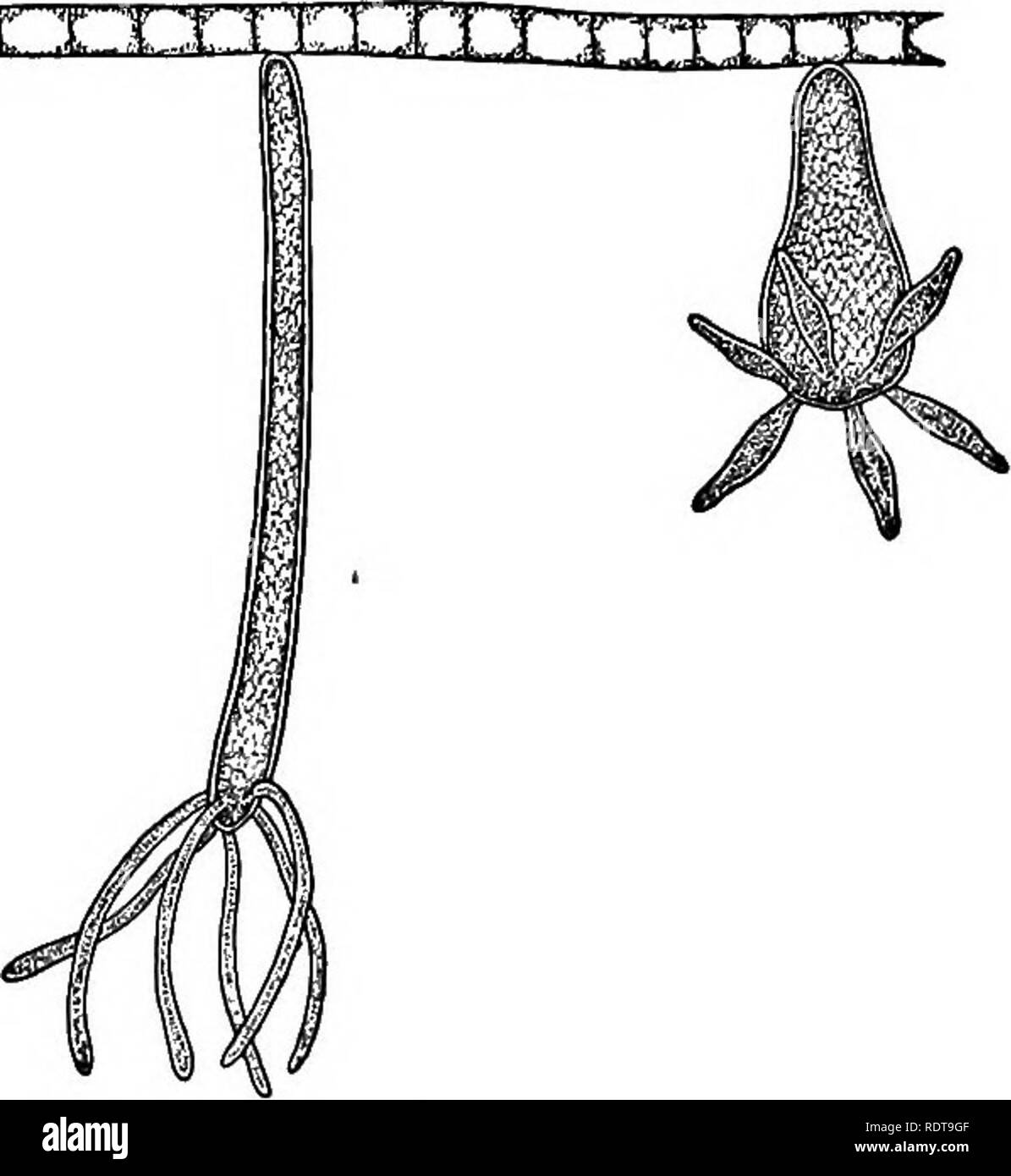 . A laboratory guide for beginners in zoology. Laboratory animals. T1..J rr T T' r 11 -i. t ;i. The Hydra. Magnified. Bring in from various situations in ponds or sluggish streams small quantities of water-weeds, — as Nitella, Spirogyra, or Vaucheria, — collecting them with a small quantity of water in jars or cans, and labelling each so as to know where it came from. Place the material of each collection in a jar of clear water near a window, and let it stand for a day or two. Then examine the jar carefully, especially the sides near the window, for hydras. In this a reading glass is very hel Stock Photohttps://www.alamy.com/image-license-details/?v=1https://www.alamy.com/a-laboratory-guide-for-beginners-in-zoology-laboratory-animals-t1j-rr-t-t-r-11-i-t-i-the-hydra-magnified-bring-in-from-various-situations-in-ponds-or-sluggish-streams-small-quantities-of-water-weeds-as-nitella-spirogyra-or-vaucheria-collecting-them-with-a-small-quantity-of-water-in-jars-or-cans-and-labelling-each-so-as-to-know-where-it-came-from-place-the-material-of-each-collection-in-a-jar-of-clear-water-near-a-window-and-let-it-stand-for-a-day-or-two-then-examine-the-jar-carefully-especially-the-sides-near-the-window-for-hydras-in-this-a-reading-glass-is-very-hel-image232259679.html
RMRDT9GF–. A laboratory guide for beginners in zoology. Laboratory animals. T1..J rr T T' r 11 -i. t ;i. The Hydra. Magnified. Bring in from various situations in ponds or sluggish streams small quantities of water-weeds, — as Nitella, Spirogyra, or Vaucheria, — collecting them with a small quantity of water in jars or cans, and labelling each so as to know where it came from. Place the material of each collection in a jar of clear water near a window, and let it stand for a day or two. Then examine the jar carefully, especially the sides near the window, for hydras. In this a reading glass is very hel
 . Text-book of botany, morphological and physiological. Botany. FIG. 199-—Nitella Jlexilis. A fertile branch (natural size) ; i internode, b leaves ; B upper part of a fertile leaf b with the node K; on the node are two lateral leaves n b, and two very young- carpogonia S; a an antheridium ; C older leaf with two leaflets, a ripe antheridium a, and two unripe carpogonia S; D a half-ripe carpogonium more strongly magnified. FIG. t$.—Xitella Jlexilis. A an almost ripe antheridium at the end of the primary leaf, by its side two lateral leaflets, i neutral lines; the arrows indicate the direction Stock Photohttps://www.alamy.com/image-license-details/?v=1https://www.alamy.com/text-book-of-botany-morphological-and-physiological-botany-fig-199-nitella-jlexilis-a-fertile-branch-natural-size-i-internode-b-leaves-b-upper-part-of-a-fertile-leaf-b-with-the-node-k-on-the-node-are-two-lateral-leaves-n-b-and-two-very-young-carpogonia-s-a-an-antheridium-c-older-leaf-with-two-leaflets-a-ripe-antheridium-a-and-two-unripe-carpogonia-s-d-a-half-ripe-carpogonium-more-strongly-magnified-fig-txitella-jlexilis-a-an-almost-ripe-antheridium-at-the-end-of-the-primary-leaf-by-its-side-two-lateral-leaflets-i-neutral-lines-the-arrows-indicate-the-direction-image237845044.html
RMRPXNNT–. Text-book of botany, morphological and physiological. Botany. FIG. 199-—Nitella Jlexilis. A fertile branch (natural size) ; i internode, b leaves ; B upper part of a fertile leaf b with the node K; on the node are two lateral leaves n b, and two very young- carpogonia S; a an antheridium ; C older leaf with two leaflets, a ripe antheridium a, and two unripe carpogonia S; D a half-ripe carpogonium more strongly magnified. FIG. t$.—Xitella Jlexilis. A an almost ripe antheridium at the end of the primary leaf, by its side two lateral leaflets, i neutral lines; the arrows indicate the direction
 . Contributions from the Botanical Laboratory, vol. 12. Botany; Botany. 114 The Fresh Water Algae of Plate V PASQUE IS. Numher Name ^^«^*^^ ^^ ^^^ 216PASQUE-A Southeast 217 PASQUE-B Southeast 218 PASQUE-C Southwest 219 PASQUE-D (NITELLA POND) Southwest 220 PASQUE-E - Northeast 221 PASQUE-F (HOURGLASS POND) Northeast 222 PASQUE-G (SKATING POND) Northeast 223 PASQUE-H Northeast 224 PASQUE-I (ICEHOUSE POND) Northeast 225 PASQUE-J Southwest 226PASQUE-K Southwest 227 PASQUE>L Northwest 228 PASQUE-M Northwest 229 PASQUE-N Northwest 230PASQUE-O Northwest 231 PASQUE-P Northwest 232 PASQUE-Q Northwe Stock Photohttps://www.alamy.com/image-license-details/?v=1https://www.alamy.com/contributions-from-the-botanical-laboratory-vol-12-botany-botany-114-the-fresh-water-algae-of-plate-v-pasque-is-numher-name-216pasque-a-southeast-217-pasque-b-southeast-218-pasque-c-southwest-219-pasque-d-nitella-pond-southwest-220-pasque-e-northeast-221-pasque-f-hourglass-pond-northeast-222-pasque-g-skating-pond-northeast-223-pasque-h-northeast-224-pasque-i-icehouse-pond-northeast-225-pasque-j-southwest-226pasque-k-southwest-227-pasquegtl-northwest-228-pasque-m-northwest-229-pasque-n-northwest-230pasque-o-northwest-231-pasque-p-northwest-232-pasque-q-northwe-image232562494.html
RMREA3RA–. Contributions from the Botanical Laboratory, vol. 12. Botany; Botany. 114 The Fresh Water Algae of Plate V PASQUE IS. Numher Name ^^«^*^^ ^^ ^^^ 216PASQUE-A Southeast 217 PASQUE-B Southeast 218 PASQUE-C Southwest 219 PASQUE-D (NITELLA POND) Southwest 220 PASQUE-E - Northeast 221 PASQUE-F (HOURGLASS POND) Northeast 222 PASQUE-G (SKATING POND) Northeast 223 PASQUE-H Northeast 224 PASQUE-I (ICEHOUSE POND) Northeast 225 PASQUE-J Southwest 226PASQUE-K Southwest 227 PASQUE>L Northwest 228 PASQUE-M Northwest 229 PASQUE-N Northwest 230PASQUE-O Northwest 231 PASQUE-P Northwest 232 PASQUE-Q Northwe
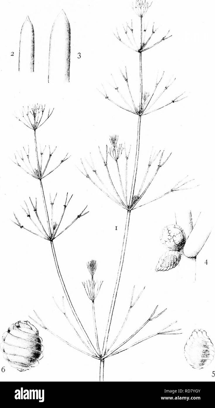 . The British Charophyta. Characeae. PLATE VIII. St. Groves del. NITELLA FLEXILIS. Please note that these images are extracted from scanned page images that may have been digitally enhanced for readability - coloration and appearance of these illustrations may not perfectly resemble the original work.. Groves, James; Bullock-Webster, George Russell, 1858- joint author. London, The Ray society Stock Photohttps://www.alamy.com/image-license-details/?v=1https://www.alamy.com/the-british-charophyta-characeae-plate-viii-st-groves-del-nitella-flexilis-please-note-that-these-images-are-extracted-from-scanned-page-images-that-may-have-been-digitally-enhanced-for-readability-coloration-and-appearance-of-these-illustrations-may-not-perfectly-resemble-the-original-work-groves-james-bullock-webster-george-russell-1858-joint-author-london-the-ray-society-image231900619.html
RMRD7YGY–. The British Charophyta. Characeae. PLATE VIII. St. Groves del. NITELLA FLEXILIS. Please note that these images are extracted from scanned page images that may have been digitally enhanced for readability - coloration and appearance of these illustrations may not perfectly resemble the original work.. Groves, James; Bullock-Webster, George Russell, 1858- joint author. London, The Ray society
 . The British Charophyta. Characeae. PLATE XII /1 '"A. NITELLA MUCRONATA. Please note that these images are extracted from scanned page images that may have been digitally enhanced for readability - coloration and appearance of these illustrations may not perfectly resemble the original work.. Groves, James; Bullock-Webster, George Russell, 1858- joint author. London, The Ray society Stock Photohttps://www.alamy.com/image-license-details/?v=1https://www.alamy.com/the-british-charophyta-characeae-plate-xii-1-quota-nitella-mucronata-please-note-that-these-images-are-extracted-from-scanned-page-images-that-may-have-been-digitally-enhanced-for-readability-coloration-and-appearance-of-these-illustrations-may-not-perfectly-resemble-the-original-work-groves-james-bullock-webster-george-russell-1858-joint-author-london-the-ray-society-image231900605.html
RMRD7YGD–. The British Charophyta. Characeae. PLATE XII /1 '"A. NITELLA MUCRONATA. Please note that these images are extracted from scanned page images that may have been digitally enhanced for readability - coloration and appearance of these illustrations may not perfectly resemble the original work.. Groves, James; Bullock-Webster, George Russell, 1858- joint author. London, The Ray society
 . The British Charophyta. Characeae. PLATE VII. ^f. droves (M. NITELLA OPACA. Please note that these images are extracted from scanned page images that may have been digitally enhanced for readability - coloration and appearance of these illustrations may not perfectly resemble the original work.. Groves, James; Bullock-Webster, George Russell, 1858- joint author. London, The Ray society Stock Photohttps://www.alamy.com/image-license-details/?v=1https://www.alamy.com/the-british-charophyta-characeae-plate-vii-f-droves-m-nitella-opaca-please-note-that-these-images-are-extracted-from-scanned-page-images-that-may-have-been-digitally-enhanced-for-readability-coloration-and-appearance-of-these-illustrations-may-not-perfectly-resemble-the-original-work-groves-james-bullock-webster-george-russell-1858-joint-author-london-the-ray-society-image231900624.html
RMRD7YH4–. The British Charophyta. Characeae. PLATE VII. ^f. droves (M. NITELLA OPACA. Please note that these images are extracted from scanned page images that may have been digitally enhanced for readability - coloration and appearance of these illustrations may not perfectly resemble the original work.. Groves, James; Bullock-Webster, George Russell, 1858- joint author. London, The Ray society
 . The British Charophyta. Characeae. PLATE X. 0. ». B.-yr.Sr M.G.dtl. NITELLA SPANIOCLEMA. Please note that these images are extracted from scanned page images that may have been digitally enhanced for readability - coloration and appearance of these illustrations may not perfectly resemble the original work.. Groves, James; Bullock-Webster, George Russell, 1858- joint author. London, The Ray society Stock Photohttps://www.alamy.com/image-license-details/?v=1https://www.alamy.com/the-british-charophyta-characeae-plate-x-0-b-yrsr-mgdtl-nitella-spanioclema-please-note-that-these-images-are-extracted-from-scanned-page-images-that-may-have-been-digitally-enhanced-for-readability-coloration-and-appearance-of-these-illustrations-may-not-perfectly-resemble-the-original-work-groves-james-bullock-webster-george-russell-1858-joint-author-london-the-ray-society-image231900612.html
RMRD7YGM–. The British Charophyta. Characeae. PLATE X. 0. ». B.-yr.Sr M.G.dtl. NITELLA SPANIOCLEMA. Please note that these images are extracted from scanned page images that may have been digitally enhanced for readability - coloration and appearance of these illustrations may not perfectly resemble the original work.. Groves, James; Bullock-Webster, George Russell, 1858- joint author. London, The Ray society
 . The British Charophyta. Characeae. PLATE XV. Af. A- H. Grov'-f^ del. NITELLA BATRACHOSPERMA. Please note that these images are extracted from scanned page images that may have been digitally enhanced for readability - coloration and appearance of these illustrations may not perfectly resemble the original work.. Groves, James; Bullock-Webster, George Russell, 1858- joint author. London, The Ray society Stock Photohttps://www.alamy.com/image-license-details/?v=1https://www.alamy.com/the-british-charophyta-characeae-plate-xv-af-a-h-grov-f-del-nitella-batrachosperma-please-note-that-these-images-are-extracted-from-scanned-page-images-that-may-have-been-digitally-enhanced-for-readability-coloration-and-appearance-of-these-illustrations-may-not-perfectly-resemble-the-original-work-groves-james-bullock-webster-george-russell-1858-joint-author-london-the-ray-society-image231900589.html
RMRD7YFW–. The British Charophyta. Characeae. PLATE XV. Af. A- H. Grov'-f^ del. NITELLA BATRACHOSPERMA. Please note that these images are extracted from scanned page images that may have been digitally enhanced for readability - coloration and appearance of these illustrations may not perfectly resemble the original work.. Groves, James; Bullock-Webster, George Russell, 1858- joint author. London, The Ray society
 . The British Charophyta. Characeae. ,v '^ t^ ,1/ (Jn.c-.i ,1,1 -â ' 7 NITELLA CAPILLARIS. Please note that these images are extracted from scanned page images that may have been digitally enhanced for readability - coloration and appearance of these illustrations may not perfectly resemble the original work.. Groves, James; Bullock-Webster, George Russell, 1858- joint author. London, The Ray society Stock Photohttps://www.alamy.com/image-license-details/?v=1https://www.alamy.com/the-british-charophyta-characeae-v-t-1-jnc-i-11-7-nitella-capillaris-please-note-that-these-images-are-extracted-from-scanned-page-images-that-may-have-been-digitally-enhanced-for-readability-coloration-and-appearance-of-these-illustrations-may-not-perfectly-resemble-the-original-work-groves-james-bullock-webster-george-russell-1858-joint-author-london-the-ray-society-image231900628.html
RMRD7YH8–. The British Charophyta. Characeae. ,v '^ t^ ,1/ (Jn.c-.i ,1,1 -â ' 7 NITELLA CAPILLARIS. Please note that these images are extracted from scanned page images that may have been digitally enhanced for readability - coloration and appearance of these illustrations may not perfectly resemble the original work.. Groves, James; Bullock-Webster, George Russell, 1858- joint author. London, The Ray society
 . The British Charophyta. Characeae. PLATE I.. NITELLA MUCRONATA VAR. GRACILLIMA. {muck enlarged) Cluirh-^ Whittingham A- (ir,gi;.<;, Ltd.. I.ith.. Please note that these images are extracted from scanned page images that may have been digitally enhanced for readability - coloration and appearance of these illustrations may not perfectly resemble the original work.. Groves, James; Bullock-Webster, George Russell, 1858- joint author. London, The Ray society Stock Photohttps://www.alamy.com/image-license-details/?v=1https://www.alamy.com/the-british-charophyta-characeae-plate-i-nitella-mucronata-var-gracillima-muck-enlarged-cluirh-whittingham-a-irgilt-ltd-iith-please-note-that-these-images-are-extracted-from-scanned-page-images-that-may-have-been-digitally-enhanced-for-readability-coloration-and-appearance-of-these-illustrations-may-not-perfectly-resemble-the-original-work-groves-james-bullock-webster-george-russell-1858-joint-author-london-the-ray-society-image231940710.html
RMRD9PMP–. The British Charophyta. Characeae. PLATE I.. NITELLA MUCRONATA VAR. GRACILLIMA. {muck enlarged) Cluirh-^ Whittingham A- (ir,gi;.<;, Ltd.. I.ith.. Please note that these images are extracted from scanned page images that may have been digitally enhanced for readability - coloration and appearance of these illustrations may not perfectly resemble the original work.. Groves, James; Bullock-Webster, George Russell, 1858- joint author. London, The Ray society
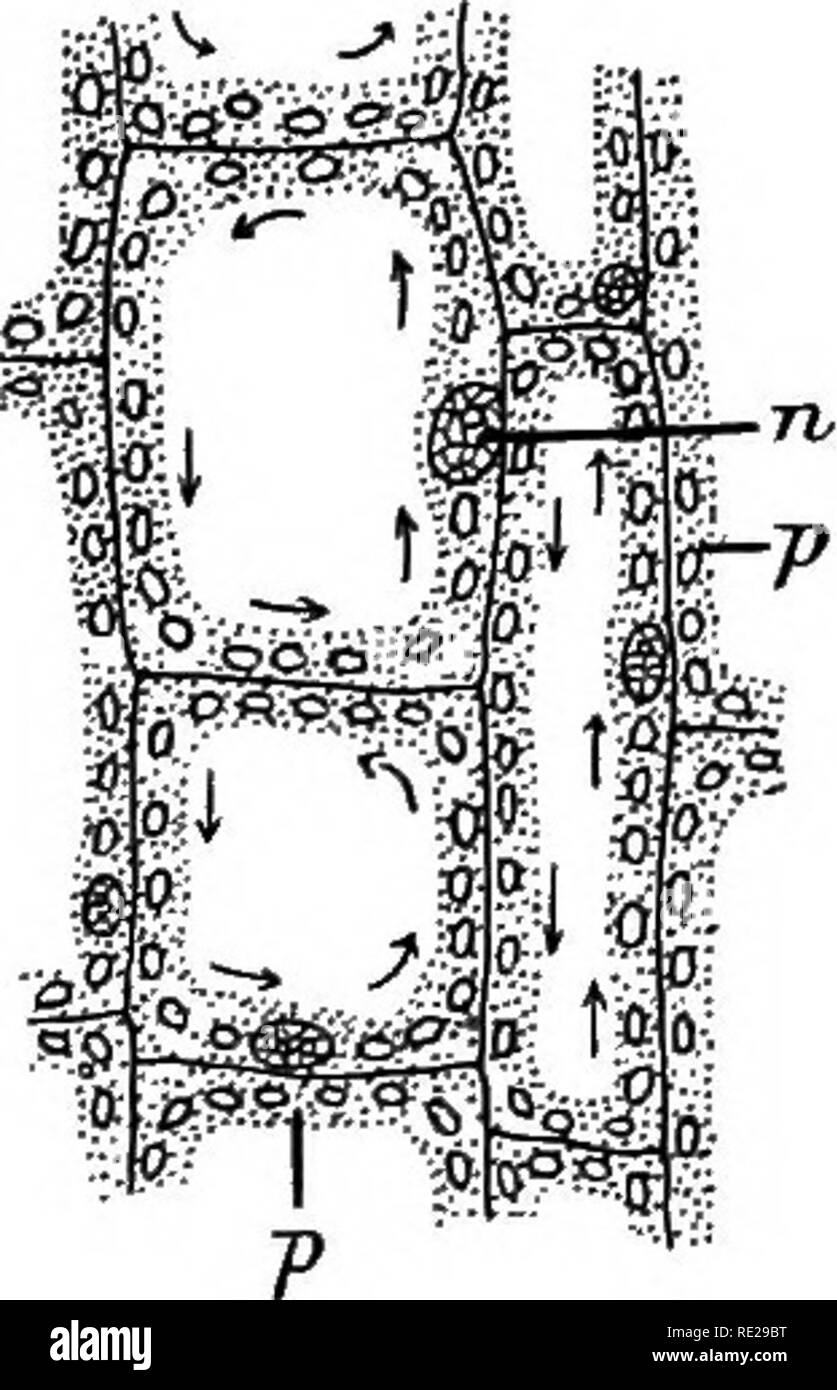 . An introduction to vegetable physiology. Plant physiology. THE GENERAL STEUCTUEE OE PLANTS neria, Nitella, Elodea (fig. 7), and others, a streaming move- ment of tlie granules the protoplasm contains can be detected. Fig. 7.—Cells ekom the Leaf of Elodea. X 500. 71, nucleos; j>, protoplasm, in which are embedded numerous chloro- plasts. The arrows show the direction of the movement of the protoplasm.. Please note that these images are extracted from scanned page images that may have been digitally enhanced for readability - coloration and appearance of these illustrations may not perfectl Stock Photohttps://www.alamy.com/image-license-details/?v=1https://www.alamy.com/an-introduction-to-vegetable-physiology-plant-physiology-the-general-steuctuee-oe-plants-neria-nitella-elodea-fig-7-and-others-a-streaming-move-ment-of-tlie-granules-the-protoplasm-contains-can-be-detected-fig-7cells-ekom-the-leaf-of-elodea-x-500-71-nucleos-jgt-protoplasm-in-which-are-embedded-numerous-chloro-plasts-the-arrows-show-the-direction-of-the-movement-of-the-protoplasm-please-note-that-these-images-are-extracted-from-scanned-page-images-that-may-have-been-digitally-enhanced-for-readability-coloration-and-appearance-of-these-illustrations-may-not-perfectl-image232391260.html
RMRE29BT–. An introduction to vegetable physiology. Plant physiology. THE GENERAL STEUCTUEE OE PLANTS neria, Nitella, Elodea (fig. 7), and others, a streaming move- ment of tlie granules the protoplasm contains can be detected. Fig. 7.—Cells ekom the Leaf of Elodea. X 500. 71, nucleos; j>, protoplasm, in which are embedded numerous chloro- plasts. The arrows show the direction of the movement of the protoplasm.. Please note that these images are extracted from scanned page images that may have been digitally enhanced for readability - coloration and appearance of these illustrations may not perfectl
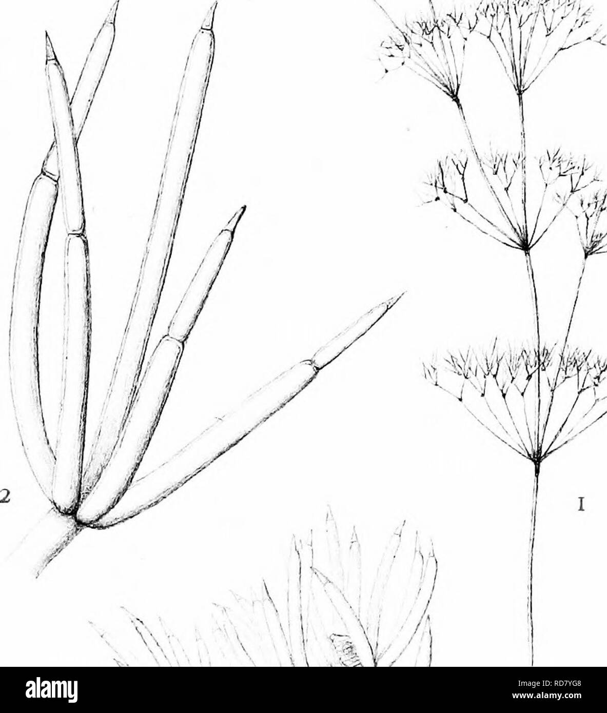 . The British Charophyta. Characeae. PLATE XIII â J^â¢^^^^ -^^r %â 3 4 #'A ^f^lffe. JJ -411, :«^i% ''^:. if-'' ^#1^ â x.:^:,^ }/ V M ^j -' i; V 6 / "J^ X. .)/. i:rn,,sihl. NITELLA GRACILIS. Please note that these images are extracted from scanned page images that may have been digitally enhanced for readability - coloration and appearance of these illustrations may not perfectly resemble the original work.. Groves, James; Bullock-Webster, George Russell, 1858- joint author. London, The Ray society Stock Photohttps://www.alamy.com/image-license-details/?v=1https://www.alamy.com/the-british-charophyta-characeae-plate-xiii-j-r-3-4-a-flffe-jj-411-i-if-1-x-v-m-j-i-v-6-quotj-x-irnsihl-nitella-gracilis-please-note-that-these-images-are-extracted-from-scanned-page-images-that-may-have-been-digitally-enhanced-for-readability-coloration-and-appearance-of-these-illustrations-may-not-perfectly-resemble-the-original-work-groves-james-bullock-webster-george-russell-1858-joint-author-london-the-ray-society-image231900600.html
RMRD7YG8–. The British Charophyta. Characeae. PLATE XIII â J^â¢^^^^ -^^r %â 3 4 #'A ^f^lffe. JJ -411, :«^i% ''^:. if-'' ^#1^ â x.:^:,^ }/ V M ^j -' i; V 6 / "J^ X. .)/. i:rn,,sihl. NITELLA GRACILIS. Please note that these images are extracted from scanned page images that may have been digitally enhanced for readability - coloration and appearance of these illustrations may not perfectly resemble the original work.. Groves, James; Bullock-Webster, George Russell, 1858- joint author. London, The Ray society