Quick filters:
Pronucleus Stock Photos and Images
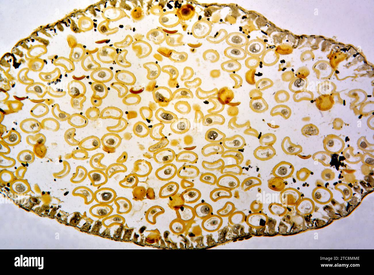 Ascaris megalocephala, pronucleus. Light microscope X150 at 10 cm wide. Stock Photohttps://www.alamy.com/image-license-details/?v=1https://www.alamy.com/ascaris-megalocephala-pronucleus-light-microscope-x150-at-10-cm-wide-image575509886.html
RF2TC8MME–Ascaris megalocephala, pronucleus. Light microscope X150 at 10 cm wide.
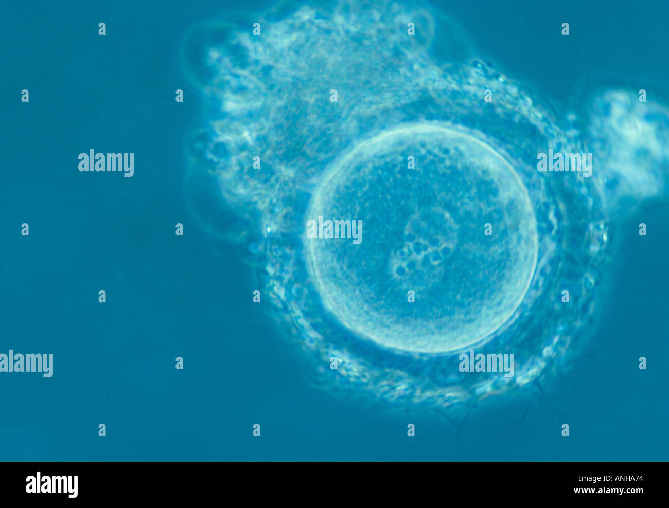 Fertilized human egg with male female pronucleus IVF In vitro fertilization Stock Photohttps://www.alamy.com/image-license-details/?v=1https://www.alamy.com/fertilized-human-egg-with-male-female-pronucleus-ivf-in-vitro-fertilization-image2873971.html
RMANHA74–Fertilized human egg with male female pronucleus IVF In vitro fertilization
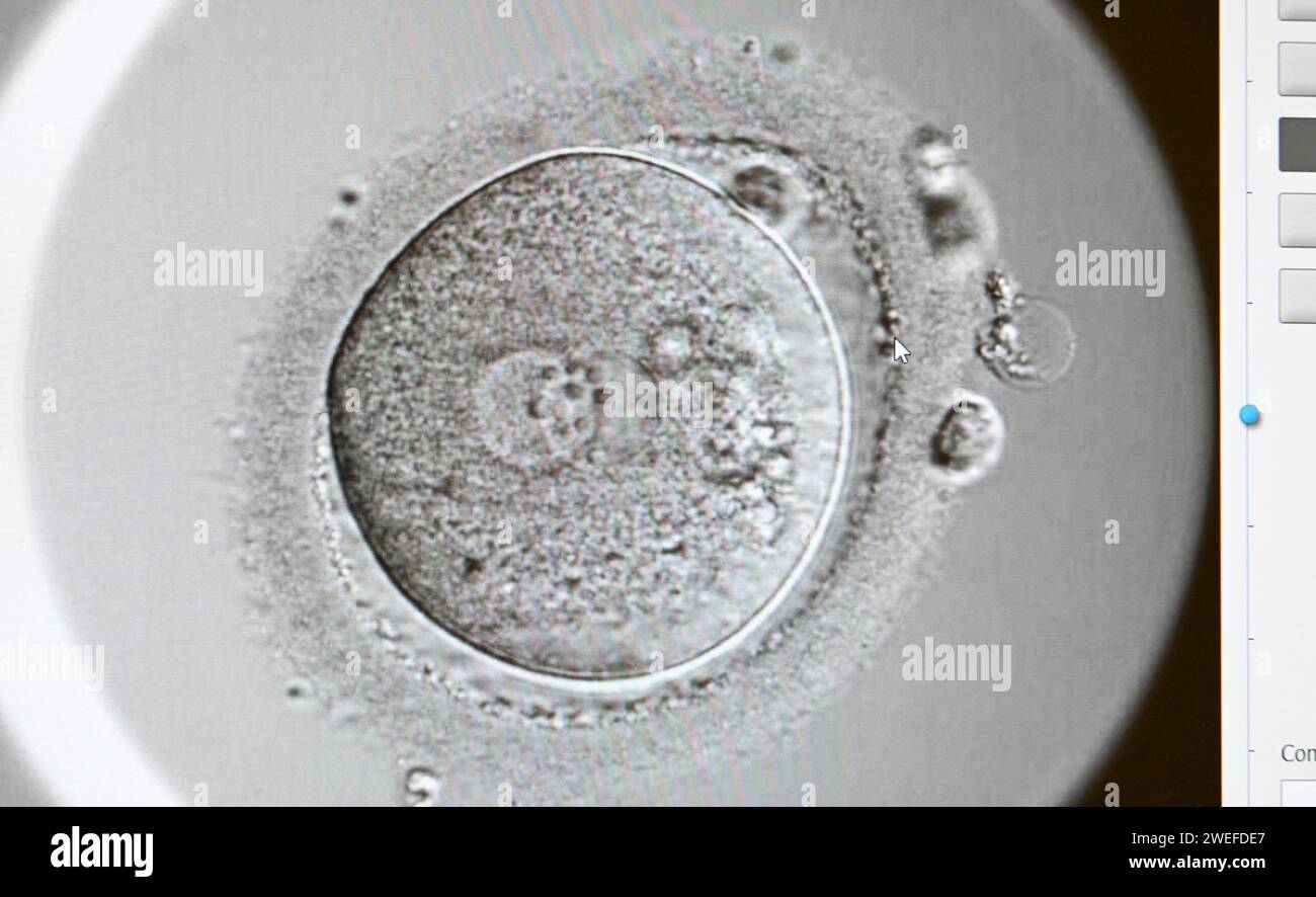 PRODUCTION - 17 January 2024, Berlin: In the cell laboratory at the Fertility Center Berlin, a two-cell organism can be seen on a screen after successful artificial insemination. Here, a paternal and maternal pronucleus fuse. Photo: Jens Kalaene/dpa Stock Photohttps://www.alamy.com/image-license-details/?v=1https://www.alamy.com/production-17-january-2024-berlin-in-the-cell-laboratory-at-the-fertility-center-berlin-a-two-cell-organism-can-be-seen-on-a-screen-after-successful-artificial-insemination-here-a-paternal-and-maternal-pronucleus-fuse-photo-jens-kalaenedpa-image594097567.html
RM2WEFDE7–PRODUCTION - 17 January 2024, Berlin: In the cell laboratory at the Fertility Center Berlin, a two-cell organism can be seen on a screen after successful artificial insemination. Here, a paternal and maternal pronucleus fuse. Photo: Jens Kalaene/dpa
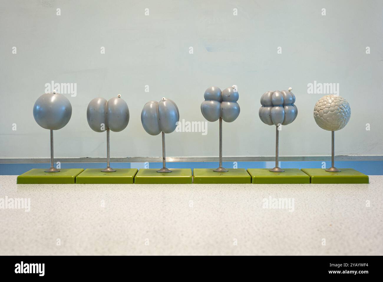 Several plastic models showing the initial stage of embryo development by cell division. Used in school biology lessons. Stock Photohttps://www.alamy.com/image-license-details/?v=1https://www.alamy.com/several-plastic-models-showing-the-initial-stage-of-embryo-development-by-cell-division-used-in-school-biology-lessons-image626332536.html
RF2YAYWF4–Several plastic models showing the initial stage of embryo development by cell division. Used in school biology lessons.
 . Journal of morphology. Fig. 5. Fig. 6. archoplasm or any centrosome in connection with the male pro-nucleus. Before this time both of the pronuclei have reached exactlythe same stage of development, and continue to keep accuratepace with each other. At first 12 very minute Diplococcus-likechromosomes may be made out in each pronucleus. Eachchromosome increases to several times its original volumeduring the growth of the pronuclei, and then breaks down intoa string of small karyo-microsomes. A few of the largespherical granules, however, remain as nucleoli (Fig. 8). In many eggs of this stage Stock Photohttps://www.alamy.com/image-license-details/?v=1https://www.alamy.com/journal-of-morphology-fig-5-fig-6-archoplasm-or-any-centrosome-in-connection-with-the-male-pro-nucleus-before-this-time-both-of-the-pronuclei-have-reached-exactlythe-same-stage-of-development-and-continue-to-keep-accuratepace-with-each-other-at-first-12-very-minute-diplococcus-likechromosomes-may-be-made-out-in-each-pronucleus-eachchromosome-increases-to-several-times-its-original-volumeduring-the-growth-of-the-pronuclei-and-then-breaks-down-intoa-string-of-small-karyo-microsomes-a-few-of-the-largespherical-granules-however-remain-as-nucleoli-fig-8-in-many-eggs-of-this-stage-image336955926.html
RM2AG5JM6–. Journal of morphology. Fig. 5. Fig. 6. archoplasm or any centrosome in connection with the male pro-nucleus. Before this time both of the pronuclei have reached exactlythe same stage of development, and continue to keep accuratepace with each other. At first 12 very minute Diplococcus-likechromosomes may be made out in each pronucleus. Eachchromosome increases to several times its original volumeduring the growth of the pronuclei, and then breaks down intoa string of small karyo-microsomes. A few of the largespherical granules, however, remain as nucleoli (Fig. 8). In many eggs of this stage
RF2KA10MB–Health zigot, illustration or icon, vector on white background.
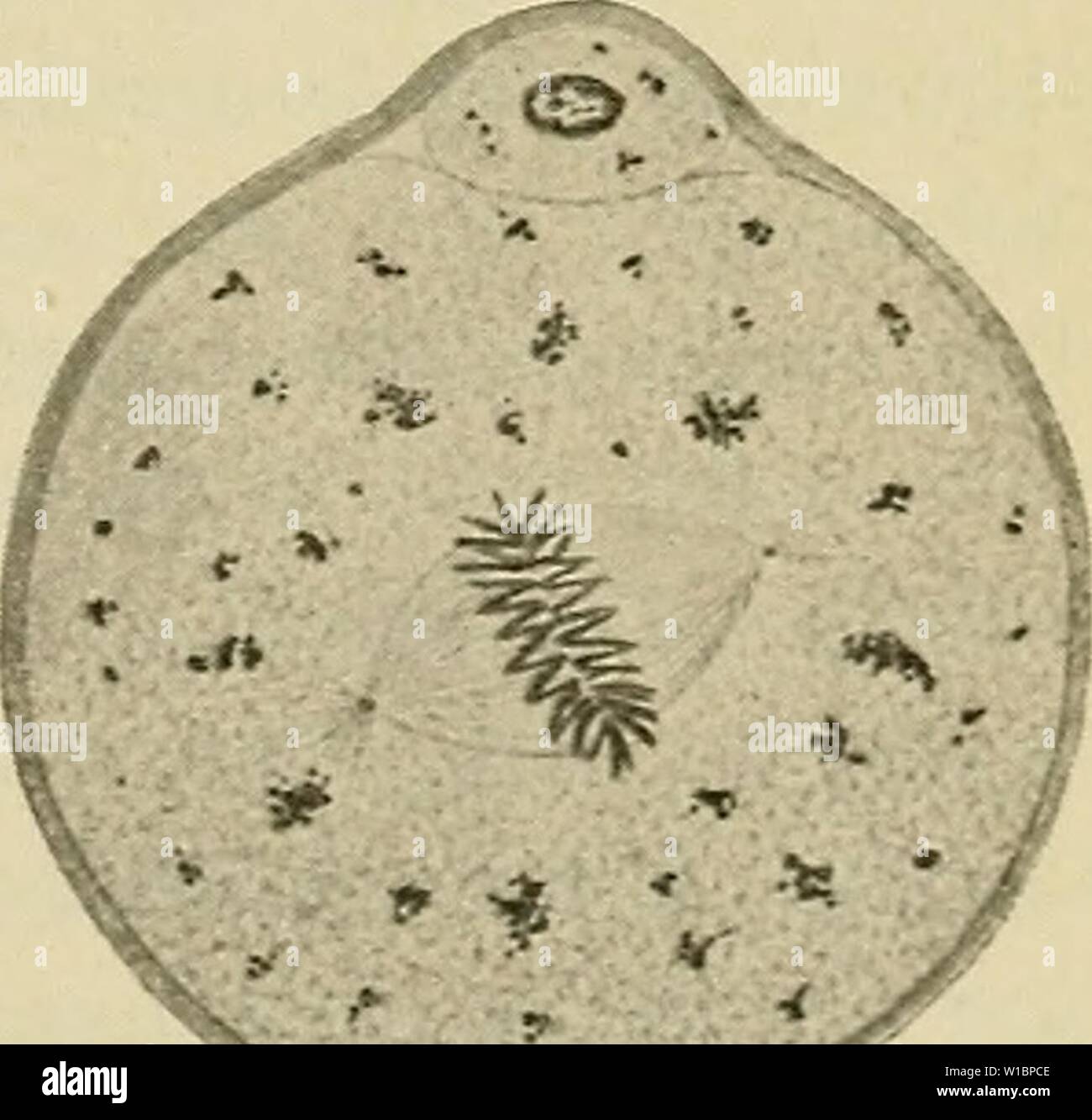 Archive image from page 44 of The development of the human. The development of the human body : a manual of human embryology . developmentofhum00mcmu Year: 1914 Fig. 16.—Six Stages in the Process of Fertilization of the Ovum of a Mouse. After the first stage figured it is impossible to determine which of the two nuclei represents the male or female pronucleus, ek, Female pronucleus; rkl and rk2, polar globules; spk, male pronucleus.—(Sobotia.) 3 Stock Photohttps://www.alamy.com/image-license-details/?v=1https://www.alamy.com/archive-image-from-page-44-of-the-development-of-the-human-the-development-of-the-human-body-a-manual-of-human-embryology-developmentofhum00mcmu-year-1914-fig-16six-stages-in-the-process-of-fertilization-of-the-ovum-of-a-mouse-after-the-first-stage-figured-it-is-impossible-to-determine-which-of-the-two-nuclei-represents-the-male-or-female-pronucleus-ek-female-pronucleus-rkl-and-rk2-polar-globules-spk-male-pronucleussobotia-3-image259029246.html
RMW1BPCE–Archive image from page 44 of The development of the human. The development of the human body : a manual of human embryology . developmentofhum00mcmu Year: 1914 Fig. 16.—Six Stages in the Process of Fertilization of the Ovum of a Mouse. After the first stage figured it is impossible to determine which of the two nuclei represents the male or female pronucleus, ek, Female pronucleus; rkl and rk2, polar globules; spk, male pronucleus.—(Sobotia.) 3
 . Readings in evolution, genetics, and eugenics. Evolution; Heredity; Eugenics. Fig. so.—Diagram to illustrate fertilization; S, male pronucleus; 9, female pronucleus; observe that the chromosomes of maternal and paternal origin respectively do not fuse. {From Guyer.) individual chromosomes do not intermingle their substance at this time, but each apparently retains its own individuality. There is considerable evidence which indicates that throughout Ufe the chro- mosomes contributed by the male parent remain distinct from those of the female parent. Inasmuch as each germ-cell, after maturatio Stock Photohttps://www.alamy.com/image-license-details/?v=1https://www.alamy.com/readings-in-evolution-genetics-and-eugenics-evolution-heredity-eugenics-fig-sodiagram-to-illustrate-fertilization-s-male-pronucleus-9-female-pronucleus-observe-that-the-chromosomes-of-maternal-and-paternal-origin-respectively-do-not-fuse-from-guyer-individual-chromosomes-do-not-intermingle-their-substance-at-this-time-but-each-apparently-retains-its-own-individuality-there-is-considerable-evidence-which-indicates-that-throughout-ufe-the-chro-mosomes-contributed-by-the-male-parent-remain-distinct-from-those-of-the-female-parent-inasmuch-as-each-germ-cell-after-maturatio-image216392531.html
RMPG1ETK–. Readings in evolution, genetics, and eugenics. Evolution; Heredity; Eugenics. Fig. so.—Diagram to illustrate fertilization; S, male pronucleus; 9, female pronucleus; observe that the chromosomes of maternal and paternal origin respectively do not fuse. {From Guyer.) individual chromosomes do not intermingle their substance at this time, but each apparently retains its own individuality. There is considerable evidence which indicates that throughout Ufe the chro- mosomes contributed by the male parent remain distinct from those of the female parent. Inasmuch as each germ-cell, after maturatio
 IVF Stock Photohttps://www.alamy.com/image-license-details/?v=1https://www.alamy.com/stock-photo-ivf-71925370.html
RFE50DF6–IVF
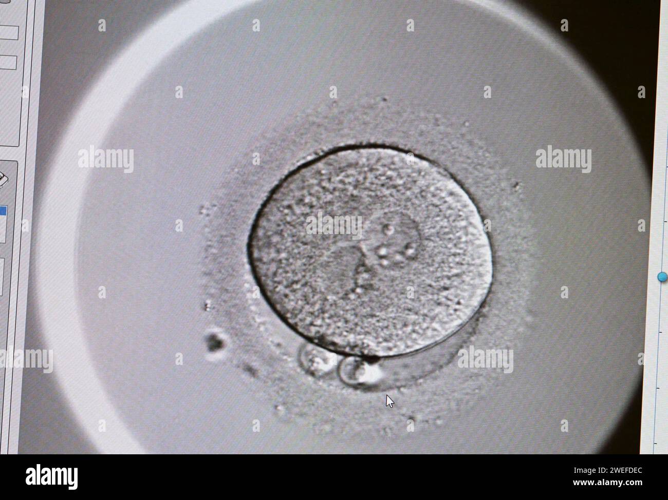 PRODUCTION - 17 January 2024, Berlin: In the cell laboratory at the Fertility Center Berlin, a two-cell organism can be seen on a screen after successful artificial insemination. Here, a paternal and maternal pronucleus fuse. Photo: Jens Kalaene/dpa Stock Photohttps://www.alamy.com/image-license-details/?v=1https://www.alamy.com/production-17-january-2024-berlin-in-the-cell-laboratory-at-the-fertility-center-berlin-a-two-cell-organism-can-be-seen-on-a-screen-after-successful-artificial-insemination-here-a-paternal-and-maternal-pronucleus-fuse-photo-jens-kalaenedpa-image594097572.html
RM2WEFDEC–PRODUCTION - 17 January 2024, Berlin: In the cell laboratory at the Fertility Center Berlin, a two-cell organism can be seen on a screen after successful artificial insemination. Here, a paternal and maternal pronucleus fuse. Photo: Jens Kalaene/dpa
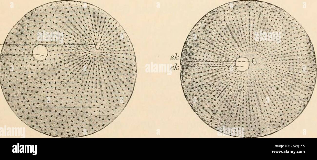 Text-book of comparative anatomy . s glacialis, after Fol (from 0. Hertwig s Lehrluchder Entwicldungsgeschichte). One of the spermatozoa which have entered the mucilaginousenvelope comes in contact with the receptive prominence. In C the yolk membrane is formed. the protoplasm of the egg; the head (the remains of the originalnucleus) increases somewhat in size. As male pronucleus, it movesforward to meet the female pronucleus. Finally they fuse and formone single nucleus, the so-called segmentation nucleus. The egg isfertilised. It seems tolerably certain that where the egg envelopes havea mic Stock Photohttps://www.alamy.com/image-license-details/?v=1https://www.alamy.com/text-book-of-comparative-anatomy-s-glacialis-after-fol-from-0-hertwig-s-lehrluchder-entwicldungsgeschichte-one-of-the-spermatozoa-which-have-entered-the-mucilaginousenvelope-comes-in-contact-with-the-receptive-prominence-in-c-the-yolk-membrane-is-formed-the-protoplasm-of-the-egg-the-head-the-remains-of-the-originalnucleus-increases-somewhat-in-size-as-male-pronucleus-it-movesforward-to-meet-the-female-pronucleus-finally-they-fuse-and-formone-single-nucleus-the-so-called-segmentation-nucleus-the-egg-isfertilised-it-seems-tolerably-certain-that-where-the-egg-envelopes-havea-mic-image342778105.html
RM2AWJTY5–Text-book of comparative anatomy . s glacialis, after Fol (from 0. Hertwig s Lehrluchder Entwicldungsgeschichte). One of the spermatozoa which have entered the mucilaginousenvelope comes in contact with the receptive prominence. In C the yolk membrane is formed. the protoplasm of the egg; the head (the remains of the originalnucleus) increases somewhat in size. As male pronucleus, it movesforward to meet the female pronucleus. Finally they fuse and formone single nucleus, the so-called segmentation nucleus. The egg isfertilised. It seems tolerably certain that where the egg envelopes havea mic
 Archive image from page 52 of The development of the chick;. The development of the chick; an introduction to embryology . developmentofchi00lill Year: 1908 DEVELOPMENT PRIOR TO LAYING 35 it rapidly withdraws from the surface of the egg to a deeper position near the center of the germinal disc. (Concerning the Fig. 12.-— Egg nucleus (female pronucleus) and polar bodies of the pigeon's egg. (After Harper.) 8.30 p.m. x 2000. E. N., Egg nucleus, p. b. 1, First polar body. p. b. 2, Second polar body. p'v. S., Perivitelline space, v. M., Vi- telline membrane. general theory of the maturation pr Stock Photohttps://www.alamy.com/image-license-details/?v=1https://www.alamy.com/archive-image-from-page-52-of-the-development-of-the-chick-the-development-of-the-chick-an-introduction-to-embryology-developmentofchi00lill-year-1908-development-prior-to-laying-35-it-rapidly-withdraws-from-the-surface-of-the-egg-to-a-deeper-position-near-the-center-of-the-germinal-disc-concerning-the-fig-12-egg-nucleus-female-pronucleus-and-polar-bodies-of-the-pigeons-egg-after-harper-830-pm-x-2000-e-n-egg-nucleus-p-b-1-first-polar-body-p-b-2-second-polar-body-pv-s-perivitelline-space-v-m-vi-telline-membrane-general-theory-of-the-maturation-pr-image259037150.html
RMW1C4EP–Archive image from page 52 of The development of the chick;. The development of the chick; an introduction to embryology . developmentofchi00lill Year: 1908 DEVELOPMENT PRIOR TO LAYING 35 it rapidly withdraws from the surface of the egg to a deeper position near the center of the germinal disc. (Concerning the Fig. 12.-— Egg nucleus (female pronucleus) and polar bodies of the pigeon's egg. (After Harper.) 8.30 p.m. x 2000. E. N., Egg nucleus, p. b. 1, First polar body. p. b. 2, Second polar body. p'v. S., Perivitelline space, v. M., Vi- telline membrane. general theory of the maturation pr
 . The development of the human body : a manual of human embryology. Embryology; Embryo, Non-Mammalian. Fig. 16.—Six Stages in the Process of Fertilization of the Ovum of a Mouse. After the first stage figured it is impossible to determine which of the two nuclei represents the male or female pronucleus, ek, Female pronucleus; rkl and rk2, polar globules; spk, male pronucleus.—(Sobotia.) 3. Please note that these images are extracted from scanned page images that may have been digitally enhanced for readability - coloration and appearance of these illustrations may not perfectly resemble the or Stock Photohttps://www.alamy.com/image-license-details/?v=1https://www.alamy.com/the-development-of-the-human-body-a-manual-of-human-embryology-embryology-embryo-non-mammalian-fig-16six-stages-in-the-process-of-fertilization-of-the-ovum-of-a-mouse-after-the-first-stage-figured-it-is-impossible-to-determine-which-of-the-two-nuclei-represents-the-male-or-female-pronucleus-ek-female-pronucleus-rkl-and-rk2-polar-globules-spk-male-pronucleussobotia-3-please-note-that-these-images-are-extracted-from-scanned-page-images-that-may-have-been-digitally-enhanced-for-readability-coloration-and-appearance-of-these-illustrations-may-not-perfectly-resemble-the-or-image215970183.html
RMPFA84R–. The development of the human body : a manual of human embryology. Embryology; Embryo, Non-Mammalian. Fig. 16.—Six Stages in the Process of Fertilization of the Ovum of a Mouse. After the first stage figured it is impossible to determine which of the two nuclei represents the male or female pronucleus, ek, Female pronucleus; rkl and rk2, polar globules; spk, male pronucleus.—(Sobotia.) 3. Please note that these images are extracted from scanned page images that may have been digitally enhanced for readability - coloration and appearance of these illustrations may not perfectly resemble the or
 In vitro Stock Photohttps://www.alamy.com/image-license-details/?v=1https://www.alamy.com/stock-photo-in-vitro-71925405.html
RFE50DGD–In vitro
 PRODUCTION - 17 January 2024, Berlin: In the cell laboratory at the Fertility Center Berlin, a two-cell organism can be seen on a screen after successful artificial insemination. Here, a paternal and maternal pronucleus fuse. Photo: Jens Kalaene/dpa Stock Photohttps://www.alamy.com/image-license-details/?v=1https://www.alamy.com/production-17-january-2024-berlin-in-the-cell-laboratory-at-the-fertility-center-berlin-a-two-cell-organism-can-be-seen-on-a-screen-after-successful-artificial-insemination-here-a-paternal-and-maternal-pronucleus-fuse-photo-jens-kalaenedpa-image594097561.html
RM2WEFDE1–PRODUCTION - 17 January 2024, Berlin: In the cell laboratory at the Fertility Center Berlin, a two-cell organism can be seen on a screen after successful artificial insemination. Here, a paternal and maternal pronucleus fuse. Photo: Jens Kalaene/dpa
 A textbook of obstetrics . Fig- 55-—A, Mature ovum of echinus : n, female pronucleus; B, immature ovarianovum of echinus (Hertwig). India impregnation has occurred before menstruation had begun ;but usually premature maternity is preceded by precocious men-struation. Ovulation has continued, as proved by impregnation,until the fifty-second, fifty-fourth, fifty-eighth, and even to the fifty- 62 PREGNANCY. ninth year ! A case is recorded of delivery at the age of fifty-nineyears and five months. An obstetrician investigating the nature ofan abdominal tumor should remember, therefore, that pregna Stock Photohttps://www.alamy.com/image-license-details/?v=1https://www.alamy.com/a-textbook-of-obstetrics-fig-55-a-mature-ovum-of-echinus-n-female-pronucleus-b-immature-ovarianovum-of-echinus-hertwig-india-impregnation-has-occurred-before-menstruation-had-begun-but-usually-premature-maternity-is-preceded-by-precocious-men-struation-ovulation-has-continued-as-proved-by-impregnationuntil-the-fifty-second-fifty-fourth-fifty-eighth-and-even-to-the-fifty-62-pregnancy-ninth-year-!-a-case-is-recorded-of-delivery-at-the-age-of-fifty-nineyears-and-five-months-an-obstetrician-investigating-the-nature-ofan-abdominal-tumor-should-remember-therefore-that-pregna-image340189935.html
RM2ANCYMF–A textbook of obstetrics . Fig- 55-—A, Mature ovum of echinus : n, female pronucleus; B, immature ovarianovum of echinus (Hertwig). India impregnation has occurred before menstruation had begun ;but usually premature maternity is preceded by precocious men-struation. Ovulation has continued, as proved by impregnation,until the fifty-second, fifty-fourth, fifty-eighth, and even to the fifty- 62 PREGNANCY. ninth year ! A case is recorded of delivery at the age of fifty-nineyears and five months. An obstetrician investigating the nature ofan abdominal tumor should remember, therefore, that pregna
 Archive image from page 52 of The development of the chick. The development of the chick : an introduction to embryology . developmentofchi02lill Year: 1936 DEVELOPMENT PRIOR TO LAYING 35 it rapidly withdraws from the surface of the egg to a deeper position near the center of the germinal disc. (Concerning the p.d.I. p.b.2. -•V-v' '.T Fig. 12.— Egg nuclous (female pronucleus) and polar bodies of the pigeon's g. (After Harper.) 8.30 p.m. x 2000. E. N., Egg nucleus, p. b. 1, First polar body. p. b. 2, Second polar body. p'v. S., Perivitelline space, v. M., Vi- telline membrane. general theor Stock Photohttps://www.alamy.com/image-license-details/?v=1https://www.alamy.com/archive-image-from-page-52-of-the-development-of-the-chick-the-development-of-the-chick-an-introduction-to-embryology-developmentofchi02lill-year-1936-development-prior-to-laying-35-it-rapidly-withdraws-from-the-surface-of-the-egg-to-a-deeper-position-near-the-center-of-the-germinal-disc-concerning-the-pdi-pb2-v-v-t-fig-12-egg-nuclous-female-pronucleus-and-polar-bodies-of-the-pigeons-g-after-harper-830-pm-x-2000-e-n-egg-nucleus-p-b-1-first-polar-body-p-b-2-second-polar-body-pv-s-perivitelline-space-v-m-vi-telline-membrane-general-theor-image259037128.html
RMW1C4E0–Archive image from page 52 of The development of the chick. The development of the chick : an introduction to embryology . developmentofchi02lill Year: 1936 DEVELOPMENT PRIOR TO LAYING 35 it rapidly withdraws from the surface of the egg to a deeper position near the center of the germinal disc. (Concerning the p.d.I. p.b.2. -•V-v' '.T Fig. 12.— Egg nuclous (female pronucleus) and polar bodies of the pigeon's g. (After Harper.) 8.30 p.m. x 2000. E. N., Egg nucleus, p. b. 1, First polar body. p. b. 2, Second polar body. p'v. S., Perivitelline space, v. M., Vi- telline membrane. general theor
 . A manual of zoology. iftern. qpro/t se^.nud Fig. 26.—Diagram illustrating the maturation and fertilisation of the ovum. A, formation of first polar globule; B, beginning of fertilisation, sperms approaching the micropyle or aperture in the enclosing membrane of the ovum through which the sperm enters; C, forma- tion of the male pronucleus; D, approximation of the male and female pronuclei; Et forma- tion of segmentation-nucleus; 9 cent, female centrosome; rf cent, male centrosome (the centrosomes are cell-structures not further referred to in this work); mem, egg-membrane; microp, micropyle; Stock Photohttps://www.alamy.com/image-license-details/?v=1https://www.alamy.com/a-manual-of-zoology-iftern-qprot-senud-fig-26diagram-illustrating-the-maturation-and-fertilisation-of-the-ovum-a-formation-of-first-polar-globule-b-beginning-of-fertilisation-sperms-approaching-the-micropyle-or-aperture-in-the-enclosing-membrane-of-the-ovum-through-which-the-sperm-enters-c-forma-tion-of-the-male-pronucleus-d-approximation-of-the-male-and-female-pronuclei-et-forma-tion-of-segmentation-nucleus-9-cent-female-centrosome-rf-cent-male-centrosome-the-centrosomes-are-cell-structures-not-further-referred-to-in-this-work-mem-egg-membrane-microp-micropyle-image216442526.html
RMPG3PJ6–. A manual of zoology. iftern. qpro/t se^.nud Fig. 26.—Diagram illustrating the maturation and fertilisation of the ovum. A, formation of first polar globule; B, beginning of fertilisation, sperms approaching the micropyle or aperture in the enclosing membrane of the ovum through which the sperm enters; C, forma- tion of the male pronucleus; D, approximation of the male and female pronuclei; Et forma- tion of segmentation-nucleus; 9 cent, female centrosome; rf cent, male centrosome (the centrosomes are cell-structures not further referred to in this work); mem, egg-membrane; microp, micropyle;
 A textbook of obstetrics . Fig. 55. — A, Mature ovum of echinus : n, female pronucleus;ovum of echinus ^Hertwig 1. immature ovarian India impregnation has occurred before menstruation had begun ;but usually premature maternity is preceded by precocious men-struation. Ovulation has continued, as proved by impregnation,until the fifty-second, fifty-fourth, fifty-eighth, and even to the six- 62 PREGNANCY. tieth year! A case is recorded of delivery at the age of fifty-nineyears and five months. An obstetrician investigating the nature ofan abdominal tumor should remember, therefore, that pregnancy Stock Photohttps://www.alamy.com/image-license-details/?v=1https://www.alamy.com/a-textbook-of-obstetrics-fig-55-a-mature-ovum-of-echinus-n-female-pronucleusovum-of-echinus-hertwig-1-immature-ovarian-india-impregnation-has-occurred-before-menstruation-had-begun-but-usually-premature-maternity-is-preceded-by-precocious-men-struation-ovulation-has-continued-as-proved-by-impregnationuntil-the-fifty-second-fifty-fourth-fifty-eighth-and-even-to-the-six-62-pregnancy-tieth-year!-a-case-is-recorded-of-delivery-at-the-age-of-fifty-nineyears-and-five-months-an-obstetrician-investigating-the-nature-ofan-abdominal-tumor-should-remember-therefore-that-pregnancy-image343105789.html
RM2AX5PX5–A textbook of obstetrics . Fig. 55. — A, Mature ovum of echinus : n, female pronucleus;ovum of echinus ^Hertwig 1. immature ovarian India impregnation has occurred before menstruation had begun ;but usually premature maternity is preceded by precocious men-struation. Ovulation has continued, as proved by impregnation,until the fifty-second, fifty-fourth, fifty-eighth, and even to the six- 62 PREGNANCY. tieth year! A case is recorded of delivery at the age of fifty-nineyears and five months. An obstetrician investigating the nature ofan abdominal tumor should remember, therefore, that pregnancy
 Elementary text-book of zoology (1884) Elementary text-book of zoology elementarytextbo0101clau Year: 1884 FERTILIZATION. the protoplasm of the ovum, is thrown out of the egg as the so-called directive bodies or polar cells (tig. 101). The part of it, however, which remains in the ovum retains its significance as a nucleus, and is known as the female pronucleus. This fuses with the single spermatozoon (male pronucleus) which has forced its way into the ovum (fig. 102); and the compound structure so formed constitutes the nucleus of the fertilized ovum, or as it is generally called, the first Stock Photohttps://www.alamy.com/image-license-details/?v=1https://www.alamy.com/elementary-text-book-of-zoology-1884-elementary-text-book-of-zoology-elementarytextbo0101clau-year-1884-fertilization-the-protoplasm-of-the-ovum-is-thrown-out-of-the-egg-as-the-so-called-directive-bodies-or-polar-cells-tig-101-the-part-of-it-however-which-remains-in-the-ovum-retains-its-significance-as-a-nucleus-and-is-known-as-the-female-pronucleus-this-fuses-with-the-single-spermatozoon-male-pronucleus-which-has-forced-its-way-into-the-ovum-fig-102-and-the-compound-structure-so-formed-constitutes-the-nucleus-of-the-fertilized-ovum-or-as-it-is-generally-called-the-first-image239582754.html
RMRWNX6X–Elementary text-book of zoology (1884) Elementary text-book of zoology elementarytextbo0101clau Year: 1884 FERTILIZATION. the protoplasm of the ovum, is thrown out of the egg as the so-called directive bodies or polar cells (tig. 101). The part of it, however, which remains in the ovum retains its significance as a nucleus, and is known as the female pronucleus. This fuses with the single spermatozoon (male pronucleus) which has forced its way into the ovum (fig. 102); and the compound structure so formed constitutes the nucleus of the fertilized ovum, or as it is generally called, the first
 . The development of the chick; an introduction to embryology. Birds -- Embryology. DEVELOPMENT PRIOR TO LAYING 35 it rapidly withdraws from the surface of the egg to a deeper position near the center of the germinal disc. (Concerning the. Fig. 12.-— Egg nucleus (female pronucleus) and polar bodies of the pigeon's egg. (After Harper.) 8.30 p.m. x 2000. E. N., Egg nucleus, p. b. 1, First polar body. p. b. 2, Second polar body. p'v. S., Perivitelline space, v. M., Vi- telline membrane. general theory of the maturation process see E. B. Wilson, "The Cell in Development and Inheritance/' the Stock Photohttps://www.alamy.com/image-license-details/?v=1https://www.alamy.com/the-development-of-the-chick-an-introduction-to-embryology-birds-embryology-development-prior-to-laying-35-it-rapidly-withdraws-from-the-surface-of-the-egg-to-a-deeper-position-near-the-center-of-the-germinal-disc-concerning-the-fig-12-egg-nucleus-female-pronucleus-and-polar-bodies-of-the-pigeons-egg-after-harper-830-pm-x-2000-e-n-egg-nucleus-p-b-1-first-polar-body-p-b-2-second-polar-body-pv-s-perivitelline-space-v-m-vi-telline-membrane-general-theory-of-the-maturation-process-see-e-b-wilson-quotthe-cell-in-development-and-inheritance-the-image215958082.html
RMPF9MMJ–. The development of the chick; an introduction to embryology. Birds -- Embryology. DEVELOPMENT PRIOR TO LAYING 35 it rapidly withdraws from the surface of the egg to a deeper position near the center of the germinal disc. (Concerning the. Fig. 12.-— Egg nucleus (female pronucleus) and polar bodies of the pigeon's egg. (After Harper.) 8.30 p.m. x 2000. E. N., Egg nucleus, p. b. 1, First polar body. p. b. 2, Second polar body. p'v. S., Perivitelline space, v. M., Vi- telline membrane. general theory of the maturation process see E. B. Wilson, "The Cell in Development and Inheritance/' the
 A system of obstetrics . ing karyokinesis; p, the first polar globule in process of extrusion,and (3) extruded; p, both polar globules extruded; f.pn, female pronucleus. poles shows a radial arrangement, the radii converging toward eachpole, while striations unite the two poles. This phenomenon is now wellknown as occurring in the phenomenon of nuclear division as prelimi-nary to ordinary cell-division: the germinal vesicle and the germ-yelkbehave exactly like the nucleus and protoplasm of any normally divid-ing cell. The phenomenon is named karyokinesis, and cannot here bedescribed in detail. Stock Photohttps://www.alamy.com/image-license-details/?v=1https://www.alamy.com/a-system-of-obstetrics-ing-karyokinesis-p-the-first-polar-globule-in-process-of-extrusionand-3-extruded-p-both-polar-globules-extruded-fpn-female-pronucleus-poles-shows-a-radial-arrangement-the-radii-converging-toward-eachpole-while-striations-unite-the-two-poles-this-phenomenon-is-now-wellknown-as-occurring-in-the-phenomenon-of-nuclear-division-as-prelimi-nary-to-ordinary-cell-division-the-germinal-vesicle-and-the-germ-yelkbehave-exactly-like-the-nucleus-and-protoplasm-of-any-normally-divid-ing-cell-the-phenomenon-is-named-karyokinesis-and-cannot-here-bedescribed-in-detail-image342826595.html
RM2AWN2PY–A system of obstetrics . ing karyokinesis; p, the first polar globule in process of extrusion,and (3) extruded; p, both polar globules extruded; f.pn, female pronucleus. poles shows a radial arrangement, the radii converging toward eachpole, while striations unite the two poles. This phenomenon is now wellknown as occurring in the phenomenon of nuclear division as prelimi-nary to ordinary cell-division: the germinal vesicle and the germ-yelkbehave exactly like the nucleus and protoplasm of any normally divid-ing cell. The phenomenon is named karyokinesis, and cannot here bedescribed in detail.
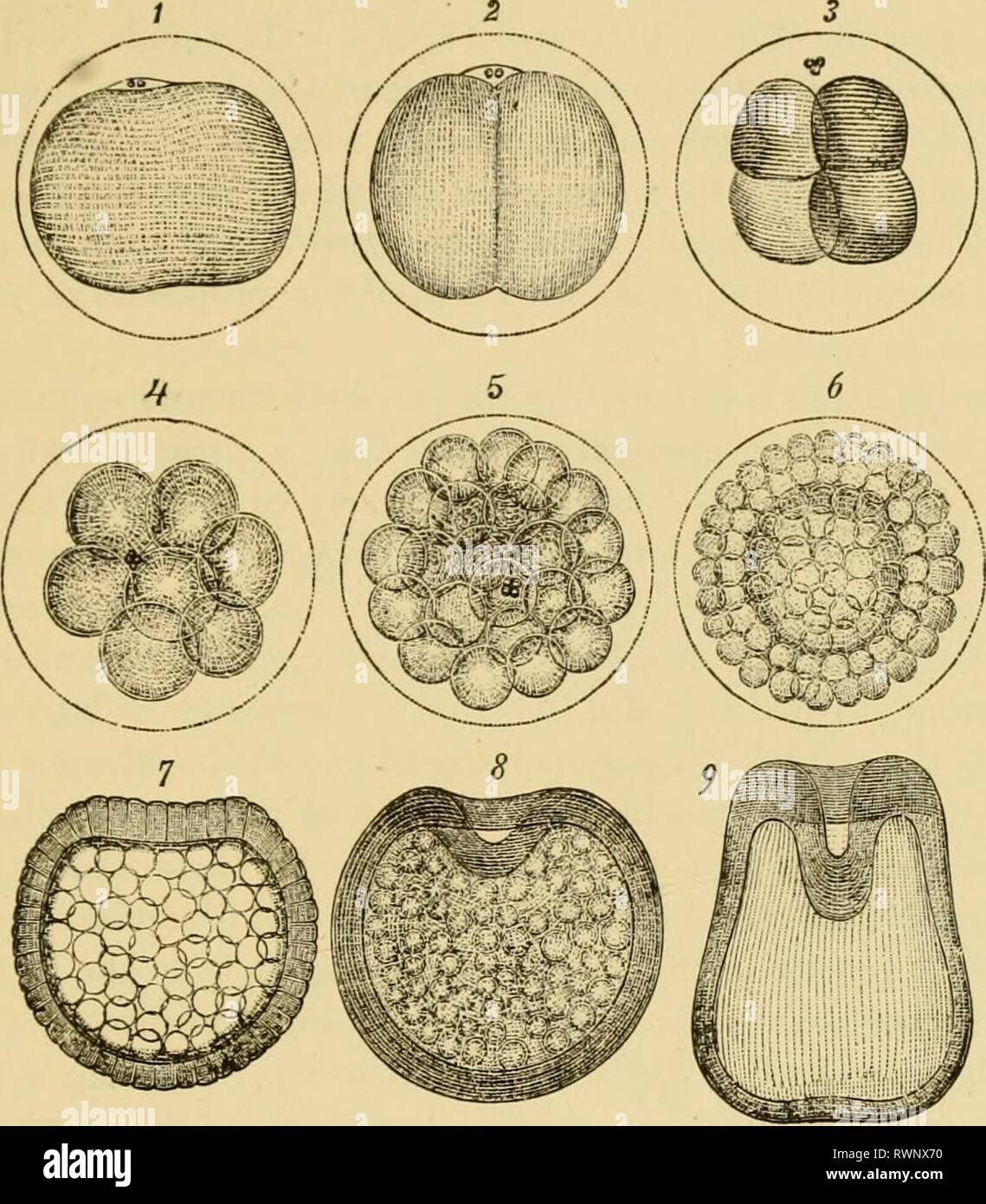 Elementary text-book of zoology, general Elementary text-book of zoology, general part and special part: protozoa to insecta elementarytextbo00clau Year: 1892 rERTILIZATION. 109 the protoplasm of the ovum, is thrown out of the egg as the so-called directive bodies or 2}olar cells (fig. 101). The part of it, however, which remains in the ovum retains its significance as a nucleus, and is known as the female pronucleus. This fu-ies with the single spermatozoon (male pronucleus) which has forced its way into the ovum (fig. 102); and the compound structure so formed constitutes the nucleus of the Stock Photohttps://www.alamy.com/image-license-details/?v=1https://www.alamy.com/elementary-text-book-of-zoology-general-elementary-text-book-of-zoology-general-part-and-special-part-protozoa-to-insecta-elementarytextbo00clau-year-1892-rertilization-109-the-protoplasm-of-the-ovum-is-thrown-out-of-the-egg-as-the-so-called-directive-bodies-or-2olar-cells-fig-101-the-part-of-it-however-which-remains-in-the-ovum-retains-its-significance-as-a-nucleus-and-is-known-as-the-female-pronucleus-this-fu-ies-with-the-single-spermatozoon-male-pronucleus-which-has-forced-its-way-into-the-ovum-fig-102-and-the-compound-structure-so-formed-constitutes-the-nucleus-of-the-image239582756.html
RMRWNX70–Elementary text-book of zoology, general Elementary text-book of zoology, general part and special part: protozoa to insecta elementarytextbo00clau Year: 1892 rERTILIZATION. 109 the protoplasm of the ovum, is thrown out of the egg as the so-called directive bodies or 2}olar cells (fig. 101). The part of it, however, which remains in the ovum retains its significance as a nucleus, and is known as the female pronucleus. This fu-ies with the single spermatozoon (male pronucleus) which has forced its way into the ovum (fig. 102); and the compound structure so formed constitutes the nucleus of the
 . The development of the chick : an introduction to embryology. Embryology; Chickens -- Embryos. DEVELOPMENT PRIOR TO LAYING 35 it rapidly withdraws from the surface of the egg to a deeper position near the center of the germinal disc. (Concerning the p.d.I. p.b.2.. -•V-v' '.^T Fig. 12.— Egg nuclous (female pronucleus) and polar bodies of the pigeon's ^g^. (After Harper.) 8.30 p.m. x 2000. E. N., Egg nucleus, p. b. 1, First polar body. p. b. 2, Second polar body. p'v. S., Perivitelline space, v. M., Vi- telline membrane. general theory of the maturation process see E. B. Wilson, "The Cell Stock Photohttps://www.alamy.com/image-license-details/?v=1https://www.alamy.com/the-development-of-the-chick-an-introduction-to-embryology-embryology-chickens-embryos-development-prior-to-laying-35-it-rapidly-withdraws-from-the-surface-of-the-egg-to-a-deeper-position-near-the-center-of-the-germinal-disc-concerning-the-pdi-pb2-v-v-t-fig-12-egg-nuclous-female-pronucleus-and-polar-bodies-of-the-pigeons-g-after-harper-830-pm-x-2000-e-n-egg-nucleus-p-b-1-first-polar-body-p-b-2-second-polar-body-pv-s-perivitelline-space-v-m-vi-telline-membrane-general-theory-of-the-maturation-process-see-e-b-wilson-quotthe-cell-image215957978.html
RMPF9MGX–. The development of the chick : an introduction to embryology. Embryology; Chickens -- Embryos. DEVELOPMENT PRIOR TO LAYING 35 it rapidly withdraws from the surface of the egg to a deeper position near the center of the germinal disc. (Concerning the p.d.I. p.b.2.. -•V-v' '.^T Fig. 12.— Egg nuclous (female pronucleus) and polar bodies of the pigeon's ^g^. (After Harper.) 8.30 p.m. x 2000. E. N., Egg nucleus, p. b. 1, First polar body. p. b. 2, Second polar body. p'v. S., Perivitelline space, v. M., Vi- telline membrane. general theory of the maturation process see E. B. Wilson, "The Cell
![. Journal of morphology. string of small karyo-microsomes. A few of the largespherical granules, however, remain as nucleoli (Fig. 8). In many eggs of this stage I have looked in vain for anytraces of archoplasm or centrosome, but whenever these struc-tures could be brought out by means of the iron-alum haema-toxylin, they were always close to the female pronucleus and No. I.] MYZOSTOMA G LAB RUM. 309 at some distance from the male. Their appearance is that ofFig. 7, where the first traces of a spindle are forming betweenthe two centrosomes — which I take to be the two originallystationed at t Stock Photo . Journal of morphology. string of small karyo-microsomes. A few of the largespherical granules, however, remain as nucleoli (Fig. 8). In many eggs of this stage I have looked in vain for anytraces of archoplasm or centrosome, but whenever these struc-tures could be brought out by means of the iron-alum haema-toxylin, they were always close to the female pronucleus and No. I.] MYZOSTOMA G LAB RUM. 309 at some distance from the male. Their appearance is that ofFig. 7, where the first traces of a spindle are forming betweenthe two centrosomes — which I take to be the two originallystationed at t Stock Photo](https://c8.alamy.com/comp/2AG5JC5/journal-of-morphology-string-of-small-karyo-microsomes-a-few-of-the-largespherical-granules-however-remain-as-nucleoli-fig-8-in-many-eggs-of-this-stage-i-have-looked-in-vain-for-anytraces-of-archoplasm-or-centrosome-but-whenever-these-struc-tures-could-be-brought-out-by-means-of-the-iron-alum-haema-toxylin-they-were-always-close-to-the-female-pronucleus-and-no-i-myzostoma-g-lab-rum-309-at-some-distance-from-the-male-their-appearance-is-that-offig-7-where-the-first-traces-of-a-spindle-are-forming-betweenthe-two-centrosomes-which-i-take-to-be-the-two-originallystationed-at-t-2AG5JC5.jpg) . Journal of morphology. string of small karyo-microsomes. A few of the largespherical granules, however, remain as nucleoli (Fig. 8). In many eggs of this stage I have looked in vain for anytraces of archoplasm or centrosome, but whenever these struc-tures could be brought out by means of the iron-alum haema-toxylin, they were always close to the female pronucleus and No. I.] MYZOSTOMA G LAB RUM. 309 at some distance from the male. Their appearance is that ofFig. 7, where the first traces of a spindle are forming betweenthe two centrosomes — which I take to be the two originallystationed at t Stock Photohttps://www.alamy.com/image-license-details/?v=1https://www.alamy.com/journal-of-morphology-string-of-small-karyo-microsomes-a-few-of-the-largespherical-granules-however-remain-as-nucleoli-fig-8-in-many-eggs-of-this-stage-i-have-looked-in-vain-for-anytraces-of-archoplasm-or-centrosome-but-whenever-these-struc-tures-could-be-brought-out-by-means-of-the-iron-alum-haema-toxylin-they-were-always-close-to-the-female-pronucleus-and-no-i-myzostoma-g-lab-rum-309-at-some-distance-from-the-male-their-appearance-is-that-offig-7-where-the-first-traces-of-a-spindle-are-forming-betweenthe-two-centrosomes-which-i-take-to-be-the-two-originallystationed-at-t-image336955701.html
. Journal of morphology. string of small karyo-microsomes. A few of the largespherical granules, however, remain as nucleoli (Fig. 8). In many eggs of this stage I have looked in vain for anytraces of archoplasm or centrosome, but whenever these struc-tures could be brought out by means of the iron-alum haema-toxylin, they were always close to the female pronucleus and No. I.] MYZOSTOMA G LAB RUM. 309 at some distance from the male. Their appearance is that ofFig. 7, where the first traces of a spindle are forming betweenthe two centrosomes — which I take to be the two originallystationed at t Stock Photohttps://www.alamy.com/image-license-details/?v=1https://www.alamy.com/journal-of-morphology-string-of-small-karyo-microsomes-a-few-of-the-largespherical-granules-however-remain-as-nucleoli-fig-8-in-many-eggs-of-this-stage-i-have-looked-in-vain-for-anytraces-of-archoplasm-or-centrosome-but-whenever-these-struc-tures-could-be-brought-out-by-means-of-the-iron-alum-haema-toxylin-they-were-always-close-to-the-female-pronucleus-and-no-i-myzostoma-g-lab-rum-309-at-some-distance-from-the-male-their-appearance-is-that-offig-7-where-the-first-traces-of-a-spindle-are-forming-betweenthe-two-centrosomes-which-i-take-to-be-the-two-originallystationed-at-t-image336955701.htmlRM2AG5JC5–. Journal of morphology. string of small karyo-microsomes. A few of the largespherical granules, however, remain as nucleoli (Fig. 8). In many eggs of this stage I have looked in vain for anytraces of archoplasm or centrosome, but whenever these struc-tures could be brought out by means of the iron-alum haema-toxylin, they were always close to the female pronucleus and No. I.] MYZOSTOMA G LAB RUM. 309 at some distance from the male. Their appearance is that ofFig. 7, where the first traces of a spindle are forming betweenthe two centrosomes — which I take to be the two originallystationed at t
![Embryology of insects and myriapods; Embryology of insects and myriapods; the developmental history of insects, centipedes, and millepedes from egg desposition [!] to hatching embryologyofinse00joha Year: 1941 Fig. 234.—Brachyrhinus. Section through oosome. (pr) Periplasm. mass, sometimes saucer shaped, can be seen lying partly in the periplasm and partly in the yolk. This is the oosome, or germinal cytoplasm. The vitelline membrane is difficult to make out, and the chorion is so thin and elastic that it can easily be removed after being pricked with a needle. The location of the pronucleus a Stock Photo Embryology of insects and myriapods; Embryology of insects and myriapods; the developmental history of insects, centipedes, and millepedes from egg desposition [!] to hatching embryologyofinse00joha Year: 1941 Fig. 234.—Brachyrhinus. Section through oosome. (pr) Periplasm. mass, sometimes saucer shaped, can be seen lying partly in the periplasm and partly in the yolk. This is the oosome, or germinal cytoplasm. The vitelline membrane is difficult to make out, and the chorion is so thin and elastic that it can easily be removed after being pricked with a needle. The location of the pronucleus a Stock Photo](https://c8.alamy.com/comp/RWW787/embryology-of-insects-and-myriapods-embryology-of-insects-and-myriapods-the-developmental-history-of-insects-centipedes-and-millepedes-from-egg-desposition-!-to-hatching-embryologyofinse00joha-year-1941-fig-234brachyrhinus-section-through-oosome-pr-periplasm-mass-sometimes-saucer-shaped-can-be-seen-lying-partly-in-the-periplasm-and-partly-in-the-yolk-this-is-the-oosome-or-germinal-cytoplasm-the-vitelline-membrane-is-difficult-to-make-out-and-the-chorion-is-so-thin-and-elastic-that-it-can-easily-be-removed-after-being-pricked-with-a-needle-the-location-of-the-pronucleus-a-RWW787.jpg) Embryology of insects and myriapods; Embryology of insects and myriapods; the developmental history of insects, centipedes, and millepedes from egg desposition [!] to hatching embryologyofinse00joha Year: 1941 Fig. 234.—Brachyrhinus. Section through oosome. (pr) Periplasm. mass, sometimes saucer shaped, can be seen lying partly in the periplasm and partly in the yolk. This is the oosome, or germinal cytoplasm. The vitelline membrane is difficult to make out, and the chorion is so thin and elastic that it can easily be removed after being pricked with a needle. The location of the pronucleus a Stock Photohttps://www.alamy.com/image-license-details/?v=1https://www.alamy.com/embryology-of-insects-and-myriapods-embryology-of-insects-and-myriapods-the-developmental-history-of-insects-centipedes-and-millepedes-from-egg-desposition-!-to-hatching-embryologyofinse00joha-year-1941-fig-234brachyrhinus-section-through-oosome-pr-periplasm-mass-sometimes-saucer-shaped-can-be-seen-lying-partly-in-the-periplasm-and-partly-in-the-yolk-this-is-the-oosome-or-germinal-cytoplasm-the-vitelline-membrane-is-difficult-to-make-out-and-the-chorion-is-so-thin-and-elastic-that-it-can-easily-be-removed-after-being-pricked-with-a-needle-the-location-of-the-pronucleus-a-image239655703.html
Embryology of insects and myriapods; Embryology of insects and myriapods; the developmental history of insects, centipedes, and millepedes from egg desposition [!] to hatching embryologyofinse00joha Year: 1941 Fig. 234.—Brachyrhinus. Section through oosome. (pr) Periplasm. mass, sometimes saucer shaped, can be seen lying partly in the periplasm and partly in the yolk. This is the oosome, or germinal cytoplasm. The vitelline membrane is difficult to make out, and the chorion is so thin and elastic that it can easily be removed after being pricked with a needle. The location of the pronucleus a Stock Photohttps://www.alamy.com/image-license-details/?v=1https://www.alamy.com/embryology-of-insects-and-myriapods-embryology-of-insects-and-myriapods-the-developmental-history-of-insects-centipedes-and-millepedes-from-egg-desposition-!-to-hatching-embryologyofinse00joha-year-1941-fig-234brachyrhinus-section-through-oosome-pr-periplasm-mass-sometimes-saucer-shaped-can-be-seen-lying-partly-in-the-periplasm-and-partly-in-the-yolk-this-is-the-oosome-or-germinal-cytoplasm-the-vitelline-membrane-is-difficult-to-make-out-and-the-chorion-is-so-thin-and-elastic-that-it-can-easily-be-removed-after-being-pricked-with-a-needle-the-location-of-the-pronucleus-a-image239655703.htmlRMRWW787–Embryology of insects and myriapods; Embryology of insects and myriapods; the developmental history of insects, centipedes, and millepedes from egg desposition [!] to hatching embryologyofinse00joha Year: 1941 Fig. 234.—Brachyrhinus. Section through oosome. (pr) Periplasm. mass, sometimes saucer shaped, can be seen lying partly in the periplasm and partly in the yolk. This is the oosome, or germinal cytoplasm. The vitelline membrane is difficult to make out, and the chorion is so thin and elastic that it can easily be removed after being pricked with a needle. The location of the pronucleus a
 A text-book of dental histology and embryology, including laboratory directions . Diagram showing stages of spermatogenesis as seen in different sections of aseminiferous tubule of a rat: s, Sertoli cell; sc1, spermatocyte of the first order;sc2, spermatocyte of the second order; sg, spermatogone; sp, spermatid; sz, sperma-tozoon. (Von Lenhosseks diagram, from McMurrich.) the male pronucleus, the other the female pronucleus. Theseboth form chromosomes, the number from each being halfthe number typical of the species. These are arranged asusual between the centrosomes. They divide longitudinall Stock Photohttps://www.alamy.com/image-license-details/?v=1https://www.alamy.com/a-text-book-of-dental-histology-and-embryology-including-laboratory-directions-diagram-showing-stages-of-spermatogenesis-as-seen-in-different-sections-of-aseminiferous-tubule-of-a-rat-s-sertoli-cell-sc1-spermatocyte-of-the-first-ordersc2-spermatocyte-of-the-second-order-sg-spermatogone-sp-spermatid-sz-sperma-tozoon-von-lenhosseks-diagram-from-mcmurrich-the-male-pronucleus-the-other-the-female-pronucleus-theseboth-form-chromosomes-the-number-from-each-being-halfthe-number-typical-of-the-species-these-are-arranged-asusual-between-the-centrosomes-they-divide-longitudinall-image338196784.html
RM2AJ65CG–A text-book of dental histology and embryology, including laboratory directions . Diagram showing stages of spermatogenesis as seen in different sections of aseminiferous tubule of a rat: s, Sertoli cell; sc1, spermatocyte of the first order;sc2, spermatocyte of the second order; sg, spermatogone; sp, spermatid; sz, sperma-tozoon. (Von Lenhosseks diagram, from McMurrich.) the male pronucleus, the other the female pronucleus. Theseboth form chromosomes, the number from each being halfthe number typical of the species. These are arranged asusual between the centrosomes. They divide longitudinall
 Elementary text-book of zoology, tr Elementary text-book of zoology, tr. and ed. by Adam Sedgwick, with the assistance of F. G. Heathcote elementarytextbo01clau Year: 1892-1893 FERTILIZATION. 109 the protoplasm of the ovum, is thrown out of the egg as the so-called directive bodies or polar cells (fig. 101). The part of it, however, which remains in the ovum retains its significance as a nucleus, and is known as the female pronucleus. This fuses with the single spermatozoon (male pronucleus) which has forced its way into the ovum (fig. 102); and the compound structure so formed constitutes th Stock Photohttps://www.alamy.com/image-license-details/?v=1https://www.alamy.com/elementary-text-book-of-zoology-tr-elementary-text-book-of-zoology-tr-and-ed-by-adam-sedgwick-with-the-assistance-of-f-g-heathcote-elementarytextbo01clau-year-1892-1893-fertilization-109-the-protoplasm-of-the-ovum-is-thrown-out-of-the-egg-as-the-so-called-directive-bodies-or-polar-cells-fig-101-the-part-of-it-however-which-remains-in-the-ovum-retains-its-significance-as-a-nucleus-and-is-known-as-the-female-pronucleus-this-fuses-with-the-single-spermatozoon-male-pronucleus-which-has-forced-its-way-into-the-ovum-fig-102-and-the-compound-structure-so-formed-constitutes-th-image239582758.html
RMRWNX72–Elementary text-book of zoology, tr Elementary text-book of zoology, tr. and ed. by Adam Sedgwick, with the assistance of F. G. Heathcote elementarytextbo01clau Year: 1892-1893 FERTILIZATION. 109 the protoplasm of the ovum, is thrown out of the egg as the so-called directive bodies or polar cells (fig. 101). The part of it, however, which remains in the ovum retains its significance as a nucleus, and is known as the female pronucleus. This fuses with the single spermatozoon (male pronucleus) which has forced its way into the ovum (fig. 102); and the compound structure so formed constitutes th
 A manual of obstetrics . -i Fig 23—formation of the polarglobule I, zona pellucida, containingspermatozoa; 2,yelk; 3,4,germinal ves-icle ; 5, the polar globulci 48 A MANUAL OF OBSTETRICS. the polar globules (Fig. 23), take place from the contractedyelk into the clear space beneath the vitelline membrane. Anew nucleus now appears from the debris of the fadedgerminal vesicle, and to it has been given the name of thefemale pronucleus. In the center of the contracted yelk aclear vesicle appears, known as the vitelline nucleus, andthis is regarded as the first sign of impregnation. The changes in t Stock Photohttps://www.alamy.com/image-license-details/?v=1https://www.alamy.com/a-manual-of-obstetrics-i-fig-23formation-of-the-polarglobule-i-zona-pellucida-containingspermatozoa-2yelk-34germinal-ves-icle-5-the-polar-globulci-48-a-manual-of-obstetrics-the-polar-globules-fig-23-take-place-from-the-contractedyelk-into-the-clear-space-beneath-the-vitelline-membrane-anew-nucleus-now-appears-from-the-debris-of-the-fadedgerminal-vesicle-and-to-it-has-been-given-the-name-of-thefemale-pronucleus-in-the-center-of-the-contracted-yelk-aclear-vesicle-appears-known-as-the-vitelline-nucleus-andthis-is-regarded-as-the-first-sign-of-impregnation-the-changes-in-t-image338192994.html
RM2AJ60H6–A manual of obstetrics . -i Fig 23—formation of the polarglobule I, zona pellucida, containingspermatozoa; 2,yelk; 3,4,germinal ves-icle ; 5, the polar globulci 48 A MANUAL OF OBSTETRICS. the polar globules (Fig. 23), take place from the contractedyelk into the clear space beneath the vitelline membrane. Anew nucleus now appears from the debris of the fadedgerminal vesicle, and to it has been given the name of thefemale pronucleus. In the center of the contracted yelk aclear vesicle appears, known as the vitelline nucleus, andthis is regarded as the first sign of impregnation. The changes in t
 Elementary text-book of zoology, tr. and ed. by Adam Sedgwick, with the assistance of F. G. Heathcote elementarytextbo01clau Year: 1892-1893 FERTILIZATION. 109 the protoplasm of the ovum, is thrown out of the egg as the so-called directive bodies or polar cells (fig. 101). The part of it, however, which remains in the ovum retains its significance as a nucleus, and is known as the female pronucleus. This fuses with the single spermatozoon (male pronucleus) which has forced its way into the ovum (fig. 102); and the compound structure so formed constitutes the nucleus of the fertilized ovum, o Stock Photohttps://www.alamy.com/image-license-details/?v=1https://www.alamy.com/elementary-text-book-of-zoology-tr-and-ed-by-adam-sedgwick-with-the-assistance-of-f-g-heathcote-elementarytextbo01clau-year-1892-1893-fertilization-109-the-protoplasm-of-the-ovum-is-thrown-out-of-the-egg-as-the-so-called-directive-bodies-or-polar-cells-fig-101-the-part-of-it-however-which-remains-in-the-ovum-retains-its-significance-as-a-nucleus-and-is-known-as-the-female-pronucleus-this-fuses-with-the-single-spermatozoon-male-pronucleus-which-has-forced-its-way-into-the-ovum-fig-102-and-the-compound-structure-so-formed-constitutes-the-nucleus-of-the-fertilized-ovum-o-image240710249.html
RMRYH8AH–Elementary text-book of zoology, tr. and ed. by Adam Sedgwick, with the assistance of F. G. Heathcote elementarytextbo01clau Year: 1892-1893 FERTILIZATION. 109 the protoplasm of the ovum, is thrown out of the egg as the so-called directive bodies or polar cells (fig. 101). The part of it, however, which remains in the ovum retains its significance as a nucleus, and is known as the female pronucleus. This fuses with the single spermatozoon (male pronucleus) which has forced its way into the ovum (fig. 102); and the compound structure so formed constitutes the nucleus of the fertilized ovum, o
 . Journal of morphology. Fig. 9. Fig. io. of the median lower protuberance, thus cutting off about one-third of the ^%^. The blastomeres become rounded off, andthe 2-cell stage may be described as having a large and a smallblastomere. Beards description ^ of this and the other cleav-age stages of Myzostoma is incorrect. The above observations on Myzostoma are of a nature torestrict the generalizations which the papers of Fol,^ Guignard,^and Conklin^ have called forth. In Myzostoma there is everyreason to believe that the female pronucleus alone is provided 1 Mitth. a. d. zool. Staz., Neapel, B Stock Photohttps://www.alamy.com/image-license-details/?v=1https://www.alamy.com/journal-of-morphology-fig-9-fig-io-of-the-median-lower-protuberance-thus-cutting-off-about-one-third-of-the-the-blastomeres-become-rounded-off-andthe-2-cell-stage-may-be-described-as-having-a-large-and-a-smallblastomere-beards-description-of-this-and-the-other-cleav-age-stages-of-myzostoma-is-incorrect-the-above-observations-on-myzostoma-are-of-a-nature-torestrict-the-generalizations-which-the-papers-of-fol-guignardand-conklin-have-called-forth-in-myzostoma-there-is-everyreason-to-believe-that-the-female-pronucleus-alone-is-provided-1-mitth-a-d-zool-staz-neapel-b-image336954990.html
RM2AG5HEP–. Journal of morphology. Fig. 9. Fig. io. of the median lower protuberance, thus cutting off about one-third of the ^%^. The blastomeres become rounded off, andthe 2-cell stage may be described as having a large and a smallblastomere. Beards description ^ of this and the other cleav-age stages of Myzostoma is incorrect. The above observations on Myzostoma are of a nature torestrict the generalizations which the papers of Fol,^ Guignard,^and Conklin^ have called forth. In Myzostoma there is everyreason to believe that the female pronucleus alone is provided 1 Mitth. a. d. zool. Staz., Neapel, B
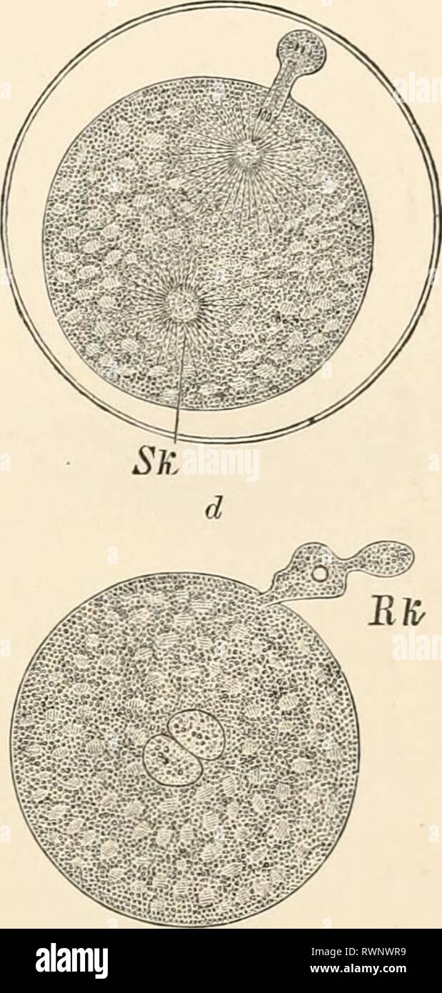 Elementary text-book of zoology (1884) Elementary text-book of zoology elementarytextbo0101clau Year: 1884 Fro. 101.—Ovum of Neplielis (after O. Hertwig). a, the ovum half-an-hour after deposition. a projection of the protoplasm indicates the commencing- formation of the first polar body ; the nuclear spindle is visible. 6, The same an hour later, with polar body extruded, and after entrance of the spermatozoon. Sk, male pronucleus. c, The same another hour later without egg membrane, and with two polar bodies and male pronucleus (Sk); d, the same an hour later with approximated female and ma Stock Photohttps://www.alamy.com/image-license-details/?v=1https://www.alamy.com/elementary-text-book-of-zoology-1884-elementary-text-book-of-zoology-elementarytextbo0101clau-year-1884-fro-101ovum-of-neplielis-after-o-hertwig-a-the-ovum-half-an-hour-after-deposition-a-projection-of-the-protoplasm-indicates-the-commencing-formation-of-the-first-polar-body-the-nuclear-spindle-is-visible-6-the-same-an-hour-later-with-polar-body-extruded-and-after-entrance-of-the-spermatozoon-sk-male-pronucleus-c-the-same-another-hour-later-without-egg-membrane-and-with-two-polar-bodies-and-male-pronucleus-sk-d-the-same-an-hour-later-with-approximated-female-and-ma-image239582429.html
RMRWNWR9–Elementary text-book of zoology (1884) Elementary text-book of zoology elementarytextbo0101clau Year: 1884 Fro. 101.—Ovum of Neplielis (after O. Hertwig). a, the ovum half-an-hour after deposition. a projection of the protoplasm indicates the commencing- formation of the first polar body ; the nuclear spindle is visible. 6, The same an hour later, with polar body extruded, and after entrance of the spermatozoon. Sk, male pronucleus. c, The same another hour later without egg membrane, and with two polar bodies and male pronucleus (Sk); d, the same an hour later with approximated female and ma
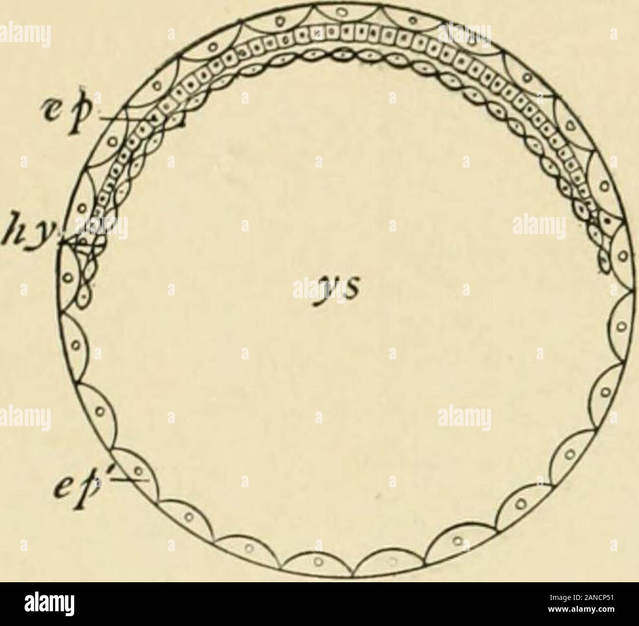 A textbook of obstetrics . t, to days after the onset of menstruation ;in the second, to days after the cessation of menstruation. The curves indicate theproportion of conceptions to copulations on each day of the menstrual month (Hensen). 72 PREGNANCY. CHANGES IN THE OVUM FOLLOWING IMPREGNATION.1 Directly after the formation of the nucleus of segmentation bythe fusion of male and female pronucleus the ovum begins to segment. The original mass di-vides itself into two cells (blasto-meres), these into four, and so onuntil the whole ovum is sur-rounded by a layer of cells inclos-ing a group of s Stock Photohttps://www.alamy.com/image-license-details/?v=1https://www.alamy.com/a-textbook-of-obstetrics-t-to-days-after-the-onset-of-menstruation-in-the-second-to-days-after-the-cessation-of-menstruation-the-curves-indicate-theproportion-of-conceptions-to-copulations-on-each-day-of-the-menstrual-month-hensen-72-pregnancy-changes-in-the-ovum-following-impregnation1-directly-after-the-formation-of-the-nucleus-of-segmentation-bythe-fusion-of-male-and-female-pronucleus-the-ovum-begins-to-segment-the-original-mass-di-vides-itself-into-two-cells-blasto-meres-these-into-four-and-so-onuntil-the-whole-ovum-is-sur-rounded-by-a-layer-of-cells-inclos-ing-a-group-of-s-image340185581.html
RM2ANCP51–A textbook of obstetrics . t, to days after the onset of menstruation ;in the second, to days after the cessation of menstruation. The curves indicate theproportion of conceptions to copulations on each day of the menstrual month (Hensen). 72 PREGNANCY. CHANGES IN THE OVUM FOLLOWING IMPREGNATION.1 Directly after the formation of the nucleus of segmentation bythe fusion of male and female pronucleus the ovum begins to segment. The original mass di-vides itself into two cells (blasto-meres), these into four, and so onuntil the whole ovum is sur-rounded by a layer of cells inclos-ing a group of s
 Elementary text-book of zoology, general Elementary text-book of zoology, general part and special part: protozoa to insecta elementarytextbo00clau Year: 1892 ^-Jm Ek Fig. 102, a, J.—Parts of the ovum of Asterias glacialis -n-ith spermatozoa, embedded in the mucilarjinous coat (after H. Fol.) c, upper part of the ovum of Potromyzon (after Calberla). Am, micropyle ; Sp, spermatozoa; Jm, path of the spermatozoon; Ek, female pronucleus; Eh, membrane of ovum ; Ehz, prominences of the snme. and a new nucleus was formed quite independently of it; and that the persistence and the participation of th Stock Photohttps://www.alamy.com/image-license-details/?v=1https://www.alamy.com/elementary-text-book-of-zoology-general-elementary-text-book-of-zoology-general-part-and-special-part-protozoa-to-insecta-elementarytextbo00clau-year-1892-jm-ek-fig-102-a-jparts-of-the-ovum-of-asterias-glacialis-n-ith-spermatozoa-embedded-in-the-mucilarjinous-coat-after-h-fol-c-upper-part-of-the-ovum-of-potromyzon-after-calberla-am-micropyle-sp-spermatozoa-jm-path-of-the-spermatozoon-ek-female-pronucleus-eh-membrane-of-ovum-ehz-prominences-of-the-snme-and-a-new-nucleus-was-formed-quite-independently-of-it-and-that-the-persistence-and-the-participation-of-th-image239582592.html
RMRWNX14–Elementary text-book of zoology, general Elementary text-book of zoology, general part and special part: protozoa to insecta elementarytextbo00clau Year: 1892 ^-Jm Ek Fig. 102, a, J.—Parts of the ovum of Asterias glacialis -n-ith spermatozoa, embedded in the mucilarjinous coat (after H. Fol.) c, upper part of the ovum of Potromyzon (after Calberla). Am, micropyle ; Sp, spermatozoa; Jm, path of the spermatozoon; Ek, female pronucleus; Eh, membrane of ovum ; Ehz, prominences of the snme. and a new nucleus was formed quite independently of it; and that the persistence and the participation of th
 A textbook of obstetrics . t, to days after the onset of menstruation ;in the second, to days after the cessation of menstruation. The curves indicate theproportion of conceptions to copulations on each day of the menstrual month (Hensen). 72 PREGNANCY. CHANGES IN THE OVUM FOLLOWING IMPREGNATION.1 Directly after the formation of the nucleus of segmentation bythe fusion of male and female pronucleus the ovum begins to segment. The original mass di-vides itself into two cells (blasto-meres), these into four, and so onuntil the whole ovum is sur-rounded by a layer of cells inclos-ing a group of s Stock Photohttps://www.alamy.com/image-license-details/?v=1https://www.alamy.com/a-textbook-of-obstetrics-t-to-days-after-the-onset-of-menstruation-in-the-second-to-days-after-the-cessation-of-menstruation-the-curves-indicate-theproportion-of-conceptions-to-copulations-on-each-day-of-the-menstrual-month-hensen-72-pregnancy-changes-in-the-ovum-following-impregnation1-directly-after-the-formation-of-the-nucleus-of-segmentation-bythe-fusion-of-male-and-female-pronucleus-the-ovum-begins-to-segment-the-original-mass-di-vides-itself-into-two-cells-blasto-meres-these-into-four-and-so-onuntil-the-whole-ovum-is-sur-rounded-by-a-layer-of-cells-inclos-ing-a-group-of-s-image340186400.html
RM2ANCR68–A textbook of obstetrics . t, to days after the onset of menstruation ;in the second, to days after the cessation of menstruation. The curves indicate theproportion of conceptions to copulations on each day of the menstrual month (Hensen). 72 PREGNANCY. CHANGES IN THE OVUM FOLLOWING IMPREGNATION.1 Directly after the formation of the nucleus of segmentation bythe fusion of male and female pronucleus the ovum begins to segment. The original mass di-vides itself into two cells (blasto-meres), these into four, and so onuntil the whole ovum is sur-rounded by a layer of cells inclos-ing a group of s
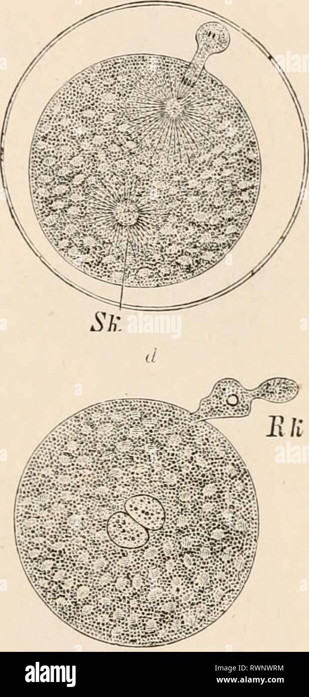 Elementary text-book of zoology, tr Elementary text-book of zoology, tr. and ed. by Adam Sedgwick, with the assistance of F. G. Heathcote elementarytextbo01clau Year: 1892-1893 FIG. 101.—Ovum of Nsphelis (after O. Hertwis). a, the ovum half-au-hour after deposition. a projection of the protoplasm indicates the commencing f jrmation of the first polar body ; the nuclear spindle is visible. 6, The same an hour later, with polar body extruded, and after entrance of the spermatozoon. Sk, male pronucleus. c, The same another hour later without egg membrane, and with two polar bodies and male pronu Stock Photohttps://www.alamy.com/image-license-details/?v=1https://www.alamy.com/elementary-text-book-of-zoology-tr-elementary-text-book-of-zoology-tr-and-ed-by-adam-sedgwick-with-the-assistance-of-f-g-heathcote-elementarytextbo01clau-year-1892-1893-fig-101ovum-of-nsphelis-after-o-hertwis-a-the-ovum-half-au-hour-after-deposition-a-projection-of-the-protoplasm-indicates-the-commencing-f-jrmation-of-the-first-polar-body-the-nuclear-spindle-is-visible-6-the-same-an-hour-later-with-polar-body-extruded-and-after-entrance-of-the-spermatozoon-sk-male-pronucleus-c-the-same-another-hour-later-without-egg-membrane-and-with-two-polar-bodies-and-male-pronu-image239582440.html
RMRWNWRM–Elementary text-book of zoology, tr Elementary text-book of zoology, tr. and ed. by Adam Sedgwick, with the assistance of F. G. Heathcote elementarytextbo01clau Year: 1892-1893 FIG. 101.—Ovum of Nsphelis (after O. Hertwis). a, the ovum half-au-hour after deposition. a projection of the protoplasm indicates the commencing f jrmation of the first polar body ; the nuclear spindle is visible. 6, The same an hour later, with polar body extruded, and after entrance of the spermatozoon. Sk, male pronucleus. c, The same another hour later without egg membrane, and with two polar bodies and male pronu
 A manual of obstetrics . ,.-M.PN. F.IN.- FiG. 22.—Fertilizalion of the ovum of a moUusk (Elysia viridis) : A, ovum sending up aprotuberance to meet the spermatozoon (5); £, approach of male pronucleus {M. PN.) tomeet the female pronucleus [F. PN.). Chang-es in the Ovum Prior to its Lodg-ement in theUterus. — An interesting series of phenomena occurwithin the ovum immediately subsequent to impregnation.Shortly after penetration of the ovum the vibratile extremityof the spermatozoid is absorbed, leaving the head only,/which is known as the malepromiclais (Fig. 22). Thisunites with the female pr Stock Photohttps://www.alamy.com/image-license-details/?v=1https://www.alamy.com/a-manual-of-obstetrics-mpn-fin-fig-22fertilizalion-of-the-ovum-of-a-mouusk-elysia-viridis-a-ovum-sending-up-aprotuberance-to-meet-the-spermatozoon-5-approach-of-male-pronucleus-m-pn-tomeet-the-female-pronucleus-f-pn-chang-es-in-the-ovum-prior-to-its-lodg-ement-in-theuterus-an-interesting-series-of-phenomena-occurwithin-the-ovum-immediately-subsequent-to-impregnationshortly-after-penetration-of-the-ovum-the-vibratile-extremityof-the-spermatozoid-is-absorbed-leaving-the-head-onlywhich-is-known-as-the-malepromiclais-fig-22-thisunites-with-the-female-pr-image338194045.html
RM2AJ61XN–A manual of obstetrics . ,.-M.PN. F.IN.- FiG. 22.—Fertilizalion of the ovum of a moUusk (Elysia viridis) : A, ovum sending up aprotuberance to meet the spermatozoon (5); £, approach of male pronucleus {M. PN.) tomeet the female pronucleus [F. PN.). Chang-es in the Ovum Prior to its Lodg-ement in theUterus. — An interesting series of phenomena occurwithin the ovum immediately subsequent to impregnation.Shortly after penetration of the ovum the vibratile extremityof the spermatozoid is absorbed, leaving the head only,/which is known as the malepromiclais (Fig. 22). Thisunites with the female pr
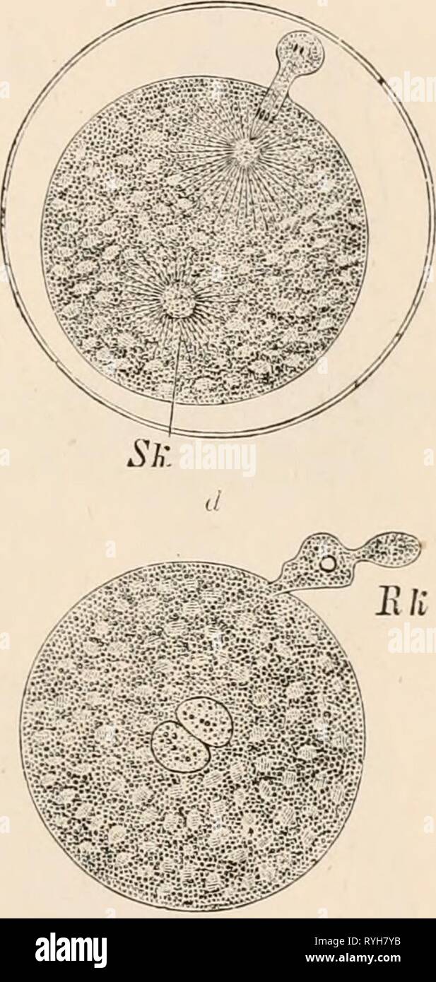 Elementary text-book of zoology, tr. and ed. by Adam Sedgwick, with the assistance of F. G. Heathcote elementarytextbo01clau Year: 1892-1893 FIG. 101.—Ovum of Nsphelis (after O. Hertwis). a, the ovum half-au-hour after deposition. a projection of the protoplasm indicates the commencing f jrmation of the first polar body ; the nuclear spindle is visible. 6, The same an hour later, with polar body extruded, and after entrance of the spermatozoon. Sk, male pronucleus. c, The same another hour later without egg membrane, and with two polar bodies and male pronucleus (Sir); d, the same an hour Stock Photohttps://www.alamy.com/image-license-details/?v=1https://www.alamy.com/elementary-text-book-of-zoology-tr-and-ed-by-adam-sedgwick-with-the-assistance-of-f-g-heathcote-elementarytextbo01clau-year-1892-1893-fig-101ovum-of-nsphelis-after-o-hertwis-a-the-ovum-half-au-hour-after-deposition-a-projection-of-the-protoplasm-indicates-the-commencing-f-jrmation-of-the-first-polar-body-the-nuclear-spindle-is-visible-6-the-same-an-hour-later-with-polar-body-extruded-and-after-entrance-of-the-spermatozoon-sk-male-pronucleus-c-the-same-another-hour-later-without-egg-membrane-and-with-two-polar-bodies-and-male-pronucleus-sir-d-the-same-an-hour-image240709935.html
RMRYH7YB–Elementary text-book of zoology, tr. and ed. by Adam Sedgwick, with the assistance of F. G. Heathcote elementarytextbo01clau Year: 1892-1893 FIG. 101.—Ovum of Nsphelis (after O. Hertwis). a, the ovum half-au-hour after deposition. a projection of the protoplasm indicates the commencing f jrmation of the first polar body ; the nuclear spindle is visible. 6, The same an hour later, with polar body extruded, and after entrance of the spermatozoon. Sk, male pronucleus. c, The same another hour later without egg membrane, and with two polar bodies and male pronucleus (Sir); d, the same an hour
 A textbook of obstetrics . Fig- 59-—A, Fertilized ova of echinus : The male a, and the female pronucleus,b, are approaching; in B, they have almost fused ; C, ovum of echinus after com-pletion of fertilization ; s.n., segmentation-nucleus (Hertwig). The Average Date of Conception after Marriage Nor-mally, impregnation should succeed the first menstruationfollowing marriage, but marriages are only called sterile aftereighteen months have elapsed without conception. Pregnancyis possible, however, after years of sterility. I have had undermy care women who conceived for the first time nine, thirt Stock Photohttps://www.alamy.com/image-license-details/?v=1https://www.alamy.com/a-textbook-of-obstetrics-fig-59-a-fertilized-ova-of-echinus-the-male-a-and-the-female-pronucleusb-are-approaching-in-b-they-have-almost-fused-c-ovum-of-echinus-after-com-pletion-of-fertilization-sn-segmentation-nucleus-hertwig-the-average-date-of-conception-after-marriage-nor-mally-impregnation-should-succeed-the-first-menstruationfollowing-marriage-but-marriages-are-only-called-sterile-aftereighteen-months-have-elapsed-without-conception-pregnancyis-possible-however-after-years-of-sterility-i-have-had-undermy-care-women-who-conceived-for-the-first-time-nine-thirt-image340186898.html
RM2ANCRT2–A textbook of obstetrics . Fig- 59-—A, Fertilized ova of echinus : The male a, and the female pronucleus,b, are approaching; in B, they have almost fused ; C, ovum of echinus after com-pletion of fertilization ; s.n., segmentation-nucleus (Hertwig). The Average Date of Conception after Marriage Nor-mally, impregnation should succeed the first menstruationfollowing marriage, but marriages are only called sterile aftereighteen months have elapsed without conception. Pregnancyis possible, however, after years of sterility. I have had undermy care women who conceived for the first time nine, thirt
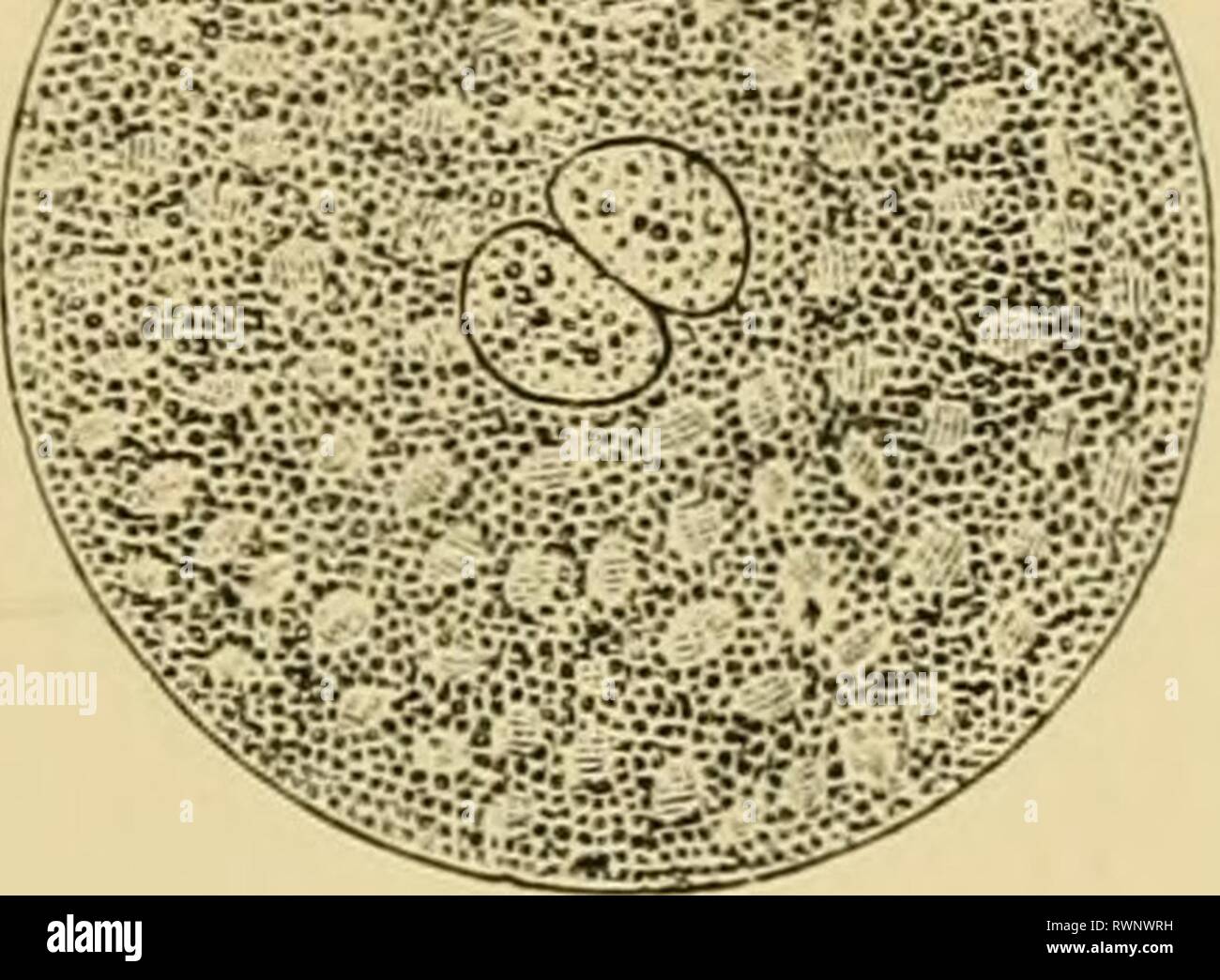 Elementary text-book of zoology, general Elementary text-book of zoology, general part and special part: protozoa to insecta elementarytextbo00clau Year: 1892 Fig. 101.—0 im of N'ophelis (ifter O Ucrtwir^). a, tlie ovum half-an-hour after deposition. a projecti juot tlie protoplasm iiidic ites the commencing f jrmation of the first polar body ; the nuclca. „p.„Jl ....Lle. I, TL., same an hour later, with polar body extruded, and after entrance of the spermatozoon. Sh, male pronucleus, e. The same another hour later without egg membrane, and with two polar bodies and male pronucleus (Si-); d Stock Photohttps://www.alamy.com/image-license-details/?v=1https://www.alamy.com/elementary-text-book-of-zoology-general-elementary-text-book-of-zoology-general-part-and-special-part-protozoa-to-insecta-elementarytextbo00clau-year-1892-fig-1010-im-of-nophelis-ifter-o-ucrtwir-a-tlie-ovum-half-an-hour-after-deposition-a-projecti-juot-tlie-protoplasm-iiidic-ites-the-commencing-f-jrmation-of-the-first-polar-body-the-nuclca-pjl-lle-i-tl-same-an-hour-later-with-polar-body-extruded-and-after-entrance-of-the-spermatozoon-sh-male-pronucleus-e-the-same-another-hour-later-without-egg-membrane-and-with-two-polar-bodies-and-male-pronucleus-si-d-image239582437.html
RMRWNWRH–Elementary text-book of zoology, general Elementary text-book of zoology, general part and special part: protozoa to insecta elementarytextbo00clau Year: 1892 Fig. 101.—0 im of N'ophelis (ifter O Ucrtwir^). a, tlie ovum half-an-hour after deposition. a projecti juot tlie protoplasm iiidic ites the commencing f jrmation of the first polar body ; the nuclca. „p.„Jl ....Lle. I, TL., same an hour later, with polar body extruded, and after entrance of the spermatozoon. Sh, male pronucleus, e. The same another hour later without egg membrane, and with two polar bodies and male pronucleus (Si-); d
 . The cell; outlines of general anatomy and physiology. Fig. 16.—An egg of Ascaris megalocephala,which has just been fertilised (after Van Bene-den; from O. Hevtwig, Fig. 22): sfc spermato-zoon, with its nucleus which has just entered ;/ glistening fatty material of spermatozoon;leb female pronucleus. Fig. 17. — Goblet-cell from thebladder epithelium of Squatlna vul-garis, hardened in Midlers fluid.(Afcer List, Plate I., Fig. 9.) from time to time by the cell, through a small opening at its freeend, and transformed into mucin. The protoplasm traverses themass of secretion in the form of fine t Stock Photohttps://www.alamy.com/image-license-details/?v=1https://www.alamy.com/the-cell-outlines-of-general-anatomy-and-physiology-fig-16an-egg-of-ascaris-megalocephalawhich-has-just-been-fertilised-after-van-bene-den-from-o-hevtwig-fig-22-sfc-spermato-zoon-with-its-nucleus-which-has-just-entered-glistening-fatty-material-of-spermatozoonleb-female-pronucleus-fig-17-goblet-cell-from-thebladder-epithelium-of-squatlna-vul-garis-hardened-in-midlers-fluidafcer-list-plate-i-fig-9-from-time-to-time-by-the-cell-through-a-small-opening-at-its-freeend-and-transformed-into-mucin-the-protoplasm-traverses-themass-of-secretion-in-the-form-of-fine-t-image336861101.html
RM2AG19NH–. The cell; outlines of general anatomy and physiology. Fig. 16.—An egg of Ascaris megalocephala,which has just been fertilised (after Van Bene-den; from O. Hevtwig, Fig. 22): sfc spermato-zoon, with its nucleus which has just entered ;/ glistening fatty material of spermatozoon;leb female pronucleus. Fig. 17. — Goblet-cell from thebladder epithelium of Squatlna vul-garis, hardened in Midlers fluid.(Afcer List, Plate I., Fig. 9.) from time to time by the cell, through a small opening at its freeend, and transformed into mucin. The protoplasm traverses themass of secretion in the form of fine t
 Traité d'anatomie humaine : anatomie descriptive, histologie, développement . certains végétaux. Il sagit du rôle que lescentrosomes des cellules sexuelles jouent dans la fécondation, rôle déjà soupçonnépar Fle.mming, Vejdowsky, Rabl, Boveri, mais élucidé par Fol. Voici ce qui sepasse. Le pronucléus femelle est accompagné dun centrosome que nous appel-lerons avec lauteur Vovocentre; le pronocléus mâle amène aussi avec lui uncentrosome propre, le spermocentre. Au moment de la conjugaison des pronucléi,ces centrosomes se divisent chacun en deux, de sorte quil y a deux demi-ovo-centres et deux de Stock Photohttps://www.alamy.com/image-license-details/?v=1https://www.alamy.com/trait-danatomie-humaine-anatomie-descriptive-histologie-dveloppement-certains-vgtaux-il-sagit-du-rle-que-lescentrosomes-des-cellules-sexuelles-jouent-dans-la-fcondation-rle-dj-souponnpar-flemming-vejdowsky-rabl-boveri-mais-lucid-par-fol-voici-ce-qui-sepasse-le-pronuclus-femelle-est-accompagn-dun-centrosome-que-nous-appel-lerons-avec-lauteur-vovocentre-le-pronoclus-mle-amne-aussi-avec-lui-uncentrosome-propre-le-spermocentre-au-moment-de-la-conjugaison-des-pronuclices-centrosomes-se-divisent-chacun-en-deux-de-sorte-quil-y-a-deux-demi-ovo-centres-et-deux-de-image338490141.html
RM2AJKFHH–Traité d'anatomie humaine : anatomie descriptive, histologie, développement . certains végétaux. Il sagit du rôle que lescentrosomes des cellules sexuelles jouent dans la fécondation, rôle déjà soupçonnépar Fle.mming, Vejdowsky, Rabl, Boveri, mais élucidé par Fol. Voici ce qui sepasse. Le pronucléus femelle est accompagné dun centrosome que nous appel-lerons avec lauteur Vovocentre; le pronocléus mâle amène aussi avec lui uncentrosome propre, le spermocentre. Au moment de la conjugaison des pronucléi,ces centrosomes se divisent chacun en deux, de sorte quil y a deux demi-ovo-centres et deux de
 . An American text-book of obstetrics. For practitioners and students. Fig. Of).—A, fertilized ova of echinus (Hertwig): the male (a) and the female pronucleus (6) areapproaching; in B they have almost fused; C, ovum of echinus after completion of fertilization (Hert-wig): s.n., segmentation-nucleus. , which the original egg-cell gives rise to an extended series of generations,leading to the production of the blastoderm. Since the youngest human embryo carefully examined and recorded—thatof Reichert—was already probably twelve days old, the early phenomena ofimpregnation and segmentation have Stock Photohttps://www.alamy.com/image-license-details/?v=1https://www.alamy.com/an-american-text-book-of-obstetrics-for-practitioners-and-students-fig-ofa-fertilized-ova-of-echinus-hertwig-the-male-a-and-the-female-pronucleus-6-areapproaching-in-b-they-have-almost-fused-c-ovum-of-echinus-after-completion-of-fertilization-hert-wig-sn-segmentation-nucleus-which-the-original-egg-cell-gives-rise-to-an-extended-series-of-generationsleading-to-the-production-of-the-blastoderm-since-the-youngest-human-embryo-carefully-examined-and-recordedthatof-reichertwas-already-probably-twelve-days-old-the-early-phenomena-ofimpregnation-and-segmentation-have-image370599873.html
RM2CEX7WN–. An American text-book of obstetrics. For practitioners and students. Fig. Of).—A, fertilized ova of echinus (Hertwig): the male (a) and the female pronucleus (6) areapproaching; in B they have almost fused; C, ovum of echinus after completion of fertilization (Hert-wig): s.n., segmentation-nucleus. , which the original egg-cell gives rise to an extended series of generations,leading to the production of the blastoderm. Since the youngest human embryo carefully examined and recorded—thatof Reichert—was already probably twelve days old, the early phenomena ofimpregnation and segmentation have
 . The science and art of midwifery . earmatter from the vitellus collects. A separation of the lines connectingthe poles next takes place, and two new nuclei surrounded by radiatemasses of yelk matter result. These have a star-like arrangement.The upper pole is then extruded and the first polar globule is formed.The process is then repeated, and the second polar globule is perfected.Finally the persistent portion of the original nucleus recedes from thesurface. It resembles in appearance the original germinal vesicle withits nucleolus, and is known as foe female pronucleus. The formation of th Stock Photohttps://www.alamy.com/image-license-details/?v=1https://www.alamy.com/the-science-and-art-of-midwifery-earmatter-from-the-vitellus-collects-a-separation-of-the-lines-connectingthe-poles-next-takes-place-and-two-new-nuclei-surrounded-by-radiatemasses-of-yelk-matter-result-these-have-a-star-like-arrangementthe-upper-pole-is-then-extruded-and-the-first-polar-globule-is-formedthe-process-is-then-repeated-and-the-second-polar-globule-is-perfectedfinally-the-persistent-portion-of-the-original-nucleus-recedes-from-thesurface-it-resembles-in-appearance-the-original-germinal-vesicle-withits-nucleolus-and-is-known-as-foe-female-pronucleus-the-formation-of-th-image372588319.html
RM2CJ4T5K–. The science and art of midwifery . earmatter from the vitellus collects. A separation of the lines connectingthe poles next takes place, and two new nuclei surrounded by radiatemasses of yelk matter result. These have a star-like arrangement.The upper pole is then extruded and the first polar globule is formed.The process is then repeated, and the second polar globule is perfected.Finally the persistent portion of the original nucleus recedes from thesurface. It resembles in appearance the original germinal vesicle withits nucleolus, and is known as foe female pronucleus. The formation of th
 . Journal of morphology . are in a medium where any of them that becomes the successful malepronucleus will divide after union with the female pronucleus.Therefore, the supernumerary sperm nuclei in the egg cytoplasmmay divide. The accessory cleavage of the selachians and birdsQgg is certainly comparable to the division of fertilized enucleatedfragments of the sea urchins egg or nemertian egg. Inwandering Follicular Cells.—Harper (04) illustrates aninwandering follicular cell. (Harper, 04, PI. II, Eig. Y i). Ihave found a large number of cells in the perivitelline fluid in theegg represented i Stock Photohttps://www.alamy.com/image-license-details/?v=1https://www.alamy.com/journal-of-morphology-are-in-a-medium-where-any-of-them-that-becomes-the-successful-malepronucleus-will-divide-after-union-with-the-female-pronucleustherefore-the-supernumerary-sperm-nuclei-in-the-egg-cytoplasmmay-divide-the-accessory-cleavage-of-the-selachians-and-birdsqgg-is-certainly-comparable-to-the-division-of-fertilized-enucleatedfragments-of-the-sea-urchins-egg-or-nemertian-egg-inwandering-follicular-cellsharper-04-illustrates-aninwandering-follicular-cell-harper-04-pi-ii-eig-y-i-ihave-found-a-large-number-of-cells-in-the-perivitelline-fluid-in-theegg-represented-i-image369780180.html
RM2CDGXB0–. Journal of morphology . are in a medium where any of them that becomes the successful malepronucleus will divide after union with the female pronucleus.Therefore, the supernumerary sperm nuclei in the egg cytoplasmmay divide. The accessory cleavage of the selachians and birdsQgg is certainly comparable to the division of fertilized enucleatedfragments of the sea urchins egg or nemertian egg. Inwandering Follicular Cells.—Harper (04) illustrates aninwandering follicular cell. (Harper, 04, PI. II, Eig. Y i). Ihave found a large number of cells in the perivitelline fluid in theegg represented i
 . A text-book of comparative physiology for students and practitioners of comparative (veterinary) medicine . ria? round female pronucleus, asseen in the living egg (E. F, H, and I from picric acid preparations); L, expulsionof the first polar cell. (Haddon.) then, are simply expelled ; they take no part in the developmentof the ovum ; and their extrusion is to be regarded as a prepar-ation for the progress of the cell, whether this event follows orprecedes the entrance of the male cell into the ovum. It is wor-thy of note that the ovum may become amceboid in the regionfrom which the polar glo Stock Photohttps://www.alamy.com/image-license-details/?v=1https://www.alamy.com/a-text-book-of-comparative-physiology-for-students-and-practitioners-of-comparative-veterinary-medicine-ria-round-female-pronucleus-asseen-in-the-living-egg-e-f-h-and-i-from-picric-acid-preparations-l-expulsionof-the-first-polar-cell-haddon-then-are-simply-expelled-they-take-no-part-in-the-developmentof-the-ovum-and-their-extrusion-is-to-be-regarded-as-a-prepar-ation-for-the-progress-of-the-cell-whether-this-event-follows-orprecedes-the-entrance-of-the-male-cell-into-the-ovum-it-is-wor-thy-of-note-that-the-ovum-may-become-amceboid-in-the-regionfrom-which-the-polar-glo-image372620407.html
RM2CJ693K–. A text-book of comparative physiology for students and practitioners of comparative (veterinary) medicine . ria? round female pronucleus, asseen in the living egg (E. F, H, and I from picric acid preparations); L, expulsionof the first polar cell. (Haddon.) then, are simply expelled ; they take no part in the developmentof the ovum ; and their extrusion is to be regarded as a prepar-ation for the progress of the cell, whether this event follows orprecedes the entrance of the male cell into the ovum. It is wor-thy of note that the ovum may become amceboid in the regionfrom which the polar glo
 . An American text-book of obstetrics. For practitioners and students. £ ??,-/•?? L-,i--.VAv. Fig. Of).—A, fertilized ova of echinus (Hertwig): the male (a) and the female pronucleus (6) areapproaching; in B they have almost fused; C, ovum of echinus after completion of fertilization (Hert-wig): s.n., segmentation-nucleus. , which the original egg-cell gives rise to an extended series of generations,leading to the production of the blastoderm. Since the youngest human embryo carefully examined and recorded—thatof Reichert—was already probably twelve days old, the early phenomena ofimpregnatio Stock Photohttps://www.alamy.com/image-license-details/?v=1https://www.alamy.com/an-american-text-book-of-obstetrics-for-practitioners-and-students-l-i-vav-fig-ofa-fertilized-ova-of-echinus-hertwig-the-male-a-and-the-female-pronucleus-6-areapproaching-in-b-they-have-almost-fused-c-ovum-of-echinus-after-completion-of-fertilization-hert-wig-sn-segmentation-nucleus-which-the-original-egg-cell-gives-rise-to-an-extended-series-of-generationsleading-to-the-production-of-the-blastoderm-since-the-youngest-human-embryo-carefully-examined-and-recordedthatof-reichertwas-already-probably-twelve-days-old-the-early-phenomena-ofimpregnatio-image370599926.html
RM2CEX7YJ–. An American text-book of obstetrics. For practitioners and students. £ ??,-/•?? L-,i--.VAv. Fig. Of).—A, fertilized ova of echinus (Hertwig): the male (a) and the female pronucleus (6) areapproaching; in B they have almost fused; C, ovum of echinus after completion of fertilization (Hert-wig): s.n., segmentation-nucleus. , which the original egg-cell gives rise to an extended series of generations,leading to the production of the blastoderm. Since the youngest human embryo carefully examined and recorded—thatof Reichert—was already probably twelve days old, the early phenomena ofimpregnatio
 . The science and art of midwifery . the female pro-nucleus. F. P-V, female pronucleus ; M. PX. male pronucleus : >. BpermatozoOn. Almost immediately after the production of the segmentation nucleusit divides into two nuclei. By a similar process of cleavage the vitellus likewise divides into two halves. The nuclei acl as central points,around which collect the molecular and viscid portion- of the pro-toplasm. In this manner the ovum is divided into two new cells, whichdiffer somewhat in size, and which lie near together within the zona •Haeckel, Anthropogenic, Leipsic, 1874, pp. 100 etseq. Stock Photohttps://www.alamy.com/image-license-details/?v=1https://www.alamy.com/the-science-and-art-of-midwifery-the-female-pro-nucleus-f-p-v-female-pronucleus-m-px-male-pronucleus-gt-bpermatozoon-almost-immediately-after-the-production-of-the-segmentation-nucleusit-divides-into-two-nuclei-by-a-similar-process-of-cleavage-the-vitellus-likewise-divides-into-two-halves-the-nuclei-acl-as-central-pointsaround-which-collect-the-molecular-and-viscid-portion-of-the-pro-toplasm-in-this-manner-the-ovum-is-divided-into-two-new-cells-whichdiffer-somewhat-in-size-and-which-lie-near-together-within-the-zona-haeckel-anthropogenic-leipsic-1874-pp-100-etseq-image372587407.html
RM2CJ4R13–. The science and art of midwifery . the female pro-nucleus. F. P-V, female pronucleus ; M. PX. male pronucleus : >. BpermatozoOn. Almost immediately after the production of the segmentation nucleusit divides into two nuclei. By a similar process of cleavage the vitellus likewise divides into two halves. The nuclei acl as central points,around which collect the molecular and viscid portion- of the pro-toplasm. In this manner the ovum is divided into two new cells, whichdiffer somewhat in size, and which lie near together within the zona •Haeckel, Anthropogenic, Leipsic, 1874, pp. 100 etseq.
 . A text-book of comparative physiology for students and practitioners of comparative (veterinary) medicine . F.PKt. -M.PN. Fig. 6:2.—Fertilization of ovum of a mollusk (Elysia viridis). A. Ovum sending up aprotuberance to meet the spermatozoon. B. Approach of male pronucleus tomeet the female pronucleus. F. FN, female pronucleus; M. FN, male pronucleus.S. spermatozoon. ovum undergoes changes similar to those that the nucleus of theovum underwent, and thus becomes fitted for its special func-tions as a fertilizer ; or perhaps it would be more correct to saythat these altered masses of nuclear Stock Photohttps://www.alamy.com/image-license-details/?v=1https://www.alamy.com/a-text-book-of-comparative-physiology-for-students-and-practitioners-of-comparative-veterinary-medicine-fpkt-mpn-fig-62fertilization-of-ovum-of-a-mollusk-elysia-viridis-a-ovum-sending-up-aprotuberance-to-meet-the-spermatozoon-b-approach-of-male-pronucleus-tomeet-the-female-pronucleus-f-fn-female-pronucleus-m-fn-male-pronucleuss-spermatozoon-ovum-undergoes-changes-similar-to-those-that-the-nucleus-of-theovum-underwent-and-thus-becomes-fitted-for-its-special-func-tions-as-a-fertilizer-or-perhaps-it-would-be-more-correct-to-saythat-these-altered-masses-of-nuclear-image372618575.html
RM2CJ66P7–. A text-book of comparative physiology for students and practitioners of comparative (veterinary) medicine . F.PKt. -M.PN. Fig. 6:2.—Fertilization of ovum of a mollusk (Elysia viridis). A. Ovum sending up aprotuberance to meet the spermatozoon. B. Approach of male pronucleus tomeet the female pronucleus. F. FN, female pronucleus; M. FN, male pronucleus.S. spermatozoon. ovum undergoes changes similar to those that the nucleus of theovum underwent, and thus becomes fitted for its special func-tions as a fertilizer ; or perhaps it would be more correct to saythat these altered masses of nuclear
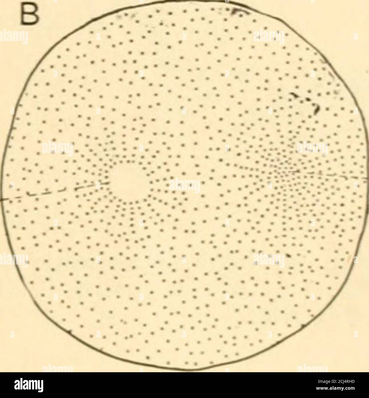 . The science and art of midwifery . F.PXr. M.PX. Fig. 33.—Fertilization of ovum of a mollusk (Elysia viridia). A, ovum sending up a prntuber-ancetomeet the spermatozoon. B. approach of male pronucleus to meet the female pro-nucleus. F. P-V, female pronucleus ; M. PX. male pronucleus : >. BpermatozoOn. Almost immediately after the production of the segmentation nucleusit divides into two nuclei. By a similar process of cleavage the vitellus likewise divides into two halves. The nuclei acl as central points,around which collect the molecular and viscid portion- of the pro-toplasm. In this ma Stock Photohttps://www.alamy.com/image-license-details/?v=1https://www.alamy.com/the-science-and-art-of-midwifery-fpxr-mpx-fig-33fertilization-of-ovum-of-a-mollusk-elysia-viridia-a-ovum-sending-up-a-prntuber-ancetomeet-the-spermatozoon-b-approach-of-male-pronucleus-to-meet-the-female-pro-nucleus-f-p-v-female-pronucleus-m-px-male-pronucleus-gt-bpermatozoon-almost-immediately-after-the-production-of-the-segmentation-nucleusit-divides-into-two-nuclei-by-a-similar-process-of-cleavage-the-vitellus-likewise-divides-into-two-halves-the-nuclei-acl-as-central-pointsaround-which-collect-the-molecular-and-viscid-portion-of-the-pro-toplasm-in-this-ma-image372587865.html
RM2CJ4RHD–. The science and art of midwifery . F.PXr. M.PX. Fig. 33.—Fertilization of ovum of a mollusk (Elysia viridia). A, ovum sending up a prntuber-ancetomeet the spermatozoon. B. approach of male pronucleus to meet the female pro-nucleus. F. P-V, female pronucleus ; M. PX. male pronucleus : >. BpermatozoOn. Almost immediately after the production of the segmentation nucleusit divides into two nuclei. By a similar process of cleavage the vitellus likewise divides into two halves. The nuclei acl as central points,around which collect the molecular and viscid portion- of the pro-toplasm. In this ma
 . Journal of morphology . ^U taken from the oviduct at (?$ CO nP —Transverse section of a pigeons egg m., about 3l^ hours after fertilization. Reference marks are the for Fig. 3. Leitz, 4/2. Tube length, 140 mm.. Fig. 3.—Diagram of Fig. 2. A central transverse section of pigeonsegg obtained at 11 :?>0 p. m., SV2 hours after fertilization. The nuclei arereconstructed from seven successive sections. 1 pb, first polar body; 2 pb, second polar body; ^, male pronucleus;9 , female pronucleus; sn, supernumerary sperm nuclei; mp, marginal peri-blast ; cp., central periblast; wy, white yolk; n P, nu Stock Photohttps://www.alamy.com/image-license-details/?v=1https://www.alamy.com/journal-of-morphology-u-taken-from-the-oviduct-at-co-np-transverse-section-of-a-pigeons-egg-m-about-3l-hours-after-fertilization-reference-marks-are-the-for-fig-3-leitz-42-tube-length-140-mm-fig-3diagram-of-fig-2-a-central-transverse-section-of-pigeonsegg-obtained-at-11-gt0-p-m-sv2-hours-after-fertilization-the-nuclei-arereconstructed-from-seven-successive-sections-1-pb-first-polar-body-2-pb-second-polar-body-male-pronucleus9-female-pronucleus-sn-supernumerary-sperm-nuclei-mp-marginal-peri-blast-cp-central-periblast-wy-white-yolk-n-p-nu-image369781930.html
RM2CDH0HE–. Journal of morphology . ^U taken from the oviduct at (?$ CO nP —Transverse section of a pigeons egg m., about 3l^ hours after fertilization. Reference marks are the for Fig. 3. Leitz, 4/2. Tube length, 140 mm.. Fig. 3.—Diagram of Fig. 2. A central transverse section of pigeonsegg obtained at 11 :?>0 p. m., SV2 hours after fertilization. The nuclei arereconstructed from seven successive sections. 1 pb, first polar body; 2 pb, second polar body; ^, male pronucleus;9 , female pronucleus; sn, supernumerary sperm nuclei; mp, marginal peri-blast ; cp., central periblast; wy, white yolk; n P, nu
 . The physiology of reproduction. Reproduction. J88 THE PHYSIOLOGY OF REPRODUCTION centre of the cell, where it unites with the male pronucleus which generally becomes somewhat enlarged. The middle- piece of the spermatozoon also enters the egg, and, according to Boveri,' induces the formation of a centrosome, which, after the completion of fertihsation, initiates the process of cell division.. Please note that these images are extracted from scanned page images that may have been digitally enhanced for readability - coloration and appearance of these illustrations may not perfectly resemble t Stock Photohttps://www.alamy.com/image-license-details/?v=1https://www.alamy.com/the-physiology-of-reproduction-reproduction-j88-the-physiology-of-reproduction-centre-of-the-cell-where-it-unites-with-the-male-pronucleus-which-generally-becomes-somewhat-enlarged-the-middle-piece-of-the-spermatozoon-also-enters-the-egg-and-according-to-boveri-induces-the-formation-of-a-centrosome-which-after-the-completion-of-fertihsation-initiates-the-process-of-cell-division-please-note-that-these-images-are-extracted-from-scanned-page-images-that-may-have-been-digitally-enhanced-for-readability-coloration-and-appearance-of-these-illustrations-may-not-perfectly-resemble-t-image232353814.html
RMRE0HJE–. The physiology of reproduction. Reproduction. J88 THE PHYSIOLOGY OF REPRODUCTION centre of the cell, where it unites with the male pronucleus which generally becomes somewhat enlarged. The middle- piece of the spermatozoon also enters the egg, and, according to Boveri,' induces the formation of a centrosome, which, after the completion of fertihsation, initiates the process of cell division.. Please note that these images are extracted from scanned page images that may have been digitally enhanced for readability - coloration and appearance of these illustrations may not perfectly resemble t
 . The beginnings of embryonic development : A symposium organized by the Section on Zoological Sciences of the American Association for the Advancement of Science, cosponsored by the American Society of Zoologists and the Association of Southeastern Biologists, and presented at the Atlanta meeting, December 27, 1955. Embryology. 184 NUCLEOCYTOPLASMIC RELATIONS Boveri (1915) studied them and concluded that the male parts were inherited from the mother and the female parts from both parents. He suggested that the sperm had not united with the egg pronucleus but with one of the cleavage nuclei (F Stock Photohttps://www.alamy.com/image-license-details/?v=1https://www.alamy.com/the-beginnings-of-embryonic-development-a-symposium-organized-by-the-section-on-zoological-sciences-of-the-american-association-for-the-advancement-of-science-cosponsored-by-the-american-society-of-zoologists-and-the-association-of-southeastern-biologists-and-presented-at-the-atlanta-meeting-december-27-1955-embryology-184-nucleocytoplasmic-relations-boveri-1915-studied-them-and-concluded-that-the-male-parts-were-inherited-from-the-mother-and-the-female-parts-from-both-parents-he-suggested-that-the-sperm-had-not-united-with-the-egg-pronucleus-but-with-one-of-the-cleavage-nuclei-f-image234793259.html
RMRHYN5F–. The beginnings of embryonic development : A symposium organized by the Section on Zoological Sciences of the American Association for the Advancement of Science, cosponsored by the American Society of Zoologists and the Association of Southeastern Biologists, and presented at the Atlanta meeting, December 27, 1955. Embryology. 184 NUCLEOCYTOPLASMIC RELATIONS Boveri (1915) studied them and concluded that the male parts were inherited from the mother and the female parts from both parents. He suggested that the sperm had not united with the egg pronucleus but with one of the cleavage nuclei (F
 . The Biological bulletin. Biology; Zoology; Biology; Marine Biology. TEXT FIG. 1. Relative positions of the polar bodies and the female pronucleus in twelve eggs selected at random from those showing the polar bodies at the periphery of the egg as seen in surface view. sufficient to influence the position of the nucleus apparently is attained only by the use of the centrifuge. The results of these observations demonstrate that at that stage in the unfertilized ovum of Arbacia punctulata which follows closely the second meiotic division, the female pronucleus is located in the more superficial Stock Photohttps://www.alamy.com/image-license-details/?v=1https://www.alamy.com/the-biological-bulletin-biology-zoology-biology-marine-biology-text-fig-1-relative-positions-of-the-polar-bodies-and-the-female-pronucleus-in-twelve-eggs-selected-at-random-from-those-showing-the-polar-bodies-at-the-periphery-of-the-egg-as-seen-in-surface-view-sufficient-to-influence-the-position-of-the-nucleus-apparently-is-attained-only-by-the-use-of-the-centrifuge-the-results-of-these-observations-demonstrate-that-at-that-stage-in-the-unfertilized-ovum-of-arbacia-punctulata-which-follows-closely-the-second-meiotic-division-the-female-pronucleus-is-located-in-the-more-superficial-image234671129.html
RMRHP5BN–. The Biological bulletin. Biology; Zoology; Biology; Marine Biology. TEXT FIG. 1. Relative positions of the polar bodies and the female pronucleus in twelve eggs selected at random from those showing the polar bodies at the periphery of the egg as seen in surface view. sufficient to influence the position of the nucleus apparently is attained only by the use of the centrifuge. The results of these observations demonstrate that at that stage in the unfertilized ovum of Arbacia punctulata which follows closely the second meiotic division, the female pronucleus is located in the more superficial
![. Embryology of insects and myriapods; the developmental history of insects, centipedes, and millepedes from egg desposition [!] to hatching. Embryology -- Insects; Embryology -- Myriapoda. Fig. 234.—Brachyrhinus. Section through oosome. (pr) Periplasm. mass, sometimes saucer shaped, can be seen lying partly in the periplasm and partly in the yolk. This is the oosome, or germinal cytoplasm. The vitelline membrane is difficult to make out, and the chorion is so thin and elastic that it can easily be removed after being pricked with a needle. The location of the pronucleus at the time of oviposi Stock Photo . Embryology of insects and myriapods; the developmental history of insects, centipedes, and millepedes from egg desposition [!] to hatching. Embryology -- Insects; Embryology -- Myriapoda. Fig. 234.—Brachyrhinus. Section through oosome. (pr) Periplasm. mass, sometimes saucer shaped, can be seen lying partly in the periplasm and partly in the yolk. This is the oosome, or germinal cytoplasm. The vitelline membrane is difficult to make out, and the chorion is so thin and elastic that it can easily be removed after being pricked with a needle. The location of the pronucleus at the time of oviposi Stock Photo](https://c8.alamy.com/comp/REYJ9F/embryology-of-insects-and-myriapods-the-developmental-history-of-insects-centipedes-and-millepedes-from-egg-desposition-!-to-hatching-embryology-insects-embryology-myriapoda-fig-234brachyrhinus-section-through-oosome-pr-periplasm-mass-sometimes-saucer-shaped-can-be-seen-lying-partly-in-the-periplasm-and-partly-in-the-yolk-this-is-the-oosome-or-germinal-cytoplasm-the-vitelline-membrane-is-difficult-to-make-out-and-the-chorion-is-so-thin-and-elastic-that-it-can-easily-be-removed-after-being-pricked-with-a-needle-the-location-of-the-pronucleus-at-the-time-of-oviposi-REYJ9F.jpg) . Embryology of insects and myriapods; the developmental history of insects, centipedes, and millepedes from egg desposition [!] to hatching. Embryology -- Insects; Embryology -- Myriapoda. Fig. 234.—Brachyrhinus. Section through oosome. (pr) Periplasm. mass, sometimes saucer shaped, can be seen lying partly in the periplasm and partly in the yolk. This is the oosome, or germinal cytoplasm. The vitelline membrane is difficult to make out, and the chorion is so thin and elastic that it can easily be removed after being pricked with a needle. The location of the pronucleus at the time of oviposi Stock Photohttps://www.alamy.com/image-license-details/?v=1https://www.alamy.com/embryology-of-insects-and-myriapods-the-developmental-history-of-insects-centipedes-and-millepedes-from-egg-desposition-!-to-hatching-embryology-insects-embryology-myriapoda-fig-234brachyrhinus-section-through-oosome-pr-periplasm-mass-sometimes-saucer-shaped-can-be-seen-lying-partly-in-the-periplasm-and-partly-in-the-yolk-this-is-the-oosome-or-germinal-cytoplasm-the-vitelline-membrane-is-difficult-to-make-out-and-the-chorion-is-so-thin-and-elastic-that-it-can-easily-be-removed-after-being-pricked-with-a-needle-the-location-of-the-pronucleus-at-the-time-of-oviposi-image232947051.html
. Embryology of insects and myriapods; the developmental history of insects, centipedes, and millepedes from egg desposition [!] to hatching. Embryology -- Insects; Embryology -- Myriapoda. Fig. 234.—Brachyrhinus. Section through oosome. (pr) Periplasm. mass, sometimes saucer shaped, can be seen lying partly in the periplasm and partly in the yolk. This is the oosome, or germinal cytoplasm. The vitelline membrane is difficult to make out, and the chorion is so thin and elastic that it can easily be removed after being pricked with a needle. The location of the pronucleus at the time of oviposi Stock Photohttps://www.alamy.com/image-license-details/?v=1https://www.alamy.com/embryology-of-insects-and-myriapods-the-developmental-history-of-insects-centipedes-and-millepedes-from-egg-desposition-!-to-hatching-embryology-insects-embryology-myriapoda-fig-234brachyrhinus-section-through-oosome-pr-periplasm-mass-sometimes-saucer-shaped-can-be-seen-lying-partly-in-the-periplasm-and-partly-in-the-yolk-this-is-the-oosome-or-germinal-cytoplasm-the-vitelline-membrane-is-difficult-to-make-out-and-the-chorion-is-so-thin-and-elastic-that-it-can-easily-be-removed-after-being-pricked-with-a-needle-the-location-of-the-pronucleus-at-the-time-of-oviposi-image232947051.htmlRMREYJ9F–. Embryology of insects and myriapods; the developmental history of insects, centipedes, and millepedes from egg desposition [!] to hatching. Embryology -- Insects; Embryology -- Myriapoda. Fig. 234.—Brachyrhinus. Section through oosome. (pr) Periplasm. mass, sometimes saucer shaped, can be seen lying partly in the periplasm and partly in the yolk. This is the oosome, or germinal cytoplasm. The vitelline membrane is difficult to make out, and the chorion is so thin and elastic that it can easily be removed after being pricked with a needle. The location of the pronucleus at the time of oviposi
 . A text-book of comparative physiology for students and practitioners of comparative (veterinary) medicine. Physiology, Comparative. REPRODUCTION. 63 of the male cell. Once more we are led to see the importance of this structure in the life of the cell. Fertilization of the Ovum.—The spermatozoon, lashing its way along, when it meets the ovum, enters it either through a special minute gateway (micropyle), or, if this be not present- as it is not in the ova of all animals—actually penetrates the membranes and substance of the female cell, and continues act- ive till the female pronucleus is re Stock Photohttps://www.alamy.com/image-license-details/?v=1https://www.alamy.com/a-text-book-of-comparative-physiology-for-students-and-practitioners-of-comparative-veterinary-medicine-physiology-comparative-reproduction-63-of-the-male-cell-once-more-we-are-led-to-see-the-importance-of-this-structure-in-the-life-of-the-cell-fertilization-of-the-ovumthe-spermatozoon-lashing-its-way-along-when-it-meets-the-ovum-enters-it-either-through-a-special-minute-gateway-micropyle-or-if-this-be-not-present-as-it-is-not-in-the-ova-of-all-animalsactually-penetrates-the-membranes-and-substance-of-the-female-cell-and-continues-act-ive-till-the-female-pronucleus-is-re-image232343768.html
RMRE04RM–. A text-book of comparative physiology for students and practitioners of comparative (veterinary) medicine. Physiology, Comparative. REPRODUCTION. 63 of the male cell. Once more we are led to see the importance of this structure in the life of the cell. Fertilization of the Ovum.—The spermatozoon, lashing its way along, when it meets the ovum, enters it either through a special minute gateway (micropyle), or, if this be not present- as it is not in the ova of all animals—actually penetrates the membranes and substance of the female cell, and continues act- ive till the female pronucleus is re
 . The comparative anatomy of the domesticated animals. Veterinary anatomy. fecundation ; and although we have not to treat of fecundation itself, yet it ia well to bring forward the essential fact resulting from modern investigations on this point, showing that this great act consists in the fusion of two germs —the female pronucleus, arising from the division of the germinal vesicle ; and the male pro)iucleus, due to the transformation and migration of the spermatozoid. These two elements, by their fusion, originate the i/olk- nucleus which, by its double origin, contains the material element Stock Photohttps://www.alamy.com/image-license-details/?v=1https://www.alamy.com/the-comparative-anatomy-of-the-domesticated-animals-veterinary-anatomy-fecundation-and-although-we-have-not-to-treat-of-fecundation-itself-yet-it-ia-well-to-bring-forward-the-essential-fact-resulting-from-modern-investigations-on-this-point-showing-that-this-great-act-consists-in-the-fusion-of-two-germs-the-female-pronucleus-arising-from-the-division-of-the-germinal-vesicle-and-the-male-proiucleus-due-to-the-transformation-and-migration-of-the-spermatozoid-these-two-elements-by-their-fusion-originate-the-iolk-nucleus-which-by-its-double-origin-contains-the-material-element-image232677318.html
RMREFA86–. The comparative anatomy of the domesticated animals. Veterinary anatomy. fecundation ; and although we have not to treat of fecundation itself, yet it ia well to bring forward the essential fact resulting from modern investigations on this point, showing that this great act consists in the fusion of two germs —the female pronucleus, arising from the division of the germinal vesicle ; and the male pro)iucleus, due to the transformation and migration of the spermatozoid. These two elements, by their fusion, originate the i/olk- nucleus which, by its double origin, contains the material element
 . The biology of the Protozoa. Protozoa; Protozoa. PHENOMENA ACCOMPANYING FERTILIZATION 529 there a fusion prior to the degeneration of other pronuclei of the stationary pronucleus with one of the "male" pronuclei of which there may be as many as four in each conjugant? Or did the sta- tionary pronucleus degenerate, its place being taken by one of the. Please note that these images are extracted from scanned page images that may have been digitally enhanced for readability - coloration and appearance of these illustrations may not perfectly resemble the original work.. Calkins, Gary Stock Photohttps://www.alamy.com/image-license-details/?v=1https://www.alamy.com/the-biology-of-the-protozoa-protozoa-protozoa-phenomena-accompanying-fertilization-529-there-a-fusion-prior-to-the-degeneration-of-other-pronuclei-of-the-stationary-pronucleus-with-one-of-the-quotmalequot-pronuclei-of-which-there-may-be-as-many-as-four-in-each-conjugant-or-did-the-sta-tionary-pronucleus-degenerate-its-place-being-taken-by-one-of-the-please-note-that-these-images-are-extracted-from-scanned-page-images-that-may-have-been-digitally-enhanced-for-readability-coloration-and-appearance-of-these-illustrations-may-not-perfectly-resemble-the-original-work-calkins-gary-image234600602.html
RMRHJYCX–. The biology of the Protozoa. Protozoa; Protozoa. PHENOMENA ACCOMPANYING FERTILIZATION 529 there a fusion prior to the degeneration of other pronuclei of the stationary pronucleus with one of the "male" pronuclei of which there may be as many as four in each conjugant? Or did the sta- tionary pronucleus degenerate, its place being taken by one of the. Please note that these images are extracted from scanned page images that may have been digitally enhanced for readability - coloration and appearance of these illustrations may not perfectly resemble the original work.. Calkins, Gary
 . The comparative anatomy of the domesticated animals. Horses; Veterinary anatomy. fecundation ; and although we have not to treat of fecundation itself, yet it ia well to bring forward the essential fact resulting from modern investigations on this point, showing that this great act consists in the fusion of two germs —the fpniale pronucleus, arising from the division of the germinal vesicle ; and the male pronucleus, due to the transformation and migration of the spermatozoid. These two elements, by their fusion, originate the yolk- nucleus which, by its double origin, contains the material Stock Photohttps://www.alamy.com/image-license-details/?v=1https://www.alamy.com/the-comparative-anatomy-of-the-domesticated-animals-horses-veterinary-anatomy-fecundation-and-although-we-have-not-to-treat-of-fecundation-itself-yet-it-ia-well-to-bring-forward-the-essential-fact-resulting-from-modern-investigations-on-this-point-showing-that-this-great-act-consists-in-the-fusion-of-two-germs-the-fpniale-pronucleus-arising-from-the-division-of-the-germinal-vesicle-and-the-male-pronucleus-due-to-the-transformation-and-migration-of-the-spermatozoid-these-two-elements-by-their-fusion-originate-the-yolk-nucleus-which-by-its-double-origin-contains-the-material-image232676648.html
RMREF9C8–. The comparative anatomy of the domesticated animals. Horses; Veterinary anatomy. fecundation ; and although we have not to treat of fecundation itself, yet it ia well to bring forward the essential fact resulting from modern investigations on this point, showing that this great act consists in the fusion of two germs —the fpniale pronucleus, arising from the division of the germinal vesicle ; and the male pronucleus, due to the transformation and migration of the spermatozoid. These two elements, by their fusion, originate the yolk- nucleus which, by its double origin, contains the material
 . The comparative anatomy of the domesticated animals. Horses; Veterinary anatomy. 1006 EMBRYOLOGY. Fi£j. 542.. fecundation ; and although we have not to treat of fecundation itself, yet it ia well to bring forward the essential fact resulting from modern investigations on this point, showing that this great act consists in the fusion of two germs —the fpniale pronucleus, arising from the division of the germinal vesicle ; and the male pronucleus, due to the transformation and migration of the spermatozoid. These two elements, by their fusion, originate the yolk- nucleus which, by its double o Stock Photohttps://www.alamy.com/image-license-details/?v=1https://www.alamy.com/the-comparative-anatomy-of-the-domesticated-animals-horses-veterinary-anatomy-1006-embryology-fij-542-fecundation-and-although-we-have-not-to-treat-of-fecundation-itself-yet-it-ia-well-to-bring-forward-the-essential-fact-resulting-from-modern-investigations-on-this-point-showing-that-this-great-act-consists-in-the-fusion-of-two-germs-the-fpniale-pronucleus-arising-from-the-division-of-the-germinal-vesicle-and-the-male-pronucleus-due-to-the-transformation-and-migration-of-the-spermatozoid-these-two-elements-by-their-fusion-originate-the-yolk-nucleus-which-by-its-double-o-image232676657.html
RMREF9CH–. The comparative anatomy of the domesticated animals. Horses; Veterinary anatomy. 1006 EMBRYOLOGY. Fi£j. 542.. fecundation ; and although we have not to treat of fecundation itself, yet it ia well to bring forward the essential fact resulting from modern investigations on this point, showing that this great act consists in the fusion of two germs —the fpniale pronucleus, arising from the division of the germinal vesicle ; and the male pronucleus, due to the transformation and migration of the spermatozoid. These two elements, by their fusion, originate the yolk- nucleus which, by its double o
 . The Protozoa. Protozoa. 230 THE PROTOZOA brane, and three of the resulting nuclei are eliminated, while one fuses with the female pronucleus, thus giving a striking analogue of the. A^^-%,. Please note that these images are extracted from scanned page images that may have been digitally enhanced for readability - coloration and appearance of these illustrations may not perfectly resemble the original work.. Calkins, Gary N. (Gary Nathan), b. 1869. New York, The Macmillan company; London, Mamcillan & co. , ltd. Stock Photohttps://www.alamy.com/image-license-details/?v=1https://www.alamy.com/the-protozoa-protozoa-230-the-protozoa-brane-and-three-of-the-resulting-nuclei-are-eliminated-while-one-fuses-with-the-female-pronucleus-thus-giving-a-striking-analogue-of-the-a-please-note-that-these-images-are-extracted-from-scanned-page-images-that-may-have-been-digitally-enhanced-for-readability-coloration-and-appearance-of-these-illustrations-may-not-perfectly-resemble-the-original-work-calkins-gary-n-gary-nathan-b-1869-new-york-the-macmillan-company-london-mamcillan-amp-co-ltd-image232346920.html
RMRE08T8–. The Protozoa. Protozoa. 230 THE PROTOZOA brane, and three of the resulting nuclei are eliminated, while one fuses with the female pronucleus, thus giving a striking analogue of the. A^^-%,. Please note that these images are extracted from scanned page images that may have been digitally enhanced for readability - coloration and appearance of these illustrations may not perfectly resemble the original work.. Calkins, Gary N. (Gary Nathan), b. 1869. New York, The Macmillan company; London, Mamcillan & co. , ltd.
 . The beginnings of embryonic development : A symposium organized by the Section on Zoological Sciences of the American Association for the Advancement of Science, cosponsored by the American Society of Zoologists and the Association of Southeastern Biologists, and presented at the Atlanta meeting, December 27, 1955. Embryology. FUSION NUCLEUS AND PRONUCLEUS CLEAVAGE X PERIPHERAL MIGRATION YOLK PHAGOCYTE FORMATION Fig. 5. Diagrammatic representation of nuclear activities during meiosis, nuclear cleavage, and peripheral migration in the Habrohracon egg. now resides in the uterine sac, and the o Stock Photohttps://www.alamy.com/image-license-details/?v=1https://www.alamy.com/the-beginnings-of-embryonic-development-a-symposium-organized-by-the-section-on-zoological-sciences-of-the-american-association-for-the-advancement-of-science-cosponsored-by-the-american-society-of-zoologists-and-the-association-of-southeastern-biologists-and-presented-at-the-atlanta-meeting-december-27-1955-embryology-fusion-nucleus-and-pronucleus-cleavage-x-peripheral-migration-yolk-phagocyte-formation-fig-5-diagrammatic-representation-of-nuclear-activities-during-meiosis-nuclear-cleavage-and-peripheral-migration-in-the-habrohracon-egg-now-resides-in-the-uterine-sac-and-the-o-image234793287.html
RMRHYN6F–. The beginnings of embryonic development : A symposium organized by the Section on Zoological Sciences of the American Association for the Advancement of Science, cosponsored by the American Society of Zoologists and the Association of Southeastern Biologists, and presented at the Atlanta meeting, December 27, 1955. Embryology. FUSION NUCLEUS AND PRONUCLEUS CLEAVAGE X PERIPHERAL MIGRATION YOLK PHAGOCYTE FORMATION Fig. 5. Diagrammatic representation of nuclear activities during meiosis, nuclear cleavage, and peripheral migration in the Habrohracon egg. now resides in the uterine sac, and the o
 . Readings in evolution, genetics, and eugenics. Evolution; Heredity; Eugenics. Fig. so.—Diagram to illustrate fertilization; S, male pronucleus; 9, female pronucleus; observe that the chromosomes of maternal and paternal origin respectively do not fuse. {From Guyer.) individual chromosomes do not intermingle their substance at this time, but each apparently retains its own individuality. There is considerable evidence which indicates that throughout Ufe the chro- mosomes contributed by the male parent remain distinct from those of the female parent. Inasmuch as each germ-cell, after maturatio Stock Photohttps://www.alamy.com/image-license-details/?v=1https://www.alamy.com/readings-in-evolution-genetics-and-eugenics-evolution-heredity-eugenics-fig-sodiagram-to-illustrate-fertilization-s-male-pronucleus-9-female-pronucleus-observe-that-the-chromosomes-of-maternal-and-paternal-origin-respectively-do-not-fuse-from-guyer-individual-chromosomes-do-not-intermingle-their-substance-at-this-time-but-each-apparently-retains-its-own-individuality-there-is-considerable-evidence-which-indicates-that-throughout-ufe-the-chro-mosomes-contributed-by-the-male-parent-remain-distinct-from-those-of-the-female-parent-inasmuch-as-each-germ-cell-after-maturatio-image231999902.html
RMRDCE6P–. Readings in evolution, genetics, and eugenics. Evolution; Heredity; Eugenics. Fig. so.—Diagram to illustrate fertilization; S, male pronucleus; 9, female pronucleus; observe that the chromosomes of maternal and paternal origin respectively do not fuse. {From Guyer.) individual chromosomes do not intermingle their substance at this time, but each apparently retains its own individuality. There is considerable evidence which indicates that throughout Ufe the chro- mosomes contributed by the male parent remain distinct from those of the female parent. Inasmuch as each germ-cell, after maturatio
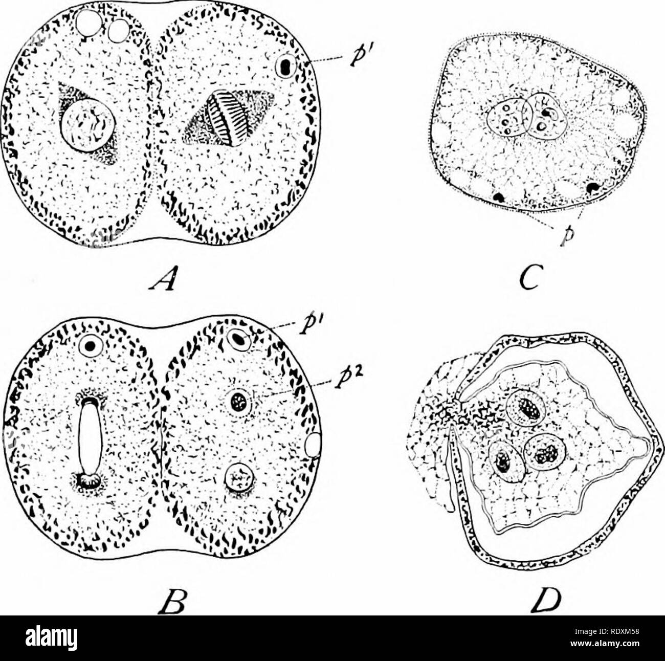 . Protozoo?logy. Protozoa; Protozoa, Pathogenic. FERTILIZATION BY ENDOGAMY 149 nuclei remaining (from one to twenty). Each of these daughter cysts secretes a gelatinous envelope about itself, and the nucleus of each divides by mitosis. This mitotic division is followed by division of the cytospore into two daughter cells (cytospores No. 2), and in these there are two successive nuclear divisions resulting in four nuclei. Three of these nuclei degenerate ("polar bodies") and one remains as a pronucleus. The cytospores of the second order next unite again, reforming the cytospores No. Stock Photohttps://www.alamy.com/image-license-details/?v=1https://www.alamy.com/protozoology-protozoa-protozoa-pathogenic-fertilization-by-endogamy-149-nuclei-remaining-from-one-to-twenty-each-of-these-daughter-cysts-secretes-a-gelatinous-envelope-about-itself-and-the-nucleus-of-each-divides-by-mitosis-this-mitotic-division-is-followed-by-division-of-the-cytospore-into-two-daughter-cells-cytospores-no-2-and-in-these-there-are-two-successive-nuclear-divisions-resulting-in-four-nuclei-three-of-these-nuclei-degenerate-quotpolar-bodiesquot-and-one-remains-as-a-pronucleus-the-cytospores-of-the-second-order-next-unite-again-reforming-the-cytospores-no-image232311892.html
RMRDXM58–. Protozoo?logy. Protozoa; Protozoa, Pathogenic. FERTILIZATION BY ENDOGAMY 149 nuclei remaining (from one to twenty). Each of these daughter cysts secretes a gelatinous envelope about itself, and the nucleus of each divides by mitosis. This mitotic division is followed by division of the cytospore into two daughter cells (cytospores No. 2), and in these there are two successive nuclear divisions resulting in four nuclei. Three of these nuclei degenerate ("polar bodies") and one remains as a pronucleus. The cytospores of the second order next unite again, reforming the cytospores No.
![. A textbook of invertebrate morphology [microform]. Invertebrates; Morphology (Animals); Invertébrés; Morphologie (Animaux). C D Fm. 23.—Diagrams to Illustkate the Piiknomena of Fertilizatiox. (From flf?ures by E. B. Wilson.) A, the approximation of the male and female pronuclei. B, division of tiie arclioplasm. V, separation of the arclioplasm spheres. D, fusion of the pronuclei, and formation of the segmentation spindle. //) = female pronucleus. a = arclioplasm. mp = male pronucleus. sn = segmentation nucleus. the spermatozoon is taken into tlie ovum and seems to ho completely absorbed, the Stock Photo . A textbook of invertebrate morphology [microform]. Invertebrates; Morphology (Animals); Invertébrés; Morphologie (Animaux). C D Fm. 23.—Diagrams to Illustkate the Piiknomena of Fertilizatiox. (From flf?ures by E. B. Wilson.) A, the approximation of the male and female pronuclei. B, division of tiie arclioplasm. V, separation of the arclioplasm spheres. D, fusion of the pronuclei, and formation of the segmentation spindle. //) = female pronucleus. a = arclioplasm. mp = male pronucleus. sn = segmentation nucleus. the spermatozoon is taken into tlie ovum and seems to ho completely absorbed, the Stock Photo](https://c8.alamy.com/comp/REPJ20/a-textbook-of-invertebrate-morphology-microform-invertebrates-morphology-animals-invertbrs-morphologie-animaux-c-d-fm-23diagrams-to-illustkate-the-piiknomena-of-fertilizatiox-from-flfures-by-e-b-wilson-a-the-approximation-of-the-male-and-female-pronuclei-b-division-of-tiie-arclioplasm-v-separation-of-the-arclioplasm-spheres-d-fusion-of-the-pronuclei-and-formation-of-the-segmentation-spindle-=-female-pronucleus-a-=-arclioplasm-mp-=-male-pronucleus-sn-=-segmentation-nucleus-the-spermatozoon-is-taken-into-tlie-ovum-and-seems-to-ho-completely-absorbed-the-REPJ20.jpg) . A textbook of invertebrate morphology [microform]. Invertebrates; Morphology (Animals); Invertébrés; Morphologie (Animaux). C D Fm. 23.—Diagrams to Illustkate the Piiknomena of Fertilizatiox. (From flf?ures by E. B. Wilson.) A, the approximation of the male and female pronuclei. B, division of tiie arclioplasm. V, separation of the arclioplasm spheres. D, fusion of the pronuclei, and formation of the segmentation spindle. //) = female pronucleus. a = arclioplasm. mp = male pronucleus. sn = segmentation nucleus. the spermatozoon is taken into tlie ovum and seems to ho completely absorbed, the Stock Photohttps://www.alamy.com/image-license-details/?v=1https://www.alamy.com/a-textbook-of-invertebrate-morphology-microform-invertebrates-morphology-animals-invertbrs-morphologie-animaux-c-d-fm-23diagrams-to-illustkate-the-piiknomena-of-fertilizatiox-from-flfures-by-e-b-wilson-a-the-approximation-of-the-male-and-female-pronuclei-b-division-of-tiie-arclioplasm-v-separation-of-the-arclioplasm-spheres-d-fusion-of-the-pronuclei-and-formation-of-the-segmentation-spindle-=-female-pronucleus-a-=-arclioplasm-mp-=-male-pronucleus-sn-=-segmentation-nucleus-the-spermatozoon-is-taken-into-tlie-ovum-and-seems-to-ho-completely-absorbed-the-image232837080.html
. A textbook of invertebrate morphology [microform]. Invertebrates; Morphology (Animals); Invertébrés; Morphologie (Animaux). C D Fm. 23.—Diagrams to Illustkate the Piiknomena of Fertilizatiox. (From flf?ures by E. B. Wilson.) A, the approximation of the male and female pronuclei. B, division of tiie arclioplasm. V, separation of the arclioplasm spheres. D, fusion of the pronuclei, and formation of the segmentation spindle. //) = female pronucleus. a = arclioplasm. mp = male pronucleus. sn = segmentation nucleus. the spermatozoon is taken into tlie ovum and seems to ho completely absorbed, the Stock Photohttps://www.alamy.com/image-license-details/?v=1https://www.alamy.com/a-textbook-of-invertebrate-morphology-microform-invertebrates-morphology-animals-invertbrs-morphologie-animaux-c-d-fm-23diagrams-to-illustkate-the-piiknomena-of-fertilizatiox-from-flfures-by-e-b-wilson-a-the-approximation-of-the-male-and-female-pronuclei-b-division-of-tiie-arclioplasm-v-separation-of-the-arclioplasm-spheres-d-fusion-of-the-pronuclei-and-formation-of-the-segmentation-spindle-=-female-pronucleus-a-=-arclioplasm-mp-=-male-pronucleus-sn-=-segmentation-nucleus-the-spermatozoon-is-taken-into-tlie-ovum-and-seems-to-ho-completely-absorbed-the-image232837080.htmlRMREPJ20–. A textbook of invertebrate morphology [microform]. Invertebrates; Morphology (Animals); Invertébrés; Morphologie (Animaux). C D Fm. 23.—Diagrams to Illustkate the Piiknomena of Fertilizatiox. (From flf?ures by E. B. Wilson.) A, the approximation of the male and female pronuclei. B, division of tiie arclioplasm. V, separation of the arclioplasm spheres. D, fusion of the pronuclei, and formation of the segmentation spindle. //) = female pronucleus. a = arclioplasm. mp = male pronucleus. sn = segmentation nucleus. the spermatozoon is taken into tlie ovum and seems to ho completely absorbed, the
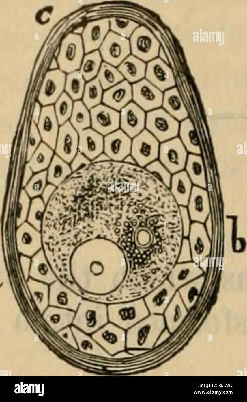 . The comparative anatomy of the domesticated animals. Veterinary anatomy. 1006 EMBRYOLOGY.. fecundation ; and although we have not to treat of fecundation itself, yet it ia well to bring forward the essential fact resulting from modern investigations on this point, showing that this great act consists in the fusion of two germs —the female pronucleus, arising from the division of the germinal vesicle ; and the male pro)iucleus, due to the transformation and migration of the spermatozoid. These two elements, by their fusion, originate the i/olk- nucleus which, by its double origin, contains th Stock Photohttps://www.alamy.com/image-license-details/?v=1https://www.alamy.com/the-comparative-anatomy-of-the-domesticated-animals-veterinary-anatomy-1006-embryology-fecundation-and-although-we-have-not-to-treat-of-fecundation-itself-yet-it-ia-well-to-bring-forward-the-essential-fact-resulting-from-modern-investigations-on-this-point-showing-that-this-great-act-consists-in-the-fusion-of-two-germs-the-female-pronucleus-arising-from-the-division-of-the-germinal-vesicle-and-the-male-proiucleus-due-to-the-transformation-and-migration-of-the-spermatozoid-these-two-elements-by-their-fusion-originate-the-iolk-nucleus-which-by-its-double-origin-contains-th-image232677326.html
RMREFA8E–. The comparative anatomy of the domesticated animals. Veterinary anatomy. 1006 EMBRYOLOGY.. fecundation ; and although we have not to treat of fecundation itself, yet it ia well to bring forward the essential fact resulting from modern investigations on this point, showing that this great act consists in the fusion of two germs —the female pronucleus, arising from the division of the germinal vesicle ; and the male pro)iucleus, due to the transformation and migration of the spermatozoid. These two elements, by their fusion, originate the i/olk- nucleus which, by its double origin, contains th
 . The Biological bulletin. Biology; Zoology; Biology; Marine Biology. GYNANDROMORPHS IN HABROBRACON. 115 The evidence does not prove although it is consistent with the theory that male parts are from one ootid, female parts from a fusion of another ootid and the male pronucleus. Intersexes. The fundamental distinction between gynandromorphs and intersexes is that the former are genetic mosaics while in the latter all parts of the body are presumed to be of similar genetic constitution. Male and female parts of gynandromorphs occur in distinct regions while intersexes are either male with a gre Stock Photohttps://www.alamy.com/image-license-details/?v=1https://www.alamy.com/the-biological-bulletin-biology-zoology-biology-marine-biology-gynandromorphs-in-habrobracon-115-the-evidence-does-not-prove-although-it-is-consistent-with-the-theory-that-male-parts-are-from-one-ootid-female-parts-from-a-fusion-of-another-ootid-and-the-male-pronucleus-intersexes-the-fundamental-distinction-between-gynandromorphs-and-intersexes-is-that-the-former-are-genetic-mosaics-while-in-the-latter-all-parts-of-the-body-are-presumed-to-be-of-similar-genetic-constitution-male-and-female-parts-of-gynandromorphs-occur-in-distinct-regions-while-intersexes-are-either-male-with-a-gre-image234672760.html
RMRHP7E0–. The Biological bulletin. Biology; Zoology; Biology; Marine Biology. GYNANDROMORPHS IN HABROBRACON. 115 The evidence does not prove although it is consistent with the theory that male parts are from one ootid, female parts from a fusion of another ootid and the male pronucleus. Intersexes. The fundamental distinction between gynandromorphs and intersexes is that the former are genetic mosaics while in the latter all parts of the body are presumed to be of similar genetic constitution. Male and female parts of gynandromorphs occur in distinct regions while intersexes are either male with a gre
![. Biological lectures delivered at the Marine Biological Laboratory of Wood's Holl [sic]. Biology. THE FERTILIZATION OF THE OVUM. 27 little smaller than those of the egg cell, and they frequently remain slightly smaller as long as they can be distinguished. In cases where there is no appreciable difference in size between the two pronuclei, the one may be distinguished from the other, as long as they remain distinct, by the position of the polar bodies which lie directly over the female pronucleus. After the two pronuclei have met, the two asters begin to move apart. They continue to separate, Stock Photo . Biological lectures delivered at the Marine Biological Laboratory of Wood's Holl [sic]. Biology. THE FERTILIZATION OF THE OVUM. 27 little smaller than those of the egg cell, and they frequently remain slightly smaller as long as they can be distinguished. In cases where there is no appreciable difference in size between the two pronuclei, the one may be distinguished from the other, as long as they remain distinct, by the position of the polar bodies which lie directly over the female pronucleus. After the two pronuclei have met, the two asters begin to move apart. They continue to separate, Stock Photo](https://c8.alamy.com/comp/RHM5BX/biological-lectures-delivered-at-the-marine-biological-laboratory-of-woods-holl-sic-biology-the-fertilization-of-the-ovum-27-little-smaller-than-those-of-the-egg-cell-and-they-frequently-remain-slightly-smaller-as-long-as-they-can-be-distinguished-in-cases-where-there-is-no-appreciable-difference-in-size-between-the-two-pronuclei-the-one-may-be-distinguished-from-the-other-as-long-as-they-remain-distinct-by-the-position-of-the-polar-bodies-which-lie-directly-over-the-female-pronucleus-after-the-two-pronuclei-have-met-the-two-asters-begin-to-move-apart-they-continue-to-separate-RHM5BX.jpg) . Biological lectures delivered at the Marine Biological Laboratory of Wood's Holl [sic]. Biology. THE FERTILIZATION OF THE OVUM. 27 little smaller than those of the egg cell, and they frequently remain slightly smaller as long as they can be distinguished. In cases where there is no appreciable difference in size between the two pronuclei, the one may be distinguished from the other, as long as they remain distinct, by the position of the polar bodies which lie directly over the female pronucleus. After the two pronuclei have met, the two asters begin to move apart. They continue to separate, Stock Photohttps://www.alamy.com/image-license-details/?v=1https://www.alamy.com/biological-lectures-delivered-at-the-marine-biological-laboratory-of-woods-holl-sic-biology-the-fertilization-of-the-ovum-27-little-smaller-than-those-of-the-egg-cell-and-they-frequently-remain-slightly-smaller-as-long-as-they-can-be-distinguished-in-cases-where-there-is-no-appreciable-difference-in-size-between-the-two-pronuclei-the-one-may-be-distinguished-from-the-other-as-long-as-they-remain-distinct-by-the-position-of-the-polar-bodies-which-lie-directly-over-the-female-pronucleus-after-the-two-pronuclei-have-met-the-two-asters-begin-to-move-apart-they-continue-to-separate-image234627230.html
. Biological lectures delivered at the Marine Biological Laboratory of Wood's Holl [sic]. Biology. THE FERTILIZATION OF THE OVUM. 27 little smaller than those of the egg cell, and they frequently remain slightly smaller as long as they can be distinguished. In cases where there is no appreciable difference in size between the two pronuclei, the one may be distinguished from the other, as long as they remain distinct, by the position of the polar bodies which lie directly over the female pronucleus. After the two pronuclei have met, the two asters begin to move apart. They continue to separate, Stock Photohttps://www.alamy.com/image-license-details/?v=1https://www.alamy.com/biological-lectures-delivered-at-the-marine-biological-laboratory-of-woods-holl-sic-biology-the-fertilization-of-the-ovum-27-little-smaller-than-those-of-the-egg-cell-and-they-frequently-remain-slightly-smaller-as-long-as-they-can-be-distinguished-in-cases-where-there-is-no-appreciable-difference-in-size-between-the-two-pronuclei-the-one-may-be-distinguished-from-the-other-as-long-as-they-remain-distinct-by-the-position-of-the-polar-bodies-which-lie-directly-over-the-female-pronucleus-after-the-two-pronuclei-have-met-the-two-asters-begin-to-move-apart-they-continue-to-separate-image234627230.htmlRMRHM5BX–. Biological lectures delivered at the Marine Biological Laboratory of Wood's Holl [sic]. Biology. THE FERTILIZATION OF THE OVUM. 27 little smaller than those of the egg cell, and they frequently remain slightly smaller as long as they can be distinguished. In cases where there is no appreciable difference in size between the two pronuclei, the one may be distinguished from the other, as long as they remain distinct, by the position of the polar bodies which lie directly over the female pronucleus. After the two pronuclei have met, the two asters begin to move apart. They continue to separate,
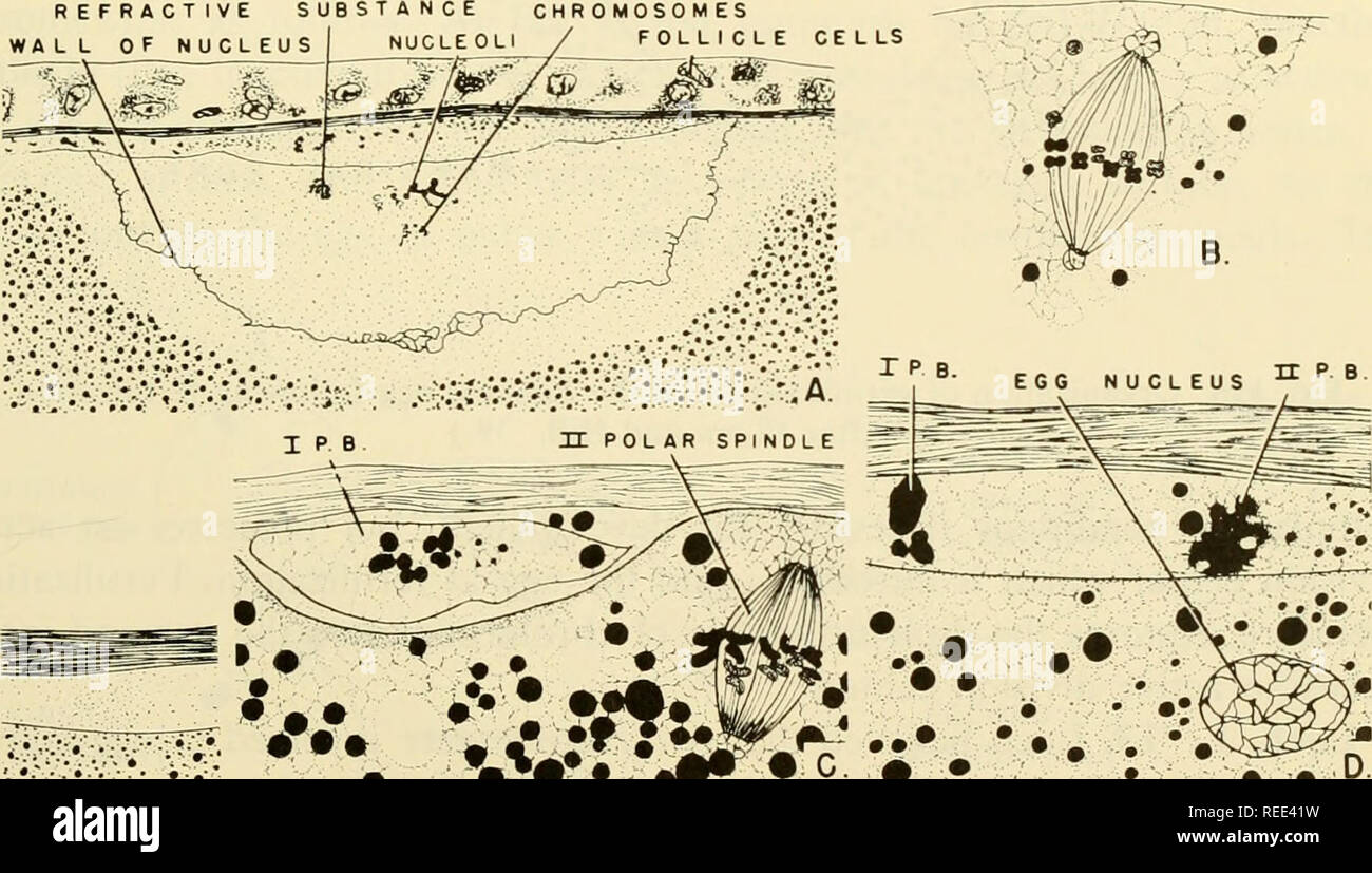 . Comparative embryology of the vertebrates; with 2057 drawings and photos. grouped as 380 illus. Vertebrates -- Embryology; Comparative embryology. THEORIES OF FERTILIZATION 267 These parthenogenetic merogons develop to the blastula stage only. Gyno- merogony is the parthenogenetic development of an egg fragment containing the egg pronucleus. J. Theories of Fertilization and Egg Activation Boveri, T., 1887, 1895. In somatic mitoses, the division center or centro- some is handed down from cell to cell. In the development of the female gamete, the division center degenerates or becomes physiolo Stock Photohttps://www.alamy.com/image-license-details/?v=1https://www.alamy.com/comparative-embryology-of-the-vertebrates-with-2057-drawings-and-photos-grouped-as-380-illus-vertebrates-embryology-comparative-embryology-theories-of-fertilization-267-these-parthenogenetic-merogons-develop-to-the-blastula-stage-only-gyno-merogony-is-the-parthenogenetic-development-of-an-egg-fragment-containing-the-egg-pronucleus-j-theories-of-fertilization-and-egg-activation-boveri-t-1887-1895-in-somatic-mitoses-the-division-center-or-centro-some-is-handed-down-from-cell-to-cell-in-the-development-of-the-female-gamete-the-division-center-degenerates-or-becomes-physiolo-image232650485.html
RMREE41W–. Comparative embryology of the vertebrates; with 2057 drawings and photos. grouped as 380 illus. Vertebrates -- Embryology; Comparative embryology. THEORIES OF FERTILIZATION 267 These parthenogenetic merogons develop to the blastula stage only. Gyno- merogony is the parthenogenetic development of an egg fragment containing the egg pronucleus. J. Theories of Fertilization and Egg Activation Boveri, T., 1887, 1895. In somatic mitoses, the division center or centro- some is handed down from cell to cell. In the development of the female gamete, the division center degenerates or becomes physiolo
 . A manual of elementary zoology . Zoology. 136 MANUAL OF ELEMENTARY ZOOLOGY place. The male pronucleus of each conjugant passes over into the other and fuses with the female pronucleus of the latter. The body which belonged to each conjugant comes thus to contain a micronucleus of mixed origin. It is, in fact, a zygote. The zygotes separate and are known as exconjugants. Immediately after separa- tion the meganucleus degenerates, splitting up into shreds, which disappear. Thus the meganucleus resem- bles in the fact of its mortality the body-cells of the frog, though the body as a whole has t Stock Photohttps://www.alamy.com/image-license-details/?v=1https://www.alamy.com/a-manual-of-elementary-zoology-zoology-136-manual-of-elementary-zoology-place-the-male-pronucleus-of-each-conjugant-passes-over-into-the-other-and-fuses-with-the-female-pronucleus-of-the-latter-the-body-which-belonged-to-each-conjugant-comes-thus-to-contain-a-micronucleus-of-mixed-origin-it-is-in-fact-a-zygote-the-zygotes-separate-and-are-known-as-exconjugants-immediately-after-separa-tion-the-meganucleus-degenerates-splitting-up-into-shreds-which-disappear-thus-the-meganucleus-resem-bles-in-the-fact-of-its-mortality-the-body-cells-of-the-frog-though-the-body-as-a-whole-has-t-image232123641.html
RMRDJ421–. A manual of elementary zoology . Zoology. 136 MANUAL OF ELEMENTARY ZOOLOGY place. The male pronucleus of each conjugant passes over into the other and fuses with the female pronucleus of the latter. The body which belonged to each conjugant comes thus to contain a micronucleus of mixed origin. It is, in fact, a zygote. The zygotes separate and are known as exconjugants. Immediately after separa- tion the meganucleus degenerates, splitting up into shreds, which disappear. Thus the meganucleus resem- bles in the fact of its mortality the body-cells of the frog, though the body as a whole has t
 . A manual of zoology. Zoology. GENERAL EMBRYOLOGY. 149 of fertilization, for even liere but one spermatozoon fuses with tlie egg- nucleus, the others degenerating sooner or later. Essential Feature of Fertilization.—After the spermatozoon has penetrated into the egg, the head and the middle piece which contains the centrosome can still he recognized, as the chromatic and acliromatic parts of the si^ermatozoou or sperm-nncleus (male pronucleus), while the tail and the slight amount of protoplasm disappear in the yolk. In the cytoplasm of tlie egg the centrosome of the sperm-nucleus gives rise Stock Photohttps://www.alamy.com/image-license-details/?v=1https://www.alamy.com/a-manual-of-zoology-zoology-general-embryology-149-of-fertilization-for-even-liere-but-one-spermatozoon-fuses-with-tlie-egg-nucleus-the-others-degenerating-sooner-or-later-essential-feature-of-fertilizationafter-the-spermatozoon-has-penetrated-into-the-egg-the-head-and-the-middle-piece-which-contains-the-centrosome-can-still-he-recognized-as-the-chromatic-and-acliromatic-parts-of-the-siermatozoou-or-sperm-nncleus-male-pronucleus-while-the-tail-and-the-slight-amount-of-protoplasm-disappear-in-the-yolk-in-the-cytoplasm-of-tlie-egg-the-centrosome-of-the-sperm-nucleus-gives-rise-image232348015.html
RMRE0A7B–. A manual of zoology. Zoology. GENERAL EMBRYOLOGY. 149 of fertilization, for even liere but one spermatozoon fuses with tlie egg- nucleus, the others degenerating sooner or later. Essential Feature of Fertilization.—After the spermatozoon has penetrated into the egg, the head and the middle piece which contains the centrosome can still he recognized, as the chromatic and acliromatic parts of the si^ermatozoou or sperm-nncleus (male pronucleus), while the tail and the slight amount of protoplasm disappear in the yolk. In the cytoplasm of tlie egg the centrosome of the sperm-nucleus gives rise
 . Comparative embryology of the vertebrates; with 2057 drawings and photos. grouped as 380 illus. Vertebrates -- Embryology; Comparative embryology. BEHAVIOR OF THE GAMETES 251 In demersal eggs, that is, eggs which sink to the bottom, the protoplasmic cap tends to assume an uppermost position. In pelagic eggs, i.e., eggs which float in the water, the protoplasmic disc turns downward since it is the heaviest part of the egg. After the polar bodies are given off, the egg-chromatin material reforms the female pronucleus. The latter and the sperm pronucleus migrate to a position near the center of Stock Photohttps://www.alamy.com/image-license-details/?v=1https://www.alamy.com/comparative-embryology-of-the-vertebrates-with-2057-drawings-and-photos-grouped-as-380-illus-vertebrates-embryology-comparative-embryology-behavior-of-the-gametes-251-in-demersal-eggs-that-is-eggs-which-sink-to-the-bottom-the-protoplasmic-cap-tends-to-assume-an-uppermost-position-in-pelagic-eggs-ie-eggs-which-float-in-the-water-the-protoplasmic-disc-turns-downward-since-it-is-the-heaviest-part-of-the-egg-after-the-polar-bodies-are-given-off-the-egg-chromatin-material-reforms-the-female-pronucleus-the-latter-and-the-sperm-pronucleus-migrate-to-a-position-near-the-center-of-image232650570.html
RMREE44X–. Comparative embryology of the vertebrates; with 2057 drawings and photos. grouped as 380 illus. Vertebrates -- Embryology; Comparative embryology. BEHAVIOR OF THE GAMETES 251 In demersal eggs, that is, eggs which sink to the bottom, the protoplasmic cap tends to assume an uppermost position. In pelagic eggs, i.e., eggs which float in the water, the protoplasmic disc turns downward since it is the heaviest part of the egg. After the polar bodies are given off, the egg-chromatin material reforms the female pronucleus. The latter and the sperm pronucleus migrate to a position near the center of
 . A text-book of animal physiology, with introductory chapters on general biology and a full treatment of reproduction ... Physiology, Comparative. 60 ANIMAL PHYSIOLOGY. etc.), then, are simply expelled; they take no part in the devel- opment of the ovum; and their extrusion is to be regarded as a preparation for the progress of the cell, whether this event fol- lows or precedes the entrance of the male cell into the ovum. It is worthy of note that the ovum may become amoeboid in the region from which the polar globules are expelled. The remainder of the nucleus {female pronucleus) now passes Stock Photohttps://www.alamy.com/image-license-details/?v=1https://www.alamy.com/a-text-book-of-animal-physiology-with-introductory-chapters-on-general-biology-and-a-full-treatment-of-reproduction-physiology-comparative-60-animal-physiology-etc-then-are-simply-expelled-they-take-no-part-in-the-devel-opment-of-the-ovum-and-their-extrusion-is-to-be-regarded-as-a-preparation-for-the-progress-of-the-cell-whether-this-event-fol-lows-or-precedes-the-entrance-of-the-male-cell-into-the-ovum-it-is-worthy-of-note-that-the-ovum-may-become-amoeboid-in-the-region-from-which-the-polar-globules-are-expelled-the-remainder-of-the-nucleus-female-pronucleus-now-passes-image232426015.html
RMRE3WN3–. A text-book of animal physiology, with introductory chapters on general biology and a full treatment of reproduction ... Physiology, Comparative. 60 ANIMAL PHYSIOLOGY. etc.), then, are simply expelled; they take no part in the devel- opment of the ovum; and their extrusion is to be regarded as a preparation for the progress of the cell, whether this event fol- lows or precedes the entrance of the male cell into the ovum. It is worthy of note that the ovum may become amoeboid in the region from which the polar globules are expelled. The remainder of the nucleus {female pronucleus) now passes
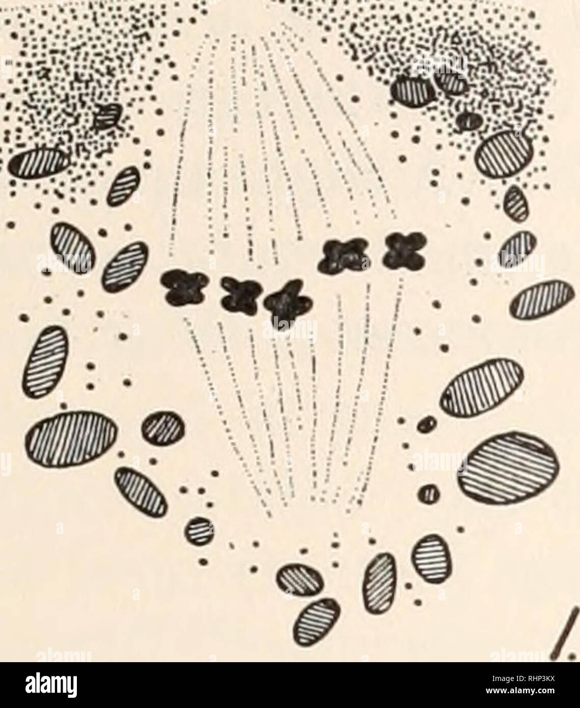 . The Biological bulletin. Biology; Zoology; Biology; Marine Biology. ANDROGENETIC DEVELOPMENT OF FROG EGG 235 awaiting the entrance of the sperm before continuing in the production of the second polar body and the female pronucleus. Sections through the egg in this stage of maturation reveal the relation of the spindle to the egg surface. (Fig. 1). It is to be noted that it lies close to the surface and is almost completely covered over by pigment granules. As the second maturation division proceeds this relationship is altered.. Please note that these images are extracted from scanned page i Stock Photohttps://www.alamy.com/image-license-details/?v=1https://www.alamy.com/the-biological-bulletin-biology-zoology-biology-marine-biology-androgenetic-development-of-frog-egg-235-awaiting-the-entrance-of-the-sperm-before-continuing-in-the-production-of-the-second-polar-body-and-the-female-pronucleus-sections-through-the-egg-in-this-stage-of-maturation-reveal-the-relation-of-the-spindle-to-the-egg-surface-fig-1-it-is-to-be-noted-that-it-lies-close-to-the-surface-and-is-almost-completely-covered-over-by-pigment-granules-as-the-second-maturation-division-proceeds-this-relationship-is-altered-please-note-that-these-images-are-extracted-from-scanned-page-i-image234669790.html
RMRHP3KX–. The Biological bulletin. Biology; Zoology; Biology; Marine Biology. ANDROGENETIC DEVELOPMENT OF FROG EGG 235 awaiting the entrance of the sperm before continuing in the production of the second polar body and the female pronucleus. Sections through the egg in this stage of maturation reveal the relation of the spindle to the egg surface. (Fig. 1). It is to be noted that it lies close to the surface and is almost completely covered over by pigment granules. As the second maturation division proceeds this relationship is altered.. Please note that these images are extracted from scanned page i
 . The biology of the protozoa. Protozoa; Protozoa. 308 BIOLOGY OF THE PROTOZOA in the protoplasm. The cell then divides into as many daughter cysts as there are nuclei and these Hertwig calls cystospores No. 1, each of which secretes a gelatinous envelope about itself. The nucleus then divides by mitosis followed by division of the cell into two daughter cells which he calls cytospores No. 2. The nuclei of the latter undergo two successive "maturation" divisions resulting in one pronucleus and two "polar bodies" in each (Fig. 156), the latter degenerating and disappearing. Stock Photohttps://www.alamy.com/image-license-details/?v=1https://www.alamy.com/the-biology-of-the-protozoa-protozoa-protozoa-308-biology-of-the-protozoa-in-the-protoplasm-the-cell-then-divides-into-as-many-daughter-cysts-as-there-are-nuclei-and-these-hertwig-calls-cystospores-no-1-each-of-which-secretes-a-gelatinous-envelope-about-itself-the-nucleus-then-divides-by-mitosis-followed-by-division-of-the-cell-into-two-daughter-cells-which-he-calls-cytospores-no-2-the-nuclei-of-the-latter-undergo-two-successive-quotmaturationquot-divisions-resulting-in-one-pronucleus-and-two-quotpolar-bodiesquot-in-each-fig-156-the-latter-degenerating-and-disappearing-image234602057.html
RMRHK18W–. The biology of the protozoa. Protozoa; Protozoa. 308 BIOLOGY OF THE PROTOZOA in the protoplasm. The cell then divides into as many daughter cysts as there are nuclei and these Hertwig calls cystospores No. 1, each of which secretes a gelatinous envelope about itself. The nucleus then divides by mitosis followed by division of the cell into two daughter cells which he calls cytospores No. 2. The nuclei of the latter undergo two successive "maturation" divisions resulting in one pronucleus and two "polar bodies" in each (Fig. 156), the latter degenerating and disappearing.
 . The Biological bulletin. Biology; Zoology; Biology; Marine Biology. 13. polar body is coming off. The mitotic figure at the pole is very small at this time, although before passing to the pole it was quite extensive. On the other hand, the second lobe is large. At the time of its begin- ning the second polar body is being extruded. The mitotic figure of the second polar body is no larger than that of the first. This lobe continues to grow larger, during which time the egg-pronucleus is de-. Please note that these images are extracted from scanned page images that may have been digitally enha Stock Photohttps://www.alamy.com/image-license-details/?v=1https://www.alamy.com/the-biological-bulletin-biology-zoology-biology-marine-biology-13-polar-body-is-coming-off-the-mitotic-figure-at-the-pole-is-very-small-at-this-time-although-before-passing-to-the-pole-it-was-quite-extensive-on-the-other-hand-the-second-lobe-is-large-at-the-time-of-its-begin-ning-the-second-polar-body-is-being-extruded-the-mitotic-figure-of-the-second-polar-body-is-no-larger-than-that-of-the-first-this-lobe-continues-to-grow-larger-during-which-time-the-egg-pronucleus-is-de-please-note-that-these-images-are-extracted-from-scanned-page-images-that-may-have-been-digitally-enha-image234669821.html
RMRHP3N1–. The Biological bulletin. Biology; Zoology; Biology; Marine Biology. 13. polar body is coming off. The mitotic figure at the pole is very small at this time, although before passing to the pole it was quite extensive. On the other hand, the second lobe is large. At the time of its begin- ning the second polar body is being extruded. The mitotic figure of the second polar body is no larger than that of the first. This lobe continues to grow larger, during which time the egg-pronucleus is de-. Please note that these images are extracted from scanned page images that may have been digitally enha
 . The Biological bulletin. Biology; Zoology; Biology; Marine Biology. FKRTII.IZATinX IN SPIROCODON SAI.TATRIX 413 siderable care must be exercised to ensure the presence of freshly shed sperm in the sea water into which the eggs are to be shed. As soon as eggs were observed in the container, a sample was removed to a slide and observed with tin- phase contrast microscope. Mature, unfertilized eggs (Fig. 1) are slightly oval with a depression at the animal pole, where the polar bodies have just been extruded, and at the base of which the pronucleus lies in contact with the egg surface. One or b Stock Photohttps://www.alamy.com/image-license-details/?v=1https://www.alamy.com/the-biological-bulletin-biology-zoology-biology-marine-biology-fkrtiiizatinx-in-spirocodon-saitatrix-413-siderable-care-must-be-exercised-to-ensure-the-presence-of-freshly-shed-sperm-in-the-sea-water-into-which-the-eggs-are-to-be-shed-as-soon-as-eggs-were-observed-in-the-container-a-sample-was-removed-to-a-slide-and-observed-with-tin-phase-contrast-microscope-mature-unfertilized-eggs-fig-1-are-slightly-oval-with-a-depression-at-the-animal-pole-where-the-polar-bodies-have-just-been-extruded-and-at-the-base-of-which-the-pronucleus-lies-in-contact-with-the-egg-surface-one-or-b-image234665545.html
RMRHNX89–. The Biological bulletin. Biology; Zoology; Biology; Marine Biology. FKRTII.IZATinX IN SPIROCODON SAI.TATRIX 413 siderable care must be exercised to ensure the presence of freshly shed sperm in the sea water into which the eggs are to be shed. As soon as eggs were observed in the container, a sample was removed to a slide and observed with tin- phase contrast microscope. Mature, unfertilized eggs (Fig. 1) are slightly oval with a depression at the animal pole, where the polar bodies have just been extruded, and at the base of which the pronucleus lies in contact with the egg surface. One or b
 . Text-book of embryology. Embryology. 12 INVEETEBEATA CHAP. of the egg results; in most cases this takes the form of simultaneous division into four equal parts. In the case of large eggs, like those of birds, it appears that normally a considerable number of spermatozoa enter the egg. One only unites with the female pronucleus and forms the zygote nucleus, the rest divide independently and form groups of small cells which are produced by the aggregation of the cytoplasm round the products of their division. Soon, however, the cells formed round the daughters of the zygote nucleus crush out a Stock Photohttps://www.alamy.com/image-license-details/?v=1https://www.alamy.com/text-book-of-embryology-embryology-12-inveetebeata-chap-of-the-egg-results-in-most-cases-this-takes-the-form-of-simultaneous-division-into-four-equal-parts-in-the-case-of-large-eggs-like-those-of-birds-it-appears-that-normally-a-considerable-number-of-spermatozoa-enter-the-egg-one-only-unites-with-the-female-pronucleus-and-forms-the-zygote-nucleus-the-rest-divide-independently-and-form-groups-of-small-cells-which-are-produced-by-the-aggregation-of-the-cytoplasm-round-the-products-of-their-division-soon-however-the-cells-formed-round-the-daughters-of-the-zygote-nucleus-crush-out-a-image232128428.html
RMRDJA50–. Text-book of embryology. Embryology. 12 INVEETEBEATA CHAP. of the egg results; in most cases this takes the form of simultaneous division into four equal parts. In the case of large eggs, like those of birds, it appears that normally a considerable number of spermatozoa enter the egg. One only unites with the female pronucleus and forms the zygote nucleus, the rest divide independently and form groups of small cells which are produced by the aggregation of the cytoplasm round the products of their division. Soon, however, the cells formed round the daughters of the zygote nucleus crush out a
![. A text-book of embryology for students of medicine [electronic resource]. Embryology; Embryology. 34 TEXT-BOOK OF EMBRYOLOGY. nuclear spindle, a second one is formed, which in the same manner extrudes the second polar body. What remains of the nucleus now moves toward the center of the cell and is known as the female pronucleus. The position of the female pronucleus is nearly or absolutely central. The egg is now ready for fertilization.. Fig. 13.—A, mature ovum of echinus; n, female pronucleus; B, immature ovarian ovum of echinus (Hertwig). For some time after their extrusion, and pending t Stock Photo . A text-book of embryology for students of medicine [electronic resource]. Embryology; Embryology. 34 TEXT-BOOK OF EMBRYOLOGY. nuclear spindle, a second one is formed, which in the same manner extrudes the second polar body. What remains of the nucleus now moves toward the center of the cell and is known as the female pronucleus. The position of the female pronucleus is nearly or absolutely central. The egg is now ready for fertilization.. Fig. 13.—A, mature ovum of echinus; n, female pronucleus; B, immature ovarian ovum of echinus (Hertwig). For some time after their extrusion, and pending t Stock Photo](https://c8.alamy.com/comp/RJNF4Y/a-text-book-of-embryology-for-students-of-medicine-electronic-resource-embryology-embryology-34-text-book-of-embryology-nuclear-spindle-a-second-one-is-formed-which-in-the-same-manner-extrudes-the-second-polar-body-what-remains-of-the-nucleus-now-moves-toward-the-center-of-the-cell-and-is-known-as-the-female-pronucleus-the-position-of-the-female-pronucleus-is-nearly-or-absolutely-central-the-egg-is-now-ready-for-fertilization-fig-13a-mature-ovum-of-echinus-n-female-pronucleus-b-immature-ovarian-ovum-of-echinus-hertwig-for-some-time-after-their-extrusion-and-pending-t-RJNF4Y.jpg) . A text-book of embryology for students of medicine [electronic resource]. Embryology; Embryology. 34 TEXT-BOOK OF EMBRYOLOGY. nuclear spindle, a second one is formed, which in the same manner extrudes the second polar body. What remains of the nucleus now moves toward the center of the cell and is known as the female pronucleus. The position of the female pronucleus is nearly or absolutely central. The egg is now ready for fertilization.. Fig. 13.—A, mature ovum of echinus; n, female pronucleus; B, immature ovarian ovum of echinus (Hertwig). For some time after their extrusion, and pending t Stock Photohttps://www.alamy.com/image-license-details/?v=1https://www.alamy.com/a-text-book-of-embryology-for-students-of-medicine-electronic-resource-embryology-embryology-34-text-book-of-embryology-nuclear-spindle-a-second-one-is-formed-which-in-the-same-manner-extrudes-the-second-polar-body-what-remains-of-the-nucleus-now-moves-toward-the-center-of-the-cell-and-is-known-as-the-female-pronucleus-the-position-of-the-female-pronucleus-is-nearly-or-absolutely-central-the-egg-is-now-ready-for-fertilization-fig-13a-mature-ovum-of-echinus-n-female-pronucleus-b-immature-ovarian-ovum-of-echinus-hertwig-for-some-time-after-their-extrusion-and-pending-t-image235271483.html
. A text-book of embryology for students of medicine [electronic resource]. Embryology; Embryology. 34 TEXT-BOOK OF EMBRYOLOGY. nuclear spindle, a second one is formed, which in the same manner extrudes the second polar body. What remains of the nucleus now moves toward the center of the cell and is known as the female pronucleus. The position of the female pronucleus is nearly or absolutely central. The egg is now ready for fertilization.. Fig. 13.—A, mature ovum of echinus; n, female pronucleus; B, immature ovarian ovum of echinus (Hertwig). For some time after their extrusion, and pending t Stock Photohttps://www.alamy.com/image-license-details/?v=1https://www.alamy.com/a-text-book-of-embryology-for-students-of-medicine-electronic-resource-embryology-embryology-34-text-book-of-embryology-nuclear-spindle-a-second-one-is-formed-which-in-the-same-manner-extrudes-the-second-polar-body-what-remains-of-the-nucleus-now-moves-toward-the-center-of-the-cell-and-is-known-as-the-female-pronucleus-the-position-of-the-female-pronucleus-is-nearly-or-absolutely-central-the-egg-is-now-ready-for-fertilization-fig-13a-mature-ovum-of-echinus-n-female-pronucleus-b-immature-ovarian-ovum-of-echinus-hertwig-for-some-time-after-their-extrusion-and-pending-t-image235271483.htmlRMRJNF4Y–. A text-book of embryology for students of medicine [electronic resource]. Embryology; Embryology. 34 TEXT-BOOK OF EMBRYOLOGY. nuclear spindle, a second one is formed, which in the same manner extrudes the second polar body. What remains of the nucleus now moves toward the center of the cell and is known as the female pronucleus. The position of the female pronucleus is nearly or absolutely central. The egg is now ready for fertilization.. Fig. 13.—A, mature ovum of echinus; n, female pronucleus; B, immature ovarian ovum of echinus (Hertwig). For some time after their extrusion, and pending t
 . The physiology of reproduction. Reproduction. Fig. 50.—Successive stages in the fertilisation of an ovum of Eolivnus esciderUus, showing the entrance of the spermatozoon. (From Bryce.) Cytoplasmic filaments arrange themselves around the centro- some in the form of a star, the sperm-aster, which accompanies the male pronucleus, and afterwards comes to he alongside of > Boveri, Zellen Stiidien IV., Ueber die Natur der Centrosomen, Jena, 1901. Jenkinson, " Observations on the Maturation and Fertilisation of the Egg of the Axolotl," Qtiar. Jour. Micr. Science, vol. xlviii., 1904, ha Stock Photohttps://www.alamy.com/image-license-details/?v=1https://www.alamy.com/the-physiology-of-reproduction-reproduction-fig-50successive-stages-in-the-fertilisation-of-an-ovum-of-eolivnus-escideruus-showing-the-entrance-of-the-spermatozoon-from-bryce-cytoplasmic-filaments-arrange-themselves-around-the-centro-some-in-the-form-of-a-star-the-sperm-aster-which-accompanies-the-male-pronucleus-and-afterwards-comes-to-he-alongside-of-gt-boveri-zellen-stiidien-iv-ueber-die-natur-der-centrosomen-jena-1901-jenkinson-quot-observations-on-the-maturation-and-fertilisation-of-the-egg-of-the-axolotlquot-qtiar-jour-micr-science-vol-xlviii-1904-ha-image232353810.html
RMRE0HJA–. The physiology of reproduction. Reproduction. Fig. 50.—Successive stages in the fertilisation of an ovum of Eolivnus esciderUus, showing the entrance of the spermatozoon. (From Bryce.) Cytoplasmic filaments arrange themselves around the centro- some in the form of a star, the sperm-aster, which accompanies the male pronucleus, and afterwards comes to he alongside of > Boveri, Zellen Stiidien IV., Ueber die Natur der Centrosomen, Jena, 1901. Jenkinson, " Observations on the Maturation and Fertilisation of the Egg of the Axolotl," Qtiar. Jour. Micr. Science, vol. xlviii., 1904, ha
 . The Biological bulletin. Biology; Zoology; Biology; Marine Biology. DIAGRAMS 1 (left) and 2 (right). Showing the relative frequency in which the female pronucleus appears in different parts of the egg cytoplasm in relation to the position of the polar bodies; 100 eggs. this difference may not be significant. On the other hand, the interval between the second meiotic division and the time of the observations may have been so short that the majority of the nuclei still remain slightly nearer the pole. In addition to the counts made above, twelve eggs, chosen at random, were sketched to show th Stock Photohttps://www.alamy.com/image-license-details/?v=1https://www.alamy.com/the-biological-bulletin-biology-zoology-biology-marine-biology-diagrams-1-left-and-2-right-showing-the-relative-frequency-in-which-the-female-pronucleus-appears-in-different-parts-of-the-egg-cytoplasm-in-relation-to-the-position-of-the-polar-bodies-100-eggs-this-difference-may-not-be-significant-on-the-other-hand-the-interval-between-the-second-meiotic-division-and-the-time-of-the-observations-may-have-been-so-short-that-the-majority-of-the-nuclei-still-remain-slightly-nearer-the-pole-in-addition-to-the-counts-made-above-twelve-eggs-chosen-at-random-were-sketched-to-show-th-image234671284.html
RMRHP5H8–. The Biological bulletin. Biology; Zoology; Biology; Marine Biology. DIAGRAMS 1 (left) and 2 (right). Showing the relative frequency in which the female pronucleus appears in different parts of the egg cytoplasm in relation to the position of the polar bodies; 100 eggs. this difference may not be significant. On the other hand, the interval between the second meiotic division and the time of the observations may have been so short that the majority of the nuclei still remain slightly nearer the pole. In addition to the counts made above, twelve eggs, chosen at random, were sketched to show th
![. The Biological bulletin. Biology; Zoology; Biology; Marine Biology. No. 4.] BULL A SOLITARIA.. FIG. 6. — The female pro- inner pole of the second maturation spindle. A few astral rays are pres- ent. Zwischenkorper of second polar body present. The structure of the two pronuclei when they have come together (Fig. 6) is the same. The male pronucleus is usu- ally regular in outline and slightly smaller. The irregularities in the outline of the female pronucleus often persist until the central corpuscles of the first cleavage appear. The chromatin stains very slightly and is connected by delicat Stock Photo . The Biological bulletin. Biology; Zoology; Biology; Marine Biology. No. 4.] BULL A SOLITARIA.. FIG. 6. — The female pro- inner pole of the second maturation spindle. A few astral rays are pres- ent. Zwischenkorper of second polar body present. The structure of the two pronuclei when they have come together (Fig. 6) is the same. The male pronucleus is usu- ally regular in outline and slightly smaller. The irregularities in the outline of the female pronucleus often persist until the central corpuscles of the first cleavage appear. The chromatin stains very slightly and is connected by delicat Stock Photo](https://c8.alamy.com/comp/RHRB5G/the-biological-bulletin-biology-zoology-biology-marine-biology-no-4-bull-a-solitaria-fig-6-the-female-pro-inner-pole-of-the-second-maturation-spindle-a-few-astral-rays-are-pres-ent-zwischenkorper-of-second-polar-body-present-the-structure-of-the-two-pronuclei-when-they-have-come-together-fig-6-is-the-same-the-male-pronucleus-is-usu-ally-regular-in-outline-and-slightly-smaller-the-irregularities-in-the-outline-of-the-female-pronucleus-often-persist-until-the-central-corpuscles-of-the-first-cleavage-appear-the-chromatin-stains-very-slightly-and-is-connected-by-delicat-RHRB5G.jpg) . The Biological bulletin. Biology; Zoology; Biology; Marine Biology. No. 4.] BULL A SOLITARIA.. FIG. 6. — The female pro- inner pole of the second maturation spindle. A few astral rays are pres- ent. Zwischenkorper of second polar body present. The structure of the two pronuclei when they have come together (Fig. 6) is the same. The male pronucleus is usu- ally regular in outline and slightly smaller. The irregularities in the outline of the female pronucleus often persist until the central corpuscles of the first cleavage appear. The chromatin stains very slightly and is connected by delicat Stock Photohttps://www.alamy.com/image-license-details/?v=1https://www.alamy.com/the-biological-bulletin-biology-zoology-biology-marine-biology-no-4-bull-a-solitaria-fig-6-the-female-pro-inner-pole-of-the-second-maturation-spindle-a-few-astral-rays-are-pres-ent-zwischenkorper-of-second-polar-body-present-the-structure-of-the-two-pronuclei-when-they-have-come-together-fig-6-is-the-same-the-male-pronucleus-is-usu-ally-regular-in-outline-and-slightly-smaller-the-irregularities-in-the-outline-of-the-female-pronucleus-often-persist-until-the-central-corpuscles-of-the-first-cleavage-appear-the-chromatin-stains-very-slightly-and-is-connected-by-delicat-image234697612.html
. The Biological bulletin. Biology; Zoology; Biology; Marine Biology. No. 4.] BULL A SOLITARIA.. FIG. 6. — The female pro- inner pole of the second maturation spindle. A few astral rays are pres- ent. Zwischenkorper of second polar body present. The structure of the two pronuclei when they have come together (Fig. 6) is the same. The male pronucleus is usu- ally regular in outline and slightly smaller. The irregularities in the outline of the female pronucleus often persist until the central corpuscles of the first cleavage appear. The chromatin stains very slightly and is connected by delicat Stock Photohttps://www.alamy.com/image-license-details/?v=1https://www.alamy.com/the-biological-bulletin-biology-zoology-biology-marine-biology-no-4-bull-a-solitaria-fig-6-the-female-pro-inner-pole-of-the-second-maturation-spindle-a-few-astral-rays-are-pres-ent-zwischenkorper-of-second-polar-body-present-the-structure-of-the-two-pronuclei-when-they-have-come-together-fig-6-is-the-same-the-male-pronucleus-is-usu-ally-regular-in-outline-and-slightly-smaller-the-irregularities-in-the-outline-of-the-female-pronucleus-often-persist-until-the-central-corpuscles-of-the-first-cleavage-appear-the-chromatin-stains-very-slightly-and-is-connected-by-delicat-image234697612.htmlRMRHRB5G–. The Biological bulletin. Biology; Zoology; Biology; Marine Biology. No. 4.] BULL A SOLITARIA.. FIG. 6. — The female pro- inner pole of the second maturation spindle. A few astral rays are pres- ent. Zwischenkorper of second polar body present. The structure of the two pronuclei when they have come together (Fig. 6) is the same. The male pronucleus is usu- ally regular in outline and slightly smaller. The irregularities in the outline of the female pronucleus often persist until the central corpuscles of the first cleavage appear. The chromatin stains very slightly and is connected by delicat
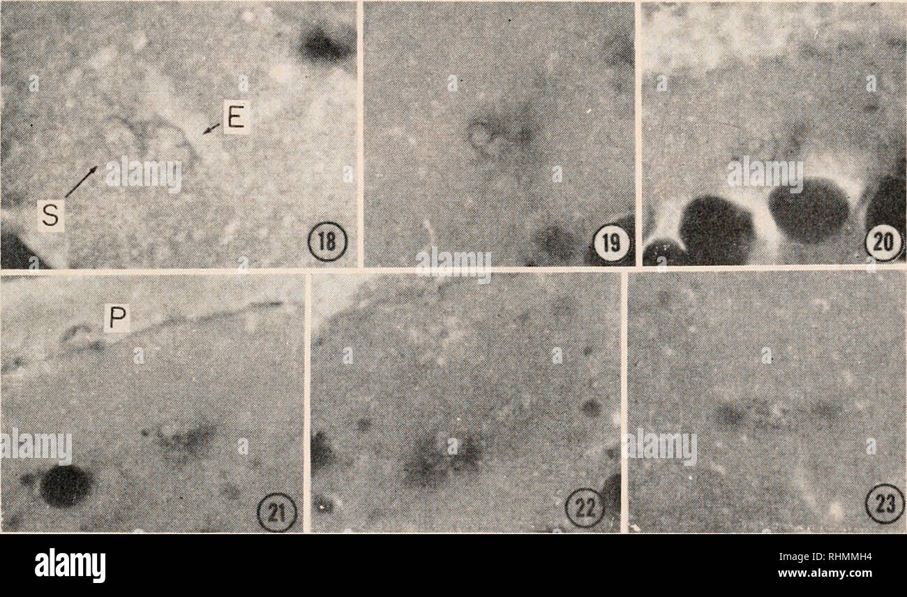 . The Biological bulletin. Biology; Zoology; Biology; Marine Biology. FERTILIZATION IN FUNA AND LOACH CROSS 119 mental eggs. The unfertilized eggs remained without advancing further in de- velopment. A similar situation was reported by Yamamoto (1951) to occur in unfertilized eggs of the dog-salmon. 4. Migration of the male and female pronuclei It is a matter of special note that the migration is more striking in the male pronucleus than in the female pronucleus. The penetration of the spermatozoon occurred always around the top of the animal pole where the micropyle exists. At first the sperm Stock Photohttps://www.alamy.com/image-license-details/?v=1https://www.alamy.com/the-biological-bulletin-biology-zoology-biology-marine-biology-fertilization-in-funa-and-loach-cross-119-mental-eggs-the-unfertilized-eggs-remained-without-advancing-further-in-de-velopment-a-similar-situation-was-reported-by-yamamoto-1951-to-occur-in-unfertilized-eggs-of-the-dog-salmon-4-migration-of-the-male-and-female-pronuclei-it-is-a-matter-of-special-note-that-the-migration-is-more-striking-in-the-male-pronucleus-than-in-the-female-pronucleus-the-penetration-of-the-spermatozoon-occurred-always-around-the-top-of-the-animal-pole-where-the-micropyle-exists-at-first-the-sperm-image234639136.html
RMRHMMH4–. The Biological bulletin. Biology; Zoology; Biology; Marine Biology. FERTILIZATION IN FUNA AND LOACH CROSS 119 mental eggs. The unfertilized eggs remained without advancing further in de- velopment. A similar situation was reported by Yamamoto (1951) to occur in unfertilized eggs of the dog-salmon. 4. Migration of the male and female pronuclei It is a matter of special note that the migration is more striking in the male pronucleus than in the female pronucleus. The penetration of the spermatozoon occurred always around the top of the animal pole where the micropyle exists. At first the sperm
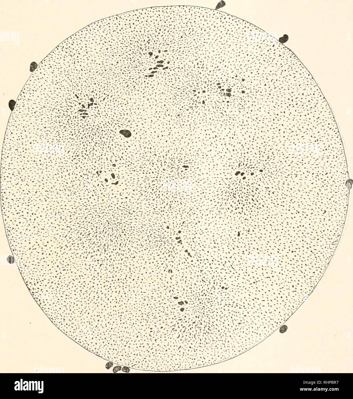 . The Biological bulletin. Biology; Zoology; Biology; Marine Biology. FERTILIZATION AFTER INITIATION OF DEVELOPMENT. 281 show more than one sperm nucleus and this has usually under- gone the characteristic swelling and vacuolization changes of a male pronucleus and is accompanied by an aster. After a longer exposure to this concentration of hypertonic sea-water the resemblance to sections of normal fertilization gradually disappears and is superseded by a general condition of poly- spermy and later by a non-reactive state of the egg substances,. FIG. 3. X 3,000. Section of an egg exposed to th Stock Photohttps://www.alamy.com/image-license-details/?v=1https://www.alamy.com/the-biological-bulletin-biology-zoology-biology-marine-biology-fertilization-after-initiation-of-development-281-show-more-than-one-sperm-nucleus-and-this-has-usually-under-gone-the-characteristic-swelling-and-vacuolization-changes-of-a-male-pronucleus-and-is-accompanied-by-an-aster-after-a-longer-exposure-to-this-concentration-of-hypertonic-sea-water-the-resemblance-to-sections-of-normal-fertilization-gradually-disappears-and-is-superseded-by-a-general-condition-of-poly-spermy-and-later-by-a-non-reactive-state-of-the-egg-substances-fig-3-x-3000-section-of-an-egg-exposed-to-th-image234676155.html
RMRHPBR7–. The Biological bulletin. Biology; Zoology; Biology; Marine Biology. FERTILIZATION AFTER INITIATION OF DEVELOPMENT. 281 show more than one sperm nucleus and this has usually under- gone the characteristic swelling and vacuolization changes of a male pronucleus and is accompanied by an aster. After a longer exposure to this concentration of hypertonic sea-water the resemblance to sections of normal fertilization gradually disappears and is superseded by a general condition of poly- spermy and later by a non-reactive state of the egg substances,. FIG. 3. X 3,000. Section of an egg exposed to th
 . The Biological bulletin. Biology; Zoology; Biology; Marine Biology. FIG. 6. — The female pro- inner pole of the second maturation spindle. A few astral rays are pres- ent. Zwischenkorper of second polar body present. The structure of the two pronuclei when they have come together (Fig. 6) is the same. The male pronucleus is usu- ally regular in outline and slightly smaller. The irregularities in the outline of the female pronucleus often persist until the central corpuscles of the first cleavage appear. The chromatin stains very slightly and is connected by delicate linin threads. The change Stock Photohttps://www.alamy.com/image-license-details/?v=1https://www.alamy.com/the-biological-bulletin-biology-zoology-biology-marine-biology-fig-6-the-female-pro-inner-pole-of-the-second-maturation-spindle-a-few-astral-rays-are-pres-ent-zwischenkorper-of-second-polar-body-present-the-structure-of-the-two-pronuclei-when-they-have-come-together-fig-6-is-the-same-the-male-pronucleus-is-usu-ally-regular-in-outline-and-slightly-smaller-the-irregularities-in-the-outline-of-the-female-pronucleus-often-persist-until-the-central-corpuscles-of-the-first-cleavage-appear-the-chromatin-stains-very-slightly-and-is-connected-by-delicate-linin-threads-the-change-image234697594.html
RMRHRB4X–. The Biological bulletin. Biology; Zoology; Biology; Marine Biology. FIG. 6. — The female pro- inner pole of the second maturation spindle. A few astral rays are pres- ent. Zwischenkorper of second polar body present. The structure of the two pronuclei when they have come together (Fig. 6) is the same. The male pronucleus is usu- ally regular in outline and slightly smaller. The irregularities in the outline of the female pronucleus often persist until the central corpuscles of the first cleavage appear. The chromatin stains very slightly and is connected by delicate linin threads. The change
 . Bulletin of the Museum of Comparative Zoology at Harvard College. Zoology; Zoology. 590 BULLETIN OF THE The male pronucleus arises when a living zoosperm penetrates into a mature and living vitellus. In certain cases (sea-urchin) it is not much larger than the " body " of a zodsperm, and one might then entertain the oj)inion that it is only such a " body " swollen. It then forms the centre of an aster. In other cases (Heteropoda) it becomes as large as tlie female pronucleus, and is not sur- rounded by a radial figure.* This growth is not a simple inflation of the body of Stock Photohttps://www.alamy.com/image-license-details/?v=1https://www.alamy.com/bulletin-of-the-museum-of-comparative-zoology-at-harvard-college-zoology-zoology-590-bulletin-of-the-the-male-pronucleus-arises-when-a-living-zoosperm-penetrates-into-a-mature-and-living-vitellus-in-certain-cases-sea-urchin-it-is-not-much-larger-than-the-quot-body-quot-of-a-zodsperm-and-one-might-then-entertain-the-ojinion-that-it-is-only-such-a-quot-body-quot-swollen-it-then-forms-the-centre-of-an-aster-in-other-cases-heteropoda-it-becomes-as-large-as-tlie-female-pronucleus-and-is-not-sur-rounded-by-a-radial-figure-this-growth-is-not-a-simple-inflation-of-the-body-of-image233922306.html
RMRGG282–. Bulletin of the Museum of Comparative Zoology at Harvard College. Zoology; Zoology. 590 BULLETIN OF THE The male pronucleus arises when a living zoosperm penetrates into a mature and living vitellus. In certain cases (sea-urchin) it is not much larger than the " body " of a zodsperm, and one might then entertain the oj)inion that it is only such a " body " swollen. It then forms the centre of an aster. In other cases (Heteropoda) it becomes as large as tlie female pronucleus, and is not sur- rounded by a radial figure.* This growth is not a simple inflation of the body of
![. A text-book of embryology for students of medicine [electronic resource]. Embryology; Embryology. 44 TEXT-BOOK OF EMBRYOLOGY. made the basis of a theory of heredity formulated by Hert- wig and independently advanced by Strasburger. A. Fig. 18.-A, fertilized ovum of echinus (Hertwig): the male (a) and the female pronucleus (b) are approaching; in B they have almost fused; C, ovum of echinus after completion of fertilization (Hertwig): s.n., segmentation-nucleus. Artificial fertilization, or the bringing about of the develop- ment of the ovum by artificial (chemical) means, without the partici Stock Photo . A text-book of embryology for students of medicine [electronic resource]. Embryology; Embryology. 44 TEXT-BOOK OF EMBRYOLOGY. made the basis of a theory of heredity formulated by Hert- wig and independently advanced by Strasburger. A. Fig. 18.-A, fertilized ovum of echinus (Hertwig): the male (a) and the female pronucleus (b) are approaching; in B they have almost fused; C, ovum of echinus after completion of fertilization (Hertwig): s.n., segmentation-nucleus. Artificial fertilization, or the bringing about of the develop- ment of the ovum by artificial (chemical) means, without the partici Stock Photo](https://c8.alamy.com/comp/RJNF2E/a-text-book-of-embryology-for-students-of-medicine-electronic-resource-embryology-embryology-44-text-book-of-embryology-made-the-basis-of-a-theory-of-heredity-formulated-by-hert-wig-and-independently-advanced-by-strasburger-a-fig-18-a-fertilized-ovum-of-echinus-hertwig-the-male-a-and-the-female-pronucleus-b-are-approaching-in-b-they-have-almost-fused-c-ovum-of-echinus-after-completion-of-fertilization-hertwig-sn-segmentation-nucleus-artificial-fertilization-or-the-bringing-about-of-the-develop-ment-of-the-ovum-by-artificial-chemical-means-without-the-partici-RJNF2E.jpg) . A text-book of embryology for students of medicine [electronic resource]. Embryology; Embryology. 44 TEXT-BOOK OF EMBRYOLOGY. made the basis of a theory of heredity formulated by Hert- wig and independently advanced by Strasburger. A. Fig. 18.-A, fertilized ovum of echinus (Hertwig): the male (a) and the female pronucleus (b) are approaching; in B they have almost fused; C, ovum of echinus after completion of fertilization (Hertwig): s.n., segmentation-nucleus. Artificial fertilization, or the bringing about of the develop- ment of the ovum by artificial (chemical) means, without the partici Stock Photohttps://www.alamy.com/image-license-details/?v=1https://www.alamy.com/a-text-book-of-embryology-for-students-of-medicine-electronic-resource-embryology-embryology-44-text-book-of-embryology-made-the-basis-of-a-theory-of-heredity-formulated-by-hert-wig-and-independently-advanced-by-strasburger-a-fig-18-a-fertilized-ovum-of-echinus-hertwig-the-male-a-and-the-female-pronucleus-b-are-approaching-in-b-they-have-almost-fused-c-ovum-of-echinus-after-completion-of-fertilization-hertwig-sn-segmentation-nucleus-artificial-fertilization-or-the-bringing-about-of-the-develop-ment-of-the-ovum-by-artificial-chemical-means-without-the-partici-image235271414.html
. A text-book of embryology for students of medicine [electronic resource]. Embryology; Embryology. 44 TEXT-BOOK OF EMBRYOLOGY. made the basis of a theory of heredity formulated by Hert- wig and independently advanced by Strasburger. A. Fig. 18.-A, fertilized ovum of echinus (Hertwig): the male (a) and the female pronucleus (b) are approaching; in B they have almost fused; C, ovum of echinus after completion of fertilization (Hertwig): s.n., segmentation-nucleus. Artificial fertilization, or the bringing about of the develop- ment of the ovum by artificial (chemical) means, without the partici Stock Photohttps://www.alamy.com/image-license-details/?v=1https://www.alamy.com/a-text-book-of-embryology-for-students-of-medicine-electronic-resource-embryology-embryology-44-text-book-of-embryology-made-the-basis-of-a-theory-of-heredity-formulated-by-hert-wig-and-independently-advanced-by-strasburger-a-fig-18-a-fertilized-ovum-of-echinus-hertwig-the-male-a-and-the-female-pronucleus-b-are-approaching-in-b-they-have-almost-fused-c-ovum-of-echinus-after-completion-of-fertilization-hertwig-sn-segmentation-nucleus-artificial-fertilization-or-the-bringing-about-of-the-develop-ment-of-the-ovum-by-artificial-chemical-means-without-the-partici-image235271414.htmlRMRJNF2E–. A text-book of embryology for students of medicine [electronic resource]. Embryology; Embryology. 44 TEXT-BOOK OF EMBRYOLOGY. made the basis of a theory of heredity formulated by Hert- wig and independently advanced by Strasburger. A. Fig. 18.-A, fertilized ovum of echinus (Hertwig): the male (a) and the female pronucleus (b) are approaching; in B they have almost fused; C, ovum of echinus after completion of fertilization (Hertwig): s.n., segmentation-nucleus. Artificial fertilization, or the bringing about of the develop- ment of the ovum by artificial (chemical) means, without the partici
![. Biological lectures delivered at the Marine Biological Laboratory of Wood's Holl [sic]. Biology. 54 BIOLOGICAL LECTURES. bodies (one or more of which apparently take part in forming the centrosome) do not appear to be merely thickenings of the cyto- plasmic threads (this, you remember, is an essential element of Watase's theory); on the contrary, many of them appear to be scattered throughout the cytoplasm in a relatively independent. Fig. 5. —Optical section through entire egg, showing telophase of second maturation spindle and head of spermatozoon forming male pronucleus. Male attraction s Stock Photo . Biological lectures delivered at the Marine Biological Laboratory of Wood's Holl [sic]. Biology. 54 BIOLOGICAL LECTURES. bodies (one or more of which apparently take part in forming the centrosome) do not appear to be merely thickenings of the cyto- plasmic threads (this, you remember, is an essential element of Watase's theory); on the contrary, many of them appear to be scattered throughout the cytoplasm in a relatively independent. Fig. 5. —Optical section through entire egg, showing telophase of second maturation spindle and head of spermatozoon forming male pronucleus. Male attraction s Stock Photo](https://c8.alamy.com/comp/RHM4MB/biological-lectures-delivered-at-the-marine-biological-laboratory-of-woods-holl-sic-biology-54-biological-lectures-bodies-one-or-more-of-which-apparently-take-part-in-forming-the-centrosome-do-not-appear-to-be-merely-thickenings-of-the-cyto-plasmic-threads-this-you-remember-is-an-essential-element-of-watases-theory-on-the-contrary-many-of-them-appear-to-be-scattered-throughout-the-cytoplasm-in-a-relatively-independent-fig-5-optical-section-through-entire-egg-showing-telophase-of-second-maturation-spindle-and-head-of-spermatozoon-forming-male-pronucleus-male-attraction-s-RHM4MB.jpg) . Biological lectures delivered at the Marine Biological Laboratory of Wood's Holl [sic]. Biology. 54 BIOLOGICAL LECTURES. bodies (one or more of which apparently take part in forming the centrosome) do not appear to be merely thickenings of the cyto- plasmic threads (this, you remember, is an essential element of Watase's theory); on the contrary, many of them appear to be scattered throughout the cytoplasm in a relatively independent. Fig. 5. —Optical section through entire egg, showing telophase of second maturation spindle and head of spermatozoon forming male pronucleus. Male attraction s Stock Photohttps://www.alamy.com/image-license-details/?v=1https://www.alamy.com/biological-lectures-delivered-at-the-marine-biological-laboratory-of-woods-holl-sic-biology-54-biological-lectures-bodies-one-or-more-of-which-apparently-take-part-in-forming-the-centrosome-do-not-appear-to-be-merely-thickenings-of-the-cyto-plasmic-threads-this-you-remember-is-an-essential-element-of-watases-theory-on-the-contrary-many-of-them-appear-to-be-scattered-throughout-the-cytoplasm-in-a-relatively-independent-fig-5-optical-section-through-entire-egg-showing-telophase-of-second-maturation-spindle-and-head-of-spermatozoon-forming-male-pronucleus-male-attraction-s-image234626683.html
. Biological lectures delivered at the Marine Biological Laboratory of Wood's Holl [sic]. Biology. 54 BIOLOGICAL LECTURES. bodies (one or more of which apparently take part in forming the centrosome) do not appear to be merely thickenings of the cyto- plasmic threads (this, you remember, is an essential element of Watase's theory); on the contrary, many of them appear to be scattered throughout the cytoplasm in a relatively independent. Fig. 5. —Optical section through entire egg, showing telophase of second maturation spindle and head of spermatozoon forming male pronucleus. Male attraction s Stock Photohttps://www.alamy.com/image-license-details/?v=1https://www.alamy.com/biological-lectures-delivered-at-the-marine-biological-laboratory-of-woods-holl-sic-biology-54-biological-lectures-bodies-one-or-more-of-which-apparently-take-part-in-forming-the-centrosome-do-not-appear-to-be-merely-thickenings-of-the-cyto-plasmic-threads-this-you-remember-is-an-essential-element-of-watases-theory-on-the-contrary-many-of-them-appear-to-be-scattered-throughout-the-cytoplasm-in-a-relatively-independent-fig-5-optical-section-through-entire-egg-showing-telophase-of-second-maturation-spindle-and-head-of-spermatozoon-forming-male-pronucleus-male-attraction-s-image234626683.htmlRMRHM4MB–. Biological lectures delivered at the Marine Biological Laboratory of Wood's Holl [sic]. Biology. 54 BIOLOGICAL LECTURES. bodies (one or more of which apparently take part in forming the centrosome) do not appear to be merely thickenings of the cyto- plasmic threads (this, you remember, is an essential element of Watase's theory); on the contrary, many of them appear to be scattered throughout the cytoplasm in a relatively independent. Fig. 5. —Optical section through entire egg, showing telophase of second maturation spindle and head of spermatozoon forming male pronucleus. Male attraction s
 . The Biological bulletin. Biology; Zoology; Biology; Marine Biology. Figures 15-20. Pronuclear formation and male pronucleus migration in Memhriinipura mcmhranacea zygotes stained with bisbenzimide H33342. Figure 15. A zygote at first meiotic anaphase shown in side view. The first polar body nucleus (FBI) is located at the animal pole. SN, sperm nucleus; ON, secondary oocyte nucleus. Scale bar = 25 ^m.. Please note that these images are extracted from scanned page images that may have been digitally enhanced for readability - coloration and appearance of these illustrations may not perfectly Stock Photohttps://www.alamy.com/image-license-details/?v=1https://www.alamy.com/the-biological-bulletin-biology-zoology-biology-marine-biology-figures-15-20-pronuclear-formation-and-male-pronucleus-migration-in-memhriinipura-mcmhranacea-zygotes-stained-with-bisbenzimide-h33342-figure-15-a-zygote-at-first-meiotic-anaphase-shown-in-side-view-the-first-polar-body-nucleus-fbi-is-located-at-the-animal-pole-sn-sperm-nucleus-on-secondary-oocyte-nucleus-scale-bar-=-25-m-please-note-that-these-images-are-extracted-from-scanned-page-images-that-may-have-been-digitally-enhanced-for-readability-coloration-and-appearance-of-these-illustrations-may-not-perfectly-image234632184.html
RMRHMBMT–. The Biological bulletin. Biology; Zoology; Biology; Marine Biology. Figures 15-20. Pronuclear formation and male pronucleus migration in Memhriinipura mcmhranacea zygotes stained with bisbenzimide H33342. Figure 15. A zygote at first meiotic anaphase shown in side view. The first polar body nucleus (FBI) is located at the animal pole. SN, sperm nucleus; ON, secondary oocyte nucleus. Scale bar = 25 ^m.. Please note that these images are extracted from scanned page images that may have been digitally enhanced for readability - coloration and appearance of these illustrations may not perfectly
 . The Biological bulletin. Biology; Zoology; Biology; Marine Biology. THE FERTILIZATION PROCESS IX THE SXAIL. 91 (a) ten chromosomes is the usual number found in young first polocytes, and an equal number normally remains in the egg; (b) the anaphase of the second maturation division usually shows ten chromosomes at each pole and (c) the observed maximum number of karyomeres which go to form the definitive egg pronucleus i ten. X. CONCLUSIONS. From the evidence brought out in this paper it appears that self-fertilization is the normal method of reproduction in L. s. apprcssa and that in this s Stock Photohttps://www.alamy.com/image-license-details/?v=1https://www.alamy.com/the-biological-bulletin-biology-zoology-biology-marine-biology-the-fertilization-process-ix-the-sxail-91-a-ten-chromosomes-is-the-usual-number-found-in-young-first-polocytes-and-an-equal-number-normally-remains-in-the-egg-b-the-anaphase-of-the-second-maturation-division-usually-shows-ten-chromosomes-at-each-pole-and-c-the-observed-maximum-number-of-karyomeres-which-go-to-form-the-definitive-egg-pronucleus-i-ten-x-conclusions-from-the-evidence-brought-out-in-this-paper-it-appears-that-self-fertilization-is-the-normal-method-of-reproduction-in-l-s-apprcssa-and-that-in-this-s-image234642888.html
RMRHMWB4–. The Biological bulletin. Biology; Zoology; Biology; Marine Biology. THE FERTILIZATION PROCESS IX THE SXAIL. 91 (a) ten chromosomes is the usual number found in young first polocytes, and an equal number normally remains in the egg; (b) the anaphase of the second maturation division usually shows ten chromosomes at each pole and (c) the observed maximum number of karyomeres which go to form the definitive egg pronucleus i ten. X. CONCLUSIONS. From the evidence brought out in this paper it appears that self-fertilization is the normal method of reproduction in L. s. apprcssa and that in this s
 . An elementary course of practical zoology. Zoology. mcfrv. 5 ptort se^.fuuZ Fig. 140. The maiui-ation and iinj>regnation of the animal ovum (diagrammatic). A, the ovum, surrounded by the vitelline membrane ijnem in the act of forming the first polar cell (Poi); ^ cent, centrosome. B, both polai- cells (pof) are formed, the female pronucleus (9/n?/^.) lies near the centre of the ovum, and one of the several sperms is shown making its way into the ovum at the micropyle (^inicrof). C, the head of the sperm has become the male pronucleus {hpron Its intermediate piece the male centrosome (cJ Stock Photohttps://www.alamy.com/image-license-details/?v=1https://www.alamy.com/an-elementary-course-of-practical-zoology-zoology-mcfrv-5-ptort-sefuuz-fig-140-the-maiui-ation-and-iinjgtregnation-of-the-animal-ovum-diagrammatic-a-the-ovum-surrounded-by-the-vitelline-membrane-ijnem-in-the-act-of-forming-the-first-polar-cell-poi-cent-centrosome-b-both-polai-cells-pof-are-formed-the-female-pronucleus-9n-lies-near-the-centre-of-the-ovum-and-one-of-the-several-sperms-is-shown-making-its-way-into-the-ovum-at-the-micropyle-inicrof-c-the-head-of-the-sperm-has-become-the-male-pronucleus-hpron-its-intermediate-piece-the-male-centrosome-cj-image232113640.html
RMRDHK8T–. An elementary course of practical zoology. Zoology. mcfrv. 5 ptort se^.fuuZ Fig. 140. The maiui-ation and iinj>regnation of the animal ovum (diagrammatic). A, the ovum, surrounded by the vitelline membrane ijnem in the act of forming the first polar cell (Poi); ^ cent, centrosome. B, both polai- cells (pof) are formed, the female pronucleus (9/n?/^.) lies near the centre of the ovum, and one of the several sperms is shown making its way into the ovum at the micropyle (^inicrof). C, the head of the sperm has become the male pronucleus {hpron Its intermediate piece the male centrosome (cJ
 . The cell in development and inheritance. Cells. 5 pronucleus (Fig. ^2, C-H). Each animal now contains a cleavage- nucleus equally derived from both the conjugating animals, and the latter soon separate. The cleavage-nucleus in each divides three times successively, and of the eight resulting bodies four become macronuclei and four micronuclei (Fig. 82, H-K). By two suc- ceeding fissions the four macronuclei are then distributed, one to each of the four resulting individuals. In some other species the micro- nuclei are equally dis- tributed in like man- ner, but in /'. cauda- tuui the process Stock Photohttps://www.alamy.com/image-license-details/?v=1https://www.alamy.com/the-cell-in-development-and-inheritance-cells-5-pronucleus-fig-2-c-h-each-animal-now-contains-a-cleavage-nucleus-equally-derived-from-both-the-conjugating-animals-and-the-latter-soon-separate-the-cleavage-nucleus-in-each-divides-three-times-successively-and-of-the-eight-resulting-bodies-four-become-macronuclei-and-four-micronuclei-fig-82-h-k-by-two-suc-ceeding-fissions-the-four-macronuclei-are-then-distributed-one-to-each-of-the-four-resulting-individuals-in-some-other-species-the-micro-nuclei-are-equally-dis-tributed-in-like-man-ner-but-in-cauda-tuui-the-process-image235050494.html
RMRJBD8E–. The cell in development and inheritance. Cells. 5 pronucleus (Fig. ^2, C-H). Each animal now contains a cleavage- nucleus equally derived from both the conjugating animals, and the latter soon separate. The cleavage-nucleus in each divides three times successively, and of the eight resulting bodies four become macronuclei and four micronuclei (Fig. 82, H-K). By two suc- ceeding fissions the four macronuclei are then distributed, one to each of the four resulting individuals. In some other species the micro- nuclei are equally dis- tributed in like man- ner, but in /'. cauda- tuui the process
 . Text-book of embryology. Embryology. -Foul' stagea iu the maturation aud fertilization of tlie egg of Grepidula plana. (After Conklin.) A, formation of first polar body; the spermatozoon has entered the egg and has begun to swell up into the male pronucleus. B, formation of second polar body ; the first polar body has divided into two. C, the male and female pronuclei have come together. D, formation of the first cleavage spindle ; the female pronucleus above and the male pronucleus below are still clearly distinguishable from one another, fp, Female pronucleus ; mp, male pronucleus; pi (in Stock Photohttps://www.alamy.com/image-license-details/?v=1https://www.alamy.com/text-book-of-embryology-embryology-foul-stagea-iu-the-maturation-aud-fertilization-of-tlie-egg-of-grepidula-plana-after-conklin-a-formation-of-first-polar-body-the-spermatozoon-has-entered-the-egg-and-has-begun-to-swell-up-into-the-male-pronucleus-b-formation-of-second-polar-body-the-first-polar-body-has-divided-into-two-c-the-male-and-female-pronuclei-have-come-together-d-formation-of-the-first-cleavage-spindle-the-female-pronucleus-above-and-the-male-pronucleus-below-are-still-clearly-distinguishable-from-one-another-fp-female-pronucleus-mp-male-pronucleus-pi-in-image232128435.html
RMRDJA57–. Text-book of embryology. Embryology. -Foul' stagea iu the maturation aud fertilization of tlie egg of Grepidula plana. (After Conklin.) A, formation of first polar body; the spermatozoon has entered the egg and has begun to swell up into the male pronucleus. B, formation of second polar body ; the first polar body has divided into two. C, the male and female pronuclei have come together. D, formation of the first cleavage spindle ; the female pronucleus above and the male pronucleus below are still clearly distinguishable from one another, fp, Female pronucleus ; mp, male pronucleus; pi (in
 . The Biological bulletin. Biology; Zoology; Biology; Marine Biology. FERTILIZATION PROCESSES IN OYSTER EGGS 209. Figure 12. Antituhulin-and 7-AAD-stained specimens depict!ngthe development of microtuhule arrays (a-d) and the morphogenesis of the maternally and paternally derived chromatin following pronuclear apposition (e-h). (a-c) Stages in the development of the mitotic spindle; the developing spindle poles are shown at the arrows, (d) Polyspermic egg in which each male pronucleus is associated with a developing sperm aster (arrows), (e, f) Apposed male and female pronuclei undergoing chro Stock Photohttps://www.alamy.com/image-license-details/?v=1https://www.alamy.com/the-biological-bulletin-biology-zoology-biology-marine-biology-fertilization-processes-in-oyster-eggs-209-figure-12-antituhulin-and-7-aad-stained-specimens-depict!ngthe-development-of-microtuhule-arrays-a-d-and-the-morphogenesis-of-the-maternally-and-paternally-derived-chromatin-following-pronuclear-apposition-e-h-a-c-stages-in-the-development-of-the-mitotic-spindle-the-developing-spindle-poles-are-shown-at-the-arrows-d-polyspermic-egg-in-which-each-male-pronucleus-is-associated-with-a-developing-sperm-aster-arrows-e-f-apposed-male-and-female-pronuclei-undergoing-chro-image234634145.html
RMRHME6W–. The Biological bulletin. Biology; Zoology; Biology; Marine Biology. FERTILIZATION PROCESSES IN OYSTER EGGS 209. Figure 12. Antituhulin-and 7-AAD-stained specimens depict!ngthe development of microtuhule arrays (a-d) and the morphogenesis of the maternally and paternally derived chromatin following pronuclear apposition (e-h). (a-c) Stages in the development of the mitotic spindle; the developing spindle poles are shown at the arrows, (d) Polyspermic egg in which each male pronucleus is associated with a developing sperm aster (arrows), (e, f) Apposed male and female pronuclei undergoing chro
 . Comparative embryology of the vertebrates; with 2057 drawings and photos. grouped as 380 illus. Vertebrates -- Embryology; Comparative embryology. 270 FERTILIZATION. Fig. 138. Polyspermy in the European newt, Triton, fAfter Fankhauser, '48.) (A) Ten minutes after insemination at 23° C. Metaphase of second maturation division; four sperm have entered the egg, one of which is at the vegetal pole of the egg, and another between the two poles of the egg. (B) One hour and 30 minutes; second polar body given off; small egg pronucleus moves toward nearest sperm nucleus. The latter will become the p Stock Photohttps://www.alamy.com/image-license-details/?v=1https://www.alamy.com/comparative-embryology-of-the-vertebrates-with-2057-drawings-and-photos-grouped-as-380-illus-vertebrates-embryology-comparative-embryology-270-fertilization-fig-138-polyspermy-in-the-european-newt-triton-fafter-fankhauser-48-a-ten-minutes-after-insemination-at-23-c-metaphase-of-second-maturation-division-four-sperm-have-entered-the-egg-one-of-which-is-at-the-vegetal-pole-of-the-egg-and-another-between-the-two-poles-of-the-egg-b-one-hour-and-30-minutes-second-polar-body-given-off-small-egg-pronucleus-moves-toward-nearest-sperm-nucleus-the-latter-will-become-the-p-image232650460.html
RMREE410–. Comparative embryology of the vertebrates; with 2057 drawings and photos. grouped as 380 illus. Vertebrates -- Embryology; Comparative embryology. 270 FERTILIZATION. Fig. 138. Polyspermy in the European newt, Triton, fAfter Fankhauser, '48.) (A) Ten minutes after insemination at 23° C. Metaphase of second maturation division; four sperm have entered the egg, one of which is at the vegetal pole of the egg, and another between the two poles of the egg. (B) One hour and 30 minutes; second polar body given off; small egg pronucleus moves toward nearest sperm nucleus. The latter will become the p
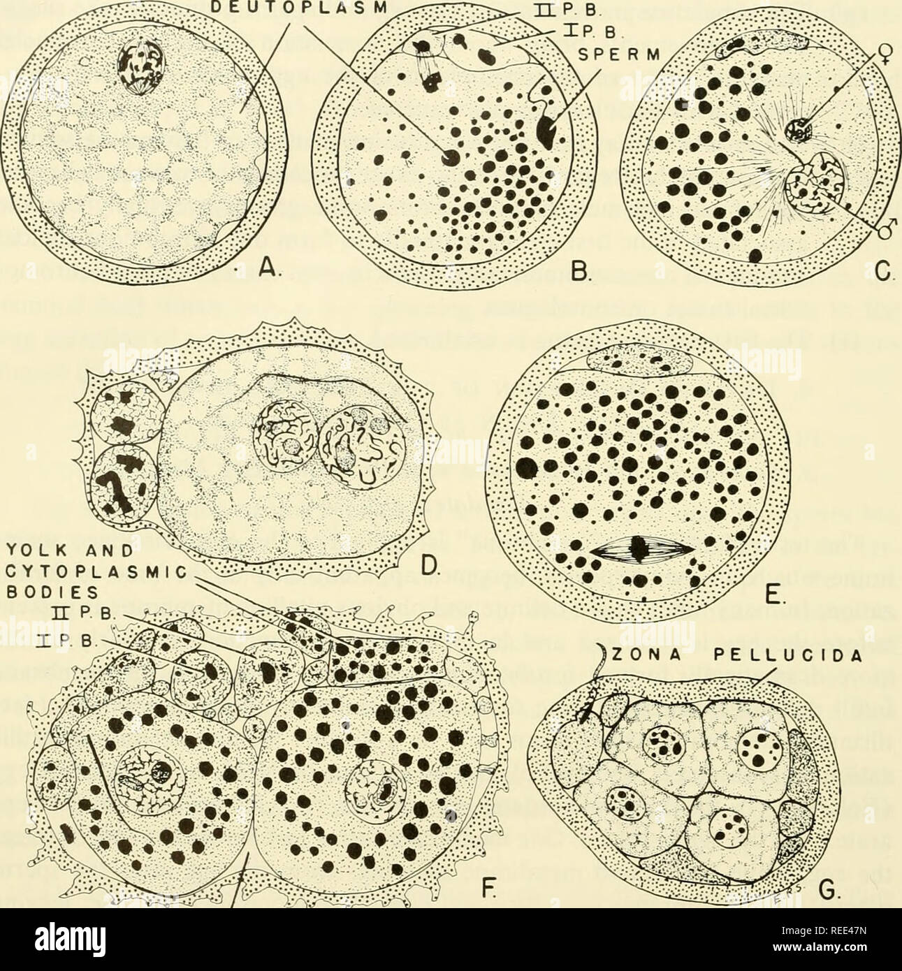 . Comparative embryology of the vertebrates; with 2057 drawings and photos. grouped as 380 illus. Vertebrates -- Embryology; Comparative embryology. 236 FERTILIZATION DEUTOPLASM. PERIVITELLiN E Fig. 118. Fertilization in the guinea pig. (After Lams, Arch. Biol., Paris, 28, figures slightly modified.) (A) Spindle of first maturation division. (B) Second maturation division completed; head of sperm in cytoplasm beginning to swell. (C) Sperm pro- nucleus, with tail still attached, greatly enlarged; female pronucleus small. (D) Pronuclei ready to fuse; chromatin material (chromosomes) evident with Stock Photohttps://www.alamy.com/image-license-details/?v=1https://www.alamy.com/comparative-embryology-of-the-vertebrates-with-2057-drawings-and-photos-grouped-as-380-illus-vertebrates-embryology-comparative-embryology-236-fertilization-deutoplasm-perivitellin-e-fig-118-fertilization-in-the-guinea-pig-after-lams-arch-biol-paris-28-figures-slightly-modified-a-spindle-of-first-maturation-division-b-second-maturation-division-completed-head-of-sperm-in-cytoplasm-beginning-to-swell-c-sperm-pro-nucleus-with-tail-still-attached-greatly-enlarged-female-pronucleus-small-d-pronuclei-ready-to-fuse-chromatin-material-chromosomes-evident-with-image232650649.html
RMREE47N–. Comparative embryology of the vertebrates; with 2057 drawings and photos. grouped as 380 illus. Vertebrates -- Embryology; Comparative embryology. 236 FERTILIZATION DEUTOPLASM. PERIVITELLiN E Fig. 118. Fertilization in the guinea pig. (After Lams, Arch. Biol., Paris, 28, figures slightly modified.) (A) Spindle of first maturation division. (B) Second maturation division completed; head of sperm in cytoplasm beginning to swell. (C) Sperm pro- nucleus, with tail still attached, greatly enlarged; female pronucleus small. (D) Pronuclei ready to fuse; chromatin material (chromosomes) evident with
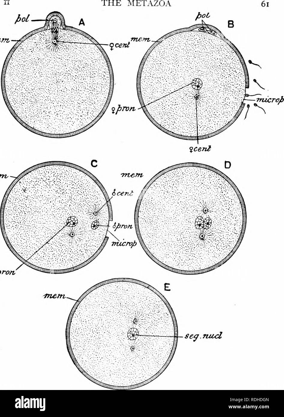 . A manual of zoology. iftern. qpro/t se^.nud Fig. 26.—Diagram illustrating the maturation and fertilisation of the ovum. A, formation of first polar globule; B, beginning of fertilisation, sperms approaching the micropyle or aperture in the enclosing membrane of the ovum through which the sperm enters; C, forma- tion of the male pronucleus; D, approximation of the male and female pronuclei; Et forma- tion of segmentation-nucleus; 9 cent, female centrosome; rf cent, male centrosome (the centrosomes are cell-structures not further referred to in this work); mem, egg-membrane; microp, micropyle; Stock Photohttps://www.alamy.com/image-license-details/?v=1https://www.alamy.com/a-manual-of-zoology-iftern-qprot-senud-fig-26diagram-illustrating-the-maturation-and-fertilisation-of-the-ovum-a-formation-of-first-polar-globule-b-beginning-of-fertilisation-sperms-approaching-the-micropyle-or-aperture-in-the-enclosing-membrane-of-the-ovum-through-which-the-sperm-enters-c-forma-tion-of-the-male-pronucleus-d-approximation-of-the-male-and-female-pronuclei-et-forma-tion-of-segmentation-nucleus-9-cent-female-centrosome-rf-cent-male-centrosome-the-centrosomes-are-cell-structures-not-further-referred-to-in-this-work-mem-egg-membrane-microp-micropyle-image232109157.html
RMRDHDGN–. A manual of zoology. iftern. qpro/t se^.nud Fig. 26.—Diagram illustrating the maturation and fertilisation of the ovum. A, formation of first polar globule; B, beginning of fertilisation, sperms approaching the micropyle or aperture in the enclosing membrane of the ovum through which the sperm enters; C, forma- tion of the male pronucleus; D, approximation of the male and female pronuclei; Et forma- tion of segmentation-nucleus; 9 cent, female centrosome; rf cent, male centrosome (the centrosomes are cell-structures not further referred to in this work); mem, egg-membrane; microp, micropyle;
 . The germ-cell cycle in animals . Cells. 56 GERM-CELL CYCLE IN ANIMALS disintegrate and disappear, apparently without performing any function. As in most other animals, these polar bodies may be considered abortive eggs. The female pronucleus moves into the central an- terior part of the egg where it becomes em- jiMp bedded in the cytoplasmic mass near the nurse chamber. It may now be designated as the cleavage nucleus, since the eggs of Miastor develop without ferti- lization and hence no male pronucleus is P PJ present to unite with it. The Fig. 15.—Miastor metraloas. Three of the four clca Stock Photohttps://www.alamy.com/image-license-details/?v=1https://www.alamy.com/the-germ-cell-cycle-in-animals-cells-56-germ-cell-cycle-in-animals-disintegrate-and-disappear-apparently-without-performing-any-function-as-in-most-other-animals-these-polar-bodies-may-be-considered-abortive-eggs-the-female-pronucleus-moves-into-the-central-an-terior-part-of-the-egg-where-it-becomes-em-jimp-bedded-in-the-cytoplasmic-mass-near-the-nurse-chamber-it-may-now-be-designated-as-the-cleavage-nucleus-since-the-eggs-of-miastor-develop-without-ferti-lization-and-hence-no-male-pronucleus-is-p-pj-present-to-unite-with-it-the-fig-15miastor-metraloas-three-of-the-four-clca-image232352833.html
RMRE0GBD–. The germ-cell cycle in animals . Cells. 56 GERM-CELL CYCLE IN ANIMALS disintegrate and disappear, apparently without performing any function. As in most other animals, these polar bodies may be considered abortive eggs. The female pronucleus moves into the central an- terior part of the egg where it becomes em- jiMp bedded in the cytoplasmic mass near the nurse chamber. It may now be designated as the cleavage nucleus, since the eggs of Miastor develop without ferti- lization and hence no male pronucleus is P PJ present to unite with it. The Fig. 15.—Miastor metraloas. Three of the four clca
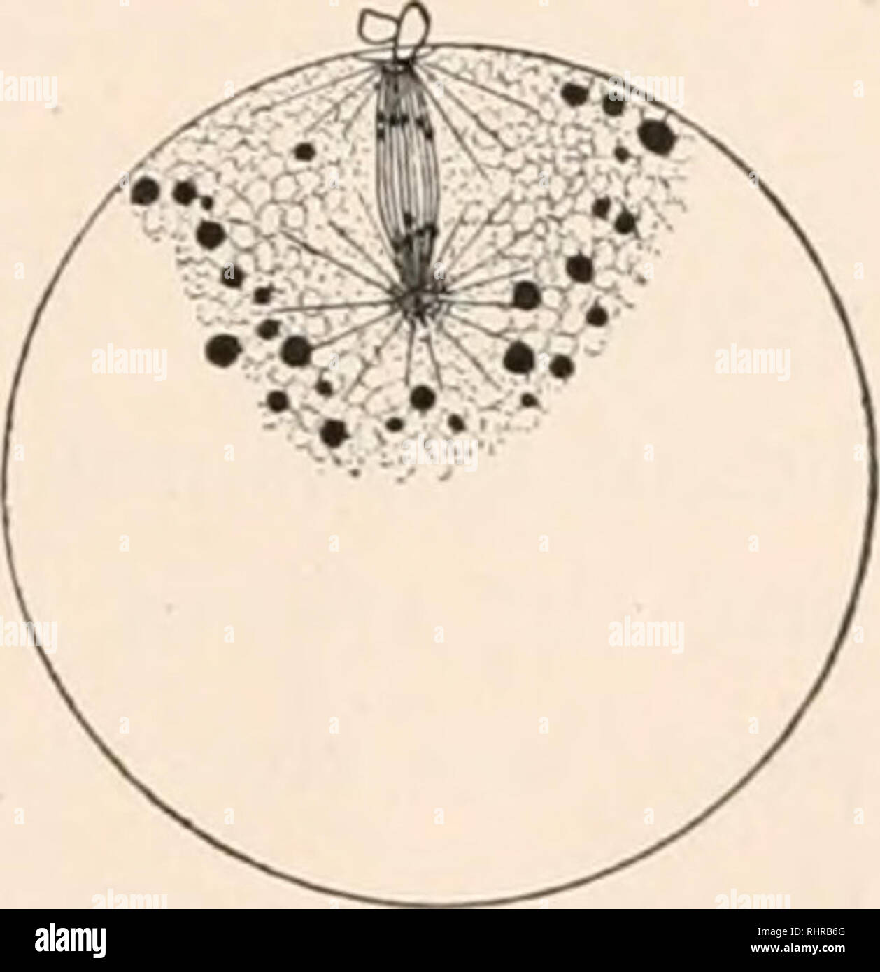 . The Biological bulletin. Biology; Zoology; Biology; Marine Biology. The metakinesis of the second polar spin- The chromo- m.uuration spindle. Wall of centrosome broken in pieces. Central corpuscles . connected by a spindle, die takes place very rapidly. New cortical zone form- SQmQS elonate divide transversely, and as ing. Chromosomes are J partly hoiiow. A few in- they move toward the poles, they assume a terzonal fibers are present. . , roundish form, and change into vesicular bodies which fuse to form the female pronucleus. During the time when they are fusing, the rays can be traced dir Stock Photohttps://www.alamy.com/image-license-details/?v=1https://www.alamy.com/the-biological-bulletin-biology-zoology-biology-marine-biology-the-metakinesis-of-the-second-polar-spin-the-chromo-muuration-spindle-wall-of-centrosome-broken-in-pieces-central-corpuscles-connected-by-a-spindle-die-takes-place-very-rapidly-new-cortical-zone-form-sqmqs-elonate-divide-transversely-and-as-ing-chromosomes-are-j-partly-hoiiow-a-few-in-they-move-toward-the-poles-they-assume-a-terzonal-fibers-are-present-roundish-form-and-change-into-vesicular-bodies-which-fuse-to-form-the-female-pronucleus-during-the-time-when-they-are-fusing-the-rays-can-be-traced-dir-image234697640.html
RMRHRB6G–. The Biological bulletin. Biology; Zoology; Biology; Marine Biology. The metakinesis of the second polar spin- The chromo- m.uuration spindle. Wall of centrosome broken in pieces. Central corpuscles . connected by a spindle, die takes place very rapidly. New cortical zone form- SQmQS elonate divide transversely, and as ing. Chromosomes are J partly hoiiow. A few in- they move toward the poles, they assume a terzonal fibers are present. . , roundish form, and change into vesicular bodies which fuse to form the female pronucleus. During the time when they are fusing, the rays can be traced dir
 . The Biological bulletin. Biology; Zoology; Biology; Marine Biology. pnorO O. FIG. 3. Camera lucida drawing of the egg nucleus, the larger circle, and the female pronucleus, the smaller circle. TV/, nucleus ; pronl. 9 > female pronucleus. FIG. 2. Outline drawing to show that sometimes the Hydractinian polyp branches. B, base of polyp. Reconstructed from several sections. this time on until new ova arise in a new polyp the chromatin does not possess such distinctness. A loose network is distributed through the nucleus with conspicuous masses where the threads cross. The achromatic substance Stock Photohttps://www.alamy.com/image-license-details/?v=1https://www.alamy.com/the-biological-bulletin-biology-zoology-biology-marine-biology-pnoro-o-fig-3-camera-lucida-drawing-of-the-egg-nucleus-the-larger-circle-and-the-female-pronucleus-the-smaller-circle-tv-nucleus-pronl-9-gt-female-pronucleus-fig-2-outline-drawing-to-show-that-sometimes-the-hydractinian-polyp-branches-b-base-of-polyp-reconstructed-from-several-sections-this-time-on-until-new-ova-arise-in-a-new-polyp-the-chromatin-does-not-possess-such-distinctness-a-loose-network-is-distributed-through-the-nucleus-with-conspicuous-masses-where-the-threads-cross-the-achromatic-substance-image234696256.html
RMRHR9D4–. The Biological bulletin. Biology; Zoology; Biology; Marine Biology. pnorO O. FIG. 3. Camera lucida drawing of the egg nucleus, the larger circle, and the female pronucleus, the smaller circle. TV/, nucleus ; pronl. 9 > female pronucleus. FIG. 2. Outline drawing to show that sometimes the Hydractinian polyp branches. B, base of polyp. Reconstructed from several sections. this time on until new ova arise in a new polyp the chromatin does not possess such distinctness. A loose network is distributed through the nucleus with conspicuous masses where the threads cross. The achromatic substance