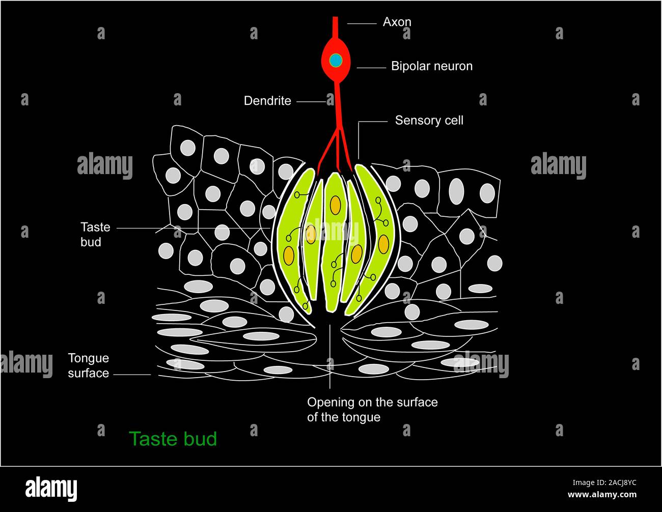
Taste bud anatomy. Diagram of the anatomical structure of a taste bud on a tongue. The surface of the tongue is at bottom, with the tastebud (rounded

RMID:Image ID:2ACJ8YC
Image details
Contributor:
Science Photo Library / Alamy Stock PhotoImage ID:
2ACJ8YCFile size:
50.4 MB (627.6 KB Compressed download)Releases:
Model - no | Property - noDo I need a release?Dimensions:
5000 x 3525 px | 42.3 x 29.8 cm | 16.7 x 11.8 inches | 300dpiDate taken:
28 February 2011Photographer:
FRANCIS LEROY, BIOCOSMOS/SCIENCE PHOTO LIBRARYMore information:
Taste bud anatomy. Diagram of the anatomical structure of a taste bud on a tongue. The surface of the tongue is at bottom, with the tastebud (rounded structure) at centre. Either side are tongue epithelial cells. Chemicals to be sensed enter the taste bud through the opening (lower centre), and trigger a response in the sensory cells (green). They then pass that response to the nerve dendrites and axon (red). At top (red and blue) is a bipolar neuron (bipolar nerve cell). The signals are passed to the brain and interpreted as taste sensations.