···
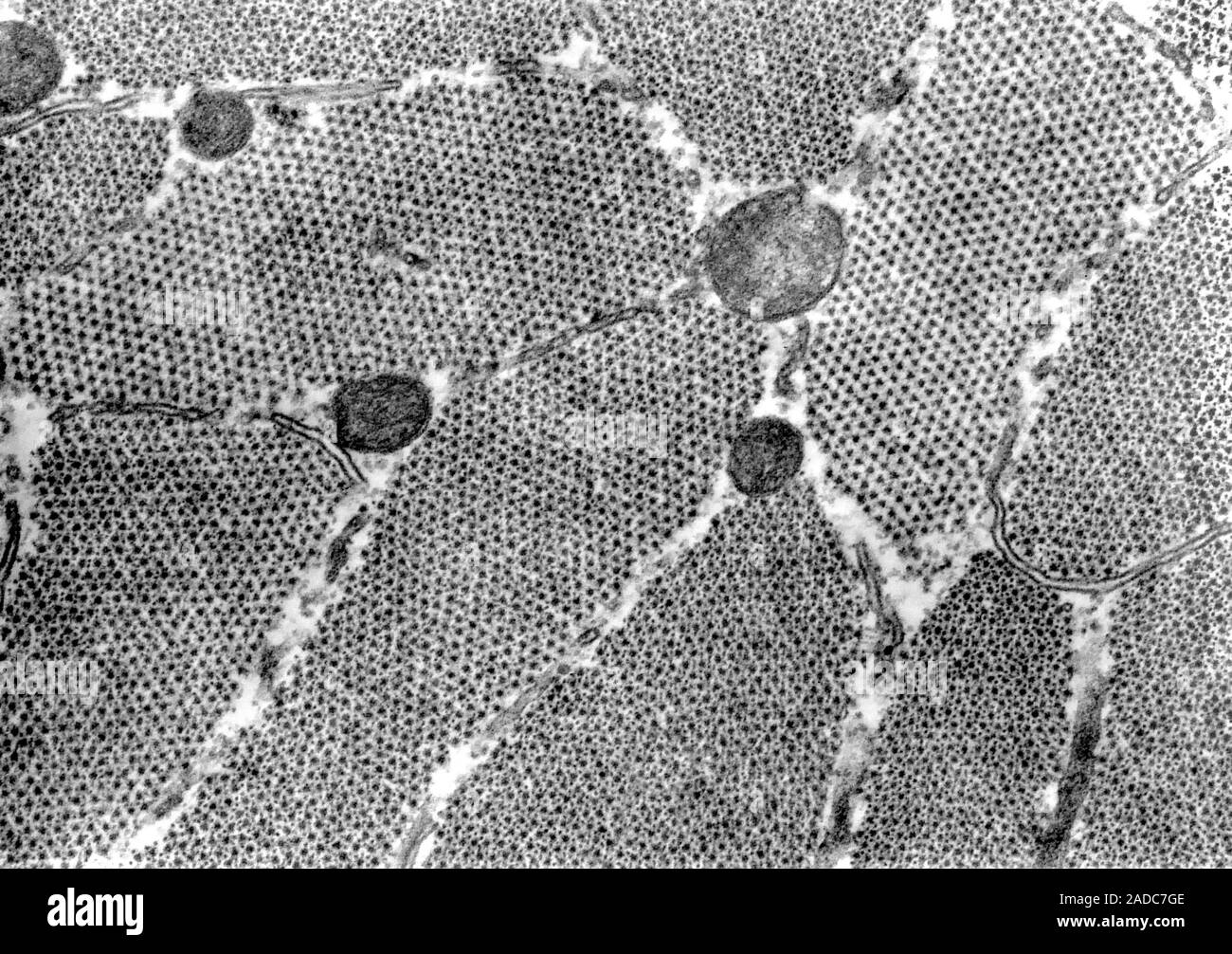
Transmission electron micrograph (TEM) showing cross-sectioned muscle myofibrils at the A-band level. Two myofibrils are sectioned at the H-band level Image details File size:
26.1 MB (1.9 MB Compressed download)
Open your image file to the full size using image processing software.
Dimensions:
3599 x 2537 px | 30.5 x 21.5 cm | 12 x 8.5 inches | 300dpi
Date taken:
6 February 2017
More information:
Transmission electron micrograph (TEM) showing cross-sectioned muscle myofibrils at the A-band level. Two myofibrils are sectioned at the H-band level, showing only myosin filaments. The elongated profiles are sarcoplasmic reticulum.
Search stock photos by tags
Similar stock images Transmission Electron Micrograph (TEM) showing Toxoplasma cyst. Toxoplasma is a parasitic protozoan that causes the disease Toxoplasmosis. Magnification unknown. Stock Photo https://www.alamy.com/image-license-details/?v=1 https://www.alamy.com/transmission-electron-micrograph-tem-showing-toxoplasma-cyst-toxoplasma-is-a-parasitic-protozoan-that-causes-the-disease-toxoplasmosis-magnification-unknown-image352825709.html RM 2BE0GP5 – Transmission Electron Micrograph (TEM) showing Toxoplasma cyst. Toxoplasma is a parasitic protozoan that causes the disease Toxoplasmosis. Magnification unknown. Transmission electron microscope (TEM) micrograph showing the labelling of vesicles of pinocytosis with an electron dense marker, the ruthenium red. Stock Photo https://www.alamy.com/image-license-details/?v=1 https://www.alamy.com/transmission-electron-microscope-tem-micrograph-showing-the-labelling-of-vesicles-of-pinocytosis-with-an-electron-dense-marker-the-ruthenium-red-image401738258.html RF 2E9GN6X – Transmission electron microscope (TEM) micrograph showing the labelling of vesicles of pinocytosis with an electron dense marker, the ruthenium red. Transmission Electron Micrograph (TEM) showing mature SIV (Simian Immunodeficiency Virus) particles. Magnification unknown. Stock Photo https://www.alamy.com/image-license-details/?v=1 https://www.alamy.com/transmission-electron-micrograph-tem-showing-mature-siv-simian-immunodeficiency-virus-particles-magnification-unknown-image352826237.html RM 2BE0HD1 – Transmission Electron Micrograph (TEM) showing mature SIV (Simian Immunodeficiency Virus) particles. Magnification unknown. Highly magnified, digitally colorized transmission electron microscopic (TEM) image showing ultrastructural details exhibited by three, spherical shaped, Middle East respiratory syndrome coronavirus (MERS-CoV) virions, 2014. MERs is in the same family as the novel coronavirus which began to infect patients in Wuhan, China in early 2020. Courtesy National Institute of Allergy and Infectious Diseases (NIAID)/CDC. Image produced in 2014. () Stock Photo https://www.alamy.com/image-license-details/?v=1 https://www.alamy.com/highly-magnified-digitally-colorized-transmission-electron-microscopic-tem-image-showing-ultrastructural-details-exhibited-by-three-spherical-shaped-middle-east-respiratory-syndrome-coronavirus-mers-cov-virions-2014-mers-is-in-the-same-family-as-the-novel-coronavirus-which-began-to-infect-patients-in-wuhan-china-in-early-2020-courtesy-national-institute-of-allergy-and-infectious-diseases-niaidcdc-image-produced-in-2014-image342265402.html RM 2ATRF0A – Highly magnified, digitally colorized transmission electron microscopic (TEM) image showing ultrastructural details exhibited by three, spherical shaped, Middle East respiratory syndrome coronavirus (MERS-CoV) virions, 2014. MERs is in the same family as the novel coronavirus which began to infect patients in Wuhan, China in early 2020. Courtesy National Institute of Allergy and Infectious Diseases (NIAID)/CDC. Image produced in 2014. () Transmission electron micrograph showing vesicular stomatitis virus (VSV) particles (yellow) budding from infected cells (burgundy). Stock Photo https://www.alamy.com/image-license-details/?v=1 https://www.alamy.com/transmission-electron-micrograph-showing-vesicular-stomatitis-virus-vsv-particles-yellow-budding-from-infected-cells-burgundy-image627689965.html RM 2YD5MXN – Transmission electron micrograph showing vesicular stomatitis virus (VSV) particles (yellow) budding from infected cells (burgundy). Transmission electron micrograph showing vesicular stomatitis virus VSV particles yellow budding from infected cells burgundy. Vesicular Stomatitis Virus VSV 016867 075 Stock Photo https://www.alamy.com/image-license-details/?v=1 https://www.alamy.com/transmission-electron-micrograph-showing-vesicular-stomatitis-virus-vsv-particles-yellow-budding-from-infected-cells-burgundy-vesicular-stomatitis-virus-vsv-016867-075-image627779009.html RM 2YD9PEW – Transmission electron micrograph showing vesicular stomatitis virus VSV particles yellow budding from infected cells burgundy. Vesicular Stomatitis Virus VSV 016867 075 A transmission electron micrograph (TEM) showing numerous hepatitis virions, of an unknown strain. Stock Photo https://www.alamy.com/image-license-details/?v=1 https://www.alamy.com/stock-photo-a-transmission-electron-micrograph-tem-showing-numerous-hepatitis-14688903.html RM AJBYJG – A transmission electron micrograph (TEM) showing numerous hepatitis virions, of an unknown strain. Created by Centers for Disease Control and Prevention (CDC) microbiologist Cynthia Goldsmith, this transmission electron micrograph (TEM) revealed some of the ultrastructural morphology displayed by a number of Ebola virus virions. Ebola is a severe, often-fatal disease in humans and nonhuman primates (monkeys, gorillas, and chimpanzees) that has appeared sporadically since its initial recognition in 1976. Image courtesy CDC/Cynthia Goldsmith. 1990. Stock Photo https://www.alamy.com/image-license-details/?v=1 https://www.alamy.com/created-by-centers-for-disease-control-and-prevention-cdc-microbiologist-image155841480.html RM K1F5B4 – Created by Centers for Disease Control and Prevention (CDC) microbiologist Cynthia Goldsmith, this transmission electron micrograph (TEM) revealed some of the ultrastructural morphology displayed by a number of Ebola virus virions. Ebola is a severe, often-fatal disease in humans and nonhuman primates (monkeys, gorillas, and chimpanzees) that has appeared sporadically since its initial recognition in 1976. Image courtesy CDC/Cynthia Goldsmith. 1990. Transmission electron microscope showing mitochondria. Stock Photo https://www.alamy.com/image-license-details/?v=1 https://www.alamy.com/stock-photo-transmission-electron-microscope-showing-mitochondria-76787883.html RM ECWYMB – Transmission electron microscope showing mitochondria. Under a high magnification of 446, 428X, this negatively-stained transmission electron micrograph (TEM) revealed some of the ultrastructural morphology displayed by numbers of rotavirus particles. A Reoviridae family member with an RNA core surrounded by a three-layered icosahedral protein capsid, the rotavirus is not enveloped, and measures 76.5nm in diameter. Rotavirus is a virus that causes gastroenteritis (inflammation of the stomach and intestines). The rotavirus disease causes severe watery diarrhea, often with vomiting, fever, and abdominal pain. In babies and young children, it can lea Stock Photo https://www.alamy.com/image-license-details/?v=1 https://www.alamy.com/under-a-high-magnification-of-446-428x-this-negatively-stained-transmission-image155848621.html RM K1FEE5 – Under a high magnification of 446, 428X, this negatively-stained transmission electron micrograph (TEM) revealed some of the ultrastructural morphology displayed by numbers of rotavirus particles. A Reoviridae family member with an RNA core surrounded by a three-layered icosahedral protein capsid, the rotavirus is not enveloped, and measures 76.5nm in diameter. Rotavirus is a virus that causes gastroenteritis (inflammation of the stomach and intestines). The rotavirus disease causes severe watery diarrhea, often with vomiting, fever, and abdominal pain. In babies and young children, it can lea . The Biological bulletin. Biology; Zoology; Biology; Marine Biology. 40 L R. PAGE. Figure 18. Transmission electron micrograph (TEM) showing different myolilament arrangements in myocytes of the left pedal muscle (LP) and the larval retractor muscle (LRM). Scale, I ^m. Figure 19. TEM of a frontal section through a 12-day larva, showing the ipsilateral and contralateral distal branches (arrows) of the left pedal muscle. Unlike the LRM. the left pedal muscle does not show obvious striations. BM = buccal mass. Scale, 10 pm. left pedal muscle bifurcates into two distal branches (Fig. 19). One bra Stock Photo https://www.alamy.com/image-license-details/?v=1 https://www.alamy.com/the-biological-bulletin-biology-zoology-biology-marine-biology-40-l-r-page-figure-18-transmission-electron-micrograph-tem-showing-different-myolilament-arrangements-in-myocytes-of-the-left-pedal-muscle-lp-and-the-larval-retractor-muscle-lrm-scale-i-m-figure-19-tem-of-a-frontal-section-through-a-12-day-larva-showing-the-ipsilateral-and-contralateral-distal-branches-arrows-of-the-left-pedal-muscle-unlike-the-lrm-the-left-pedal-muscle-does-not-show-obvious-striations-bm-=-buccal-mass-scale-10-pm-left-pedal-muscle-bifurcates-into-two-distal-branches-fig-19-one-bra-image234618082.html RM RHKNN6 – . The Biological bulletin. Biology; Zoology; Biology; Marine Biology. 40 L R. PAGE. Figure 18. Transmission electron micrograph (TEM) showing different myolilament arrangements in myocytes of the left pedal muscle (LP) and the larval retractor muscle (LRM). Scale, I ^m. Figure 19. TEM of a frontal section through a 12-day larva, showing the ipsilateral and contralateral distal branches (arrows) of the left pedal muscle. Unlike the LRM. the left pedal muscle does not show obvious striations. BM = buccal mass. Scale, 10 pm. left pedal muscle bifurcates into two distal branches (Fig. 19). One bra This 1976 negative stained transmission electron micrograph (TEM) depicted the ultrastructural features displayed by the mumps virus. Image courtesy CDC/Dr. F. A. Murphy, 1976. Stock Photo https://www.alamy.com/image-license-details/?v=1 https://www.alamy.com/this-1976-negative-stained-transmission-electron-micrograph-tem-depicted-image155841200.html RM K1F514 – This 1976 negative stained transmission electron micrograph (TEM) depicted the ultrastructural features displayed by the mumps virus. Image courtesy CDC/Dr. F. A. Murphy, 1976. Created by CDC microbiologist Cynthia Goldsmith, this transmission electron micrograph (TEM) revealed some of the ultrastructural morphology displayed by an Ebola virus virion. See PHIL 10816 for a colorized version of this image. Ebola is a severe, often-fatal disease in humans and nonhuman primates (monkeys, gorillas, and chimpanzees) that has appeared sporadically since its initial recognition in 1976. Image courtesy CDC/Cynthia Goldsmith. 1990. Stock Photo https://www.alamy.com/image-license-details/?v=1 https://www.alamy.com/created-by-cdc-microbiologist-cynthia-goldsmith-this-transmission-image155841496.html RM K1F5BM – Created by CDC microbiologist Cynthia Goldsmith, this transmission electron micrograph (TEM) revealed some of the ultrastructural morphology displayed by an Ebola virus virion. See PHIL 10816 for a colorized version of this image. Ebola is a severe, often-fatal disease in humans and nonhuman primates (monkeys, gorillas, and chimpanzees) that has appeared sporadically since its initial recognition in 1976. Image courtesy CDC/Cynthia Goldsmith. 1990. 