. Cunningham's Text-book of anatomy. Anatomy. Pars ossea of external acoustic meatu Recessus epitympanicus Malleus Cochlea Cavum tympani Membrana tympani Internal carotid artery Crus antihelicis inferior = Cyrnba conchse Crus helicis Pars cartilaginea of external acoustic meatus Cavum conchse Lower boundary of incisura intertragica Fig. 707. Frontal Section of Eight Ear : Anterior Half of Section, viewed from behind (natural size). finger into the meatus, and then alternately opening and shutting the mouth. The condyle of the mandible lies in front of the pars ossea, while between the condyle
Image details
Contributor:
Central Historic Books / Alamy Stock PhotoImage ID:
PFYA28File size:
7.1 MB (310.8 KB Compressed download)Releases:
Model - no | Property - noDo I need a release?Dimensions:
1751 x 1427 px | 29.7 x 24.2 cm | 11.7 x 9.5 inches | 150dpiMore information:
This image is a public domain image, which means either that copyright has expired in the image or the copyright holder has waived their copyright. Alamy charges you a fee for access to the high resolution copy of the image.
This image could have imperfections as it’s either historical or reportage.
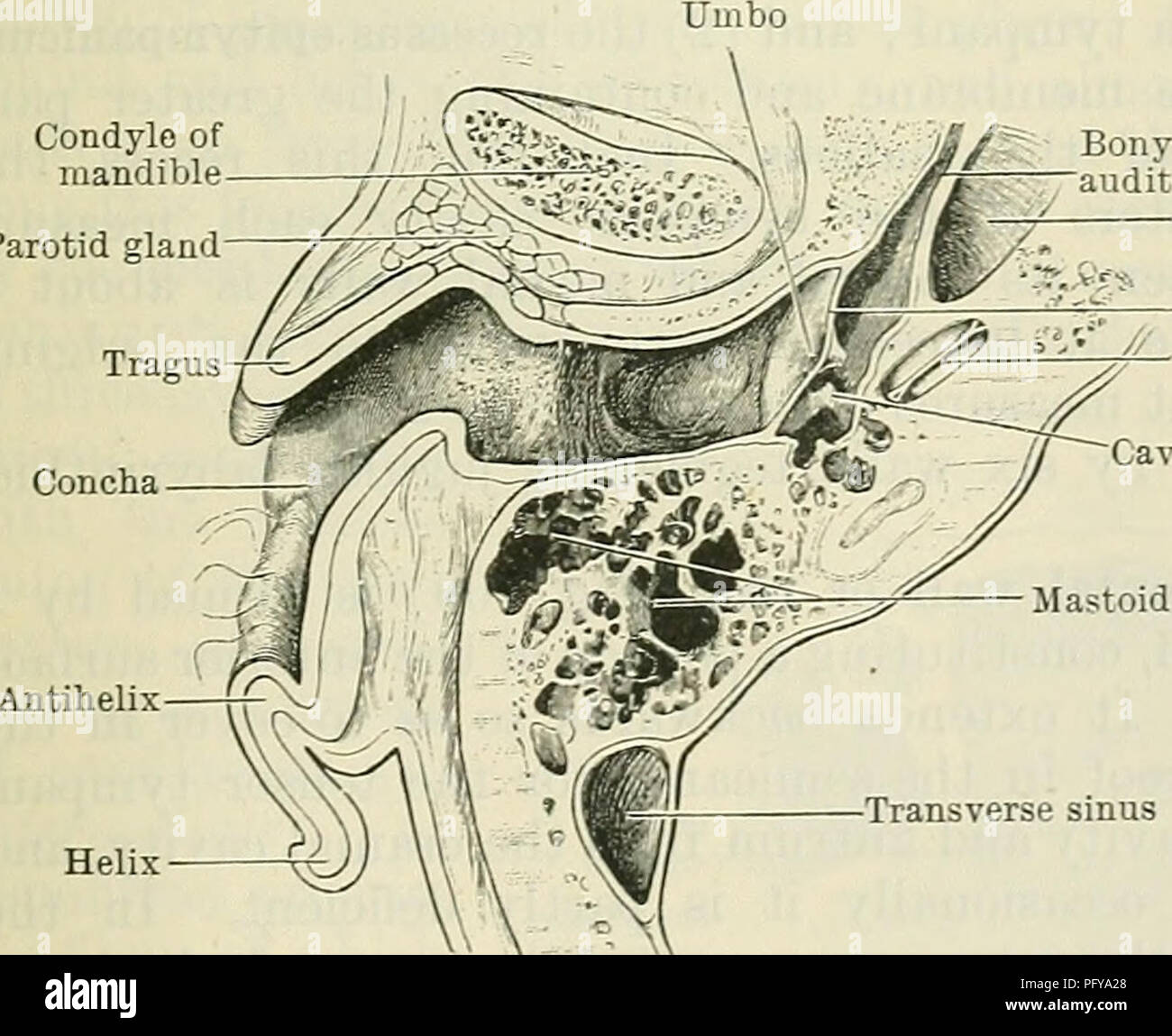
. Cunningham's Text-book of anatomy. Anatomy. Pars ossea of external acoustic meatu Recessus epitympanicus Malleus Cochlea Cavum tympani Membrana tympani Internal carotid artery Crus antihelicis inferior =~ Cyrnba conchse Crus helicis Pars cartilaginea of external acoustic meatus Cavum conchse Lower boundary of incisura intertragica Fig. 707. Frontal Section of Eight Ear : Anterior Half of Section, viewed from behind (natural size). finger into the meatus, and then alternately opening and shutting the mouth. The condyle of the mandible lies in front of the pars ossea, while between the condyle and the pars cartilaginea a portion of the parotid gland is sometimes present. Behind the pars ossea, and separated from it by a thin plate of bone, are the mastoid air-cells. Structure of the Meatus.—The cartilage of the meatus, directly continuous with that of the auricula, is folded on itself to form a groove, opening upwards and backwards, the margins of which are connected by fibrous tissue. The medial end of the cartilaginous tube is firmly fixed to the lateral margin of the bony meatus, whilst its lateral extremity is continuous with the cartilage of the tragus (p. 829). Two fissures exist in the anterior portion of the pars cartilaginea, and are filled by fibrous tissue. In> the lateral part of the meatus the cartil- age forms about three- fourths of the circum- ference of the tube; but, near the medial end of the pars cartil- aginea the cartilage forms merely a part of the anterior and lower boundaries of the canal. Condyle of mandible Parotid gland. Concha Bony part of auditory tube Internal carotid artery Membrana tympani First turn of cochlea Cavum tympani Mastoid air-cells Transverse sinus FIG. 708. —Horizontal Section through Right Ear; Upper Half of Section, seen from below (natural size). The pars ossea of the meatus is described on p. 127 ; but it may be well to state here that in the new-born child it is represented only by an incomplete ring of bone,