Quick filters:
Obturator artery Stock Photos and Images
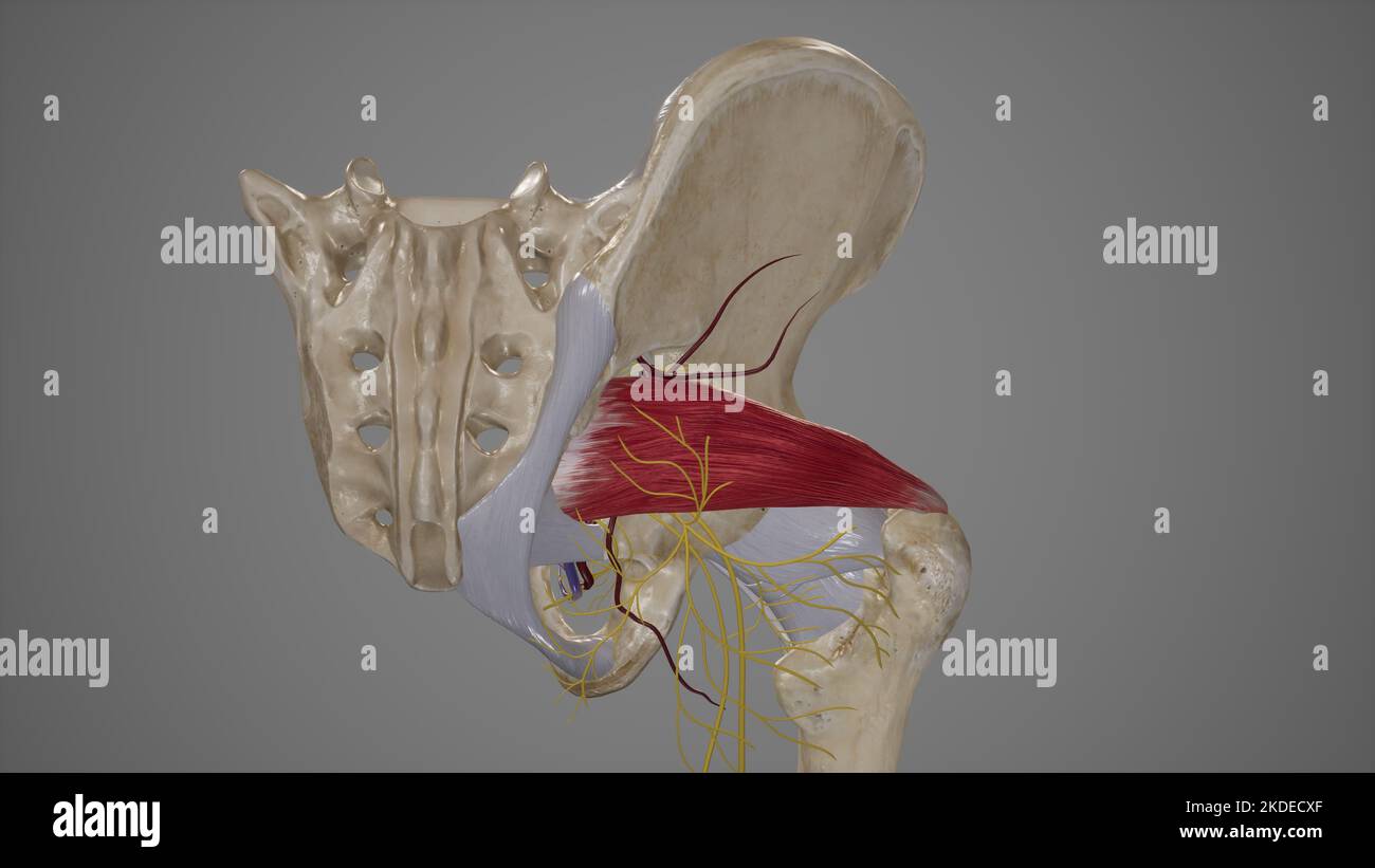
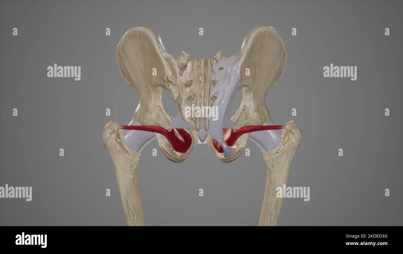 Medical Illustration of Obturator Internus Muscle Stock Photohttps://www.alamy.com/image-license-details/?v=1https://www.alamy.com/medical-illustration-of-obturator-internus-muscle-image490198452.html
RF2KDED3G–Medical Illustration of Obturator Internus Muscle
 The obturator artery is a branch of the anterior division of the internal iliac artery 3d illustration Stock Photohttps://www.alamy.com/image-license-details/?v=1https://www.alamy.com/the-obturator-artery-is-a-branch-of-the-anterior-division-of-the-internal-iliac-artery-3d-illustration-image596590591.html
RF2WJH1AR–The obturator artery is a branch of the anterior division of the internal iliac artery 3d illustration
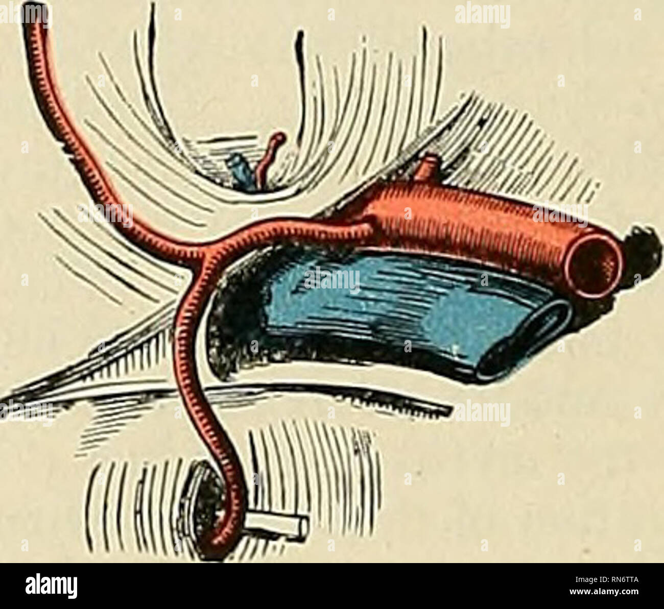 . Anatomy, descriptive and applied. Anatomy. Figs. 474 and 475.—Variations in origin and course of the obturator artery. When the obturator artery arises at the front of the pelvis from the deep epigastric, it descends almost vertically to the upper part of the obturator foramen. The artery in this course usually lies in contact with the external iliac vein and on the outer side of the femoral ring (Fig. 474); in such cases it would not be endangered in the operation for femoral hernia. Occasionally, however, it curves inward along the free margin of Gimbernat's ligament (Fig. 475), and under Stock Photohttps://www.alamy.com/image-license-details/?v=1https://www.alamy.com/anatomy-descriptive-and-applied-anatomy-figs-474-and-475variations-in-origin-and-course-of-the-obturator-artery-when-the-obturator-artery-arises-at-the-front-of-the-pelvis-from-the-deep-epigastric-it-descends-almost-vertically-to-the-upper-part-of-the-obturator-foramen-the-artery-in-this-course-usually-lies-in-contact-with-the-external-iliac-vein-and-on-the-outer-side-of-the-femoral-ring-fig-474-in-such-cases-it-would-not-be-endangered-in-the-operation-for-femoral-hernia-occasionally-however-it-curves-inward-along-the-free-margin-of-gimbernats-ligament-fig-475-and-under-image236793770.html
RMRN6TTA–. Anatomy, descriptive and applied. Anatomy. Figs. 474 and 475.—Variations in origin and course of the obturator artery. When the obturator artery arises at the front of the pelvis from the deep epigastric, it descends almost vertically to the upper part of the obturator foramen. The artery in this course usually lies in contact with the external iliac vein and on the outer side of the femoral ring (Fig. 474); in such cases it would not be endangered in the operation for femoral hernia. Occasionally, however, it curves inward along the free margin of Gimbernat's ligament (Fig. 475), and under
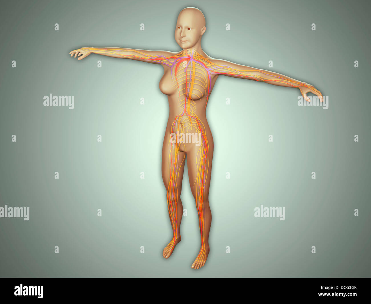 Anatomy of female body with arteries, veins and nervous system. Stock Photohttps://www.alamy.com/image-license-details/?v=1https://www.alamy.com/stock-photo-anatomy-of-female-body-with-arteries-veins-and-nervous-system-59361027.html
RFDCG3GK–Anatomy of female body with arteries, veins and nervous system.
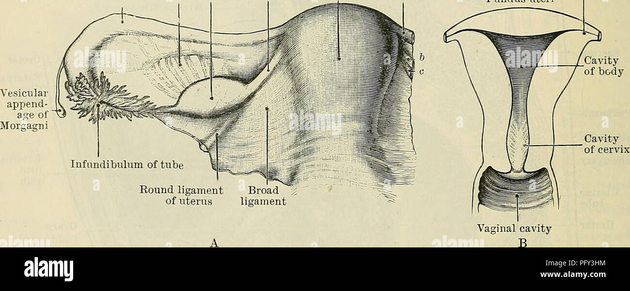 . Cunningham's Text-book of anatomy. Anatomy. 131: THE UKINO-GENITAL SYSTEM. against it, is depressed to form a little fossa termed the fossa ovarii, within which the ovary is placed. In the floor of this fossa are the obturator nerve and vessels. The tubal extremity of the ovary lies below the level of the external iliac vessels, and its uterine extremity is placed just above the level of the peritoneum covering the pelvic floor. The fossa ovarii, in which the ovary lies, extends as far forwards as the obliterated umbilical artery, and backwards as far as the ureter and uterine vessels. Thus Stock Photohttps://www.alamy.com/image-license-details/?v=1https://www.alamy.com/cunninghams-text-book-of-anatomy-anatomy-131-the-ukino-genital-system-against-it-is-depressed-to-form-a-little-fossa-termed-the-fossa-ovarii-within-which-the-ovary-is-placed-in-the-floor-of-this-fossa-are-the-obturator-nerve-and-vessels-the-tubal-extremity-of-the-ovary-lies-below-the-level-of-the-external-iliac-vessels-and-its-uterine-extremity-is-placed-just-above-the-level-of-the-peritoneum-covering-the-pelvic-floor-the-fossa-ovarii-in-which-the-ovary-lies-extends-as-far-forwards-as-the-obliterated-umbilical-artery-and-backwards-as-far-as-the-ureter-and-uterine-vessels-thus-image216339808.html
RMPFY3HM–. Cunningham's Text-book of anatomy. Anatomy. 131: THE UKINO-GENITAL SYSTEM. against it, is depressed to form a little fossa termed the fossa ovarii, within which the ovary is placed. In the floor of this fossa are the obturator nerve and vessels. The tubal extremity of the ovary lies below the level of the external iliac vessels, and its uterine extremity is placed just above the level of the peritoneum covering the pelvic floor. The fossa ovarii, in which the ovary lies, extends as far forwards as the obliterated umbilical artery, and backwards as far as the ureter and uterine vessels. Thus
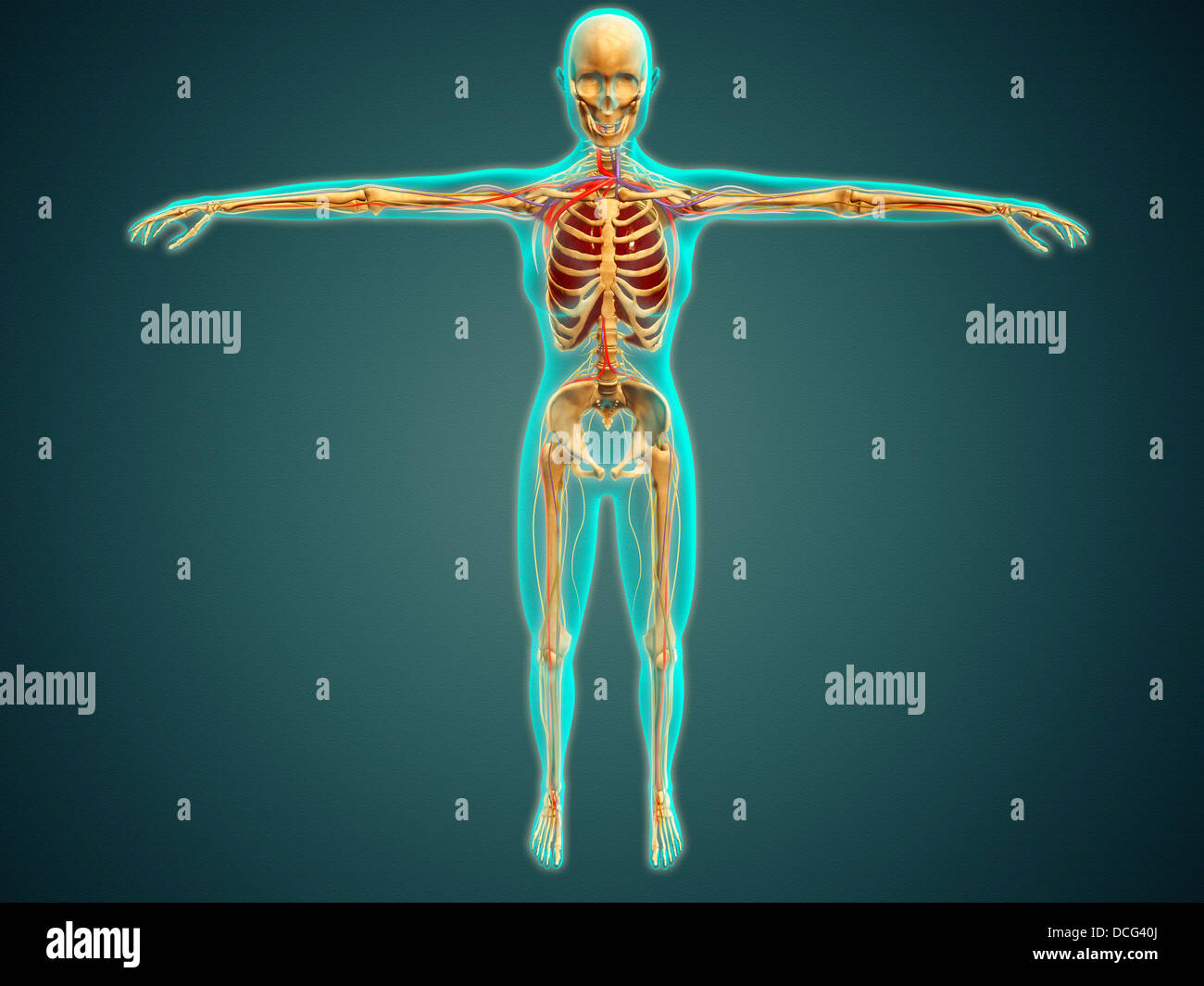 Medical illustration of human body showing skeletal system, arteries, veins, and nervous system. Stock Photohttps://www.alamy.com/image-license-details/?v=1https://www.alamy.com/stock-photo-medical-illustration-of-human-body-showing-skeletal-system-arteries-59361362.html
RFDCG40J–Medical illustration of human body showing skeletal system, arteries, veins, and nervous system.
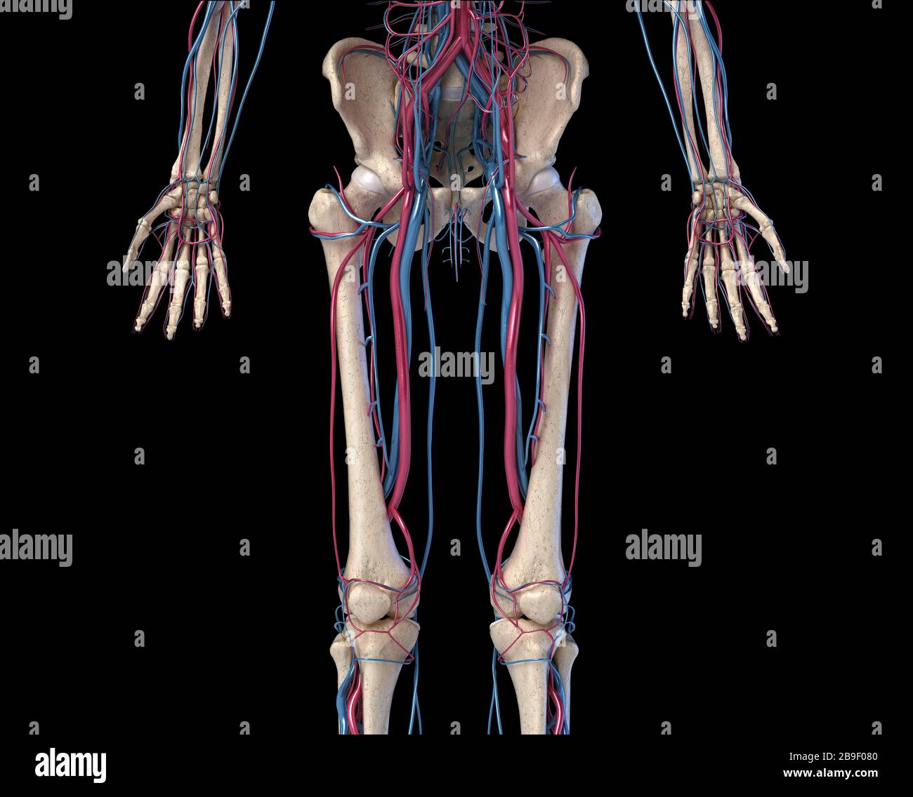 Front view of hip, limbs and hands of skeletal system with veins and arteries, black background. Stock Photohttps://www.alamy.com/image-license-details/?v=1https://www.alamy.com/front-view-of-hip-limbs-and-hands-of-skeletal-system-with-veins-and-arteries-black-background-image350068768.html
RF2B9F080–Front view of hip, limbs and hands of skeletal system with veins and arteries, black background.
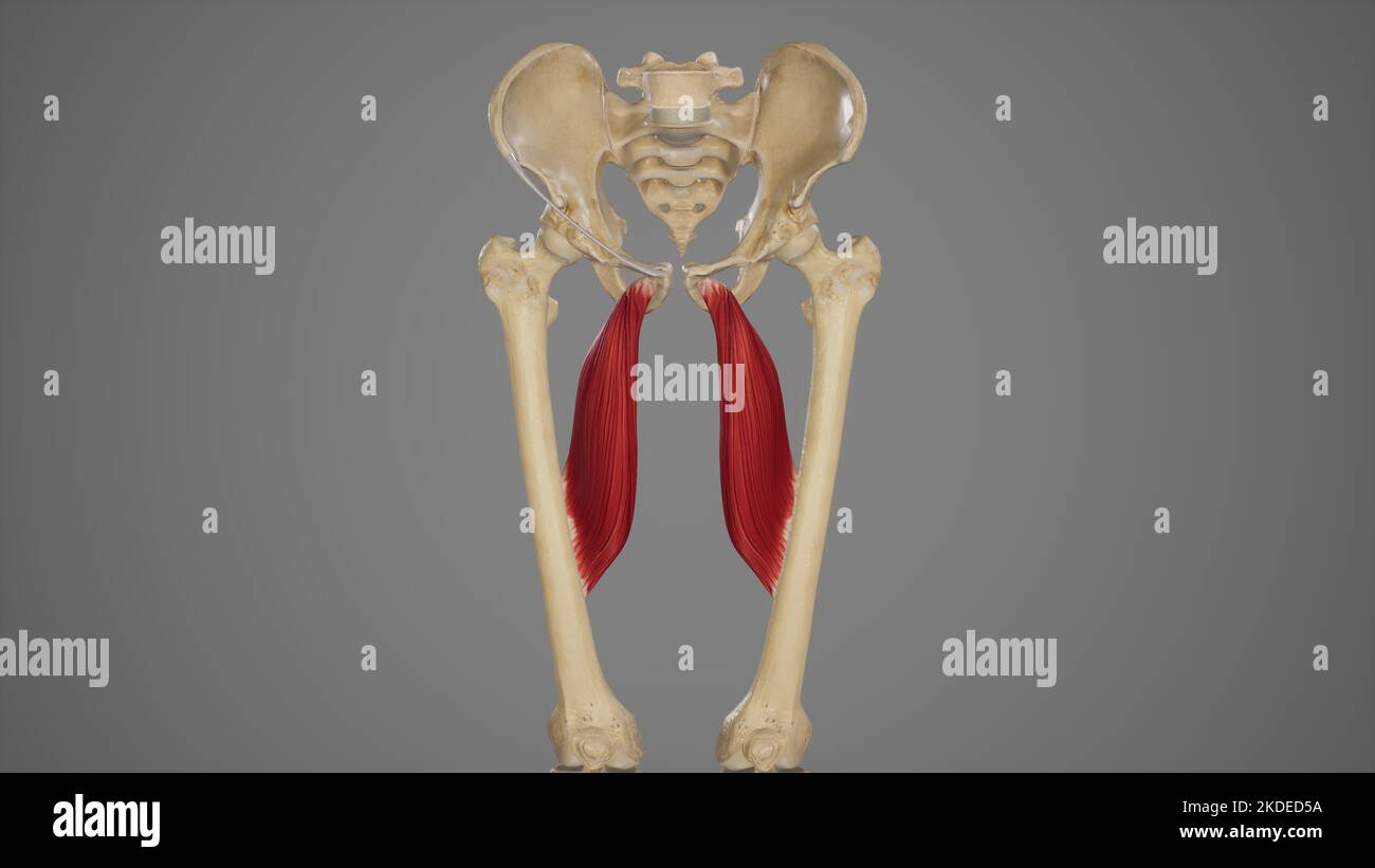 Medical Accurate Illustration of Adductor Longus Stock Photohttps://www.alamy.com/image-license-details/?v=1https://www.alamy.com/medical-accurate-illustration-of-adductor-longus-image490198502.html
RF2KDED5A–Medical Accurate Illustration of Adductor Longus
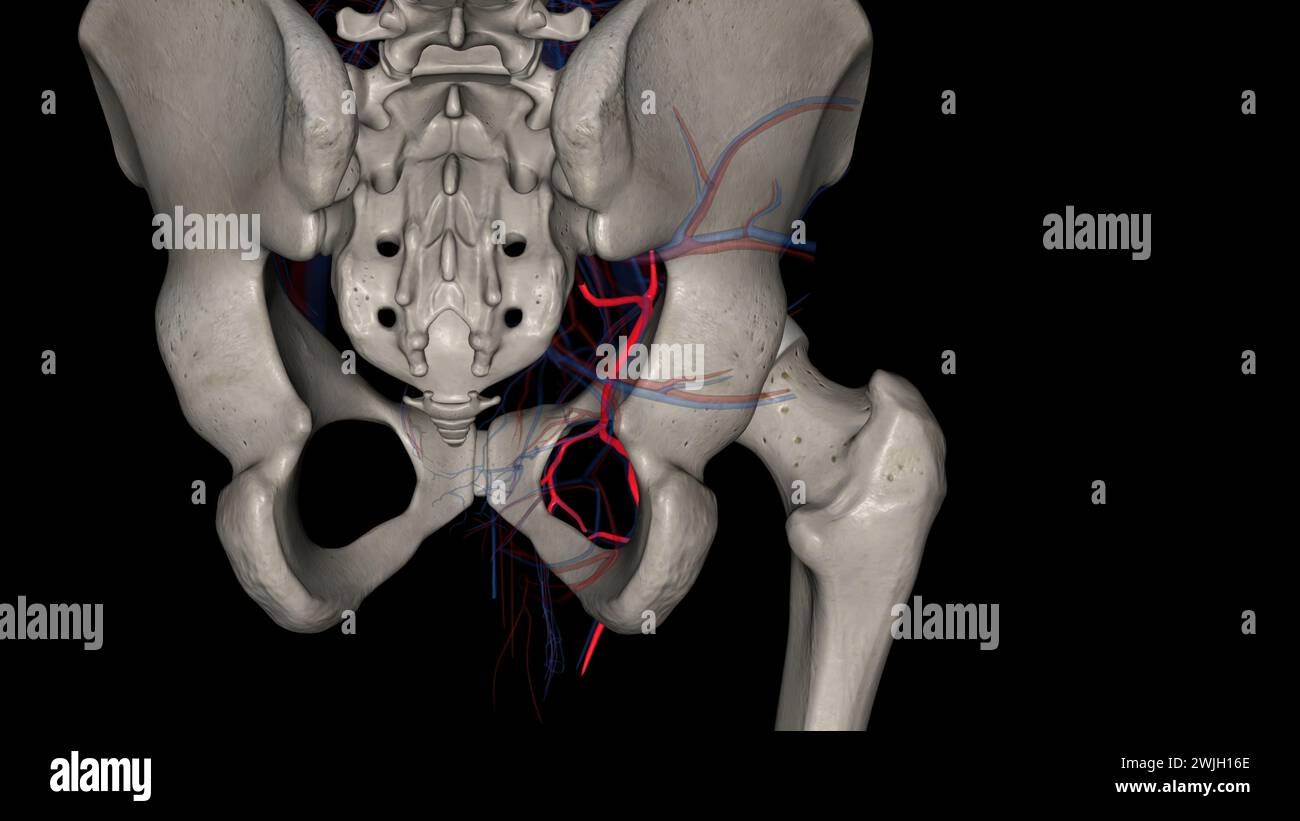 The obturator artery is a branch of the anterior division of the internal iliac artery 3d illustration Stock Photohttps://www.alamy.com/image-license-details/?v=1https://www.alamy.com/the-obturator-artery-is-a-branch-of-the-anterior-division-of-the-internal-iliac-artery-3d-illustration-image596590470.html
RF2WJH16E–The obturator artery is a branch of the anterior division of the internal iliac artery 3d illustration
 Operative surgery . are, within, Gimber-nats ligament, and without, the femoral vein (Fig. 1117), surrounded by its sheath. Throughout the course of thisthe femoral vein lies at the outerside. The distinctive coverings of thisprotrusion are the cribriform fascia,crural sheath, and septumcrurale. The importantvascular relations are thoseof the femoral vein andthe obturator artery. Taxis should be em-ployed with greater cau-tion and for a shorter timein femoral than in ingui-nal hernia, since the con-stricting influences aregreater, and the neck ofthe sac much smaller inthe former. The fact, is Stock Photohttps://www.alamy.com/image-license-details/?v=1https://www.alamy.com/operative-surgery-are-within-gimber-nats-ligament-and-without-the-femoral-vein-fig-1117-surrounded-by-its-sheath-throughout-the-course-of-thisthe-femoral-vein-lies-at-the-outerside-the-distinctive-coverings-of-thisprotrusion-are-the-cribriform-fasciacrural-sheath-and-septumcrurale-the-importantvascular-relations-are-thoseof-the-femoral-vein-andthe-obturator-artery-taxis-should-be-em-ployed-with-greater-cau-tion-and-for-a-shorter-timein-femoral-than-in-ingui-nal-hernia-since-the-con-stricting-influences-aregreater-and-the-neck-ofthe-sac-much-smaller-inthe-former-the-fact-is-image342668318.html
RM2AWDTX6–Operative surgery . are, within, Gimber-nats ligament, and without, the femoral vein (Fig. 1117), surrounded by its sheath. Throughout the course of thisthe femoral vein lies at the outerside. The distinctive coverings of thisprotrusion are the cribriform fascia,crural sheath, and septumcrurale. The importantvascular relations are thoseof the femoral vein andthe obturator artery. Taxis should be em-ployed with greater cau-tion and for a shorter timein femoral than in ingui-nal hernia, since the con-stricting influences aregreater, and the neck ofthe sac much smaller inthe former. The fact, is
 . Cunningham's Text-book of anatomy. Anatomy. 988 THE VASCULAE SYSTEM. — Superficial epigastric vein Superficial circumflex —" iliac vein Superficial external pudendal vein Femoral vein Great saphenous vein Lateral superficial femoral vein Medial superficial femoral vein artery, whilst just before its termination it crosses the lateral side of the hypo- gastric artery, and separates that vessel from the medial border of the psoas major muscle. In its whole course the vein lies anterior to the obturator nerve. It is usually provided with one bicuspid valve ; sometimes there are two, but bo Stock Photohttps://www.alamy.com/image-license-details/?v=1https://www.alamy.com/cunninghams-text-book-of-anatomy-anatomy-988-the-vasculae-system-superficial-epigastric-vein-superficial-circumflex-quot-iliac-vein-superficial-external-pudendal-vein-femoral-vein-great-saphenous-vein-lateral-superficial-femoral-vein-medial-superficial-femoral-vein-artery-whilst-just-before-its-termination-it-crosses-the-lateral-side-of-the-hypo-gastric-artery-and-separates-that-vessel-from-the-medial-border-of-the-psoas-major-muscle-in-its-whole-course-the-vein-lies-anterior-to-the-obturator-nerve-it-is-usually-provided-with-one-bicuspid-valve-sometimes-there-are-two-but-bo-image216334416.html
RMPFXTN4–. Cunningham's Text-book of anatomy. Anatomy. 988 THE VASCULAE SYSTEM. — Superficial epigastric vein Superficial circumflex —" iliac vein Superficial external pudendal vein Femoral vein Great saphenous vein Lateral superficial femoral vein Medial superficial femoral vein artery, whilst just before its termination it crosses the lateral side of the hypo- gastric artery, and separates that vessel from the medial border of the psoas major muscle. In its whole course the vein lies anterior to the obturator nerve. It is usually provided with one bicuspid valve ; sometimes there are two, but bo
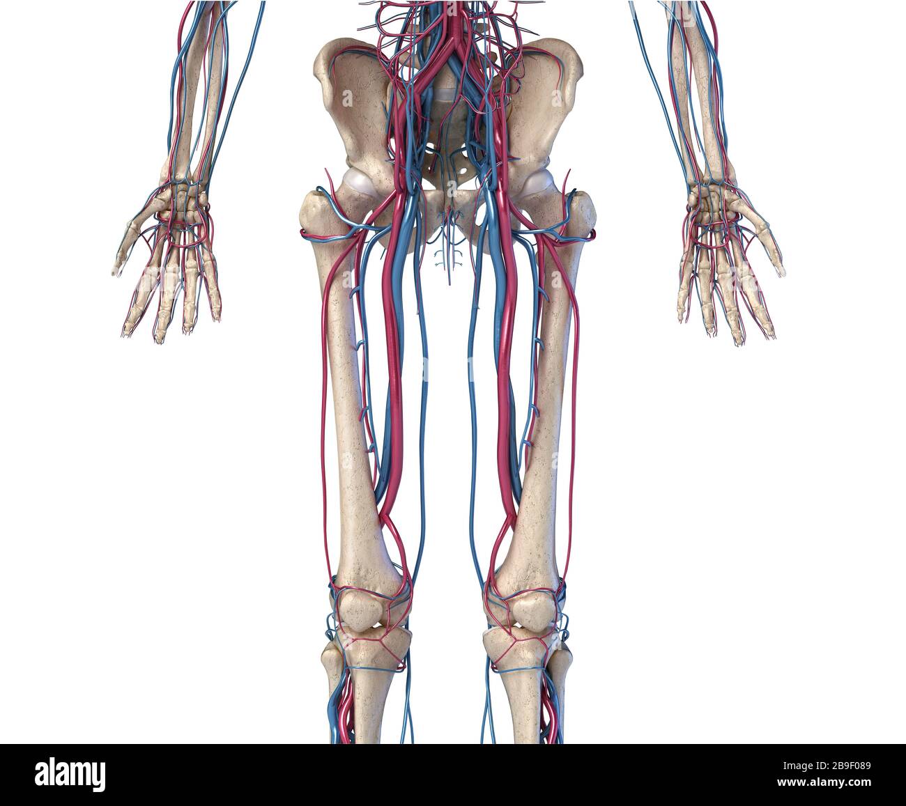 Front view of hip, limbs and hands of skeletal system with veins and arteries, white background. Stock Photohttps://www.alamy.com/image-license-details/?v=1https://www.alamy.com/front-view-of-hip-limbs-and-hands-of-skeletal-system-with-veins-and-arteries-white-background-image350068777.html
RF2B9F089–Front view of hip, limbs and hands of skeletal system with veins and arteries, white background.
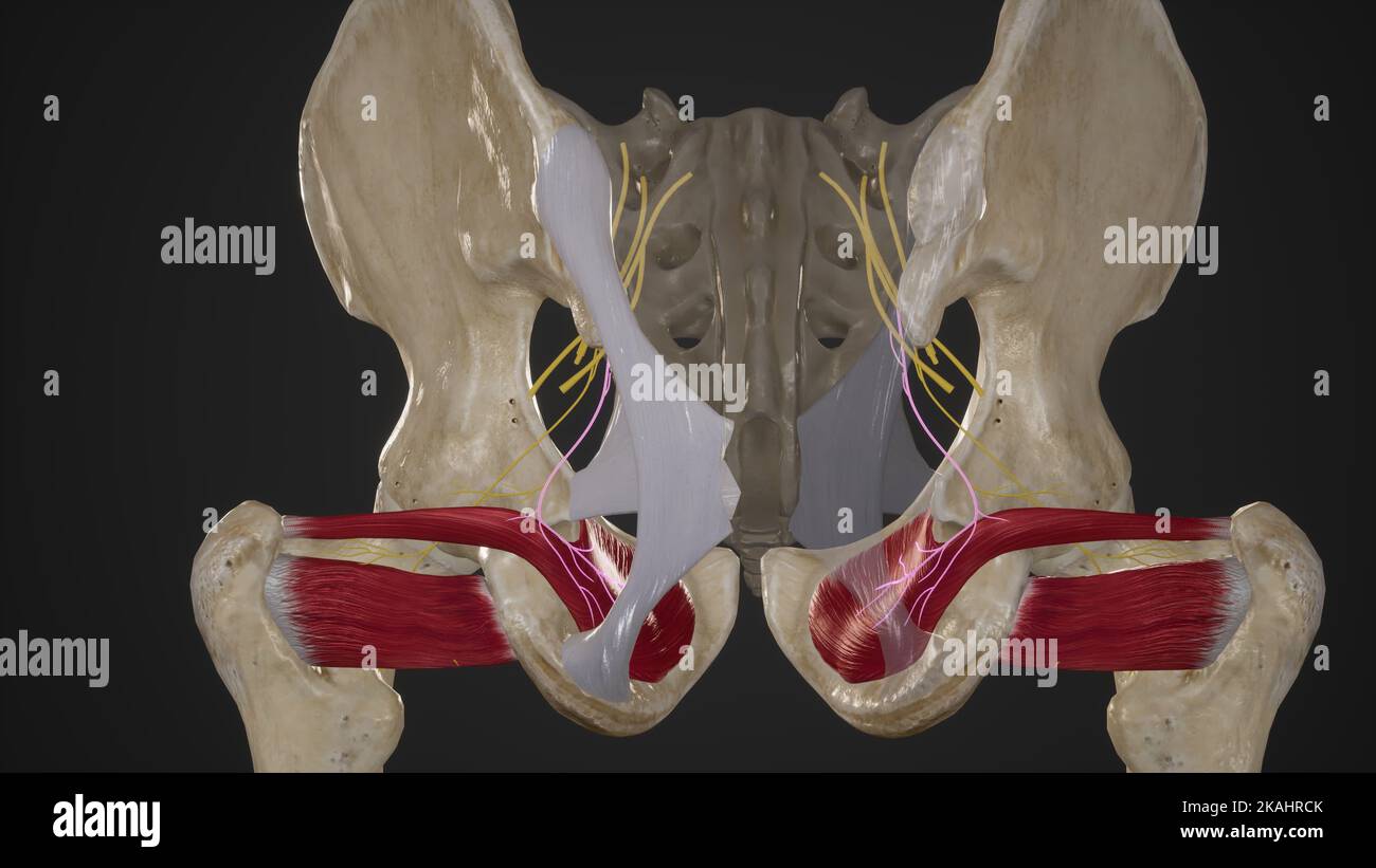 Nerve To Obturator Internus Stock Photohttps://www.alamy.com/image-license-details/?v=1https://www.alamy.com/nerve-to-obturator-internus-image488428435.html
RF2KAHRCK–Nerve To Obturator Internus
 The obturator artery is a branch of the anterior division of the internal iliac artery 3d illustration Stock Photohttps://www.alamy.com/image-license-details/?v=1https://www.alamy.com/the-obturator-artery-is-a-branch-of-the-anterior-division-of-the-internal-iliac-artery-3d-illustration-image596592832.html
RF2WJH46T–The obturator artery is a branch of the anterior division of the internal iliac artery 3d illustration
 Plastic surgery; its principles and practice . Fig. 812.—Arteries of the skin of the buttock and posterior surface of the thigh (Man-chot).—I. Cutaneous branches of the gluteal artery. 2. Cutaneous branches of the comesnervi ischiadic! artery. 3. Cutaneous branches of the internal pudic artery. 4. Cuta-neous branches of the lateral sacral arteries. ,5. Cutaneous branches of the ileo4umbaTand the last lumbar arteries. 6. Cutaneous branches of the obturator artery. 7. Cuta-neous branches of the internal circumflex artery. 8. Cutaneous branches of the profundafemoris artery. 9. Cutaneous branches Stock Photohttps://www.alamy.com/image-license-details/?v=1https://www.alamy.com/plastic-surgery-its-principles-and-practice-fig-812arteries-of-the-skin-of-the-buttock-and-posterior-surface-of-the-thigh-man-choti-cutaneous-branches-of-the-gluteal-artery-2-cutaneous-branches-of-the-comesnervi-ischiadic!-artery-3-cutaneous-branches-of-the-internal-pudic-artery-4-cuta-neous-branches-of-the-lateral-sacral-arteries-5-cutaneous-branches-of-the-ileo4umbatand-the-last-lumbar-arteries-6-cutaneous-branches-of-the-obturator-artery-7-cuta-neous-branches-of-the-internal-circumflex-artery-8-cutaneous-branches-of-the-profundafemoris-artery-9-cutaneous-branches-image338180164.html
RM2AJ5C70–Plastic surgery; its principles and practice . Fig. 812.—Arteries of the skin of the buttock and posterior surface of the thigh (Man-chot).—I. Cutaneous branches of the gluteal artery. 2. Cutaneous branches of the comesnervi ischiadic! artery. 3. Cutaneous branches of the internal pudic artery. 4. Cuta-neous branches of the lateral sacral arteries. ,5. Cutaneous branches of the ileo4umbaTand the last lumbar arteries. 6. Cutaneous branches of the obturator artery. 7. Cuta-neous branches of the internal circumflex artery. 8. Cutaneous branches of the profundafemoris artery. 9. Cutaneous branches
 . The surgical anatomy of the horse ... Horses. -18. Plate XXIII.—Obtukatok and Anterior Crural Nerves I. External iliac artery. 2. E.xternal iliac vein. j. Obturator externus. 4. Filaments of obturator nerve. 5. Obturator foramen. 6. Cotyloid cavity. 7, Obturator internus. 8. Obturator nerve. 9. Bladder (distended). 10. Internal iliac artery. 1 i Recium. 12. Posterior aorta. 13 Circumflex-iliac artery. 14. Psoas parvus, cut through to expose anterior crural nerve. 15. Psoas magnus. i5. Anterior crural nerve. 17. Vastus internus. 18. Rectus femoris.. Please note that these images are extracted Stock Photohttps://www.alamy.com/image-license-details/?v=1https://www.alamy.com/the-surgical-anatomy-of-the-horse-horses-18-plate-xxiiiobtukatok-and-anterior-crural-nerves-i-external-iliac-artery-2-external-iliac-vein-j-obturator-externus-4-filaments-of-obturator-nerve-5-obturator-foramen-6-cotyloid-cavity-7-obturator-internus-8-obturator-nerve-9-bladder-distended-10-internal-iliac-artery-1-i-recium-12-posterior-aorta-13-circumflex-iliac-artery-14-psoas-parvus-cut-through-to-expose-anterior-crural-nerve-15-psoas-magnus-i5-anterior-crural-nerve-17-vastus-internus-18-rectus-femoris-please-note-that-these-images-are-extracted-image216394405.html
RMPG1H7H–. The surgical anatomy of the horse ... Horses. -18. Plate XXIII.—Obtukatok and Anterior Crural Nerves I. External iliac artery. 2. E.xternal iliac vein. j. Obturator externus. 4. Filaments of obturator nerve. 5. Obturator foramen. 6. Cotyloid cavity. 7, Obturator internus. 8. Obturator nerve. 9. Bladder (distended). 10. Internal iliac artery. 1 i Recium. 12. Posterior aorta. 13 Circumflex-iliac artery. 14. Psoas parvus, cut through to expose anterior crural nerve. 15. Psoas magnus. i5. Anterior crural nerve. 17. Vastus internus. 18. Rectus femoris.. Please note that these images are extracted
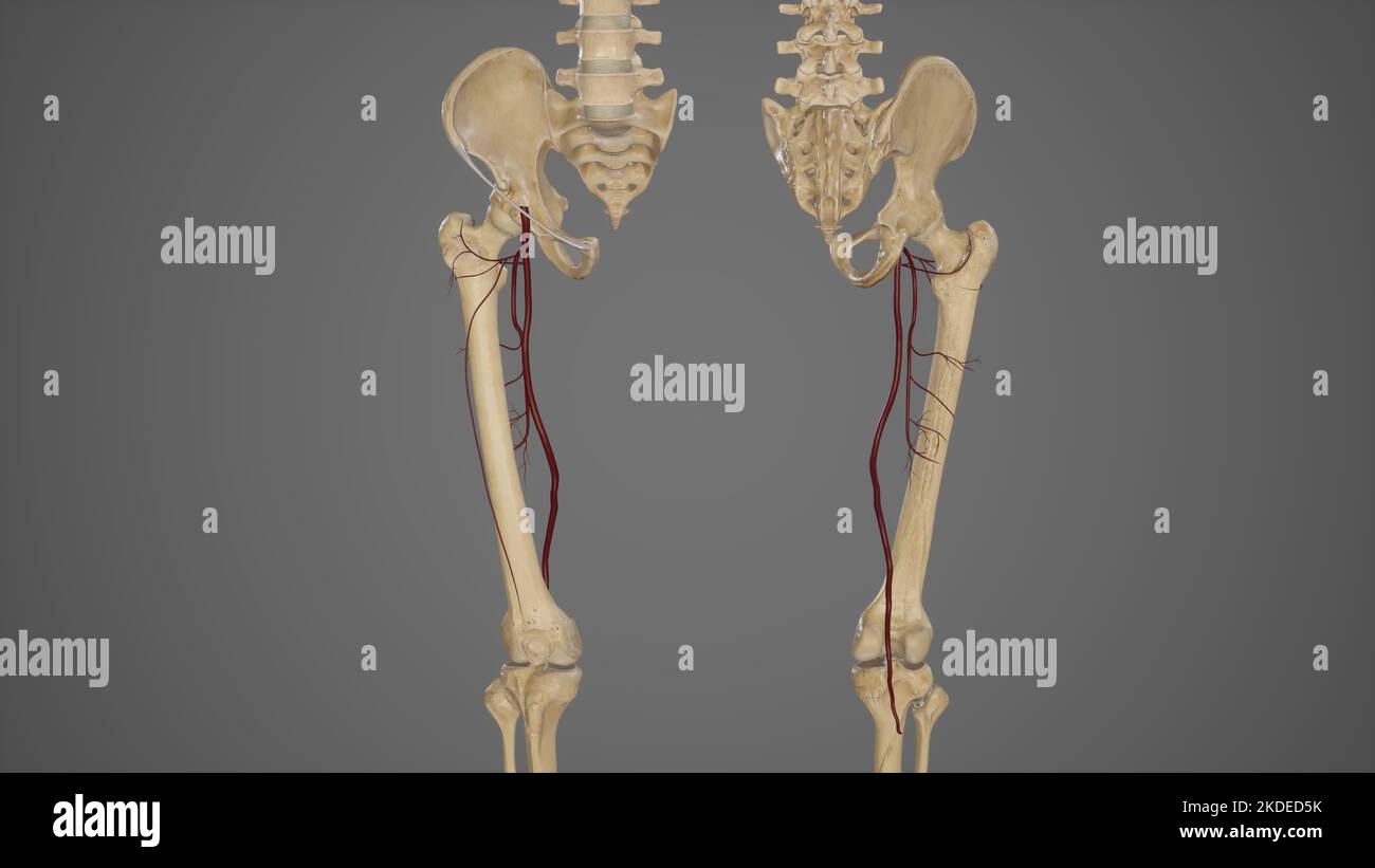 Anterior and Posterior View of Profunda Femoris Artery and Its Branches Stock Photohttps://www.alamy.com/image-license-details/?v=1https://www.alamy.com/anterior-and-posterior-view-of-profunda-femoris-artery-and-its-branches-image490198511.html
RF2KDED5K–Anterior and Posterior View of Profunda Femoris Artery and Its Branches
 The obturator artery is a branch of the anterior division of the internal iliac artery 3d illustration Stock Photohttps://www.alamy.com/image-license-details/?v=1https://www.alamy.com/the-obturator-artery-is-a-branch-of-the-anterior-division-of-the-internal-iliac-artery-3d-illustration-image596590225.html
RF2WJH0WN–The obturator artery is a branch of the anterior division of the internal iliac artery 3d illustration
 An atlas of human anatomy for students and physicians . unis Internal iliac arteryA. hypogastuca Sacral promontory Promontorium Anterior superior spine of the iliumSpina iliaca anterior superior iPouparts ligament (superficial femoral arch)I Lig- inguinale (Iouparti) I Internal or deep abdominal ring- fi)1 Deep or inferior epigastric arteryi and vein ? A et Vv. epi;-;astric;i- inferiores Vestige of the obliterated hypogastric artery, orexternal umbilical ligament (•;!,- Obturator artery and vein ;L V. ol.turatoria ^Rectal fascia^ Iri^ ia diaphragmatis pelvis superior ^ Pubic symphysis vmphysis Stock Photohttps://www.alamy.com/image-license-details/?v=1https://www.alamy.com/an-atlas-of-human-anatomy-for-students-and-physicians-unis-internal-iliac-arterya-hypogastuca-sacral-promontory-promontorium-anterior-superior-spine-of-the-iliumspina-iliaca-anterior-superior-ipouparts-ligament-superficial-femoral-archi-lig-inguinale-iouparti-i-internal-or-deep-abdominal-ring-fi1-deep-or-inferior-epigastric-arteryi-and-vein-a-et-vv-epi-astrici-inferiores-vestige-of-the-obliterated-hypogastric-artery-orexternal-umbilical-ligament-!-obturator-artery-and-vein-l-v-olturatoria-rectal-fascia-iri-ia-diaphragmatis-pelvis-superior-pubic-symphysis-vmphysis-image338272564.html
RM2AJ9J30–An atlas of human anatomy for students and physicians . unis Internal iliac arteryA. hypogastuca Sacral promontory Promontorium Anterior superior spine of the iliumSpina iliaca anterior superior iPouparts ligament (superficial femoral arch)I Lig- inguinale (Iouparti) I Internal or deep abdominal ring- fi)1 Deep or inferior epigastric arteryi and vein ? A et Vv. epi;-;astric;i- inferiores Vestige of the obliterated hypogastric artery, orexternal umbilical ligament (•;!,- Obturator artery and vein ;L V. ol.turatoria ^Rectal fascia^ Iri^ ia diaphragmatis pelvis superior ^ Pubic symphysis vmphysis
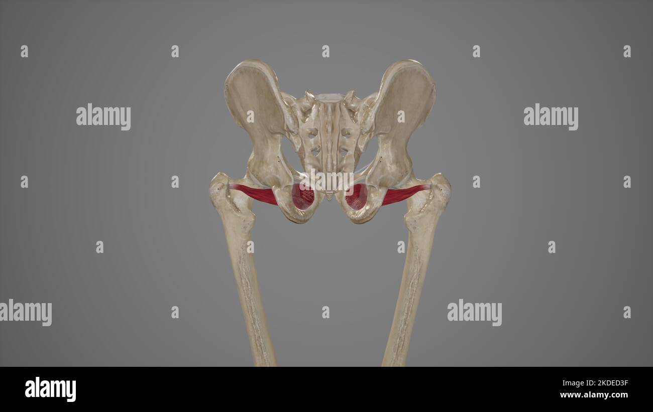 Medical Accurate Illustration of Obturator Externus Stock Photohttps://www.alamy.com/image-license-details/?v=1https://www.alamy.com/medical-accurate-illustration-of-obturator-externus-image490198451.html
RF2KDED3F–Medical Accurate Illustration of Obturator Externus
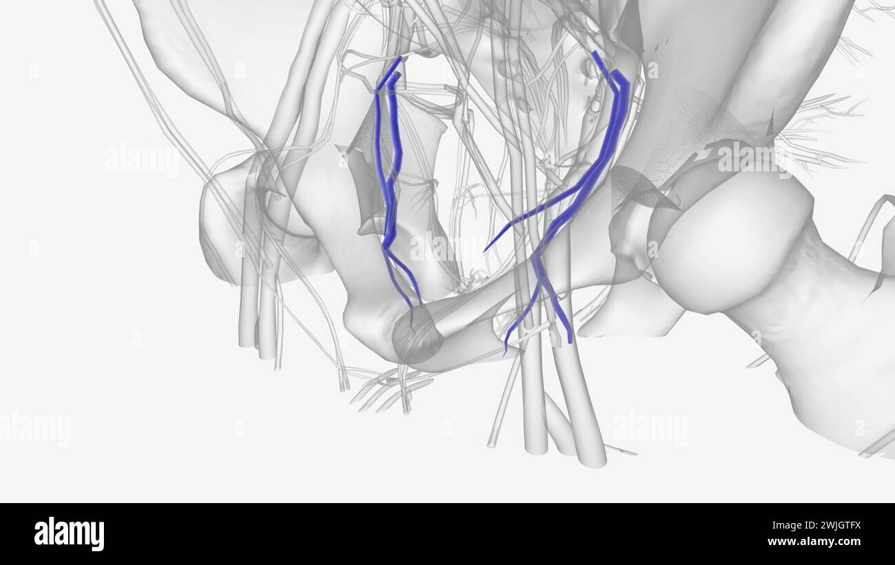 The obturator vein begins in the upper portion of the adductor region of the thigh 3d medical Stock Photohttps://www.alamy.com/image-license-details/?v=1https://www.alamy.com/the-obturator-vein-begins-in-the-upper-portion-of-the-adductor-region-of-the-thigh-3d-medical-image596586814.html
RF2WJGTFX–The obturator vein begins in the upper portion of the adductor region of the thigh 3d medical
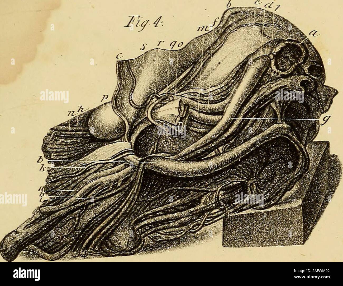 . The anatomy and surgical treatment of abdominal hernia. IT French, del. inndalrs Llth EXPLANATION OF PLATE XIX. 413 q. Common trunk of the epigastric and obturator arteries. r. Obturator artery passing before and on the inner side ofthe neck of the sac, in its course to the obturator fora-men, and situated a little above the posterior edge of theexternal oblique muscle. s. Epigastric artery. An engraving of this preparation has been published in an in-genious Thesis on Crural Hernia, by Dr. James Sanders, Edin-burgh, 1805. PLATE XX. Shows three umbilical herniae, one of which is curious on a Stock Photohttps://www.alamy.com/image-license-details/?v=1https://www.alamy.com/the-anatomy-and-surgical-treatment-of-abdominal-hernia-it-french-del-inndalrs-llth-explanation-of-plate-xix-413-q-common-trunk-of-the-epigastric-and-obturator-arteries-r-obturator-artery-passing-before-and-on-the-inner-side-ofthe-neck-of-the-sac-in-its-course-to-the-obturator-fora-men-and-situated-a-little-above-the-posterior-edge-of-theexternal-oblique-muscle-s-epigastric-artery-an-engraving-of-this-preparation-has-been-published-in-an-in-genious-thesis-on-crural-hernia-by-dr-james-sanders-edin-burgh-1805-plate-xx-shows-three-umbilical-herniae-one-of-which-is-curious-on-a-image336781566.html
RM2AFWM92–. The anatomy and surgical treatment of abdominal hernia. IT French, del. inndalrs Llth EXPLANATION OF PLATE XIX. 413 q. Common trunk of the epigastric and obturator arteries. r. Obturator artery passing before and on the inner side ofthe neck of the sac, in its course to the obturator fora-men, and situated a little above the posterior edge of theexternal oblique muscle. s. Epigastric artery. An engraving of this preparation has been published in an in-genious Thesis on Crural Hernia, by Dr. James Sanders, Edin-burgh, 1805. PLATE XX. Shows three umbilical herniae, one of which is curious on a
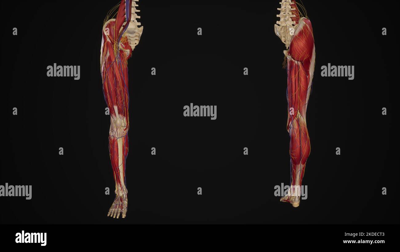 Lower limb with muscles, blood vessels and nerves anterior and posterior view Stock Photohttps://www.alamy.com/image-license-details/?v=1https://www.alamy.com/lower-limb-with-muscles-blood-vessels-and-nerves-anterior-and-posterior-view-image490198243.html
RF2KDECT3–Lower limb with muscles, blood vessels and nerves anterior and posterior view
 The obturator artery is a branch of the anterior division of the internal iliac artery 3d illustration Stock Photohttps://www.alamy.com/image-license-details/?v=1https://www.alamy.com/the-obturator-artery-is-a-branch-of-the-anterior-division-of-the-internal-iliac-artery-3d-illustration-image596588465.html
RF2WJGXJW–The obturator artery is a branch of the anterior division of the internal iliac artery 3d illustration
 . The anatomy and surgical treatment of abdominal hernia. valis.)A view of a perineal hernia in the possession of Mr. Cutcliffe, of Barn-staple. Also a hernia congenita in the female, and a crural hernia sentme by Mr. Allan Burns, surgeon, of Glasgow. Fig. 1. Thyroideal hernia. a. Symphysis pubis. b. Spine of the ilium. c. Abdominal muscles. d. Acetabulum. e. Tuberosity of the ischium. /. Ligament of the obturator, or thyroid foramen. g. Crural artery. h. Artera circumflexa ilii. i. Spermatic vein. k. Obturator artery. /. Inguinal hernia drawn aside. m. Thyroideal hernia situated just behind t Stock Photohttps://www.alamy.com/image-license-details/?v=1https://www.alamy.com/the-anatomy-and-surgical-treatment-of-abdominal-hernia-valisa-view-of-a-perineal-hernia-in-the-possession-of-mr-cutcliffe-of-barn-staple-also-a-hernia-congenita-in-the-female-and-a-crural-hernia-sentme-by-mr-allan-burns-surgeon-of-glasgow-fig-1-thyroideal-hernia-a-symphysis-pubis-b-spine-of-the-ilium-c-abdominal-muscles-d-acetabulum-e-tuberosity-of-the-ischium-ligament-of-the-obturator-or-thyroid-foramen-g-crural-artery-h-artera-circumflexa-ilii-i-spermatic-vein-k-obturator-artery-inguinal-hernia-drawn-aside-m-thyroideal-hernia-situated-just-behind-t-image336779969.html
RM2AFWJ81–. The anatomy and surgical treatment of abdominal hernia. valis.)A view of a perineal hernia in the possession of Mr. Cutcliffe, of Barn-staple. Also a hernia congenita in the female, and a crural hernia sentme by Mr. Allan Burns, surgeon, of Glasgow. Fig. 1. Thyroideal hernia. a. Symphysis pubis. b. Spine of the ilium. c. Abdominal muscles. d. Acetabulum. e. Tuberosity of the ischium. /. Ligament of the obturator, or thyroid foramen. g. Crural artery. h. Artera circumflexa ilii. i. Spermatic vein. k. Obturator artery. /. Inguinal hernia drawn aside. m. Thyroideal hernia situated just behind t
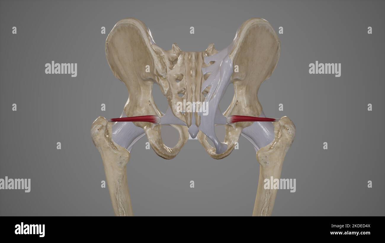 Medical Illustration of Superior Gemellus Muscle Stock Photohttps://www.alamy.com/image-license-details/?v=1https://www.alamy.com/medical-illustration-of-superior-gemellus-muscle-image490198490.html
RF2KDED4X–Medical Illustration of Superior Gemellus Muscle
 The obturator artery is a branch of the anterior division of the internal iliac artery 3d illustration Stock Photohttps://www.alamy.com/image-license-details/?v=1https://www.alamy.com/the-obturator-artery-is-a-branch-of-the-anterior-division-of-the-internal-iliac-artery-3d-illustration-image596586995.html
RF2WJGTPB–The obturator artery is a branch of the anterior division of the internal iliac artery 3d illustration
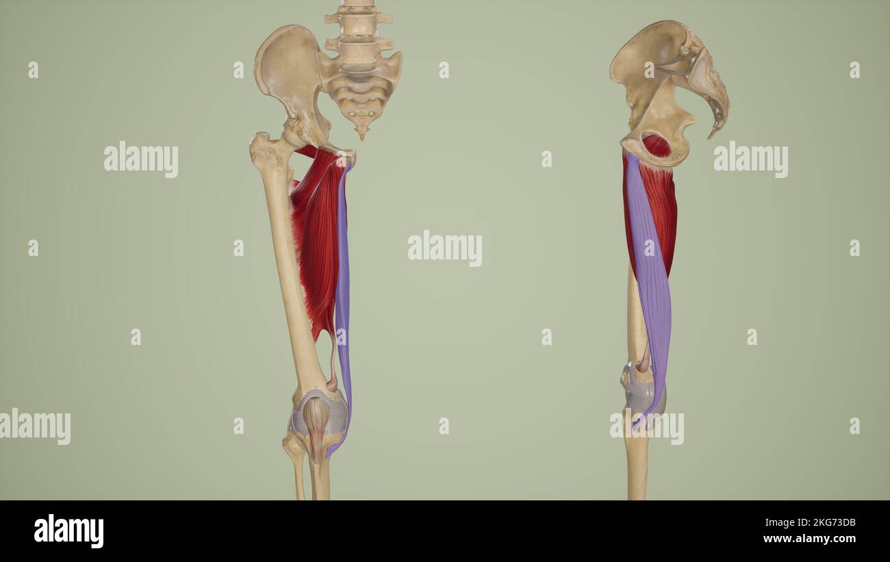 Gracilis Anterior and Lateral View Stock Photohttps://www.alamy.com/image-license-details/?v=1https://www.alamy.com/gracilis-anterior-and-lateral-view-image491881191.html
RF2KG73DB–Gracilis Anterior and Lateral View
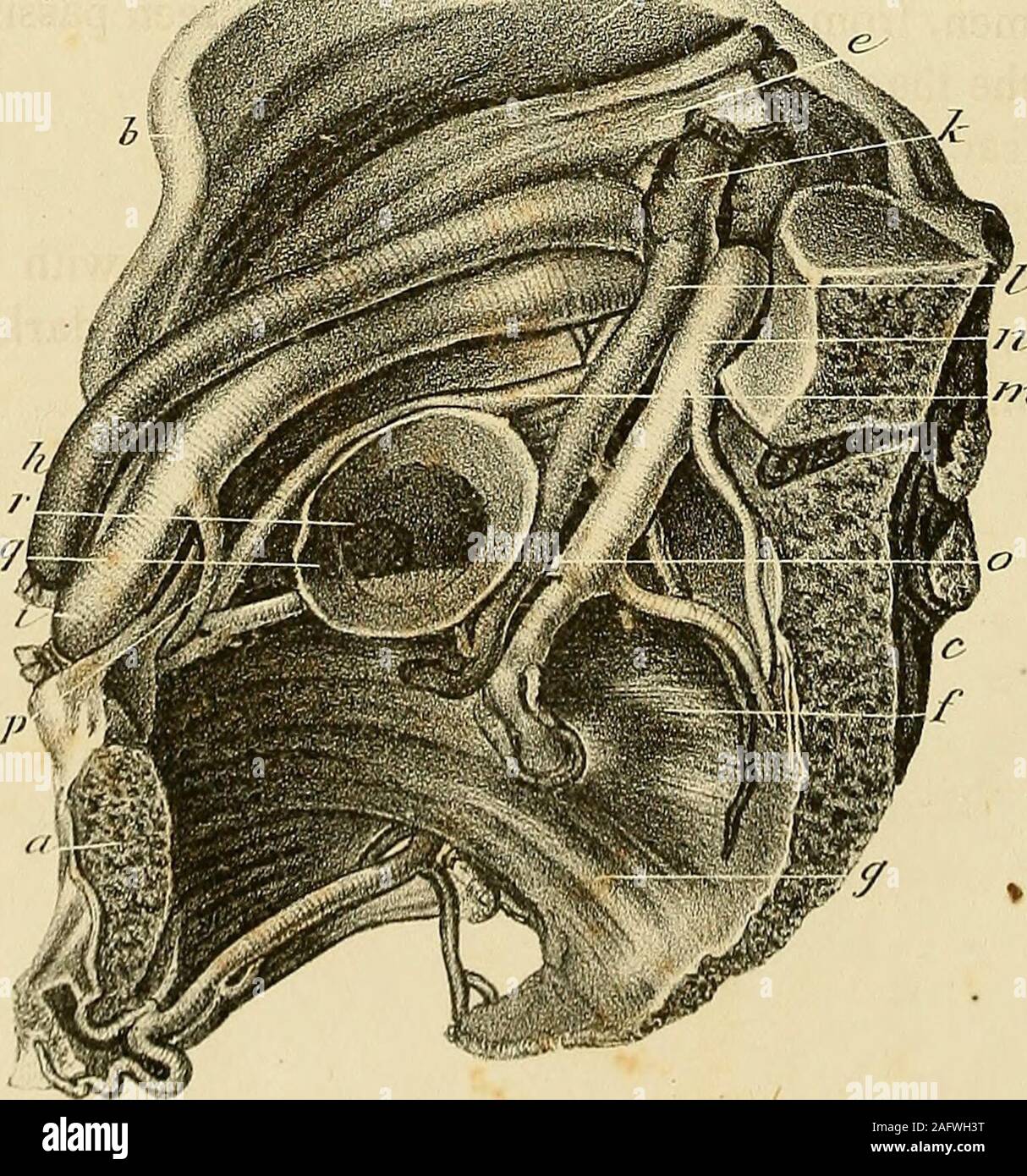 . The anatomy and surgical treatment of abdominal hernia. &??* d. > fjn-^dcl SmcZnu s Iiitii PLATE XX III.—Fig. 1. Gives an internal view of the ischiatic hernia, from Dr. Joness patient.The preparation is in the anatomical collection at Saint Thomass Hos-pital. a. Section of the pubes. b. Spinous process of the ilium. c. Sacrum. d. Iliacus internus muscle. e. Psoas muscle. /. Pyriformis muscle. g. Coccygeus muscle. h. Termination of the external iliac artery in the crural. i. Beginning of the crural vein. k. Trunk of the common iliac artery. /. Internal iliac artery. m. Obturator artery, w Stock Photohttps://www.alamy.com/image-license-details/?v=1https://www.alamy.com/the-anatomy-and-surgical-treatment-of-abdominal-hernia-d-gt-fjn-dcl-smcznu-s-iiitii-plate-xx-iiifig-1-gives-an-internal-view-of-the-ischiatic-hernia-from-dr-joness-patientthe-preparation-is-in-the-anatomical-collection-at-saint-thomass-hos-pital-a-section-of-the-pubes-b-spinous-process-of-the-ilium-c-sacrum-d-iliacus-internus-muscle-e-psoas-muscle-pyriformis-muscle-g-coccygeus-muscle-h-termination-of-the-external-iliac-artery-in-the-crural-i-beginning-of-the-crural-vein-k-trunk-of-the-common-iliac-artery-internal-iliac-artery-m-obturator-artery-w-image336779068.html
RM2AFWH3T–. The anatomy and surgical treatment of abdominal hernia. &??* d. > fjn-^dcl SmcZnu s Iiitii PLATE XX III.—Fig. 1. Gives an internal view of the ischiatic hernia, from Dr. Joness patient.The preparation is in the anatomical collection at Saint Thomass Hos-pital. a. Section of the pubes. b. Spinous process of the ilium. c. Sacrum. d. Iliacus internus muscle. e. Psoas muscle. /. Pyriformis muscle. g. Coccygeus muscle. h. Termination of the external iliac artery in the crural. i. Beginning of the crural vein. k. Trunk of the common iliac artery. /. Internal iliac artery. m. Obturator artery, w
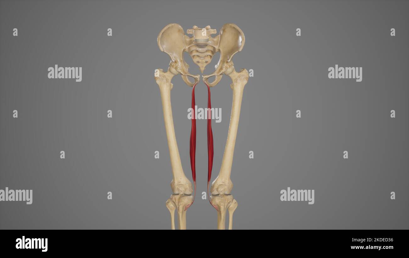 Medical Illustration of Gracilis Muscle Stock Photohttps://www.alamy.com/image-license-details/?v=1https://www.alamy.com/medical-illustration-of-gracilis-muscle-image490198442.html
RF2KDED36–Medical Illustration of Gracilis Muscle
 The obturator artery is a branch of the anterior division of the internal iliac artery 3d illustration Stock Photohttps://www.alamy.com/image-license-details/?v=1https://www.alamy.com/the-obturator-artery-is-a-branch-of-the-anterior-division-of-the-internal-iliac-artery-3d-illustration-image596587461.html
RF2WJGWB1–The obturator artery is a branch of the anterior division of the internal iliac artery 3d illustration
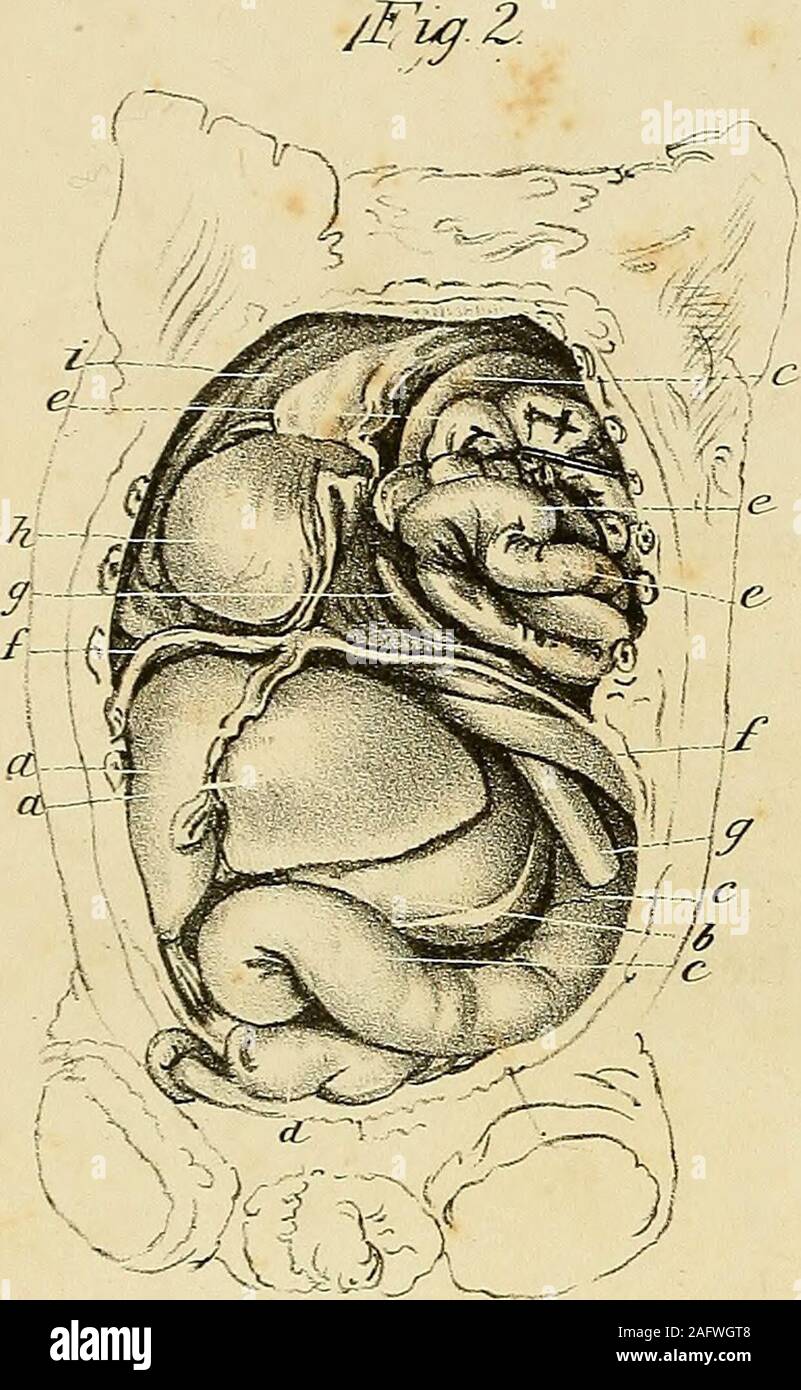 . The anatomy and surgical treatment of abdominal hernia. the ilium. c. Sacrum. d. Iliacus internus muscle. e. Psoas muscle. /. Pyriformis muscle. g. Coccygeus muscle. h. Termination of the external iliac artery in the crural. i. Beginning of the crural vein. k. Trunk of the common iliac artery. /. Internal iliac artery. m. Obturator artery, which may be traced before the sac as faras the obturator foramen. n. Internal iliac vein. o. Obturator vein passing behind the hernia to the obturator fora-men, from which another vein (p) is seen passing intothe iliac vein. q. Hernial sac. r. Its orifice Stock Photohttps://www.alamy.com/image-license-details/?v=1https://www.alamy.com/the-anatomy-and-surgical-treatment-of-abdominal-hernia-the-ilium-c-sacrum-d-iliacus-internus-muscle-e-psoas-muscle-pyriformis-muscle-g-coccygeus-muscle-h-termination-of-the-external-iliac-artery-in-the-crural-i-beginning-of-the-crural-vein-k-trunk-of-the-common-iliac-artery-internal-iliac-artery-m-obturator-artery-which-may-be-traced-before-the-sac-as-faras-the-obturator-foramen-n-internal-iliac-vein-o-obturator-vein-passing-behind-the-hernia-to-the-obturator-fora-men-from-which-another-vein-p-is-seen-passing-intothe-iliac-vein-q-hernial-sac-r-its-orifice-image336778856.html
RM2AFWGT8–. The anatomy and surgical treatment of abdominal hernia. the ilium. c. Sacrum. d. Iliacus internus muscle. e. Psoas muscle. /. Pyriformis muscle. g. Coccygeus muscle. h. Termination of the external iliac artery in the crural. i. Beginning of the crural vein. k. Trunk of the common iliac artery. /. Internal iliac artery. m. Obturator artery, which may be traced before the sac as faras the obturator foramen. n. Internal iliac vein. o. Obturator vein passing behind the hernia to the obturator fora-men, from which another vein (p) is seen passing intothe iliac vein. q. Hernial sac. r. Its orifice
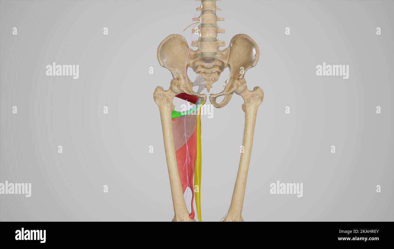 Posterior Branch of Obturator Nerve Stock Photohttps://www.alamy.com/image-license-details/?v=1https://www.alamy.com/posterior-branch-of-obturator-nerve-image488428499.html
RF2KAHREY–Posterior Branch of Obturator Nerve
 The obturator artery is a branch of the anterior division of the internal iliac artery 3d illustration Stock Photohttps://www.alamy.com/image-license-details/?v=1https://www.alamy.com/the-obturator-artery-is-a-branch-of-the-anterior-division-of-the-internal-iliac-artery-3d-illustration-image596592017.html
RF2WJH35N–The obturator artery is a branch of the anterior division of the internal iliac artery 3d illustration
 . Plates of the arteries of the human body. rare origin of the cjngastric and obtu-rator arteries, from the eomnion femoral lielowPouparts ligament in the body of a woman. 1, Gluteus medius. 2, 2. Tensor vaginje femoris. 3, Vastus externus. 4, 4. Rectus femoris. 5, 5, 5. Sartorius. 6, 6. Iliacus internus. 7, 7. Psoas magnus. 8, Pectineus. 9, 9. Adductor longus. 10, 10, 10. Gracilis. 11, 11. Ligament of Poupart. 12, 12. Common femora! artery. 13. Circumflex iliac artery. 14. Trunk of the epigastric and obturator ar- teries. 15. Epigastric artery. 16. Obturator artery. 17, 17. Twig to the psoas Stock Photohttps://www.alamy.com/image-license-details/?v=1https://www.alamy.com/plates-of-the-arteries-of-the-human-body-rare-origin-of-the-cjngastric-and-obtu-rator-arteries-from-the-eomnion-femoral-lielowpouparts-ligament-in-the-body-of-a-woman-1-gluteus-medius-2-2-tensor-vaginje-femoris-3-vastus-externus-4-4-rectus-femoris-5-5-5-sartorius-6-6-iliacus-internus-7-7-psoas-magnus-8-pectineus-9-9-adductor-longus-10-10-10-gracilis-11-11-ligament-of-poupart-12-12-common-femora!-artery-13-circumflex-iliac-artery-14-trunk-of-the-epigastric-and-obturator-ar-teries-15-epigastric-artery-16-obturator-artery-17-17-twig-to-the-psoas-image336720158.html
RM2AFPWYX–. Plates of the arteries of the human body. rare origin of the cjngastric and obtu-rator arteries, from the eomnion femoral lielowPouparts ligament in the body of a woman. 1, Gluteus medius. 2, 2. Tensor vaginje femoris. 3, Vastus externus. 4, 4. Rectus femoris. 5, 5, 5. Sartorius. 6, 6. Iliacus internus. 7, 7. Psoas magnus. 8, Pectineus. 9, 9. Adductor longus. 10, 10, 10. Gracilis. 11, 11. Ligament of Poupart. 12, 12. Common femora! artery. 13. Circumflex iliac artery. 14. Trunk of the epigastric and obturator ar- teries. 15. Epigastric artery. 16. Obturator artery. 17, 17. Twig to the psoas
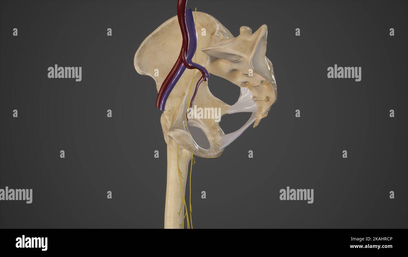 Anatomical Illustration of Obturator Canal Stock Photohttps://www.alamy.com/image-license-details/?v=1https://www.alamy.com/anatomical-illustration-of-obturator-canal-image488428438.html
RF2KAHRCP–Anatomical Illustration of Obturator Canal
 The obturator artery is a branch of the anterior division of the internal iliac artery 3d illustration Stock Photohttps://www.alamy.com/image-license-details/?v=1https://www.alamy.com/the-obturator-artery-is-a-branch-of-the-anterior-division-of-the-internal-iliac-artery-3d-illustration-image596587213.html
RF2WJGW25–The obturator artery is a branch of the anterior division of the internal iliac artery 3d illustration
 . The anatomy and surgical treatment of hernia. ision inward is the very fre-quent disposition of the obturator artery, which oftenembraces very closely the neck of the sac upon theinner side. The division of this artery would beserious, because of the difficulty of securing it thusdeeply situated. However, it has frequently happened in my ex-perience that it seemed the wise procedure to divideonly the fibers of Gimbernats ligament, and it is oftensurprising to note that the division of a few of thesefibers is sufficient to double the capacity of the ring,and thereby liberate the imprisoned co Stock Photohttps://www.alamy.com/image-license-details/?v=1https://www.alamy.com/the-anatomy-and-surgical-treatment-of-hernia-ision-inward-is-the-very-fre-quent-disposition-of-the-obturator-artery-which-oftenembraces-very-closely-the-neck-of-the-sac-upon-theinner-side-the-division-of-this-artery-would-beserious-because-of-the-difficulty-of-securing-it-thusdeeply-situated-however-it-has-frequently-happened-in-my-ex-perience-that-it-seemed-the-wise-procedure-to-divideonly-the-fibers-of-gimbernats-ligament-and-it-is-oftensurprising-to-note-that-the-division-of-a-few-of-thesefibers-is-sufficient-to-double-the-capacity-of-the-ringand-thereby-liberate-the-imprisoned-co-image370331508.html
RM2CEE1H8–. The anatomy and surgical treatment of hernia. ision inward is the very fre-quent disposition of the obturator artery, which oftenembraces very closely the neck of the sac upon theinner side. The division of this artery would beserious, because of the difficulty of securing it thusdeeply situated. However, it has frequently happened in my ex-perience that it seemed the wise procedure to divideonly the fibers of Gimbernats ligament, and it is oftensurprising to note that the division of a few of thesefibers is sufficient to double the capacity of the ring,and thereby liberate the imprisoned co
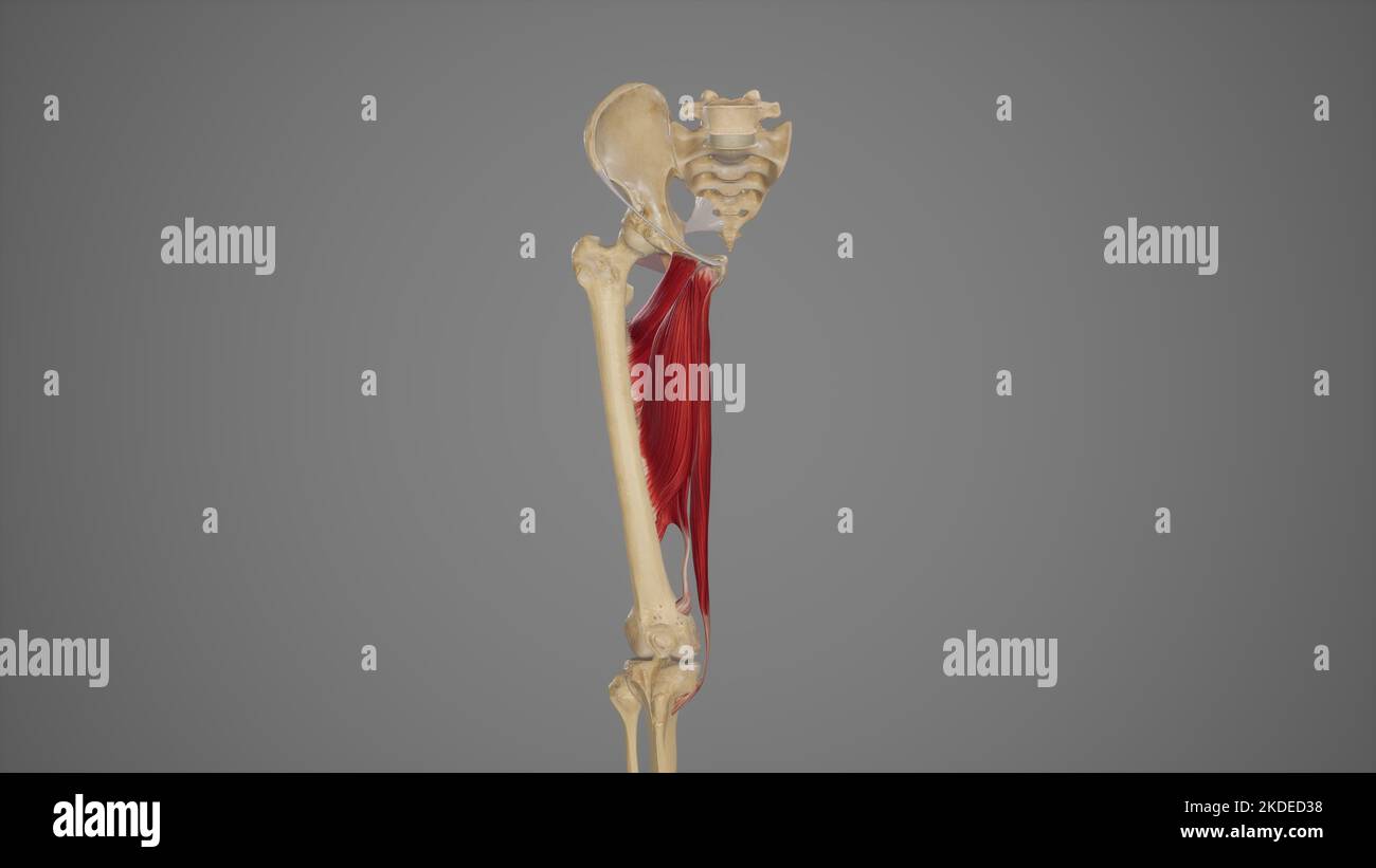 Medical Illustration of Hip Adductor Muscles Stock Photohttps://www.alamy.com/image-license-details/?v=1https://www.alamy.com/medical-illustration-of-hip-adductor-muscles-image490198444.html
RF2KDED38–Medical Illustration of Hip Adductor Muscles
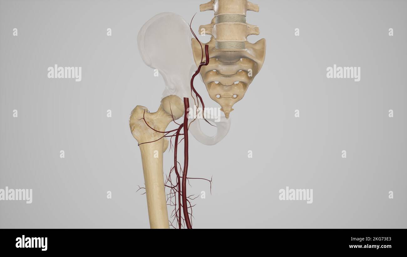 Medical Illustration of Cruciate Anastomosis Stock Photohttps://www.alamy.com/image-license-details/?v=1https://www.alamy.com/medical-illustration-of-cruciate-anastomosis-image491881211.html
RF2KG73E3–Medical Illustration of Cruciate Anastomosis
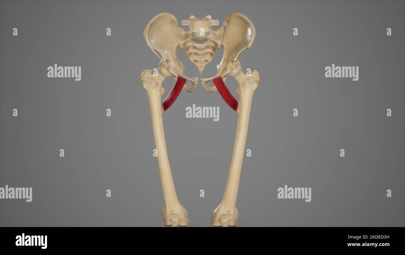 Medical Accurate Illustration of Pectineus Stock Photohttps://www.alamy.com/image-license-details/?v=1https://www.alamy.com/medical-accurate-illustration-of-pectineus-image490198453.html
RF2KDED3H–Medical Accurate Illustration of Pectineus
 The obturator vein begins in the upper portion of the adductor region of the thigh 3d medical Stock Photohttps://www.alamy.com/image-license-details/?v=1https://www.alamy.com/the-obturator-vein-begins-in-the-upper-portion-of-the-adductor-region-of-the-thigh-3d-medical-image596588636.html
RF2WJGXW0–The obturator vein begins in the upper portion of the adductor region of the thigh 3d medical
 . Journal of anatomy . epigastric and deepcircumtlex iliac, and the proximal half of the internal iliac arteries on theleft side were represented by fibrous cords. The femoral artery was patent, receiving blood by means of ananastomosis between the last lumbar and the deep circumflex iliac arteries.The distal half of the internal iliac artery was also patent, receiving bloodV)y an anastomosis between the middle sacral and several large lateralsacral arteries. The usual branches arose from the internal iliac artery, except that theilio-lumbar artery was absent: the obturator artery arose from t Stock Photohttps://www.alamy.com/image-license-details/?v=1https://www.alamy.com/journal-of-anatomy-epigastric-and-deepcircumtlex-iliac-and-the-proximal-half-of-the-internal-iliac-arteries-on-theleft-side-were-represented-by-fibrous-cords-the-femoral-artery-was-patent-receiving-blood-by-means-of-ananastomosis-between-the-last-lumbar-and-the-deep-circumflex-iliac-arteriesthe-distal-half-of-the-internal-iliac-artery-was-also-patent-receiving-bloodvy-an-anastomosis-between-the-middle-sacral-and-several-large-lateralsacral-arteries-the-usual-branches-arose-from-the-internal-iliac-artery-except-that-theilio-lumbar-artery-was-absent-the-obturator-artery-arose-from-t-image370165588.html
RM2CE6DYG–. Journal of anatomy . epigastric and deepcircumtlex iliac, and the proximal half of the internal iliac arteries on theleft side were represented by fibrous cords. The femoral artery was patent, receiving blood by means of ananastomosis between the last lumbar and the deep circumflex iliac arteries.The distal half of the internal iliac artery was also patent, receiving bloodV)y an anastomosis between the middle sacral and several large lateralsacral arteries. The usual branches arose from the internal iliac artery, except that theilio-lumbar artery was absent: the obturator artery arose from t
 Medical Accurate Illustration of Adductor Brevis Stock Photohttps://www.alamy.com/image-license-details/?v=1https://www.alamy.com/medical-accurate-illustration-of-adductor-brevis-image490198500.html
RF2KDED58–Medical Accurate Illustration of Adductor Brevis
 . The anatomy and surgical treatment of hernia. o the bone; and below, appeared the muscles and ligaments of the pelvis. PLATE XLV* Gives an internal view of the ischiatic hernia from Dr. Joness patient. The preparation is in theanatomical collection at St. Thomass Hospital. a. Section of the pubes. m. Obturator artery, which may be traced be- b. Spinous process of the ilium. fore the sac as far as the obturator foramen. c. Sacrum. n. Internal iliac vein. d. Iliacus internus muscle. o. Obturator vein passing behind the hernia e. Psoas muscle. to the obturator foramen, from which another/. Pyri Stock Photohttps://www.alamy.com/image-license-details/?v=1https://www.alamy.com/the-anatomy-and-surgical-treatment-of-hernia-o-the-bone-and-below-appeared-the-muscles-and-ligaments-of-the-pelvis-plate-xlv-gives-an-internal-view-of-the-ischiatic-hernia-from-dr-joness-patient-the-preparation-is-in-theanatomical-collection-at-st-thomass-hospital-a-section-of-the-pubes-m-obturator-artery-which-may-be-traced-be-b-spinous-process-of-the-ilium-fore-the-sac-as-far-as-the-obturator-foramen-c-sacrum-n-internal-iliac-vein-d-iliacus-internus-muscle-o-obturator-vein-passing-behind-the-hernia-e-psoas-muscle-to-the-obturator-foramen-from-which-another-pyri-image370330253.html
RM2CEE00D–. The anatomy and surgical treatment of hernia. o the bone; and below, appeared the muscles and ligaments of the pelvis. PLATE XLV* Gives an internal view of the ischiatic hernia from Dr. Joness patient. The preparation is in theanatomical collection at St. Thomass Hospital. a. Section of the pubes. m. Obturator artery, which may be traced be- b. Spinous process of the ilium. fore the sac as far as the obturator foramen. c. Sacrum. n. Internal iliac vein. d. Iliacus internus muscle. o. Obturator vein passing behind the hernia e. Psoas muscle. to the obturator foramen, from which another/. Pyri
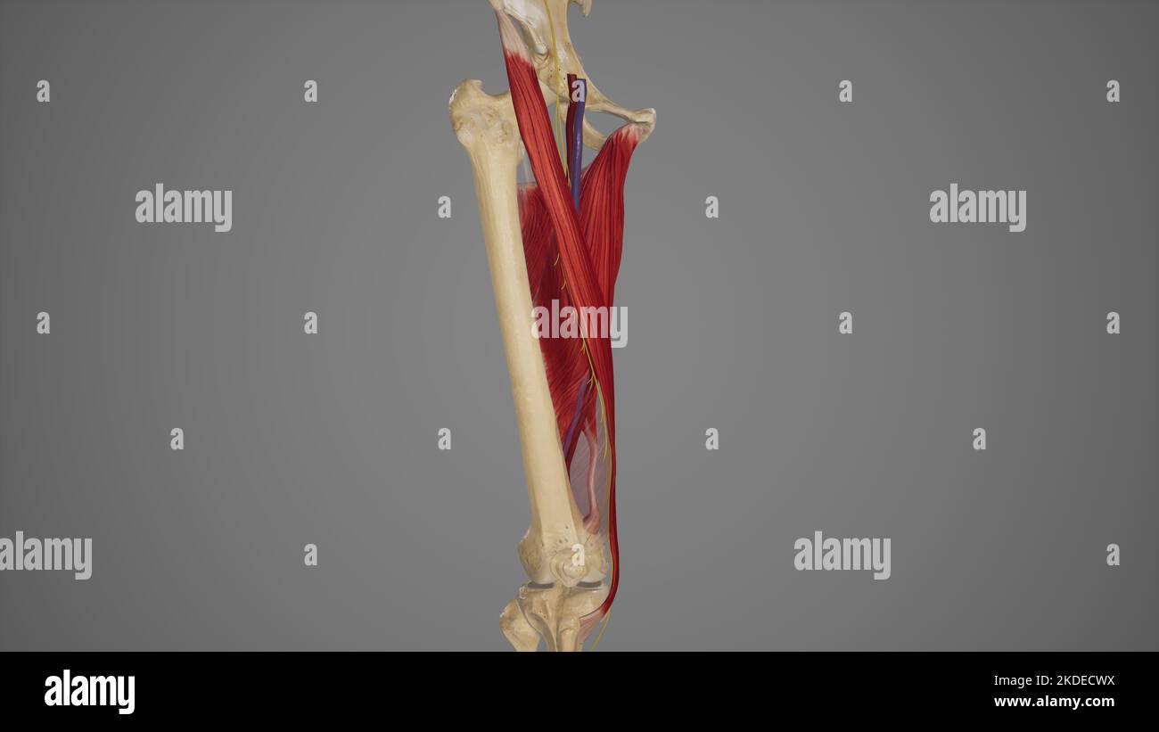 Anatomical Illustration of Adductor Canal Stock Photohttps://www.alamy.com/image-license-details/?v=1https://www.alamy.com/anatomical-illustration-of-adductor-canal-image490198294.html
RF2KDECWX–Anatomical Illustration of Adductor Canal
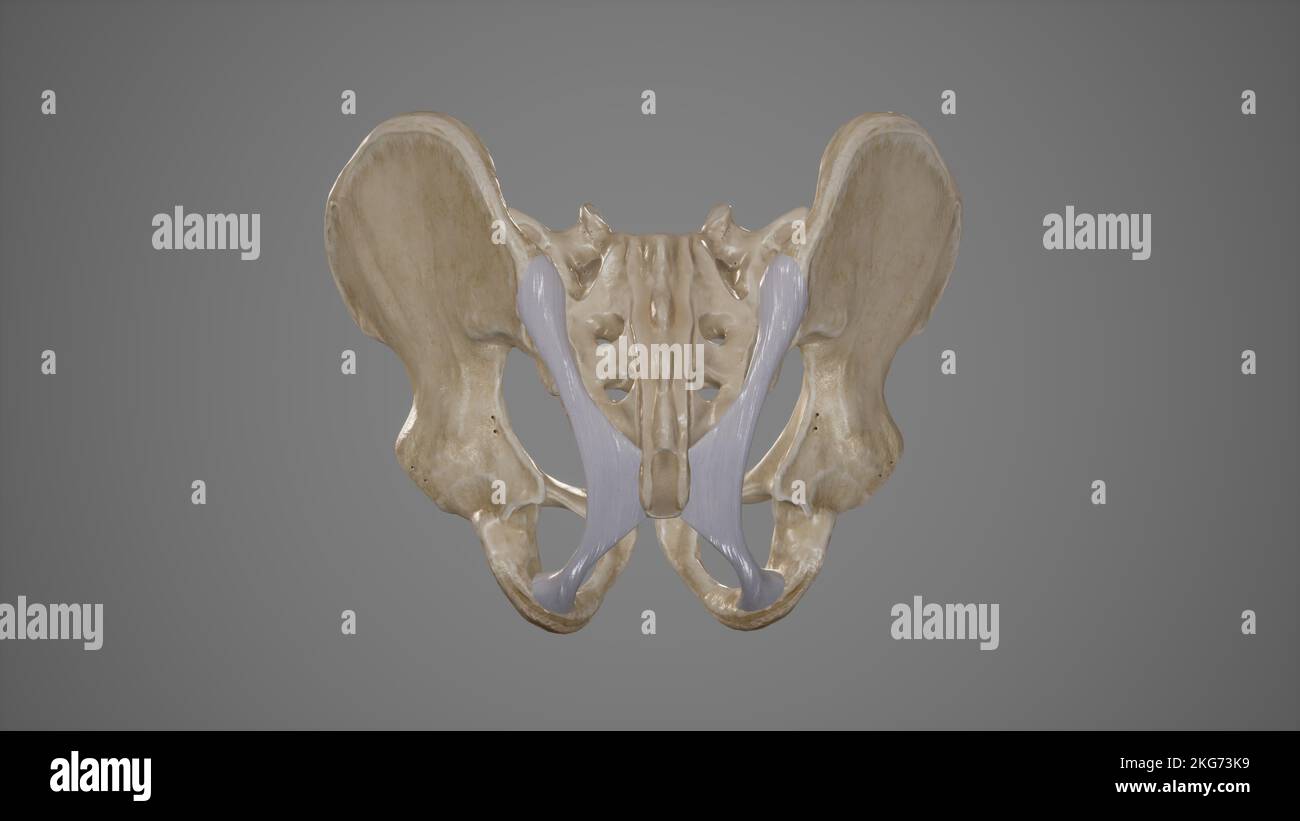 Medical Illustration of Sacrotuberous Ligament Stock Photohttps://www.alamy.com/image-license-details/?v=1https://www.alamy.com/medical-illustration-of-sacrotuberous-ligament-image491881357.html
RF2KG73K9–Medical Illustration of Sacrotuberous Ligament
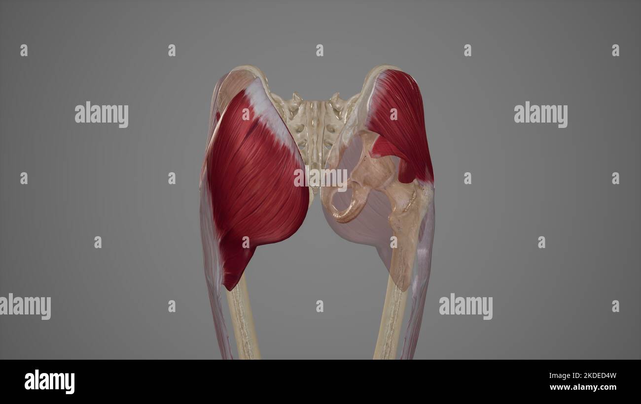 Superficial Muscles of the Gluteal Region Stock Photohttps://www.alamy.com/image-license-details/?v=1https://www.alamy.com/superficial-muscles-of-the-gluteal-region-image490198489.html
RF2KDED4W–Superficial Muscles of the Gluteal Region
 . The anatomy and surgical treatment of hernia. ^^ay-sa^^. OBTURATOR, OR HERNIA OF THE FORAMEN OVALE. 159 Figure i. Thyroideal hernia. a. Symphysis pubis. b. Spine of the ilium. c. Abdominal muscles. d. Acetabulum. e. Tuberosity of the ischium. /. Ligament of the obturator, or thyroid fora-men. g. Crural artery. h. Artera circumflexa ilii. /. Spermatic vein. k. Obturator artery. /. Inguinal hernia drawn aside. m. Thyroideal hernia situated just behind thepubes. Figu7-e 2. Posterior view of the same preparation. a. Symphysis pubis. b. Tuberosity of the ischium. c. Sacro-sciatic ligaments. d. L Stock Photohttps://www.alamy.com/image-license-details/?v=1https://www.alamy.com/the-anatomy-and-surgical-treatment-of-hernia-ay-sa-obturator-or-hernia-of-the-foramen-ovale-159-figure-i-thyroideal-hernia-a-symphysis-pubis-b-spine-of-the-ilium-c-abdominal-muscles-d-acetabulum-e-tuberosity-of-the-ischium-ligament-of-the-obturator-or-thyroid-fora-men-g-crural-artery-h-artera-circumflexa-ilii-spermatic-vein-k-obturator-artery-inguinal-hernia-drawn-aside-m-thyroideal-hernia-situated-just-behind-thepubes-figu7-e-2-posterior-view-of-the-same-preparation-a-symphysis-pubis-b-tuberosity-of-the-ischium-c-sacro-sciatic-ligaments-d-l-image370330199.html
RM2CEDYXF–. The anatomy and surgical treatment of hernia. ^^ay-sa^^. OBTURATOR, OR HERNIA OF THE FORAMEN OVALE. 159 Figure i. Thyroideal hernia. a. Symphysis pubis. b. Spine of the ilium. c. Abdominal muscles. d. Acetabulum. e. Tuberosity of the ischium. /. Ligament of the obturator, or thyroid fora-men. g. Crural artery. h. Artera circumflexa ilii. /. Spermatic vein. k. Obturator artery. /. Inguinal hernia drawn aside. m. Thyroideal hernia situated just behind thepubes. Figu7-e 2. Posterior view of the same preparation. a. Symphysis pubis. b. Tuberosity of the ischium. c. Sacro-sciatic ligaments. d. L
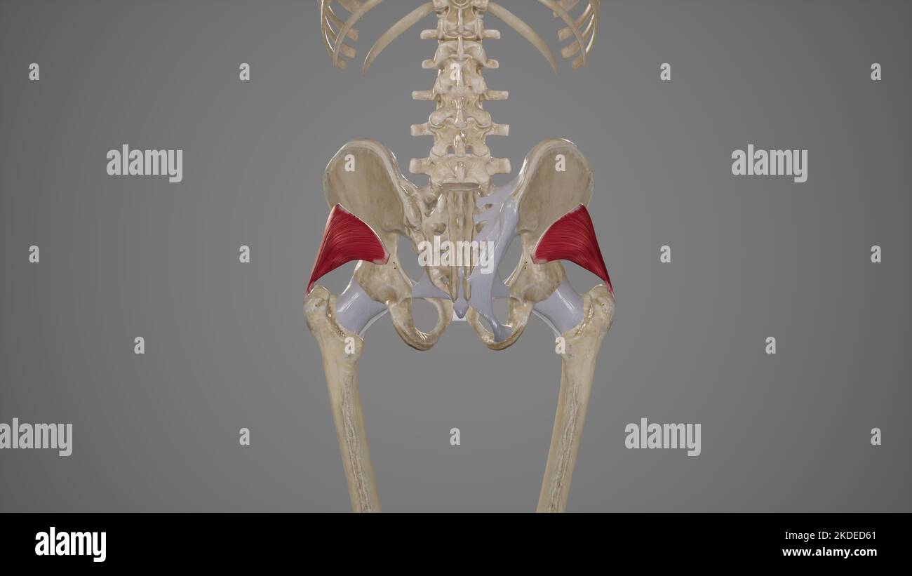 Medical Illustration of Gluteus Minimus Muscle Stock Photohttps://www.alamy.com/image-license-details/?v=1https://www.alamy.com/medical-illustration-of-gluteus-minimus-muscle-image490198521.html
RF2KDED61–Medical Illustration of Gluteus Minimus Muscle
 . Text-book of operative surgery . (2) Fascia lata.Obturator v.Obturator a. Femoral v. Obturator ext. m. ^ Fascia covcring obturatoj| ext. m. Pectineus m.Int. saplienous v. Fascia covering adductor m.Femoral a. Profunda femoris a.Int. saplienous v. Adductor longus m. Sartorius m. Fig. 54.—(1) Ligature of profunda femoris artery and external circumflex artery. (2) Ligature of obturator artery. (3) Ligature of profunda femoris artery. 9c 136 OPERATIVE SURGERY situated between tlie peritoiieum and tlie obturator internus muscle. Its pubicbranch, which ascends on the back of the pubis to the inne Stock Photohttps://www.alamy.com/image-license-details/?v=1https://www.alamy.com/text-book-of-operative-surgery-2-fascia-lataobturator-vobturator-a-femoral-v-obturator-ext-m-fascia-covcring-obturatoj-ext-m-pectineus-mint-saplienous-v-fascia-covering-adductor-mfemoral-a-profunda-femoris-aint-saplienous-v-adductor-longus-m-sartorius-m-fig-541-ligature-of-profunda-femoris-artery-and-external-circumflex-artery-2-ligature-of-obturator-artery-3-ligature-of-profunda-femoris-artery-9c-136-operative-surgery-situated-between-tlie-peritoiieum-and-tlie-obturator-internus-muscle-its-pubicbranch-which-ascends-on-the-back-of-the-pubis-to-the-inne-image372314860.html
RM2CHMBB8–. Text-book of operative surgery . (2) Fascia lata.Obturator v.Obturator a. Femoral v. Obturator ext. m. ^ Fascia covcring obturatoj| ext. m. Pectineus m.Int. saplienous v. Fascia covering adductor m.Femoral a. Profunda femoris a.Int. saplienous v. Adductor longus m. Sartorius m. Fig. 54.—(1) Ligature of profunda femoris artery and external circumflex artery. (2) Ligature of obturator artery. (3) Ligature of profunda femoris artery. 9c 136 OPERATIVE SURGERY situated between tlie peritoiieum and tlie obturator internus muscle. Its pubicbranch, which ascends on the back of the pubis to the inne
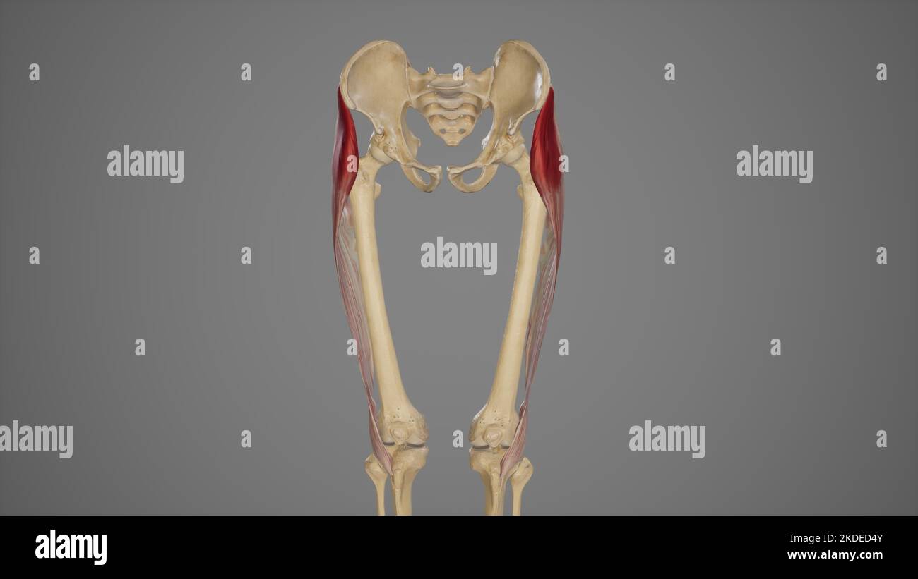 Medical Illustration of Tensor Fascia Lata Muscle Stock Photohttps://www.alamy.com/image-license-details/?v=1https://www.alamy.com/medical-illustration-of-tensor-fascia-lata-muscle-image490198491.html
RF2KDED4Y–Medical Illustration of Tensor Fascia Lata Muscle
 . Anatomy, descriptive and applied. Anatomy. THE EXTERNAL ILIAC ARTERY 681 spermatic cord, anastomosing with the spermatic artery in the male, and which accompanies the round ligament in the female; a pubic branch {ramus puhicu.i), which runs along Poupart's ligament, and then descends behind the os pubis to the inner side of the femoral ring, and anastomoses with offshoots from the obturator artery; muscular branches, some of which are distributed to the Abdominal muscles and peritoneum, anastomosing with the lumbar and circumflex iliac arteries; cutaneous branches, which perforate the tendon Stock Photohttps://www.alamy.com/image-license-details/?v=1https://www.alamy.com/anatomy-descriptive-and-applied-anatomy-the-external-iliac-artery-681-spermatic-cord-anastomosing-with-the-spermatic-artery-in-the-male-and-which-accompanies-the-round-ligament-in-the-female-a-pubic-branch-ramus-puhicui-which-runs-along-pouparts-ligament-and-then-descends-behind-the-os-pubis-to-the-inner-side-of-the-femoral-ring-and-anastomoses-with-offshoots-from-the-obturator-artery-muscular-branches-some-of-which-are-distributed-to-the-abdominal-muscles-and-peritoneum-anastomosing-with-the-lumbar-and-circumflex-iliac-arteries-cutaneous-branches-which-perforate-the-tendon-image236793697.html
RMRN6TNN–. Anatomy, descriptive and applied. Anatomy. THE EXTERNAL ILIAC ARTERY 681 spermatic cord, anastomosing with the spermatic artery in the male, and which accompanies the round ligament in the female; a pubic branch {ramus puhicu.i), which runs along Poupart's ligament, and then descends behind the os pubis to the inner side of the femoral ring, and anastomoses with offshoots from the obturator artery; muscular branches, some of which are distributed to the Abdominal muscles and peritoneum, anastomosing with the lumbar and circumflex iliac arteries; cutaneous branches, which perforate the tendon
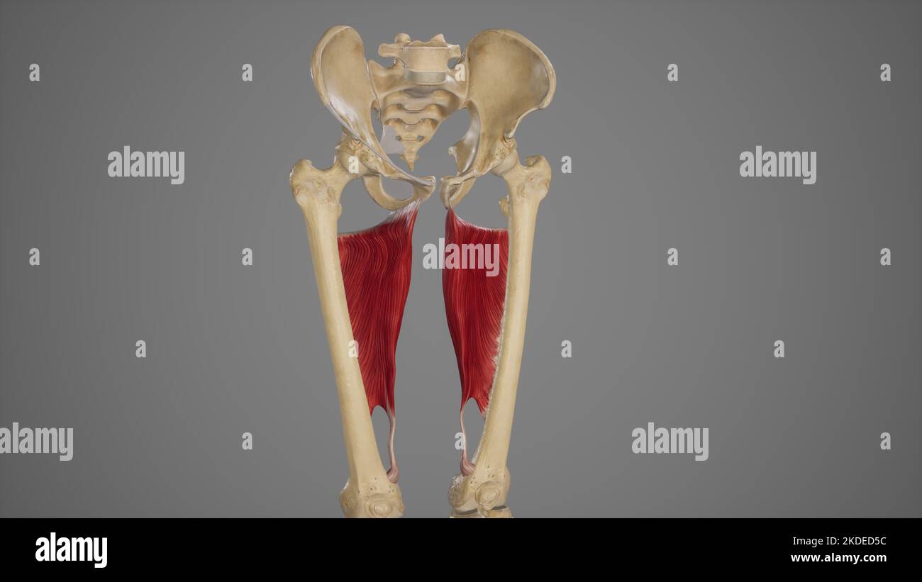 Medical Acurate Illustration of Adductor Magnus Stock Photohttps://www.alamy.com/image-license-details/?v=1https://www.alamy.com/medical-acurate-illustration-of-adductor-magnus-image490198504.html
RF2KDED5C–Medical Acurate Illustration of Adductor Magnus
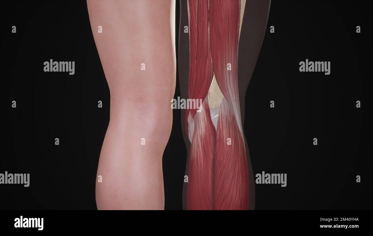 Boundaries of Popliteal Fossa Stock Photohttps://www.alamy.com/image-license-details/?v=1https://www.alamy.com/boundaries-of-popliteal-fossa-image501580950.html
RF2M40YHA–Boundaries of Popliteal Fossa
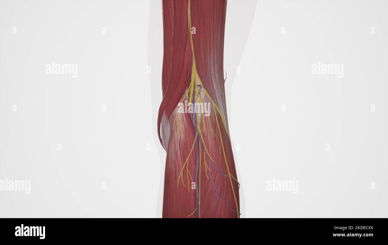 Anatomical Illustration of Popliteal Fossa Stock Photohttps://www.alamy.com/image-license-details/?v=1https://www.alamy.com/anatomical-illustration-of-popliteal-fossa-image490198322.html
RF2KDECXX–Anatomical Illustration of Popliteal Fossa
 . Atlas of applied (topographical) human anatomy for students and practitioners. Anatomy. Rectus Kfmi>ris Muscle (llutrus Mctlius ;ind Minimus Muscles Kxteriiiil Abdominal Oblique Muscle Anterior Inferior Iliac Spine Internal Abdominal Oblique Muscle Transversalis Abdominis Muscle Ilio-Psoas Muscle Anterior Crural Nerve Head of Femur liio-Pectineal Bursa Fascia Transversalis Extcrnil Inguin il Fossa Deep Epiga'^tric Vrtcrj Internal Inguinal Fossa Cremaster Muscle Obturator Artery, Femoral Ring Gimbgrhat's Ligament ^„:^^^ .-x Pectineus Muscle Ilio-Pectlneal Eminence—-^ Edge of Glenoid Fossa Stock Photohttps://www.alamy.com/image-license-details/?v=1https://www.alamy.com/atlas-of-applied-topographical-human-anatomy-for-students-and-practitioners-anatomy-rectus-kfmigtris-muscle-llutrus-mctlius-ind-minimus-muscles-kxteriiiil-abdominal-oblique-muscle-anterior-inferior-iliac-spine-internal-abdominal-oblique-muscle-transversalis-abdominis-muscle-ilio-psoas-muscle-anterior-crural-nerve-head-of-femur-liio-pectineal-bursa-fascia-transversalis-extcrnil-inguin-il-fossa-deep-epigatric-vrtcrj-internal-inguinal-fossa-cremaster-muscle-obturator-artery-femoral-ring-gimbgrhats-ligament-x-pectineus-muscle-ilio-pectlneal-eminence-edge-of-glenoid-fossa-image235390674.html
RMRJXY5P–. Atlas of applied (topographical) human anatomy for students and practitioners. Anatomy. Rectus Kfmi>ris Muscle (llutrus Mctlius ;ind Minimus Muscles Kxteriiiil Abdominal Oblique Muscle Anterior Inferior Iliac Spine Internal Abdominal Oblique Muscle Transversalis Abdominis Muscle Ilio-Psoas Muscle Anterior Crural Nerve Head of Femur liio-Pectineal Bursa Fascia Transversalis Extcrnil Inguin il Fossa Deep Epiga'^tric Vrtcrj Internal Inguinal Fossa Cremaster Muscle Obturator Artery, Femoral Ring Gimbgrhat's Ligament ^„:^^^ .-x Pectineus Muscle Ilio-Pectlneal Eminence—-^ Edge of Glenoid Fossa
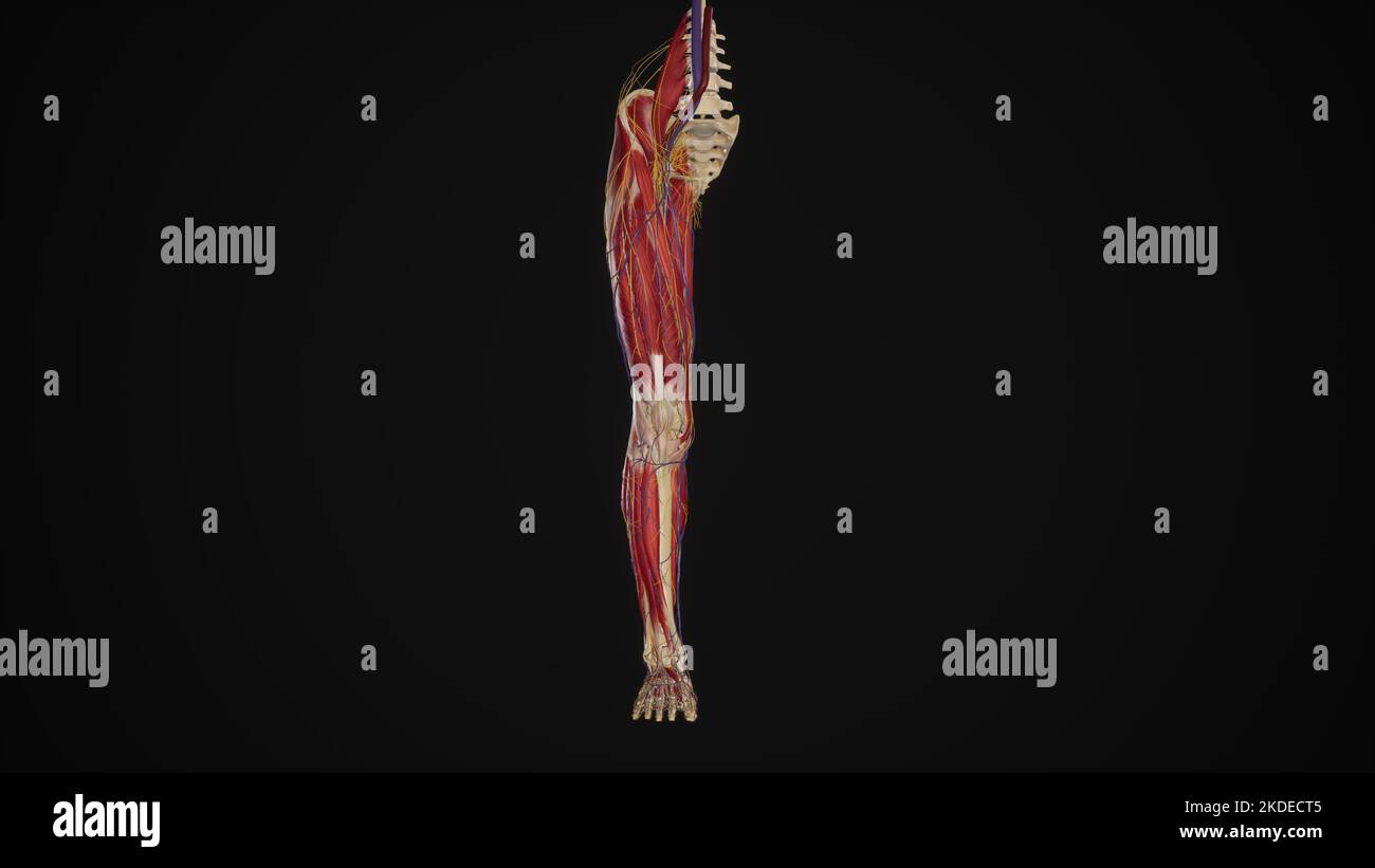 Lower limb with muscles, blood vessels and nerves Stock Photohttps://www.alamy.com/image-license-details/?v=1https://www.alamy.com/lower-limb-with-muscles-blood-vessels-and-nerves-image490198245.html
RF2KDECT5–Lower limb with muscles, blood vessels and nerves
 . A compend of equine anatomy and physiology. Horses. 78 EQUINE ANATOMY. Fig. 9.. MUSCLES ON INNER ASPECT OF LEFT POSTERIOR LIMB. i, Crest of the ilium; 2, Section through it; 3, Sacro-ischiatic ligament; 4, Pyriformis ; 5, Posterior portion of sacro-ischiatic ligament; 6, Tuberosity of ischium; 7, Anterior portion of ischium, sawn through; 8, Pubis ; 9, Obturator foramen; 10, External iliac artery and vein, 11; 12, Obturator artery and vein; the figures are placed on the internal obturator muscle ; 13, Long adductor of the leg, or sartorius; 14, Small adduc- tor of the thigh, or adductor brev Stock Photohttps://www.alamy.com/image-license-details/?v=1https://www.alamy.com/a-compend-of-equine-anatomy-and-physiology-horses-78-equine-anatomy-fig-9-muscles-on-inner-aspect-of-left-posterior-limb-i-crest-of-the-ilium-2-section-through-it-3-sacro-ischiatic-ligament-4-pyriformis-5-posterior-portion-of-sacro-ischiatic-ligament-6-tuberosity-of-ischium-7-anterior-portion-of-ischium-sawn-through-8-pubis-9-obturator-foramen-10-external-iliac-artery-and-vein-11-12-obturator-artery-and-vein-the-figures-are-placed-on-the-internal-obturator-muscle-13-long-adductor-of-the-leg-or-sartorius-14-small-adduc-tor-of-the-thigh-or-adductor-brev-image232648315.html
RMREE18B–. A compend of equine anatomy and physiology. Horses. 78 EQUINE ANATOMY. Fig. 9.. MUSCLES ON INNER ASPECT OF LEFT POSTERIOR LIMB. i, Crest of the ilium; 2, Section through it; 3, Sacro-ischiatic ligament; 4, Pyriformis ; 5, Posterior portion of sacro-ischiatic ligament; 6, Tuberosity of ischium; 7, Anterior portion of ischium, sawn through; 8, Pubis ; 9, Obturator foramen; 10, External iliac artery and vein, 11; 12, Obturator artery and vein; the figures are placed on the internal obturator muscle ; 13, Long adductor of the leg, or sartorius; 14, Small adduc- tor of the thigh, or adductor brev
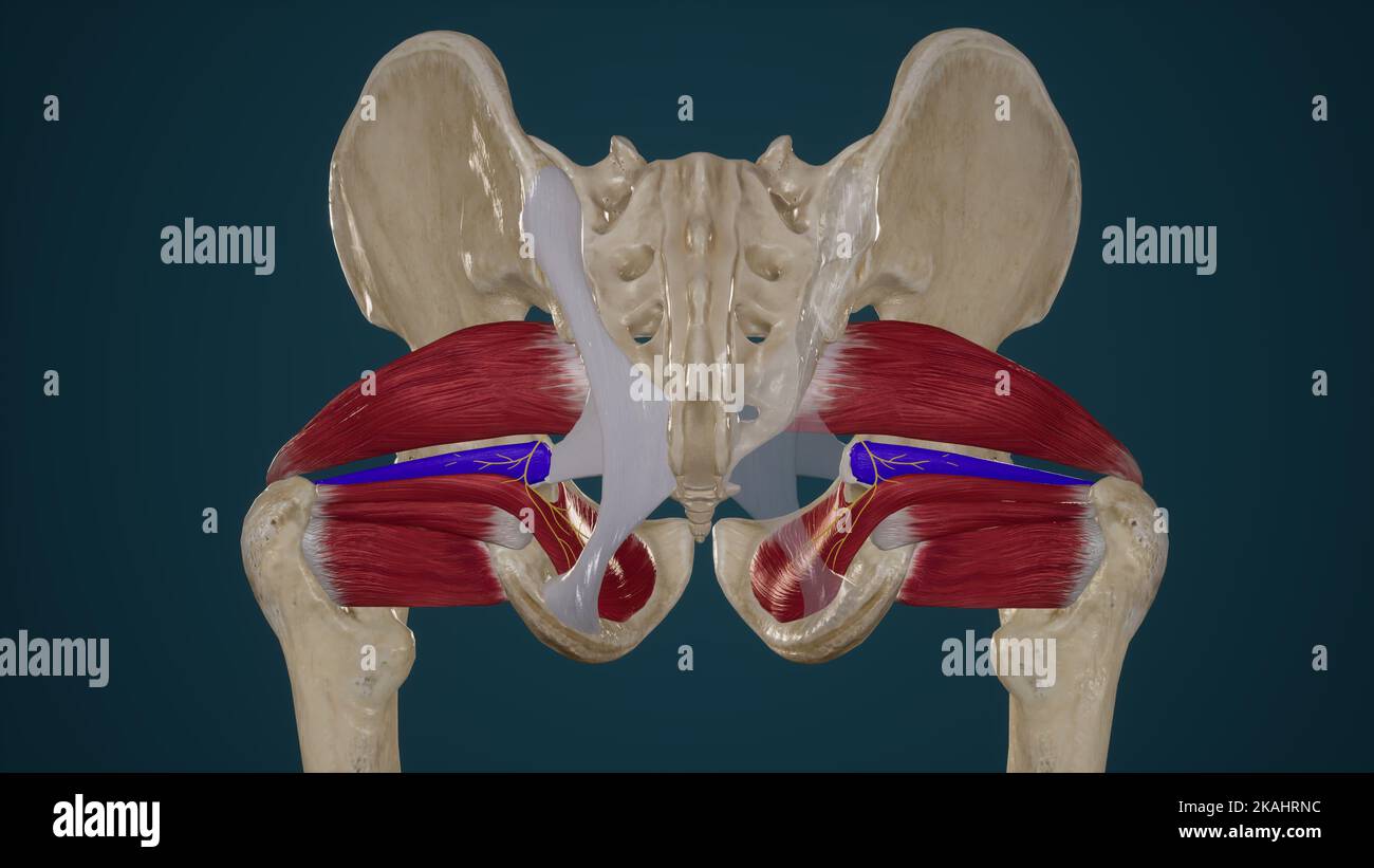 Lateral Rotators of Hip-Superior Gemellus Stock Photohttps://www.alamy.com/image-license-details/?v=1https://www.alamy.com/lateral-rotators-of-hip-superior-gemellus-image488428680.html
RF2KAHRNC–Lateral Rotators of Hip-Superior Gemellus
![. An atlas of human anatomy for students and physicians. Anatomy. External iliac artery^. A. iliaca externa External iliac vein V. iliaca externa Ante] lor superior spine of the ilium ""Spina iliaca anterior superior ,Poupart's ligament (superficial femoral arch) ILg inguinile ( r lupirti) 1 Internal or deep abdominal ring- (i) I Deep or inferior epigastric artery —k 1 and vein ^ ' ^ et V epi-,astric I inferiores Vestige of the obliterated hypogastric artery, or 'external umbilical ligament" (•;) Obturator artery and vein I 1 1 jturia ^Rectal fascia' L liaphragmatis pelvis Stock Photo . An atlas of human anatomy for students and physicians. Anatomy. External iliac artery^. A. iliaca externa External iliac vein V. iliaca externa Ante] lor superior spine of the ilium ""Spina iliaca anterior superior ,Poupart's ligament (superficial femoral arch) ILg inguinile ( r lupirti) 1 Internal or deep abdominal ring- (i) I Deep or inferior epigastric artery —k 1 and vein ^ ' ^ et V epi-,astric I inferiores Vestige of the obliterated hypogastric artery, or 'external umbilical ligament" (•;) Obturator artery and vein I 1 1 jturia ^Rectal fascia' L liaphragmatis pelvis Stock Photo](https://c8.alamy.com/comp/RJY2W0/an-atlas-of-human-anatomy-for-students-and-physicians-anatomy-external-iliac-artery-a-iliaca-externa-external-iliac-vein-v-iliaca-externa-ante-lor-superior-spine-of-the-ilium-quotquotspina-iliaca-anterior-superior-pouparts-ligament-superficial-femoral-arch-ilg-inguinile-r-lupirti-1-internal-or-deep-abdominal-ring-i-i-deep-or-inferior-epigastric-artery-k-1-and-vein-et-v-epi-astric-i-inferiores-vestige-of-the-obliterated-hypogastric-artery-or-external-umbilical-ligamentquot-obturator-artery-and-vein-i-1-1-jturia-rectal-fascia-l-liaphragmatis-pelvis-RJY2W0.jpg) . An atlas of human anatomy for students and physicians. Anatomy. External iliac artery^. A. iliaca externa External iliac vein V. iliaca externa Ante] lor superior spine of the ilium ""Spina iliaca anterior superior ,Poupart's ligament (superficial femoral arch) ILg inguinile ( r lupirti) 1 Internal or deep abdominal ring- (i) I Deep or inferior epigastric artery —k 1 and vein ^ ' ^ et V epi-,astric I inferiores Vestige of the obliterated hypogastric artery, or 'external umbilical ligament" (•;) Obturator artery and vein I 1 1 jturia ^Rectal fascia' L liaphragmatis pelvis Stock Photohttps://www.alamy.com/image-license-details/?v=1https://www.alamy.com/an-atlas-of-human-anatomy-for-students-and-physicians-anatomy-external-iliac-artery-a-iliaca-externa-external-iliac-vein-v-iliaca-externa-ante-lor-superior-spine-of-the-ilium-quotquotspina-iliaca-anterior-superior-pouparts-ligament-superficial-femoral-arch-ilg-inguinile-r-lupirti-1-internal-or-deep-abdominal-ring-i-i-deep-or-inferior-epigastric-artery-k-1-and-vein-et-v-epi-astric-i-inferiores-vestige-of-the-obliterated-hypogastric-artery-or-external-umbilical-ligamentquot-obturator-artery-and-vein-i-1-1-jturia-rectal-fascia-l-liaphragmatis-pelvis-image235393564.html
. An atlas of human anatomy for students and physicians. Anatomy. External iliac artery^. A. iliaca externa External iliac vein V. iliaca externa Ante] lor superior spine of the ilium ""Spina iliaca anterior superior ,Poupart's ligament (superficial femoral arch) ILg inguinile ( r lupirti) 1 Internal or deep abdominal ring- (i) I Deep or inferior epigastric artery —k 1 and vein ^ ' ^ et V epi-,astric I inferiores Vestige of the obliterated hypogastric artery, or 'external umbilical ligament" (•;) Obturator artery and vein I 1 1 jturia ^Rectal fascia' L liaphragmatis pelvis Stock Photohttps://www.alamy.com/image-license-details/?v=1https://www.alamy.com/an-atlas-of-human-anatomy-for-students-and-physicians-anatomy-external-iliac-artery-a-iliaca-externa-external-iliac-vein-v-iliaca-externa-ante-lor-superior-spine-of-the-ilium-quotquotspina-iliaca-anterior-superior-pouparts-ligament-superficial-femoral-arch-ilg-inguinile-r-lupirti-1-internal-or-deep-abdominal-ring-i-i-deep-or-inferior-epigastric-artery-k-1-and-vein-et-v-epi-astric-i-inferiores-vestige-of-the-obliterated-hypogastric-artery-or-external-umbilical-ligamentquot-obturator-artery-and-vein-i-1-1-jturia-rectal-fascia-l-liaphragmatis-pelvis-image235393564.htmlRMRJY2W0–. An atlas of human anatomy for students and physicians. Anatomy. External iliac artery^. A. iliaca externa External iliac vein V. iliaca externa Ante] lor superior spine of the ilium ""Spina iliaca anterior superior ,Poupart's ligament (superficial femoral arch) ILg inguinile ( r lupirti) 1 Internal or deep abdominal ring- (i) I Deep or inferior epigastric artery —k 1 and vein ^ ' ^ et V epi-,astric I inferiores Vestige of the obliterated hypogastric artery, or 'external umbilical ligament" (•;) Obturator artery and vein I 1 1 jturia ^Rectal fascia' L liaphragmatis pelvis
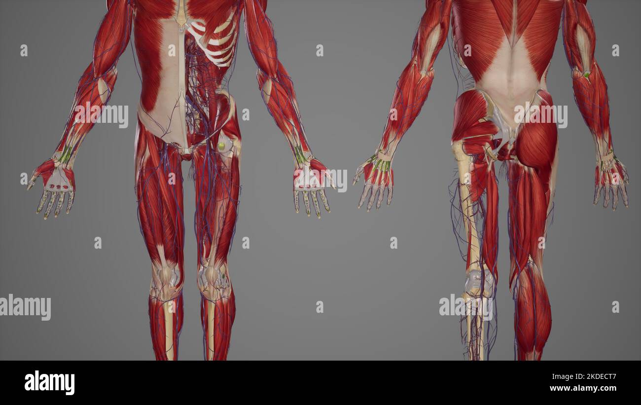 Lower limb anatomy, skeletal, muscular and cardiovascular systems, with sublayers muscles Stock Photohttps://www.alamy.com/image-license-details/?v=1https://www.alamy.com/lower-limb-anatomy-skeletal-muscular-and-cardiovascular-systems-with-sublayers-muscles-image490198247.html
RF2KDECT7–Lower limb anatomy, skeletal, muscular and cardiovascular systems, with sublayers muscles
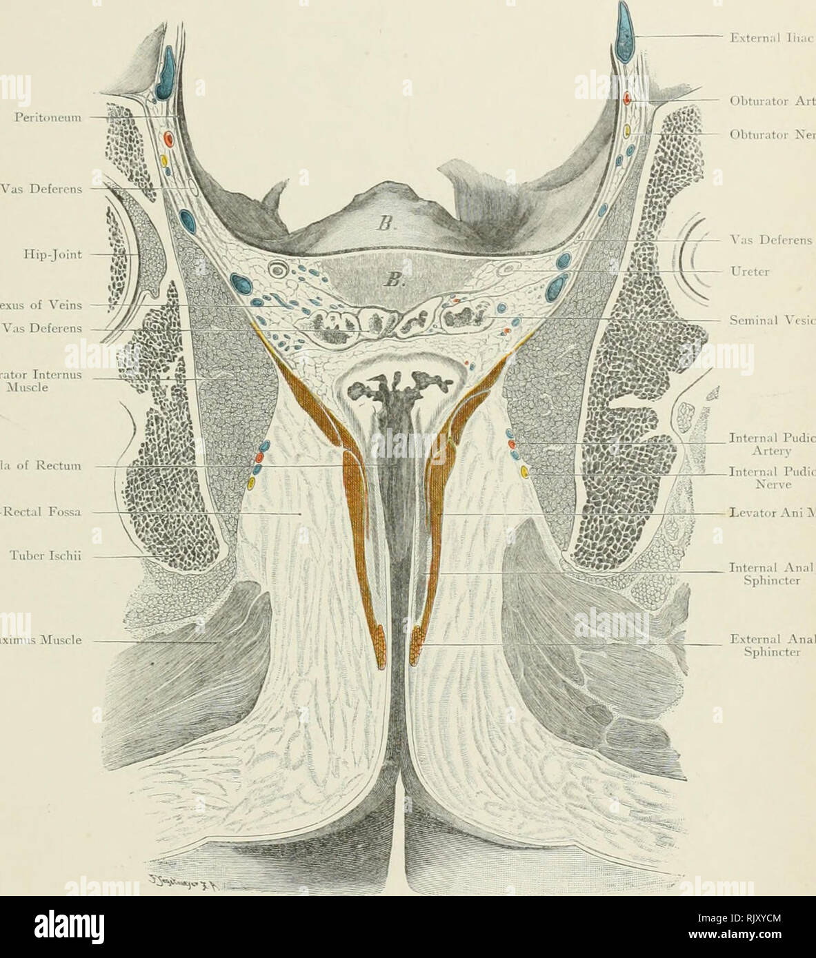 . Atlas of applied (topographical) human anatomy for students and practitioners. Anatomy. Hip-Joint â* Vesica! Plexus of Veins â^^ ^/*:-fWt t Ampulla of Vas Deferens ây^rpW3|S Obturator Internus "ff^KV . g Extern;!! Iliac 'eiii Obturator Artery â Obturator Nerve Deferens Ureter Seinina! ^'esI(â ltâ. Ampulla of Rectum Ischio-Roctal Fossa Tuber Isch Gluteus Afaximu^ ilusc!! Levator Ani ^Muscle Fig. 153. Frontal Section through Male Pelvis â Levator Ani Muscle. Seen from behind. â -/s Nat. Size. Rinjman Limileil, Loiulon. Rcbmaii Company, New York.. Please note that these images are e Stock Photohttps://www.alamy.com/image-license-details/?v=1https://www.alamy.com/atlas-of-applied-topographical-human-anatomy-for-students-and-practitioners-anatomy-hip-joint-vesica!-plexus-of-veins-fwt-t-ampulla-of-vas-deferens-yrpw3s-obturator-internus-quotffkv-g-extern!!-iliac-eiii-obturator-artery-obturator-nerve-deferens-ureter-seinina!-esi-lt-ampulla-of-rectum-ischio-roctal-fossa-tuber-isch-gluteus-afaximu-ilusc!!-levator-ani-muscle-fig-153-frontal-section-through-male-pelvis-levator-ani-muscle-seen-from-behind-s-nat-size-rinjman-limileil-loiulon-rcbmaii-company-new-york-please-note-that-these-images-are-e-image235390868.html
RMRJXYCM–. Atlas of applied (topographical) human anatomy for students and practitioners. Anatomy. Hip-Joint â* Vesica! Plexus of Veins â^^ ^/*:-fWt t Ampulla of Vas Deferens ây^rpW3|S Obturator Internus "ff^KV . g Extern;!! Iliac 'eiii Obturator Artery â Obturator Nerve Deferens Ureter Seinina! ^'esI(â ltâ. Ampulla of Rectum Ischio-Roctal Fossa Tuber Isch Gluteus Afaximu^ ilusc!! Levator Ani ^Muscle Fig. 153. Frontal Section through Male Pelvis â Levator Ani Muscle. Seen from behind. â -/s Nat. Size. Rinjman Limileil, Loiulon. Rcbmaii Company, New York.. Please note that these images are e
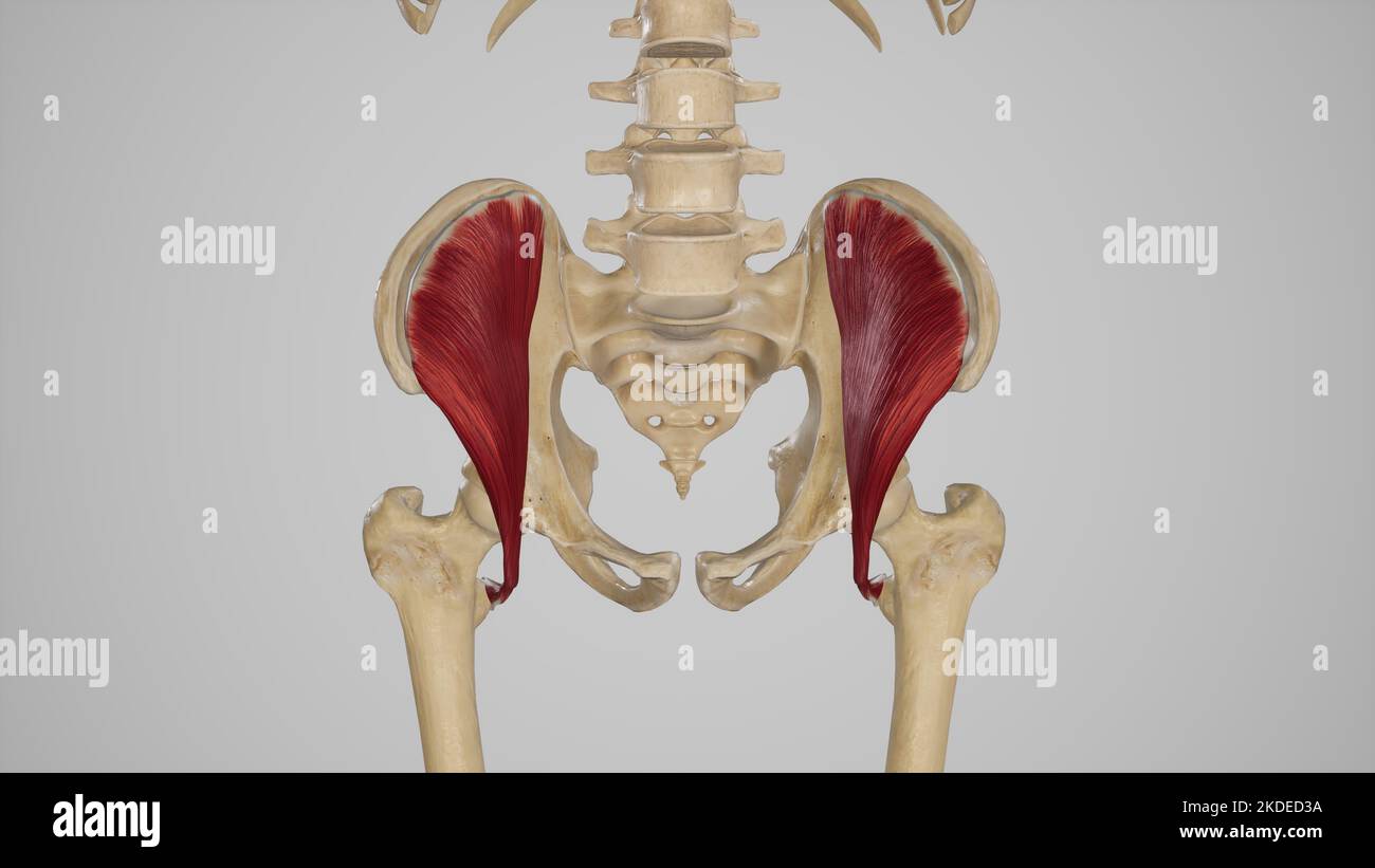 Medical Illustration of Iliacus Muscle Stock Photohttps://www.alamy.com/image-license-details/?v=1https://www.alamy.com/medical-illustration-of-iliacus-muscle-image490198446.html
RF2KDED3A–Medical Illustration of Iliacus Muscle
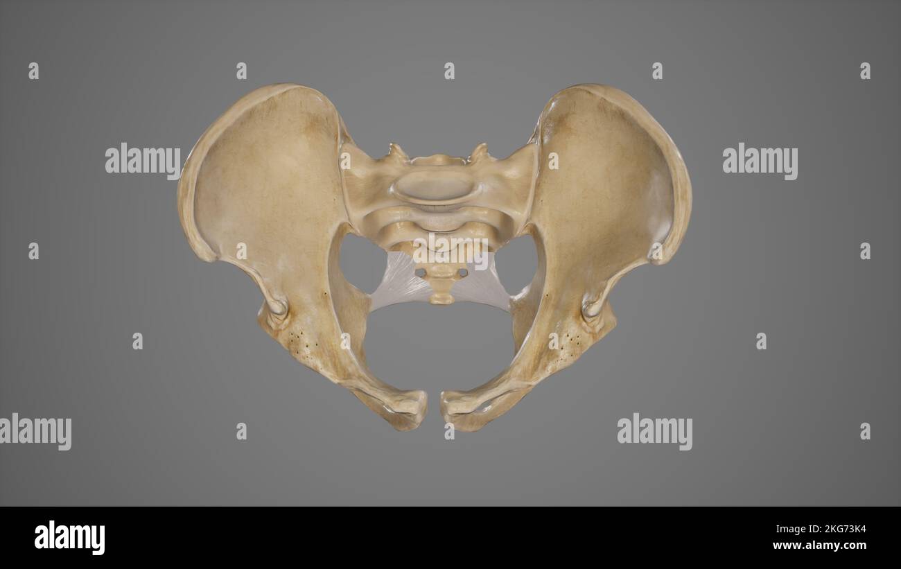 Medical Illustration of Sacrospinous Ligament Stock Photohttps://www.alamy.com/image-license-details/?v=1https://www.alamy.com/medical-illustration-of-sacrospinous-ligament-image491881352.html
RF2KG73K4–Medical Illustration of Sacrospinous Ligament
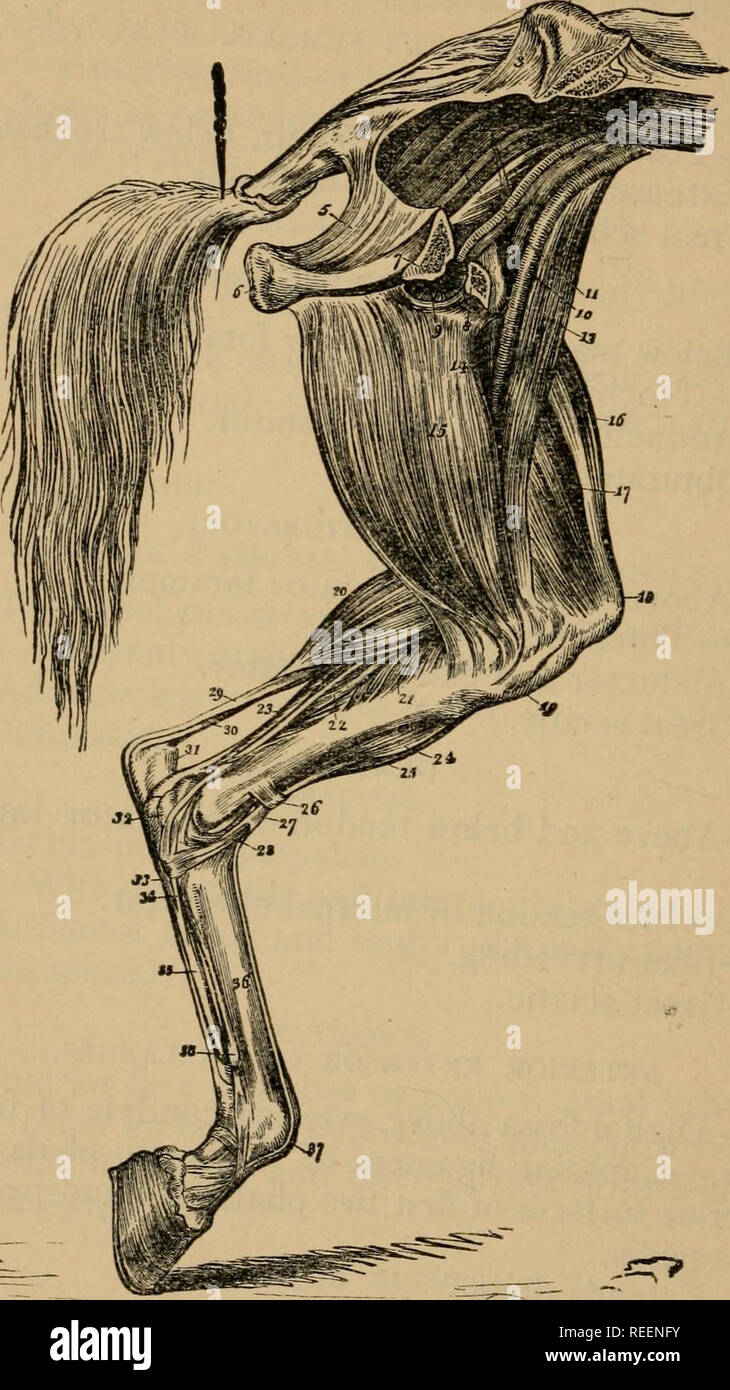 . A compend of equine anatomy and physiology. Horses; Horses -- Anatomy. 78 EQUINE ANATOMY. Fig. 9.. MUSCLES ON INNER ASPECT OK LEFT POSTERIOR LIMB I, Crest of the ilium; 2, Section through if o Sarrn i^rhi..- v Posterior portion of sacro-ischiatic ligament- 6 TnWnll 'PT"^' 4' Pyriformis; 5, tion of ischium, sawn through; 8, PubiT: o ObturLtoXV^ '''^'T= 7- Anterior por-' and vem, 11; 12, Obturator artery and v?in the fi^nS ""^"j ^°'External iliac artery "l-^^o^niuscle; 13, Long adducto^ of the leg or sa^^or^,"'^'^'c"^ °,? '^^ ^"^^'""^I ob^ th Stock Photohttps://www.alamy.com/image-license-details/?v=1https://www.alamy.com/a-compend-of-equine-anatomy-and-physiology-horses-horses-anatomy-78-equine-anatomy-fig-9-muscles-on-inner-aspect-ok-left-posterior-limb-i-crest-of-the-ilium-2-section-through-if-o-sarrn-irhi-v-posterior-portion-of-sacro-ischiatic-ligament-6-tnwnll-ptquot-4-pyriformis-5-tion-of-ischium-sawn-through-8-pubit-o-obturltoxv-t=-7-anterior-por-and-vem-11-12-obturator-artery-and-vin-the-fins-quotquotquotj-external-iliac-artery-quotl-oniuscle-13-long-adducto-of-the-leg-or-saorquotcquot-quotquotquoti-ob-th-image232664207.html
RMREENFY–. A compend of equine anatomy and physiology. Horses; Horses -- Anatomy. 78 EQUINE ANATOMY. Fig. 9.. MUSCLES ON INNER ASPECT OK LEFT POSTERIOR LIMB I, Crest of the ilium; 2, Section through if o Sarrn i^rhi..- v Posterior portion of sacro-ischiatic ligament- 6 TnWnll 'PT"^' 4' Pyriformis; 5, tion of ischium, sawn through; 8, PubiT: o ObturLtoXV^ '''^'T= 7- Anterior por-' and vem, 11; 12, Obturator artery and v?in the fi^nS ""^"j ^°'External iliac artery "l-^^o^niuscle; 13, Long adducto^ of the leg or sa^^or^,"'^'^'c"^ °,? '^^ ^"^^'""^I ob^ th
 Medical Illustration of Sacrotuberous and Sacrospinous Ligaments Stock Photohttps://www.alamy.com/image-license-details/?v=1https://www.alamy.com/medical-illustration-of-sacrotuberous-and-sacrospinous-ligaments-image491881355.html
RF2KG73K7–Medical Illustration of Sacrotuberous and Sacrospinous Ligaments
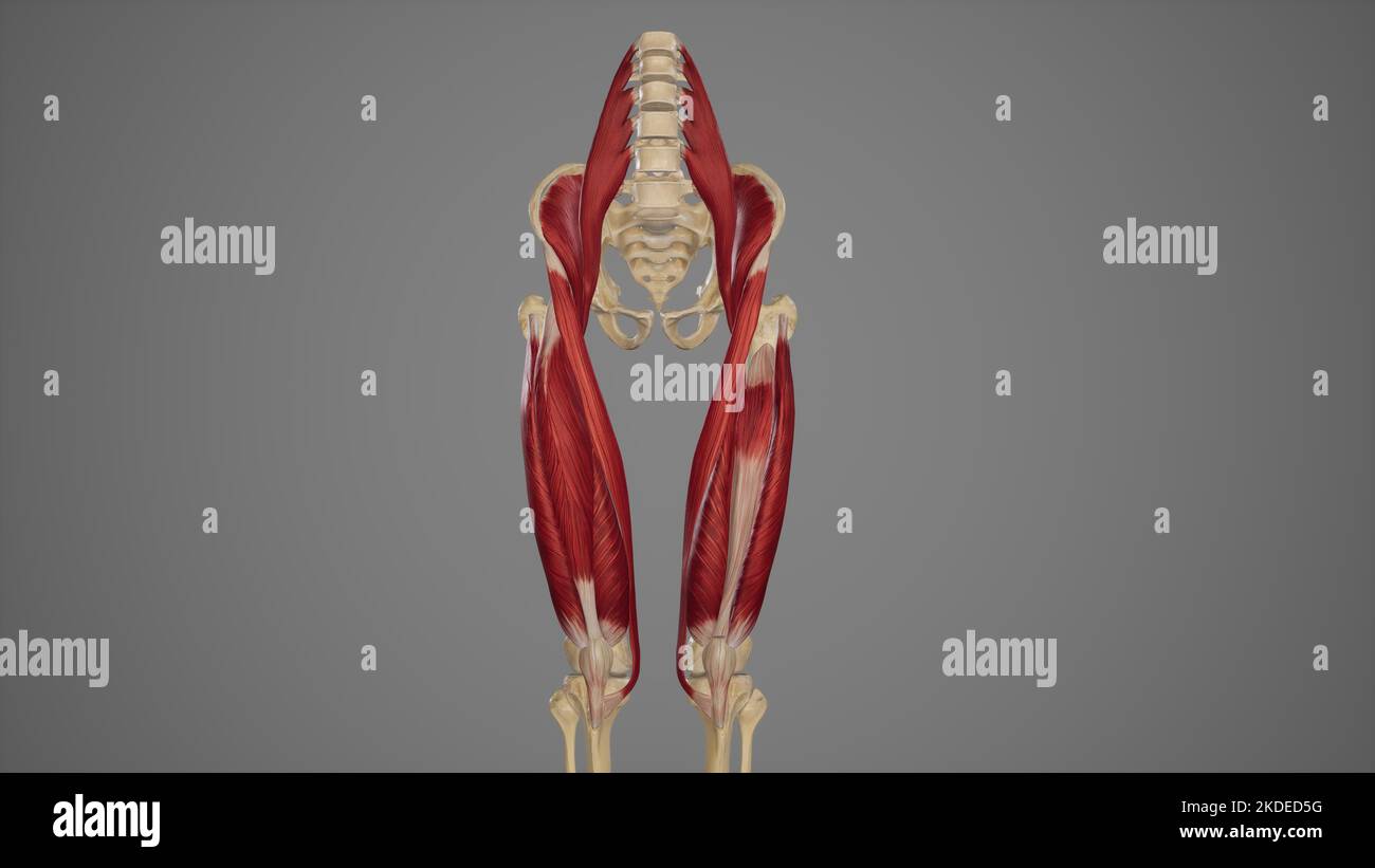 Anterior View of Anterior Thigh Muscles Stock Photohttps://www.alamy.com/image-license-details/?v=1https://www.alamy.com/anterior-view-of-anterior-thigh-muscles-image490198508.html
RF2KDED5G–Anterior View of Anterior Thigh Muscles
 Surgery; its theory and practice . 104) is carriedon chiefly by the anastomosisbetween the internal mammaryand deep epigastric ; the iho-lumbar and circumflex iliac ; thegluteal and external circumflex;the obturator and internal cir-cumflex ; the sciatic and thesuperior perforating and internalcircumflex. The cojbion iliac artery hasbeen hgatured for aneurysm ofthe external iliac and for glutealaneurysm; the internal iliacartery, also for gluteal aneur-ysm. Both may be reached byprolonging the incision for liga-ture of the external iliac, andboth have recently been tiedthrough the peritoneum. Stock Photohttps://www.alamy.com/image-license-details/?v=1https://www.alamy.com/surgery-its-theory-and-practice-104-is-carriedon-chiefly-by-the-anastomosisbetween-the-internal-mammaryand-deep-epigastric-the-iho-lumbar-and-circumflex-iliac-thegluteal-and-external-circumflexthe-obturator-and-internal-cir-cumflex-the-sciatic-and-thesuperior-perforating-and-internalcircumflex-the-cojbion-iliac-artery-hasbeen-hgatured-for-aneurysm-ofthe-external-iliac-and-for-glutealaneurysm-the-internal-iliacartery-also-for-gluteal-aneur-ysm-both-may-be-reached-byprolonging-the-incision-for-liga-ture-of-the-external-iliac-andboth-have-recently-been-tiedthrough-the-peritoneum-image343119675.html
RM2AX6CJ3–Surgery; its theory and practice . 104) is carriedon chiefly by the anastomosisbetween the internal mammaryand deep epigastric ; the iho-lumbar and circumflex iliac ; thegluteal and external circumflex;the obturator and internal cir-cumflex ; the sciatic and thesuperior perforating and internalcircumflex. The cojbion iliac artery hasbeen hgatured for aneurysm ofthe external iliac and for glutealaneurysm; the internal iliacartery, also for gluteal aneur-ysm. Both may be reached byprolonging the incision for liga-ture of the external iliac, andboth have recently been tiedthrough the peritoneum.
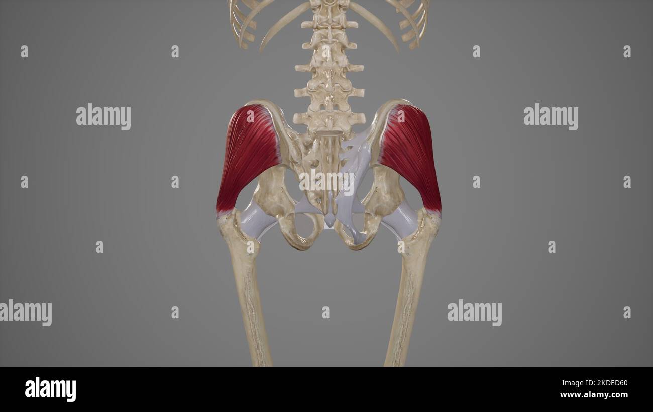 Medical Illustration of Gluteus Medius Muscle Stock Photohttps://www.alamy.com/image-license-details/?v=1https://www.alamy.com/medical-illustration-of-gluteus-medius-muscle-image490198520.html
RF2KDED60–Medical Illustration of Gluteus Medius Muscle
 Operative surgery . , and the patient recovered. After returning the protrusion thewound is closed and dressed antiseptically. Femoral herniae do not always follow the course just described ; theytake, though infrequently, anomalous courses, sometimes appearing at theouter side, or behind the femoral vessels. They have been known to passthrough Gimbernats ligament. It is important to know that in all theanomalous cases the neck of the sac lies closely associated with the epi-gastric artery alone, or together with the obturator, and troublesome andeven fatal hsemorrhages may be caused unless ca Stock Photohttps://www.alamy.com/image-license-details/?v=1https://www.alamy.com/operative-surgery-and-the-patient-recovered-after-returning-the-protrusion-thewound-is-closed-and-dressed-antiseptically-femoral-herniae-do-not-always-follow-the-course-just-described-theytake-though-infrequently-anomalous-courses-sometimes-appearing-at-theouter-side-or-behind-the-femoral-vessels-they-have-been-known-to-passthrough-gimbernats-ligament-it-is-important-to-know-that-in-all-theanomalous-cases-the-neck-of-the-sac-lies-closely-associated-with-the-epi-gastric-artery-alone-or-together-with-the-obturator-and-troublesome-andeven-fatal-hsemorrhages-may-be-caused-unless-ca-image342667985.html
RM2AWDTE9–Operative surgery . , and the patient recovered. After returning the protrusion thewound is closed and dressed antiseptically. Femoral herniae do not always follow the course just described ; theytake, though infrequently, anomalous courses, sometimes appearing at theouter side, or behind the femoral vessels. They have been known to passthrough Gimbernats ligament. It is important to know that in all theanomalous cases the neck of the sac lies closely associated with the epi-gastric artery alone, or together with the obturator, and troublesome andeven fatal hsemorrhages may be caused unless ca
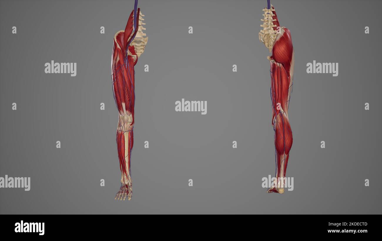 ower limb with muscles, blood vessels anterior and posterior view Stock Photohttps://www.alamy.com/image-license-details/?v=1https://www.alamy.com/ower-limb-with-muscles-blood-vessels-anterior-and-posterior-view-image490198253.html
RF2KDECTD–ower limb with muscles, blood vessels anterior and posterior view
 A series of engravings, explaining the course of the nerves : with an address to young physicians on the study of the nerves . e ligature with it, during operations. 9. The last subdivision of this Nerve to the Muscles, viz. to the Triceps and Gracilis. 65 10. The Obturator Nerve, or middle Nerve of the Thigh. Itcomes down through the Thyroid Hole, and is distributeddeep amongst the Adductor Muscles of the Thigh. 11. A Branch of the Obturator Nerve, which passes down before the Triceps Muscle to the inside of the Knee. 12. 12. The continued Branch 8, which passes down with theFemoral Artery th Stock Photohttps://www.alamy.com/image-license-details/?v=1https://www.alamy.com/a-series-of-engravings-explaining-the-course-of-the-nerves-with-an-address-to-young-physicians-on-the-study-of-the-nerves-e-ligature-with-it-during-operations-9-the-last-subdivision-of-this-nerve-to-the-muscles-viz-to-the-triceps-and-gracilis-65-10-the-obturator-nerve-or-middle-nerve-of-the-thigh-itcomes-down-through-the-thyroid-hole-and-is-distributeddeep-amongst-the-adductor-muscles-of-the-thigh-11-a-branch-of-the-obturator-nerve-which-passes-down-before-the-triceps-muscle-to-the-inside-of-the-knee-12-12-the-continued-branch-8-which-passes-down-with-thefemoral-artery-th-image343186385.html
RM2AX9DMH–A series of engravings, explaining the course of the nerves : with an address to young physicians on the study of the nerves . e ligature with it, during operations. 9. The last subdivision of this Nerve to the Muscles, viz. to the Triceps and Gracilis. 65 10. The Obturator Nerve, or middle Nerve of the Thigh. Itcomes down through the Thyroid Hole, and is distributeddeep amongst the Adductor Muscles of the Thigh. 11. A Branch of the Obturator Nerve, which passes down before the Triceps Muscle to the inside of the Knee. 12. 12. The continued Branch 8, which passes down with theFemoral Artery th
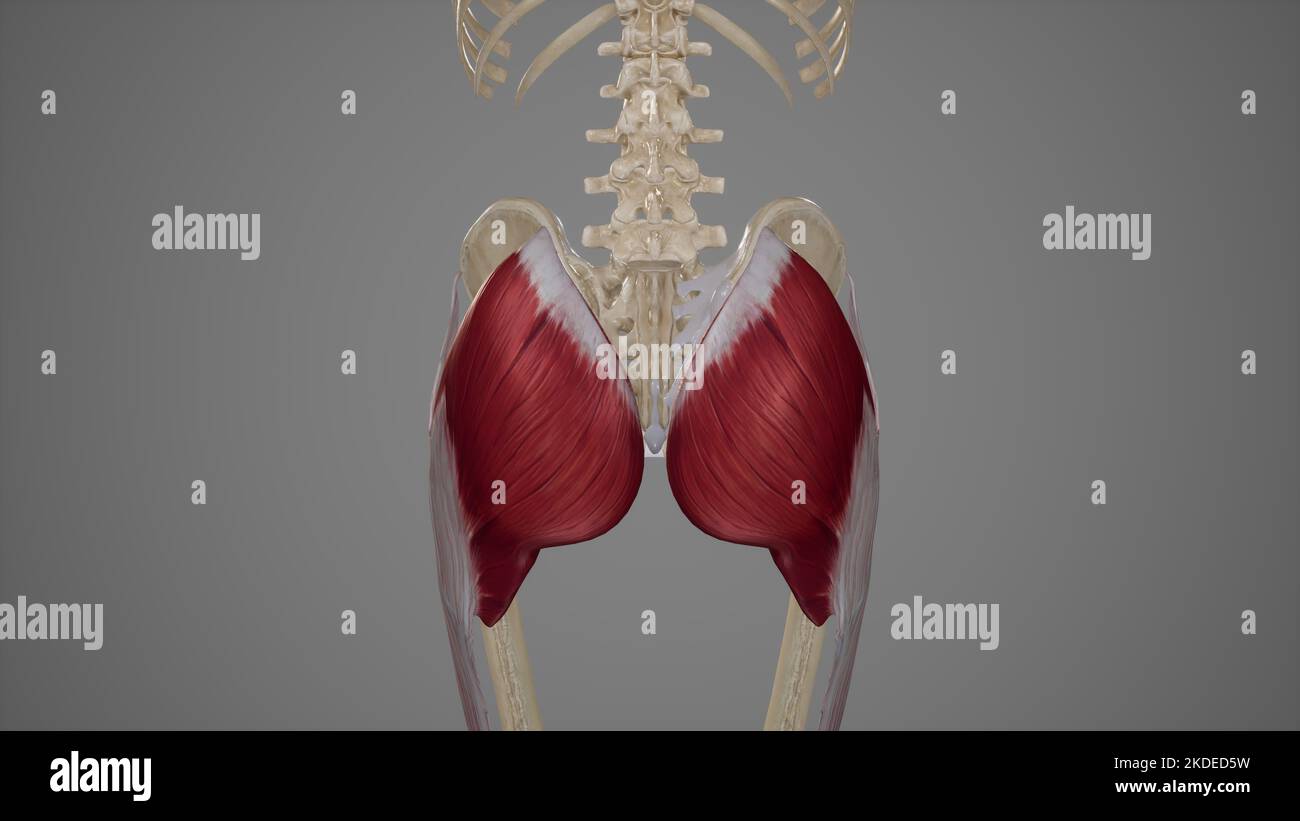 Medical Illustration of Gluteus Maximus Muscle Stock Photohttps://www.alamy.com/image-license-details/?v=1https://www.alamy.com/medical-illustration-of-gluteus-maximus-muscle-image490198517.html
RF2KDED5W–Medical Illustration of Gluteus Maximus Muscle
 Annual and analytical cyclopaedia of practical medicine . 446 IIKIIXIA. KARE FORMS. HERPES. Winckel, who found 6 cases in 5600patients examined by him, recommendsa radical operation through the perinealtissues. Obtueatoe IIerxia.—This is a rarevariety of hernia, which protrudesthrough the obturator foramen betweenobturator externus and pectineus, push-ing before it the obturator fascia. Thefemoral artery and vein pass externallyand in front of it, the adductor longusforming the opposite wall. The obtura-tor artery and vein may lie to the inneror outer side of the hernia, especially feature, in Stock Photohttps://www.alamy.com/image-license-details/?v=1https://www.alamy.com/annual-and-analytical-cyclopaedia-of-practical-medicine-446-iikiixia-kare-forms-herpes-winckel-who-found-6-cases-in-5600patients-examined-by-him-recommendsa-radical-operation-through-the-perinealtissues-obtueatoe-iierxiathis-is-a-rarevariety-of-hernia-which-protrudesthrough-the-obturator-foramen-betweenobturator-externus-and-pectineus-push-ing-before-it-the-obturator-fascia-thefemoral-artery-and-vein-pass-externallyand-in-front-of-it-the-adductor-longusforming-the-opposite-wall-the-obtura-tor-artery-and-vein-may-lie-to-the-inneror-outer-side-of-the-hernia-especially-feature-in-image338302035.html
RM2AJAYKF–Annual and analytical cyclopaedia of practical medicine . 446 IIKIIXIA. KARE FORMS. HERPES. Winckel, who found 6 cases in 5600patients examined by him, recommendsa radical operation through the perinealtissues. Obtueatoe IIerxia.—This is a rarevariety of hernia, which protrudesthrough the obturator foramen betweenobturator externus and pectineus, push-ing before it the obturator fascia. Thefemoral artery and vein pass externallyand in front of it, the adductor longusforming the opposite wall. The obtura-tor artery and vein may lie to the inneror outer side of the hernia, especially feature, in
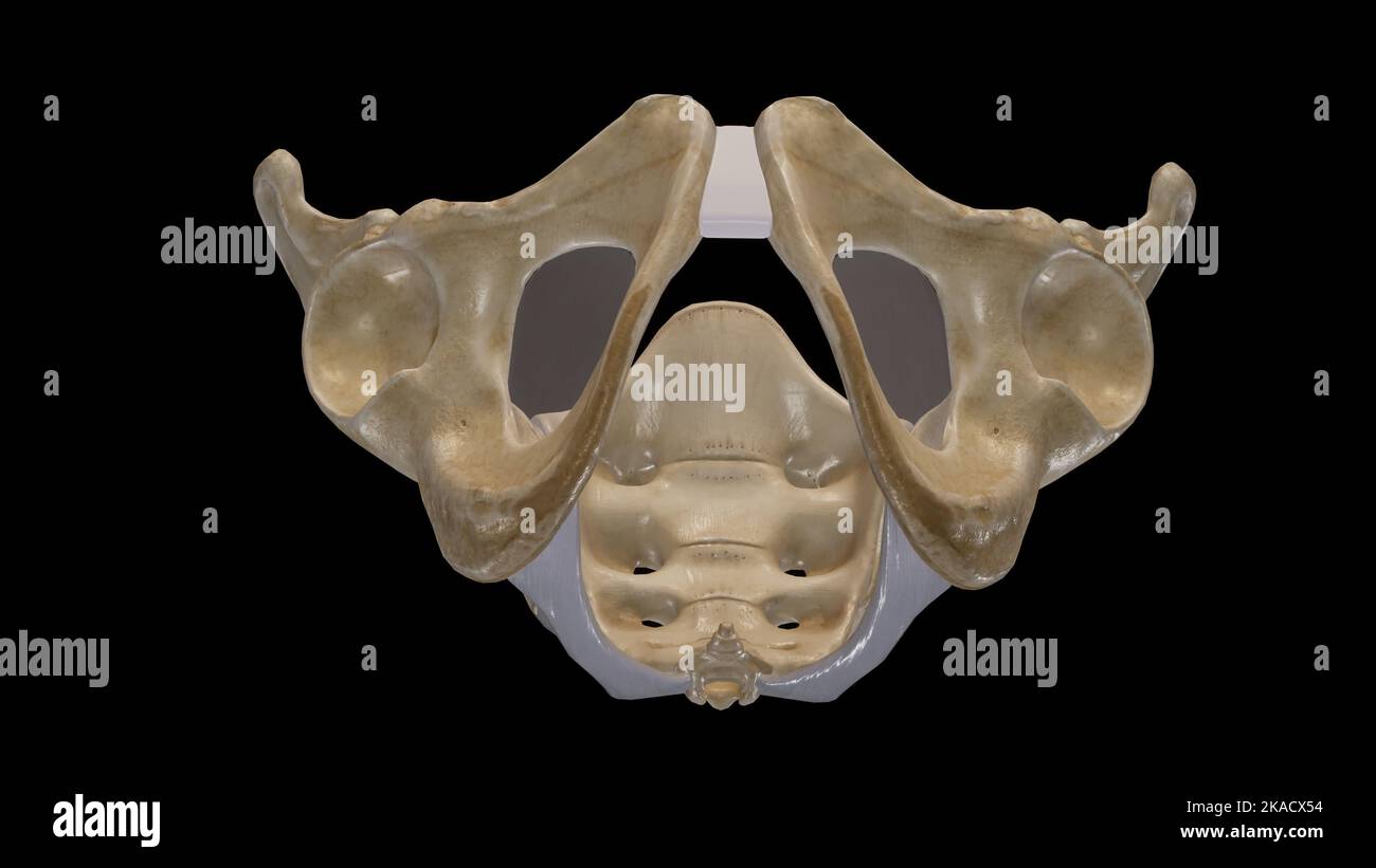 The Pelvic Girdle and Pelvic Outlet Stock Photohttps://www.alamy.com/image-license-details/?v=1https://www.alamy.com/the-pelvic-girdle-and-pelvic-outlet-image488320816.html
RF2KACX54–The Pelvic Girdle and Pelvic Outlet
![Practical human anatomy [electronic resource] : a working-guide for students of medicine and a ready-reference for surgeons and physicians . iformis muscle; it continues interiorly, upon thebone (ischium), anterior to the gemellus superior, obturator in-ternus, and gemellus inferior muscles ; it sends a branch to thegemellus inferior muscle, and its terminal portion enters theanterior surface of the quadratus femoris muscle. It is accom-panied by a small branch from the sciatic artery. 53. Parts Emerging at the Great Saero-Seiatie Foramen,Plate 100 —The parts emerging from this foramen are : t Stock Photo Practical human anatomy [electronic resource] : a working-guide for students of medicine and a ready-reference for surgeons and physicians . iformis muscle; it continues interiorly, upon thebone (ischium), anterior to the gemellus superior, obturator in-ternus, and gemellus inferior muscles ; it sends a branch to thegemellus inferior muscle, and its terminal portion enters theanterior surface of the quadratus femoris muscle. It is accom-panied by a small branch from the sciatic artery. 53. Parts Emerging at the Great Saero-Seiatie Foramen,Plate 100 —The parts emerging from this foramen are : t Stock Photo](https://c8.alamy.com/comp/2AX5ADG/practical-human-anatomy-electronic-resource-a-working-guide-for-students-of-medicine-and-a-ready-reference-for-surgeons-and-physicians-iformis-muscle-it-continues-interiorly-upon-thebone-ischium-anterior-to-the-gemellus-superior-obturator-in-ternus-and-gemellus-inferior-muscles-it-sends-a-branch-to-thegemellus-inferior-muscle-and-its-terminal-portion-enters-theanterior-surface-of-the-quadratus-femoris-muscle-it-is-accom-panied-by-a-small-branch-from-the-sciatic-artery-53-parts-emerging-at-the-great-saero-seiatie-foramenplate-100-the-parts-emerging-from-this-foramen-are-t-2AX5ADG.jpg) Practical human anatomy [electronic resource] : a working-guide for students of medicine and a ready-reference for surgeons and physicians . iformis muscle; it continues interiorly, upon thebone (ischium), anterior to the gemellus superior, obturator in-ternus, and gemellus inferior muscles ; it sends a branch to thegemellus inferior muscle, and its terminal portion enters theanterior surface of the quadratus femoris muscle. It is accom-panied by a small branch from the sciatic artery. 53. Parts Emerging at the Great Saero-Seiatie Foramen,Plate 100 —The parts emerging from this foramen are : t Stock Photohttps://www.alamy.com/image-license-details/?v=1https://www.alamy.com/practical-human-anatomy-electronic-resource-a-working-guide-for-students-of-medicine-and-a-ready-reference-for-surgeons-and-physicians-iformis-muscle-it-continues-interiorly-upon-thebone-ischium-anterior-to-the-gemellus-superior-obturator-in-ternus-and-gemellus-inferior-muscles-it-sends-a-branch-to-thegemellus-inferior-muscle-and-its-terminal-portion-enters-theanterior-surface-of-the-quadratus-femoris-muscle-it-is-accom-panied-by-a-small-branch-from-the-sciatic-artery-53-parts-emerging-at-the-great-saero-seiatie-foramenplate-100-the-parts-emerging-from-this-foramen-are-t-image343096028.html
Practical human anatomy [electronic resource] : a working-guide for students of medicine and a ready-reference for surgeons and physicians . iformis muscle; it continues interiorly, upon thebone (ischium), anterior to the gemellus superior, obturator in-ternus, and gemellus inferior muscles ; it sends a branch to thegemellus inferior muscle, and its terminal portion enters theanterior surface of the quadratus femoris muscle. It is accom-panied by a small branch from the sciatic artery. 53. Parts Emerging at the Great Saero-Seiatie Foramen,Plate 100 —The parts emerging from this foramen are : t Stock Photohttps://www.alamy.com/image-license-details/?v=1https://www.alamy.com/practical-human-anatomy-electronic-resource-a-working-guide-for-students-of-medicine-and-a-ready-reference-for-surgeons-and-physicians-iformis-muscle-it-continues-interiorly-upon-thebone-ischium-anterior-to-the-gemellus-superior-obturator-in-ternus-and-gemellus-inferior-muscles-it-sends-a-branch-to-thegemellus-inferior-muscle-and-its-terminal-portion-enters-theanterior-surface-of-the-quadratus-femoris-muscle-it-is-accom-panied-by-a-small-branch-from-the-sciatic-artery-53-parts-emerging-at-the-great-saero-seiatie-foramenplate-100-the-parts-emerging-from-this-foramen-are-t-image343096028.htmlRM2AX5ADG–Practical human anatomy [electronic resource] : a working-guide for students of medicine and a ready-reference for surgeons and physicians . iformis muscle; it continues interiorly, upon thebone (ischium), anterior to the gemellus superior, obturator in-ternus, and gemellus inferior muscles ; it sends a branch to thegemellus inferior muscle, and its terminal portion enters theanterior surface of the quadratus femoris muscle. It is accom-panied by a small branch from the sciatic artery. 53. Parts Emerging at the Great Saero-Seiatie Foramen,Plate 100 —The parts emerging from this foramen are : t
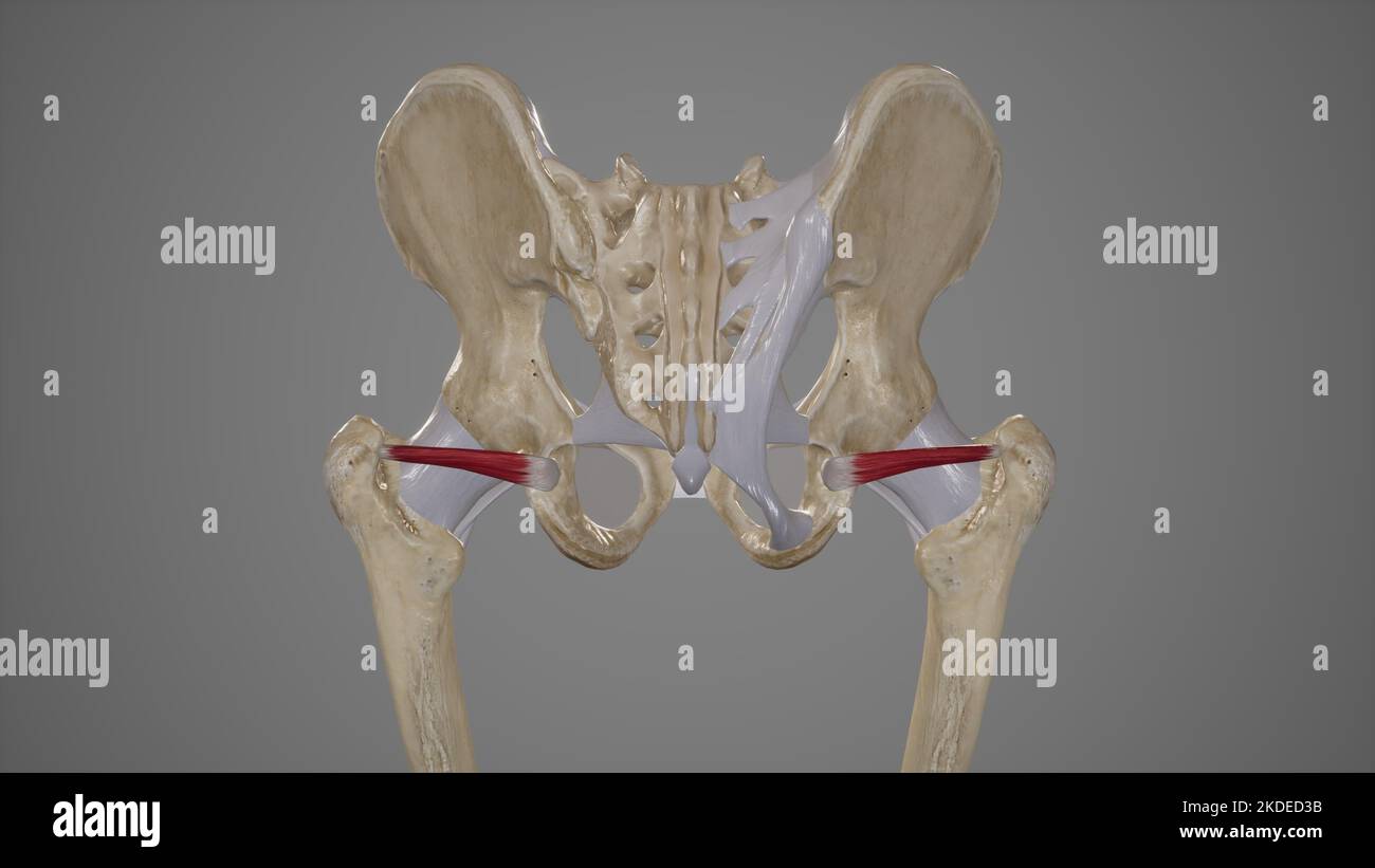 Medical Illustration of Inferior Gemellus Muscle Stock Photohttps://www.alamy.com/image-license-details/?v=1https://www.alamy.com/medical-illustration-of-inferior-gemellus-muscle-image490198447.html
RF2KDED3B–Medical Illustration of Inferior Gemellus Muscle
 . Manual of operative surgery. Selig (Arch. f. Klin.Chir., ciii, 994) advocates division of the obturator nerve before its entranceinto the obturator canal. The fact that the adductor magnus gains part of itsnerve supply from the sciatic nerve explains why after section of the obturatornerve, while spastic contraction is prevented, active contraction remains pos-sible. The obturator nerve arises from the second, third and fourth lumbarnerves, crosses the sacro-iliac joint and the internal iliac artery to find its way DIVISION OBTURATOR NERVE 77.3 along the lateral wall of the true pelvis until Stock Photohttps://www.alamy.com/image-license-details/?v=1https://www.alamy.com/manual-of-operative-surgery-selig-arch-f-klinchir-ciii-994-advocates-division-of-the-obturator-nerve-before-its-entranceinto-the-obturator-canal-the-fact-that-the-adductor-magnus-gains-part-of-itsnerve-supply-from-the-sciatic-nerve-explains-why-after-section-of-the-obturatornerve-while-spastic-contraction-is-prevented-active-contraction-remains-pos-sible-the-obturator-nerve-arises-from-the-second-third-and-fourth-lumbarnerves-crosses-the-sacro-iliac-joint-and-the-internal-iliac-artery-to-find-its-way-division-obturator-nerve-773-along-the-lateral-wall-of-the-true-pelvis-until-image336814898.html
RM2AFY6RE–. Manual of operative surgery. Selig (Arch. f. Klin.Chir., ciii, 994) advocates division of the obturator nerve before its entranceinto the obturator canal. The fact that the adductor magnus gains part of itsnerve supply from the sciatic nerve explains why after section of the obturatornerve, while spastic contraction is prevented, active contraction remains pos-sible. The obturator nerve arises from the second, third and fourth lumbarnerves, crosses the sacro-iliac joint and the internal iliac artery to find its way DIVISION OBTURATOR NERVE 77.3 along the lateral wall of the true pelvis until
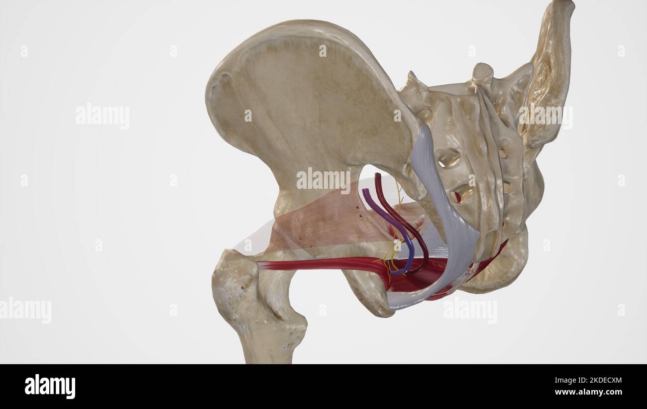 Anatomy of Lesser Sciatic Foramen Stock Photohttps://www.alamy.com/image-license-details/?v=1https://www.alamy.com/anatomy-of-lesser-sciatic-foramen-image490198316.html
RF2KDECXM–Anatomy of Lesser Sciatic Foramen
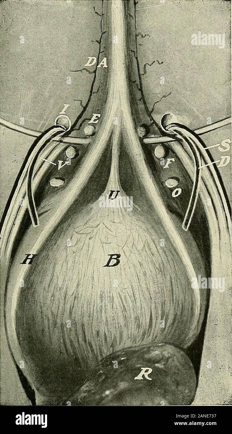 A text-book of clinical anatomy : for students and practitioners . Fig. 78.—Location of various forms of abdominal hernia; (diagrammatic). U,Umbilical hernia. D, Direct inguinal hernia. B, Indirect incomplete inguinal hernia.O, Complete or scrotal inguinal hernia. F, Femoral hernia. 241. - *.-*¥& Fig. 79.—View of inner aspect of anterior wall of abdomen to show internal orifices ofinguinal, femoral,, and obturator hernias. DA, Deep epigastric artery. E, Middle in-guinal fossa, corresponding externally to external abdominal ring. A direct inguinalhernia passes directly outward through this de Stock Photohttps://www.alamy.com/image-license-details/?v=1https://www.alamy.com/a-text-book-of-clinical-anatomy-for-students-and-practitioners-fig-78location-of-various-forms-of-abdominal-hernia-diagrammatic-uumbilical-hernia-d-direct-inguinal-hernia-b-indirect-incomplete-inguinal-herniao-complete-or-scrotal-inguinal-hernia-f-femoral-hernia-241-fig-79view-of-inner-aspect-of-anterior-wall-of-abdomen-to-show-internal-orifices-ofinguinal-femoral-and-obturator-hernias-da-deep-epigastric-artery-e-middle-in-guinal-fossa-corresponding-externally-to-external-abdominal-ring-a-direct-inguinalhernia-passes-directly-outward-through-this-de-image340217675.html
RM2ANE737–A text-book of clinical anatomy : for students and practitioners . Fig. 78.—Location of various forms of abdominal hernia; (diagrammatic). U,Umbilical hernia. D, Direct inguinal hernia. B, Indirect incomplete inguinal hernia.O, Complete or scrotal inguinal hernia. F, Femoral hernia. 241. - *.-*¥& Fig. 79.—View of inner aspect of anterior wall of abdomen to show internal orifices ofinguinal, femoral,, and obturator hernias. DA, Deep epigastric artery. E, Middle in-guinal fossa, corresponding externally to external abdominal ring. A direct inguinalhernia passes directly outward through this de
 Anatomy of Greater Sciatic Foramen Stock Photohttps://www.alamy.com/image-license-details/?v=1https://www.alamy.com/anatomy-of-greater-sciatic-foramen-image490198311.html
RF2KDECXF–Anatomy of Greater Sciatic Foramen
 . Plates of the arteries of the human body. pudic artery. Twigs to the obturator internus and gemellimuscles. External haemorrhoidal artery. 76. Twigs to the tuberosity of the ischium. 77- First jierforating artery. Twigs communicating with the external cir-cumflex artery of the thigh. Twig of the external circumflex artery of thethigh. Twig to the ischiatic nerve. 81, 81, 81. Muscular twigs. 1^2. Second perforating artery- 83, 83. Third perforating artery. 84, 84, 84, 84. Popliteal artery. 85, 85, 85, 85,85, 85, 85. Twigs to the muscles. 86, 86. Superficial internal superior articular ar- ter Stock Photohttps://www.alamy.com/image-license-details/?v=1https://www.alamy.com/plates-of-the-arteries-of-the-human-body-pudic-artery-twigs-to-the-obturator-internus-and-gemellimuscles-external-haemorrhoidal-artery-76-twigs-to-the-tuberosity-of-the-ischium-77-first-jierforating-artery-twigs-communicating-with-the-external-cir-cumflex-artery-of-the-thigh-twig-of-the-external-circumflex-artery-of-thethigh-twig-to-the-ischiatic-nerve-81-81-81-muscular-twigs-12-second-perforating-artery-83-83-third-perforating-artery-84-84-84-84-popliteal-artery-85-85-85-8585-85-85-twigs-to-the-muscles-86-86-superficial-internal-superior-articular-ar-ter-image336720662.html
RM2AFPXHX–. Plates of the arteries of the human body. pudic artery. Twigs to the obturator internus and gemellimuscles. External haemorrhoidal artery. 76. Twigs to the tuberosity of the ischium. 77- First jierforating artery. Twigs communicating with the external cir-cumflex artery of the thigh. Twig of the external circumflex artery of thethigh. Twig to the ischiatic nerve. 81, 81, 81. Muscular twigs. 1^2. Second perforating artery- 83, 83. Third perforating artery. 84, 84, 84, 84. Popliteal artery. 85, 85, 85, 85,85, 85, 85. Twigs to the muscles. 86, 86. Superficial internal superior articular ar- ter
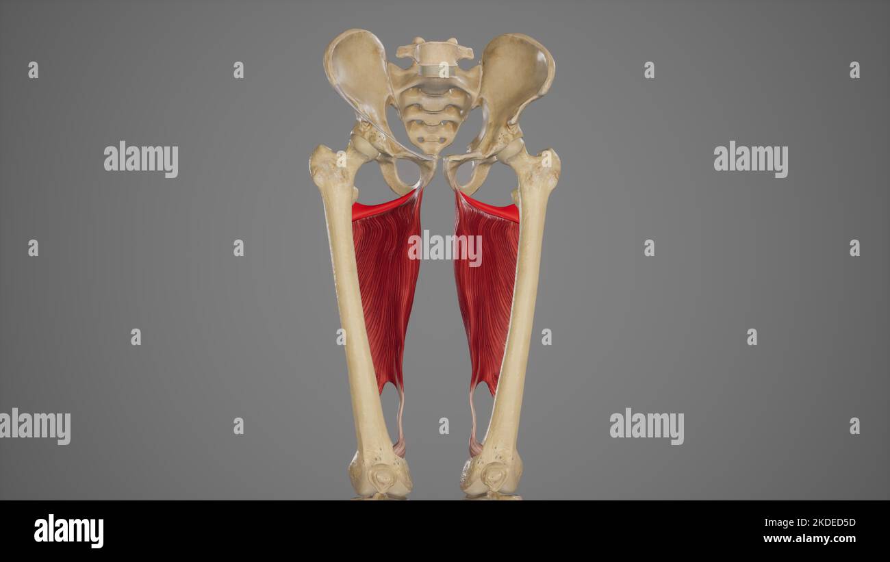 Medical Acurate Illustration of Adductor Minimus Stock Photohttps://www.alamy.com/image-license-details/?v=1https://www.alamy.com/medical-acurate-illustration-of-adductor-minimus-image490198505.html
RF2KDED5D–Medical Acurate Illustration of Adductor Minimus
 Atlas and text-book of topographic and applied anatomy . Obturator nerve(anterior branch)M. adductor longus ^ M. adductor ma M. vastus medial Femoral artei Long ! iaphenous nerv al vein (almost entire cone ealed) M. sartorh Anastomotica magnTendon of adductor Pouparts ligame Femoral veFemoralSuperficial external pudarteryProfunda femoris Fascia lat. THE THIGH. 159 Fig. 79.—The anterior femoral region. Fig. 80.—The exposure of the femoral artery before its entrance into Hunters canal. Fig. 81.—The subperitoneal exposure of the external iliac artery. Below Pouparts ligament the femoral vesselsha Stock Photohttps://www.alamy.com/image-license-details/?v=1https://www.alamy.com/atlas-and-text-book-of-topographic-and-applied-anatomy-obturator-nerveanterior-branchm-adductor-longus-m-adductor-ma-m-vastus-medial-femoral-artei-long-!-iaphenous-nerv-al-vein-almost-entire-cone-ealed-m-sartorh-anastomotica-magntendon-of-adductor-pouparts-ligame-femoral-vefemoralsuperficial-external-pudarteryprofunda-femoris-fascia-lat-the-thigh-159-fig-79the-anterior-femoral-region-fig-80the-exposure-of-the-femoral-artery-before-its-entrance-into-hunters-canal-fig-81the-subperitoneal-exposure-of-the-external-iliac-artery-below-pouparts-ligament-the-femoral-vesselsha-image338233293.html
RM2AJ7T0D–Atlas and text-book of topographic and applied anatomy . Obturator nerve(anterior branch)M. adductor longus ^ M. adductor ma M. vastus medial Femoral artei Long ! iaphenous nerv al vein (almost entire cone ealed) M. sartorh Anastomotica magnTendon of adductor Pouparts ligame Femoral veFemoralSuperficial external pudarteryProfunda femoris Fascia lat. THE THIGH. 159 Fig. 79.—The anterior femoral region. Fig. 80.—The exposure of the femoral artery before its entrance into Hunters canal. Fig. 81.—The subperitoneal exposure of the external iliac artery. Below Pouparts ligament the femoral vesselsha
 . Plates of the arteries of the human body. Morbid Anatomyof the Human (luUet, &c. p. 427.) I have a specimen inwhich the epigastric artery takes its rise from the obturator andpasses upwards and inwards to the rectus muscle, J. K. Hes-selbach (I. c. Tab. 2.) has delineated it. 10. Obturator internus. 11. Levator ani muscle. 12. Smaller sacvo-sciatic ligament. 13. 13. Origin of the pyriform muscle. 14. 14. Obturator nerve. 15. Fifth lumbar nerve.IG, 16, 16. Sacral nerves.17- Aorta. 18. 18. Middle sacral artery. 19. Fifth lumbar artery. 20. Left iliac artery. 21. 21. Right iliac artery. 22. 22. Stock Photohttps://www.alamy.com/image-license-details/?v=1https://www.alamy.com/plates-of-the-arteries-of-the-human-body-morbid-anatomyof-the-human-luuet-c-p-427-i-have-a-specimen-inwhich-the-epigastric-artery-takes-its-rise-from-the-obturator-andpasses-upwards-and-inwards-to-the-rectus-muscle-j-k-hes-selbach-i-c-tab-2-has-delineated-it-10-obturator-internus-11-levator-ani-muscle-12-smaller-sacvo-sciatic-ligament-13-13-origin-of-the-pyriform-muscle-14-14-obturator-nerve-15-fifth-lumbar-nerveig-16-16-sacral-nerves17-aorta-18-18-middle-sacral-artery-19-fifth-lumbar-artery-20-left-iliac-artery-21-21-right-iliac-artery-22-22-image336721279.html
RM2AFPYBY–. Plates of the arteries of the human body. Morbid Anatomyof the Human (luUet, &c. p. 427.) I have a specimen inwhich the epigastric artery takes its rise from the obturator andpasses upwards and inwards to the rectus muscle, J. K. Hes-selbach (I. c. Tab. 2.) has delineated it. 10. Obturator internus. 11. Levator ani muscle. 12. Smaller sacvo-sciatic ligament. 13. 13. Origin of the pyriform muscle. 14. 14. Obturator nerve. 15. Fifth lumbar nerve.IG, 16, 16. Sacral nerves.17- Aorta. 18. 18. Middle sacral artery. 19. Fifth lumbar artery. 20. Left iliac artery. 21. 21. Right iliac artery. 22. 22.
 . Plates of the arteries of the human body. th lumbar artery of each side. 31, 31. Middle sacral arterv. 32, 32. Fifth lumbar artery of each side. 33, 33. Sacral branches. 34, 34, 34, 34. Common iliac arteries. 35, 35. Internal iliac arteries. 36, 36, 36, 36, 36, 36. Uterine arteries. 37, .37, 37, .37, 37, 37. Tortuous branches go- ing to the posterior surface of the uterus. 38, 38. Umbilical arteries. 39, 39. Lateral sacral arteries. 40, 40. Gluteal arteries.41,41. Obturator arteries. 42, 42. Internal pudic arteries. 43, 43. Ischiatic arteries. 44, 44, 44, 44. External iliac arteries. 45, 45, Stock Photohttps://www.alamy.com/image-license-details/?v=1https://www.alamy.com/plates-of-the-arteries-of-the-human-body-th-lumbar-artery-of-each-side-31-31-middle-sacral-arterv-32-32-fifth-lumbar-artery-of-each-side-33-33-sacral-branches-34-34-34-34-common-iliac-arteries-35-35-internal-iliac-arteries-36-36-36-36-36-36-uterine-arteries-37-37-37-37-37-37-tortuous-branches-go-ing-to-the-posterior-surface-of-the-uterus-38-38-umbilical-arteries-39-39-lateral-sacral-arteries-40-40-gluteal-arteries4141-obturator-arteries-42-42-internal-pudic-arteries-43-43-ischiatic-arteries-44-44-44-44-external-iliac-arteries-45-45-image336722225.html
RM2AFR0HN–. Plates of the arteries of the human body. th lumbar artery of each side. 31, 31. Middle sacral arterv. 32, 32. Fifth lumbar artery of each side. 33, 33. Sacral branches. 34, 34, 34, 34. Common iliac arteries. 35, 35. Internal iliac arteries. 36, 36, 36, 36, 36, 36. Uterine arteries. 37, .37, 37, .37, 37, 37. Tortuous branches go- ing to the posterior surface of the uterus. 38, 38. Umbilical arteries. 39, 39. Lateral sacral arteries. 40, 40. Gluteal arteries.41,41. Obturator arteries. 42, 42. Internal pudic arteries. 43, 43. Ischiatic arteries. 44, 44, 44, 44. External iliac arteries. 45, 45,
 A system of gynecology . le; d, internal obturator muscle; c, e, psoasmuscle; /, linea alba ; ;/. r;. ureters; ft, obturator nerve ; i, internal inguinal ring: 1, abdom-inal aorta: 2, inferior mesenteric artery ; 3,3, common iliac arteries : 1. external iliac artery;5,vena cava; 6, renal veins; 7,7, common iliac veins; 8, external iliac vein: 9, internaliliac artery: 10, gluteal; 11, ileo-lumbar; 12, sciatic; 18, pudic; it. obturator: IS, epigastricveins; 17. uterine veins; is, vagino-vesical venous cete; 19, spermatic veins: 20, bulb ofovary; 21, vein to round ligament; JJ, Fallopian veins. n Stock Photohttps://www.alamy.com/image-license-details/?v=1https://www.alamy.com/a-system-of-gynecology-le-d-internal-obturator-muscle-c-e-psoasmuscle-linea-alba-r-ureters-ft-obturator-nerve-i-internal-inguinal-ring-1-abdom-inal-aorta-2-inferior-mesenteric-artery-33-common-iliac-arteries-1-external-iliac-artery5vena-cava-6-renal-veins-77-common-iliac-veins-8-external-iliac-vein-9-internaliliac-artery-10-gluteal-11-ileo-lumbar-12-sciatic-18-pudic-it-obturator-is-epigastricveins-17-uterine-veins-is-vagino-vesical-venous-cete-19-spermatic-veins-20-bulb-ofovary-21-vein-to-round-ligament-jj-fallopian-veins-n-image338502089.html
RM2AJM2T9–A system of gynecology . le; d, internal obturator muscle; c, e, psoasmuscle; /, linea alba ; ;/. r;. ureters; ft, obturator nerve ; i, internal inguinal ring: 1, abdom-inal aorta: 2, inferior mesenteric artery ; 3,3, common iliac arteries : 1. external iliac artery;5,vena cava; 6, renal veins; 7,7, common iliac veins; 8, external iliac vein: 9, internaliliac artery: 10, gluteal; 11, ileo-lumbar; 12, sciatic; 18, pudic; it. obturator: IS, epigastricveins; 17. uterine veins; is, vagino-vesical venous cete; 19, spermatic veins: 20, bulb ofovary; 21, vein to round ligament; JJ, Fallopian veins. n
 . Anatomy, descriptive and surgical. tructures are enclosed in a sheath formed by the obturator fascia, thepudic nerve lying below the artery (Fig. 402, p. 588). Crossing the space trans-versely about its centre are the inferior hemorrhoidal vessels and nerves, branchesof the internal pudic ; they are distributed to the integument of the anus and to themuscles of the lower end of the rectum. These vessels are occasionally of largesize, and may give rise to troublesome hemorrhage when divided in the operationof lithotomy or of fistula in ano. At the back part of this space may be seen abranch o Stock Photohttps://www.alamy.com/image-license-details/?v=1https://www.alamy.com/anatomy-descriptive-and-surgical-tructures-are-enclosed-in-a-sheath-formed-by-the-obturator-fascia-thepudic-nerve-lying-below-the-artery-fig-402-p-588-crossing-the-space-trans-versely-about-its-centre-are-the-inferior-hemorrhoidal-vessels-and-nerves-branchesof-the-internal-pudic-they-are-distributed-to-the-integument-of-the-anus-and-to-themuscles-of-the-lower-end-of-the-rectum-these-vessels-are-occasionally-of-largesize-and-may-give-rise-to-troublesome-hemorrhage-when-divided-in-the-operationof-lithotomy-or-of-fistula-in-ano-at-the-back-part-of-this-space-may-be-seen-abranch-o-image370500169.html
RM2CENMMW–. Anatomy, descriptive and surgical. tructures are enclosed in a sheath formed by the obturator fascia, thepudic nerve lying below the artery (Fig. 402, p. 588). Crossing the space trans-versely about its centre are the inferior hemorrhoidal vessels and nerves, branchesof the internal pudic ; they are distributed to the integument of the anus and to themuscles of the lower end of the rectum. These vessels are occasionally of largesize, and may give rise to troublesome hemorrhage when divided in the operationof lithotomy or of fistula in ano. At the back part of this space may be seen abranch o
 . A practical treatise on fractures and dislocations. taken as the typical form ; in itthe limb is but slightly, if at all, abducted,markedly everted, and somewhat shortened(Fig. 342), and the head of the femur can befelt more or less distinctly in the groin, withthe artery pulsating directly in front of it or to its inner side. Whenthe head is displaced further toward the median line the limb isabducted and flexed as well as everted, and its position is more likethat of an obturator dislocation; the capital difference is the positionof the head on the pubis where it can be distinctly, felt an Stock Photohttps://www.alamy.com/image-license-details/?v=1https://www.alamy.com/a-practical-treatise-on-fractures-and-dislocations-taken-as-the-typical-form-in-itthe-limb-is-but-slightly-if-at-all-abductedmarkedly-everted-and-somewhat-shortenedfig-342-and-the-head-of-the-femur-can-befelt-more-or-less-distinctly-in-the-groin-withthe-artery-pulsating-directly-in-front-of-it-or-to-its-inner-side-whenthe-head-is-displaced-further-toward-the-median-line-the-limb-isabducted-and-flexed-as-well-as-everted-and-its-position-is-more-likethat-of-an-obturator-dislocation-the-capital-difference-is-the-positionof-the-head-on-the-pubis-where-it-can-be-distinctly-felt-an-image370604734.html
RM2CEXE3A–. A practical treatise on fractures and dislocations. taken as the typical form ; in itthe limb is but slightly, if at all, abducted,markedly everted, and somewhat shortened(Fig. 342), and the head of the femur can befelt more or less distinctly in the groin, withthe artery pulsating directly in front of it or to its inner side. Whenthe head is displaced further toward the median line the limb isabducted and flexed as well as everted, and its position is more likethat of an obturator dislocation; the capital difference is the positionof the head on the pubis where it can be distinctly, felt an
 . Anatomy, descriptive and surgical. s a very thin mem-brane in front of the Pyriformis muscle and sacral nerves, behind the branches ofthe internal iliac artery and vein (which perforate it), to the front of the sacrum.In front it follows the attachment of the Obturator internus to the bone, archesbeneath the obturator vessels, completing the orifice of the obturator canal, and atthe front of the pelvis is attached to the lower part of the symphysis pubis. At thelevel of a line extending from the lower part of the symphysis pubis to the spine ofthe ischium is a thickened whitish band; this ma Stock Photohttps://www.alamy.com/image-license-details/?v=1https://www.alamy.com/anatomy-descriptive-and-surgical-s-a-very-thin-mem-brane-in-front-of-the-pyriformis-muscle-and-sacral-nerves-behind-the-branches-ofthe-internal-iliac-artery-and-vein-which-perforate-it-to-the-front-of-the-sacrumin-front-it-follows-the-attachment-of-the-obturator-internus-to-the-bone-archesbeneath-the-obturator-vessels-completing-the-orifice-of-the-obturator-canal-and-atthe-front-of-the-pelvis-is-attached-to-the-lower-part-of-the-symphysis-pubis-at-thelevel-of-a-line-extending-from-the-lower-part-of-the-symphysis-pubis-to-the-spine-ofthe-ischium-is-a-thickened-whitish-band-this-ma-image370499080.html
RM2CENKA0–. Anatomy, descriptive and surgical. s a very thin mem-brane in front of the Pyriformis muscle and sacral nerves, behind the branches ofthe internal iliac artery and vein (which perforate it), to the front of the sacrum.In front it follows the attachment of the Obturator internus to the bone, archesbeneath the obturator vessels, completing the orifice of the obturator canal, and atthe front of the pelvis is attached to the lower part of the symphysis pubis. At thelevel of a line extending from the lower part of the symphysis pubis to the spine ofthe ischium is a thickened whitish band; this ma
 . The anatomy and surgical treatment of hernia. l tumor is exceptional. Mr. Birkett, who has especially studied the subject, writes:* After passing alongthe obturator canal, the hernial tumor emerges upon the thigh, below the horizontalramus of the pubes to the inner side of the capsule of the hip-joint, behind and a little tothe inner side of the femoral artery and vein, and to the outer side of the tendon of theadductor longus. The tumor formed by the protrusion is covered by the pectineusmuscle. It may be distinguished, therefore, from crural hernia, by observing the relativepositions of th Stock Photohttps://www.alamy.com/image-license-details/?v=1https://www.alamy.com/the-anatomy-and-surgical-treatment-of-hernia-l-tumor-is-exceptional-mr-birkett-who-has-especially-studied-the-subject-writes-after-passing-alongthe-obturator-canal-the-hernial-tumor-emerges-upon-the-thigh-below-the-horizontalramus-of-the-pubes-to-the-inner-side-of-the-capsule-of-the-hip-joint-behind-and-a-little-tothe-inner-side-of-the-femoral-artery-and-vein-and-to-the-outer-side-of-the-tendon-of-theadductor-longus-the-tumor-formed-by-the-protrusion-is-covered-by-the-pectineusmuscle-it-may-be-distinguished-therefore-from-crural-hernia-by-observing-the-relativepositions-of-th-image370330572.html
RM2CEE0BT–. The anatomy and surgical treatment of hernia. l tumor is exceptional. Mr. Birkett, who has especially studied the subject, writes:* After passing alongthe obturator canal, the hernial tumor emerges upon the thigh, below the horizontalramus of the pubes to the inner side of the capsule of the hip-joint, behind and a little tothe inner side of the femoral artery and vein, and to the outer side of the tendon of theadductor longus. The tumor formed by the protrusion is covered by the pectineusmuscle. It may be distinguished, therefore, from crural hernia, by observing the relativepositions of th
 . American practice of surgery ; a complete system of the science and art of surgery . ^ a strong septum,the ol^turator nicinbrane, which is usually in two layers separated by lightareolar tissue. The obturator internus and externus nuiscles, which springfrom th(^ inner and outer surfaces of this membrane, pass toward the trochantermajor as extcn-nal rotators of the femur. The obturator canal or sulcus permitsthe passage of the obturator nerve, artery, and vein in the order named, from. Fig. 24.5.—The Drawing Shows the First Stage of Mayos Transverse Suturing of the UmbilicalRing bv the Imbric Stock Photohttps://www.alamy.com/image-license-details/?v=1https://www.alamy.com/american-practice-of-surgery-a-complete-system-of-the-science-and-art-of-surgery-a-strong-septumthe-olturator-nicinbrane-which-is-usually-in-two-layers-separated-by-lightareolar-tissue-the-obturator-internus-and-externus-nuiscles-which-springfrom-th-inner-and-outer-surfaces-of-this-membrane-pass-toward-the-trochantermajor-as-extcn-nal-rotators-of-the-femur-the-obturator-canal-or-sulcus-permitsthe-passage-of-the-obturator-nerve-artery-and-vein-in-the-order-named-from-fig-245the-drawing-shows-the-first-stage-of-mayos-transverse-suturing-of-the-umbilicalring-bv-the-imbric-image372373931.html
RM2CHR2MY–. American practice of surgery ; a complete system of the science and art of surgery . ^ a strong septum,the ol^turator nicinbrane, which is usually in two layers separated by lightareolar tissue. The obturator internus and externus nuiscles, which springfrom th(^ inner and outer surfaces of this membrane, pass toward the trochantermajor as extcn-nal rotators of the femur. The obturator canal or sulcus permitsthe passage of the obturator nerve, artery, and vein in the order named, from. Fig. 24.5.—The Drawing Shows the First Stage of Mayos Transverse Suturing of the UmbilicalRing bv the Imbric
 . The anatomy and surgical treatment of hernia. reserved. 20. Ramus of the pubis. 21. Nutritive vessels separated from the fem-oral vessels. 22. Obturator vessels and nerves seen behindthe posterior aponeurosis of the sheath. J. Muscular sheath of the first abductor. 2j, 2j. Nutritive vessels freed from the fem-oral artery. 24. Nerve branch of the same muscle fur-nished by the obturator. 2j. Trunk of the obturator nerve seen throughfrom behind the posterior sheath. K. Superior extremity of the layer of the vastusinternus. L. Superior extremity of the sartorius. jM. Aponeurosis of the anterior Stock Photohttps://www.alamy.com/image-license-details/?v=1https://www.alamy.com/the-anatomy-and-surgical-treatment-of-hernia-reserved-20-ramus-of-the-pubis-21-nutritive-vessels-separated-from-the-fem-oral-vessels-22-obturator-vessels-and-nerves-seen-behindthe-posterior-aponeurosis-of-the-sheath-j-muscular-sheath-of-the-first-abductor-2j-2j-nutritive-vessels-freed-from-the-fem-oral-artery-24-nerve-branch-of-the-same-muscle-fur-nished-by-the-obturator-2j-trunk-of-the-obturator-nerve-seen-throughfrom-behind-the-posterior-sheath-k-superior-extremity-of-the-layer-of-the-vastusinternus-l-superior-extremity-of-the-sartorius-jm-aponeurosis-of-the-anterior-image370336673.html
RM2CEE85N–. The anatomy and surgical treatment of hernia. reserved. 20. Ramus of the pubis. 21. Nutritive vessels separated from the fem-oral vessels. 22. Obturator vessels and nerves seen behindthe posterior aponeurosis of the sheath. J. Muscular sheath of the first abductor. 2j, 2j. Nutritive vessels freed from the fem-oral artery. 24. Nerve branch of the same muscle fur-nished by the obturator. 2j. Trunk of the obturator nerve seen throughfrom behind the posterior sheath. K. Superior extremity of the layer of the vastusinternus. L. Superior extremity of the sartorius. jM. Aponeurosis of the anterior
 . Anatomy, descriptive and applied. Anatomy. 674 THE VASCULAB SYSTEMS The internal branch {ramus anterior) curves backward along the inner margin of the obturator foramen, lying between it and the Obturator externus muscle; it distributes branches to the Obturator externus, Pectineus, Adductors and Gracilis, and anastomoses with the external branch, and with the internal circumflex artery. The external branch {ramus posterior) curves backward around the outer margin of the obturator foramen, also lying between the obturator foramen and the Obturator externus muscle, to the space between the Ge Stock Photohttps://www.alamy.com/image-license-details/?v=1https://www.alamy.com/anatomy-descriptive-and-applied-anatomy-674-the-vasculab-systems-the-internal-branch-ramus-anterior-curves-backward-along-the-inner-margin-of-the-obturator-foramen-lying-between-it-and-the-obturator-externus-muscle-it-distributes-branches-to-the-obturator-externus-pectineus-adductors-and-gracilis-and-anastomoses-with-the-external-branch-and-with-the-internal-circumflex-artery-the-external-branch-ramus-posterior-curves-backward-around-the-outer-margin-of-the-obturator-foramen-also-lying-between-the-obturator-foramen-and-the-obturator-externus-muscle-to-the-space-between-the-ge-image236793798.html
RMRN6TWA–. Anatomy, descriptive and applied. Anatomy. 674 THE VASCULAB SYSTEMS The internal branch {ramus anterior) curves backward along the inner margin of the obturator foramen, lying between it and the Obturator externus muscle; it distributes branches to the Obturator externus, Pectineus, Adductors and Gracilis, and anastomoses with the external branch, and with the internal circumflex artery. The external branch {ramus posterior) curves backward around the outer margin of the obturator foramen, also lying between the obturator foramen and the Obturator externus muscle, to the space between the Ge
 . The anatomy of the human body. Human anatomy; Anatomy. 276 MYOLOGY. F^. 127. Relations.—The pectineus is covered by the deep layer of the femoral fascia, and by the femoral vessels. It covers the capsular ligament of the joint, the smaU deep adductor, and the obturator externus, from which it is separated by the ob- turator vessels and nerves. Its outer border is parallel with the inner border of the conjoined portions of the psoas and iliacus, and is separated from them by a cellular interval, over which the femoral artery passes ; so that, were it not for the projection of this outer borde Stock Photohttps://www.alamy.com/image-license-details/?v=1https://www.alamy.com/the-anatomy-of-the-human-body-human-anatomy-anatomy-276-myology-f-127-relationsthe-pectineus-is-covered-by-the-deep-layer-of-the-femoral-fascia-and-by-the-femoral-vessels-it-covers-the-capsular-ligament-of-the-joint-the-smau-deep-adductor-and-the-obturator-externus-from-which-it-is-separated-by-the-ob-turator-vessels-and-nerves-its-outer-border-is-parallel-with-the-inner-border-of-the-conjoined-portions-of-the-psoas-and-iliacus-and-is-separated-from-them-by-a-cellular-interval-over-which-the-femoral-artery-passes-so-that-were-it-not-for-the-projection-of-this-outer-borde-image236797580.html
RMRN71MC–. The anatomy of the human body. Human anatomy; Anatomy. 276 MYOLOGY. F^. 127. Relations.—The pectineus is covered by the deep layer of the femoral fascia, and by the femoral vessels. It covers the capsular ligament of the joint, the smaU deep adductor, and the obturator externus, from which it is separated by the ob- turator vessels and nerves. Its outer border is parallel with the inner border of the conjoined portions of the psoas and iliacus, and is separated from them by a cellular interval, over which the femoral artery passes ; so that, were it not for the projection of this outer borde
 . The anatomy of the domestic animals. Veterinary anatomy. 324 FASCIA AND MUSCLES OF THE HORSE Relations.—Laterally, the skin and fascia, the biceps, and the medial head of the gastrocnemius; medially, the coccygeal fascia, the sacro-sciatic ligament, the semimembranosus; anteriorly, the biceps femoris, branches of the femoral artery, and the great sciatic nerve. Blood-supply.—Posterior gluteal, obturator, and posterior femoral arteries. Nerve-supply.—Posterior gluteal and great sciatic nerves. Origin nf nhliqunx ah- fioniini.^ iidirinix Inguinal liyamcitt (part removed) Iliacus Tensor faciw l Stock Photohttps://www.alamy.com/image-license-details/?v=1https://www.alamy.com/the-anatomy-of-the-domestic-animals-veterinary-anatomy-324-fascia-and-muscles-of-the-horse-relationslaterally-the-skin-and-fascia-the-biceps-and-the-medial-head-of-the-gastrocnemius-medially-the-coccygeal-fascia-the-sacro-sciatic-ligament-the-semimembranosus-anteriorly-the-biceps-femoris-branches-of-the-femoral-artery-and-the-great-sciatic-nerve-blood-supplyposterior-gluteal-obturator-and-posterior-femoral-arteries-nerve-supplyposterior-gluteal-and-great-sciatic-nerves-origin-nf-nhliqunx-ah-fioniini-iidirinix-inguinal-liyamcitt-part-removed-iliacus-tensor-faciw-l-image236800913.html
RMRN75YD–. The anatomy of the domestic animals. Veterinary anatomy. 324 FASCIA AND MUSCLES OF THE HORSE Relations.—Laterally, the skin and fascia, the biceps, and the medial head of the gastrocnemius; medially, the coccygeal fascia, the sacro-sciatic ligament, the semimembranosus; anteriorly, the biceps femoris, branches of the femoral artery, and the great sciatic nerve. Blood-supply.—Posterior gluteal, obturator, and posterior femoral arteries. Nerve-supply.—Posterior gluteal and great sciatic nerves. Origin nf nhliqunx ah- fioniini.^ iidirinix Inguinal liyamcitt (part removed) Iliacus Tensor faciw l
 . The anatomy of the domestic animals . Veterinary anatomy. 324 FASCIA AND MUSCLES OF THE HORSE Relations.—Laterally, the skin and fascia, the biceps, and the medial head of the gastrocnemius; medially, the coccygeal fascia, the sacro-sciatic ligament, the semimembranosus; anteriorly, the biceps femoris, branches of the femoral artery, and the great sciatic nerve. Blood-supply.—FosterioT gluteal, obturator, and posterior femoral arteries. Nerve-supply.—Posterior gluteal and great sciatic nerves. Origin of ohliquus ab- dominis intern us Inguinal ligament {part removed) Iliacus Tensor facia laic Stock Photohttps://www.alamy.com/image-license-details/?v=1https://www.alamy.com/the-anatomy-of-the-domestic-animals-veterinary-anatomy-324-fascia-and-muscles-of-the-horse-relationslaterally-the-skin-and-fascia-the-biceps-and-the-medial-head-of-the-gastrocnemius-medially-the-coccygeal-fascia-the-sacro-sciatic-ligament-the-semimembranosus-anteriorly-the-biceps-femoris-branches-of-the-femoral-artery-and-the-great-sciatic-nerve-blood-supplyfosteriot-gluteal-obturator-and-posterior-femoral-arteries-nerve-supplyposterior-gluteal-and-great-sciatic-nerves-origin-of-ohliquus-ab-dominis-intern-us-inguinal-ligament-part-removed-iliacus-tensor-facia-laic-image232325939.html
RMRDYA2Y–. The anatomy of the domestic animals . Veterinary anatomy. 324 FASCIA AND MUSCLES OF THE HORSE Relations.—Laterally, the skin and fascia, the biceps, and the medial head of the gastrocnemius; medially, the coccygeal fascia, the sacro-sciatic ligament, the semimembranosus; anteriorly, the biceps femoris, branches of the femoral artery, and the great sciatic nerve. Blood-supply.—FosterioT gluteal, obturator, and posterior femoral arteries. Nerve-supply.—Posterior gluteal and great sciatic nerves. Origin of ohliquus ab- dominis intern us Inguinal ligament {part removed) Iliacus Tensor facia laic
 . Cunningham's Text-book of anatomy. Anatomy. 131: THE UKINO-GENITAL SYSTEM. against it, is depressed to form a little fossa termed the fossa ovarii, within which the ovary is placed. In the floor of this fossa are the obturator nerve and vessels. The tubal extremity of the ovary lies below the level of the external iliac vessels, and its uterine extremity is placed just above the level of the peritoneum covering the pelvic floor. The fossa ovarii, in which the ovary lies, extends as far forwards as the obliterated umbilical artery, and backwards as far as the ureter and uterine vessels. Thus Stock Photohttps://www.alamy.com/image-license-details/?v=1https://www.alamy.com/cunninghams-text-book-of-anatomy-anatomy-131-the-ukino-genital-system-against-it-is-depressed-to-form-a-little-fossa-termed-the-fossa-ovarii-within-which-the-ovary-is-placed-in-the-floor-of-this-fossa-are-the-obturator-nerve-and-vessels-the-tubal-extremity-of-the-ovary-lies-below-the-level-of-the-external-iliac-vessels-and-its-uterine-extremity-is-placed-just-above-the-level-of-the-peritoneum-covering-the-pelvic-floor-the-fossa-ovarii-in-which-the-ovary-lies-extends-as-far-forwards-as-the-obliterated-umbilical-artery-and-backwards-as-far-as-the-ureter-and-uterine-vessels-thus-image231867949.html
RMRD6DX5–. Cunningham's Text-book of anatomy. Anatomy. 131: THE UKINO-GENITAL SYSTEM. against it, is depressed to form a little fossa termed the fossa ovarii, within which the ovary is placed. In the floor of this fossa are the obturator nerve and vessels. The tubal extremity of the ovary lies below the level of the external iliac vessels, and its uterine extremity is placed just above the level of the peritoneum covering the pelvic floor. The fossa ovarii, in which the ovary lies, extends as far forwards as the obliterated umbilical artery, and backwards as far as the ureter and uterine vessels. Thus
 . Anatomy, descriptive and applied. Anatomy. THE BEEP VETNS OF THE LOWER EXTREMITY 74.3 and joins the external iliac vein about three-quarters of an inch above Poupart's ligament. The pubic vein communicates with the obturator em in the obturator fora- men, and ascends on the back of the pubis to terminate in the external iliac -ein. The internal iliac vein {v. hypoc/astrica) commences near the upper part of the great sacrosciatic foramen, passes upward behind and slightly to the inner side of the internal iliac artery, and at the brim of the pelvis joins with the external iliac to form the Stock Photohttps://www.alamy.com/image-license-details/?v=1https://www.alamy.com/anatomy-descriptive-and-applied-anatomy-the-beep-vetns-of-the-lower-extremity-743-and-joins-the-external-iliac-vein-about-three-quarters-of-an-inch-above-pouparts-ligament-the-pubic-vein-communicates-with-the-obturator-em-in-the-obturator-fora-men-and-ascends-on-the-back-of-the-pubis-to-terminate-in-the-external-iliac-ein-the-internal-iliac-vein-v-hypocastrica-commences-near-the-upper-part-of-the-great-sacrosciatic-foramen-passes-upward-behind-and-slightly-to-the-inner-side-of-the-internal-iliac-artery-and-at-the-brim-of-the-pelvis-joins-with-the-external-iliac-to-form-the-image236773020.html
RMRN5XB8–. Anatomy, descriptive and applied. Anatomy. THE BEEP VETNS OF THE LOWER EXTREMITY 74.3 and joins the external iliac vein about three-quarters of an inch above Poupart's ligament. The pubic vein communicates with the obturator em in the obturator fora- men, and ascends on the back of the pubis to terminate in the external iliac -ein. The internal iliac vein {v. hypoc/astrica) commences near the upper part of the great sacrosciatic foramen, passes upward behind and slightly to the inner side of the internal iliac artery, and at the brim of the pelvis joins with the external iliac to form the