Quick filters:
Odontoid Stock Photos and Images
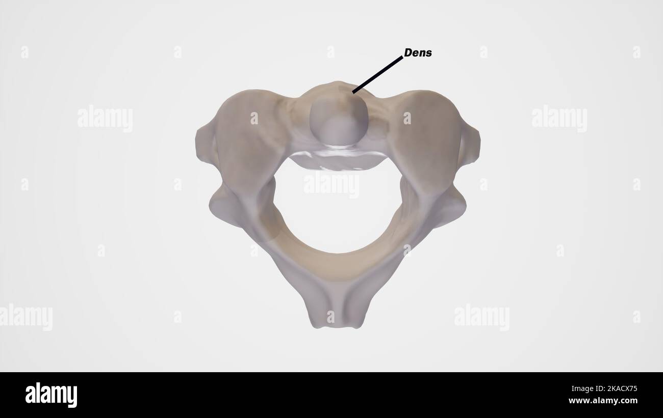 Cervical spine,second cervical vertebrae (Axis)-superior view labeled Stock Photohttps://www.alamy.com/image-license-details/?v=1https://www.alamy.com/cervical-spinesecond-cervical-vertebrae-axis-superior-view-labeled-image488320873.html
RF2KACX75–Cervical spine,second cervical vertebrae (Axis)-superior view labeled
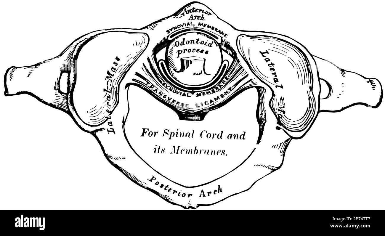 This illustration represents Odontoid Process, vintage line drawing or engraving illustration. Stock Vectorhttps://www.alamy.com/image-license-details/?v=1https://www.alamy.com/this-illustration-represents-odontoid-process-vintage-line-drawing-or-engraving-illustration-image348617255.html
RF2B74TT7–This illustration represents Odontoid Process, vintage line drawing or engraving illustration.
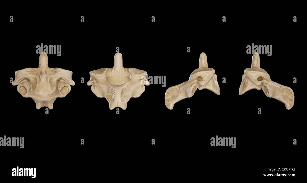 Second Cervical Vertebra (Axis) -Multiple Views.jpg Stock Photohttps://www.alamy.com/image-license-details/?v=1https://www.alamy.com/second-cervical-vertebra-axis-multiple-viewsjpg-image491879602.html
RF2KG71CJ–Second Cervical Vertebra (Axis) -Multiple Views.jpg
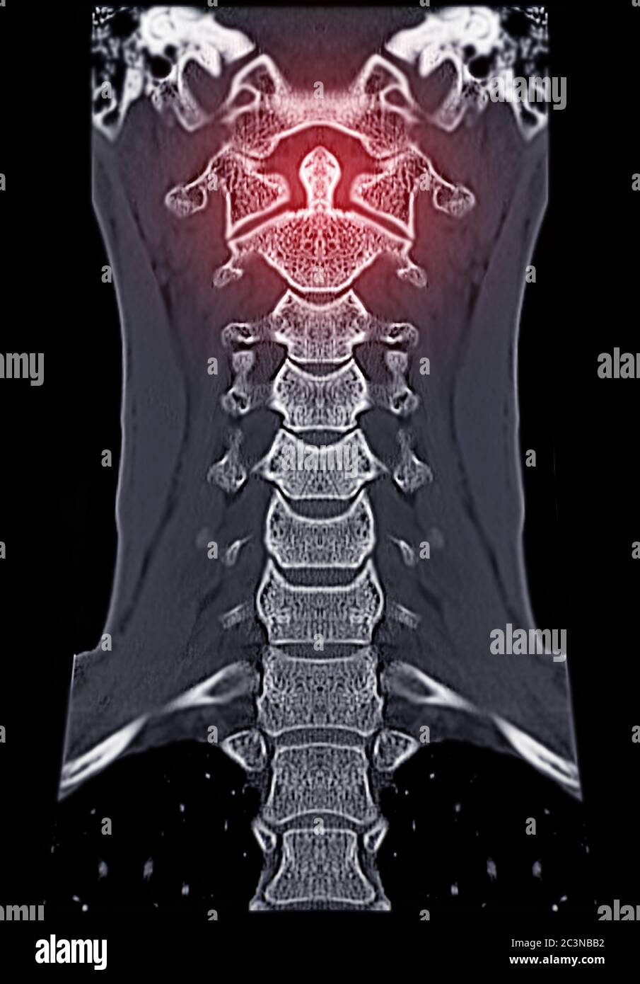 CT C-Spine or Cervical spine Coronal view in patient trauma cervical spine injury showing odontoid process or dens. Stock Photohttps://www.alamy.com/image-license-details/?v=1https://www.alamy.com/ct-c-spine-or-cervical-spine-coronal-view-in-patient-trauma-cervical-spine-injury-showing-odontoid-process-or-dens-image363731622.html
RF2C3NBB2–CT C-Spine or Cervical spine Coronal view in patient trauma cervical spine injury showing odontoid process or dens.
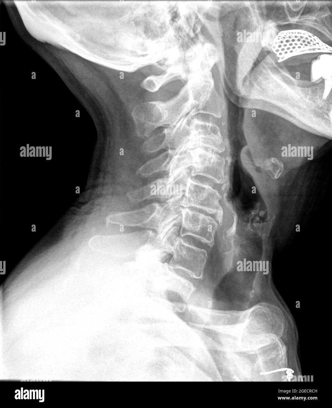 Cervical spine x-ray of a 72 year old female patient side view Stock Photohttps://www.alamy.com/image-license-details/?v=1https://www.alamy.com/cervical-spine-x-ray-of-a-72-year-old-female-patient-side-view-image439146193.html
RM2GECRCH–Cervical spine x-ray of a 72 year old female patient side view
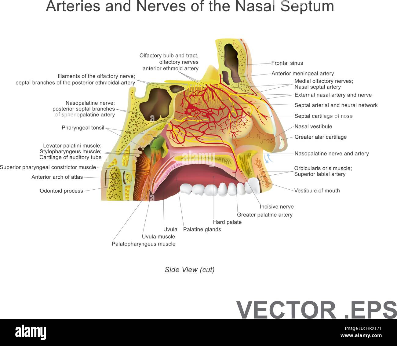 The nasal cavity (or nasal fossa) is a large air filled space above and behind the nose in the middle of the face. Each cavity is the continuation of Stock Vectorhttps://www.alamy.com/image-license-details/?v=1https://www.alamy.com/stock-photo-the-nasal-cavity-or-nasal-fossa-is-a-large-air-filled-space-above-135199429.html
RFHRXT71–The nasal cavity (or nasal fossa) is a large air filled space above and behind the nose in the middle of the face. Each cavity is the continuation of
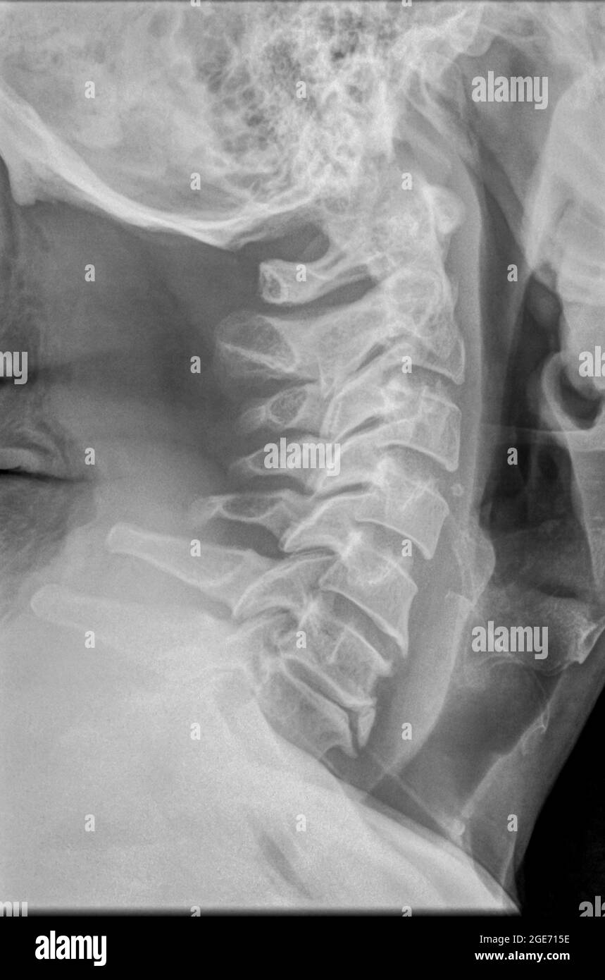 Cervical spine x-ray with a fractured Dens. A 50 year old male patient side View Stock Photohttps://www.alamy.com/image-license-details/?v=1https://www.alamy.com/cervical-spine-x-ray-with-a-fractured-dens-a-50-year-old-male-patient-side-view-image439018986.html
RM2GE715E–Cervical spine x-ray with a fractured Dens. A 50 year old male patient side View
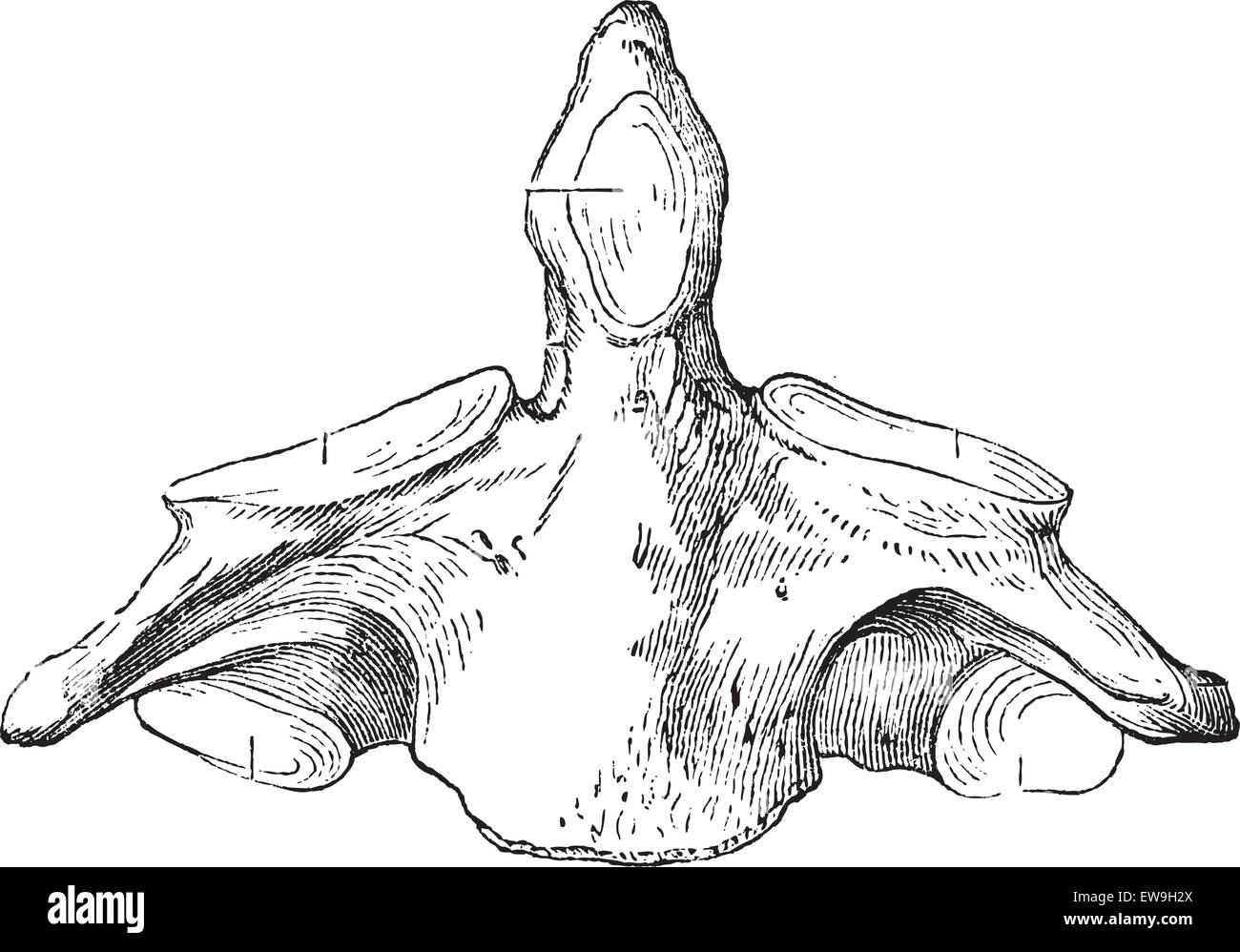 Fig. 136. Axis (second cervical vertebra), vintage engraved illustration. Magasin Pittoresque 1875. Stock Vectorhttps://www.alamy.com/image-license-details/?v=1https://www.alamy.com/stock-photo-fig-136-axis-second-cervical-vertebra-vintage-engraved-illustration-84418850.html
RFEW9H2X–Fig. 136. Axis (second cervical vertebra), vintage engraved illustration. Magasin Pittoresque 1875.
 Argentine Horned Frog (Ceratophrys ornata) Stock Photohttps://www.alamy.com/image-license-details/?v=1https://www.alamy.com/argentine-horned-frog-ceratophrys-ornata-image208165211.html
RMP2JMRR–Argentine Horned Frog (Ceratophrys ornata)
 The anatomist's vade mecum : a system of human anatomy . e upon the axis; the perfect freedom ofmovement between these bones being ensured by the two synovialmembranes. The lower part of the ring, formed by the transverseligament with the atlas, is smaller than the upper, while the summitof the odontoid process is larger than its base; so that the process isretained in its position by the transverse ligament, when the otherligaments are cut through. The extent to which the rotation of thehead upon the axis can be carried, is determined by the odontoid liga-ments. The odontoid process with its Stock Photohttps://www.alamy.com/image-license-details/?v=1https://www.alamy.com/the-anatomists-vade-mecum-a-system-of-human-anatomy-e-upon-the-axis-the-perfect-freedom-ofmovement-between-these-bones-being-ensured-by-the-two-synovialmembranes-the-lower-part-of-the-ring-formed-by-the-transverseligament-with-the-atlas-is-smaller-than-the-upper-while-the-summitof-the-odontoid-process-is-larger-than-its-base-so-that-the-process-isretained-in-its-position-by-the-transverse-ligament-when-the-otherligaments-are-cut-through-the-extent-to-which-the-rotation-of-thehead-upon-the-axis-can-be-carried-is-determined-by-the-odontoid-liga-ments-the-odontoid-process-with-its-image342767794.html
RM2AWJBPX–The anatomist's vade mecum : a system of human anatomy . e upon the axis; the perfect freedom ofmovement between these bones being ensured by the two synovialmembranes. The lower part of the ring, formed by the transverseligament with the atlas, is smaller than the upper, while the summitof the odontoid process is larger than its base; so that the process isretained in its position by the transverse ligament, when the otherligaments are cut through. The extent to which the rotation of thehead upon the axis can be carried, is determined by the odontoid liga-ments. The odontoid process with its
 . The cyclopædia of anatomy and physiology. Anatomy; Physiology; Zoology. 520 RUMINANTIA. (fig. 344.). The atlas in the Camels is not thus modified. In all other ruminants, in- cluding the Giraffe, an opening exists in this bone, which is placed at the fore part of the superior ring. The odontoid process of the axis or dentata is well marked and prominent in the short-necked ruminantia, but the Giraffe and Camels have it very small and incorporated with the articular end of the body ; in them, also, very slight traces of transverse " apophyses " are detectable. The dorsal vertebrae a Stock Photohttps://www.alamy.com/image-license-details/?v=1https://www.alamy.com/the-cyclopdia-of-anatomy-and-physiology-anatomy-physiology-zoology-520-ruminantia-fig-344-the-atlas-in-the-camels-is-not-thus-modified-in-all-other-ruminants-in-cluding-the-giraffe-an-opening-exists-in-this-bone-which-is-placed-at-the-fore-part-of-the-superior-ring-the-odontoid-process-of-the-axis-or-dentata-is-well-marked-and-prominent-in-the-short-necked-ruminantia-but-the-giraffe-and-camels-have-it-very-small-and-incorporated-with-the-articular-end-of-the-body-in-them-also-very-slight-traces-of-transverse-quot-apophyses-quot-are-detectable-the-dorsal-vertebrae-a-image216209377.html
RMPFN57D–. The cyclopædia of anatomy and physiology. Anatomy; Physiology; Zoology. 520 RUMINANTIA. (fig. 344.). The atlas in the Camels is not thus modified. In all other ruminants, in- cluding the Giraffe, an opening exists in this bone, which is placed at the fore part of the superior ring. The odontoid process of the axis or dentata is well marked and prominent in the short-necked ruminantia, but the Giraffe and Camels have it very small and incorporated with the articular end of the body ; in them, also, very slight traces of transverse " apophyses " are detectable. The dorsal vertebrae a
 Archive image from page 456 of Denkschriften - Österreichische Akademie der. Denkschriften - Österreichische Akademie der Wissenschaften denkschriftens971921akad Year: 1850 Halswirbel von Dorygnalhtts; a 3.—8. Wirbel des Wiener Originales von oben; b Rekonstruktion der ganzen Serie. Atlas und Axis nach RA. Kßkcni Plieninger (1907, Taf. XVI, Fig. 7 -10) - Pia Proatlati, na Neurale, ic Inlcircnlrum od Odontoid. Beim Exemplar des Dimorphodon niacrouyx Bück 1. sp., welches Owen in Foss. Rept. Lias. Format, pt. III, p. 45, Taf. 18 c aus dem unteren Lias beschrieb, ist eine Serie von 4 Halswirbeln Stock Photohttps://www.alamy.com/image-license-details/?v=1https://www.alamy.com/archive-image-from-page-456-of-denkschriften-sterreichische-akademie-der-denkschriften-sterreichische-akademie-der-wissenschaften-denkschriftens971921akad-year-1850-halswirbel-von-dorygnalhtts-a-38-wirbel-des-wiener-originales-von-oben-b-rekonstruktion-der-ganzen-serie-atlas-und-axis-nach-ra-kkcni-plieninger-1907-taf-xvi-fig-7-10-pia-proatlati-na-neurale-ic-inlcircnlrum-od-odontoid-beim-exemplar-des-dimorphodon-niacrouyx-bck-1-sp-welches-owen-in-foss-rept-lias-format-pt-iii-p-45-taf-18-c-aus-dem-unteren-lias-beschrieb-ist-eine-serie-von-4-halswirbeln-image259305012.html
RMW1TA58–Archive image from page 456 of Denkschriften - Österreichische Akademie der. Denkschriften - Österreichische Akademie der Wissenschaften denkschriftens971921akad Year: 1850 Halswirbel von Dorygnalhtts; a 3.—8. Wirbel des Wiener Originales von oben; b Rekonstruktion der ganzen Serie. Atlas und Axis nach RA. Kßkcni Plieninger (1907, Taf. XVI, Fig. 7 -10) - Pia Proatlati, na Neurale, ic Inlcircnlrum od Odontoid. Beim Exemplar des Dimorphodon niacrouyx Bück 1. sp., welches Owen in Foss. Rept. Lias. Format, pt. III, p. 45, Taf. 18 c aus dem unteren Lias beschrieb, ist eine Serie von 4 Halswirbeln
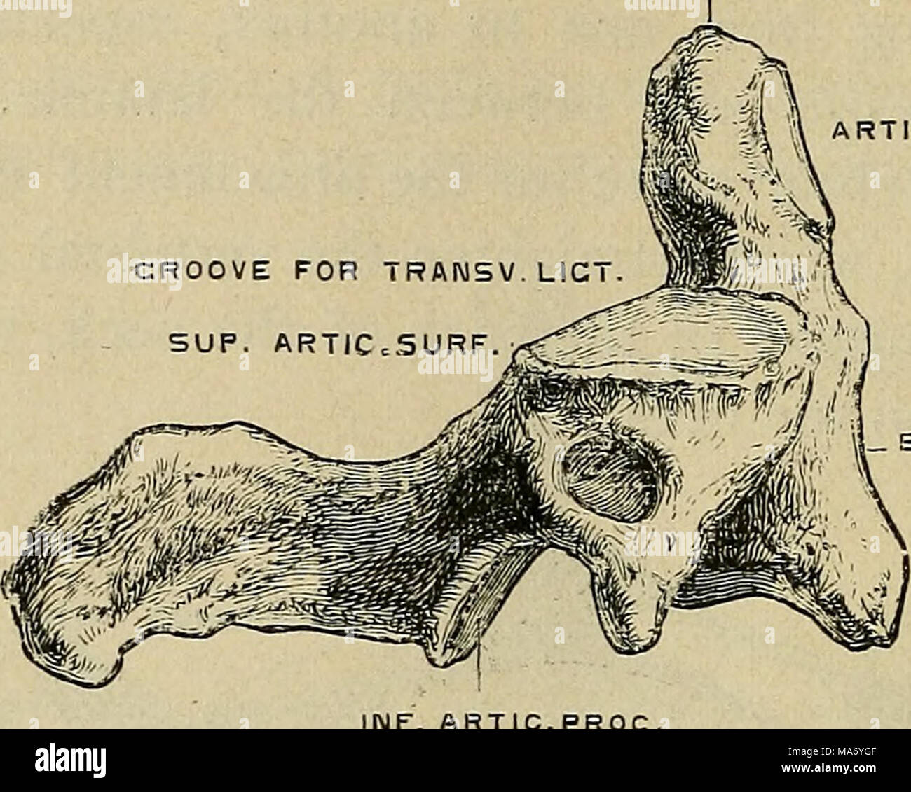 . Elementary physiology . NF, ARTIC.PROC. Fig. io.—Axis, from the right side. (Drawn by D. Gunn.) of the axis projects through, round which, as on a pivot, the atlas carrying the head can turn. The strong transverse ligmnent passing behind the odontoid process completes the socket. On the upper surface of the atlas are two articular facets ODONTOID PSOCESS IMF ARTIC PROCESS Stock Photohttps://www.alamy.com/image-license-details/?v=1https://www.alamy.com/elementary-physiology-nf-articproc-fig-ioaxis-from-the-right-side-drawn-by-d-gunn-of-the-axis-projects-through-round-which-as-on-a-pivot-the-atlas-carrying-the-head-can-turn-the-strong-transverse-ligmnent-passing-behind-the-odontoid-process-completes-the-socket-on-the-upper-surface-of-the-atlas-are-two-articular-facets-odontoid-psocess-imf-artic-process-image178403583.html
RMMA6YGF–. Elementary physiology . NF, ARTIC.PROC. Fig. io.—Axis, from the right side. (Drawn by D. Gunn.) of the axis projects through, round which, as on a pivot, the atlas carrying the head can turn. The strong transverse ligmnent passing behind the odontoid process completes the socket. On the upper surface of the atlas are two articular facets ODONTOID PSOCESS IMF ARTIC PROCESS
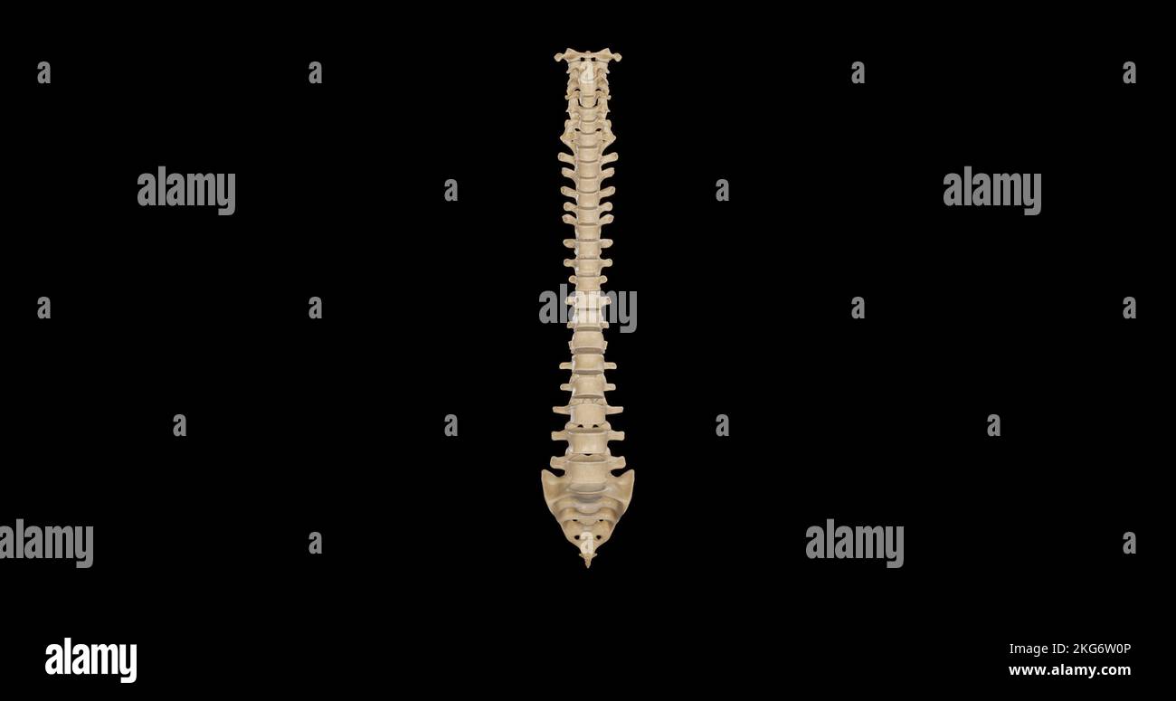 Anterior view of Vertebral Column Stock Photohttps://www.alamy.com/image-license-details/?v=1https://www.alamy.com/anterior-view-of-vertebral-column-image491876134.html
RF2KG6W0P–Anterior view of Vertebral Column
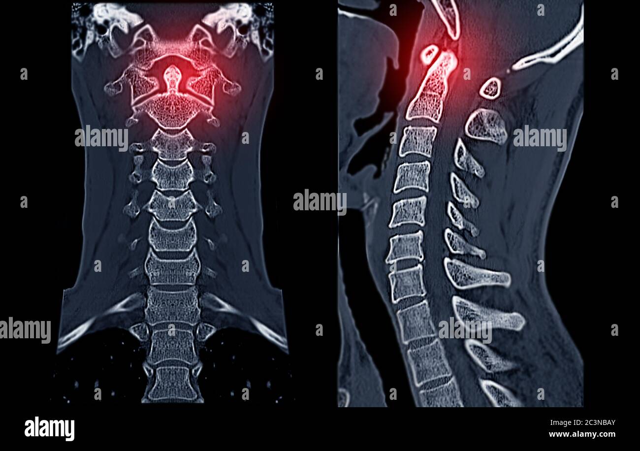 CT C-Spine or Cervical spine Coronal view in patient trauma cervical spine injury showing odontoid process or dens. Stock Photohttps://www.alamy.com/image-license-details/?v=1https://www.alamy.com/ct-c-spine-or-cervical-spine-coronal-view-in-patient-trauma-cervical-spine-injury-showing-odontoid-process-or-dens-image363731619.html
RF2C3NBAY–CT C-Spine or Cervical spine Coronal view in patient trauma cervical spine injury showing odontoid process or dens.
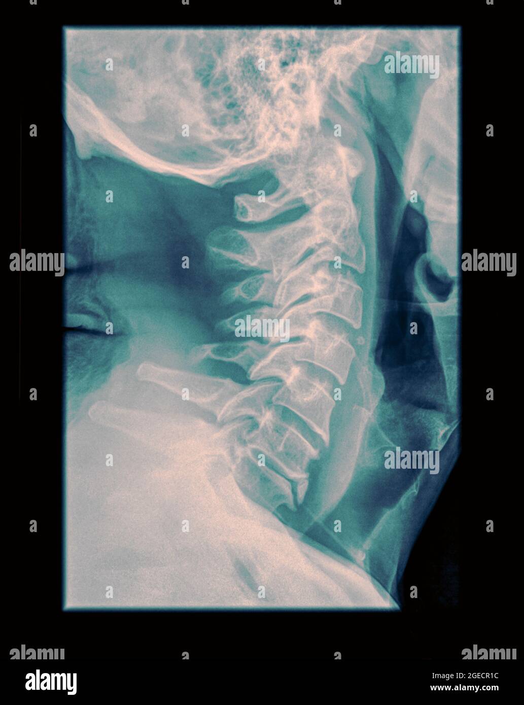 Cervical spine x-ray with a fractured Dens. A 50 year old male patient side View Stock Photohttps://www.alamy.com/image-license-details/?v=1https://www.alamy.com/cervical-spine-x-ray-with-a-fractured-dens-a-50-year-old-male-patient-side-view-image439145880.html
RM2GECR1C–Cervical spine x-ray with a fractured Dens. A 50 year old male patient side View
 Cervical spine x-ray with a fractured Dens. A 50 year old male patient Front View Stock Photohttps://www.alamy.com/image-license-details/?v=1https://www.alamy.com/cervical-spine-x-ray-with-a-fractured-dens-a-50-year-old-male-patient-front-view-image439018640.html
RM2GE70N4–Cervical spine x-ray with a fractured Dens. A 50 year old male patient Front View
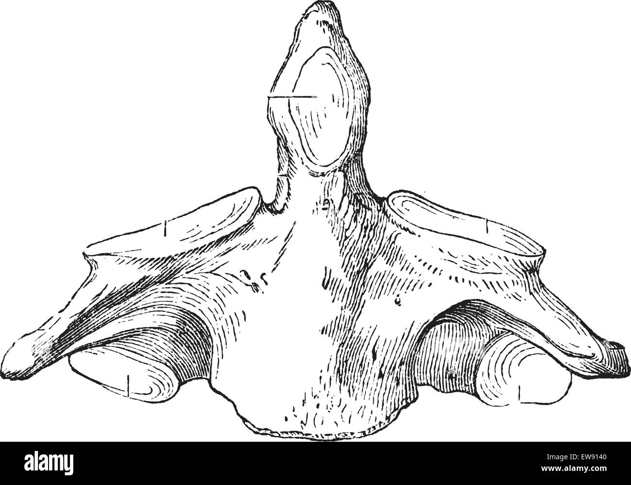 Fig. 136. Axis (second cervical vertebra), vintage engraved illustration. Magasin Pittoresque 1875. Stock Vectorhttps://www.alamy.com/image-license-details/?v=1https://www.alamy.com/stock-photo-fig-136-axis-second-cervical-vertebra-vintage-engraved-illustration-84406336.html
RFEW9140–Fig. 136. Axis (second cervical vertebra), vintage engraved illustration. Magasin Pittoresque 1875.
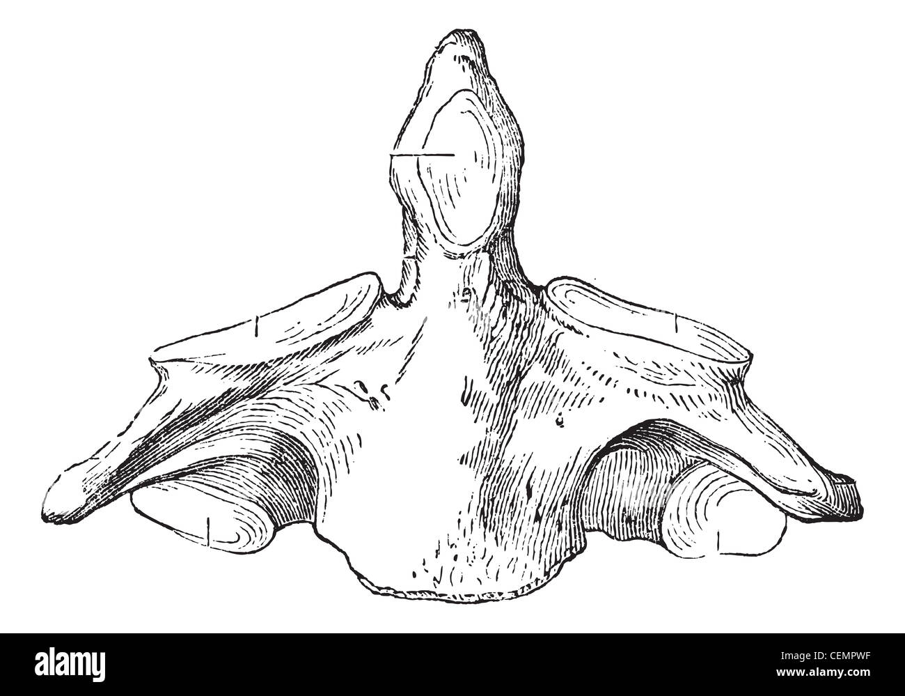 Fig. 136. Axis (second cervical vertebra), vintage engraved illustration. Magasin Pittoresque 1875. Stock Photohttps://www.alamy.com/image-license-details/?v=1https://www.alamy.com/stock-photo-fig-136-axis-second-cervical-vertebra-vintage-engraved-illustration-43482923.html
RFCEMPWF–Fig. 136. Axis (second cervical vertebra), vintage engraved illustration. Magasin Pittoresque 1875.
 Carpenter's principles of human physiology . er arch, but with the body of the axisforming the odontoid process of this vertebra. The ossification of thevertebrae begins at the end of the 2nd or the beginning of the 3rd month,with three centres for each vertebra, one for the body and one for thearch on each side. These parts, however, do not unite till the secondyear after birth. Accessory centres of ossification are subsequently formedat the tips of the spinous and transverse processes, and in the form ofthin leaves at the upper and lower surfaces of the bodies, which resemble theepiphyses of Stock Photohttps://www.alamy.com/image-license-details/?v=1https://www.alamy.com/carpenters-principles-of-human-physiology-er-arch-but-with-the-body-of-the-axisforming-the-odontoid-process-of-this-vertebra-the-ossification-of-thevertebrae-begins-at-the-end-of-the-2nd-or-the-beginning-of-the-3rd-monthwith-three-centres-for-each-vertebra-one-for-the-body-and-one-for-thearch-on-each-side-these-parts-however-do-not-unite-till-the-secondyear-after-birth-accessory-centres-of-ossification-are-subsequently-formedat-the-tips-of-the-spinous-and-transverse-processes-and-in-the-form-ofthin-leaves-at-the-upper-and-lower-surfaces-of-the-bodies-which-resemble-theepiphyses-of-image339967366.html
RM2AN2RRJ–Carpenter's principles of human physiology . er arch, but with the body of the axisforming the odontoid process of this vertebra. The ossification of thevertebrae begins at the end of the 2nd or the beginning of the 3rd month,with three centres for each vertebra, one for the body and one for thearch on each side. These parts, however, do not unite till the secondyear after birth. Accessory centres of ossification are subsequently formedat the tips of the spinous and transverse processes, and in the form ofthin leaves at the upper and lower surfaces of the bodies, which resemble theepiphyses of
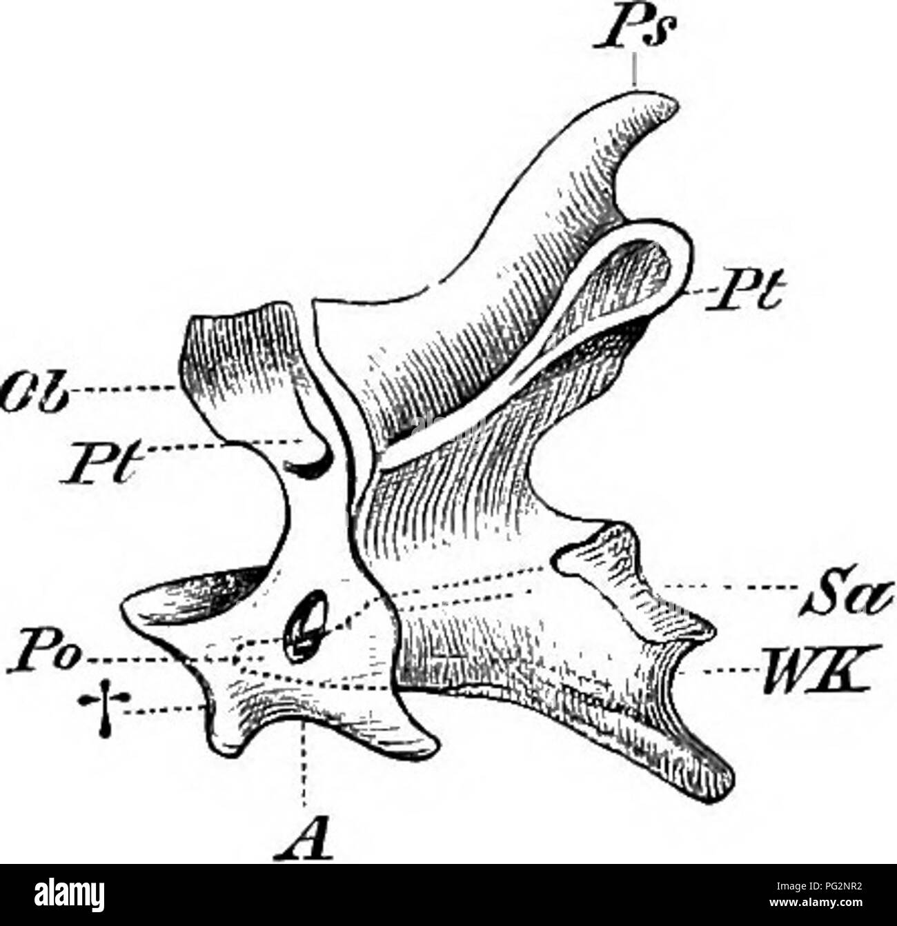 . Elements of the comparative anatomy of vertebrates. Anatomy, Comparative. 48 COMPARATIVE ANATOMY them throughout life by sutures, as is the case iu certain Reptiles: even the ligament which keeps the odontoid process of the axis in its place may become ossified. Fibro-cartilaginous discs or menisci are present between the centra. In the cervical region, which is extremely flexible and often very long, the centra are in nearly all cases connected by means of saddle-shaped synovial articulations; the upper part of the bifurcated transverse processes arises from the arch, the lower from the cen Stock Photohttps://www.alamy.com/image-license-details/?v=1https://www.alamy.com/elements-of-the-comparative-anatomy-of-vertebrates-anatomy-comparative-48-comparative-anatomy-them-throughout-life-by-sutures-as-is-the-case-iu-certain-reptiles-even-the-ligament-which-keeps-the-odontoid-process-of-the-axis-in-its-place-may-become-ossified-fibro-cartilaginous-discs-or-menisci-are-present-between-the-centra-in-the-cervical-region-which-is-extremely-flexible-and-often-very-long-the-centra-are-in-nearly-all-cases-connected-by-means-of-saddle-shaped-synovial-articulations-the-upper-part-of-the-bifurcated-transverse-processes-arises-from-the-arch-the-lower-from-the-cen-image216419926.html
RMPG2NR2–. Elements of the comparative anatomy of vertebrates. Anatomy, Comparative. 48 COMPARATIVE ANATOMY them throughout life by sutures, as is the case iu certain Reptiles: even the ligament which keeps the odontoid process of the axis in its place may become ossified. Fibro-cartilaginous discs or menisci are present between the centra. In the cervical region, which is extremely flexible and often very long, the centra are in nearly all cases connected by means of saddle-shaped synovial articulations; the upper part of the bifurcated transverse processes arises from the arch, the lower from the cen
![Elementary entomology ([c1912]) Elementary entomology elementaryentomo00sand Year: [c1912] THE BEETLES 157 to the larvae of the locust-beetle (Odontoid dorsalis). On morning- glory and sweet-potato vines are found some striking little beetles, Stock Photo Elementary entomology ([c1912]) Elementary entomology elementaryentomo00sand Year: [c1912] THE BEETLES 157 to the larvae of the locust-beetle (Odontoid dorsalis). On morning- glory and sweet-potato vines are found some striking little beetles, Stock Photo](https://c8.alamy.com/comp/RWPR31/elementary-entomology-c1912-elementary-entomology-elementaryentomo00sand-year-c1912-the-beetles-157-to-the-larvae-of-the-locust-beetle-odontoid-dorsalis-on-morning-glory-and-sweet-potato-vines-are-found-some-striking-little-beetles-RWPR31.jpg) Elementary entomology ([c1912]) Elementary entomology elementaryentomo00sand Year: [c1912] THE BEETLES 157 to the larvae of the locust-beetle (Odontoid dorsalis). On morning- glory and sweet-potato vines are found some striking little beetles, Stock Photohttps://www.alamy.com/image-license-details/?v=1https://www.alamy.com/elementary-entomology-c1912-elementary-entomology-elementaryentomo00sand-year-c1912-the-beetles-157-to-the-larvae-of-the-locust-beetle-odontoid-dorsalis-on-morning-glory-and-sweet-potato-vines-are-found-some-striking-little-beetles-image239602245.html
Elementary entomology ([c1912]) Elementary entomology elementaryentomo00sand Year: [c1912] THE BEETLES 157 to the larvae of the locust-beetle (Odontoid dorsalis). On morning- glory and sweet-potato vines are found some striking little beetles, Stock Photohttps://www.alamy.com/image-license-details/?v=1https://www.alamy.com/elementary-entomology-c1912-elementary-entomology-elementaryentomo00sand-year-c1912-the-beetles-157-to-the-larvae-of-the-locust-beetle-odontoid-dorsalis-on-morning-glory-and-sweet-potato-vines-are-found-some-striking-little-beetles-image239602245.htmlRMRWPR31–Elementary entomology ([c1912]) Elementary entomology elementaryentomo00sand Year: [c1912] THE BEETLES 157 to the larvae of the locust-beetle (Odontoid dorsalis). On morning- glory and sweet-potato vines are found some striking little beetles,
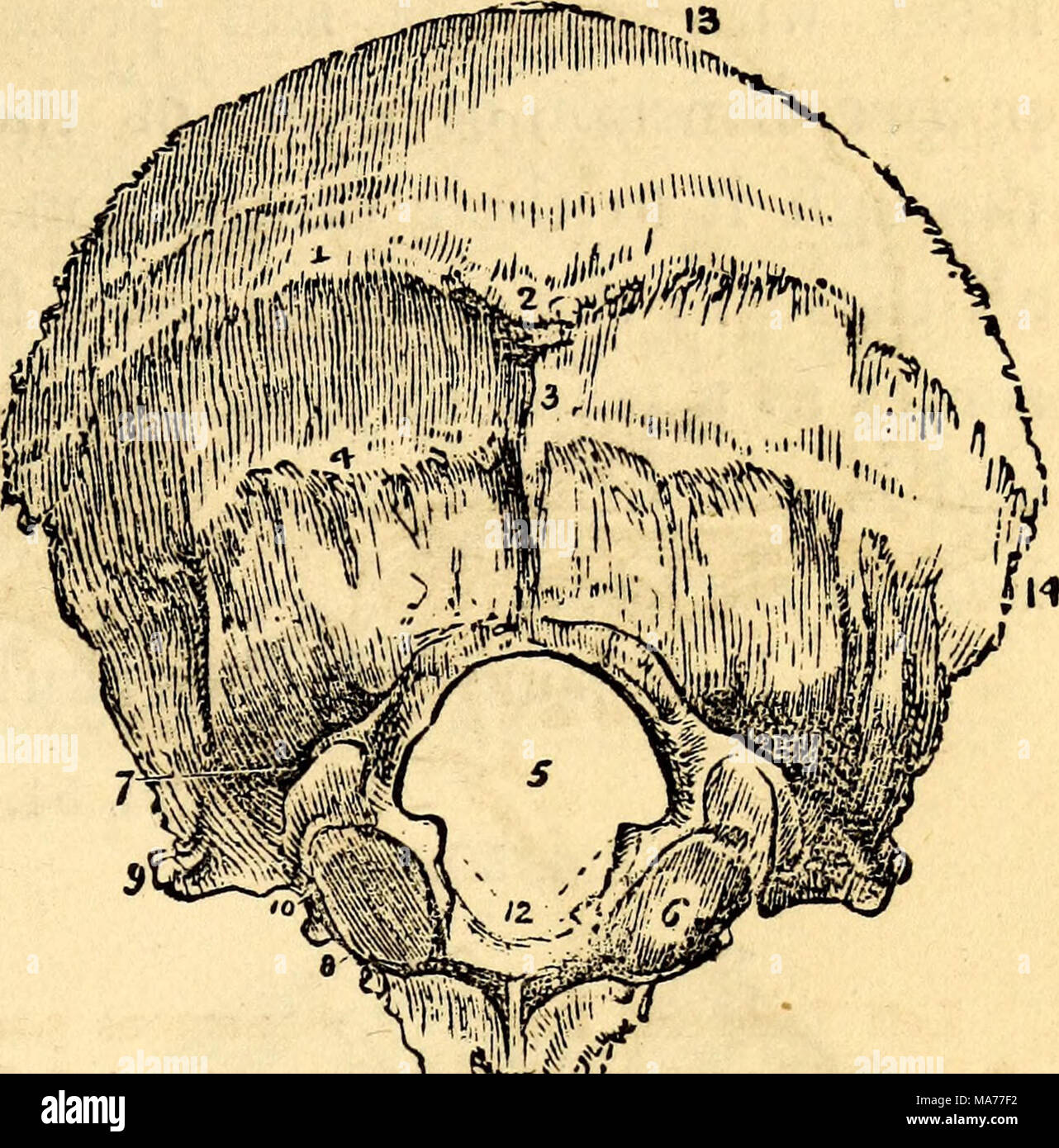 . Elementary anatomy and physiology : for colleges, academies, and other schools . External surface of Occipital Bone. 1 and 4, Semicircular Ridges. 2, Occipital Protube- rance. 3, Attachment of ligamentum nuche. 5, Foramen for Medulla oblongata. 6, Condyle of right side. 7 and 8, Condyloid Foramina. 9, Jugular Eminence. 10, Jugular Foramen. II, Basilar process. 12, Points of attachment for odontoid ligaments. 13, Edge for attach- ment with Parietal bone. 14, Point of attach- ment for Temporal bone. FIG. 49. Stock Photohttps://www.alamy.com/image-license-details/?v=1https://www.alamy.com/elementary-anatomy-and-physiology-for-colleges-academies-and-other-schools-external-surface-of-occipital-bone-1-and-4-semicircular-ridges-2-occipital-protube-rance-3-attachment-of-ligamentum-nuche-5-foramen-for-medulla-oblongata-6-condyle-of-right-side-7-and-8-condyloid-foramina-9-jugular-eminence-10-jugular-foramen-ii-basilar-process-12-points-of-attachment-for-odontoid-ligaments-13-edge-for-attach-ment-with-parietal-bone-14-point-of-attach-ment-for-temporal-bone-fig-49-image178409814.html
RMMA77F2–. Elementary anatomy and physiology : for colleges, academies, and other schools . External surface of Occipital Bone. 1 and 4, Semicircular Ridges. 2, Occipital Protube- rance. 3, Attachment of ligamentum nuche. 5, Foramen for Medulla oblongata. 6, Condyle of right side. 7 and 8, Condyloid Foramina. 9, Jugular Eminence. 10, Jugular Foramen. II, Basilar process. 12, Points of attachment for odontoid ligaments. 13, Edge for attach- ment with Parietal bone. 14, Point of attach- ment for Temporal bone. FIG. 49.
 Posterior view of Vertebral Column Stock Photohttps://www.alamy.com/image-license-details/?v=1https://www.alamy.com/posterior-view-of-vertebral-column-image491876713.html
RF2KG6WND–Posterior view of Vertebral Column
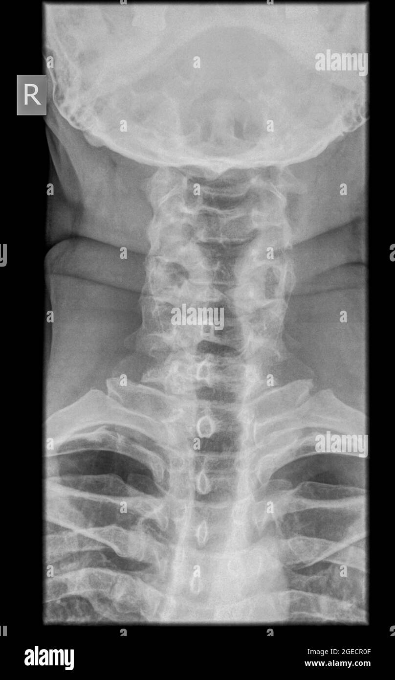 Cervical spine x-ray with a fractured Dens. A 50 year old male patient Front View Stock Photohttps://www.alamy.com/image-license-details/?v=1https://www.alamy.com/cervical-spine-x-ray-with-a-fractured-dens-a-50-year-old-male-patient-front-view-image439145855.html
RM2GECR0F–Cervical spine x-ray with a fractured Dens. A 50 year old male patient Front View
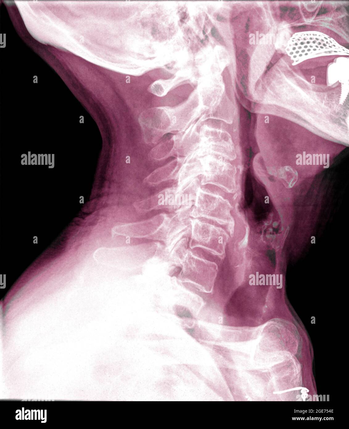 Cervical spine x-ray of a 72 year old female patient side view Stock Photohttps://www.alamy.com/image-license-details/?v=1https://www.alamy.com/cervical-spine-x-ray-of-a-72-year-old-female-patient-side-view-image439022094.html
RM2GE754E–Cervical spine x-ray of a 72 year old female patient side view
 A hanged man A. soft palate ; B, wall of the pharynx ; C, tongue ; D, tip of the tongue pressed between the teeth ; E, body of the hyoid bone ; F, groove made by the rope ; G, anterior portion of the atlas ; H, odontoid process of the axis. from Forensic medicine and toxicology by Mann, J. Dixon (John Dixon), 1840-1912 Publication date 1898 published by Charles Griffin Stock Photohttps://www.alamy.com/image-license-details/?v=1https://www.alamy.com/a-hanged-man-a-soft-palate-b-wall-of-the-pharynx-c-tongue-d-tip-of-the-tongue-pressed-between-the-teeth-e-body-of-the-hyoid-bone-f-groove-made-by-the-rope-g-anterior-portion-of-the-atlas-h-odontoid-process-of-the-axis-from-forensic-medicine-and-toxicology-by-mann-j-dixon-john-dixon-1840-1912-publication-date-1898-published-by-charles-griffin-image622669109.html
RM2Y510PD–A hanged man A. soft palate ; B, wall of the pharynx ; C, tongue ; D, tip of the tongue pressed between the teeth ; E, body of the hyoid bone ; F, groove made by the rope ; G, anterior portion of the atlas ; H, odontoid process of the axis. from Forensic medicine and toxicology by Mann, J. Dixon (John Dixon), 1840-1912 Publication date 1898 published by Charles Griffin
 A manual of anatomy . Transverse process j Foramen transversarium Lateral mass Facet for odontoid process Anterior tnbercleFig. 7.—The atlas seen from above. {Sobotta and McMurrich.) divided into two portions by the transverse ligament that extendsbetween the two median tubercles. The smaller ventral divisioncontains the dens of the epistropheus while the larger dorsal portionaccommodates the spinal cord. The Epistropheus.—This is the second cervical vertebra and it Transverse ,-.process Odontoid process/ ^ . - {anterior articularfacet) Superior articular facet. Inferior articular facet Body P Stock Photohttps://www.alamy.com/image-license-details/?v=1https://www.alamy.com/a-manual-of-anatomy-transverse-process-j-foramen-transversarium-lateral-mass-facet-for-odontoid-process-anterior-tnberclefig-7the-atlas-seen-from-above-sobotta-and-mcmurrich-divided-into-two-portions-by-the-transverse-ligament-that-extendsbetween-the-two-median-tubercles-the-smaller-ventral-divisioncontains-the-dens-of-the-epistropheus-while-the-larger-dorsal-portionaccommodates-the-spinal-cord-the-epistropheusthis-is-the-second-cervical-vertebra-and-it-transverse-process-odontoid-process-anterior-articularfacet-superior-articular-facet-inferior-articular-facet-body-p-image343400888.html
RM2AXK79C–A manual of anatomy . Transverse process j Foramen transversarium Lateral mass Facet for odontoid process Anterior tnbercleFig. 7.—The atlas seen from above. {Sobotta and McMurrich.) divided into two portions by the transverse ligament that extendsbetween the two median tubercles. The smaller ventral divisioncontains the dens of the epistropheus while the larger dorsal portionaccommodates the spinal cord. The Epistropheus.—This is the second cervical vertebra and it Transverse ,-.process Odontoid process/ ^ . - {anterior articularfacet) Superior articular facet. Inferior articular facet Body P
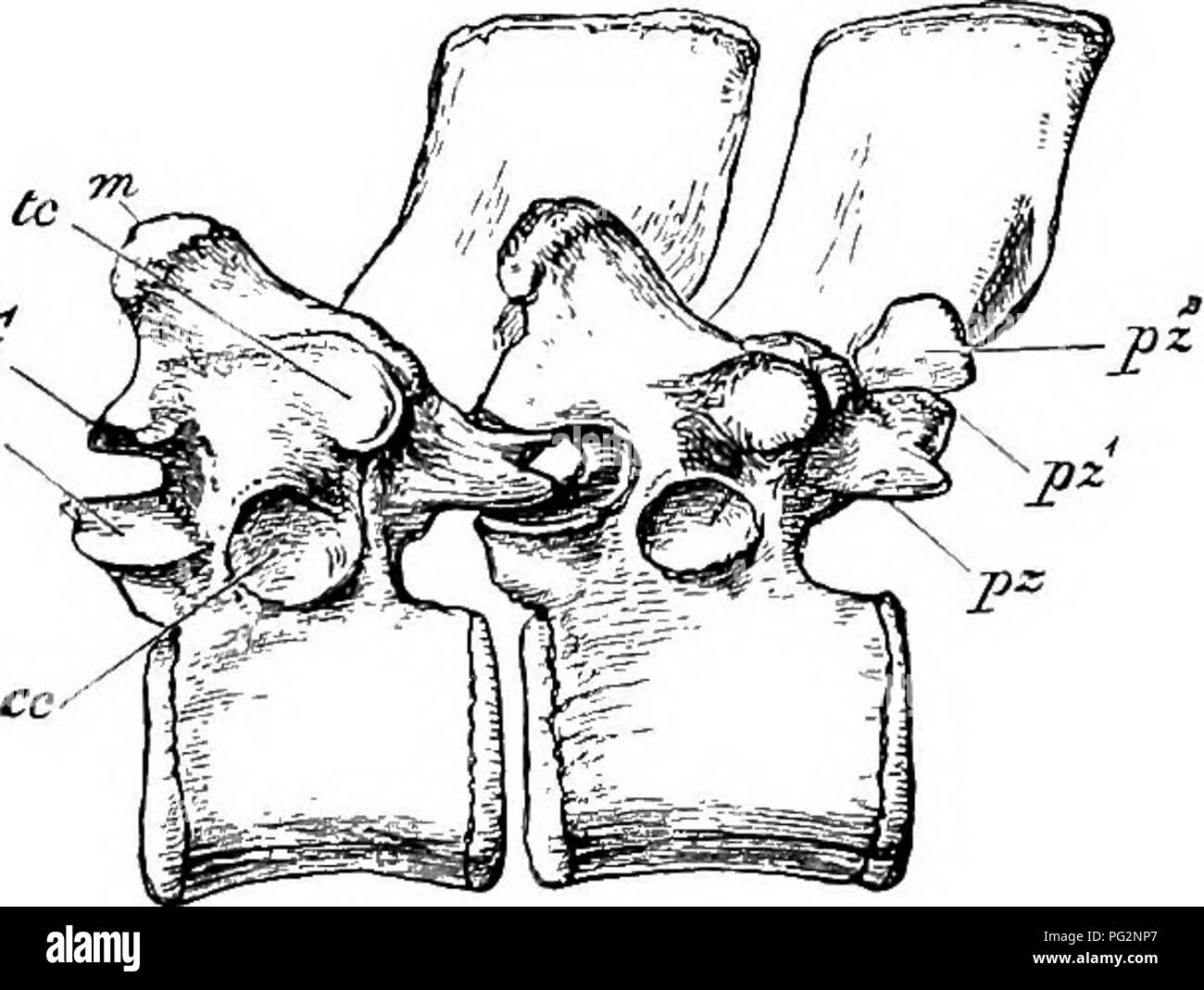 . Elements of the comparative anatomy of vertebrates. Anatomy, Comparative. Fig. 39a.—Diagram showing mode or Ossi- fication OF Human Axis. (Ventral surface.) From Flower's Osteology of the Mammalia. o, odontoid process, or centrum of atlas ; t, proper centrum of axis ; na, neural arch ; an, anterior articular surface; c, e, e, e, epi- physes, completing the ends of the centra, The transverse processes are simple in all but the cervical region and arise from the base of the arch : in the thoracic region they are tipped with cartilage on the ventral side of their distal ends for articulation wi Stock Photohttps://www.alamy.com/image-license-details/?v=1https://www.alamy.com/elements-of-the-comparative-anatomy-of-vertebrates-anatomy-comparative-fig-39adiagram-showing-mode-or-ossi-fication-of-human-axis-ventral-surface-from-flowers-osteology-of-the-mammalia-o-odontoid-process-or-centrum-of-atlas-t-proper-centrum-of-axis-na-neural-arch-an-anterior-articular-surface-c-e-e-e-epi-physes-completing-the-ends-of-the-centra-the-transverse-processes-are-simple-in-all-but-the-cervical-region-and-arise-from-the-base-of-the-arch-in-the-thoracic-region-they-are-tipped-with-cartilage-on-the-ventral-side-of-their-distal-ends-for-articulation-wi-image216419903.html
RMPG2NP7–. Elements of the comparative anatomy of vertebrates. Anatomy, Comparative. Fig. 39a.—Diagram showing mode or Ossi- fication OF Human Axis. (Ventral surface.) From Flower's Osteology of the Mammalia. o, odontoid process, or centrum of atlas ; t, proper centrum of axis ; na, neural arch ; an, anterior articular surface; c, e, e, e, epi- physes, completing the ends of the centra, The transverse processes are simple in all but the cervical region and arise from the base of the arch : in the thoracic region they are tipped with cartilage on the ventral side of their distal ends for articulation wi
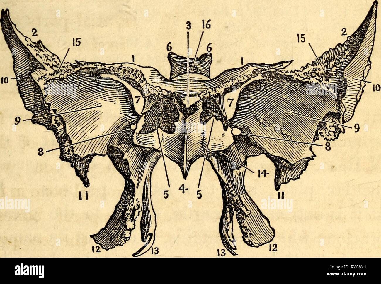 Elementary anatomy and physiology : for colleges, academies, and other schools elementaryanato00hitc Year: 1869 External surface of Occipital Bone. 1 and 4, Semicircular Ridges. 2, Occipital Protube- rance. 3, Attachment of ligamentum nuche. 5, Foramen for Medulla oblongata. 6, Condyle of right side. 7 and 8, Condyloid Foramina. 9, Jugular Eminence. 10, Jugular Foramen. II, Basilar process. 12, Points of attachment for odontoid ligaments. 13, Edge for attach- ment with Parietal bone. 14, Point of attach- ment for Temporal bone. FIG. 49. The Anterior and Inferior Surface of the Sphenoid Bo Stock Photohttps://www.alamy.com/image-license-details/?v=1https://www.alamy.com/elementary-anatomy-and-physiology-for-colleges-academies-and-other-schools-elementaryanato00hitc-year-1869-external-surface-of-occipital-bone-1-and-4-semicircular-ridges-2-occipital-protube-rance-3-attachment-of-ligamentum-nuche-5-foramen-for-medulla-oblongata-6-condyle-of-right-side-7-and-8-condyloid-foramina-9-jugular-eminence-10-jugular-foramen-ii-basilar-process-12-points-of-attachment-for-odontoid-ligaments-13-edge-for-attach-ment-with-parietal-bone-14-point-of-attach-ment-for-temporal-bone-fig-49-the-anterior-and-inferior-surface-of-the-sphenoid-bo-image240688773.html
RMRYG8YH–Elementary anatomy and physiology : for colleges, academies, and other schools elementaryanato00hitc Year: 1869 External surface of Occipital Bone. 1 and 4, Semicircular Ridges. 2, Occipital Protube- rance. 3, Attachment of ligamentum nuche. 5, Foramen for Medulla oblongata. 6, Condyle of right side. 7 and 8, Condyloid Foramina. 9, Jugular Eminence. 10, Jugular Foramen. II, Basilar process. 12, Points of attachment for odontoid ligaments. 13, Edge for attach- ment with Parietal bone. 14, Point of attach- ment for Temporal bone. FIG. 49. The Anterior and Inferior Surface of the Sphenoid Bo
 . Elementary physiology . TUBERCLE FOR TRANSV. LIGAMCNT ANTERIOR ARCH ARTIC. SURF. FOR ODONTOID PROC. Fig. 9.—Atlas, from above. (Drawn by D. Gunn.) The position of the transverse ligament is indicated by dotted lines. The seven vertebrae at the upper end of the column are known as the cervical vertebrae; these form the bones of the neck. They are much more slenderly built and lighter than the lower members of the column, and are capable of moving on one another to a much greater extent, so as to allow move- ments of the neck. The two upper members of the cervical series are much modified in s Stock Photohttps://www.alamy.com/image-license-details/?v=1https://www.alamy.com/elementary-physiology-tubercle-for-transv-ligamcnt-anterior-arch-artic-surf-for-odontoid-proc-fig-9atlas-from-above-drawn-by-d-gunn-the-position-of-the-transverse-ligament-is-indicated-by-dotted-lines-the-seven-vertebrae-at-the-upper-end-of-the-column-are-known-as-the-cervical-vertebrae-these-form-the-bones-of-the-neck-they-are-much-more-slenderly-built-and-lighter-than-the-lower-members-of-the-column-and-are-capable-of-moving-on-one-another-to-a-much-greater-extent-so-as-to-allow-move-ments-of-the-neck-the-two-upper-members-of-the-cervical-series-are-much-modified-in-s-image178403582.html
RMMA6YGE–. Elementary physiology . TUBERCLE FOR TRANSV. LIGAMCNT ANTERIOR ARCH ARTIC. SURF. FOR ODONTOID PROC. Fig. 9.—Atlas, from above. (Drawn by D. Gunn.) The position of the transverse ligament is indicated by dotted lines. The seven vertebrae at the upper end of the column are known as the cervical vertebrae; these form the bones of the neck. They are much more slenderly built and lighter than the lower members of the column, and are capable of moving on one another to a much greater extent, so as to allow move- ments of the neck. The two upper members of the cervical series are much modified in s
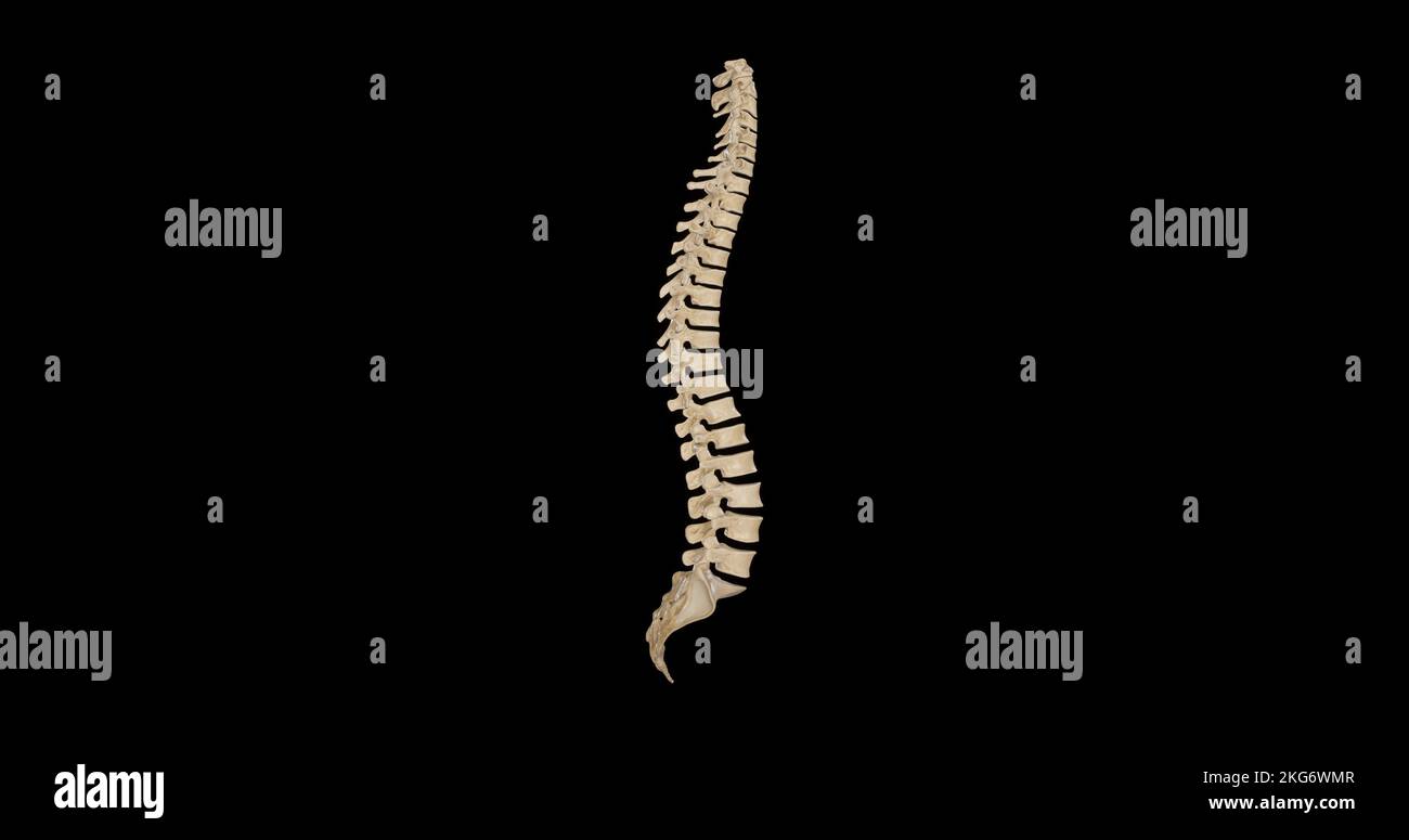 Lateral view of Vertebral Column Stock Photohttps://www.alamy.com/image-license-details/?v=1https://www.alamy.com/lateral-view-of-vertebral-column-image491876695.html
RF2KG6WMR–Lateral view of Vertebral Column
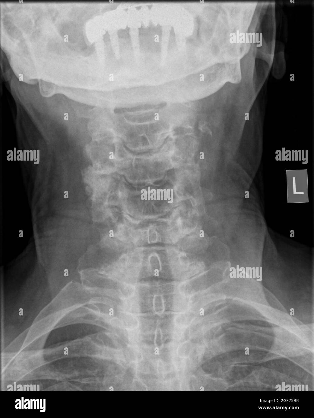 Cervical spine x-ray of a 72 year old female patient front view Stock Photohttps://www.alamy.com/image-license-details/?v=1https://www.alamy.com/cervical-spine-x-ray-of-a-72-year-old-female-patient-front-view-image439022299.html
RM2GE75BR–Cervical spine x-ray of a 72 year old female patient front view
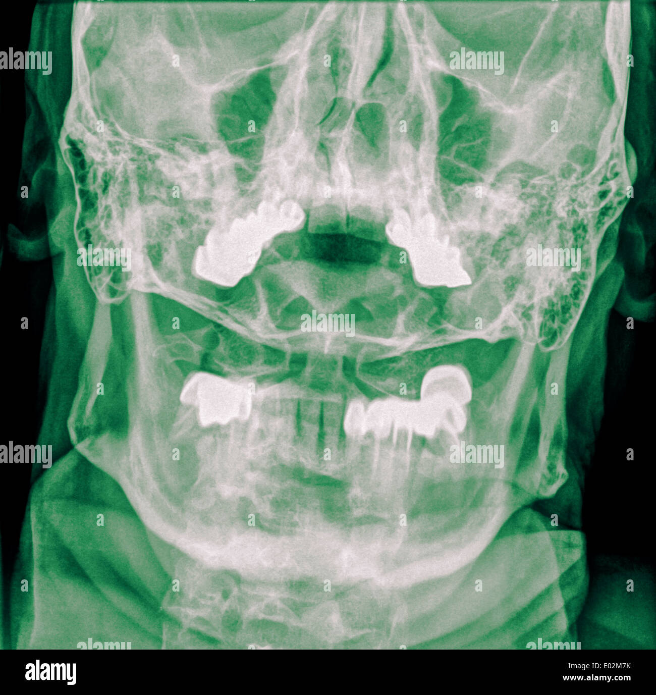 Cervical spine x-ray with a fractured Dens. A 50 year old male patient Front View Stock Photohttps://www.alamy.com/image-license-details/?v=1https://www.alamy.com/cervical-spine-x-ray-with-a-fractured-dens-a-50-year-old-male-patient-image68901271.html
RME02M7K–Cervical spine x-ray with a fractured Dens. A 50 year old male patient Front View
![A practical treatise on fractures and dislocations . hich had been part ofthe right occipital condyle. The axis was intact, except the odontoid process,which was denuded of cartilage and roughened on one side; the cheek ligamentsseemed to have been disorganized; the right transverse process of the atlas wasfound destroyed by caries, and the articular surface of its lateral mass about halfabsorbed, due to the chronic inflammatory process to which the region hadevidently been subjected.1] CHAPTER XVI. FRACTURES OF THE VERTEBRAE. It will be convenient to divide fractures of the vertebrae into fra Stock Photo A practical treatise on fractures and dislocations . hich had been part ofthe right occipital condyle. The axis was intact, except the odontoid process,which was denuded of cartilage and roughened on one side; the cheek ligamentsseemed to have been disorganized; the right transverse process of the atlas wasfound destroyed by caries, and the articular surface of its lateral mass about halfabsorbed, due to the chronic inflammatory process to which the region hadevidently been subjected.1] CHAPTER XVI. FRACTURES OF THE VERTEBRAE. It will be convenient to divide fractures of the vertebrae into fra Stock Photo](https://c8.alamy.com/comp/2ANHTR3/a-practical-treatise-on-fractures-and-dislocations-hich-had-been-part-ofthe-right-occipital-condyle-the-axis-was-intact-except-the-odontoid-processwhich-was-denuded-of-cartilage-and-roughened-on-one-side-the-cheek-ligamentsseemed-to-have-been-disorganized-the-right-transverse-process-of-the-atlas-wasfound-destroyed-by-caries-and-the-articular-surface-of-its-lateral-mass-about-halfabsorbed-due-to-the-chronic-inflammatory-process-to-which-the-region-hadevidently-been-subjected1-chapter-xvi-fractures-of-the-vertebrae-it-will-be-convenient-to-divide-fractures-of-the-vertebrae-into-fra-2ANHTR3.jpg) A practical treatise on fractures and dislocations . hich had been part ofthe right occipital condyle. The axis was intact, except the odontoid process,which was denuded of cartilage and roughened on one side; the cheek ligamentsseemed to have been disorganized; the right transverse process of the atlas wasfound destroyed by caries, and the articular surface of its lateral mass about halfabsorbed, due to the chronic inflammatory process to which the region hadevidently been subjected.1] CHAPTER XVI. FRACTURES OF THE VERTEBRAE. It will be convenient to divide fractures of the vertebrae into fra Stock Photohttps://www.alamy.com/image-license-details/?v=1https://www.alamy.com/a-practical-treatise-on-fractures-and-dislocations-hich-had-been-part-ofthe-right-occipital-condyle-the-axis-was-intact-except-the-odontoid-processwhich-was-denuded-of-cartilage-and-roughened-on-one-side-the-cheek-ligamentsseemed-to-have-been-disorganized-the-right-transverse-process-of-the-atlas-wasfound-destroyed-by-caries-and-the-articular-surface-of-its-lateral-mass-about-halfabsorbed-due-to-the-chronic-inflammatory-process-to-which-the-region-hadevidently-been-subjected1-chapter-xvi-fractures-of-the-vertebrae-it-will-be-convenient-to-divide-fractures-of-the-vertebrae-into-fra-image340297415.html
A practical treatise on fractures and dislocations . hich had been part ofthe right occipital condyle. The axis was intact, except the odontoid process,which was denuded of cartilage and roughened on one side; the cheek ligamentsseemed to have been disorganized; the right transverse process of the atlas wasfound destroyed by caries, and the articular surface of its lateral mass about halfabsorbed, due to the chronic inflammatory process to which the region hadevidently been subjected.1] CHAPTER XVI. FRACTURES OF THE VERTEBRAE. It will be convenient to divide fractures of the vertebrae into fra Stock Photohttps://www.alamy.com/image-license-details/?v=1https://www.alamy.com/a-practical-treatise-on-fractures-and-dislocations-hich-had-been-part-ofthe-right-occipital-condyle-the-axis-was-intact-except-the-odontoid-processwhich-was-denuded-of-cartilage-and-roughened-on-one-side-the-cheek-ligamentsseemed-to-have-been-disorganized-the-right-transverse-process-of-the-atlas-wasfound-destroyed-by-caries-and-the-articular-surface-of-its-lateral-mass-about-halfabsorbed-due-to-the-chronic-inflammatory-process-to-which-the-region-hadevidently-been-subjected1-chapter-xvi-fractures-of-the-vertebrae-it-will-be-convenient-to-divide-fractures-of-the-vertebrae-into-fra-image340297415.htmlRM2ANHTR3–A practical treatise on fractures and dislocations . hich had been part ofthe right occipital condyle. The axis was intact, except the odontoid process,which was denuded of cartilage and roughened on one side; the cheek ligamentsseemed to have been disorganized; the right transverse process of the atlas wasfound destroyed by caries, and the articular surface of its lateral mass about halfabsorbed, due to the chronic inflammatory process to which the region hadevidently been subjected.1] CHAPTER XVI. FRACTURES OF THE VERTEBRAE. It will be convenient to divide fractures of the vertebrae into fra
 . The development of the human body : a manual of human embryology. Embryology; Embryo, Non-Mammalian. Fig. ioo.—A, Upper Surface of the First Sacral Vertebra, and B, Ventral View of the Sacrum showing Primary Centers of Ossification. c, Body; na, vertebral arch; r, rib center.—(Sappey.) atlas a double center appears in the persisting hypochordal bar, and the body which corresponds to the atlas, after developing the termi- nal epiphysial disks, fuses with the body of the epistropheus (axis) to form its odontoid process, this vertebra consequently possessing, in addition to the typical centers, Stock Photohttps://www.alamy.com/image-license-details/?v=1https://www.alamy.com/the-development-of-the-human-body-a-manual-of-human-embryology-embryology-embryo-non-mammalian-fig-iooa-upper-surface-of-the-first-sacral-vertebra-and-b-ventral-view-of-the-sacrum-showing-primary-centers-of-ossification-c-body-na-vertebral-arch-r-rib-centersappey-atlas-a-double-center-appears-in-the-persisting-hypochordal-bar-and-the-body-which-corresponds-to-the-atlas-after-developing-the-termi-nal-epiphysial-disks-fuses-with-the-body-of-the-epistropheus-axis-to-form-its-odontoid-process-this-vertebra-consequently-possessing-in-addition-to-the-typical-centers-image215969742.html
RMPFA7H2–. The development of the human body : a manual of human embryology. Embryology; Embryo, Non-Mammalian. Fig. ioo.—A, Upper Surface of the First Sacral Vertebra, and B, Ventral View of the Sacrum showing Primary Centers of Ossification. c, Body; na, vertebral arch; r, rib center.—(Sappey.) atlas a double center appears in the persisting hypochordal bar, and the body which corresponds to the atlas, after developing the termi- nal epiphysial disks, fuses with the body of the epistropheus (axis) to form its odontoid process, this vertebra consequently possessing, in addition to the typical centers,
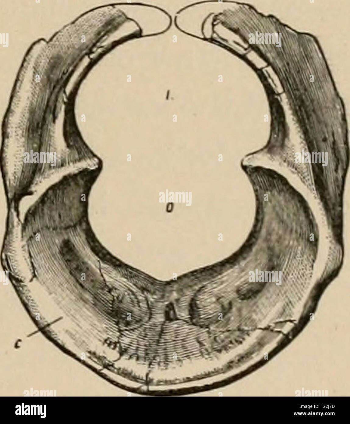 Archive image from page 18 of Diplodocus (Marsh) its osteology, Diplodocus (Marsh) : its osteology, taxonomy, and probable habits, with a restoration of the skeleton diplodocusmarshi11hatc Year: 1901 Fig. -t. Atlas of Diplodocus longus; side view; one halt' natural size, n, articular lace for axis ; e, cup; r, face for rib ; z, posterior zygapopliyses. After Marsh. Fig. 5. Atlas of Diplodocus longus; front view ; one half natural size ; c, cup ; o, cavity for odontoid process ; /, neural canal. After jNIarsh. than the distance from its ventral surface to the top of the neural spine. Promi- Stock Photohttps://www.alamy.com/image-license-details/?v=1https://www.alamy.com/archive-image-from-page-18-of-diplodocus-marsh-its-osteology-diplodocus-marsh-its-osteology-taxonomy-and-probable-habits-with-a-restoration-of-the-skeleton-diplodocusmarshi11hatc-year-1901-fig-t-atlas-of-diplodocus-longus-side-view-one-halt-natural-size-n-articular-lace-for-axis-e-cup-r-face-for-rib-z-posterior-zygapopliyses-after-marsh-fig-5-atlas-of-diplodocus-longus-front-view-one-half-natural-size-c-cup-o-cavity-for-odontoid-process-neural-canal-after-jniarsh-than-the-distance-from-its-ventral-surface-to-the-top-of-the-neural-spine-promi-image242232689.html
RMT22J7D–Archive image from page 18 of Diplodocus (Marsh) its osteology, Diplodocus (Marsh) : its osteology, taxonomy, and probable habits, with a restoration of the skeleton diplodocusmarshi11hatc Year: 1901 Fig. -t. Atlas of Diplodocus longus; side view; one halt' natural size, n, articular lace for axis ; e, cup; r, face for rib ; z, posterior zygapopliyses. After Marsh. Fig. 5. Atlas of Diplodocus longus; front view ; one half natural size ; c, cup ; o, cavity for odontoid process ; /, neural canal. After jNIarsh. than the distance from its ventral surface to the top of the neural spine. Promi-
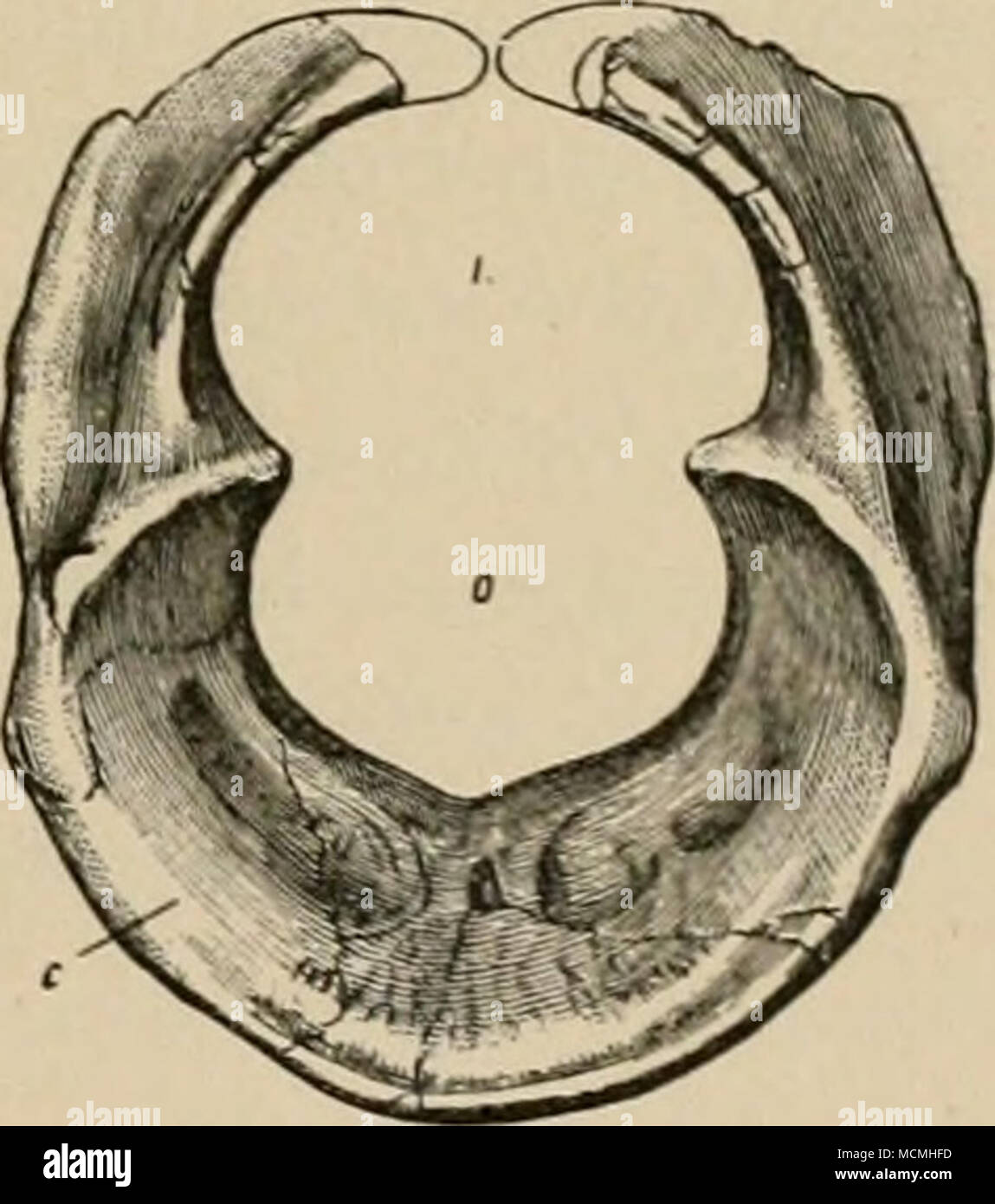 . Fig. -t. Atlas of Diplodocus longus; side view; one halt' natural size, n, articular lace for axis ; e, cup; r, face for rib ; z, posterior zygapopliyses. After Marsh. Fig. 5. Atlas of Diplodocus longus; front view ; one half natural size ; c, cup ; o, cavity for odontoid process ; /, neural canal. After jNIarsh. than the distance from its ventral surface to the top of the neural spine. Promi- nent postzygapophysial laminie spring from the posterior and superior border of the neural arch. These diverge and extend upward and backward until they reach the summits of the postzygapophyses, when Stock Photohttps://www.alamy.com/image-license-details/?v=1https://www.alamy.com/fig-t-atlas-of-diplodocus-longus-side-view-one-halt-natural-size-n-articular-lace-for-axis-e-cup-r-face-for-rib-z-posterior-zygapopliyses-after-marsh-fig-5-atlas-of-diplodocus-longus-front-view-one-half-natural-size-c-cup-o-cavity-for-odontoid-process-neural-canal-after-jniarsh-than-the-distance-from-its-ventral-surface-to-the-top-of-the-neural-spine-promi-nent-postzygapophysial-laminie-spring-from-the-posterior-and-superior-border-of-the-neural-arch-these-diverge-and-extend-upward-and-backward-until-they-reach-the-summits-of-the-postzygapophyses-when-image179932353.html
RMMCMHFD–. Fig. -t. Atlas of Diplodocus longus; side view; one halt' natural size, n, articular lace for axis ; e, cup; r, face for rib ; z, posterior zygapopliyses. After Marsh. Fig. 5. Atlas of Diplodocus longus; front view ; one half natural size ; c, cup ; o, cavity for odontoid process ; /, neural canal. After jNIarsh. than the distance from its ventral surface to the top of the neural spine. Promi- nent postzygapophysial laminie spring from the posterior and superior border of the neural arch. These diverge and extend upward and backward until they reach the summits of the postzygapophyses, when
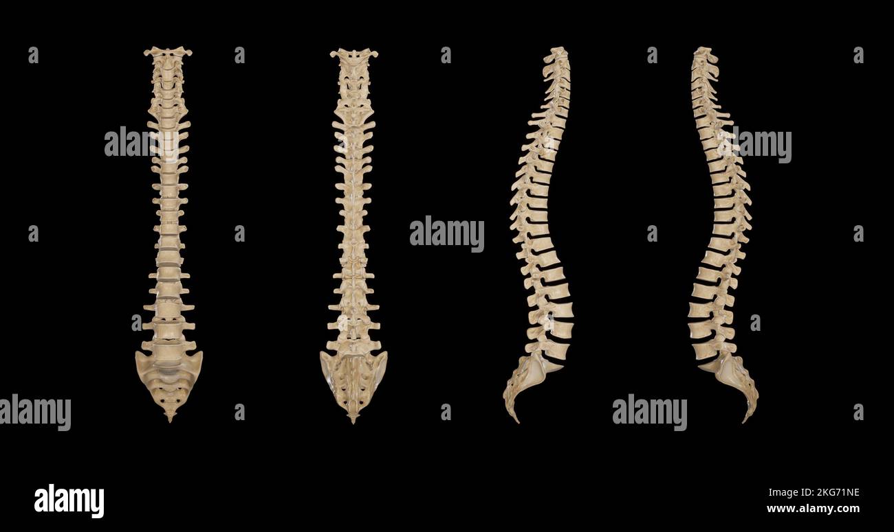 Vertebral Column-Multiple Views Stock Photohttps://www.alamy.com/image-license-details/?v=1https://www.alamy.com/vertebral-column-multiple-views-image491879850.html
RF2KG71NE–Vertebral Column-Multiple Views
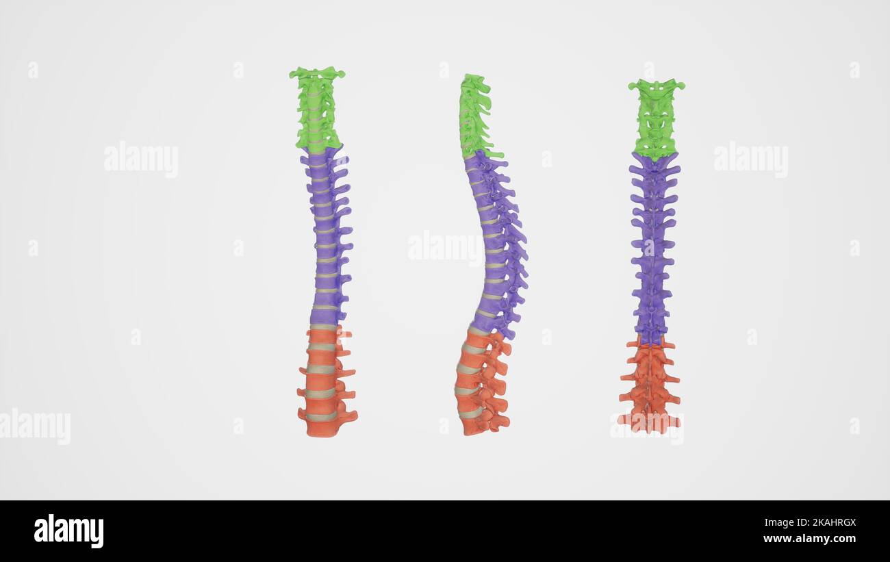 Medical Ilustration of Colored Cervical,Thoracic,and Lumbar Spines-Anterior,posterior,and Back views Stock Photohttps://www.alamy.com/image-license-details/?v=1https://www.alamy.com/medical-ilustration-of-colored-cervicalthoracicand-lumbar-spines-anteriorposteriorand-back-views-image488428554.html
RF2KAHRGX–Medical Ilustration of Colored Cervical,Thoracic,and Lumbar Spines-Anterior,posterior,and Back views
 The hydropathic encyclopedia: a system of hydropathy and hygiene .. . rocess of theaxis in connection with the anterior arch of the atlas. Where itcrosses the odontoid process, some fibres pass downward to be attachedto the body of the axis, and others are sent upward to the basilar pro-cess of the occipital bone. This disposition enables the atlas, andwith it the whole head, to rotate upon the axis, its extent of rotat:onbeing limited by the odontoid ligaments. Fig. 25 is a posterior view of the ligamentsconnecting the atlas, axis, and occipitalbone. The back part of the occipitis andthe arch Stock Photohttps://www.alamy.com/image-license-details/?v=1https://www.alamy.com/the-hydropathic-encyclopedia-a-system-of-hydropathy-and-hygiene-rocess-of-theaxis-in-connection-with-the-anterior-arch-of-the-atlas-where-itcrosses-the-odontoid-process-some-fibres-pass-downward-to-be-attachedto-the-body-of-the-axis-and-others-are-sent-upward-to-the-basilar-pro-cess-of-the-occipital-bone-this-disposition-enables-the-atlas-andwith-it-the-whole-head-to-rotate-upon-the-axis-its-extent-of-rotatonbeing-limited-by-the-odontoid-ligaments-fig-25-is-a-posterior-view-of-the-ligamentsconnecting-the-atlas-axis-and-occipitalbone-the-back-part-of-the-occipitis-andthe-arch-image340106854.html
RM2AN95NA–The hydropathic encyclopedia: a system of hydropathy and hygiene .. . rocess of theaxis in connection with the anterior arch of the atlas. Where itcrosses the odontoid process, some fibres pass downward to be attachedto the body of the axis, and others are sent upward to the basilar pro-cess of the occipital bone. This disposition enables the atlas, andwith it the whole head, to rotate upon the axis, its extent of rotat:onbeing limited by the odontoid ligaments. Fig. 25 is a posterior view of the ligamentsconnecting the atlas, axis, and occipitalbone. The back part of the occipitis andthe arch
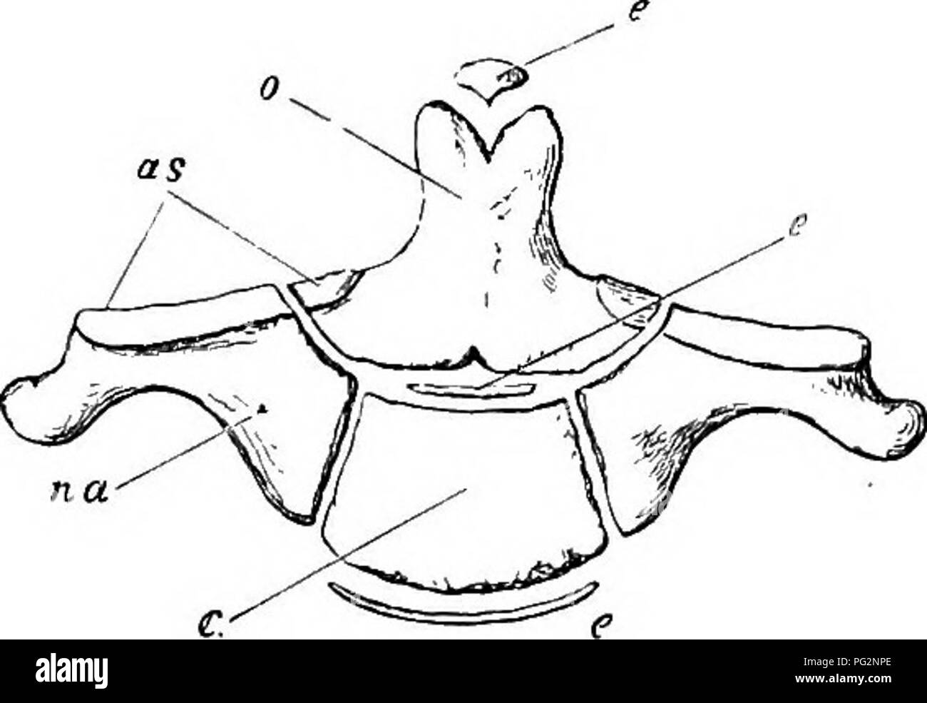 . Elements of the comparative anatomy of vertebrates. Anatomy, Comparative. VERTEBRAL COLUMN 51. Fig. 39a.—Diagram showing mode or Ossi- fication OF Human Axis. (Ventral surface.) From Flower's Osteology of the Mammalia. o, odontoid process, or centrum of atlas ; t, proper centrum of axis ; na, neural arch ; an, anterior articular surface; c, e, e, e, epi- physes, completing the ends of the centra, The transverse processes are simple in all but the cervical region and arise from the base of the arch : in the thoracic region they are tipped with cartilage on the ventral side of their distal end Stock Photohttps://www.alamy.com/image-license-details/?v=1https://www.alamy.com/elements-of-the-comparative-anatomy-of-vertebrates-anatomy-comparative-vertebral-column-51-fig-39adiagram-showing-mode-or-ossi-fication-of-human-axis-ventral-surface-from-flowers-osteology-of-the-mammalia-o-odontoid-process-or-centrum-of-atlas-t-proper-centrum-of-axis-na-neural-arch-an-anterior-articular-surface-c-e-e-e-epi-physes-completing-the-ends-of-the-centra-the-transverse-processes-are-simple-in-all-but-the-cervical-region-and-arise-from-the-base-of-the-arch-in-the-thoracic-region-they-are-tipped-with-cartilage-on-the-ventral-side-of-their-distal-end-image216419910.html
RMPG2NPE–. Elements of the comparative anatomy of vertebrates. Anatomy, Comparative. VERTEBRAL COLUMN 51. Fig. 39a.—Diagram showing mode or Ossi- fication OF Human Axis. (Ventral surface.) From Flower's Osteology of the Mammalia. o, odontoid process, or centrum of atlas ; t, proper centrum of axis ; na, neural arch ; an, anterior articular surface; c, e, e, e, epi- physes, completing the ends of the centra, The transverse processes are simple in all but the cervical region and arise from the base of the arch : in the thoracic region they are tipped with cartilage on the ventral side of their distal end
![. Fig. 86. FiGURR 84.—Albs of TmOLCiat, giaiicL, Mu^h (No ]040); front view. Figure 8.5.—The same veitebia, back view FiGDEK Bfi.—The same vertebra; bottom view. a. face for axis; 6. fare Air odontoid process of a.xis; c. face for occipital condyle of skull; d. foramen in neural arch for spinal nerve; nc. neural canal. All the fignres are one-fourtli natural size. The following- measfirements give the princijial dimensions of the atlas in one specimen of Dlnoceras : Measurements of Atlas. {Dhiooeras miraUe, No. 12.51.) m. Greatest transverse diameter of atlas,. .26.5 Greatest vertical diamete Stock Photo . Fig. 86. FiGURR 84.—Albs of TmOLCiat, giaiicL, Mu^h (No ]040); front view. Figure 8.5.—The same veitebia, back view FiGDEK Bfi.—The same vertebra; bottom view. a. face for axis; 6. fare Air odontoid process of a.xis; c. face for occipital condyle of skull; d. foramen in neural arch for spinal nerve; nc. neural canal. All the fignres are one-fourtli natural size. The following- measfirements give the princijial dimensions of the atlas in one specimen of Dlnoceras : Measurements of Atlas. {Dhiooeras miraUe, No. 12.51.) m. Greatest transverse diameter of atlas,. .26.5 Greatest vertical diamete Stock Photo](https://c8.alamy.com/comp/MCMJAD/fig-86-figurr-84albs-of-tmolciat-giaiicl-muh-no-040-front-view-figure-85the-same-veitebia-back-view-figdek-bfithe-same-vertebra-bottom-view-a-face-for-axis-6-fare-air-odontoid-process-of-axis-c-face-for-occipital-condyle-of-skull-d-foramen-in-neural-arch-for-spinal-nerve-nc-neural-canal-all-the-fignres-are-one-fourtli-natural-size-the-following-measfirements-give-the-princijial-dimensions-of-the-atlas-in-one-specimen-of-dlnoceras-measurements-of-atlas-dhiooeras-miraue-no-1251-m-greatest-transverse-diameter-of-atlas-265-greatest-vertical-diamete-MCMJAD.jpg) . Fig. 86. FiGURR 84.—Albs of TmOLCiat, giaiicL, Mu^h (No ]040); front view. Figure 8.5.—The same veitebia, back view FiGDEK Bfi.—The same vertebra; bottom view. a. face for axis; 6. fare Air odontoid process of a.xis; c. face for occipital condyle of skull; d. foramen in neural arch for spinal nerve; nc. neural canal. All the fignres are one-fourtli natural size. The following- measfirements give the princijial dimensions of the atlas in one specimen of Dlnoceras : Measurements of Atlas. {Dhiooeras miraUe, No. 12.51.) m. Greatest transverse diameter of atlas,. .26.5 Greatest vertical diamete Stock Photohttps://www.alamy.com/image-license-details/?v=1https://www.alamy.com/fig-86-figurr-84albs-of-tmolciat-giaiicl-muh-no-040-front-view-figure-85the-same-veitebia-back-view-figdek-bfithe-same-vertebra-bottom-view-a-face-for-axis-6-fare-air-odontoid-process-of-axis-c-face-for-occipital-condyle-of-skull-d-foramen-in-neural-arch-for-spinal-nerve-nc-neural-canal-all-the-fignres-are-one-fourtli-natural-size-the-following-measfirements-give-the-princijial-dimensions-of-the-atlas-in-one-specimen-of-dlnoceras-measurements-of-atlas-dhiooeras-miraue-no-1251-m-greatest-transverse-diameter-of-atlas-265-greatest-vertical-diamete-image179932997.html
. Fig. 86. FiGURR 84.—Albs of TmOLCiat, giaiicL, Mu^h (No ]040); front view. Figure 8.5.—The same veitebia, back view FiGDEK Bfi.—The same vertebra; bottom view. a. face for axis; 6. fare Air odontoid process of a.xis; c. face for occipital condyle of skull; d. foramen in neural arch for spinal nerve; nc. neural canal. All the fignres are one-fourtli natural size. The following- measfirements give the princijial dimensions of the atlas in one specimen of Dlnoceras : Measurements of Atlas. {Dhiooeras miraUe, No. 12.51.) m. Greatest transverse diameter of atlas,. .26.5 Greatest vertical diamete Stock Photohttps://www.alamy.com/image-license-details/?v=1https://www.alamy.com/fig-86-figurr-84albs-of-tmolciat-giaiicl-muh-no-040-front-view-figure-85the-same-veitebia-back-view-figdek-bfithe-same-vertebra-bottom-view-a-face-for-axis-6-fare-air-odontoid-process-of-axis-c-face-for-occipital-condyle-of-skull-d-foramen-in-neural-arch-for-spinal-nerve-nc-neural-canal-all-the-fignres-are-one-fourtli-natural-size-the-following-measfirements-give-the-princijial-dimensions-of-the-atlas-in-one-specimen-of-dlnoceras-measurements-of-atlas-dhiooeras-miraue-no-1251-m-greatest-transverse-diameter-of-atlas-265-greatest-vertical-diamete-image179932997.htmlRMMCMJAD–. Fig. 86. FiGURR 84.—Albs of TmOLCiat, giaiicL, Mu^h (No ]040); front view. Figure 8.5.—The same veitebia, back view FiGDEK Bfi.—The same vertebra; bottom view. a. face for axis; 6. fare Air odontoid process of a.xis; c. face for occipital condyle of skull; d. foramen in neural arch for spinal nerve; nc. neural canal. All the fignres are one-fourtli natural size. The following- measfirements give the princijial dimensions of the atlas in one specimen of Dlnoceras : Measurements of Atlas. {Dhiooeras miraUe, No. 12.51.) m. Greatest transverse diameter of atlas,. .26.5 Greatest vertical diamete
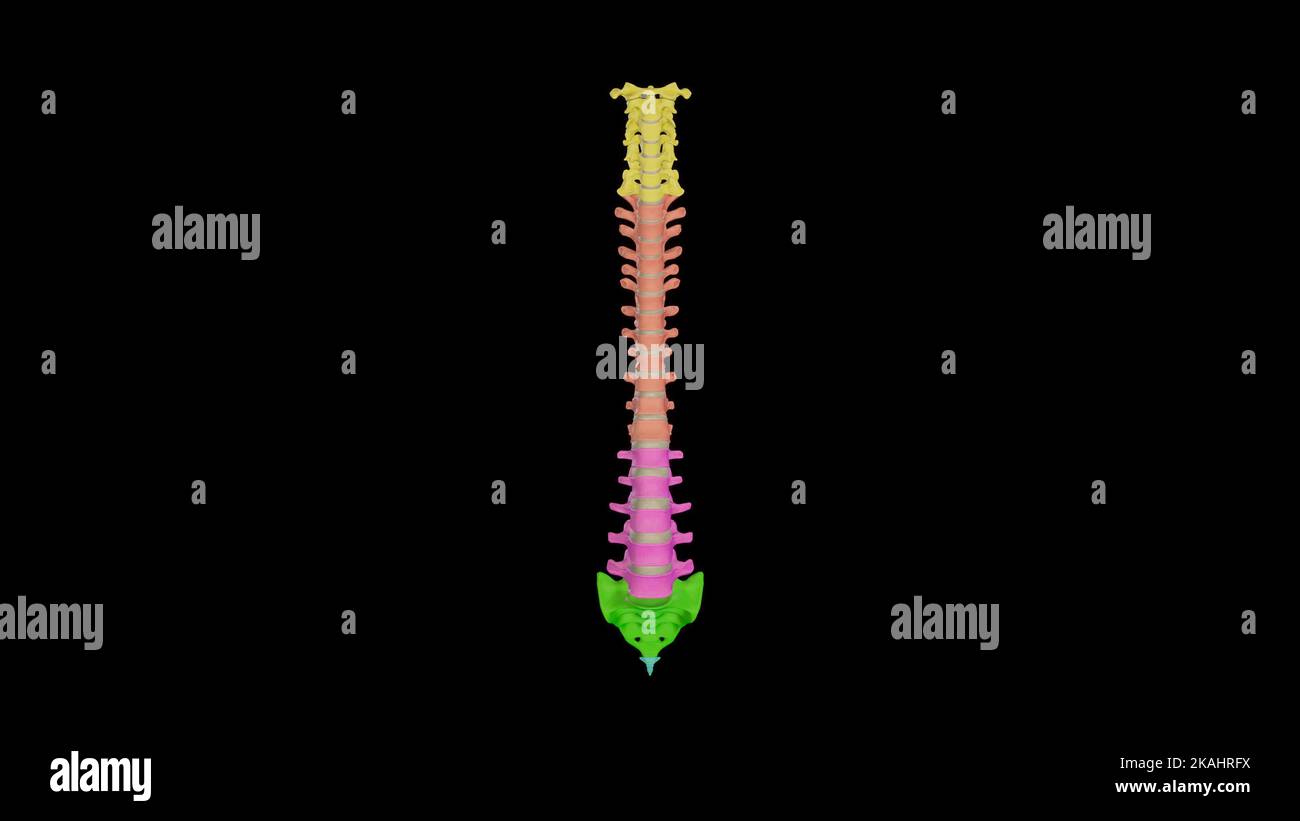 Medical Ilustration of Vertebral Column with Colored spines Stock Photohttps://www.alamy.com/image-license-details/?v=1https://www.alamy.com/medical-ilustration-of-vertebral-column-with-colored-spines-image488428526.html
RF2KAHRFX–Medical Ilustration of Vertebral Column with Colored spines
 The anatomist's vade mecum : a system of human anatomy . ls. It is bounded infront by the upper border of the petrous portion and dorsum ephippii,and along its posterior circumference by the groove for the lateralsinuses; it gives support to the pons Varolii, meduUa oblongata, andcerebellum. In the centre of this fossa is the foramen magnum,bounded on each side by a rough tubercle, which gives attachment tothe odontoid ligament, and by the anterior condyloid foramen. In 58 BASE OP THE SKULL. front of the foramen magnum is tlie concave smface (clivus Blumen-bachii) wliich. supports the medulla Stock Photohttps://www.alamy.com/image-license-details/?v=1https://www.alamy.com/the-anatomists-vade-mecum-a-system-of-human-anatomy-ls-it-is-bounded-infront-by-the-upper-border-of-the-petrous-portion-and-dorsum-ephippiiand-along-its-posterior-circumference-by-the-groove-for-the-lateralsinuses-it-gives-support-to-the-pons-varolii-meduua-oblongata-andcerebellum-in-the-centre-of-this-fossa-is-the-foramen-magnumbounded-on-each-side-by-a-rough-tubercle-which-gives-attachment-tothe-odontoid-ligament-and-by-the-anterior-condyloid-foramen-in-58-base-op-the-skull-front-of-the-foramen-magnum-is-tlie-concave-smface-clivus-blumen-bachii-wliich-supports-the-medulla-image342778483.html
RM2AWJWCK–The anatomist's vade mecum : a system of human anatomy . ls. It is bounded infront by the upper border of the petrous portion and dorsum ephippii,and along its posterior circumference by the groove for the lateralsinuses; it gives support to the pons Varolii, meduUa oblongata, andcerebellum. In the centre of this fossa is the foramen magnum,bounded on each side by a rough tubercle, which gives attachment tothe odontoid ligament, and by the anterior condyloid foramen. In 58 BASE OP THE SKULL. front of the foramen magnum is tlie concave smface (clivus Blumen-bachii) wliich. supports the medulla
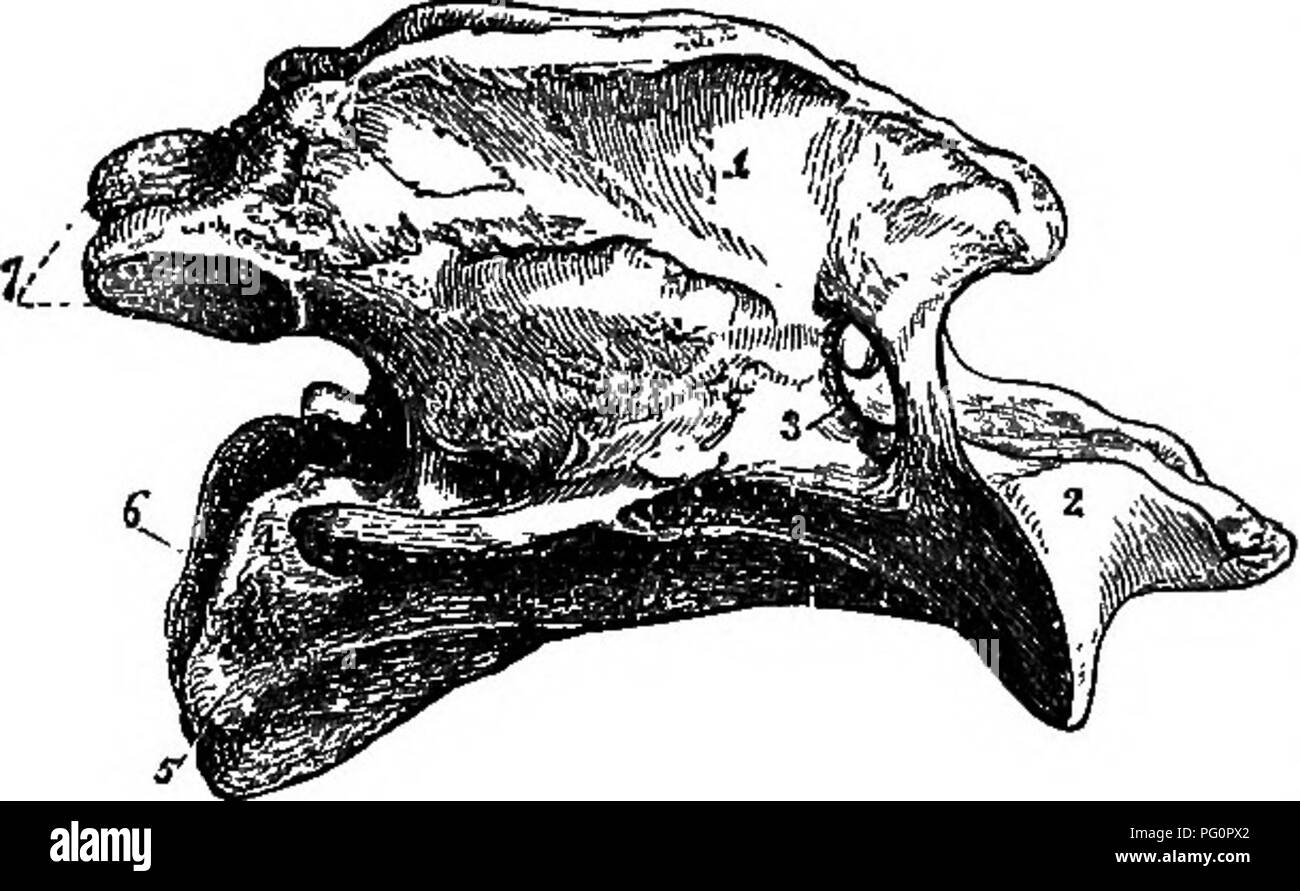 . A text-book of agricultural zoology. Zoology, Economic. 288 SKELETON 01 THE HOKSE.. number. This is the general number in all mammals; even the long neck of the giraffe has only the same number as the short neck of the pig.' The cervical vertebrae are cubical in form, the first two being modified. The first is known as the atlas (fig. 151), which is simply a bony ring with which the skuU articur lates in front; there is Fig. 160.—Axis. (Lateral view.) nO centrum to this Ver- 1, Superior spinous process; 2, odontoid pro- tebra at aU. The secOlld cess; 3, intervertebral foramen; 4, body; 5, in Stock Photohttps://www.alamy.com/image-license-details/?v=1https://www.alamy.com/a-text-book-of-agricultural-zoology-zoology-economic-288-skeleton-01-the-hokse-number-this-is-the-general-number-in-all-mammals-even-the-long-neck-of-the-giraffe-has-only-the-same-number-as-the-short-neck-of-the-pig-the-cervical-vertebrae-are-cubical-in-form-the-first-two-being-modified-the-first-is-known-as-the-atlas-fig-151-which-is-simply-a-bony-ring-with-which-the-skuu-articur-lates-in-front-there-is-fig-160axis-lateral-view-no-centrum-to-this-ver-1-superior-spinous-process-2-odontoid-pro-tebra-at-au-the-secolld-cess-3-intervertebral-foramen-4-body-5-in-image216376890.html
RMPG0PX2–. A text-book of agricultural zoology. Zoology, Economic. 288 SKELETON 01 THE HOKSE.. number. This is the general number in all mammals; even the long neck of the giraffe has only the same number as the short neck of the pig.' The cervical vertebrae are cubical in form, the first two being modified. The first is known as the atlas (fig. 151), which is simply a bony ring with which the skuU articur lates in front; there is Fig. 160.—Axis. (Lateral view.) nO centrum to this Ver- 1, Superior spinous process; 2, odontoid pro- tebra at aU. The secOlld cess; 3, intervertebral foramen; 4, body; 5, in
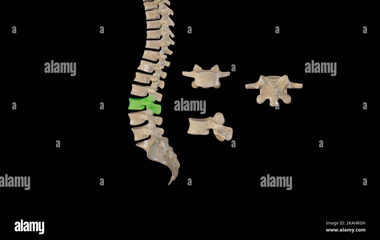 Medical Ilustration of Third Lumbar Vertebrae-Multiple Views Stock Photohttps://www.alamy.com/image-license-details/?v=1https://www.alamy.com/medical-ilustration-of-third-lumbar-vertebrae-multiple-views-image488428545.html
RF2KAHRGH–Medical Ilustration of Third Lumbar Vertebrae-Multiple Views
RM2AGD4MK–. Outlines of zoology. or coccyx. The first vertebra bears two facets for the two condyles ofthe skull, and an odontoid process which lies between thecondyles. It has no transverse processes, and its arch isincompletely ossified. Each of thenext six has an anteriorly concave orprocoelous centrum, a neural arch sur-rounding the spinal cord, a transverseprocess from each side of the base ofthe arch, an anterior and a posteriorpair of articular processes, and a shortneural spine. The eighth vertebra hasa biconcave or amphicoelous centrum.The ninth is convex in front, with twoconvex tubercles behi
 . A text-book of agricultural zoology. Zoology, Economic. number. This is the general number in all mammals; even the long neck of the giraffe has only the same number as the short neck of the pig.' The cervical vertebrae are cubical in form, the first two being modified. The first is known as the atlas (fig. 151), which is simply a bony ring with which the skuU articur lates in front; there is Fig. 160.âAxis. (Lateral view.) nO centrum to this Ver- 1, Superior spinous process; 2, odontoid pro- tebra at aU. The secOlld cess; 3, intervertebral foramen; 4, body; 5, in- ferior spinous process; 6, Stock Photohttps://www.alamy.com/image-license-details/?v=1https://www.alamy.com/a-text-book-of-agricultural-zoology-zoology-economic-number-this-is-the-general-number-in-all-mammals-even-the-long-neck-of-the-giraffe-has-only-the-same-number-as-the-short-neck-of-the-pig-the-cervical-vertebrae-are-cubical-in-form-the-first-two-being-modified-the-first-is-known-as-the-atlas-fig-151-which-is-simply-a-bony-ring-with-which-the-skuu-articur-lates-in-front-there-is-fig-160axis-lateral-view-no-centrum-to-this-ver-1-superior-spinous-process-2-odontoid-pro-tebra-at-au-the-secolld-cess-3-intervertebral-foramen-4-body-5-in-ferior-spinous-process-6-image216376888.html
RMPG0PX0–. A text-book of agricultural zoology. Zoology, Economic. number. This is the general number in all mammals; even the long neck of the giraffe has only the same number as the short neck of the pig.' The cervical vertebrae are cubical in form, the first two being modified. The first is known as the atlas (fig. 151), which is simply a bony ring with which the skuU articur lates in front; there is Fig. 160.âAxis. (Lateral view.) nO centrum to this Ver- 1, Superior spinous process; 2, odontoid pro- tebra at aU. The secOlld cess; 3, intervertebral foramen; 4, body; 5, in- ferior spinous process; 6,
 A practical treatise on fractures and dislocations . tion of the occipito-axoid ligament,which would otherwise have protected thecontents of the spinal cavity, must have fa-vored this result.1 Dr. Philip Bevan presented to the SurgicalSociety of Ireland, in 1862, a specimen ob-tained from the dead-room, and which was sup-posed to be an epiphyseal separation of the odontoid process, occur-ring in early life. The history of the case is not known, althoughthe woman was forty years old when she died. It does not appearvery clear to us whether this was really an epiphyseal separation, orthe result Stock Photohttps://www.alamy.com/image-license-details/?v=1https://www.alamy.com/a-practical-treatise-on-fractures-and-dislocations-tion-of-the-occipito-axoid-ligamentwhich-would-otherwise-have-protected-thecontents-of-the-spinal-cavity-must-have-fa-vored-this-result1-dr-philip-bevan-presented-to-the-surgicalsociety-of-ireland-in-1862-a-specimen-ob-tained-from-the-dead-room-and-which-was-sup-posed-to-be-an-epiphyseal-separation-of-the-odontoid-process-occur-ring-in-early-life-the-history-of-the-case-is-not-known-althoughthe-woman-was-forty-years-old-when-she-died-it-does-not-appearvery-clear-to-us-whether-this-was-really-an-epiphyseal-separation-orthe-result-image343260052.html
RM2AXCRKG–A practical treatise on fractures and dislocations . tion of the occipito-axoid ligament,which would otherwise have protected thecontents of the spinal cavity, must have fa-vored this result.1 Dr. Philip Bevan presented to the SurgicalSociety of Ireland, in 1862, a specimen ob-tained from the dead-room, and which was sup-posed to be an epiphyseal separation of the odontoid process, occur-ring in early life. The history of the case is not known, althoughthe woman was forty years old when she died. It does not appearvery clear to us whether this was really an epiphyseal separation, orthe result
 . Denkschriften - Österreichische Akademie der Wissenschaften. . Halswirbel von Dorygnalhtts; a 3.—8. Wirbel des Wiener Originales von oben; b Rekonstruktion der ganzen Serie. Atlas und Axis nach RA. Kßkcni Plieninger (1907, Taf. XVI, Fig. 7 -10) - Pia Proatlati, na Neurale, ic Inlcircnlrum od Odontoid. Beim Exemplar des Dimorphodon niacrouyx Bück 1. sp., welches Owen in Foss. Rept. Lias. Format, pt. III, p. 45, Taf. 18 c aus dem unteren Lias beschrieb, ist eine Serie von 4 Halswirbeln und ein 5., abgetrennt liegender Wirbel erhalten. Die Folge beginnt bei Atlas und Axis, welche Owen als einen Stock Photohttps://www.alamy.com/image-license-details/?v=1https://www.alamy.com/denkschriften-sterreichische-akademie-der-wissenschaften-halswirbel-von-dorygnalhtts-a-38-wirbel-des-wiener-originales-von-oben-b-rekonstruktion-der-ganzen-serie-atlas-und-axis-nach-ra-kkcni-plieninger-1907-taf-xvi-fig-7-10-pia-proatlati-na-neurale-ic-inlcircnlrum-od-odontoid-beim-exemplar-des-dimorphodon-niacrouyx-bck-1-sp-welches-owen-in-foss-rept-lias-format-pt-iii-p-45-taf-18-c-aus-dem-unteren-lias-beschrieb-ist-eine-serie-von-4-halswirbeln-und-ein-5-abgetrennt-liegender-wirbel-erhalten-die-folge-beginnt-bei-atlas-und-axis-welche-owen-als-einen-image216048590.html
RMPFDT52–. Denkschriften - Österreichische Akademie der Wissenschaften. . Halswirbel von Dorygnalhtts; a 3.—8. Wirbel des Wiener Originales von oben; b Rekonstruktion der ganzen Serie. Atlas und Axis nach RA. Kßkcni Plieninger (1907, Taf. XVI, Fig. 7 -10) - Pia Proatlati, na Neurale, ic Inlcircnlrum od Odontoid. Beim Exemplar des Dimorphodon niacrouyx Bück 1. sp., welches Owen in Foss. Rept. Lias. Format, pt. III, p. 45, Taf. 18 c aus dem unteren Lias beschrieb, ist eine Serie von 4 Halswirbeln und ein 5., abgetrennt liegender Wirbel erhalten. Die Folge beginnt bei Atlas und Axis, welche Owen als einen
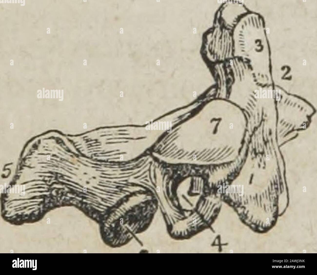 A system of human anatomy, general and special . e-neath. The transverse processes are quite rudimentary, not bifid, * The upper surface of the atlas. 1. The anterior tubercle projecting from the ante-rior arch. 2. The articular surface for the odontoid process upon the posterior surfaceof the anterior arch. 3. The posterior arch, with its rudimentary spinous process. 4. Theintervertebral notch. 5. The transverse process. 6. The vertebral foramen. -7. Supe-rior articular surface. 8. The tubercle for the attachment of the transverse li<rament.The tubercle referred to is just above the head o Stock Photohttps://www.alamy.com/image-license-details/?v=1https://www.alamy.com/a-system-of-human-anatomy-general-and-special-e-neath-the-transverse-processes-are-quite-rudimentary-not-bifid-the-upper-surface-of-the-atlas-1-the-anterior-tubercle-projecting-from-the-ante-rior-arch-2-the-articular-surface-for-the-odontoid-process-upon-the-posterior-surfaceof-the-anterior-arch-3-the-posterior-arch-with-its-rudimentary-spinous-process-4-theintervertebral-notch-5-the-transverse-process-6-the-vertebral-foramen-7-supe-rior-articular-surface-8-the-tubercle-for-the-attachment-of-the-transverse-liltramentthe-tubercle-referred-to-is-just-above-the-head-o-image342761487.html
RM2AWJ3NK–A system of human anatomy, general and special . e-neath. The transverse processes are quite rudimentary, not bifid, * The upper surface of the atlas. 1. The anterior tubercle projecting from the ante-rior arch. 2. The articular surface for the odontoid process upon the posterior surfaceof the anterior arch. 3. The posterior arch, with its rudimentary spinous process. 4. Theintervertebral notch. 5. The transverse process. 6. The vertebral foramen. -7. Supe-rior articular surface. 8. The tubercle for the attachment of the transverse li<rament.The tubercle referred to is just above the head o
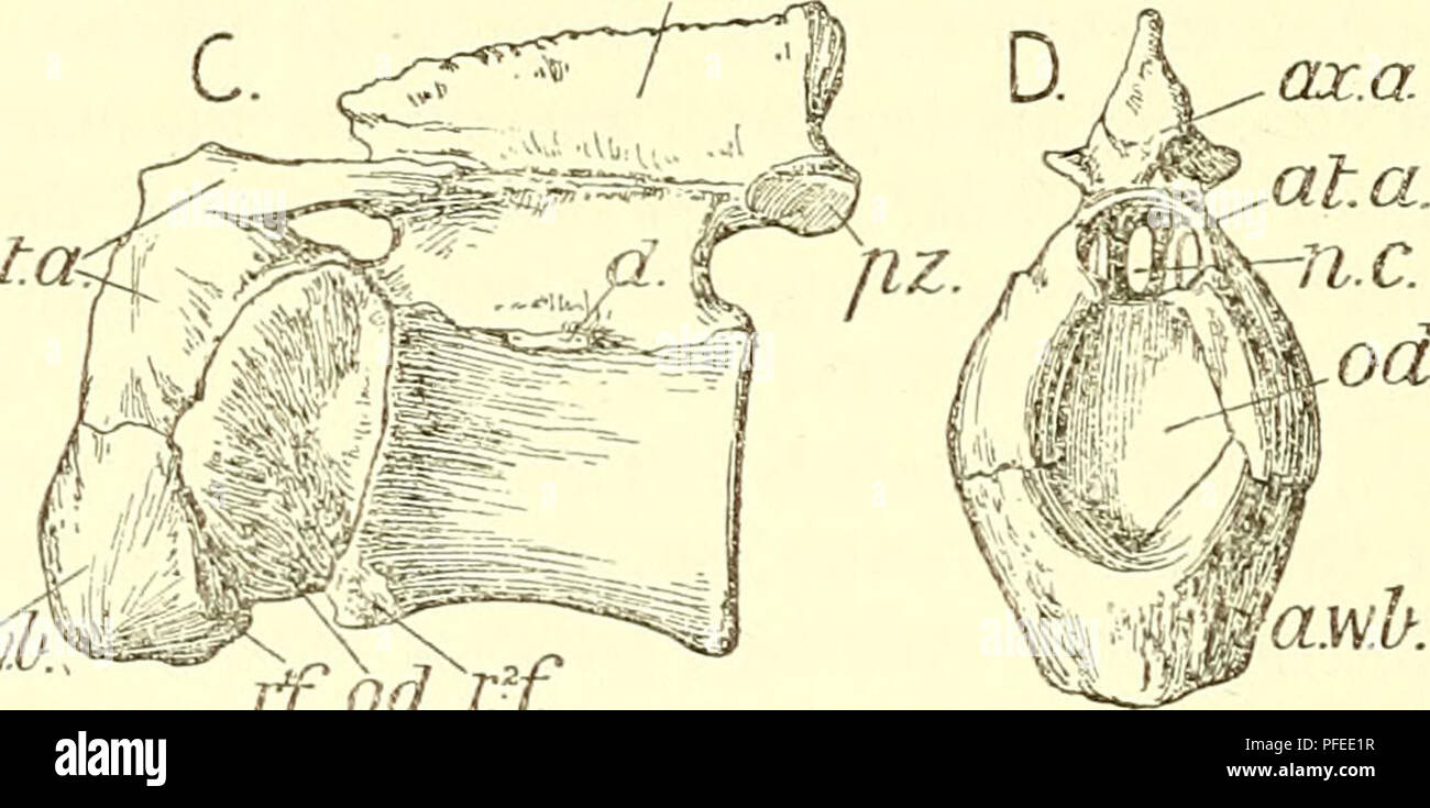 . A descriptive catalogue of the marine reptiles of the Oxford clay. Based on the Leeds Collection in the British Museum (Natural History), London ... Reptiles, Fossil. ax. a. n.c. a.w.b. r.'f iff Gx..a. C. at.ck awj. wA rfodrf Atlas and axis of Steneosaurus leedsi: A, from the left side; B, from behind. (R. 3806, nat. size.) Also of Steneosaurus durobrivensis : C, from left side ; D, from front. (R. 3701, £ nat. size.) at.a., neural arch of atlas ; a.to.b., anterior wedge-bone (hypocentrum) ; ax.a., neural arch of axis ; </., diapophysis of axis ; n.c., neural canal; od., odontoid (centr Stock Photohttps://www.alamy.com/image-license-details/?v=1https://www.alamy.com/a-descriptive-catalogue-of-the-marine-reptiles-of-the-oxford-clay-based-on-the-leeds-collection-in-the-british-museum-natural-history-london-reptiles-fossil-ax-a-nc-awb-rf-iff-gxa-c-atck-awj-wa-rfodrf-atlas-and-axis-of-steneosaurus-leedsi-a-from-the-left-side-b-from-behind-r-3806-nat-size-also-of-steneosaurus-durobrivensis-c-from-left-side-d-from-front-r-3701-nat-size-ata-neural-arch-of-atlas-atob-anterior-wedge-bone-hypocentrum-axa-neural-arch-of-axis-lt-diapophysis-of-axis-nc-neural-canal-od-odontoid-centr-image216062611.html
RMPFEE1R–. A descriptive catalogue of the marine reptiles of the Oxford clay. Based on the Leeds Collection in the British Museum (Natural History), London ... Reptiles, Fossil. ax. a. n.c. a.w.b. r.'f iff Gx..a. C. at.ck awj. wA rfodrf Atlas and axis of Steneosaurus leedsi: A, from the left side; B, from behind. (R. 3806, nat. size.) Also of Steneosaurus durobrivensis : C, from left side ; D, from front. (R. 3701, £ nat. size.) at.a., neural arch of atlas ; a.to.b., anterior wedge-bone (hypocentrum) ; ax.a., neural arch of axis ; </., diapophysis of axis ; n.c., neural canal; od., odontoid (centr
 The anatomist's vade mecum : a system of human anatomy . le by the projection of the odontoid process.6. Lateral and capsular ligament of the occipito-atloid articulation. 7. Cap-sular ligament between the articulating processes of the atlas and axis. 134 LIGAMENTS OP THE VERTEBRAL C0LU5[N. 4. Articulation of the Atlas with the Axis.—The ligaments of thisarticulation ^x^five in number,— Anterior atlo-axoid, Two capsular. Posterior atlo-axoid, Transverse. The anterior ligament consists of ligamentous fibres, which passfrom the anterior tubercle and arch of the atlas to the base of theodontoid p Stock Photohttps://www.alamy.com/image-license-details/?v=1https://www.alamy.com/the-anatomists-vade-mecum-a-system-of-human-anatomy-le-by-the-projection-of-the-odontoid-process6-lateral-and-capsular-ligament-of-the-occipito-atloid-articulation-7-cap-sular-ligament-between-the-articulating-processes-of-the-atlas-and-axis-134-ligaments-op-the-vertebral-c0lu5-n-4-articulation-of-the-atlas-with-the-axisthe-ligaments-of-thisarticulation-xfive-in-number-anterior-atlo-axoid-two-capsular-posterior-atlo-axoid-transverse-the-anterior-ligament-consists-of-ligamentous-fibres-which-passfrom-the-anterior-tubercle-and-arch-of-the-atlas-to-the-base-of-theodontoid-p-image342768937.html
RM2AWJD7N–The anatomist's vade mecum : a system of human anatomy . le by the projection of the odontoid process.6. Lateral and capsular ligament of the occipito-atloid articulation. 7. Cap-sular ligament between the articulating processes of the atlas and axis. 134 LIGAMENTS OP THE VERTEBRAL C0LU5[N. 4. Articulation of the Atlas with the Axis.—The ligaments of thisarticulation ^x^five in number,— Anterior atlo-axoid, Two capsular. Posterior atlo-axoid, Transverse. The anterior ligament consists of ligamentous fibres, which passfrom the anterior tubercle and arch of the atlas to the base of theodontoid p
 The anatomist's vade mecum : a system of human anatomy . o named from having a process uponwhich the head turns as on a pivot. The body is of large size, andsupports a strong process, the odontoid, which rises perpendicularlyfrom its upper surface. The odontoid process presents two articulatingsurfaces; one on its anterior face, to articulate with the anterior archof the atlas; the other on its posterior face, for the transverse liga- * The upper surface of the atlas. 1. The anterior tubercle projecting fromthe anterior arch. 2. The articular surface for tlie odontoid process uponthe posterior Stock Photohttps://www.alamy.com/image-license-details/?v=1https://www.alamy.com/the-anatomists-vade-mecum-a-system-of-human-anatomy-o-named-from-having-a-process-uponwhich-the-head-turns-as-on-a-pivot-the-body-is-of-large-size-andsupports-a-strong-process-the-odontoid-which-rises-perpendicularlyfrom-its-upper-surface-the-odontoid-process-presents-two-articulatingsurfaces-one-on-its-anterior-face-to-articulate-with-the-anterior-archof-the-atlas-the-other-on-its-posterior-face-for-the-transverse-liga-the-upper-surface-of-the-atlas-1-the-anterior-tubercle-projecting-fromthe-anterior-arch-2-the-articular-surface-for-tlie-odontoid-process-uponthe-posterior-image342785276.html
RM2AWK638–The anatomist's vade mecum : a system of human anatomy . o named from having a process uponwhich the head turns as on a pivot. The body is of large size, andsupports a strong process, the odontoid, which rises perpendicularlyfrom its upper surface. The odontoid process presents two articulatingsurfaces; one on its anterior face, to articulate with the anterior archof the atlas; the other on its posterior face, for the transverse liga- * The upper surface of the atlas. 1. The anterior tubercle projecting fromthe anterior arch. 2. The articular surface for tlie odontoid process uponthe posterior
 The anatomist's vade mecum : a system of human anatomy . mes the anterior tubercle represents a small butdistinct rib. Dorsal Vertelra.—The hody of a dorsal vertebra is as long frombefore backwards as from side to side, particularly in the middle of thedorsal region; it is thicker behind than before, and marked on eachside by two half-articulating surfaces for the heads of two ribs. The * A lateral view of the axis. 1. The body; the figure is placed on thedepression which gives attachment to the longus colli. 2. The odontoid pro-cess. 3. The smooth facet on the anterior surface of the odontoid Stock Photohttps://www.alamy.com/image-license-details/?v=1https://www.alamy.com/the-anatomists-vade-mecum-a-system-of-human-anatomy-mes-the-anterior-tubercle-represents-a-small-butdistinct-rib-dorsal-vertelrathe-hody-of-a-dorsal-vertebra-is-as-long-frombefore-backwards-as-from-side-to-side-particularly-in-the-middle-of-thedorsal-region-it-is-thicker-behind-than-before-and-marked-on-eachside-by-two-half-articulating-surfaces-for-the-heads-of-two-ribs-the-a-lateral-view-of-the-axis-1-the-body-the-figure-is-placed-on-thedepression-which-gives-attachment-to-the-longus-colli-2-the-odontoid-pro-cess-3-the-smooth-facet-on-the-anterior-surface-of-the-odontoid-image342785019.html
RM2AWK5P3–The anatomist's vade mecum : a system of human anatomy . mes the anterior tubercle represents a small butdistinct rib. Dorsal Vertelra.—The hody of a dorsal vertebra is as long frombefore backwards as from side to side, particularly in the middle of thedorsal region; it is thicker behind than before, and marked on eachside by two half-articulating surfaces for the heads of two ribs. The * A lateral view of the axis. 1. The body; the figure is placed on thedepression which gives attachment to the longus colli. 2. The odontoid pro-cess. 3. The smooth facet on the anterior surface of the odontoid
 The common frog . Fig. 34.—The Atlas VerteHra of Man. s, rudiment of neural spine ; d. tubercularprocess; /, capitular process ; a, articular surface for skul ; hy, plate of boaeholding the pbce oi a craaium, and articulating with the odontoid process ofthe axis vertebra. the axis, and provided with a toothlike [odontoidYprocess, round which, as round a pivot, the atlas works. Nothing of the kind exists in any fish.. Fig 35.—Lateral, Dorsal, and Vencial view of first Vertebra of Ampkiutna. In the frog (and in all its class) we find but a singlevertebra representing these two, but in some allie Stock Photohttps://www.alamy.com/image-license-details/?v=1https://www.alamy.com/the-common-frog-fig-34the-atlas-vertehra-of-man-s-rudiment-of-neural-spine-d-tubercularprocess-capitular-process-a-articular-surface-for-skul-hy-plate-of-boaeholding-the-pbce-oi-a-craaium-and-articulating-with-the-odontoid-process-ofthe-axis-vertebra-the-axis-and-provided-with-a-toothlike-odontoidyprocess-round-which-as-round-a-pivot-the-atlas-works-nothing-of-the-kind-exists-in-any-fish-fig-35lateral-dorsal-and-vencial-view-of-first-vertebra-of-ampkiutna-in-the-frog-and-in-all-its-class-we-find-but-a-singlevertebra-representing-these-two-but-in-some-allie-image339152543.html
RM2AKNMER–The common frog . Fig. 34.—The Atlas VerteHra of Man. s, rudiment of neural spine ; d. tubercularprocess; /, capitular process ; a, articular surface for skul ; hy, plate of boaeholding the pbce oi a craaium, and articulating with the odontoid process ofthe axis vertebra. the axis, and provided with a toothlike [odontoidYprocess, round which, as round a pivot, the atlas works. Nothing of the kind exists in any fish.. Fig 35.—Lateral, Dorsal, and Vencial view of first Vertebra of Ampkiutna. In the frog (and in all its class) we find but a singlevertebra representing these two, but in some allie
 A practical treatise on fractures and dislocations . place, doubtless, from the displace-ment of the process during some unfortunatemovement of the head, by which pressure wasmade upon the cord. The destruction of theoccipito-axoid ligament, which would otherwisehave protected the contents of the spinal cavity,Fracture of the odontoid must have favored this result.1process of the axis. Parkers j)r. Philip Bevan presented to the Surgical case. A. Broken surface. B. 0 . , nr i i • i o/>r» • i, • j odontoid process. society of Ireland, m 1862, a specimen obtained from the dead-room, and which Stock Photohttps://www.alamy.com/image-license-details/?v=1https://www.alamy.com/a-practical-treatise-on-fractures-and-dislocations-place-doubtless-from-the-displace-ment-of-the-process-during-some-unfortunatemovement-of-the-head-by-which-pressure-wasmade-upon-the-cord-the-destruction-of-theoccipito-axoid-ligament-which-would-otherwisehave-protected-the-contents-of-the-spinal-cavityfracture-of-the-odontoid-must-have-favored-this-result1process-of-the-axis-parkers-jr-philip-bevan-presented-to-the-surgical-case-a-broken-surface-b-0-nr-i-i-i-ogtr-i-j-odontoid-process-society-of-ireland-m-1862-a-specimen-obtained-from-the-dead-room-and-which-image338280927.html
RM2AJA0NK–A practical treatise on fractures and dislocations . place, doubtless, from the displace-ment of the process during some unfortunatemovement of the head, by which pressure wasmade upon the cord. The destruction of theoccipito-axoid ligament, which would otherwisehave protected the contents of the spinal cavity,Fracture of the odontoid must have favored this result.1process of the axis. Parkers j)r. Philip Bevan presented to the Surgical case. A. Broken surface. B. 0 . , nr i i • i o/>r» • i, • j odontoid process. society of Ireland, m 1862, a specimen obtained from the dead-room, and which
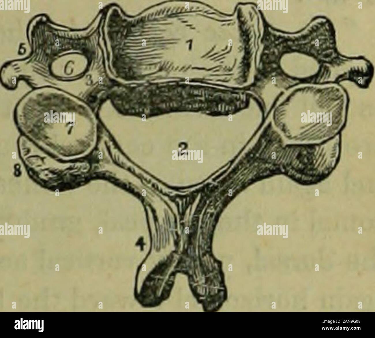 The hydropathic encyclopedia: a system of hydropathy and hygiene .. . al notch. 4. Spinousprocess, its extremity bifur-cated. 5. Transverse pro-cess. 6. Vertebral foramen.7. Superior articular pro-cess. 8. Inferior articularprocess. The first cervical vertebra supports the head,from which circumstance it is called the atlas.It is a simple ring of bone, and moves laterally, aswell as forward and backward to some extent onthe second cervical, which is called the axis. The axis has a large body, and a strong, tooth-7 like process, called odontoid, which rises perpen-dicularly, and is articulated Stock Photohttps://www.alamy.com/image-license-details/?v=1https://www.alamy.com/the-hydropathic-encyclopedia-a-system-of-hydropathy-and-hygiene-al-notch-4-spinousprocess-its-extremity-bifur-cated-5-transverse-pro-cess-6-vertebral-foramen7-superior-articular-pro-cess-8-inferior-articularprocess-the-first-cervical-vertebra-supports-the-headfrom-which-circumstance-it-is-called-the-atlasit-is-a-simple-ring-of-bone-and-moves-laterally-aswell-as-forward-and-backward-to-some-extent-onthe-second-cervical-which-is-called-the-axis-the-axis-has-a-large-body-and-a-strong-tooth-7-like-process-called-odontoid-which-rises-perpen-dicularly-and-is-articulated-image340114888.html
RM2AN9G08–The hydropathic encyclopedia: a system of hydropathy and hygiene .. . al notch. 4. Spinousprocess, its extremity bifur-cated. 5. Transverse pro-cess. 6. Vertebral foramen.7. Superior articular pro-cess. 8. Inferior articularprocess. The first cervical vertebra supports the head,from which circumstance it is called the atlas.It is a simple ring of bone, and moves laterally, aswell as forward and backward to some extent onthe second cervical, which is called the axis. The axis has a large body, and a strong, tooth-7 like process, called odontoid, which rises perpen-dicularly, and is articulated
 Anatomy, physiology and hygiene for high schools . ull, atlas, and axis are bound together by ligamentsin a set of loose joints which allow the free motion of thehead. The skull moves back-ward and forward upon itsjoints with the atlas, the atlasbeing held firm on the axis.When the head moves around?the atlas moves w^th it, rotat-ing upon the pivot process ofthe atlas. Two ligaments fromthe odontoid process to theskull check this rotary mo-tion. The sacnun lies at the baseof the column. Its five verte-brae are grown together into one bone in the adult. Itsupports the pelvic girdle. Below it ca Stock Photohttps://www.alamy.com/image-license-details/?v=1https://www.alamy.com/anatomy-physiology-and-hygiene-for-high-schools-ull-atlas-and-axis-are-bound-together-by-ligamentsin-a-set-of-loose-joints-which-allow-the-free-motion-of-thehead-the-skull-moves-back-ward-and-forward-upon-itsjoints-with-the-atlas-the-atlasbeing-held-firm-on-the-axiswhen-the-head-moves-aroundthe-atlas-moves-wth-it-rotat-ing-upon-the-pivot-process-ofthe-atlas-two-ligaments-fromthe-odontoid-process-to-theskull-check-this-rotary-mo-tion-the-sacnun-lies-at-the-baseof-the-column-its-five-verte-brae-are-grown-together-into-one-bone-in-the-adult-itsupports-the-pelvic-girdle-below-it-ca-image340052162.html
RM2AN6M02–Anatomy, physiology and hygiene for high schools . ull, atlas, and axis are bound together by ligamentsin a set of loose joints which allow the free motion of thehead. The skull moves back-ward and forward upon itsjoints with the atlas, the atlasbeing held firm on the axis.When the head moves around?the atlas moves w^th it, rotat-ing upon the pivot process ofthe atlas. Two ligaments fromthe odontoid process to theskull check this rotary mo-tion. The sacnun lies at the baseof the column. Its five verte-brae are grown together into one bone in the adult. Itsupports the pelvic girdle. Below it ca
 The treatment of fractures . i y mm 4. m Fig. 83.—Lesion of spine between sixthand seventh cervical vertebrae. Position incase of complete transverse destruction ofthe cord just below nuclei for subscapularis ;areas of anesthesia shown (after Thor-burn). Fig. 84.—Atlas, axis, and third cervicalvertebra from the front. Case: man, thirty-eight years of age ; fell from a cart. Frac-ture of odontoid process. Slight hemor-rhage into the medulla. Death after forty-eight hours (Cabot). figure 82. The characteristic attitude in lesions between thesixth and seventh cervical vertebrae is also shown in Stock Photohttps://www.alamy.com/image-license-details/?v=1https://www.alamy.com/the-treatment-of-fractures-i-y-mm-4-m-fig-83lesion-of-spine-between-sixthand-seventh-cervical-vertebrae-position-incase-of-complete-transverse-destruction-ofthe-cord-just-below-nuclei-for-subscapularis-areas-of-anesthesia-shown-after-thor-burn-fig-84atlas-axis-and-third-cervicalvertebra-from-the-front-case-man-thirty-eight-years-of-age-fell-from-a-cart-frac-ture-of-odontoid-process-slight-hemor-rhage-into-the-medulla-death-after-forty-eight-hours-cabot-figure-82-the-characteristic-attitude-in-lesions-between-thesixth-and-seventh-cervical-vertebrae-is-also-shown-in-image340047850.html
RM2AN6EE2–The treatment of fractures . i y mm 4. m Fig. 83.—Lesion of spine between sixthand seventh cervical vertebrae. Position incase of complete transverse destruction ofthe cord just below nuclei for subscapularis ;areas of anesthesia shown (after Thor-burn). Fig. 84.—Atlas, axis, and third cervicalvertebra from the front. Case: man, thirty-eight years of age ; fell from a cart. Frac-ture of odontoid process. Slight hemor-rhage into the medulla. Death after forty-eight hours (Cabot). figure 82. The characteristic attitude in lesions between thesixth and seventh cervical vertebrae is also shown in
 Bones, molars, and briefs . .Vdvt—Frederick Express Co 108 Clubs ... 10.) North Carolina CUib, (Photo).. ... 110 Page. IVniisylvania Dental (71nb. (Photo) 112 Smith Carolin.i Club. (Photo) 114 West irginia Chil). (Photo) 116 Bugicide 122 Xaini Cairan 124 (iirl With Feet 125 Knights of Rest 127 llE.NT.M.. Deiu.il 1 )ii)anini-iii. igo.? 131 Dental Facidty. ( Photo) 132 .? Odontoid Congratulation 13Q Class Officers. 1903 140 Class Nfembers. iqo3 142 to 144 The Whisper of Cnpid 146 ••. Xight Oflf 158 Prof James II. Harris 160 IVeshnian .rrival. Senior Departure 167 Jnnior Class .Members. ( Phot Stock Photohttps://www.alamy.com/image-license-details/?v=1https://www.alamy.com/bones-molars-and-briefs-vdvtfrederick-express-co-108-clubs-10-north-carolina-cuib-photo-110-page-ivniisylvania-dental-71nb-photo-112-smith-carolini-club-photo-114-west-irginia-chil-photo-116-bugicide-122-xaini-cairan-124-iirl-with-feet-125-knights-of-rest-127-llentm-deiuil-1-iianini-iii-igo-131-dental-facidty-photo-132-odontoid-congratulation-13q-class-officers-1903-140-class-nfembers-iqo3-142-to-144-the-whisper-of-cnpid-146-xight-oflf-158-prof-james-ii-harris-160-iveshnian-rrival-senior-departure-167-jnnior-class-members-phot-image342820421.html
RM2AWMPXD–Bones, molars, and briefs . .Vdvt—Frederick Express Co 108 Clubs ... 10.) North Carolina CUib, (Photo).. ... 110 Page. IVniisylvania Dental (71nb. (Photo) 112 Smith Carolin.i Club. (Photo) 114 West irginia Chil). (Photo) 116 Bugicide 122 Xaini Cairan 124 (iirl With Feet 125 Knights of Rest 127 llE.NT.M.. Deiu.il 1 )ii)anini-iii. igo.? 131 Dental Facidty. ( Photo) 132 .? Odontoid Congratulation 13Q Class Officers. 1903 140 Class Nfembers. iqo3 142 to 144 The Whisper of Cnpid 146 ••. Xight Oflf 158 Prof James II. Harris 160 IVeshnian .rrival. Senior Departure 167 Jnnior Class .Members. ( Phot
 . The structure and classification of birds . Fia. 66.—Atlas ofEmu (aftek Mivabt). ac, articular surface;V, vertebrarterialcanal; hp, hyper-apophyses.. Fio. 67.—Axis of Emu (aftkkMivakt). 0, odontoid process ; nsy neural spine ;az, anterior zygapoptiyses; pi,pleural lamella; pc, articular sur-face ; hif, hypapophysis ; hp, hyper-apophysis. Stock Photohttps://www.alamy.com/image-license-details/?v=1https://www.alamy.com/the-structure-and-classification-of-birds-fia-66atlas-ofemu-aftek-mivabt-ac-articular-surfacev-vertebrarterialcanal-hp-hyper-apophyses-fio-67axis-of-emu-aftkkmivakt-0-odontoid-process-nsy-neural-spine-az-anterior-zygapoptiyses-pipleural-lamella-pc-articular-sur-face-hif-hypapophysis-hp-hyper-apophysis-image375198076.html
RM2CPBMY8–. The structure and classification of birds . Fia. 66.—Atlas ofEmu (aftek Mivabt). ac, articular surface;V, vertebrarterialcanal; hp, hyper-apophyses.. Fio. 67.—Axis of Emu (aftkkMivakt). 0, odontoid process ; nsy neural spine ;az, anterior zygapoptiyses; pi,pleural lamella; pc, articular sur-face ; hif, hypapophysis ; hp, hyper-apophysis.
 . Human physiology. Fig. 42.—Section through theFivot Joint formed by theAtlas and the Axis. 1, section through the odontoid peg,showing synovial cavities before andbehind ; 2, cut portion of the atlas :3, the transverse ligament whichholds the peg ; 4, surface of the atlaswhich articulates ivith the skull.. Fig. 43.—A Lumbar Vertebra, Viewed from above, showing the inter-vertebral disc of cartilage. Stock Photohttps://www.alamy.com/image-license-details/?v=1https://www.alamy.com/human-physiology-fig-42section-through-thefivot-joint-formed-by-theatlas-and-the-axis-1-section-through-the-odontoid-pegshowing-synovial-cavities-before-andbehind-2-cut-portion-of-the-atlas-3-the-transverse-ligament-whichholds-the-peg-4-surface-of-the-atlaswhich-articulates-ivith-the-skull-fig-43a-lumbar-vertebra-viewed-from-above-showing-the-inter-vertebral-disc-of-cartilage-image370648987.html
RM2CF0EFR–. Human physiology. Fig. 42.—Section through theFivot Joint formed by theAtlas and the Axis. 1, section through the odontoid peg,showing synovial cavities before andbehind ; 2, cut portion of the atlas :3, the transverse ligament whichholds the peg ; 4, surface of the atlaswhich articulates ivith the skull.. Fig. 43.—A Lumbar Vertebra, Viewed from above, showing the inter-vertebral disc of cartilage.
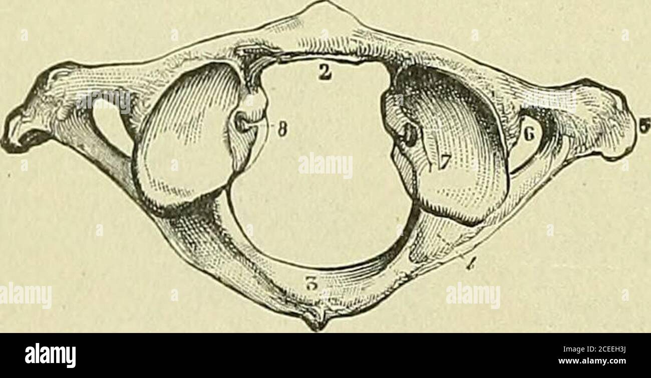 . Text-book of anatomy and physiology for nurses. Pedicle Fig. 31.—Cervical Vertebea, Showing Bifid Spinous Process.—(Morris.). Fig. 32.—Atlas, Superior Surface.I, Tubercle of anterior arch; 2,articular facet for odontoid processof axis; 3, posterior arch and posteriortubercle; 4, groove for vertebral arteryand first cervical nerve; 5, transverseprocess; 6, transverse foramen; 7,superior articular process; 8, tuberclefor attachment of transverse ligament.—(Goulds Dictionary.) Stock Photohttps://www.alamy.com/image-license-details/?v=1https://www.alamy.com/text-book-of-anatomy-and-physiology-for-nurses-pedicle-fig-31cervical-vertebea-showing-bifid-spinous-processmorris-fig-32atlas-superior-surfacei-tubercle-of-anterior-arch-2articular-facet-for-odontoid-processof-axis-3-posterior-arch-and-posteriortubercle-4-groove-for-vertebral-arteryand-first-cervical-nerve-5-transverseprocess-6-transverse-foramen-7superior-articular-process-8-tuberclefor-attachment-of-transverse-ligamentgoulds-dictionary-image370343670.html
RM2CEEH3J–. Text-book of anatomy and physiology for nurses. Pedicle Fig. 31.—Cervical Vertebea, Showing Bifid Spinous Process.—(Morris.). Fig. 32.—Atlas, Superior Surface.I, Tubercle of anterior arch; 2,articular facet for odontoid processof axis; 3, posterior arch and posteriortubercle; 4, groove for vertebral arteryand first cervical nerve; 5, transverseprocess; 6, transverse foramen; 7,superior articular process; 8, tuberclefor attachment of transverse ligament.—(Goulds Dictionary.)
 . A dictionary of birds . 2 SKELETON The Second Cervical, known as the Axis or Epistropheus, as beingthe pivot on which the Atlas and Head turn, is composed of sevenseparate elements, the first of which is really the centrum of theAtlas, but fused with the second, the centrum of the Axis, so as toform the odontoid process. The thinl and fourth are the pair ofpieces which form the neural arcli, and generally bear a prominentspinous process. The fifth and sixth are a pair of rib-elements, eachof which is perforated by a transverse arterial foramen, and fuses withthe antero-lateral portion of the Stock Photohttps://www.alamy.com/image-license-details/?v=1https://www.alamy.com/a-dictionary-of-birds-2-skeleton-the-second-cervical-known-as-the-axis-or-epistropheus-as-beingthe-pivot-on-which-the-atlas-and-head-turn-is-composed-of-sevenseparate-elements-the-first-of-which-is-really-the-centrum-of-theatlas-but-fused-with-the-second-the-centrum-of-the-axis-so-as-toform-the-odontoid-process-the-thinl-and-fourth-are-the-pair-ofpieces-which-form-the-neural-arcli-and-generally-bear-a-prominentspinous-process-the-fifth-and-sixth-are-a-pair-of-rib-elements-eachof-which-is-perforated-by-a-transverse-arterial-foramen-and-fuses-withthe-antero-lateral-portion-of-the-image375007588.html
RM2CP3204–. A dictionary of birds . 2 SKELETON The Second Cervical, known as the Axis or Epistropheus, as beingthe pivot on which the Atlas and Head turn, is composed of sevenseparate elements, the first of which is really the centrum of theAtlas, but fused with the second, the centrum of the Axis, so as toform the odontoid process. The thinl and fourth are the pair ofpieces which form the neural arcli, and generally bear a prominentspinous process. The fifth and sixth are a pair of rib-elements, eachof which is perforated by a transverse arterial foramen, and fuses withthe antero-lateral portion of the
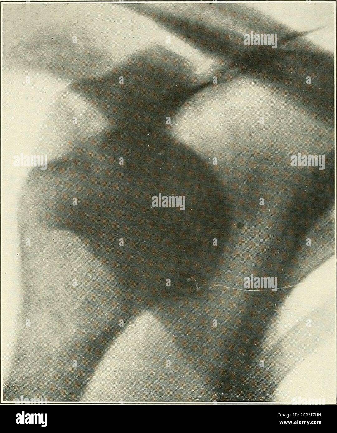 . Roentgen interpretation; a manual for students and practitioners . Fig. 22.—Double congenital dislocation of the hip. DISLOCATIONS. Dislocations of the spine are usually accompanied by fracture.They are most common in the cervical region. The first cervicalvertebra may be displaced backward on the second with fractureof the odontoid or, more rarely, rotated upon the second withoutfracture of the odontoid. The most frequent injury is a forwarddisplacement of the upper cervical vertebrae upon the ones belowin the region of the third to the seventh. 46 FRACTURES AND DISLOCATIONS. u,. --■;.- ni Stock Photohttps://www.alamy.com/image-license-details/?v=1https://www.alamy.com/roentgen-interpretation-a-manual-for-students-and-practitioners-fig-22double-congenital-dislocation-of-the-hip-dislocations-dislocations-of-the-spine-are-usually-accompanied-by-fracturethey-are-most-common-in-the-cervical-region-the-first-cervicalvertebra-may-be-displaced-backward-on-the-second-with-fractureof-the-odontoid-or-more-rarely-rotated-upon-the-second-withoutfracture-of-the-odontoid-the-most-frequent-injury-is-a-forwarddisplacement-of-the-upper-cervical-vertebrae-upon-the-ones-belowin-the-region-of-the-third-to-the-seventh-46-fractures-and-dislocations-u-ni-image375999841.html
RM2CRM7HN–. Roentgen interpretation; a manual for students and practitioners . Fig. 22.—Double congenital dislocation of the hip. DISLOCATIONS. Dislocations of the spine are usually accompanied by fracture.They are most common in the cervical region. The first cervicalvertebra may be displaced backward on the second with fractureof the odontoid or, more rarely, rotated upon the second withoutfracture of the odontoid. The most frequent injury is a forwarddisplacement of the upper cervical vertebrae upon the ones belowin the region of the third to the seventh. 46 FRACTURES AND DISLOCATIONS. u,. --■;.- ni
 . A dictionary of birds . SKELETON The Second Cervical, known as the Axis or Epistroiiheus, as beingthe pivot on which the Atlas and Head turn, is composed of sevenseparate elements, the first of which is really the centrum of theAtlas, but fused with the second, the centrum of the Axis, so as toform the odontoid process. The third and fourth are the pair ofpieces which form the neural arcli, and generally bear a prominentsjiinous process. The fifth and sixth are a pair of rib-elements, eachof which is perforated by a transverse arterial foramen, and fuses withthe antero-lateral portion of the Stock Photohttps://www.alamy.com/image-license-details/?v=1https://www.alamy.com/a-dictionary-of-birds-skeleton-the-second-cervical-known-as-the-axis-or-epistroiiheus-as-beingthe-pivot-on-which-the-atlas-and-head-turn-is-composed-of-sevenseparate-elements-the-first-of-which-is-really-the-centrum-of-theatlas-but-fused-with-the-second-the-centrum-of-the-axis-so-as-toform-the-odontoid-process-the-third-and-fourth-are-the-pair-ofpieces-which-form-the-neural-arcli-and-generally-bear-a-prominentsjiinous-process-the-fifth-and-sixth-are-a-pair-of-rib-elements-eachof-which-is-perforated-by-a-transverse-arterial-foramen-and-fuses-withthe-antero-lateral-portion-of-the-image375275548.html
RM2CPF7P4–. A dictionary of birds . SKELETON The Second Cervical, known as the Axis or Epistroiiheus, as beingthe pivot on which the Atlas and Head turn, is composed of sevenseparate elements, the first of which is really the centrum of theAtlas, but fused with the second, the centrum of the Axis, so as toform the odontoid process. The third and fourth are the pair ofpieces which form the neural arcli, and generally bear a prominentsjiinous process. The fifth and sixth are a pair of rib-elements, eachof which is perforated by a transverse arterial foramen, and fuses withthe antero-lateral portion of the
 . Quain's Elements of anatomy. It is attached on each side to the tuljercle below the inner border of the supeiiorarticular process. It is arched backwards behind the neck of the odontoid process,and is broadened out in its central part. From the middle of its posterior surfacea short thin Imndle of fibres passes down to be attached to the body of the axis,while another passes up to the basilar process. These form, with the transverseportion, the figure of a cross, and from this arrangement is derived the termcruciform, wliich is sometimes applied to the transverse ligament and its appendagest Stock Photohttps://www.alamy.com/image-license-details/?v=1https://www.alamy.com/quains-elements-of-anatomy-it-is-attached-on-each-side-to-the-tuljercle-below-the-inner-border-of-the-supeiiorarticular-process-it-is-arched-backwards-behind-the-neck-of-the-odontoid-processand-is-broadened-out-in-its-central-part-from-the-middle-of-its-posterior-surfacea-short-thin-imndle-of-fibres-passes-down-to-be-attached-to-the-body-of-the-axiswhile-another-passes-up-to-the-basilar-process-these-form-with-the-transverseportion-the-figure-of-a-cross-and-from-this-arrangement-is-derived-the-termcruciform-wliich-is-sometimes-applied-to-the-transverse-ligament-and-its-appendagest-image370773999.html
RM2CF660F–. Quain's Elements of anatomy. It is attached on each side to the tuljercle below the inner border of the supeiiorarticular process. It is arched backwards behind the neck of the odontoid process,and is broadened out in its central part. From the middle of its posterior surfacea short thin Imndle of fibres passes down to be attached to the body of the axis,while another passes up to the basilar process. These form, with the transverseportion, the figure of a cross, and from this arrangement is derived the termcruciform, wliich is sometimes applied to the transverse ligament and its appendagest
![. An introduction to the osteology of the mammalia . ghtlycurved bar of bone, with a smooth facet on its neural orupper surface, for articulation with the odontoid process ofthe axis. The second cervical vertebra, axis, epistrophens, or vertebradsntata (Fig. 10), has a body terminating anteriorly in alarge subconical median projection, the odontoid process(0), which is received into, and articulates with, the con-cavity of the inferior arch of the atlas. It is retained in IV.] MAN. 35 its place by means of a strong transverse ligament passingbetween the lateral masses of that bone, and separat Stock Photo . An introduction to the osteology of the mammalia . ghtlycurved bar of bone, with a smooth facet on its neural orupper surface, for articulation with the odontoid process ofthe axis. The second cervical vertebra, axis, epistrophens, or vertebradsntata (Fig. 10), has a body terminating anteriorly in alarge subconical median projection, the odontoid process(0), which is received into, and articulates with, the con-cavity of the inferior arch of the atlas. It is retained in IV.] MAN. 35 its place by means of a strong transverse ligament passingbetween the lateral masses of that bone, and separat Stock Photo](https://c8.alamy.com/comp/2CDFB61/an-introduction-to-the-osteology-of-the-mammalia-ghtlycurved-bar-of-bone-with-a-smooth-facet-on-its-neural-orupper-surface-for-articulation-with-the-odontoid-process-ofthe-axis-the-second-cervical-vertebra-axis-epistrophens-or-vertebradsntata-fig-10-has-a-body-terminating-anteriorly-in-alarge-subconical-median-projection-the-odontoid-process0-which-is-received-into-and-articulates-with-the-con-cavity-of-the-inferior-arch-of-the-atlas-it-is-retained-in-iv-man-35-its-place-by-means-of-a-strong-transverse-ligament-passingbetween-the-lateral-masses-of-that-bone-and-separat-2CDFB61.jpg) . An introduction to the osteology of the mammalia . ghtlycurved bar of bone, with a smooth facet on its neural orupper surface, for articulation with the odontoid process ofthe axis. The second cervical vertebra, axis, epistrophens, or vertebradsntata (Fig. 10), has a body terminating anteriorly in alarge subconical median projection, the odontoid process(0), which is received into, and articulates with, the con-cavity of the inferior arch of the atlas. It is retained in IV.] MAN. 35 its place by means of a strong transverse ligament passingbetween the lateral masses of that bone, and separat Stock Photohttps://www.alamy.com/image-license-details/?v=1https://www.alamy.com/an-introduction-to-the-osteology-of-the-mammalia-ghtlycurved-bar-of-bone-with-a-smooth-facet-on-its-neural-orupper-surface-for-articulation-with-the-odontoid-process-ofthe-axis-the-second-cervical-vertebra-axis-epistrophens-or-vertebradsntata-fig-10-has-a-body-terminating-anteriorly-in-alarge-subconical-median-projection-the-odontoid-process0-which-is-received-into-and-articulates-with-the-con-cavity-of-the-inferior-arch-of-the-atlas-it-is-retained-in-iv-man-35-its-place-by-means-of-a-strong-transverse-ligament-passingbetween-the-lateral-masses-of-that-bone-and-separat-image369746329.html
. An introduction to the osteology of the mammalia . ghtlycurved bar of bone, with a smooth facet on its neural orupper surface, for articulation with the odontoid process ofthe axis. The second cervical vertebra, axis, epistrophens, or vertebradsntata (Fig. 10), has a body terminating anteriorly in alarge subconical median projection, the odontoid process(0), which is received into, and articulates with, the con-cavity of the inferior arch of the atlas. It is retained in IV.] MAN. 35 its place by means of a strong transverse ligament passingbetween the lateral masses of that bone, and separat Stock Photohttps://www.alamy.com/image-license-details/?v=1https://www.alamy.com/an-introduction-to-the-osteology-of-the-mammalia-ghtlycurved-bar-of-bone-with-a-smooth-facet-on-its-neural-orupper-surface-for-articulation-with-the-odontoid-process-ofthe-axis-the-second-cervical-vertebra-axis-epistrophens-or-vertebradsntata-fig-10-has-a-body-terminating-anteriorly-in-alarge-subconical-median-projection-the-odontoid-process0-which-is-received-into-and-articulates-with-the-con-cavity-of-the-inferior-arch-of-the-atlas-it-is-retained-in-iv-man-35-its-place-by-means-of-a-strong-transverse-ligament-passingbetween-the-lateral-masses-of-that-bone-and-separat-image369746329.htmlRM2CDFB61–. An introduction to the osteology of the mammalia . ghtlycurved bar of bone, with a smooth facet on its neural orupper surface, for articulation with the odontoid process ofthe axis. The second cervical vertebra, axis, epistrophens, or vertebradsntata (Fig. 10), has a body terminating anteriorly in alarge subconical median projection, the odontoid process(0), which is received into, and articulates with, the con-cavity of the inferior arch of the atlas. It is retained in IV.] MAN. 35 its place by means of a strong transverse ligament passingbetween the lateral masses of that bone, and separat
 . The horse, its treatment in health and disease with a complete guide to breeding, training and management . 2. Supero-anterior foramen. 3. Postero-inferior foramen. 4. Surface for artJciilaiion w Inferior iiiberclespinons snrface.Spinal canal.. Fig. 2. ATLAS Untero-inferi Fig. 3. AXIS (side view) 1. Superior spinous process. 4. Odontoid process. 2. Intervertebral foramen. 5. Inferior spinous process. 3. Transverse process. 6. Posterior articular face of body. 7. Oblique process. Stock Photohttps://www.alamy.com/image-license-details/?v=1https://www.alamy.com/the-horse-its-treatment-in-health-and-disease-with-a-complete-guide-to-breeding-training-and-management-2-supero-anterior-foramen-3-postero-inferior-foramen-4-surface-for-artjciilaiion-w-inferior-iiiberclespinons-snrfacespinal-canal-fig-2-atlas-untero-inferi-fig-3-axis-side-view-1-superior-spinous-process-4-odontoid-process-2-intervertebral-foramen-5-inferior-spinous-process-3-transverse-process-6-posterior-articular-face-of-body-7-oblique-process-image370069488.html
RM2CE23BC–. The horse, its treatment in health and disease with a complete guide to breeding, training and management . 2. Supero-anterior foramen. 3. Postero-inferior foramen. 4. Surface for artJciilaiion w Inferior iiiberclespinons snrface.Spinal canal.. Fig. 2. ATLAS Untero-inferi Fig. 3. AXIS (side view) 1. Superior spinous process. 4. Odontoid process. 2. Intervertebral foramen. 5. Inferior spinous process. 3. Transverse process. 6. Posterior articular face of body. 7. Oblique process.
 . Quain's Elements of anatomy. passes ARTICULATIONS OF THE ATLAS, AXIS, AND OCCIPITAL BONE. 157 directly upwards from the summit of the odontoid process to the centre of theanterior margin of the foramen magnum. The central odontoid lirrament is developed around the notochord in the interval betweenthe basioccipital and first vertebral centrum, and may therefore be said to represent an inter-vertebral disc. The transverse ligament of the atlas and the lateral odontoid ligaments arederived from ligamenta conju<ralia costarum (p. 1.59). The posterior occipito-azial ligament is a strong wide b Stock Photohttps://www.alamy.com/image-license-details/?v=1https://www.alamy.com/quains-elements-of-anatomy-passes-articulations-of-the-atlas-axis-and-occipital-bone-157-directly-upwards-from-the-summit-of-the-odontoid-process-to-the-centre-of-theanterior-margin-of-the-foramen-magnum-the-central-odontoid-lirrament-is-developed-around-the-notochord-in-the-interval-betweenthe-basioccipital-and-first-vertebral-centrum-and-may-therefore-be-said-to-represent-an-inter-vertebral-disc-the-transverse-ligament-of-the-atlas-and-the-lateral-odontoid-ligaments-arederived-from-ligamenta-conjultralia-costarum-p-159-the-posterior-occipito-azial-ligament-is-a-strong-wide-b-image370773194.html
RM2CF64YP–. Quain's Elements of anatomy. passes ARTICULATIONS OF THE ATLAS, AXIS, AND OCCIPITAL BONE. 157 directly upwards from the summit of the odontoid process to the centre of theanterior margin of the foramen magnum. The central odontoid lirrament is developed around the notochord in the interval betweenthe basioccipital and first vertebral centrum, and may therefore be said to represent an inter-vertebral disc. The transverse ligament of the atlas and the lateral odontoid ligaments arederived from ligamenta conju<ralia costarum (p. 1.59). The posterior occipito-azial ligament is a strong wide b
 . Quain's Elements of anatomy. TRANSV. PROC. TRANSV. PROO.Mr. ARTIC. PROCESS Fig. 3.—Atlas and axis, froji before. (Drawn by D. Grunn.) responds to the foramina of the other vertebrjE ; its narrower anterior part isoccupied by the odontoid process of the axis, and in the recent state is separatedfrom the posterior by the transverse ligament. In front of the ring is the anterioiarch, on the anterior aspect of which is a small tubercle, and on the posterior asmooth surface for articulation with the odontoid process. At the sides of the ringare the lateral masses, which are thick and strong, Ijea Stock Photohttps://www.alamy.com/image-license-details/?v=1https://www.alamy.com/quains-elements-of-anatomy-transv-proc-transv-proomr-artic-process-fig-3atlas-and-axis-froji-before-drawn-by-d-grunn-responds-to-the-foramina-of-the-other-vertebrje-its-narrower-anterior-part-isoccupied-by-the-odontoid-process-of-the-axis-and-in-the-recent-state-is-separatedfrom-the-posterior-by-the-transverse-ligament-in-front-of-the-ring-is-the-anterioiarch-on-the-anterior-aspect-of-which-is-a-small-tubercle-and-on-the-posterior-asmooth-surface-for-articulation-with-the-odontoid-process-at-the-sides-of-the-ringare-the-lateral-masses-which-are-thick-and-strong-ijea-image370428827.html
RM2CEJDMY–. Quain's Elements of anatomy. TRANSV. PROC. TRANSV. PROO.Mr. ARTIC. PROCESS Fig. 3.—Atlas and axis, froji before. (Drawn by D. Grunn.) responds to the foramina of the other vertebrjE ; its narrower anterior part isoccupied by the odontoid process of the axis, and in the recent state is separatedfrom the posterior by the transverse ligament. In front of the ring is the anterioiarch, on the anterior aspect of which is a small tubercle, and on the posterior asmooth surface for articulation with the odontoid process. At the sides of the ringare the lateral masses, which are thick and strong, Ijea
 . The structure and classification of birds . FIG. 247.—PELVIS OF CASSOWARY (AFTER MIVAKT). pl, pelvic rib. Other letters as in fig. 243. for the odontoid process of the axis. The catapophyses ofthe seventeenth cervical unite. The sternum has a raised and flattened tract posteriorly,which may be the equivalent of the keel; it has two posteriorlateral processes, which extend beyond the median portion ofthe bone. 1 On the Axial Skeleton of the Ostrich, TV. Z. S. viii. p. 385. 518 STRUCTURE AND CLASSIFICATION OF BIRDS The pelvis is remarkable for the symphysis of the pubes,which is shown in the a Stock Photohttps://www.alamy.com/image-license-details/?v=1https://www.alamy.com/the-structure-and-classification-of-birds-fig-247pelvis-of-cassowary-after-mivakt-pl-pelvic-rib-other-letters-as-in-fig-243-for-the-odontoid-process-of-the-axis-the-catapophyses-ofthe-seventeenth-cervical-unite-the-sternum-has-a-raised-and-flattened-tract-posteriorlywhich-may-be-the-equivalent-of-the-keel-it-has-two-posteriorlateral-processes-which-extend-beyond-the-median-portion-ofthe-bone-1-on-the-axial-skeleton-of-the-ostrich-tv-z-s-viii-p-385-518-structure-and-classification-of-birds-the-pelvis-is-remarkable-for-the-symphysis-of-the-pubeswhich-is-shown-in-the-a-image375339702.html
RM2CPJ5HA–. The structure and classification of birds . FIG. 247.—PELVIS OF CASSOWARY (AFTER MIVAKT). pl, pelvic rib. Other letters as in fig. 243. for the odontoid process of the axis. The catapophyses ofthe seventeenth cervical unite. The sternum has a raised and flattened tract posteriorly,which may be the equivalent of the keel; it has two posteriorlateral processes, which extend beyond the median portion ofthe bone. 1 On the Axial Skeleton of the Ostrich, TV. Z. S. viii. p. 385. 518 STRUCTURE AND CLASSIFICATION OF BIRDS The pelvis is remarkable for the symphysis of the pubes,which is shown in the a
 . A practical treatise on fractures and dislocations . fter death by Dr. C. E.Isaacs,in presence of several medical gentlemen. Mus-cular development uncommonly fine. An un-usual prominence discovered in the region ofthe axis and atlas. On making an incisionfrom the occiput along the spines of the cervicalvertebras, the parts were found to be very vas-cular. These vertebras were removed en masse,and a careful examination instituted. Thetransverse, the odontoid (ligamenta modera-toria), as also all the ligaments of this region,excepting the occipito-axoideum, were in a stateof perfect integrity; Stock Photohttps://www.alamy.com/image-license-details/?v=1https://www.alamy.com/a-practical-treatise-on-fractures-and-dislocations-fter-death-by-dr-c-eisaacsin-presence-of-several-medical-gentlemen-mus-cular-development-uncommonly-fine-an-un-usual-prominence-discovered-in-the-region-ofthe-axis-and-atlas-on-making-an-incisionfrom-the-occiput-along-the-spines-of-the-cervicalvertebras-the-parts-were-found-to-be-very-vas-cular-these-vertebras-were-removed-en-masseand-a-careful-examination-instituted-thetransverse-the-odontoid-ligamenta-modera-toria-as-also-all-the-ligaments-of-this-regionexcepting-the-occipito-axoideum-were-in-a-stateof-perfect-integrity-image370025638.html
RM2CE03DA–. A practical treatise on fractures and dislocations . fter death by Dr. C. E.Isaacs,in presence of several medical gentlemen. Mus-cular development uncommonly fine. An un-usual prominence discovered in the region ofthe axis and atlas. On making an incisionfrom the occiput along the spines of the cervicalvertebras, the parts were found to be very vas-cular. These vertebras were removed en masse,and a careful examination instituted. Thetransverse, the odontoid (ligamenta modera-toria), as also all the ligaments of this region,excepting the occipito-axoideum, were in a stateof perfect integrity;
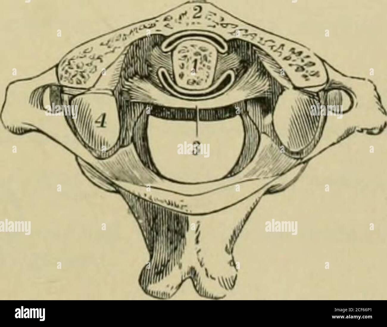 . Quain's Elements of anatomy. Fig. 180.—Horizontal section through the odonto-ATLANTAL ARTICULATION, (Allen Thomson.) ^ 1, cut surface of the odontoid process ; 2, cut surface of theanterior arch of the atlas ; 3, transverse ligament; between 1and 2, the anterior synovial cavity, and between 1 and 3, theposterior synovial cavity of the ai-ticuhition ; 4, is placed on theback part of the left superior articular jji-oce-ss of the atlas ;the anterior part, has been partly removed by the section. Forthe sake of distinctness, the .synovial spaces are representedwider than natural.. It is attached Stock Photohttps://www.alamy.com/image-license-details/?v=1https://www.alamy.com/quains-elements-of-anatomy-fig-180horizontal-section-through-the-odonto-atlantal-articulation-allen-thomson-1-cut-surface-of-the-odontoid-process-2-cut-surface-of-theanterior-arch-of-the-atlas-3-transverse-ligament-between-1and-2-the-anterior-synovial-cavity-and-between-1-and-3-theposterior-synovial-cavity-of-the-ai-ticuhition-4-is-placed-on-theback-part-of-the-left-superior-articular-jji-oce-ss-of-the-atlas-the-anterior-part-has-been-partly-removed-by-the-section-forthe-sake-of-distinctness-the-synovial-spaces-are-representedwider-than-natural-it-is-attached-image370774601.html
RM2CF66P1–. Quain's Elements of anatomy. Fig. 180.—Horizontal section through the odonto-ATLANTAL ARTICULATION, (Allen Thomson.) ^ 1, cut surface of the odontoid process ; 2, cut surface of theanterior arch of the atlas ; 3, transverse ligament; between 1and 2, the anterior synovial cavity, and between 1 and 3, theposterior synovial cavity of the ai-ticuhition ; 4, is placed on theback part of the left superior articular jji-oce-ss of the atlas ;the anterior part, has been partly removed by the section. Forthe sake of distinctness, the .synovial spaces are representedwider than natural.. It is attached
 . Quain's Elements of anatomy. of the body resembles that of the succeeding vertebrge. Its anterior surfaceis marked by a low median vertical ridge, with a depression on each side. The superior «rf/f«/«r surfaces, placed like those of the atlas in front of the notch,lie close to the base of the odontoid process, partly on the body and partly on thepedicles of the vertebra. These surfaces look upwards and slightly outwards. Theinferior articular processes are similar in form and position to those of the succeedingvertebrae. The spinous process is very large, rough, deeply bifid, and grooved Stock Photohttps://www.alamy.com/image-license-details/?v=1https://www.alamy.com/quains-elements-of-anatomy-of-the-body-resembles-that-of-the-succeeding-vertebrge-its-anterior-surfaceis-marked-by-a-low-median-vertical-ridge-with-a-depression-on-each-side-the-superior-rffr-surfaces-placed-like-those-of-the-atlas-in-front-of-the-notchlie-close-to-the-base-of-the-odontoid-process-partly-on-the-body-and-partly-on-thepedicles-of-the-vertebra-these-surfaces-look-upwards-and-slightly-outwards-theinferior-articular-processes-are-similar-in-form-and-position-to-those-of-the-succeedingvertebrae-the-spinous-process-is-very-large-rough-deeply-bifid-and-grooved-image370426766.html
RM2CEJB3A–. Quain's Elements of anatomy. of the body resembles that of the succeeding vertebrge. Its anterior surfaceis marked by a low median vertical ridge, with a depression on each side. The superior «rf/f«/«r surfaces, placed like those of the atlas in front of the notch,lie close to the base of the odontoid process, partly on the body and partly on thepedicles of the vertebra. These surfaces look upwards and slightly outwards. Theinferior articular processes are similar in form and position to those of the succeedingvertebrae. The spinous process is very large, rough, deeply bifid, and grooved
 . The structure and classification of birds . Fio. 247.—Pelvis op Cassowaey (after Mivaet).pl, pelvic rib. Other letters as in fig. 243. for the odontoid process of the axis. The catapophyses ofthe seventeenth cervical unite. The sternum has a raised and flattened tract posteriorly,which may be the equivalent of the keel; it has two posteriorlateral processes, which extend beyond the median portion ofthe bone. *- i On the Axial Skeleton of the Ostrich, Tr. Z, S. viii. p. 385. 618 STRUCTURE AND CLASSIFICATION OF BIRDS The pelvis is remarkable for the symphysis of the pubes,which is shown in the Stock Photohttps://www.alamy.com/image-license-details/?v=1https://www.alamy.com/the-structure-and-classification-of-birds-fio-247pelvis-op-cassowaey-after-mivaetpl-pelvic-rib-other-letters-as-in-fig-243-for-the-odontoid-process-of-the-axis-the-catapophyses-ofthe-seventeenth-cervical-unite-the-sternum-has-a-raised-and-flattened-tract-posteriorlywhich-may-be-the-equivalent-of-the-keel-it-has-two-posteriorlateral-processes-which-extend-beyond-the-median-portion-ofthe-bone-i-on-the-axial-skeleton-of-the-ostrich-tr-z-s-viii-p-385-618-structure-and-classification-of-birds-the-pelvis-is-remarkable-for-the-symphysis-of-the-pubeswhich-is-shown-in-the-image375178897.html
RM2CPATE9–. The structure and classification of birds . Fio. 247.—Pelvis op Cassowaey (after Mivaet).pl, pelvic rib. Other letters as in fig. 243. for the odontoid process of the axis. The catapophyses ofthe seventeenth cervical unite. The sternum has a raised and flattened tract posteriorly,which may be the equivalent of the keel; it has two posteriorlateral processes, which extend beyond the median portion ofthe bone. *- i On the Axial Skeleton of the Ostrich, Tr. Z, S. viii. p. 385. 618 STRUCTURE AND CLASSIFICATION OF BIRDS The pelvis is remarkable for the symphysis of the pubes,which is shown in the
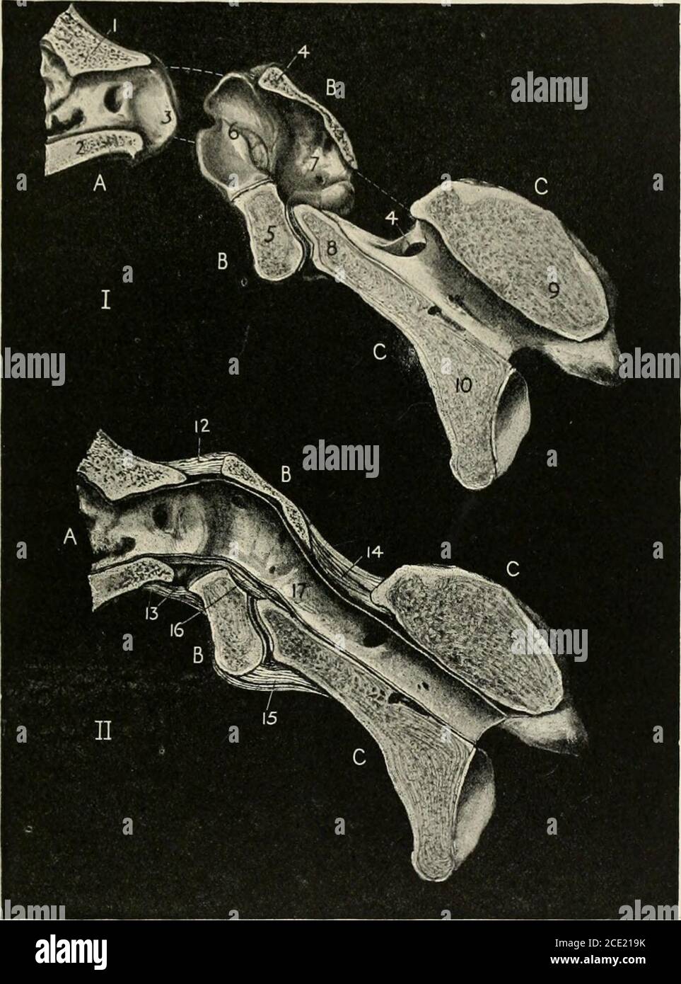 . The horse, its treatment in health and disease with a complete guide to breeding, training and management . Fig. 350.—Occipito-Atloid Articulation 1, Occipital Bone. 2, Atlas (the upper wall removed to show the odontoid ligament). 3, Axisor Dentata. 4, Third cervical vertebra. 5, Capsular ligament. 6, Odontoid ligament. 7, Fibrouscapsule (partly removed). 8, Interspinous ligament. 9, Fibrous capsule, uniting the articularprocesses of the vertebree. odontoid process of the axis into the ring of the atlas, where it is retainedby the odontoid ligament (fig. 351). Other ligaments, the superior a Stock Photohttps://www.alamy.com/image-license-details/?v=1https://www.alamy.com/the-horse-its-treatment-in-health-and-disease-with-a-complete-guide-to-breeding-training-and-management-fig-350occipito-atloid-articulation-1-occipital-bone-2-atlas-the-upper-wall-removed-to-show-the-odontoid-ligament-3-axisor-dentata-4-third-cervical-vertebra-5-capsular-ligament-6-odontoid-ligament-7-fibrouscapsule-partly-removed-8-interspinous-ligament-9-fibrous-capsule-uniting-the-articularprocesses-of-the-vertebree-odontoid-process-of-the-axis-into-the-ring-of-the-atlas-where-it-is-retainedby-the-odontoid-ligament-fig-351-other-ligaments-the-superior-a-image370067871.html
RM2CE219K–. The horse, its treatment in health and disease with a complete guide to breeding, training and management . Fig. 350.—Occipito-Atloid Articulation 1, Occipital Bone. 2, Atlas (the upper wall removed to show the odontoid ligament). 3, Axisor Dentata. 4, Third cervical vertebra. 5, Capsular ligament. 6, Odontoid ligament. 7, Fibrouscapsule (partly removed). 8, Interspinous ligament. 9, Fibrous capsule, uniting the articularprocesses of the vertebree. odontoid process of the axis into the ring of the atlas, where it is retainedby the odontoid ligament (fig. 351). Other ligaments, the superior a
 . The structure and classification of birds . Fia. 245.—Pelvis of Emu (aftek Mivaet). Lettebs as in Fio. 243. closing of the notch for the odontoid process may be com-pleted, as is shown in the cut (fig. 68, p. US). The shouldergirdle is very Hke that of the emu, possessing also rudimen-tary clavicles. The membrana coracoidea may, however,be ossified, and there are two foramina. One of these liesbetween the membrana coracoidea and the coracoid, andis therefore apparently the homologue of the foramen in 1 W. H. Flowee, On the Skeleton of the AustraUan Cassowary, P. Z. S.1871, p. 32. L L. 2 616 Stock Photohttps://www.alamy.com/image-license-details/?v=1https://www.alamy.com/the-structure-and-classification-of-birds-fia-245pelvis-of-emu-aftek-mivaet-lettebs-as-in-fio-243-closing-of-the-notch-for-the-odontoid-process-may-be-com-pleted-as-is-shown-in-the-cut-fig-68-p-us-the-shouldergirdle-is-very-hke-that-of-the-emu-possessing-also-rudimen-tary-clavicles-the-membrana-coracoidea-may-howeverbe-ossified-and-there-are-two-foramina-one-of-these-liesbetween-the-membrana-coracoidea-and-the-coracoid-andis-therefore-apparently-the-homologue-of-the-foramen-in-1-w-h-flowee-on-the-skeleton-of-the-austrauan-cassowary-p-z-s1871-p-32-l-l-2-616-image375179299.html
RM2CPAW0K–. The structure and classification of birds . Fia. 245.—Pelvis of Emu (aftek Mivaet). Lettebs as in Fio. 243. closing of the notch for the odontoid process may be com-pleted, as is shown in the cut (fig. 68, p. US). The shouldergirdle is very Hke that of the emu, possessing also rudimen-tary clavicles. The membrana coracoidea may, however,be ossified, and there are two foramina. One of these liesbetween the membrana coracoidea and the coracoid, andis therefore apparently the homologue of the foramen in 1 W. H. Flowee, On the Skeleton of the AustraUan Cassowary, P. Z. S.1871, p. 32. L L. 2 616
 . Radiography and radio-therapeutics . FRACTURES OF THE VERTEBRAE 241. Fici. 213.— Fracturf of base of odontoid process. Radio-gram obtained by use of extension tube centred overthe nioutb. Fractures of the Nasal Bones.—These are occasionally fractured on one or both sides. A plate on the injured side is generally sufficient to show the injury. An antero-posterior view is also useful. Stereoscopic pictures may be necessary. A small piece of X-ray film placed in contact with the side of the nose will give a sharp picture. Injuries of the Cervical Vertebrae.—Two positions have already been descr Stock Photohttps://www.alamy.com/image-license-details/?v=1https://www.alamy.com/radiography-and-radio-therapeutics-fractures-of-the-vertebrae-241-fici-213-fracturf-of-base-of-odontoid-process-radio-gram-obtained-by-use-of-extension-tube-centred-overthe-nioutb-fractures-of-the-nasal-bonesthese-are-occasionally-fractured-on-one-or-both-sides-a-plate-on-the-injured-side-is-generally-sufficient-to-show-the-injury-an-antero-posterior-view-is-also-useful-stereoscopic-pictures-may-be-necessary-a-small-piece-of-x-ray-film-placed-in-contact-with-the-side-of-the-nose-will-give-a-sharp-picture-injuries-of-the-cervical-vertebraetwo-positions-have-already-been-descr-image376015846.html
RM2CRN01A–. Radiography and radio-therapeutics . FRACTURES OF THE VERTEBRAE 241. Fici. 213.— Fracturf of base of odontoid process. Radio-gram obtained by use of extension tube centred overthe nioutb. Fractures of the Nasal Bones.—These are occasionally fractured on one or both sides. A plate on the injured side is generally sufficient to show the injury. An antero-posterior view is also useful. Stereoscopic pictures may be necessary. A small piece of X-ray film placed in contact with the side of the nose will give a sharp picture. Injuries of the Cervical Vertebrae.—Two positions have already been descr
 . Radiography and radio-therapeutics . FRACTURES OF THE VERTEBRA 241. Fig. 213.—Fracture of base of odontoid process. Radio-gram obtained by use of extension tube centred overthe mouth. Fractures of the Nasal Bones.—These are occasionally fractured on one or both sides. A plate on the injured side is generally sufficient to show the injury. An antero-posterior view is also useful. Stereoscopic pictures may be necessary. A small piece of X-ray film placed in contact with the side of the nose will give a sharp picture. Injuries of the Cervical Vertebrae.—Two positions have already been described Stock Photohttps://www.alamy.com/image-license-details/?v=1https://www.alamy.com/radiography-and-radio-therapeutics-fractures-of-the-vertebra-241-fig-213fracture-of-base-of-odontoid-process-radio-gram-obtained-by-use-of-extension-tube-centred-overthe-mouth-fractures-of-the-nasal-bonesthese-are-occasionally-fractured-on-one-or-both-sides-a-plate-on-the-injured-side-is-generally-sufficient-to-show-the-injury-an-antero-posterior-view-is-also-useful-stereoscopic-pictures-may-be-necessary-a-small-piece-of-x-ray-film-placed-in-contact-with-the-side-of-the-nose-will-give-a-sharp-picture-injuries-of-the-cervical-vertebraetwo-positions-have-already-been-described-image376025076.html
RM2CRNBR0–. Radiography and radio-therapeutics . FRACTURES OF THE VERTEBRA 241. Fig. 213.—Fracture of base of odontoid process. Radio-gram obtained by use of extension tube centred overthe mouth. Fractures of the Nasal Bones.—These are occasionally fractured on one or both sides. A plate on the injured side is generally sufficient to show the injury. An antero-posterior view is also useful. Stereoscopic pictures may be necessary. A small piece of X-ray film placed in contact with the side of the nose will give a sharp picture. Injuries of the Cervical Vertebrae.—Two positions have already been described
 . An introduction to the osteology of the mammalia . FIG. 13.—Inferior surface of atlas of Red Deer (Cervus etaphns). sn foramen forfirst spinal nerve; sii foramen for inferior branch of the same nerve. being like a spout, or hollow half-cylinder, with a prominentsharp semicircular rim. The canal for the second cervical. FIG. 14.—Anterior surface of axis of Red Deer. f. o odontoid process ; sn foramenfor second spinal nerve ; pz posterior zygapophysis. spinal nerve pierces the lamina of the axis near its anteriorborder. The other vertebrae have more or less elongatedbodies, which are opisthoc& Stock Photohttps://www.alamy.com/image-license-details/?v=1https://www.alamy.com/an-introduction-to-the-osteology-of-the-mammalia-fig-13inferior-surface-of-atlas-of-red-deer-cervus-etaphns-sn-foramen-forfirst-spinal-nerve-sii-foramen-for-inferior-branch-of-the-same-nerve-being-like-a-spout-or-hollow-half-cylinder-with-a-prominentsharp-semicircular-rim-the-canal-for-the-second-cervical-fig-14anterior-surface-of-axis-of-red-deer-f-o-odontoid-process-sn-foramenfor-second-spinal-nerve-pz-posterior-zygapophysis-spinal-nerve-pierces-the-lamina-of-the-axis-near-its-anteriorborder-the-other-vertebrae-have-more-or-less-elongatedbodies-which-are-opisthoc-image369746024.html
RM2CDFAR4–. An introduction to the osteology of the mammalia . FIG. 13.—Inferior surface of atlas of Red Deer (Cervus etaphns). sn foramen forfirst spinal nerve; sii foramen for inferior branch of the same nerve. being like a spout, or hollow half-cylinder, with a prominentsharp semicircular rim. The canal for the second cervical. FIG. 14.—Anterior surface of axis of Red Deer. f. o odontoid process ; sn foramenfor second spinal nerve ; pz posterior zygapophysis. spinal nerve pierces the lamina of the axis near its anteriorborder. The other vertebrae have more or less elongatedbodies, which are opisthoc&
 . The structure and classification of birds . FIG. 245.—PELVIS OF EMU (AFTER MIVART). LETTERS AS IN FIG. 243. closing of the notch for the odontoid process may be com-pleted, as is shown in the cut (fig. 68, p. 118). The shouldergirdle is very like that of the emu, possessing also rudimen-tary clavicles. The membrana coracoidea may, however,be ossified, and there are two foramina. One of these liesbetween the membrana coracoidea and the coracoid, andis therefore apparently the homologue of the foramen in 1 W. H. FLOWER, On the Skeleton of the Australian Cassowary, P. Z. S.1871, p. 32. L L 2 51 Stock Photohttps://www.alamy.com/image-license-details/?v=1https://www.alamy.com/the-structure-and-classification-of-birds-fig-245pelvis-of-emu-after-mivart-letters-as-in-fig-243-closing-of-the-notch-for-the-odontoid-process-may-be-com-pleted-as-is-shown-in-the-cut-fig-68-p-118-the-shouldergirdle-is-very-like-that-of-the-emu-possessing-also-rudimen-tary-clavicles-the-membrana-coracoidea-may-howeverbe-ossified-and-there-are-two-foramina-one-of-these-liesbetween-the-membrana-coracoidea-and-the-coracoid-andis-therefore-apparently-the-homologue-of-the-foramen-in-1-w-h-flower-on-the-skeleton-of-the-australian-cassowary-p-z-s1871-p-32-l-l-2-51-image375339761.html
RM2CPJ5KD–. The structure and classification of birds . FIG. 245.—PELVIS OF EMU (AFTER MIVART). LETTERS AS IN FIG. 243. closing of the notch for the odontoid process may be com-pleted, as is shown in the cut (fig. 68, p. 118). The shouldergirdle is very like that of the emu, possessing also rudimen-tary clavicles. The membrana coracoidea may, however,be ossified, and there are two foramina. One of these liesbetween the membrana coracoidea and the coracoid, andis therefore apparently the homologue of the foramen in 1 W. H. FLOWER, On the Skeleton of the Australian Cassowary, P. Z. S.1871, p. 32. L L 2 51
 . The horse, its treatment in health and disease with a complete guide to breeding, training and management . II. —Vertical section through the same bones in their natural position, showing the ligaments. 12, Superior occipito-atloid ligament. 13. Inferior occipito-atloid ligament. 1-i, Superior atlo-axoid ligament. 15, Inferioratlo-axoid ligament. 16, Odontoid ligament. 17, Spinal canal, with the dura mater in position. (The spinal cordhas been removed.) 270 HEALTH AND DISEASE SCAPULO-HUMERAL OR SHOULDER-JOINT The shoulder-joint results from the union of the glenoid or shallowcavity on the in Stock Photohttps://www.alamy.com/image-license-details/?v=1https://www.alamy.com/the-horse-its-treatment-in-health-and-disease-with-a-complete-guide-to-breeding-training-and-management-ii-vertical-section-through-the-same-bones-in-their-natural-position-showing-the-ligaments-12-superior-occipito-atloid-ligament-13-inferior-occipito-atloid-ligament-1-i-superior-atlo-axoid-ligament-15-inferioratlo-axoid-ligament-16-odontoid-ligament-17-spinal-canal-with-the-dura-mater-in-position-the-spinal-cordhas-been-removed-270-health-and-disease-scapulo-humeral-or-shoulder-joint-the-shoulder-joint-results-from-the-union-of-the-glenoid-or-shallowcavity-on-the-in-image370067882.html
RM2CE21A2–. The horse, its treatment in health and disease with a complete guide to breeding, training and management . II. —Vertical section through the same bones in their natural position, showing the ligaments. 12, Superior occipito-atloid ligament. 13. Inferior occipito-atloid ligament. 1-i, Superior atlo-axoid ligament. 15, Inferioratlo-axoid ligament. 16, Odontoid ligament. 17, Spinal canal, with the dura mater in position. (The spinal cordhas been removed.) 270 HEALTH AND DISEASE SCAPULO-HUMERAL OR SHOULDER-JOINT The shoulder-joint results from the union of the glenoid or shallowcavity on the in
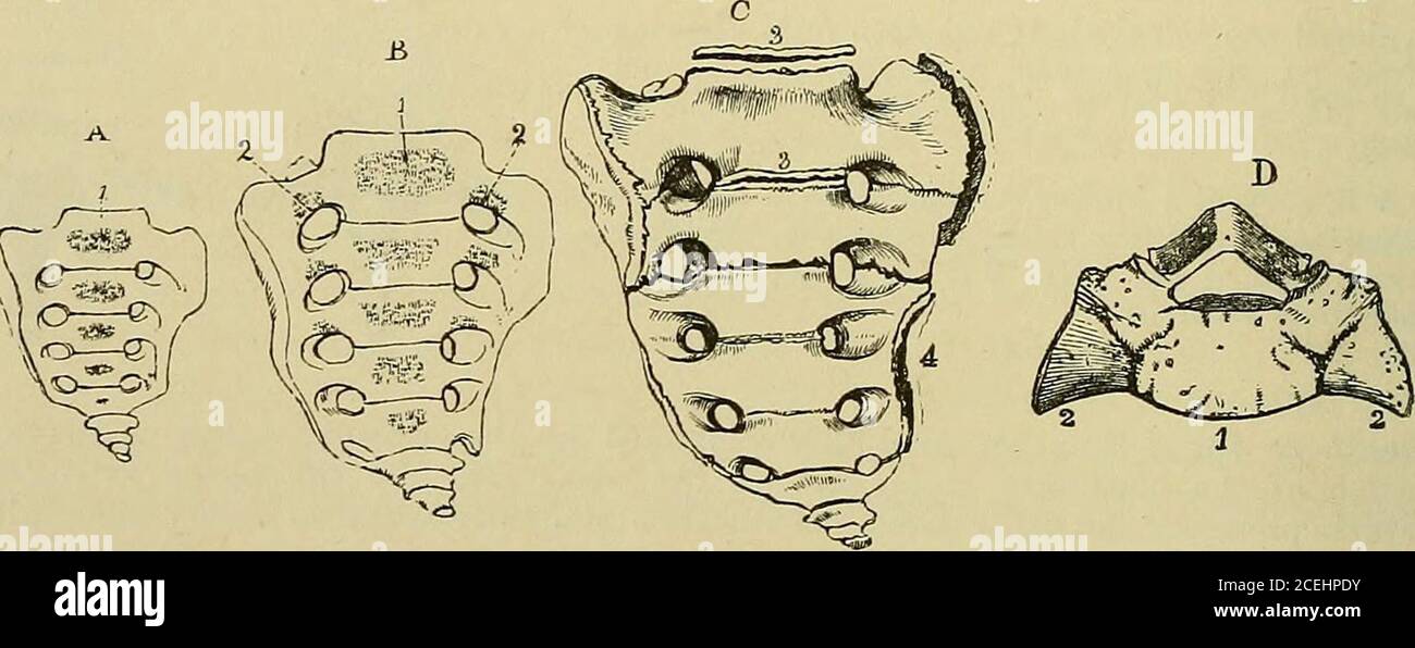 . Quain's Elements of anatomy. art of the common cartilage of the body and odontoid process. In the upper part of thiscartilage, a little later, two collateral centres appear for the odontoid process ; these soon uniteinto one, so that at birth the axis is composed of four pieces of bone. About the fourth yearthe odontoid process becomes joined to the body and the fore part of the neural arch of theaxis on each side, and a little later union occurs in front and behind. In the centre,however, a small disc of cartilage remains until advanced age.^ The apex of the odontoidprocess is formed from a Stock Photohttps://www.alamy.com/image-license-details/?v=1https://www.alamy.com/quains-elements-of-anatomy-art-of-the-common-cartilage-of-the-body-and-odontoid-process-in-the-upper-part-of-thiscartilage-a-little-later-two-collateral-centres-appear-for-the-odontoid-process-these-soon-uniteinto-one-so-that-at-birth-the-axis-is-composed-of-four-pieces-of-bone-about-the-fourth-yearthe-odontoid-process-becomes-joined-to-the-body-and-the-fore-part-of-the-neural-arch-of-theaxis-on-each-side-and-a-little-later-union-occurs-in-front-and-behind-in-the-centrehowever-a-small-disc-of-cartilage-remains-until-advanced-age-the-apex-of-the-odontoidprocess-is-formed-from-a-image370413735.html
RM2CEHPDY–. Quain's Elements of anatomy. art of the common cartilage of the body and odontoid process. In the upper part of thiscartilage, a little later, two collateral centres appear for the odontoid process ; these soon uniteinto one, so that at birth the axis is composed of four pieces of bone. About the fourth yearthe odontoid process becomes joined to the body and the fore part of the neural arch of theaxis on each side, and a little later union occurs in front and behind. In the centre,however, a small disc of cartilage remains until advanced age.^ The apex of the odontoidprocess is formed from a
 . An introduction to the osteology of the mammalia . 2. sn foramen for first spinal nerve. and lower bars, widely separated at their bases, but united attheir extremities so as to enclose a very large space betweenthem. In the seventh the upper process only exists, and the. FIG. 17.—Anterior surface of axis of common Fin Whale (Balcejioptera musculus), i . o odontoid process. lower one is occasionally imperfect in the sixth.1 In veryyoung animals these processes are formed only of cartilage • 1 Professor Turner has shown that, in a foetal Balcenoptera sibbaldii,the inferior transverse process Stock Photohttps://www.alamy.com/image-license-details/?v=1https://www.alamy.com/an-introduction-to-the-osteology-of-the-mammalia-2-sn-foramen-for-first-spinal-nerve-and-lower-bars-widely-separated-at-their-bases-but-united-attheir-extremities-so-as-to-enclose-a-very-large-space-betweenthem-in-the-seventh-the-upper-process-only-exists-and-the-fig-17anterior-surface-of-axis-of-common-fin-whale-balcejioptera-musculus-i-o-odontoid-process-lower-one-is-occasionally-imperfect-in-the-sixth1-in-veryyoung-animals-these-processes-are-formed-only-of-cartilage-1-professor-turner-has-shown-that-in-a-foetal-balcenoptera-sibbaldiithe-inferior-transverse-process-image369745700.html
RM2CDFABG–. An introduction to the osteology of the mammalia . 2. sn foramen for first spinal nerve. and lower bars, widely separated at their bases, but united attheir extremities so as to enclose a very large space betweenthem. In the seventh the upper process only exists, and the. FIG. 17.—Anterior surface of axis of common Fin Whale (Balcejioptera musculus), i . o odontoid process. lower one is occasionally imperfect in the sixth.1 In veryyoung animals these processes are formed only of cartilage • 1 Professor Turner has shown that, in a foetal Balcenoptera sibbaldii,the inferior transverse process
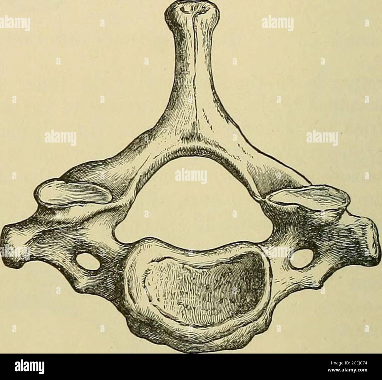 . Quain's Elements of anatomy. Fig. 5.—Axis, from the right side.by D. Gunn.) (Drawn Fig. 6.- -Seventh cervical aertebra, fromABOVE. (Drawn by D. Gunn.) odontoid (proc. dentatus). This consists of an enlarged part termed the head, and alower part or neck. It has in front a smooth surface for articulation with theatlas, and behind a smooth groove to receive the transverse hgament. The lowersurface of the body resembles that of the succeeding vertebrge. Its anterior surfaceis marked by a low median vertical ridge, with a depression on each side. The superior «rf/f«/«r surfaces, placed like those Stock Photohttps://www.alamy.com/image-license-details/?v=1https://www.alamy.com/quains-elements-of-anatomy-fig-5axis-from-the-right-sideby-d-gunn-drawn-fig-6-seventh-cervical-aertebra-fromabove-drawn-by-d-gunn-odontoid-proc-dentatus-this-consists-of-an-enlarged-part-termed-the-head-and-alower-part-or-neck-it-has-in-front-a-smooth-surface-for-articulation-with-theatlas-and-behind-a-smooth-groove-to-receive-the-transverse-hgament-the-lowersurface-of-the-body-resembles-that-of-the-succeeding-vertebrge-its-anterior-surfaceis-marked-by-a-low-median-vertical-ridge-with-a-depression-on-each-side-the-superior-rffr-surfaces-placed-like-those-image370427656.html
RM2CEJC74–. Quain's Elements of anatomy. Fig. 5.—Axis, from the right side.by D. Gunn.) (Drawn Fig. 6.- -Seventh cervical aertebra, fromABOVE. (Drawn by D. Gunn.) odontoid (proc. dentatus). This consists of an enlarged part termed the head, and alower part or neck. It has in front a smooth surface for articulation with theatlas, and behind a smooth groove to receive the transverse hgament. The lowersurface of the body resembles that of the succeeding vertebrge. Its anterior surfaceis marked by a low median vertical ridge, with a depression on each side. The superior «rf/f«/«r surfaces, placed like those
 . The horse, its treatment in health and disease with a complete guide to breeding, training and management . Fig. 2. ATLAS Untero-inferi Fig. 3. AXIS (side view) 1. Superior spinous process. 4. Odontoid process. 2. Intervertebral foramen. 5. Inferior spinous process. 3. Transverse process. 6. Posterior articular face of body. 7. Oblique process.. Fig. 4. DORSAL VERTEBRA (front viI. Superior spinous process. 2. Traiisveprocess. 3. Articulation for tubercle of 14 Articulation for head of rib. 5. Anteiarticular face of body. 6. Spinal c.inal. Stock Photohttps://www.alamy.com/image-license-details/?v=1https://www.alamy.com/the-horse-its-treatment-in-health-and-disease-with-a-complete-guide-to-breeding-training-and-management-fig-2-atlas-untero-inferi-fig-3-axis-side-view-1-superior-spinous-process-4-odontoid-process-2-intervertebral-foramen-5-inferior-spinous-process-3-transverse-process-6-posterior-articular-face-of-body-7-oblique-process-fig-4-dorsal-vertebra-front-vii-superior-spinous-process-2-traiisveprocess-3-articulation-for-tubercle-of-14-articulation-for-head-of-rib-5-anteiarticular-face-of-body-6-spinal-cinal-image370069382.html
RM2CE237J–. The horse, its treatment in health and disease with a complete guide to breeding, training and management . Fig. 2. ATLAS Untero-inferi Fig. 3. AXIS (side view) 1. Superior spinous process. 4. Odontoid process. 2. Intervertebral foramen. 5. Inferior spinous process. 3. Transverse process. 6. Posterior articular face of body. 7. Oblique process.. Fig. 4. DORSAL VERTEBRA (front viI. Superior spinous process. 2. Traiisveprocess. 3. Articulation for tubercle of 14 Articulation for head of rib. 5. Anteiarticular face of body. 6. Spinal c.inal.
 . Text-book of anatomy and physiology for nurses. Fig. t,t,.—Axis, Posterosu-perior View.I, Posterior surface ofbody; 2, odontoid process; 3,3, superior articular proc-esses; 4, 4, inferior articularprocesses; 5, 5, transverseprocesses; 6, spinous process.—(Goulds Dictionary.) The first is called the atlas. It is a mere ring but has the usual number ofprocesses (Fig. 32). The atlas is so named because it bears the weight of theskull (as Atlas, the fabled giant, bore the globe upon his shoulders). The second is the axis. A strong process projects upward from its bodyforming a pivot for the ring Stock Photohttps://www.alamy.com/image-license-details/?v=1https://www.alamy.com/text-book-of-anatomy-and-physiology-for-nurses-fig-ttaxis-posterosu-perior-viewi-posterior-surface-ofbody-2-odontoid-process-33-superior-articular-proc-esses-4-4-inferior-articularprocesses-5-5-transverseprocesses-6-spinous-processgoulds-dictionary-the-first-is-called-the-atlas-it-is-a-mere-ring-but-has-the-usual-number-ofprocesses-fig-32-the-atlas-is-so-named-because-it-bears-the-weight-of-theskull-as-atlas-the-fabled-giant-bore-the-globe-upon-his-shoulders-the-second-is-the-axis-a-strong-process-projects-upward-from-its-bodyforming-a-pivot-for-the-ring-image370343724.html
RM2CEEH5G–. Text-book of anatomy and physiology for nurses. Fig. t,t,.—Axis, Posterosu-perior View.I, Posterior surface ofbody; 2, odontoid process; 3,3, superior articular proc-esses; 4, 4, inferior articularprocesses; 5, 5, transverseprocesses; 6, spinous process.—(Goulds Dictionary.) The first is called the atlas. It is a mere ring but has the usual number ofprocesses (Fig. 32). The atlas is so named because it bears the weight of theskull (as Atlas, the fabled giant, bore the globe upon his shoulders). The second is the axis. A strong process projects upward from its bodyforming a pivot for the ring
 . An introduction to the osteology of the mammalia . e of Greenland RightWhale (Balteua mysticetus), . a articular surface for occipital condyle ; e epi-physis on posterior end of body of seventh cervical vertebra ; sn foramen in archof atlas for first spinal nerve ; i arch of atlas ,23456 conjoined arches of theaxis and four following vertebrae ; 7 arch of seventh vertebra. In the common large Fin Whale of our coasts (13.musculus) the atlas (Fig. 16) has short, stout, conical, imper-forate transverse processes. The axis (Fig. 17) has a broadoval body, high massive arch, very short odontoid p Stock Photohttps://www.alamy.com/image-license-details/?v=1https://www.alamy.com/an-introduction-to-the-osteology-of-the-mammalia-e-of-greenland-rightwhale-balteua-mysticetus-a-articular-surface-for-occipital-condyle-e-epi-physis-on-posterior-end-of-body-of-seventh-cervical-vertebra-sn-foramen-in-archof-atlas-for-first-spinal-nerve-i-arch-of-atlas-23456-conjoined-arches-of-theaxis-and-four-following-vertebrae-7-arch-of-seventh-vertebra-in-the-common-large-fin-whale-of-our-coasts-13musculus-the-atlas-fig-16-has-short-stout-conical-imper-forate-transverse-processes-the-axis-fig-17-has-a-broadoval-body-high-massive-arch-very-short-odontoid-p-image369745785.html
RM2CDFAEH–. An introduction to the osteology of the mammalia . e of Greenland RightWhale (Balteua mysticetus), . a articular surface for occipital condyle ; e epi-physis on posterior end of body of seventh cervical vertebra ; sn foramen in archof atlas for first spinal nerve ; i arch of atlas ,23456 conjoined arches of theaxis and four following vertebrae ; 7 arch of seventh vertebra. In the common large Fin Whale of our coasts (13.musculus) the atlas (Fig. 16) has short, stout, conical, imper-forate transverse processes. The axis (Fig. 17) has a broadoval body, high massive arch, very short odontoid p
 . Quain's Elements of anatomy. the superior articular surfaces of the Fig. 179—Coronal section of the lower P.RT OF THE OCCIPITAL BONE, AND THE FIRST TWO vertebra:, behind theARTICULATIONS. (Allen Thomson, afterArnold,) i 1, 1, posterior occipito-axial ligamentturned up in two layers ; 2, 2, vertical part,and 3, 3, transverse or principal part of thecruciform ligament ; x, over the neck ofthe odontoid process ; 4, 4, lateral odontoid•or check ligaments ; 5, 5, accessory ligamentsof the athinto-axial capsules ; 6, 6, capsularligaments of the condylar articulations;7, 7, capsular ligaments of t Stock Photohttps://www.alamy.com/image-license-details/?v=1https://www.alamy.com/quains-elements-of-anatomy-the-superior-articular-surfaces-of-the-fig-179coronal-section-of-the-lower-prt-of-the-occipital-bone-and-the-first-two-vertebra-behind-thearticulations-allen-thomson-afterarnold-i-1-1-posterior-occipito-axial-ligamentturned-up-in-two-layers-2-2-vertical-partand-3-3-transverse-or-principal-part-of-thecruciform-ligament-x-over-the-neck-ofthe-odontoid-process-4-4-lateral-odontoidor-check-ligaments-5-5-accessory-ligamentsof-the-athinto-axial-capsules-6-6-capsularligaments-of-the-condylar-articulations7-7-capsular-ligaments-of-t-image370775665.html
RM2CF6841–. Quain's Elements of anatomy. the superior articular surfaces of the Fig. 179—Coronal section of the lower P.RT OF THE OCCIPITAL BONE, AND THE FIRST TWO vertebra:, behind theARTICULATIONS. (Allen Thomson, afterArnold,) i 1, 1, posterior occipito-axial ligamentturned up in two layers ; 2, 2, vertical part,and 3, 3, transverse or principal part of thecruciform ligament ; x, over the neck ofthe odontoid process ; 4, 4, lateral odontoid•or check ligaments ; 5, 5, accessory ligamentsof the athinto-axial capsules ; 6, 6, capsularligaments of the condylar articulations;7, 7, capsular ligaments of t
 . Quain's Elements of anatomy. 1%;, ;|4iv Fig. 181.—Transverse section similar to that shown in FIG. 179, THE CRUCIFORM LIGAMENT HAVING BEEN RE-MOVED. (Allen Thomson.) ^ 4, alar odontoitl ligament ; 5, accessory atlanto-axial liga-ment ; 6, 7, caiDsular ligaments of the occipito-atlantal andthe atlanto-axial articulations : 9, odontoid process ; 9, 9,middle odontoid ligament. strong bundles of fibres, which extend from thesides of the summit of the odontoid process out-wards and a little upwards to be implanted intothe rough impression on the inner side of each VCHT ART ENT. ODONTOID LIGT... S Stock Photohttps://www.alamy.com/image-license-details/?v=1https://www.alamy.com/quains-elements-of-anatomy-1-4iv-fig-181transverse-section-similar-to-that-shown-in-fig-179-the-cruciform-ligament-having-been-re-moved-allen-thomson-4-alar-odontoitl-ligament-5-accessory-atlanto-axial-liga-ment-6-7-caidsular-ligaments-of-the-occipito-atlantal-andthe-atlanto-axial-articulations-9-odontoid-process-9-9middle-odontoid-ligament-strong-bundles-of-fibres-which-extend-from-thesides-of-the-summit-of-the-odontoid-process-out-wards-and-a-little-upwards-to-be-implanted-intothe-rough-impression-on-the-inner-side-of-each-vcht-art-ent-odontoid-ligt-s-image370773698.html
RM2CF65HP–. Quain's Elements of anatomy. 1%;, ;|4iv Fig. 181.—Transverse section similar to that shown in FIG. 179, THE CRUCIFORM LIGAMENT HAVING BEEN RE-MOVED. (Allen Thomson.) ^ 4, alar odontoitl ligament ; 5, accessory atlanto-axial liga-ment ; 6, 7, caiDsular ligaments of the occipito-atlantal andthe atlanto-axial articulations : 9, odontoid process ; 9, 9,middle odontoid ligament. strong bundles of fibres, which extend from thesides of the summit of the odontoid process out-wards and a little upwards to be implanted intothe rough impression on the inner side of each VCHT ART ENT. ODONTOID LIGT... S
 . An introduction to the osteology of the mammalia . FIG. 17.—Anterior surface of axis of common Fin Whale (Balcejioptera musculus), i . o odontoid process. lower one is occasionally imperfect in the sixth.1 In veryyoung animals these processes are formed only of cartilage • 1 Professor Turner has shown that, in a foetal Balcenoptera sibbaldii,the inferior transverse process of the seventh is present in a cartilagi-nous condition. (Journal of Anatomy and Physiology, May 1871.) CE K VIC A L VER TEBR&. [CHAP. and as ossification takes place gradually from within out-wards, and does not reach the Stock Photohttps://www.alamy.com/image-license-details/?v=1https://www.alamy.com/an-introduction-to-the-osteology-of-the-mammalia-fig-17anterior-surface-of-axis-of-common-fin-whale-balcejioptera-musculus-i-o-odontoid-process-lower-one-is-occasionally-imperfect-in-the-sixth1-in-veryyoung-animals-these-processes-are-formed-only-of-cartilage-1-professor-turner-has-shown-that-in-a-foetal-balcenoptera-sibbaldiithe-inferior-transverse-process-of-the-seventh-is-present-in-a-cartilagi-nous-condition-journal-of-anatomy-and-physiology-may-1871-ce-k-vic-a-l-ver-tebr-chap-and-as-ossification-takes-place-gradually-from-within-out-wards-and-does-not-reach-the-image369745522.html
RM2CDFA56–. An introduction to the osteology of the mammalia . FIG. 17.—Anterior surface of axis of common Fin Whale (Balcejioptera musculus), i . o odontoid process. lower one is occasionally imperfect in the sixth.1 In veryyoung animals these processes are formed only of cartilage • 1 Professor Turner has shown that, in a foetal Balcenoptera sibbaldii,the inferior transverse process of the seventh is present in a cartilagi-nous condition. (Journal of Anatomy and Physiology, May 1871.) CE K VIC A L VER TEBR&. [CHAP. and as ossification takes place gradually from within out-wards, and does not reach the
 . Quain's Elements of anatomy. omson.) In this figure the bones havebeen slightly separated and dis-placed so as to bring the wholeinto one view : /, frontal ; ^w,parietal ; so, supraoccipital;11, nasal; I, lachrymal; via,malar ; os, orbitosphenoid ;OS, alisphenoid ; sq, squa-mosal ; Z1J, zygomatic; ^«v,periotic ; co. exoccipital; d,ethmoturbinal; inx; maxilla ;mt, maxilloturbinal ; pm, pre-maxillary ; mc, mesethmoid ;V, vomer; ^^Z, palatal; p(,pterygoid ; jjs, presphenoid ;bs, basisphenoid ; bo, basioc-cipital ; c, bodies of 2nd, 3rd,and 4th cervical vertebras; c,odontoid process ; x , anteri Stock Photohttps://www.alamy.com/image-license-details/?v=1https://www.alamy.com/quains-elements-of-anatomy-omson-in-this-figure-the-bones-havebeen-slightly-separated-and-dis-placed-so-as-to-bring-the-wholeinto-one-view-frontal-wparietal-so-supraoccipital11-nasal-i-lachrymal-viamalar-os-orbitosphenoid-os-alisphenoid-sq-squa-mosal-z1j-zygomatic-vperiotic-co-exoccipital-dethmoturbinal-inx-maxilla-mt-maxilloturbinal-pm-pre-maxillary-mc-mesethmoid-v-vomer-z-palatal-ppterygoid-jjs-presphenoid-bs-basisphenoid-bo-basioc-cipital-c-bodies-of-2nd-3rdand-4th-cervical-vertebras-codontoid-process-x-anteri-image370364615.html
RM2CEFFRK–. Quain's Elements of anatomy. omson.) In this figure the bones havebeen slightly separated and dis-placed so as to bring the wholeinto one view : /, frontal ; ^w,parietal ; so, supraoccipital;11, nasal; I, lachrymal; via,malar ; os, orbitosphenoid ;OS, alisphenoid ; sq, squa-mosal ; Z1J, zygomatic; ^«v,periotic ; co. exoccipital; d,ethmoturbinal; inx; maxilla ;mt, maxilloturbinal ; pm, pre-maxillary ; mc, mesethmoid ;V, vomer; ^^Z, palatal; p(,pterygoid ; jjs, presphenoid ;bs, basisphenoid ; bo, basioc-cipital ; c, bodies of 2nd, 3rd,and 4th cervical vertebras; c,odontoid process ; x , anteri
 . Human physiology. Fig. 23.—The Atlas,»or First CervicalVertebra; viewed from above. i, the anterior arch; 2, the posterior arch ;3, spinal cavity ; 4, lateral processes ; 5 marksthe position of the odontoid peg of the axis :6, concave surfaces which articulate with theoccipital bone. The dotted line marks theposition of the ligament which secures thepeg. The body is absent, but is representedby the odontoid peg of the axis.. Fig. 24.—The Axis, or Second Cervical Vertebra. a, viewed from above and behind ; B, viewed from the right side ; t, odontoidprocess ; 2, body ; 3, spinal cavity ; 4, la Stock Photohttps://www.alamy.com/image-license-details/?v=1https://www.alamy.com/human-physiology-fig-23the-atlasor-first-cervicalvertebra-viewed-from-above-i-the-anterior-arch-2-the-posterior-arch-3-spinal-cavity-4-lateral-processes-5-marksthe-position-of-the-odontoid-peg-of-the-axis-6-concave-surfaces-which-articulate-with-theoccipital-bone-the-dotted-line-marks-theposition-of-the-ligament-which-secures-thepeg-the-body-is-absent-but-is-representedby-the-odontoid-peg-of-the-axis-fig-24the-axis-or-second-cervical-vertebra-a-viewed-from-above-and-behind-b-viewed-from-the-right-side-t-odontoidprocess-2-body-3-spinal-cavity-4-la-image370649756.html
RM2CF0FF8–. Human physiology. Fig. 23.—The Atlas,»or First CervicalVertebra; viewed from above. i, the anterior arch; 2, the posterior arch ;3, spinal cavity ; 4, lateral processes ; 5 marksthe position of the odontoid peg of the axis :6, concave surfaces which articulate with theoccipital bone. The dotted line marks theposition of the ligament which secures thepeg. The body is absent, but is representedby the odontoid peg of the axis.. Fig. 24.—The Axis, or Second Cervical Vertebra. a, viewed from above and behind ; B, viewed from the right side ; t, odontoidprocess ; 2, body ; 3, spinal cavity ; 4, la
 . Text-book of anatomy and physiology for nurses. Fig. 32.—Atlas, Superior Surface.I, Tubercle of anterior arch; 2,articular facet for odontoid processof axis; 3, posterior arch and posteriortubercle; 4, groove for vertebral arteryand first cervical nerve; 5, transverseprocess; 6, transverse foramen; 7,superior articular process; 8, tuberclefor attachment of transverse ligament.—(Goulds Dictionary.). Fig. t,t,.—Axis, Posterosu-perior View.I, Posterior surface ofbody; 2, odontoid process; 3,3, superior articular proc-esses; 4, 4, inferior articularprocesses; 5, 5, transverseprocesses; 6, spinou Stock Photohttps://www.alamy.com/image-license-details/?v=1https://www.alamy.com/text-book-of-anatomy-and-physiology-for-nurses-fig-32atlas-superior-surfacei-tubercle-of-anterior-arch-2articular-facet-for-odontoid-processof-axis-3-posterior-arch-and-posteriortubercle-4-groove-for-vertebral-arteryand-first-cervical-nerve-5-transverseprocess-6-transverse-foramen-7superior-articular-process-8-tuberclefor-attachment-of-transverse-ligamentgoulds-dictionary-fig-ttaxis-posterosu-perior-viewi-posterior-surface-ofbody-2-odontoid-process-33-superior-articular-proc-esses-4-4-inferior-articularprocesses-5-5-transverseprocesses-6-spinou-image370343719.html
RM2CEEH5B–. Text-book of anatomy and physiology for nurses. Fig. 32.—Atlas, Superior Surface.I, Tubercle of anterior arch; 2,articular facet for odontoid processof axis; 3, posterior arch and posteriortubercle; 4, groove for vertebral arteryand first cervical nerve; 5, transverseprocess; 6, transverse foramen; 7,superior articular process; 8, tuberclefor attachment of transverse ligament.—(Goulds Dictionary.). Fig. t,t,.—Axis, Posterosu-perior View.I, Posterior surface ofbody; 2, odontoid process; 3,3, superior articular proc-esses; 4, 4, inferior articularprocesses; 5, 5, transverseprocesses; 6, spinou
 . The American journal of roentgenology, radium therapy and nuclear medicine . ral I3th-28th 1st coccygeal 37th-40th structural arrangement 13th-16th odontoid process of axis i7th-20th Costal processes, 6th and 7th cervical. 2ist-33d 5th cervical 33d-36th 4th, 3d, 2d cervical . 37th-40thTransverse processes, cenacal and dor-sal 2ist-24th Itunbar 25th—28th Ribs and Sternum Ribs, 5th, 6th, 7th 8th-9th week 2d, 3d, 4th, 8th, 9th, loth, nth. 9th 1st loth I2th (ver irregular) loth SternumStemiim 2ist-24th week Pelvic Girdle Ilium 9th week Ischium (descending ramus) 16th-17 th Os pubis (horizontal Stock Photohttps://www.alamy.com/image-license-details/?v=1https://www.alamy.com/the-american-journal-of-roentgenology-radium-therapy-and-nuclear-medicine-ral-i3th-28th-1st-coccygeal-37th-40th-structural-arrangement-13th-16th-odontoid-process-of-axis-i7th-20th-costal-processes-6th-and-7th-cervical-2ist-33d-5th-cervical-33d-36th-4th-3d-2d-cervical-37th-40thtransverse-processes-cenacal-and-dor-sal-2ist-24th-itunbar-25th28th-ribs-and-sternum-ribs-5th-6th-7th-8th-9th-week-2d-3d-4th-8th-9th-loth-nth-9th-1st-loth-i2th-ver-irregular-loth-sternumstemiim-2ist-24th-week-pelvic-girdle-ilium-9th-week-ischium-descending-ramus-16th-17-th-os-pubis-horizontal-image376003058.html
RM2CRMBMJ–. The American journal of roentgenology, radium therapy and nuclear medicine . ral I3th-28th 1st coccygeal 37th-40th structural arrangement 13th-16th odontoid process of axis i7th-20th Costal processes, 6th and 7th cervical. 2ist-33d 5th cervical 33d-36th 4th, 3d, 2d cervical . 37th-40thTransverse processes, cenacal and dor-sal 2ist-24th Itunbar 25th—28th Ribs and Sternum Ribs, 5th, 6th, 7th 8th-9th week 2d, 3d, 4th, 8th, 9th, loth, nth. 9th 1st loth I2th (ver irregular) loth SternumStemiim 2ist-24th week Pelvic Girdle Ilium 9th week Ischium (descending ramus) 16th-17 th Os pubis (horizontal
 . The comparative anatomy of the domesticated animals. Horses; Veterinary anatomy. 188 THE ARTICULATIONS. the head, the small oblique, and the complexus. There is also the cord of the cervical ligament. Synovial nfmbrams.—These membranes are two in number—one for each condyle and coiTCsponding atloid cavity. Sustained above, below, and outwardly by the capsular ligament, they are related inwardly to the dura mater and the fibrous tractus which, from the odontoid ligament, is carried to the internal face of the occipital condyles. Movempnts.—Extension, flexion, lateral inclination, and circumdu Stock Photohttps://www.alamy.com/image-license-details/?v=1https://www.alamy.com/the-comparative-anatomy-of-the-domesticated-animals-horses-veterinary-anatomy-188-the-articulations-the-head-the-small-oblique-and-the-complexus-there-is-also-the-cord-of-the-cervical-ligament-synovial-nfmbramsthese-membranes-are-two-in-numberone-for-each-condyle-and-coitcsponding-atloid-cavity-sustained-above-below-and-outwardly-by-the-capsular-ligament-they-are-related-inwardly-to-the-dura-mater-and-the-fibrous-tractus-which-from-the-odontoid-ligament-is-carried-to-the-internal-face-of-the-occipital-condyles-movempntsextension-flexion-lateral-inclination-and-circumdu-image232667692.html
RMREEX0C–. The comparative anatomy of the domesticated animals. Horses; Veterinary anatomy. 188 THE ARTICULATIONS. the head, the small oblique, and the complexus. There is also the cord of the cervical ligament. Synovial nfmbrams.—These membranes are two in number—one for each condyle and coiTCsponding atloid cavity. Sustained above, below, and outwardly by the capsular ligament, they are related inwardly to the dura mater and the fibrous tractus which, from the odontoid ligament, is carried to the internal face of the occipital condyles. Movempnts.—Extension, flexion, lateral inclination, and circumdu
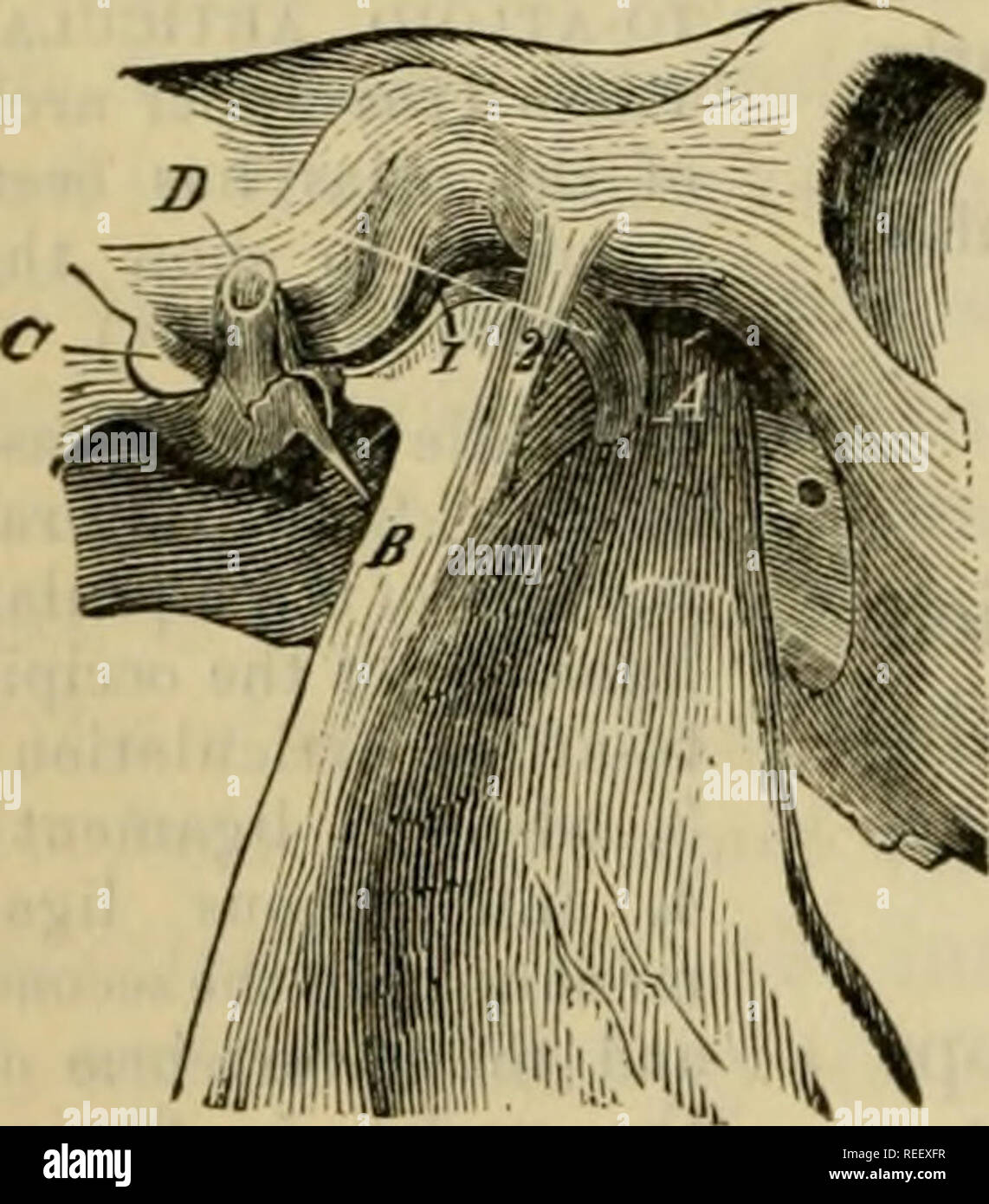 . The comparative anatomy of the domesticated animals. Veterinary anatomy. 188 THE ARTICULATIONS. the head, the small oblique, and the complexus. There is also the cord of the cervical ligament, Sijiiovial membranes.—These membranes are two in number—one for each condyle and coiTesponding atloid cavity. Sustained above, below, and outwardly by the capsular ligament, they are related inwardly to the dura mater and the fibrous tractus which, from the odontoid ligament, is carried to the internal face of the occipital condyles. Movements.—Extension, flexion, lateral inclination, and circumduction Stock Photohttps://www.alamy.com/image-license-details/?v=1https://www.alamy.com/the-comparative-anatomy-of-the-domesticated-animals-veterinary-anatomy-188-the-articulations-the-head-the-small-oblique-and-the-complexus-there-is-also-the-cord-of-the-cervical-ligament-sijiiovial-membranesthese-membranes-are-two-in-numberone-for-each-condyle-and-coitesponding-atloid-cavity-sustained-above-below-and-outwardly-by-the-capsular-ligament-they-are-related-inwardly-to-the-dura-mater-and-the-fibrous-tractus-which-from-the-odontoid-ligament-is-carried-to-the-internal-face-of-the-occipital-condyles-movementsextension-flexion-lateral-inclination-and-circumduction-image232668123.html
RMREEXFR–. The comparative anatomy of the domesticated animals. Veterinary anatomy. 188 THE ARTICULATIONS. the head, the small oblique, and the complexus. There is also the cord of the cervical ligament, Sijiiovial membranes.—These membranes are two in number—one for each condyle and coiTesponding atloid cavity. Sustained above, below, and outwardly by the capsular ligament, they are related inwardly to the dura mater and the fibrous tractus which, from the odontoid ligament, is carried to the internal face of the occipital condyles. Movements.—Extension, flexion, lateral inclination, and circumduction